Research Article Circadian Models of Serum Potassium...
9
Hindawi Publishing Corporation Computational and Mathematical Methods in Medicine Volume 2013, Article ID 429037, 8 pages http://dx.doi.org/10.1155/2013/429037 Research Article Circadian Models of Serum Potassium, Sodium, and Calcium Concentrations in Healthy Individuals and Their Application to Cardiac Electrophysiology Simulations at Individual Level Kamil Fijorek, 1 Miroslawa Puskulluoglu, 2 and Sebastian Polak 3 1 Department of Statistics, Cracow University of Economics, 27 Rakowicka Street, 31-510 Krakow, Poland 2 Department of Oncology, Jagiellonian University Medical College, 20 Grzegorzecka Street, 31-531 Krakow, Poland 3 Unit of Pharmacoepidemiology and Pharmacoeconomics, Faculty of Pharmacy, Jagiellonian University Medical College, Medyczna 9 Street, 30-688 Krakow, Poland Correspondence should be addressed to Sebastian Polak; [email protected] Received 27 May 2013; Accepted 25 July 2013 Academic Editor: Michele Migliore Copyright © 2013 Kamil Fijorek et al. is is an open access article distributed under the Creative Commons Attribution License, which permits unrestricted use, distribution, and reproduction in any medium, provided the original work is properly cited. In the article a brief description of the biological basis of the regulation of human biological clocks was presented in order to introduce the role of circadian rhythms in physiology and specifically in the pharmacological translational tools based on the computational physiology models to motivate the need to provide models of circadian fluctuation in plasma cations. e main aim of the study was to develop statistical models of the circadian rhythm of potassium, sodium, and calcium concentrations in plasma. e developed ion models were further tested by assessing their influence on QT duration (cardiac endpoint) as simulated by the biophysically detailed models of human leſt ventricular cardiomyocyte. e main results are model equations along with an electronic supplement to the article that contains a fully functional implementation of all models. 1. Introduction e crucial role of homeostasis maintenance in all living creatures is not in contradiction with the observation that various biological parameters are not static. e rhythmical changes observed in humans that occur periodically play an important role in the adaptation to the dynamic environ- ment. Chronobiology influences the activity and functions of organs and tissues and is also a driver of anatomical, physiological, and molecular changes. Classification of bio- logical rhythms depends on interval duration, starting with the very short periods expressed in seconds (e.g., electro- cardiographic changes), through ultradian periods described in minutes/hours (e.g., sleep), and circadian periods close to 24 hours, up to longer periods, including monthly (cir- catrigintan, i.e., menstrual) and yearly (circannual) rhythms [1]. It has been suggested that in humans, the physiological rhythmicity and its behavioral reflection define chronotype (e.g., morningness versus eveningness tendencies). In the following sections, a brief description of the bio- logical basis of the regulation of human biological clocks is presented in order to introduce the role of circadian rhythms in physiology and specifically in the pharmacological transla- tional tools based on the computational physiology models to motivate the need to provide models of circadian fluctuation in the main plasma cations. 2. Human Biological Clocks e center of the circadian clock is localized in the bilat- eral suprachiasmatic nuclei (SCN) in the hypothalamus [1– 4]. Also, organs known as peripheral circadian oscillators, for example, the heart, liver, kidneys, are thought to be responsible for circadian rhythmicity of human physiology, behavior, or biochemistry. Information exchange between clocks involves humoral and nervous systems and includes feedback loops. Peripheral oscillators may also show their
Transcript of Research Article Circadian Models of Serum Potassium...

Hindawi Publishing CorporationComputational and Mathematical Methods in MedicineVolume 2013, Article ID 429037, 8 pageshttp://dx.doi.org/10.1155/2013/429037
Research ArticleCircadian Models of Serum Potassium, Sodium, and CalciumConcentrations in Healthy Individuals and Their Application toCardiac Electrophysiology Simulations at Individual Level
Kamil Fijorek,1 Miroslawa Puskulluoglu,2 and Sebastian Polak3
1 Department of Statistics, Cracow University of Economics, 27 Rakowicka Street, 31-510 Krakow, Poland2Department of Oncology, Jagiellonian University Medical College, 20 Grzegorzecka Street, 31-531 Krakow, Poland3Unit of Pharmacoepidemiology and Pharmacoeconomics, Faculty of Pharmacy, Jagiellonian University Medical College,Medyczna 9 Street, 30-688 Krakow, Poland
Correspondence should be addressed to Sebastian Polak; [email protected]
Received 27 May 2013; Accepted 25 July 2013
Academic Editor: Michele Migliore
Copyright © 2013 Kamil Fijorek et al. This is an open access article distributed under the Creative Commons Attribution License,which permits unrestricted use, distribution, and reproduction in any medium, provided the original work is properly cited.
In the article a brief description of the biological basis of the regulation of human biological clocks was presented in order tointroduce the role of circadian rhythms in physiology and specifically in the pharmacological translational tools based on thecomputational physiology models to motivate the need to provide models of circadian fluctuation in plasma cations. The mainaim of the study was to develop statistical models of the circadian rhythm of potassium, sodium, and calcium concentrations inplasma.The developed ion models were further tested by assessing their influence on QT duration (cardiac endpoint) as simulatedby the biophysically detailed models of human left ventricular cardiomyocyte. The main results are model equations along with anelectronic supplement to the article that contains a fully functional implementation of all models.
1. Introduction
The crucial role of homeostasis maintenance in all livingcreatures is not in contradiction with the observation thatvarious biological parameters are not static. The rhythmicalchanges observed in humans that occur periodically play animportant role in the adaptation to the dynamic environ-ment. Chronobiology influences the activity and functionsof organs and tissues and is also a driver of anatomical,physiological, and molecular changes. Classification of bio-logical rhythms depends on interval duration, starting withthe very short periods expressed in seconds (e.g., electro-cardiographic changes), through ultradian periods describedin minutes/hours (e.g., sleep), and circadian periods closeto 24 hours, up to longer periods, including monthly (cir-catrigintan, i.e., menstrual) and yearly (circannual) rhythms[1]. It has been suggested that in humans, the physiologicalrhythmicity and its behavioral reflection define chronotype(e.g., morningness versus eveningness tendencies).
In the following sections, a brief description of the bio-logical basis of the regulation of human biological clocks ispresented in order to introduce the role of circadian rhythmsin physiology and specifically in the pharmacological transla-tional tools based on the computational physiologymodels tomotivate the need to provide models of circadian fluctuationin the main plasma cations.
2. Human Biological Clocks
The center of the circadian clock is localized in the bilat-eral suprachiasmatic nuclei (SCN) in the hypothalamus [1–4]. Also, organs known as peripheral circadian oscillators,for example, the heart, liver, kidneys, are thought to beresponsible for circadian rhythmicity of human physiology,behavior, or biochemistry. Information exchange betweenclocks involves humoral and nervous systems and includesfeedback loops. Peripheral oscillators may also show their
2 Computational and Mathematical Methods in Medicine
autonomous nature [2–6]. There are many genes that areexpressed in a clock-dependent manner in both central orperipheral clock tissues—for details see numerous reviews[1–3]. As for the cardiovascular system, circadian variationmay be seen in blood pressure, heart rate, coronary bloodflow, hormonal secretion, or electrical activity [2, 7–10]. Theclock localized in the heart influences the cells’ responseto numerous physiological or pathological conditions, forexample, ischemia [11, 12]. Examples of external and internalfactors influencing heart functioning are physical activity andautonomic nervous/humoral system (e.g., glucocorticoids,renin-angiotensin-aldosterone activity) modulating bloodpressure, for example, causing its increase in the morning[2, 3]. However, most studies on peripheral clock localizedin cardiomyocytes and factors influencing it were performedon animals. Although comprehensive data on humans arestill lacking, it was shown that almost 10% of genes in heartmuscle tissue are expressed in a clock-dependent manner[2, 9].Thus, the control and the loops between the clocks takeplace on different levels: from organs and systems throughbiochemical, cellular to molecular levels.
It is not only the physiological functioning of the heart,but also many diseases such as atrial fibrillations and otherarrhythmias (also those triggered by drugs), myocardialinfarction, or sudden cardiac death that show circadian vari-ation [5, 7, 13]. The incidence of most adverse cardiologicalevents is related to the time of the day [1, 5]. Moreover,the studies suggest that circadian oscillation (e.g., RR or QTinterval, R and T wave voltage) may play a predicting rolein cardiac death. It was shown that prolongation of the QTcinterval plays a prognostic role and also reduced circadianfluctuations are connected with poorer patients’ outcome[2, 13].
The biological rhythmicity can be influenced by geneticfactors and is frequently related to the risk of developing anumber of diseases and their further course [3, 14–16].There-fore, chronotype modification has been used as a therapeuticstrategy [17] for various diseases including cardiovasculardisturbances [2].
3. Chronopharmacology
Circadian rhythms have been actively investigated andapplied in clinical pharmacology. The accumulation ofknowledge in this area led to the formation of a new fieldcalled chronopharmaceutics, which aims at delivering drugsin a controlledmanner at themost appropriate time of the day[18], which in turn leads to the optimization of drugs dosingand their clinical effect. Various drugs and populations wereinvestigated in chronopharmacology research; for example,see Block et al. [19]. Numerous studies also confirmed thatcircadian rhythms influence both the pharmacokinetics andpharmacodynamics of drugs. Time of drug application mod-ifies drug pharmacokinetics by changing all the elements ofthe ADME pathway—absorption, distribution, metabolism,and excretion [20]. As a result, many drug pharmacokineticparameters are influenced to some degree, which in turn
impacts drug efficacy and safety, that is, the pharmacody-namic components of chronopharmacology [21–23].
4. Cardiac Electrophysiology Simulations
Chronopharmaceutics, however, is not the only area ofpharmaceutical sciences that can benefit from research intothe human rhythms. Translational tools based on the bio-physically detailedmathematicalmodels allowing for in vitro-in vivo extrapolation are gaining increasing interest and aremore and more commonly used in systems pharmacology[24]. In cardiology, simulations based on the models ofcardiac electrophysiology are utilized for the assessment ofdrugs’ cardiac safety [25, 26]. Approaches taken by differentresearch groups vary significantly with respect to the mod-elling techniques, level of models complexity, heart represen-tation (ranging from single cell up to the two- andwhole heartthree-dimensional simulations), and the simulated endpoints[27–29]. Regardless of the applied methodology, most ofthe published manuscripts report results nonspecific to anyindividual patient by only using simulations constant (usuallyaverage) values of parameters describing human biology[30]. Based on the publicly available data sources, empiricalmodels describing the distribution of each relevant physio-logical parameter in populations of human individuals canbe developed and introduced into cardiac electrophysiologysimulations. For example, in Polak et al. [31] and Polak andFijorek [32], regression models relating age to the volume ofcardiomyocyte and cell electric capacitance were developedand later used to introduce interindividual variability into thecardiac electrophysiology simulations. In Fijorek et al. [33]inter- and intraindividual variability was introduced into thecardiac electrophysiology simulations by accounting for cir-cadian rhythmicity of the heart rate. However, the circadianrhythmicity of many other physiological parameters can besimilarly modeled and introduced into the simulations.
5. Study Aims
Themain aim of the study was to develop statistical models ofthe circadian rhythm of three plasma ions, that is, potassium,sodium, and calcium.The developed ionmodels were furthertested by assessing their influence on QT duration (cardiacendpoint) as simulated by the biophysically detailed modelsof human left ventricular cardiomyocyte.The relevance of theions to cardiac electrophysiology was described previously[34].
6. Materials, Methods, and Results
The first stage included estimation of mean and standarddeviation of concentration of potassium, sodium, and cal-cium. Suitable data was obtained fromPolak et al. [34], whichis an archive of data on plasma concentration of potassium,sodium, and calcium extracted from scientific articles viaextensive literature search. The data were available for a largenumber of healthy subjects of both sexes (496, 553, and 475males; 328, 322, and 1783 females; potassium, sodium, and

Computational and Mathematical Methods in Medicine 3
calcium, resp.) and a wide range of ages (4–69, 4–103, and 2–75).
Estimation ofmean concentration and standard deviationof concentration (Table 1) was performed separately for bothsexes since statistically significant differences between themwere found. Different normal ranges of metabolic panelcomponents for men and women are already used in clinicalpractice for parameters such as: liver function tests, uricacid, creatinine, creatine kinase, and hormone levels [35,36]. However, for potassium, sodium, and calcium, separatenormal ranges for men and women have not been used so farin daily clinical practice.
Markowitz et al. [37] suggested the existence of a rela-tionship between age and ions concentration. However, theresults of meta-analysis presented in Polak et al. [34] showno significant age dependence for potassium and sodiumconcentration. In the case of calcium, there might be a weakpositive relationship with age; however, the age impact seemsto be too small to be of clinical relevance in the contextassumed in this paper, but further research in this area isneeded. As a result, none of the developed models includeage as an explanatory variable.
There is a noticeable evidence of the existence of acircadian rhythm in potassium and sodium ion concentra-tions [38–40]. Kanabrocki et al. [41] additionally studieda circadian rhythm in calcium ion concentration; however,the results were not statistically significant, probably dueto the rather small number of subjects included in thestudy. Given insufficient support for calcium rhythmicity, themodel developed for that ion does not contain a circadiancomponent.
For potassium and sodium, it was decided to base the newmodels on the models proposed by Sennels et al. [38] afterthey had been extensively modified. The main argument infavor of the Sennels’ model is that among the existingmodels,this is the one with the largest number of subjects usedfor its development (23 individuals, all men). Additionally,it is the most recently published model, so its participantsare believed to be the most similar to the presently livingpopulation of healthy people. Also, the samples were taken9 times for every individual (at 9:00, 12:00, 15:00, 18:00,21:00, 0:00, 3:00, 6:00, and 9:00), which surpasses any otherreviewed report, giving Sennels’ study more power to detecta circadian rhythm. The standard deviation of residuals (thepotassium and sodium model) was estimated from the dataextracted from the box plots presented in Sennels’ paperusing the method described by Hozo et al. [42]. The maleand female mean concentrations were taken from Table 1 andincorporated into the developed models. Due to the lack ofsuitable data, it was assumed that the circadian rhythm is thesame for men and women.
In the case of calcium, it was assumed that the con-centration values between 2.0–2.8mM are physiologicallyvalid [34]. Consequently, the assumption of normality ofcalcium concentration was dismissed, and logit-normalitywas assumed; that is, concentration is normal after a scaledlogit transformation. After transformation, mean calciumconcentration for males was 0.1 (SD = 1.3) and for females−0.5 (SD = 0.8). In the case of the potassium and sodium
Table 1: Estimation results of mean concentration and standarddeviation of concentration.
Ion Sex Number ofsubjects Mean Standard
deviation (SD)
Potassium Female 328 4.088 0.445Male 496 4.213 0.347
Sodium Female 322 138.169 4.398Male 553 140.096 3.017
Calcium Female 1783 2.313 0.136Male 475 2.418 0.199
model, such transformation was not needed since both mod-els conformed with the physiological concentration rangessatisfactorily (residuals in both models were assumed tofollow the normal distribution).Having completed the above-mentioned stages, the ionic means are calculated by thefollowing formulas:
mean potassium
= (M/F mean concentration)
+ 0.18 ∗ COS(2 ∗ PI24
∗ (Time—10 :07)) ,
mean sodium
= (M/F mean concentration)
+ 1.1 ∗ COS(2 ∗ PI24
∗ (Time—13 :08)) ,
mean logit-transformed calcium
= (M/F mean logit-transformed concentration) ,
(1)
where M/F—male or female; time—value from 0 to 24 range.In the next phase, the stochastic part of the models was
extended by incorporating a physiology-based assumptionthat for a given individual, the closer in time the concen-trations are measured, the more similar are the deviationsfrom the mean concentration. The most common way toinduce a correlation between residuals is to use an autore-gressive process. In order to estimate the parameters of thisprocess, concentration trajectories from individual subjectsare needed. These were extracted for potassium from theKanabrocki et al. [43] and Williams et al. [44] papers, forsodium from theKanabrocki et al. [43] paper, and for calciumfrom theKanabrocki et al. [43], Jubiz et al. [45], andWills [46]papers. The data were used to estimate the autocorrelationcoefficient of autoregressive process of order one—AR(1). Inall of the enumerated studies, the number of subjects was verysmall, and ionic concentrations were evaluated very sparsely.Consequently, it was found that the data was not able tosupport a more sophisticated autoregressive model. Resultsfor the AR(1) process are presented assuming a 15-minutesampling interval for ionic concentrations (Table 2).
In the next stage, the validity of the developed modelswas verified. A set of a 1000 random model paths of ionic
4 Computational and Mathematical Methods in Medicine
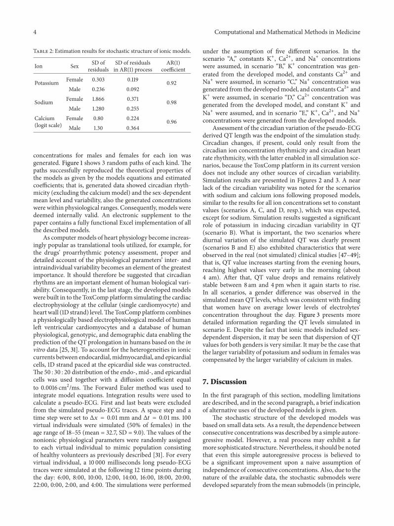
Table 2: Estimation results for stochastic structure of ionic models.
Ion Sex SD ofresiduals
SD of residualsin AR(1) process
AR(1)coefficient
Potassium Female 0.303 0.119 0.92Male 0.236 0.092
Sodium Female 1.866 0.371 0.98Male 1.280 0.255
Calcium(logit scale)
Female 0.80 0.224 0.96Male 1.30 0.364
concentrations for males and females for each ion wasgenerated. Figure 1 shows 3 random paths of each kind. Thepaths successfully reproduced the theoretical properties ofthe models as given by the models equations and estimatedcoefficients; that is, generated data showed circadian rhyth-micity (excluding the calciummodel) and the sex-dependentmean level and variability, also the generated concentrationswere within physiological ranges. Consequently, models weredeemed internally valid. An electronic supplement to thepaper contains a fully functional Excel implementation of allthe described models.
As computer models of heart physiology become increas-ingly popular as translational tools utilized, for example, forthe drugs’ proarrhythmic potency assessment, proper anddetailed account of the physiological parameters’ inter- andintraindividual variability becomes an element of the greatestimportance. It should therefore be suggested that circadianrhythms are an important element of human biological vari-ability. Consequently, in the last stage, the developed modelswere built in to the ToxComp platform simulating the cardiacelectrophysiology at the cellular (single cardiomyocyte) andheartwall (1D strand) level.TheToxCompplatform combinesa physiologically based electrophysiological model of humanleft ventricular cardiomyocytes and a database of humanphysiological, genotypic, and demographic data enabling theprediction of the QT prolongation in humans based on the invitro data [25, 31]. To account for the heterogeneities in ioniccurrents between endocardial,midmyocardial, and epicardialcells, 1D strand paced at the epicardial side was constructed.The 50 : 30 : 20 distribution of the endo-, mid-, and epicardialcells was used together with a diffusion coefficient equalto 0.0016 cm2/ms. The Forward Euler method was used tointegrate model equations. Integration results were used tocalculate a pseudo-ECG. First and last beats were excludedfrom the simulated pseudo-ECG traces. A space step and atime step were set to Δ𝑥 = 0.01mm and Δ𝑡 = 0.01ms. 100virtual individuals were simulated (50% of females) in theage range of 18–55 (mean = 32.7, SD = 9.0). The values of thenonionic physiological parameters were randomly assignedto each virtual individual to mimic population consistingof healthy volunteers as previously described [31]. For everyvirtual individual, a 10 000 milliseconds long pseudo-ECGtraces were simulated at the following 12 time points duringthe day: 6:00, 8:00, 10:00, 12:00, 14:00, 16:00, 18:00, 20:00,22:00, 0:00, 2:00, and 4:00. The simulations were performed
under the assumption of five different scenarios. In thescenario “A,” constants K+, Ca2+, and Na+ concentrationswere assumed, in scenario “B,” K+ concentration was gen-erated from the developed model, and constants Ca2+ andNa+ were assumed, in scenario “C,” Na+ concentration wasgenerated from the developedmodel, and constants Ca2+ andK+ were assumed, in scenario “D,” Ca2+ concentration wasgenerated from the developed model, and constant K+ andNa+ were assumed, and in scenario “E,” K+, Ca2+, and Na+concentrations were generated from the developed models.
Assessment of the circadian variation of the pseudo-ECGderived QT length was the endpoint of the simulation study.Circadian changes, if present, could only result from thecircadian ion concentration rhythmicity and circadian heartrate rhythmicity, with the latter enabled in all simulation sce-narios, because the ToxComp platform in its current versiondoes not include any other sources of circadian variability.Simulation results are presented in Figures 2 and 3. A nearlack of the circadian variability was noted for the scenarioswith sodium and calcium ions following proposed models,similar to the results for all ion concentrations set to constantvalues (scenarios A, C, and D, resp.), which was expected,except for sodium. Simulation results suggested a significantrole of potassium in inducing circadian variability in QT(scenario B). What is important, the two scenarios wherediurnal variation of the simulated QT was clearly present(scenarios B and E) also exhibited characteristics that wereobserved in the real (not simulated) clinical studies [47–49];that is, QT value increases starting from the evening hours,reaching highest values very early in the morning (about4 am). After that, QT value drops and remains relativelystable between 8 am and 4 pm when it again starts to rise.In all scenarios, a gender difference was observed in thesimulated mean QT levels, which was consistent with findingthat women have on average lower levels of electrolytes’concentration throughout the day. Figure 3 presents moredetailed information regarding the QT levels simulated inscenario E. Despite the fact that ionic models included sex-dependent dispersion, it may be seen that dispersion of QTvalues for both genders is very similar. It may be the case thatthe larger variability of potassium and sodium in females wascompensated by the larger variability of calcium in males.
7. Discussion
In the first paragraph of this section, modelling limitationsare described, and in the second paragraph, a brief indicationof alternative uses of the developed models is given.
The stochastic structure of the developed models wasbased on small data sets. As a result, the dependence betweenconsecutive concentrations was described by a simple autore-gressive model. However, a real process may exhibit a farmore sophisticated structure.Nevertheless, it should be notedthat even this simple autoregressive process is believed tobe a significant improvement upon a naive assumption ofindependence of consecutive concentrations. Also, due to thenature of the available data, the stochastic submodels weredeveloped separately from themean submodels (in principle,

Computational and Mathematical Methods in Medicine 5
3.0
3.5
4.0
4.5
5.0
5.5
Time of the day
Con
cent
ratio
n (m
M)
9 13 17 21 1 5 9
(a)
3.0
3.5
4.0
4.5
5.0
5.5
Time of the day
Con
cent
ratio
n (m
M)
9 13 17 21 1 5 9
(b)
130
135
140
145
150
Con
cent
ratio
n (m
M)
Time of the day9 13 17 21 1 5 9
(c)
130
135
140
145
150
Con
cent
ratio
n (m
M)
Time of the day9 13 17 21 1 5 9
(d)
2.0
2.2
2.4
2.6
2.8
Con
cent
ratio
n (m
M)
Time of the day9 13 17 21 1 5 9
(e)
2.0
2.2
2.4
2.6
2.8
Con
cent
ratio
n (m
M)
Time of the day9 13 17 21 1 5 9
(f)
Figure 1: Sample random paths of ionic concentrations: (a) potassium male, (b) potassium female, (c) sodium male, (d) sodium female, (e)calcium male, and (f) calcium female.
joint modelling should be preferred whenever possible, as itleads to a more efficient use of data). Also, the mix of theaggregated and individual data from different sources causeda major problem for the quantitative assessment of the modelfit and model validation. Consequently, external validity ofthe simulated paths of ionic concentrations was graded by
two domain experts who stated that the paths do not possessfeatures that might disqualify them from the physiologicalpoint of view. The only raised concern regarded a highintrasubject variability of potassium and calcium, which maybe a result of using AR process with short memory; however,as it was mentioned before, the data were not sufficient to

6 Computational and Mathematical Methods in Medicine
348350352354356358360362
QT
(ms)
Time of the day
06:0
0
08:0
0
10:0
0
12:0
0
14:0
0
16:0
0
18:0
0
20:0
0
22:0
0
00:0
0
02:0
0
04:0
0
(a)
348350352354356358360362
QT
(ms)
Time of the day
06:0
0
08:0
0
10:0
0
12:0
0
14:0
0
16:0
0
18:0
0
20:0
0
22:0
0
00:0
0
02:0
0
04:0
0
(b)
348350352354356358360362
QT
(ms)
Time of the day
06:0
0
08:0
0
10:0
0
12:0
0
14:0
0
16:0
0
18:0
0
20:0
0
22:0
0
00:0
0
02:0
0
04:0
0
(c)
348350352354356358360362
QT
(ms)
Time of the day
06:0
0
08:0
0
10:0
0
12:0
0
14:0
0
16:0
0
18:0
0
20:0
0
22:0
0
00:0
0
02:0
0
04:0
0
(d)
348350352354356358360362
QT
(ms)
All individualsFemalesMales
Time of the day
06:0
0
08:0
0
10:0
0
12:0
0
14:0
0
16:0
0
18:0
0
20:0
0
22:0
0
00:0
0
02:0
0
04:0
0
(e)
Figure 2: Mean QT values derived from the simulated scenarios.
support a more sophisticated autoregressive model. Furtherresearch is definitely needed to objectively resolve this issue.As it was justified in the main text, the modified Sennels’models were utilized for potassium and sodium. However,in Sennels, only male volunteers were included; therefore,the assumption of the gender independent structure of thecircadian rhythm may potentially introduce a bias. Also,it should be reiterated that sodium and potassium modelsinclude a circadian rhythm but calcium does not, due to lackof contrary evidence.
Current studies are aimed at establishing a connectionbetween pathogenesis of cardiac diseases and circadianrhythms. Such a connection might be helpful in developingnew chronotherapeutics and an early assessment of potentialsafety concerns. Circadian models of ions may be considered
in simulations of drugs’ activity, including their side effects,efficacy, and targeting ability. Also, there are suggestionsthat a synchronization of the cardiac clock with drugdelivery strategies may bring benefits in terms of avoidingcardiotoxic events. Therefore, there is a need not only forstudies presenting models useful for chronopharmaceuticalresearch, but also concerning the potential use of chemicaloscillators as biomarkers for new chronotherapeutics andchronopharmacological schedules [1, 2, 4, 50]. Additionally, itis possible that circadian models of ion levels may play a rolein researching pharmacokinetics and pharmacodynamics ofnew chronotherapeutics and “old” drugs (e.g., 𝛽-blockers,calcium channel blockers), as it is the subject of numerousstudies whether the time of the day when the medication isadministered influences its effect or toxicity [2, 51].

Computational and Mathematical Methods in Medicine 7
342346350354358362366
QT
(ms)
Time of the day
06:0
0
08:0
0
10:0
0
12:0
0
14:0
0
16:0
0
18:0
0
20:0
0
22:0
0
00:0
0
02:0
0
04:0
0
All individualsFemalesMales
Figure 3: Mean and standard deviation of QT values derived onlyfrom the scenario “E.”
Acknowledgments
The project is financed by the Polish National Center forResearch and Development, LIDER Project no. LIDER/02/187/L-1/09. Kamil Fijorek acknowledges financial supportfromThe Foundation for Polish Science.
References
[1] S. Ohdo, S. Koyanagi, and N. Matsunaga, “Chronopharma-cological strategies: intra- and inter-individual variability ofmolecular clock,” Advanced Drug Delivery Reviews, vol. 62, no.9-10, pp. 885–897, 2010.
[2] N. Takeda and K. Maemura, “Cardiovascular disease,chronopharmacotherapy, and the molecular clock,” AdvancedDrug Delivery Reviews, vol. 62, no. 9-10, pp. 956–966, 2010.
[3] S. Sukumaran, R. R. Almon, D. C. DuBois, and W. J. Jusko,“Circadian rhythms in gene expression: relationship to physiol-ogy, disease, drug disposition and drug action,” Advanced DrugDelivery Reviews, vol. 62, no. 9-10, pp. 904–917, 2010.
[4] H. Okamura, M. Doi, J. M. Fustin, Y. Yamaguchi, and M.Matsuo, “Mammalian circadian clock system: molecular mech-anisms for pharmaceutical and medical sciences,” AdvancedDrug Delivery Reviews, vol. 62, no. 9-10, pp. 876–884, 2010.
[5] S. Fournier, E. Eeckhout, F. Mangiacapra et al., “Circadianvariations of ischemic burden among patients with myocardialinfarction undergoing primary percutaneous coronary inter-vention,” American Heart Journal, vol. 163, no. 2, pp. 208–213,2012.
[6] S. Panda and J. B. Hogenesch, “It’s all in the timing:many clocks,many outputs,” Journal of Biological Rhythms, vol. 19, no. 5, pp.374–387, 2004.
[7] D. Hoyer, B. Frank, R. Baranowski, J. Zebrowski, P. Stein, andH.Schmidt, “Autonomic information flow rhythms: fromheat beatinterval to circadian variation,” IEEE Engineering in Medicineand Biology Magazine, vol. 26, no. 6, pp. 19–24, 2007.
[8] P. Stein, E. Lundequam, L. Oliveira et al., “Circadian andultradian rhythms in cardiac autonomic modulation,” IEEEEngineering in Medicine and Biology Magazine, vol. 26, no. 6,pp. 14–18, 2007.
[9] U. Albrecht and G. Eichele, “The mammalian circadian clock,”Current Opinion in Genetics and Development, vol. 13, no. 3, pp.271–277, 2003.
[10] K. Eckel-Mahan and P. Sassone-Corsi, “Epigenetic regulationof the molecular clockwork,” Progress in Molecular Biology andTranslational Science, vol. 119, pp. 29–50, 2013.
[11] D. J. Durgan and M. E. Young, “The cardiomyocyte circa-dian clock: emerging roles in health and disease,” CirculationResearch, vol. 106, no. 4, pp. 647–658, 2010.
[12] D. J. Durgan, T. Pulinilkunnil, C. Villegas-Montoya et al.,“Short communication: ischemia/reperfusion tolerance is time-of-day-dependent: mediation by the cardiomyocyte circadianclock,” Circulation Research, vol. 106, no. 3, pp. 546–550, 2010.
[13] E. Watanabe, T. Arakawa, T. Uchiyama et al., “Prognosticsignificance of circadian variability of RR and QT intervals andQT dynamicity in patients with chronic heart failure,” HeartRhythm, vol. 4, no. 8, pp. 999–1005, 2007.
[14] C. Johansson and T. Partonen, “Human genetic studies inchronobiology,” in Trends in Chronobiology Research, F. H.Columbus, Ed., pp. 63–85, Nova Science Publishers, New York,NY, USA, 2006.
[15] E. Ferraz, M. C. Borges, and E. O. Vianna, “Influence ofnocturnal asthma on chronotype,” Journal of Asthma, vol. 45,no. 10, pp. 911–915, 2008.
[16] M. P. Hidalgo, W. Caumo, M. Posser, S. B. Coccaro, A. L.Camozzato, andM. L. F. Chaves, “Relationship between depres-sive mood and chronotype in healthy subjects,” Psychiatry andClinical Neurosciences, vol. 63, no. 3, pp. 283–290, 2009.
[17] A. Wirz-Justice, F. Benedetti, and M. Terman, Chronotherapeu-tics for Affective Disorders: A Clinician’s Manual for Light andWake Therapy, Karger, Basel, Switzerland, 2012.
[18] B. B. Youan, Chronopharmaceutics: Science and Technology forBiological Rhythm-Guided Therapy and Prevention of Diseases,John Wiley & Sons, Hoboken, NJ, USA, 2009.
[19] S. L. Block, D. Kelsey, D. Coury et al., “Once-daily atomoxetinefor treating pediatric attention-deficit/ hyperactivity disorder:comparison of morning and evening dosing,” Clinical Pedi-atrics, vol. 48, no. 7, pp. 723–733, 2009.
[20] B. Lemmer, “Clinical chronopharmacology: the importance oftime in drug treatment,” Ciba Foundation Symposium, vol. 183,pp. 235–247, 1995.
[21] K. Erol, F. S. Kilic, O. S. Batu, and E. Yildirim, “Morning-evening administration time differences in digoxin kinetics inhealthy young subjects,”Chronobiology International, vol. 18, no.5, pp. 841–849, 2001.
[22] S. Nakano and L. E. Hollister, “Chronopharmacology ofamitriptyline,” Clinical Pharmacology and Therapeutics, vol. 33,no. 4, pp. 453–459, 1983.
[23] B. R. Rao and D. Rambhau, “Diurnal oscillations in the serumand salivary levels of quinidine,” Drug Research, vol. 45, no. 6,pp. 681–683, 1995.
[24] A. Rostami-Hodjegan, “Physiologically based pharmacokinet-ics joined with in vitro-in vivo extrapolation of ADME: amarriage under the arch of systems pharmacology,” ClinicalPharmacology &Therapeutics, vol. 92, pp. 50–61, 2012.
[25] K.H.W. J. Ten Tusscher, D. Noble, P. J. Noble, andA. V. Panfilov,“A model for human ventricular tissue,” American Journal ofPhysiology. Heart and Circulatory Physiology, vol. 286, no. 4, pp.H1573–H1589, 2004.

8 Computational and Mathematical Methods in Medicine
[26] G. R. Mirams, M. R. Davies, Y. Cui, P. Kohl, and D. Noble,“Application of cardiac electrophysiology simulations to pro-arrhythmic safety testing,” British Journal of Pharmacology, vol.167, pp. 932–945, 2012.
[27] G. R. Mirams, Y. Cui, A. Sher et al., “Simulation of multipleion channel block provides improved early prediction of com-pounds’ clinical torsadogenic risk,” Cardiovascular Research,vol. 91, no. 1, pp. 53–61, 2011.
[28] J. D. Moreno, Z. I. Zhu, P.-C. Yang et al., “A computationalmodel to predict the effects of class I anti-arrhythmic drugs onventricular rhythms,” Science Translational Medicine, vol. 3, no.98, Article ID 98ra83, 2011.
[29] N. Zemzemi, M. O. Bernabeu, J. Saiz et al., “Computationalassessment of drug-induced effects on the electrocardiogram:from ion channel to body surface potentials,” British Journal ofPharmacology, vol. 168, pp. 718–733, 2013.
[30] C. Obiol-Pardo, J. Gomis-Tena, F. Sanz, J. Saiz, and M. Pastor,“A multiscale simulation system for the prediction of drug-induced cardiotoxicity,” Journal of Chemical Information andModeling, vol. 51, no. 2, pp. 483–492, 2011.
[31] S. Polak, K. Fijorek, A. Glinka, B. Wisniowska, and A.Mendyk, “Virtual population generator for human cardiomy-ocytes parameters: in silico drug cardiotoxicity assessment,”Toxicology Mechanisms and Methods, vol. 22, no. 1, pp. 31–40,2012.
[32] S. Polak and K. Fijorek, “Inter-individual variability in thepre-clinical drug cardiotoxic safety assessment—analysis of theage—cardiomyocytes electric capacitance dependence,” Journalof Cardiovascular Translational Research, vol. 5, pp. 321–332,2012.
[33] K. Fijorek, N. Patel, Ł. Klima, K. Stolarz-Skrzypek, K. Kawecka-Jaszcz, and S. Polak, “Age and gender dependent heart ratecircadian model development and performance verification onthe proarrhythmic drug case study,” Theoretical Biology andMedical Modelling, vol. 10, article 7, 2013.
[34] S. Polak, K. Fijorek, M. Puskulluoglu, A. Glinka, D.Tomaszewska, and R. Tomaszewski, “Literature review ofthe serum potassium, sodium and calcium levels in healthyindividuals,” Technical Note 001, 2013, http://tox-portal.net.
[35] F. Ceriotti, J. Henny, J. Queralto et al., “Common referenceintervals for aspartate aminotransferase (AST), alanine amino-transferase (ALT) and 𝛾-glutamyl transferase (GGT) in serum:results from an IFCCmulticenter study,”Clinical Chemistry andLaboratory Medicine, vol. 48, no. 11, pp. 1593–1601, 2010.
[36] A. Kratz and K. B. Lewandrowski, “Case records of the Mas-sachusetts General Hospital. Weekly clinicopathological exer-cises. Normal reference laboratory values,” The New Englandjournal of medicine, vol. 339, no. 15, pp. 1063–1072, 1998.
[37] M. E. Markowitz, J. F. Rosen, S. Laxminarayan, and M.Mizruchi, “Circadian rhythms of blood minerals during ado-lescence,” Pediatric Research, vol. 18, no. 5, pp. 456–462, 1984.
[38] H. P. Sennels, H. L. Jørgensen, J. P. Goetze, and J. Fahrenkrug,“Rhythmic 24-hour variations of frequently used clinical bio-chemical parameters in healthy young males–the Bispebjergstudy of diurnal variations,” Scandinavian Journal of Clinical &Laboratory Investigation, vol. 72, pp. 287–295, 2012.
[39] M. Bernardi, R. DePalma, and F. Trevisani, “Serum potassiumcircadian rhythm.Relationshipwith aldosterone,”Hormone andMetabolic Research, vol. 17, no. 12, p. 695, 1985.
[40] R. B. Sothern, D. L. Vesely, E. L. Kanabrocki et al., “Circadianrelationships between circulating atrial natriuretic peptides and
serum sodium and chloride in healthy humans,” AmericanJournal of Nephrology, vol. 16, no. 6, pp. 462–470, 1996.
[41] E. L. Kanabrocki, L. E. Scheving, and F. Halberg, “Circadianvariations in presumably healthymenunder conditions of peacetime army reserve unit training,” Space Life Sciences, vol. 4, no.2, pp. 258–270, 1973.
[42] S. P. Hozo, B. Djulbegovic, and I. Hozo, “Estimating the meanand variance from the median, range, and the size of a sample,”BMCMedical Research Methodology, vol. 5, article 13, 2005.
[43] E. L. Kanabrocki, R. B. Sothern, L. E. Scheving et al., “Ten-year-replicated circadian profiles for 36 physiological, serologicaland urinary variables in healthy men,” Chronobiology Interna-tional, vol. 5, no. 3, pp. 237–284, 1988.
[44] G. H. Williams, J. P. Cain, R. G. Dluhy, and R. H. Underwood,“Studies of the control of plasma aldosterone concentration innormal man. I. Response to posture, acute and chronic volumedepletion, and sodium loading,” Journal of Clinical Investigation,vol. 51, no. 7, pp. 1731–1742, 1972.
[45] W. Jubiz, J. M. Canterbury, E. Reiss, and F. H. Tyler, “Circadianrhythm in serumparathyroid hormone concentration in humansubjects: correlation with serum calcium, phosphate, albumin,and growth hormone levels,” Journal of Clinical Investigation,vol. 51, no. 8, pp. 2040–2046, 1972.
[46] M. R. Wills, “The effect of diurnal variation on total plasmacalcium concentration in normal subjects,” Journal of ClinicalPathology, vol. 23, no. 9, pp. 772–777, 1970.
[47] J. Molnar, F. Zhang, J. Weiss, F. A. Ehlert, and J. E. Rosenthal,“Diurnal pattern of QTc interval: how long is prolonged?Possible relation to circadian triggers of cardiovascular events,”Journal of the American College of Cardiology, vol. 27, no. 1, pp.76–83, 1996.
[48] S. Ishida, M. Nakagawa, T. Fujino, H. Yonemochi, T. Saikawa,and M. Ito, “Circadian variation of QT interval dispersion: cor-relation with heart rate variability,” Journal of Electrocardiology,vol. 30, no. 3, pp. 205–210, 1997.
[49] V. K. Yeragani, R. Berger, R. Pohl, and R. Balon, “Effect of ageon diurnal changes of 24-hour QT interval variability,” PediatricCardiology, vol. 26, no. 1, pp. 39–44, 2005.
[50] B.-B. C. Youan, “Chronopharmaceutical drug delivery systems:hurdles, hype or hope?” Advanced Drug Delivery Reviews, vol.62, no. 9-10, pp. 898–903, 2010.
[51] R. C. Hermida, D. E. Ayala, A. Mojon, and J. R. Fernandez,“Role of time-of-day of hypertension treatment on the J-shapedrelationship between blood pressure and cardiovascular risk,”Chronobiology International, vol. 30, pp. 328–339, 2013.

Submit your manuscripts athttp://www.hindawi.com
Stem CellsInternational
Hindawi Publishing Corporationhttp://www.hindawi.com Volume 2014
Hindawi Publishing Corporationhttp://www.hindawi.com Volume 2014
MEDIATORSINFLAMMATION
of
Hindawi Publishing Corporationhttp://www.hindawi.com Volume 2014
Behavioural Neurology
EndocrinologyInternational Journal of
Hindawi Publishing Corporationhttp://www.hindawi.com Volume 2014
Hindawi Publishing Corporationhttp://www.hindawi.com Volume 2014
Disease Markers
Hindawi Publishing Corporationhttp://www.hindawi.com Volume 2014
BioMed Research International
OncologyJournal of
Hindawi Publishing Corporationhttp://www.hindawi.com Volume 2014
Hindawi Publishing Corporationhttp://www.hindawi.com Volume 2014
Oxidative Medicine and Cellular Longevity
Hindawi Publishing Corporationhttp://www.hindawi.com Volume 2014
PPAR Research
The Scientific World JournalHindawi Publishing Corporation http://www.hindawi.com Volume 2014
Immunology ResearchHindawi Publishing Corporationhttp://www.hindawi.com Volume 2014
Journal of
ObesityJournal of
Hindawi Publishing Corporationhttp://www.hindawi.com Volume 2014
Hindawi Publishing Corporationhttp://www.hindawi.com Volume 2014
Computational and Mathematical Methods in Medicine
OphthalmologyJournal of
Hindawi Publishing Corporationhttp://www.hindawi.com Volume 2014
Diabetes ResearchJournal of
Hindawi Publishing Corporationhttp://www.hindawi.com Volume 2014
Hindawi Publishing Corporationhttp://www.hindawi.com Volume 2014
Research and TreatmentAIDS
Hindawi Publishing Corporationhttp://www.hindawi.com Volume 2014
Gastroenterology Research and Practice
Hindawi Publishing Corporationhttp://www.hindawi.com Volume 2014
Parkinson’s Disease
Evidence-Based Complementary and Alternative Medicine
Volume 2014Hindawi Publishing Corporationhttp://www.hindawi.com