
research article Behavioral and Neurochemical Changes in ... · PDF fileAda Mitsacos, PhD;...
17
Received: June 8, 2014; Revised: November 13, 2014; Accepted: November 14, 2014 International Journal of Neuropsychopharmacology, 2015, 1–17 doi:10.1093/ijnp/pyu097 Research Article 1 This is an Open Access article distributed under the terms of the Creative Commons Attribution Non-Commercial License (http://creativecommons.org/licenses/by-nc/4.0/), which permits non-commercial re-use, distribution, and reproduction in any medium, provided the original work is properly cited. For commercial re-use, please contact [email protected] © The Author 2015. Published by Oxford University Press on behalf of CINP. research article Behavioral and Neurochemical Changes in Mesostriatal Dopaminergic Regions of the Rat after Chronic Administration of the Cannabinoid Receptor Agonist WIN55,212-2 Eleni Fanarioti, BSc; Maria Mavrikaki, PhD; George Panagis, PhD; Ada Mitsacos, PhD; George G. Nomikos, PhD; Panagiotis Giompres*, PhD University of Patras, Department of Biology, Laboratory of Human and Animal Physiology, Patras, Greece (Drs Fanarioti and Giompres); University of Crete, Department of Psychology, Laboratory of Behavioral Neuroscience, Rethymno, Crete, Greece (Drs Mavrikaki and Panagis); University of Patras, Department of Medicine, Laboratory of Physiology, Patras (Dr Mitsacos); Takeda Development Center Americas Inc., Deerfield, IL (Dr Nomikos). *Correspondence: Panagiotis Giompres, PhD, University of Patras, Department of Biology, Laboratory of Human and Animal Physiology, 26500 Patras, Greece ([email protected]). Abstract Background: The endocannabinoid system interacts extensively with other neurotransmitter systems and has been implicated in a variety of functions, including regulation of basal ganglia circuits and motor behavior. The present study examined the effects of repeated administration of the nonselective cannabinoid receptor 1 agonist WIN55,212-2 on locomotor activity and on binding and mRNA levels of dopamine receptors and transporters and GABA A receptors in mesostriatal dopaminergic regions of the rat. Methods: Rats received systemic injections of WIN55,212-2 (0, 0.1, 0.3, or 1 mg/kg, intraperitoneally) for 20 consecutive days. Locomotor activity was measured on days 1, 10, and 20. Following the last measurement, rats were euthanized and prepared for in vitro binding and in situ hybridization experiments. Results: Acutely, 0.3 and 1 mg/kg of WIN55,212-2 produced hypolocomotion, which was sustained for the next 2 measurements, compared to vehicle. Repeated administration of WIN55,212-2 decreased the mRNA levels of the D2 autoreceptors in substantia nigra and ventral tegmental area and increased D1 receptor mRNA and binding in nucleus accumbens. Furthermore, both dopamine receptor and transporter binding and mRNA levels were decreased in substantia nigra. Moreover, repeated administration of WIN55,212-2 decreased GABA A receptor binding levels in dorsal striatum and substantia nigra. Conclusions: Our data indicate that chronic WIN55,212-2 administration results in sustained effects on locomotor activity, similar to those observed after acute administration, and modulates the dopaminergic and GABAergic systems in a region-, dose-, and neurotransmitter-selective manner Keywords: WIN55,212-2, locomotor activity, dopamine transporter, dopamine receptors, GABAA receptor
Transcript of research article Behavioral and Neurochemical Changes in ... · PDF fileAda Mitsacos, PhD;...

Received: June 8, 2014; Revised: November 13, 2014; Accepted: November 14, 2014
International Journal of Neuropsychopharmacology, 2015, 1–17
doi:10.1093/ijnp/pyu097Research Article
1This is an Open Access article distributed under the terms of the Creative Commons Attribution Non-Commercial License (http://creativecommons.org/licenses/by-nc/4.0/), which permits non-commercial re-use, distribution, and reproduction in any medium, provided the original work is properly cited. For commercial re-use, please contact [email protected]
© The Author 2015. Published by Oxford University Press on behalf of CINP.
research article
Behavioral and Neurochemical Changes in Mesostriatal Dopaminergic Regions of the Rat after Chronic Administration of the Cannabinoid Receptor Agonist WIN55,212-2Eleni Fanarioti, BSc; Maria Mavrikaki, PhD; George Panagis, PhD; Ada Mitsacos, PhD; George G. Nomikos, PhD; Panagiotis Giompres*, PhD
University of Patras, Department of Biology, Laboratory of Human and Animal Physiology, Patras, Greece (Drs Fanarioti and Giompres); University of Crete, Department of Psychology, Laboratory of Behavioral Neuroscience, Rethymno, Crete, Greece (Drs Mavrikaki and Panagis); University of Patras, Department of Medicine, Laboratory of Physiology, Patras (Dr Mitsacos); Takeda Development Center Americas Inc., Deerfield, IL (Dr Nomikos).
*Correspondence: Panagiotis Giompres, PhD, University of Patras, Department of Biology, Laboratory of Human and Animal Physiology, 26500 Patras, Greece ([email protected]).
Abstract
Background: The endocannabinoid system interacts extensively with other neurotransmitter systems and has been implicated in a variety of functions, including regulation of basal ganglia circuits and motor behavior. The present study examined the effects of repeated administration of the nonselective cannabinoid receptor 1 agonist WIN55,212-2 on locomotor activity and on binding and mRNA levels of dopamine receptors and transporters and GABAA receptors in mesostriatal dopaminergic regions of the rat.Methods: Rats received systemic injections of WIN55,212-2 (0, 0.1, 0.3, or 1 mg/kg, intraperitoneally) for 20 consecutive days. Locomotor activity was measured on days 1, 10, and 20. Following the last measurement, rats were euthanized and prepared for in vitro binding and in situ hybridization experiments.Results: Acutely, 0.3 and 1 mg/kg of WIN55,212-2 produced hypolocomotion, which was sustained for the next 2 measurements, compared to vehicle. Repeated administration of WIN55,212-2 decreased the mRNA levels of the D2 autoreceptors in substantia nigra and ventral tegmental area and increased D1 receptor mRNA and binding in nucleus accumbens. Furthermore, both dopamine receptor and transporter binding and mRNA levels were decreased in substantia nigra. Moreover, repeated administration of WIN55,212-2 decreased GABAA receptor binding levels in dorsal striatum and substantia nigra.Conclusions: Our data indicate that chronic WIN55,212-2 administration results in sustained effects on locomotor activity, similar to those observed after acute administration, and modulates the dopaminergic and GABAergic systems in a region-, dose-, and neurotransmitter-selective manner
Keywords: WIN55,212-2, locomotor activity, dopamine transporter, dopamine receptors, GABAA receptor

2 | International Journal of Neuropsychopharmacology, 2015
IntroductionThe rewarding and motor effects produced by Δ9-tetrahydrocannabinol (Δ9-THC) and synthetic cannabinoid ago-nists are mediated primarily by cannabinoid 1 receptors (CB1R) (Ameri, 1999). CB1R are abundantly expressed in the basal gan-glia (Herkenham et al., 1991a), a brain region involved in motor control (Martín et al., 2008; Morera-Herreras et al., 2008). It has been shown that systemic administration of Δ9-THC and other CB1R agonists exerts biphasic effects on motor activity, with low doses increasing motor activation and higher doses producing hypolocomotion or even catalepsy (Sañudo-Peña et al., 2000; Drews et al., 2005; Shi et al., 2005; Rodvelt et al., 2007; Polissidis et al., 2010, 2013; Katsidoni et al., 2013).
It has been suggested that cannabinoids elicit their phar-macological effects in part through activation of dopaminer-gic neurons in the brain and more specifically the mesostriatal dopaminergic system with cell bodies located within the ven-tral tegmental area (VTA) and substantia nigra pars compacta (SNpc) (French et al., 1997; Rodríguez De Fonseca et al., 2001; Pan et al., 2008; Morera-Herreras et al., 2008) by enhancing dopamine release in their respective dopamine terminal fields, the nucleus accumbens (NAc) (Tanda et al., 1997) and striatum (Taylor et al., 1988).
Interestingly, CB1Rs do not appear to be expressed at dopa-minergic terminals in these main dopaminoceptive regions (Herkenham et al., 1991a, 1991b; Mailleux and Vanderhaeghen, 1992, 1993; Matsuda et al., 1993; Westlake et al., 1994; Julian et al., 2003) but rather on presynaptic GABAergic interneurons (Katona et al., 2000) and glutamatergic projecting neurons (Hermann et al., 2002), indicating that the effects of cannabi-noids on dopamine neurotransmission are mainly indirect and modulated via the function of other neurotransmitters, such as GABA and glutamate. Indeed, it is well documented that can-nabinoids affect extracellular levels of GABA in hippocampus (Katona et al., 1999), prefrontal cortex (Pistis et al., 2002), amyg-dala (Katona et al., 2001), and glutamate in cerebral (Ferraro et al., 2001) and prefrontal cortex (Pistis et al., 2002) and stri-atum (Polissidis et al., 2013). In the striatum, CB1Rs are local-ized presynaptically on GABAergic and glutamatergic terminals (Matsuda et al., 1993) and postsynaptically in the somata, den-drites, and axon terminals of striatal medium spiny neurons (Fitzgerald et al., 2012). Activation of CB1R inhibits GABAergic neurotransmission in the globus pallidus (Pertwee et al., 1988), NAc (Manzoni and Bockaert, 2001), SN (Wallmichrath and Szabo, 2002), and VTA (Szabo et al., 2002) via inhibition of adenylate cyclase (Pertwee, 2006).
Striatal medium spiny projection neurons (MSNs) expressing D1 dopamine receptors (D1DRs) form the direct pathway, while neurons expressing D2 receptors (D2DRs) form the indirect pathway. Activation of D1DR leads to stimulation of adenylate cyclase (Blandini et al., 2000) and cAMP formation (Gingrich and Caron, 1993) and, in turn, to activation of the direct path-way (van der Stelt and Di Marzo, 2003) while activation of D2DR inhibits adenylate cyclase (Blandini et al., 2000) and cAMP for-mation, leading to inhibition of striatal MSNs that project to the nuclei of the indirect pathway.
Several studies suggest an interaction between CB1R and D1/D2 dopamine receptors (D1DR/D2DR) at the cellular level and coupling to the same effector systems (Hermann et al., 2002). Simultaneous activation of both CB1R and D2DR leads to enhanced activation of adenylyl cyclase resulting in activation of striatal neurons of the indirect pathway, which in turn activates neurons of the subthalamic nuclei, resulting in hypomotility
(Glass and Felder, 1997; Kearn et al., 2005; Martín et al., 2008). Moreover, simultaneous stimulation of CB1R and D1DR leads to a net decrease in adenylyl cyclase, which in turn reduces the inhibitory activity of direct striatal projection neurons. This inhibition increases the activity of nigral neurons, resulting in a decreased motor response (Martín et al., 2008).
The present study was designed to correlate the behavioral effects of chronic WIN55,212-2 with changes in neurochemical indices. In this context and based on the key role of dopamine and GABA in the mesostriatal dopaminergic system, we investigated the effects of systemic acute and chronic administration of low and high doses of WIN55,212-2, a nonselective CB1R agonist, on motor activity patterns and characterized neurochemical altera-tions of dopaminergic and GABAergic systems. WIN55,212-2 has higher efficacy than other CB1R agonists (Kearn et al., 1999) and has been well characterized in behavioral and neurochemical studies (Manzoni and Bockaert, 2001; Wallmichrath and Szabo, 2002; Castañé et al., 2004; Vlachou et al., 2008; Moranta et al., 2009; Mavrikaki et al., 2010; Polissidis et al., 2013). We examined the dopamine transporter (DAT), dopamine, and GABA receptors in the striatum, NAc, SN, and VTA of WIN55,212-2-treated and vehicle-treated rats. In vitro binding and in situ hybridization experiments were used to evaluate binding and mRNA levels of the aforementioned receptors and transporters.
Methods
Animals
Male Sprague-Dawley rats (n = 40) weighing 250 to 300 g (postna-tal days 60 to 70) were used. The animals were housed in groups of 2 or 3 under a 12-h-light/-dark cycle with free access to food and water. Experiments were conducted in accordance with the European Communities Council Directive (86/609/EEC) and the National Institutes of Health Guide for the Care and Use of Laboratory Animals. All efforts were made to minimize animal suffering and to reduce the number of animals used.
WIN55,212-2 Treatment
WIN55,212-2 (Tocris, Westwoods Bus. Park) was dissolved in a vehicle solution containing 5% dimethylsulfoxide, 5% cremo-phor EL in 0.9% NaCl, and injected intraperitoneally (i.p.) at a volume of 3 mL/kg of body weight. Experimental animals were divided into 4 groups (n = 10 per group) receiving a single daily i.p. injection of either vehicle or WIN55,212-2 (vehicle, 0.1, 0.3, and 1 mg/kg) for 20 days. Control animals (n = 10) received i.p. the corresponding vehicle solution in the same injection volume. Αll rats were tested for locomotor activity and used for neurochem-ical studies (n = 10/group). Animals were euthanized by rapid decapitation 1.5 hours after the last injection.
Locomotor Activity
Spontaneous motor activity was measured using an activity recording system (Model 7445, Ugo Basile) consisting of an ani-mal cage and an electronic unit incorporating a counter and a printer. The rectangular animal cage (56 × 56 × 30 cm) had trans-parent sides and lid to allow observation. The cage floor had horizontal and vertical infrared sensors. The counter summed up the photocell disruptions, and a printer displayed the results at preset intervals. In our studies, a summation of photocell
Fanarioti et al. | 3
disruptions of ambulatory distance and rearing for each 5-min-ute interval period during the 1-hour observation period was registered. Behavioral testing was performed one the first, 10th, and 20th day of the drug treatment between 8:00 am and 4:00 pm, 10 minutes after drug administration. The postinjection time was selected taking into account that the behavioral effects lasted for approximately 1 hour.
Brain Sectioning for Neurochemical Studies
Rats were euthanized by rapid decapitation. Brains were isolated and quickly frozen in 2-methyl-butane. Coronal sections, 14 μm thick, were cut in a cryostat Leica (CM1850), thaw-mounted on gelatin-chromalum–coated glass slides (for autoradiography studies) or poly-l-lysine–coated slides (for in situ hybridization studies), dried at room temperature (RT), and stored at −75oC until experiments were performed.
Receptor Binding Autoradiography
DAT binding was assayed according to Dickinson et al. (1999) using 5 nM [3H]-WIN35428 (S.A. 87 Ci/mmol; PerkinElmer Life Sciences, Belgium) as radioligand. Sections were allowed to air-dry at RT, were preincubated for 30 minutes in 20 mM sodium phosphate buffer (PBS), pH 7.4, at 4oC, and were then incubated for 90 minutes in buffer containing 0.32 M sucrose, pH 7.4, at 4°C in the presence of radioligand. Sections were washed for 2 × 1 minutes in 20 mM PBS, pH 7.4, at 4oC, briefly dipped in ice-cold distilled water, and air-dried. Nonspecific binding was deter-mined in the presence of 30 μM benztropine (Sigma Aldrich, Greece).
D2- and D1-like dopamine receptors were assayed as described by Tarazi et al. (1998). Sections were preincubated at RT for 1 hour in 50 mM Tris-HCl, 120 mM NaCl, 5 mM KCl, 2 mM CaCl2, 1 mM MgCl2, pH 7.4, and incubated for 1 hour at RT in the presence of [3H]raclopride (2 nM; S.A. 62.2 Ci/mmol; PerkinElmer Life Sciences, Belgium) and [3H]SCH23390 (2.5 nM; S.A. 85Ci/mmol; PerkinElmer Life Sciences, Belgium) for D2- and D1-like receptors, respectively. For D1-like receptors, the incubation buffer contained 40 nM ketanserin (Tocris, UK) to block the 5-HT2 serotonin binding sites. After incubation, sections were rinsed 2 × 5 minutes in ice-cold buffer, briefly dipped in ice-cold distilled water, and air-dried. Nonspecific binding was determined with l μM cis-flupenthixol (Sigma-Aldrich, Greece) for both receptors.
GABAA receptor binding was performed according to Bristow and Martin (1988). Sections were preincubated at RT for 30 minutes in buffer containing 50 mM Tris-citrate, 100 mM MgCl2, pH 7.4, air-dried, and incubated with 6.5 nM [3H]-SR95531 (S.A. 55.3 Ci/mmol; PerkinElmer Life Sciences, Belgium) at 4oC for 30 minutes. Nonspecific binding was determined in the presence of 10 mM GABA (Sigma Aldrich, Greece). Sections were rinsed 3 × 5 min in ice-cold buffer, briefly dipped in ice-cold distilled water, and air-dried under a stream of cold air. The labeled sections were exposed to BioMax MR Film (Kodak) for 4 to 10 months. Tritium micro scales (Amersham, UK), calibrated as nCi/mg tissue equivalent, were exposed along with the tissue samples and used as standards.
In Situ Hybridization Histochemistry
Hybridization was carried out according to Giannakopoulou et al. (2012). Sections were air-dried at RT, fixed for 5 minutes in 4% paraformaldehyde in diethylpyrocarbonate (DEPC)-treated PBS (0.1 M, pH 7.4), rinsed in PBS, dehydrated in graded ethanol, and
air-dried at RT. The oligonucleotide sequences used are shown in the Supplementary Material.
Each probe was diluted to a concentration of 3 pmol/μL and labeled with 35S-ATP (PerkinElmer Life Sciences, Belgium) at the 3’ end, using the 3’ terminal transferase enzyme (Roche, Germany) to a specific activity of 2 × 10⁵ cpm/μL. Chromatography Sephadex G-50 columns (BioRad, Greece) were used to remove unincorporated nucleotides. Hybridization was performed in 50% formamide (vol/vol), 4 × saline-sodium citrate (SSC) buffer (1 × SSC: 0.15 M sodium chloride, 0.015 M sodium citrate), 10% dextran sulfate (wt/vol), and 10 mM dithiothreitol, with 1:100 labeled probe (0.03 pmol/μL final concentration of labeled probe). Sections were covered with 120 μL of hybridization solu-tion and incubated for 18 hours in a humid chamber at 42oC. Nonspecific signal was determined by the addition of 100-fold excess of unlabeled probe to the hybridization solution. After hybridization, sections were washed in 1 × SSC for 20 minutes at 60oC, in 0.1 × SSC for 3 minutes at RT, dehydrated in graded ethanol, and air-dried at RT. Sections were exposed to a Kodak BioMax MR film and exposure time ranged from 2 to 8 weeks depending on probe labeling.
Quantification
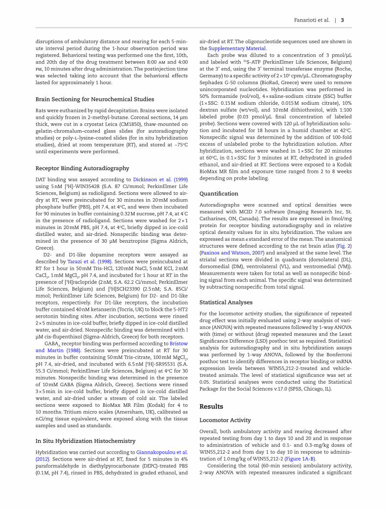
Autoradiographs were scanned and optical densities were measured with MCID 7.0 software (Imaging Research Inc, St. Catharines, ON, Canada). The results are expressed in fmol/mg protein for receptor binding autoradiography and in relative optical density values for in situ hybridization. The values are expressed as mean ± standard error of the mean. The anatomical structures were defined according to the rat brain atlas (Fig. 2) (Paxinos and Watson, 2007) and analyzed at the same level. The striatal sections were divided in quadrants (dorsolateral (DL), dorsomedial (DM), ventrolateral (VL), and ventromedial (VM)). Measurements were taken for total as well as nonspecific bind-ing signal from each animal. The specific signal was determined by subtracting nonspecific from total signal.
Statistical Analyses
For the locomotor activity studies, the significance of repeated drug effect was initially evaluated using 2-way analysis of vari-ance (ANOVA) with repeated measures followed by 1-way ANOVA with (time) or without (drug) repeated measures and the Least Significance Difference (LSD) posthoc test as required. Statistical analysis for autoradiography and in situ hybridization assays was performed by 1-way ANOVA, followed by the Bonferroni posthoc test to identify differences in receptor binding or mRNA expression levels between WIN55,212-2-treated and vehicle-treated animals. The level of statistical significance was set at 0.05. Statistical analyses were conducted using the Statistical Package for the Social Sciences v.17.0 (SPSS, Chicago, IL).
Results
Locomotor Activity
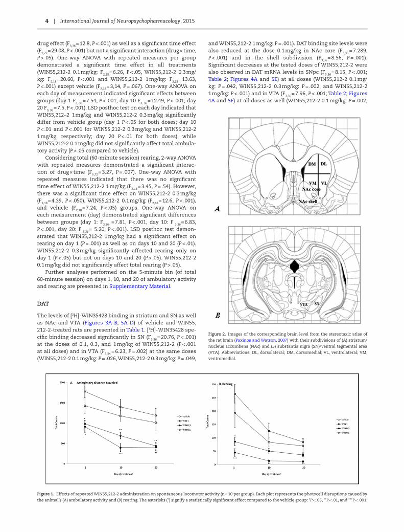
Overall, both ambulatory activity and rearing decreased after repeated testing from day 1 to days 10 and 20 and in response to administration of vehicle and 0.1- and 0.3-mg/kg doses of WIN55,212-2 and from day 1 to day 10 in response to adminis-tration of 1.0 mg/kg of WIN55,212-2 (Figure 1A-B).
Considering the total (60-min session) ambulatory activity, 2-way ANOVA with repeated measures indicated a significant
4 | International Journal of Neuropsychopharmacology, 2015
drug effect (F3,36 = 12.8, P < .001) as well as a significant time effect (F2,72 = 29.08, P < .001) but not a significant interaction (drug × time, P > .05). One-way ANOVA with repeated measures per group demonstrated a significant time effect in all treatments (WIN55,212-2 0.1 mg/kg: F2,18 = 6.26, P < .05, WIN55,212-2 0.3 mg/kg: F2,18 = 20.60, P < .001 and WIN55,212-2 1 mg/kg: F2,18 = 13.63, P < .001) except vehicle (F2,18 = 3,14, P = .067). One-way ANOVA on each day of measurement indicated significant effects between groups (day 1 F3, 36 = 7.54, P < .001; day 10 F3, 36 = 12.49, P < .001; day 20 F3, 36 = 7.5, P < .001). LSD posthoc test on each day indicated that WIN55,212–2 1 mg/kg and WIN55,212-2 0.3 mg/kg significantly differ from vehicle group (day 1 P < .05 for both doses; day 10 P < .01 and P < .001 for WIN55,212-2 0.3 mg/kg and WIN55,212-2 1 mg/kg, respectively; day 20 P < .01 for both doses), while WIN55,212-2 0.1 mg/kg did not significantly affect total ambula-tory activity (P > .05 compared to vehicle).
Considering total (60-minute session) rearing, 2-way ANOVA with repeated measures demonstrated a significant interac-tion of drug × time (F6,72 = 3.27, P = .007). One-way ANOVA with repeated measures indicated that there was no significant time effect of WIN55,212-2 1 mg/kg (F2,18 = 3.45, P = .54). However, there was a significant time effect on WIN55,212-2 0.3 mg/kg (F2,18 = 4.39, P < .050), WIN55,212-2 0.1 mg/kg (F2,18 = 12.6, P < .001), and vehicle (F2,18 = 7.24, P < .05) groups. One-way ANOVA on each measurement (day) demonstrated significant differences between groups (day 1: F3,36 = 7.81, P < .001, day 10: F 3,36 = 6.83, P < .001, day 20: F 3,36 = 5.20, P < .001). LSD posthoc test demon-strated that WIN55,212-2 1 mg/kg had a significant effect on rearing on day 1 (P = .001) as well as on days 10 and 20 (P < .01). WIN55,212-2 0.3 mg/kg significantly affected rearing only on day 1 (P < .05) but not on days 10 and 20 (P > .05). WIN55,212-2 0.1 mg/kg did not significantly affect total rearing (P > .05).
Further analyses performed on the 5-minute bin (of total 60-minute session) on days 1, 10, and 20 of ambulatory activity and rearing are presented in Supplementary Material.
DAT
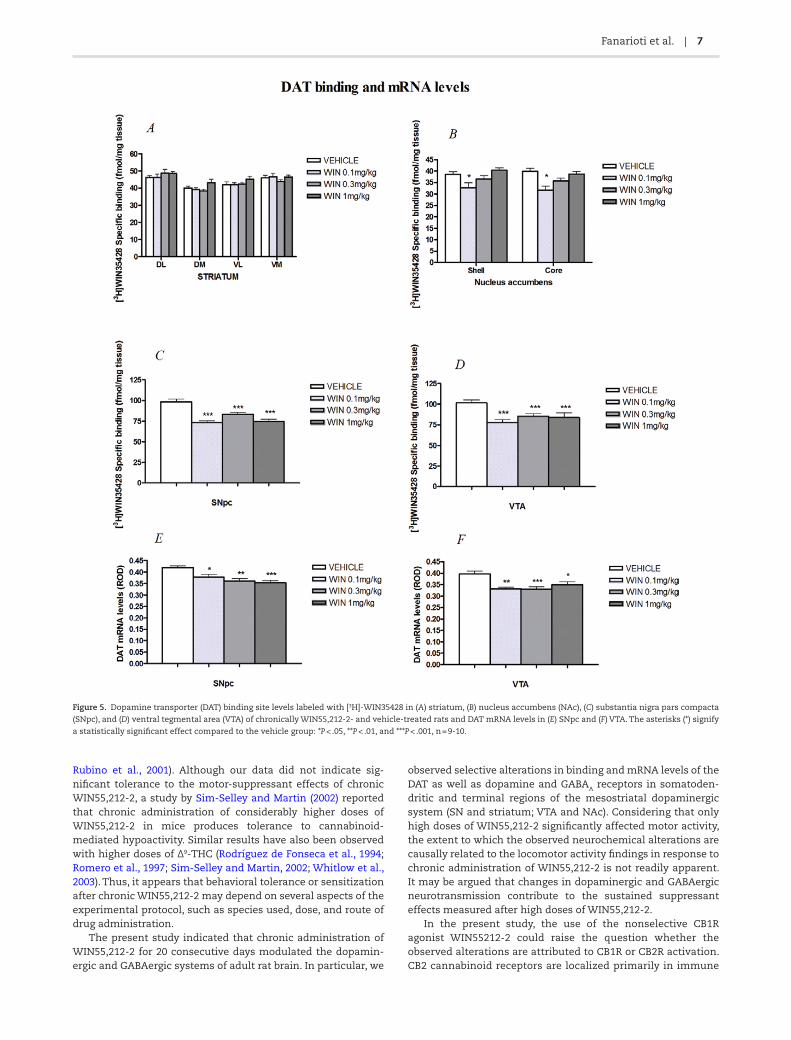
The levels of [3H]-WIN35428 binding in striatum and SN as well as NAc and VTA (Figures 3A-B, 5A-D) of vehicle and WIN55, 212-2-treated rats are presented in Table 1. [3H]-WIN35428 spe-cific binding decreased significantly in SN (F3,36 = 20.76, P < .001) at the doses of 0.1, 0.3, and 1 mg/kg of WIN55,212-2 (P < .001 at all doses) and in VTA (F3,34 = 6.23, P = .002) at the same doses (WIN55,212-2 0.1 mg/kg: P = .026, WIN55,212-2 0.3 mg/kg: P = .049,
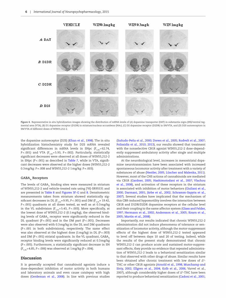
and WIN55,212-2 1 mg/kg: P = .001). DAT binding site levels were also reduced at the dose 0.1 mg/kg in NAc core (F3,36 = 7.289, P < .001) and in the shell subdivision (F3,36 = 8.56, P = .001). Significant decreases at the tested doses of WIN55,212-2 were also observed in DAT mRNA levels in SNpc (F3,36 = 8.15, P < .001; Table 2; Figures 4A and 5E) at all doses (WIN55,212-2 0.1 mg/kg: P = .042, WIN55,212-2 0.3 mg/kg: P = .002, and WIN55,212-2 1 mg/kg: P < .001) and in VTA (F3,36 = 7.96, P < .001; Table 2; Figures 4A and 5F) at all doses as well (WIN55,212-2 0.1 mg/kg: P = .002,
Figure 2. Images of the corresponding brain level from the stereotaxic atlas of
the rat brain (Paxinos and Watson, 2007) with their subdivisions of (A) striatum/
nucleus accumbens (NAc) and (B) substantia nigra (SN)/ventral tegmental area
(VTA). Abbreviations: DL, dorsolateral; DM, dorsomedial; VL, ventrolateral; VM,
ventromedial.
Figure 1. Effects of repeated WIN55,212-2 administration on spontaneous locomotor activity (n = 10 per group). Each plot represents the photocell disruptions caused by
the animal’s (A) ambulatory activity and (B) rearing. The asterisks (*) signify a statistically significant effect compared to the vehicle group: *P < .05, **P < .01, and ***P < .001.

Fanarioti et al. | 5
WIN55,212-2 0.3 mg/kg: P = .001, and WIN55,212-2 1 mg/kg: P = .032). Furthermore, no significant differences between vehi-cle- and WIN55,212-2-treated rats were observed in DAT bind-ing levels in the striatum (Table 1).
D1DRs
The levels of D1DR binding (Figures 3C and 6 A-B) and mRNA levels (Figures 4B and 6 C-D) in the terminal regions of the mesostriatal dopaminergic system of vehicle- and WIN55,212-2-treated rats are presented in Tables 3 and 4. Specific binding of [3H]-SCH23390 (F3,34 = 3.882, P = .039; Table 3) and D1DR mRNA levels (F3,15 = 13.176, P = .019, Table 4) increased significantly at the dose of 1 mg/kg in the core subdivision of NAc. No effects were observed at the other 2 doses.
D2DRs
The levels of D2DR binding, as determined by [3H]-raclopride specific binding (Figures 3D-E and 7A-D), in the mesostriatal dopaminergic regions of vehicle- and WIN55,212-2-treated rats are presented in Table 5. Significantly decreased levels of D2DR binding were observed in the medial quadrants of striatum (DM: F3,35 = 4.56, P = .008; VM: F3,35 = 4.64, P = .008) at the doses of 0.1 (DM: P = .011 and VM: P = .013) and 1 mg/kg (DM: P = .030 and VM: P = .019). However, as seen in Table 6, no significant alterations were observed in D2DR mRNA levels in SN and VTA (Figures 4C, 7 E-F). Furthermore, we examined the mRNA levels of the D2S iso-form (Figures 4D and 7 G-H), which corresponds to a splice vari-ant of the D2DR, showing an expression pattern presynaptically on dopaminergic neurons of SNpc and VTA and likely represents
Figure 3. Autoradiographic localization of (A) dopamine transporter (DAT) in striatum/nucleus accumbens (NAc) and (B) substantia nigra (SN)/ventral tegmental area
(VTA); (C) D1 dopamine receptor (D1DR) in striatum/NAc; (D) D2 dopamine receptor (D2DR) in striatum/NAc and (E) SN/VTA; (F) GABAA receptors in striatum/NAc; and
(G) SN/VTA of different doses of WIN55,212-2.
6 | International Journal of Neuropsychopharmacology, 2015
the dopamine autoreceptor (D2S) (Khan et al., 1998). The in situ hybridization histochemistry study for D2S mRNA revealed significant differences in mRNA levels in SNpc (F3,36 = 62.74, P < .001) and VTA (F3,36 = 5.93, P = .002). Particularly, statistically significant decreases were observed at all doses of WIN55,212-2 in SNpc (P < .001) as described in Table 7, while in VTA, signifi-cant decreases were observed at the higher doses (WIN55,212-2 0.3 mg/kg: P = .008 and WIN55,212-2 1 mg/kg: P = .003).
GABAA Receptors
The levels of GABAA binding sites were measured in striatum of WIN55,212-2 and vehicle-treated rats using [3H]-SR95531 and are presented in Table 8 and Figures 3F-G and 8. Densitometric measurements taken from striatum showed statistically sig-nificant decreases in DL (F3,36 = 9.95, P < .001) and DM (F3,36 = 19.42, P < .001) quadrants at all doses tested, as well as at 0.3 mg/kg in the VL subdivision (F3,36 = 5.43, P = .003). More specifically, at the lowest dose of WIN55,212-2 (0.1 mg/kg), the observed bind-ing levels of GABAA receptor were significantly reduced in the DL quadrant (P = .001) and in the DM part (P < .001). Decreases were also observed with 0.3 mg/kg in the DL and DM quadrants (P < .001 in both subdivisions), respectively. The same effect was also observed at the highest dose (1 mg/kg) in DL (P = .003) and DM (P < .001) striatal quadrants. In the VL quadrant, GABAA receptor binding levels were significantly reduced at 0.3 mg/kg (P = .005). Furthermore, a statistically significant decrease in SN (F3,36 = 4.85, P = .006) was observed at 1 mg/kg (P = .004).
Discussion
It is generally accepted that cannabinoid agonists induce a dose-dependent inhibition of motor activity in both humans and laboratory animals and even cause catalepsy with high doses (Gerdeman et al., 2008). In line with previous studies
(Sañudo-Peña et al., 2000; Drews et al., 2005; Rodvelt et al., 2007; Polissidis et al., 2010; 2013), our results showed that treatment with the nonselective CB1R agonist WIN55,212-2 dose-depend-ently suppressed ambulatory activity after single and multiple administrations.
At the neurobiological level, increases in mesostriatal dopa-mine neurotransmission have been associated with increased spontaneous locomotor activity after treatment with a variety of substances of abuse (Nestler, 2005; Lüscher and Malenka, 2011). However, most of the CNS actions of cannabinoids are mediated via CB1R (Gardner, 2005; Hashimotodani et al., 2007; Vlachou et al., 2008), and activation of these receptors in the striatum is associated with inhibition of motor behaviors (Giuliani et al., 2000; Darmani, 2001; Järbe et al., 2002; Schramm-Sapyta et al., 2007). Several studies have implicated that the mechanism of this CBR-induced hypomotility involves the interaction between CB1R and D1DR/D2DR dopamine receptors at the cellular level and their coupling to the same affector system (Glass and Felder, 1997; Hermann et al., 2002; Andersson et al., 2005; Kearn et al., 2005; Martín et al., 2008).
Importantly, our results indicated that chronic WIN55,212-2 administration did not induce phenomena of tolerance or sen-sitization of locomotor activity, although the motor-suppressant effects of the highest dose of WIN55,212-2 tested appeared to level off between days 10 and 20 of testing. Indeed, while the results of the present study demonstrated that chronic WIN55,212-2 can produce acute and sustained motor-suppres-sant effects, they provide no evidence that repeated administra-tion of WIN55,212-2 leads to a behavioral sensitization similar to that observed with other drugs of abuse. Similar results have been obtained after chronic treatment with low doses of Δ9-THC or other CB1R agonists (Arnold et al., 1998; Muschamp and Siviy, 2002; Ellgren et al., 2004; Kolb et al., 2006; Varvel et al., 2007), although considerably higher doses of Δ9-THC have been reported to produce behavioral sensitization (Cadoni et al., 2001;
Figure 4. Representative in situ hybridization images showing the distribution of mRNA levels of (A) dopamine transporter (DAT) in substantia nigra (SN)/ventral teg-
mental area (VTA), (B) D1 dopamine receptor (D1DR) in striatum/nucleus accumbens (NAc), (C) D2 dopamine receptor (D2DR) in SN/VTA, and (D) D2S autoreceptors in
SN/VTA of different doses of WIN55,212-2.
Fanarioti et al. | 7
Rubino et al., 2001). Although our data did not indicate sig-nificant tolerance to the motor-suppressant effects of chronic WIN55,212-2, a study by Sim-Selley and Martin (2002) reported that chronic administration of considerably higher doses of WIN55,212-2 in mice produces tolerance to cannabinoid-mediated hypoactivity. Similar results have also been observed with higher doses of Δ9-THC (Rodríguez de Fonseca et al., 1994; Romero et al., 1997; Sim-Selley and Martin, 2002; Whitlow et al., 2003). Thus, it appears that behavioral tolerance or sensitization after chronic WIN55,212-2 may depend on several aspects of the experimental protocol, such as species used, dose, and route of drug administration.
The present study indicated that chronic administration of WIN55,212-2 for 20 consecutive days modulated the dopamin-ergic and GABAergic systems of adult rat brain. In particular, we
observed selective alterations in binding and mRNA levels of the DAT as well as dopamine and GABAA receptors in somatoden-dritic and terminal regions of the mesostriatal dopaminergic system (SN and striatum; VTA and NAc). Considering that only high doses of WIN55,212-2 significantly affected motor activity, the extent to which the observed neurochemical alterations are causally related to the locomotor activity findings in response to chronic administration of WIN55,212-2 is not readily apparent. It may be argued that changes in dopaminergic and GABAergic neurotransmission contribute to the sustained suppressant effects measured after high doses of WIN55,212-2.
In the present study, the use of the nonselective CB1R agonist WIN55212-2 could raise the question whether the observed alterations are attributed to CB1R or CB2R activation. CB2 cannabinoid receptors are localized primarily in immune
Figure 5. Dopamine transporter (DAT) binding site levels labeled with [3H]-WIN35428 in (A) striatum, (B) nucleus accumbens (NAc), (C) substantia nigra pars compacta
(SNpc), and (D) ventral tegmental area (VTA) of chronically WIN55,212-2- and vehicle-treated rats and DAT mRNA levels in (E) SNpc and (F) VTA. The asterisks (*) signify
a statistically significant effect compared to the vehicle group: *P < .05, **P < .01, and ***P < .001, n = 9-10.

8 | International Journal of Neuropsychopharmacology, 2015
cells both in the periphery (Griffin et al., 2000) and brain micro-glia (Maresz et al., 2007), indicating that they are activated after brain damage or injury. In addition, they are expressed in neurons, including the striatum and midbrain (Gong et al., 2006), but the extent and level of expression remain contro-versial (Atwood and Mackie, 2010). However, most of the CNS actions of cannabinoids appear to be mediated via CB1R (Gardner, 2005; Hashimotodani et al., 2007; Vlachou et al., 2008). Systemic administration of CB1R agonists such as 9Δ-THC and WIN55,212-2 suppress motor activity, especially at higher doses (Darmani, 2001; Polissidis et al., 2013), and this effect is reversed by CB1R specific antagonists.
It is well known that DAT is localized to plasma membranes of axon terminals as well as dendrites of SNpc dopaminergic neurons (Nirenberg et al., 1996) and plays a role in reuptake of dopamine into dendrites and axon terminals (Cheramy et al., 1981). It has been reported that endogenous or exogenous (eg, WIN55,212-2) cannabinoids inhibit DAT activity in vitro (Chen et al., 2003; Steffens and Feuerstein, 2004), while other studies do not show any effect (Cheer et al., 2004; Köfalvi et al., 2005).
In the present study, we observed reduced DAT mRNA and binding levels in VTA and SNpc at all doses of WIN55,212-2, while DAT binding levels were not altered in striatum but were reduced in both NAc shell and core only at the lowest dose. These results suggest that the expression of DAT decreased at both the protein and mRNA levels in mesolimbic and nigrostri-atal dopaminergic neurons and this effect is not dose depend-ent, with the exception of the NAc. It is interesting to point out that the effect of chronic administration of cannabinoids on
DAT binding levels is evident only at the somatodendritic level of dopamine neurons and not at their axonal terminals. These brain regional differences may be a consequence of differential effect of CB1R density and function, as it is known that repeated treatment with cannabinoids (including WIN55,212-2) induces downregulation of the cannabinoid receptor (Oviedo et al., 1993; Breivogel et al., 1999; Sim-Selley, 2003; Tanda and Goldberg, 2003; Sim-Selley et al., 2006; Wu et al., 2008). In support of this notion, Moranta et al. (2009) reported brain regional differences in the synthesis of brain monoamines after chronic treatment with WIN55,212-2.
In vivo electrophysiological studies have shown that cannab-inoid agonists increase cell firing of the dopaminergic neurons located in SNpc and VTA (French et al., 1997; Melis et al., 2000; Wu and French, 2000). The increased activity of SNpc neurons is in agreement with in vivo microdialysis experiments showing enhanced dopamine release in the striatum after cannabinoid agonist treatment (Tanda et al., 1997; Solinas et al., 2008; Moranta et al., 2009; Polissidis et al., 2010, 2013). Taking into account the above findings and our results of decreased binding and mRNA levels of DAT in SN and VTA, we could suggest that chronic acti-vation of cannabinoid receptors may lead to decreased dopa-mine uptake by the dendrites of mesolimbic and nigrostriatal dopaminergic neurons, but not by the nigrostriatal terminals. Furthermore, the increased extracellular levels of dopamine in the striatum could be attributed to saturation of DAT (Oleson and Cheer, 2012; Tye et al., 2013) and/or to decreased inhibitory control over dopamine release due to stimulation of D2S (see below).
Table 1. DAT Binding Site Levels Labeled with [3H]-WIN35428 in Striatum, Nucleus Accumbens, SNpc, and VTA of Chronically WIN55,212-2- and Vehicle-Treated Rats
Brain Region Vehicle WIN 0.1 mg/kg WIN 0.3 mg/kg WIN 1 mg/kg
Dorsolateral striatum 46.22 ± 1.08n = 10
46.23 ± 1.93n = 10
48.70 ± 2.18n = 10
46.65 ± 1.09n = 10
Dorsomedial striatum 40.11 ± 1.01n = 10
39.33 ± 0.96n = 10
38.28 ± 1.04n = 10
43.00 ± 2.09n = 10
Ventrolateral striatum 41.99 ± 1.56n = 10
42.07 ± 0.98n = 10
42.33 ± 1.11n = 10
45.19 ± 1.68n = 10
Ventromedial striatum 46.08 ± 1.32n = 10
46.54 ± 1.97n = 10
43.79 ± 1.14n = 10
46.40 ± 1.15n = 10
Nucleus accumbens shell 38.55 ± 1.27n = 10
32.73 ± 2.14***↓15.1% n = 10
36.51 ± 1.42n = 10
40.43 ± 0.96n = 10
Nucleus accumbens core 39.92 ± 1.27n = 10
31.59 ± 1.68***↓20.9% n = 10
35.63 ± 1.34n = 10
38.74 ± 1.18n = 10
SNpc 98.48 ± 3.33n = 10
73.49 ± 1.69***↓25.4% n = 10
83.39 ± 2.33***↓15.3% n = 10
74.79 ± 2.47***↓24.05% n = 10
VTA 101.47 ± 3.61n = 9
78.10 ± 3.28*↓23.03% n = 10
85.70 ± 2.93*↓15.5% n = 10
83.85 ± 5.75***↓17.4% n = 9
Abbreviations: SNpc, substantia nigra pars compacta; VTA, ventral tegmental area. The asterisks (*) signify a statistically significant effect compared to the vehicle
group: *P < .05, **P < .01, and ***P < .001, n = 9-10.
Table 2. DAT mRNA Levels in SNpc and VTA of Chronically WIN55,212-2- and Vehicle-Treated Rats
Brain Region Vehicle WIN 0.1 mg/kg WIN 0.3 mg/kg WIN 1 mg/kg
SNpc 0.42 ± 0.008 0.378 ± 0.0122*↓10%
0.361 ± 0.010**↓14.05%
0.353 ± 0.011***↓15.95%
VTA 0.413 ± 0.011 0.332 ± 0.006**↓19.6%
0.331 ± 0.010***↓19.8%
0.349 ± 0.013*↓15.5%
Abbreviations: SNpc, substantia nigra pars compacta; VTA, ventral tegmentall area. The asterisks (*) signify a statistically significant effect compared to the vehicle
group: *P < .05, **P < .01, and ***P < .001, n = 9-10.

Fanarioti et al. | 9
Figure 6. D1 dopamine receptor (D1DR) binding site levels labeled with [3H]-SCH23390 in (A) striatum and (B) nucleus accumbens (NAc) and D1DR mRNA levels in (C)
striatum and (D) NAc of chronically WIN55,212-2- and vehicle-treated rats. The asterisk (*) denotes a statistically significant effect of WIN55,212-2 compared to the
vehicle group: *P < .05; n = 9-10 for receptor autoradiography and n = 5 per group for in situ hybridization.
Table 3. DRD1 Binding Site Levels Labeled with [3H]-SCH23390 in Striatum, Nucleus Accumbens, SNpc, and VTA of Chronically WIN55,212-2- and Vehicle-Treated Rats
Brain Region Vehicle WIN 0.1mg/kg WIN0.3mg/kg WIN1mg/kg
Dorsolateral striatum 118.45 ± 3.82n = 10
112.27 ± 1.71n = 10
121.61 ± 1.22n = 10
117.88 ± 4.28n = 10
Dorsomedial striatum 118.19 ± 3.31n = 10
115.59 ± 2.26n = 10
115.92 ± 3.10n = 10
117.36 ± 4.76n = 10
Ventrolateral striatum 121.50 ± 1.87n = 10
117.91 ± 2.76n = 10
120.15 ± 2.82n = 10
122.18 ± 3.12n = 10
Ventromedial striatum 119.36 ± 3.10n = 10
115.33 ± 1.45n = 10
123.24 ± 3.13n =10
123.32 ± 1.83n = 10
Nucleus accumbens shell 118.79 ± 3.87n = 10
125.80 ± 8.26n = 9
116.71 ± 3.56n = 10
118.05 ± 5.72n = 10
Nucleus accumbens core 102.47 ± 3.47n = 10
105.88 ± 4.49n = 9
101.72 ± 2.91n = 10
116.24 ± 2.15*↑13.44% n = 9
The asterisk (*) denotes a statistically significant effect of WIN55,212-2 compared to the vehicle group: P < .05; n = 9-10.
Table 4. DRD1 mRNA Levels in Striatum and Nucleus Accumbens of Chronically WIN55,212-2- and Vehicle-Treated Rats
Brain Region Vehicle WIN 0.1 mg/kg WIN 0.3 mg/kg WIN 1 mg/kg
Dorsolateral striatum 0.20 ± 0.0014 0.202 ± 0.0035 0.202 ± 0.0042 0.208 ± 0.0043Dorsomedial striatum 0.203 ± 0.0031 0.201 ± 0.0032 0.201 ± 0.0037 0.20872 ± 0.0043Ventrolateral striatum 0.21 ± 0.0025 0.209 ± 0.0043 0.209 ± 0.0054 0.209 ± 0.0047Ventromedial striatum 0.197 ± 0.0039 0.197 ± 0.0027 0.197 ± 0.0032 0.201 ± 0.0046Nucleus accumbens shell 0.22 ± 0.004 0.215 ± 0.007 0.22 ± 0.004 0.24 ± 0.006Nucleus accumbens core 0.18 ± 0.0018 0.18 ± 0.0031 0.172 ± 0.002 0.194 ± 0.003**
↑7.8%
The asterisk (*) signify a statistically significant effect compared to the vehicle group: **P < .01; n = 5 per group.

10 | International Journal of Neuropsychopharmacology, 2015
Figure 7. D2 dopamine receptor (D2DR) binding site levels labeled with [3H]-raclopride in (A) striatum, (B) nucleus accumbens (NAc), (C) substantia nigra (SN) and (D)
ventral tegmental area (VTA), D2DR receptor mRNA levels in (E) substantia nigra pars compacta (SNpc) and (F) VTA and D2 autoreceptor (D2S) mRNA levels in (G) SNpc
and (H) VTA of chronically WIN55,212- and vehicle-treated rats. The asterisk (*) denotes a statistically significant effect of WIN55,212-2 compared to the vehicle group:
*P < .05, **P < .01, and ***P < .001, n = 9-10.

Fanarioti et al. | 11
D2DRs exist in 2 isoforms (D2S, D2L) generated by alterna-tive splicing of the same gene (Giros et al., 1989). D2Ss are local-ized presynaptically on both the somatodendritic and terminal regions of midbrain dopaminergic neurons (Khan et al., 1998). Activation of these receptors in mesencephalic dopaminergic neurons elicits hyperpolarization and decreases firing rate (Lacey et al., 1987; Mercuri et al., 1989, 1997; Centonze et al., 2002) while inhibiting dopamine release in their somatodendritic (Cragg and Greenfield, 1997) and terminal mesostriatal regions (Starke et al., 1989; Cragg and Greenfield, 1997; Usiello et al., 2000). Thus, dopa-mine release is under the inhibitory control of D2DRs (Benoit-Marand et al., 2001). In the present study, the mRNA levels of the short isoform of D2 receptor (D2S), which corresponds to the presynaptic D2DR, were reduced in SNpc and VTA after chronic administration of the CB1R agonist, WIN55,212-2. Using [3Η]raclopride, which labels D2DR binding sites but does not dis-tinguish between presynaptic and postsynaptic localization of D2DR, we were unable to detect any changes of the D2DR at the
binding site level in SN and VTA. However, decreased D2DR bind-ing levels were found in the striatum of WIN-treated rats. This change could be correlated to the decreased mRNA expression of D2S observed in SNpc and may reflect a reduction in striatal D2S. However, further immunohistochemical studies using an anti-body specific for the D2S are required to verify this change. Our finding is in contrast to the results of Ginovart et al. (2012), who showed increased presynaptic D2/3 autoreceptor binding levels after chronic Δ9-THC administration, but are in agreement with the results of Bossong et al. (2009), who showed reduced [14C]raclopride binding in human striatum after Δ9-THC inhalation.
Overall, we could suggest that the somatodendritic regions of the mesostriatal dopaminergic system, the SN and VTA, seem to be more affected than the projection regions, the striatum and NAc, by chronic administration of WIN55,212-2. Decreased mRNA levels of D2S and DAT may lead to increased activity of SNpc and VTA neurons and enhanced release of dopamine at their somatodendritic and terminal fields. This finding may be
Table 6. DRD2 Receptor mRNA Levels in SNpc and VTA of Chronically WIN55,212-2- and Vehicle-Treated Rats
Brain Region Vehicle WIN 0.1 mg/kg WIN 0.3 mg/kg WIN 1 mg/kg
SNpc 0.411 ± 0.021n = 9
0.439 ± 0.008n = 10
0.413 ± 0.014n = 10
0.456 ± 0.0052n = 10
VTA 0.356 ± 0.031n = 9
0.375 ± 0.024n = 10
0.382 ± 0.021n = 10
0.391 ± 0.024n = 10
Abbreviations: SNpc, substantia nigra pars compacta; VTA, ventral tegmental area; n = 9-10.
Table 5. DRD2 Binding Site Levels Labeled with [3H]-raclopride in Striatum, Nucleus Accumbens, SN, and VTA of Chronically WIN55,212- and Vehicle-Treated Rats
Brain Region Vehicle WIN 0.1 mg/kg WIN 0.3 mg/kg WIN 1 mg/kg
Dorsolateral striatum 221.31 ± 5.89n = 9
207.98 ± 7.35n = 10
214.27 ± 8.47n = 10
204.46 ± 6.88n = 10
Dorsomedial striatum 210.02 ± 8.87n = 9
175.06 ± 7.29*↓16.65% n = 10
191.20 ± 4.75n = 10
178.97 ± 7.68*↓14.8% n = 10
Ventrolateral striatum 221.08 ± 2.87n = 9
191.01 ± 7.40n = 10
191.72 ± 5.34n = 10
205.10 ± 6.84n = 10
Ventromedial striatum 203.61 ± 5.06n = 9
170.47 ± 5.87*↓16.3% n = 10
182.95 ± 5.44n = 10
171.93 ± 6.14*↓15.56% n = 10
Nucleus accumbens shell 54.95 ± 1.87n = 10
53.21 ± 2.95n = 9
62.00 ± 2.43n = 9
58.07 ± 2.88n = 10
Nucleus accumbens core 56.27 ± 3.26n = 9
59.64 ± 2.68n = 10
51.94 ± 2.17n = 10
48.95 ± 2.68n = 10
SN 53.53 ± 1.63n = 10
50.42 ± 0.89n = 10
55.05 ± 1.55n = 10
57.09 ± 2.18n = 10
VTA 41.28 ± 0.83n = 10
45.37 ± 1.61n = 10
44.66 ± 1.74n = 10
46.17 ± 2.07n = 10
Abbreviations: SN, substantia nigra; VTA, ventral tegmental area. The asterisk (*) denotes a statistically significant effect of WIN55,212-2 compared to the vehicle
group: *P < .05; n = 9-10.
Table 7. D2S mRNA Levels in SNpc and VTA of Chronically WIN55,212-2- and Vehicle-Treated Rats
Brain Region Vehicle WIN 0.1mg/kg WIN0.3mg/kg WIN1mg/kg
SNpc 0.46 ± 0.007 0.359 ± 0.005***↓21.96%
0.343 ± 0.006***↓25.43%
0.353 ± 0.009***↓23.26%
VTA 0.406 ± 0.022 0.36 ± 0.008 0.344 ± 0.005**↓15.27%
0.338 ± 0.005**↓16.75%
Abbreviations: SNpc, substantia nigra pars compacta; VTA, ventral tegmental area. The asterisks (*) signify a statistically significant effect compared to the vehicle
group: *P < .05, **P < .01, and ***P < .001; n = 10 per group.

12 | International Journal of Neuropsychopharmacology, 2015
attributed to the fact that chronic exposure to WIN55,212-2 can cause neuroadaptive alterations (ie, downregulation) of CB1 receptor (density and function) which is region specific (Sim-Selley and Martin, 2002).
It is generally accepted that regulation of dopamine release in striatum is mainly the consequence of alterations in dopa-minergic cell firing in the SNpc and the VTA. However, several studies have revealed local regulation of DA release by other
Table 8. GABAA Receptor Binding Site Levels Labeled with [3H]-SR955321 in Striatum, Nucleus Accumbens, SN, and VTA of Chronically WIN55,212-2- and Vehicle-Treated Rats
Brain Region Vehicle WIN 0.1mg/kg WIN0.3mg/kg WIN1mg/kg
Dorsolateral striatum 82.81 ± 2.08n = 10
72.44 ± 0.99***↓12.5% n = 10
71.41 ± 1.68***↓13.8% n = 10
73.83 ± 1.67**↓10.84% n = 10
Dorsomedial striatum 90.81 ± 1.84n = 10
79.05 ± 1.29***↓12.9% n = 10
78.73 ± 1.23***↓13.3% n = 10
77.22 ± 1.25***↓14.96% n = 10
Ventrolateral striatum 88.98 ± 1.40n = 10
87.16 ± 1.05n = 10
80.14 ± 0.96**↓9.9% n = 10
83.25 ± 2.75n = 10
Ventromedial striatum 100.21 ± 1.59n = 10
97.21 ± 2.89n = 10
95.29 ± 1.62n = 10
103.26 ± 1.07n = 10
Nucleus accumbens shell 111.90 ± 2.49n = 10
111.15 ± 1.89n = 10
109.23 ± 2.5n = 10
121.13 ± 2.44n = 10
Nucleus accumbens core 116.24 ± 1.72n = 10
112.59 ± 3.35n = 10
115.06 ± 3.64n = 10
124.19 ± 2.91n = 10
SN 84.13 ± 1.26n = 10
77.22 ± 3.13n = 10
80.05 ± 3.32n = 10
70.39 ± 2.25**↓16.33% n = 10
VTA 60.95 ± 3.00n = 9
55.24 ± 1.66n = 10
55.51 ± 1.08n = 10
57.62 ± 2.71n = 10
Abbreviations: SN, substantia nigra; VTA, ventral tegmental area. The asterisks (*) signify a statistically significant effect compared to the vehicle group: *P < .05,
**P < .01, and ***P < .001; n = 9-10.
Figure 8. GABAA receptor binding site levels labeled with [3H]-SR955321 in (A) striatum, (B) nucleus accumbens (NAc), (C) substantia nigra (SN), and (D) ventral tegmental
area (VTA) of chronically WIN55,212-2- and vehicle-treated rats. The asterisks (*) signify a statistically significant effect compared to the vehicle group: **P < .01, and
***P < .001, n = 9-10.

Fanarioti et al. | 13
neurotransmitters and modulators, such as glutamate. Recent studies suggest that the glutamatergic regulation of dopamine release is inhibitory (Rice et al., 2011). Taking into consideration the above and that activation of CB1R receptors on corticostri-atal terminals would inhibit glutamate release, this activation may lead to increased DA release.
Our results have also shown that D1DR mRNA and binding were not altered in striatum; however, in NAc core, both mRNA and binding levels were increased only at the highest dose. This dose-dependent effect is specific for the mesolimbic pathway, and it may be related to the pronounced motor-suppressant effects of the high dose of WIN22,212-2 that persisted over the course of administration. It has been suggested that simultane-ous stimulation of CB1R and D1DR reduces the inhibitory activ-ity of direct striatal projection neurons, resulting in a decreased motor response (Martín et al., 2008).
It is well known that NAc plays a pivotal role in reward and aversive learning and learning flexibility (Graybiel, 2008). In NAc, similar to dorsal striatum, MSNs express either D1DR or D2DR along with other receptors and neuropeptides, and their distinct roles in learning have only recently been explored (Nakanishi et al., 2014). Furthermore, recent investigations suggest a dif-ferential involvement of D1-MSNs and D2-MSN cell populations in NAc in drug-related behaviors (Laviolette et al., 2008; Hikida et al., 2010; Smith et al., 2013). In particular, distinct roles of D1DR and D2DR in the core and shell of NAc have been impli-cated in the modulation of reward by nicotine (Laviolette et al., 2008) and in the acquisition of cocaine-related learning (Espana and Jones, 2013; Smith et al., 2013).
Seif et al. (2011) provided evidence that endocannabinoids mediate the ability of DA receptors to enhance action poten-tial firing in NAc core neurons in vitro, requiring coactivation of D1DR and D2DR. The selective upregulation of D1DRs in NAc core observed in the present study after high doses of chronic can-nabinoid administration suggest that the core vs shell and D1DR vs D2DR MSNs of NAc may respond differently to repeated can-nabinoid administration, and these cell-type specific alterations in NAc core may contribute to cannabinoid-related behaviors.
It has also been shown that CB1R are localized presynapti-cally on GABAergic neurons in several brain regions (Matsuda et al., 1993) and their activation inhibits GABA release (French et al., 1997; Pistis et al., 2002; Szabo et al., 2002; Lupica and Riegel, 2005; Szabo and Schlicker, 2005). More precisely, in SN pars reticulata, CB1R are located on GABAergic striatonigral terminals. Furthermore, dendrites of dopaminergic neurons in SNpc extend into the SN pars reticulate, where they form syn-apses with CB1-containing axon terminals (Fitzgerald et al., 2012). Considering this localization of CB1R, a possible mech-anism for the increased dopamine release after cannabinoid treatment may involve an indirect disinhibition of dopamine neurons (French et al., 1997; Szabo et al., 2002; Lupica and Riegel, 2005). It is thus suggested that CB1R, because of their localiza-tion, can modulate GABA release and in turn control the activity of the dopaminergic cells in the midbrain (Wu and French, 2000; Laviolette and Grace, 2006; Fernández-Ruiz et al., 2010).
Our results have indicated that GABAA receptor binding lev-els were reduced in the dorsal striatum at all doses and SN at the highest dose. Therefore, in addition to the reduction of GABA released from the striatonigral terminals of the direct pathway, the observed reduction of GABAA receptors in SN at least at the higher dose would increase the activity of the GABAergic cells of SNr, leading to hypomobility. Overall, these results indicate that the effects of WIN55,212-2 on motor activity could be mediated at least partially via GABAA receptors expressed in the nigrostriatal
pathway. In conclusion, our data indicate that chronic admin-istration of the cannabinoid agonist WIN55,212-2 did not induce phenomena of tolerance or sensitization of locomotor activity. Furthermore, repeated cannabinoid administration induced neu-roadaptive alterations of the dopaminergic and GABAergic sys-tems in a region-, dose-, and neurotransmitter-dependent manner.
Acknowledgments
E. Fanarioti was supported by K. Karatheodoris (Research Committee, University of PatrasC902) and Polembros Shipping Limited grants.
Statement of Interest
None.
ReferencesAmeri A (1999) The effects of cannabinoids on the brain. Prog
Neurobiol 58:315–348.Andersson M, Usiello A, Borgkvist A, Pozzi L, Dominguez C, Fien-
berg AA, Svenningsson P, Fredholm BB, Borrelli E, Greengard P, Fisone G (2005) Cannabinoid action depends on phospho-rylation of dopamine- and cAMP-regulated phosphoprotein of 32 kDa at the protein kinase A site in striatal projection neurons. J Neurosci 25:8432–8438.
Arnold JC, Topple AN, Hunt GE, McGregor IS (1998) Effects of pre-exposure and co-administration of the cannabinoid receptor agonist CP 55,940 on behavioral sensitization to cocaine. Eur J Pharmacol 354:9–16.
Atwood BK, Mackie K (2010) CB2: a cannabinoid receptor with an identity crisis. Br J Pharmacol 160:467–479.
Benoit-Marand M, Borrelli E, Gonon F (2001) Inhibition of dopa-mine release via presynaptic D2 receptors: time course and functional characteristics in vivo. J Neurosci 21:9134–9141.
Blandini F, Nappi G, Tassorelli C, Martignoni E (2000) Functional changes of the basal ganglia circuitry in Parkinson’s disease. Prog Neurobiol 62:63–88.
Bossong MG, van Berckel BN, Boellaard R, Zuurman L, Schuit RC, Windhorst AD, van Gerven JM, Ramsey NF, Lammertsma AA, Kahn RS (2009) Delta 9-tetrahydrocannabinol induces dopa-mine release in the human striatum. Neuropsychopharma-cology 34:759–766.
Breivogel CS, Childers SR, Deadwyler SA, Hampson RE, Vogt LJ, Sim-Selley LJ (1999) Chronic delta9-tetrahydrocannabinol treatment produces a time-dependent loss of cannabinoid receptors and cannabinoid receptor-activated G proteins in rat brain. J Neurochem 73:2447–2459.
Bristow DR, Martin IL (1988) Light microscopic autoradio-graphic localisation in rat brain of the binding sites for the GABAA receptor antagonist [3H]SR 95531: comparison with the [3H]GABAA receptor distribution. Eur J Pharmacol 148:283–288.
Bunzow JR, Van Tol HH, Grandy DK, Albert P, Salon J, Christie M, Machida CA, Neve KA, Civelli O (1988) Cloning and expression of a rat D2 dopamine receptor cDNA. Nature 336:783–787.
Cadoni C, Pisanu A, Solinas M, Acquas E, Di Chiara G (2001) Behavioural sensitization after repeated exposure to delta 9-tetrahydrocannabinol and cross-sensitization with mor-phine. Psychopharmacology (Berl) 158:259–266.
Castañé A, Maldonado R, Valverde O (2004) Role of different brain structures in the behavioural expression of WIN 55,212-2 withdrawal in mice. Br J Pharmacol 142:1309–1317.

14 | International Journal of Neuropsychopharmacology, 2015
Centonze D, Picconi B, Baunez C, Borrelli E, Pisani A, Bernardi G, Calabresi P (2002) Cocaine and amphetamine depress stri-atal GABAergic synaptic transmission through D2 dopamine receptors. Neuropsychopharmacology 26:164–175.
Cheer JF, Wassum KM, Heien ML, Phillips PE, Wightman RM (2004) Cannabinoids enhance subsecond dopamine release in the nucleus accumbens of awake rats. J Neurosci 24:4393–4400.
Chen N, Appell M, Berfield JL, Reith ME (2003) Inhibition by ara-chidonic acid and other fatty acids of dopamine uptake at the human dopamine transporter. Eur J Pharmacol 478:89–95.
Cheramy A, Leviel V, Glowinski J (1981) Dendritic release of dopa-mine in the substantia nigra. Nature 289:537–542.
Cragg SJ, Greenfield SA (1997) Differential autoreceptor control of somatodendritic and axon terminal dopamine release in substantia nigra, ventral tegmental area, and striatum. J Neu-rosci 17:5738–5746.
Darmani NA (2001) The cannabinoid CB1 receptor antagonist SR 141716A reverses the antiemetic and motor depressant actions of WIN 55, 212-2. Eur J Pharmacol 430:49–58.
Dickinson SD, Sabeti J, Larson GA, Giardina K, Rubinstein M, Kelly MA, Grandy DK, Low MJ, Gerhardt GA, Zahniser NR (1999) Dopamine D2 receptor-deficient mice exhibit decreased dopamine transporter function but no changes in dopamine release in dorsal striatum. J Neurochem 72:148–156.
Drews E, Schneider M, Koch M (2005) Effects of the cannabinoid receptor agonist WIN 55,212-2 on operant behavior and loco-motor activity in rats. Pharmacol Biochem Behav 80:145–150.
Ellgren M, Hurd YL, Franck J (2004) Amphetamine effects on dopamine levels and behavior following cannabinoid expo-sure during adolescence. Eur J Pharmacol 497:205–213.
Espana RA, Jones SR (2013) Presynaptic dopamine modulation by stimulant self-administration. Front Biosci (Schol Ed) 5:261–276.
Fernández-Ruiz J, Hernández M, Ramos J (2010) Cannabinoid-dopamine interaction in the pathophysiology and treatment of CNS disorders. CNS Neurosci Ther 16:72–91.
Ferraro L, Tomasini MC, Gessa GL, Bebe BW, Tanganelli S, Antonelli T (2001) The cannabinoid receptor agonist WIN 55,212-2 regulates glutamate transmission in rat cerebral cortex: an in vivo and in vitro study. Cereb Cortex 11:728–733.
Fitzgerald ML, Shobin E, Pickel VM (2012) Cannabinoid modula-tion of the dopaminergic circuitry: implications for limbic and striatal output. Prog Neuropsychopharmacol Biol Psy-chiatry 38:21–29.
French ED, Dillon K, Wu X (1997) Cannabinoids excite dopamine neurons in the ventral tegmentum and substantia nigra. Neuroreport 8:649–652.
Gardner EL (2005) Endocannabinoid signaling system and brain reward: emphasis on dopamine. Pharmacol Biochem Behav 81:263–284.
Gerdeman GL, Schechter JB, French ED (2008) Context-specific reversal of cocaine sensitization by the CB1 cannabinoid receptor antagonist rimonabant. Neuropsychopharmacology 33:2747–2759.
Giannakopoulou D, Daguin-Nerrière V, Mitsacos A, Kouvelas ED, Neveu I, Giompres P, Brachet P (2012) Ectopic expression of TrKA in the adult rat basal ganglia induces both nerve growth factor-dependent and -independent neuronal responses. J Neurosci Res 90:1507–1521.
Gingrich JA, Caron MG (1993) Recent advances in the molecular biology of dopamine receptors. Annu Rev Neurosci 16:299–321.
Ginovart N, Tournier BB, Moulin-Sallanon M, Steimer T, Ibanez V, Millet P (2012) Chronic Δ⁹-tetrahydrocannabinol exposure
induces a sensitization of dopamine D₂/₃ receptors in the mesoaccumbens and nigrostriatal systems. Neuropsychop-harmacology 37:2355–2367.
Giros B, Sokoloff P, Martres MP, Riou JF, Emorine LJ, Schwartz JC (1989) Alternative splicing directs the expression of two D2 dopamine receptor isoforms. Nature 342:923–926.
Giros B, el Mestikawy S, Bertrand L, Caron MG (1991) Cloning and functional characterization of a cocaine-sensitive dopamine transporter. FEBS Lett 295:149–154.
Giuliani D, Ferrari F, Ottani A (2000) The cannabinoid agonist HU 210 modifies rat behavioural responses to novelty and stress. Pharmacol Res 41:47–53.
Glass M, Felder CC (1997) Concurrent stimulation of cannabinoid CB1 and dopamine D2 receptors augments cAMP accumula-tion in striatal neurons: evidence for a Gs linkage to the CB1 receptor. J Neurosci 17:5327–5333.
Gong JP, Onaivi ES, Ishiguro H, Liu QR, Tagliaferro PA, Brusco A, Uhl GR (2006) Cannabinoid CB2 receptors: immunohisto-chemical localization in rat brain. Brain Res 1071:10–23.
Graybiel AM (2008) Habits, rituals, and the evaluative brain. Annu Rev Neurosci 31:359–387.
Griffin G, Tao Q, Abood ME (2000) Cloning and pharmacological characterization of the rat CB(2) cannabinoid receptor. J Phar-macol Exp Ther 292:886–894.
Hashimotodani Y, Ohno-Shosaku T, Kano M (2007) Endocan-nabinoids and synaptic function in the CNS. Neuroscientist 13:127–137.
Herkenham M, Lynn AB, de Costa BR, Richfield EK (1991a) Neu-ronal localization of cannabinoid receptors in the basal gan-glia of the rat. Brain Res 547:267–274.
Herkenham M, Lynn AB, Johnson MR, Melvin LS, de Costa BR, Rice KC (1991b) Characterization and localization of cannabi-noid receptors in rat brain: a quantitative in vitro autoradio-graphic study. J Neurosci 11:563–583.
Hermann H, Marsicano G, Lutz B (2002) Coexpression of the cannabinoid receptor type 1 with dopamine and serotonin receptors in distinct neuronal subpopulations of the adult mouse forebrain. Neuroscience 109:451–460.
Hikida T, Kimura K, Wada N, Funabiki K, Nakanishi S (2010) Distinct roles of synaptic transmission in direct and indirect striatal pathways to reward and aversive behavior. Neuron 66:896–907.
Järbe TU, Andrzejewski ME, DiPatrizio NV (2002) Interactions between the CB1 receptor agonist Delta 9-THC and the CB1 receptor antagonist SR-141716 in rats: open-field revisited. Pharmacol Biochem Behav 73:911–919.
Julian MD, Martin AB, Cuellar B, Rodriguez De Fonseca F, Navarro M, Moratalla R, Garcia-Segura LM (2003) Neuroanatomical relation-ship between type 1 cannabinoid receptors and dopaminergic systems in the rat basal ganglia. Neuroscience 119:309–318.
Katona I, Sperlágh B, Sík A, Käfalvi A, Vizi ES, Mackie K, Freund TF (1999) Presynaptically located CB1 cannabinoid receptors regulate GABA release from axon terminals of specific hip-pocampal interneurons. J Neurosci 19:4544–4558.
Katona I, Sperlágh B, Maglóczky Z, Sántha E, Köfalvi A, Czirják S, Mackie K, Vizi ES, Freund TF (2000) GABAergic interneurons are the targets of cannabinoid actions in the human hip-pocampus. Neuroscience 100:797–804.
Katona I, Rancz EA, Acsady L, Ledent C, Mackie K, Hajos N, Fre-und TF (2001) Distribution of CB1 cannabinoid receptors in the amygdala and their role in the control of GABAergic transmission. J Neurosci 21:9506–9518.
Katsidoni V, Kastellakis A, Panagis G (2013) Biphasic effects of Δ9-tetrahydrocannabinol on brain stimulation reward and motor activity. Int J Neuropsychopharmacol 16:2273–2284.

Fanarioti et al. | 15
Kearn CS, Greenberg MJ, DiCamelli R, Kurzawa K, Hillard CJ (1999) Relationships between ligand affinities for the cerebel-lar cannabinoid receptor CB1 and the induction of GDP/GTP exchange. J Neurochem 72:2379–2387.
Kearn CS, Blake-Palmer K, Daniel E, Mackie K, Glass M (2005) Concurrent stimulation of cannabinoid CB1 and dopamine D2 receptors enhances heterodimer formation: a mechanism for receptor cross-talk? Mol Pharmacol 67:1697–1704.
Khan ZU, Mrzljak L, Gutierrez A, De La Calle A, Goldman-Rakic PS (1998) Prominence of the dopamine D2 short isoform in dopaminergic pathways. Proc Natl Acad Sci U S A 95:7731.
Köfalvi A, Rodrigues RJ, Ledent C, Mackie K, Vizi ES, Cunha RA, Sperlágh B (2005) Involvement of cannabinoid receptors in the regulation of neurotransmitter release in the rodent stri-atum: a combined immunochemical and pharmacological analysis. J Neurosci 25:2874–2884.
Kolb B, Gorny G, Limebeer CL, Parker LA (2006) Chronic treat-ment with Delta-9-tetrahydrocannabinol alters the structure of neurons in the nucleus accumbens shell and medial pre-frontal cortex of rats. Synapse 60:429–436.
Lacey MG, Mercuri NB, North RA (1987) Dopamine acts on D2 receptors to increase potassium conductance in neurones of the rat substantia nigra zona compacta. J Physiol 392:397–416.
Laviolette SR, Grace AA (2006) The roles of cannabinoid and dopamine receptor systems in neural emotional learning cir-cuits: implications for schizophrenia and addiction. Cell Mol Life Sci 63:1597–1613.
Laviolette SR, Lauzon NM, Bishop SF, Sun N, Tan H (2008) Dopa-mine signaling through D1-like versus D2-like receptors in the nucleus accumbens core versus shell differentially mod-ulates nicotine reward sensitivity. J Neurosci 28:8025–8033.
Lüscher C, Malenka RC (2011) Drug-evoked synaptic plasticity in addiction: from molecular changes to circuit remodeling. Neuron 69:650–663.
Lupica CR, Riegel AC (2005) Endocannabinoid release from midbrain dopamine neurons: a potential substrate for can-nabinoid receptor antagonist treatment of addiction. Neu-ropharmacology 48:1105–1116.
Mailleux P, Vanderhaeghen JJ (1992) Distribution of neuronal cannabinoid receptor in the adult rat brain: a comparative receptor binding radioautography and in situ hybridization histochemistry. Neuroscience 48:655–668.
Mailleux P, Vanderhaeghen JJ (1993) Dopaminergic regulation of cannabinoid receptor mRNA levels in the rat caudate-puta-men: an in situ hybridization study. J Neurochem 61:1705–1712.
Manzoni OJ, Bockaert J (2001) Cannabinoids inhibit GABAergic synaptic transmission in mice nucleus accumbens. Eur J Pharmacol 412:R3–5.
Maresz K, Pryce G, Ponomarev ED, Marsicano G, Croxford JL, Shriver LP, Ledent C, Cheng X, Carrier EJ, Mann MK, Giovan-noni G, Pertwee RG, Yamamura T, Buckley NE, Hillard CJ, Lutz B, Baker D, Dittel BN (2007) Direct suppression of CNS auto-immune inflammation via the cannabinoid receptor CB1 on neurons and CB2 on autoreactive T cells. Nat Med 13:492–497.
Martín AB, Fernandez-Espejo E, Ferrer B, Gorriti MA, Bilbao A, Navarro M, Rodriguez de Fonseca F, Moratalla R (2008) Expres-sion and function of CB1 receptor in the rat striatum: locali-zation and effects on D1 and D2 dopamine receptor-mediated motor behaviors. Neuropsychopharmacology 33:1667–1679.
Matsuda LA, Bonner TI, Lolait SJ (1993) Localization of cannabi-noid receptor mRNA in rat brain. J Comp Neurol 327:535–550.
Mavrikaki M, Markaki E, Nomikos GG, Panagis G (2010) Chronic WIN55,212-2 elicits sustained and conditioned increases in
intracranial self-stimulation thresholds in the rat. Behav Brain Res 209:114–118.
Melis M, Gessa GL, Diana M (2000) Different mechanisms for dopaminergic excitation induced by opiates and cannabi-noids in the rat midbrain. Prog Neuropsychopharmacol Biol Psychiatry 24:993–1006.
Mercuri NB, Calabresi P, Bernardi G (1989) The mechanism of amphetamine-induced inhibition of rat substantia nigra compacta neurones investigated with intracellular recording in vitro. Br J Pharmacol 98:127–134.
Mercuri NB, Bonci A, Bernardi G (1997) Electrophysiological phar-macology of the autoreceptor-mediated responses of dopa-minergic cells to antiparkinsonian drugs. Trends Pharmacol Sci 18:232–235.
Moranta D, Esteban S, García-Sevilla JA (2009) Chronic treatment and withdrawal of the cannabinoid agonist WIN 55,212–2 modulate the sensitivity of presynaptic receptors involved in the regulation of monoamine syntheses in rat brain. Naunyn Schmiedebergs Arch Pharmacol 379:61–72.
Morera-Herreras T, Ruiz-Ortega JA, Gómez-Urquijo S, Ugedo L (2008) Involvement of subthalamic nucleus in the stimula-tory effect of Delta(9)-tetrahydrocannabinol on dopaminergic neurons. Neuroscience 151:817–823.
Muschamp JW, Siviy SM (2002) Behavioral sensitization to amphet-amine follows chronic administration of the CB1 agonist WIN 55,212-2 in Lewis rats. Pharmacol Biochem Behav 73:835–842.
Nakanishi S, Hikida T, Yawata S (2014) Distinct dopaminergic control of the direct and indirect pathways in reward-based and avoidance learning behaviors. Neuroscience in press.
Nestler EJ (2005) Is there a common molecular pathway for addiction? Nat Neurosci 8:1445–1449.
Nirenberg MJ, Vaughan RA, Uhl GR, Kuhar MJ, Pickel VM (1996) The dopamine transporter is localized to dendritic and axonal plasma membranes of nigrostriatal dopaminergic neurons. J Neurosci 16:436–447.
Oleson EB, Cheer JF (2012) A brain on cannabinoids: the role of dopamine release in reward seeking. Cold Spring Harb Per-spect Med 2:1–13.
Oviedo A, Glowa J, Herkenham M (1993) Chronic cannabinoid administration alters cannabinoid receptor binding in rat brain: a quantitative autoradiographic study. Brain Res 616:293–302.
Pan B, Hillard CJ, Liu QS (2008) D2 dopamine receptor activation facilitates endocannabinoid-mediated long-term synaptic depression of GABAergic synaptic transmission in midbrain dopamine neurons via cAMP-protein kinase A signaling. J Neurosci 28:14018–14030.
Paxinos G, Watson C (2007) The rat brain in stereotaxic coordi-nates: Academic Pr.
Pertwee RG (2006) Cannabinoid pharmacology: the first 66 years. Br J Pharmacol 147:163–171.
Pertwee RG, Greentree SG, A. SP (1988) Drugs which stimulate or facilitate central GABAergic transmission interact synergisti-cally with delta-9-tetrahydrocannabinol to produce marked catalepsy in mice. Neuropharmacology 27:1265–1270.
Pistis M, Ferraro L, Pira L, Flore G, Tanganelli S, Gessa GL, Devoto P (2002) Delta(9)-tetrahydrocannabinol decreases extracellular GABA and increases extracellular glutamate and dopamine levels in the rat prefrontal cortex: an in vivo microdialysis study. Brain Res 948:155–158.
Polissidis A, Chouliara O, Galanopoulos A, Rentesi G, Dosi M, Hyphantis T, Marselos M, Papadopoulou-Daifoti Z, Nomikos GG, Spyraki C, Tzavara ET, Antoniou K (2010) Individual dif-ferences in the effects of cannabinoids on motor activity,

16 | International Journal of Neuropsychopharmacology, 2015
dopaminergic activity and DARPP-32 phosphorylation in distinct regions of the brain. Int J Neuropsychopharmacol 13:1175–1191.
Polissidis A, Galanopoulos A, Naxakis G, Papahatjis D, Papado-poulou-Daifoti Z, Antoniou K (2013) The cannabinoid CB1 receptor biphasically modulates motor activity and regulates dopamine and glutamate release region dependently. Int J Neuropsychopharmacol 16:393–403.
Rice ME, Patel JC, Cragg SJ (2011) Dopamine release in the basal ganglia. Neuroscience 198:112–137.
Rodríguez de Fonseca F, Gorriti MA, Fernández-Ruiz JJ, Palomo T, Ramos JA (1994) Downregulation of rat brain cannabinoid binding sites after chronic delta 9-tetrahydrocannabinol treatment. Pharmacol Biochem Behav 47:33–40.
Rodríguez De Fonseca F, Gorriti MA, Bilbao A, Escuredo L, García-Segura LM, Piomelli D, Navarro M (2001) Role of the endog-enous cannabinoid system as a modulator of dopamine transmission: implications for Parkinson’s disease and schiz-ophrenia. Neurotox Res 3:23–35.
Rodvelt KR, Bumgarner DM, Putnam WC, Miller DK (2007) WIN-55,212-2 and SR-141716A alter nicotine-induced changes in locomotor activity, but do not alter nicotine-evoked [3H]dopamine release. Life Sci 80:337–344.
Romero J, Garcia-Palomero E, Castro JG, Garcia-Gil L, Ramos JA, Fernandez-Ruiz JJ (1997) Effects of chronic exposure to delta9-tetrahydrocannabinol on cannabinoid receptor bind-ing and mRNA levels in several rat brain regions. Brain Res Mol Brain Res 46:100–108.
Rubino T, Viganò D, Massi P, Parolaro D (2001) The psychoactive ingredient of marijuana induces behavioural sensitization. Eur J Neurosci 14:884–886.
Sañudo-Peña MC, Romero J, Seale GE, Fernandez-Ruiz JJ, Walker JM (2000) Activational role of cannabinoids on movement. Eur J Pharmacol 391:269–274.
Schramm-Sapyta NL, Cha YM, Chaudhry S, Wilson WA, Swart-zwelder HS, Kuhn CM (2007) Differential anxiogenic, aversive, and locomotor effects of THC in adolescent and adult rats. Psychopharmacology (Berl) 191:867–877.
Seif T, Makriyannis A, Kunos G, Bonci A, Hopf FW (2011) The endocannabinoid 2-arachidonoylglycerol mediates D1 and D2 receptor cooperative enhancement of rat nucleus accum-bens core neuron firing. Neuroscience 193:21–33.
Shi LH, Luo F, Woodward DJ, Chang JY (2005) Dose and behavio-ral context dependent inhibition of movement and basal gan-glia neural activity by Delta-9-tetrahydrocannabinol during spontaneous and treadmill locomotion tasks in rats. Synapse 55:1–16.
Sim-Selley LJ, Martin BR (2002) Effect of chronic administra-tion of R-(+)-[2,3-Dihydro-5-methyl-3-[(morpholinyl)methyl]pyrrolo[1,2,3-de]-1,4-benzoxazinyl]-(1-naphthalenyl)metha-none mesylate (WIN55,212-2) or delta(9)-tetrahydrocannabi-nol on cannabinoid receptor adaptation in mice. J Pharmacol Exp Ther 303:36–44.
Sim-Selley LJ (2003) Regulation of cannabinoid CB1 receptors in the central nervous system by chronic cannabinoids. Crit Rev Neurobiol 15:91–119.
Sim-Selley LJ, Schechter NS, Rorrer WK, Dalton GD, Hernandez J, Martin BR, Selley DE (2006) Prolonged recovery rate of CB1 receptor adaptation after cessation of long-term cannabinoid administration. Mol Pharmacol 70:986–996.
Smith RJ, Lobo MK, Spencer S, Kalivas PW (2013) Cocaine-induced adaptations in D1 and D2 accumbens projection neurons (a dichotomy not necessarily synonymous with direct and indi-rect pathways). Curr Opin Neurobiol 23:546–552.
Solinas M, Goldberg SR, Piomelli D (2008) The endocannabinoid system in brain reward processes. Br J Pharmacol 154:369–383.
Starke K, Göthert M, Kilbinger H (1989) Modulation of neuro-transmitter release by presynaptic autoreceptors. Physiol Rev 69:864–989.
Steffens M, Feuerstein TJ (2004) Receptor-independent depres-sion of DA and 5-HT uptake by cannabinoids in rat neo-cortex--involvement of Na(+)/K(+)-ATPase. Neurochem Int 44:529–538.
Sunahara RK, Niznik HB, Weiner DM, Stormann TM, Brann MR, Kennedy JL, Gelernter JE, Rozmahel R, Yang Y, Israel Y (1990) Human dopamine D1 receptor encoded by an intronless gene on chromosome 5. Nature 347:80–83.
Szabo B, Siemes S, Wallmichrath I (2002) Inhibition of GABAergic neurotransmission in the ventral tegmental area by cannabi-noids. Eur J Neurosci 15:2057–2061.
Szabo B, Schlicker E (2005) Effects of cannabinoids on neuro-transmission. Handb Exp Pharmacol 168:327–365.
Tanda G, Pontieri FE, Di Chiara G (1997) Cannabinoid and heroin activation of mesolimbic dopamine transmission by a com-mon mu1 opioid receptor mechanism. Science 276:2048–2050.
Tanda G, Goldberg SR (2003) Cannabinoids: reward, dependence, and underlying neurochemical mechanisms--a review of recent preclinical data. Psychopharmacology (Berl) 169:115–134.
Tarazi FI, Campbell A, Yeghiayan SK, Baldessarini RJ (1998) Local-ization of dopamine receptor subtypes in corpus striatum and nucleus accumbens septi of rat brain: comparison of D1-, D2-, and D4-like receptors. Neuroscience 83:169–176.
Taylor DA, Sitaram BR, Elliot-Baker S (1988) Effect of 9-tetrahy-drocannabinol on release of dopamine in the corpus striatum of the rat. In: Chesher G, Consroe P, Musty R (eds)Marijuana: An International Research Report Australian Govt Publ Ser-vice: Canberra: pp 405–408.
Tye SJ, Miller AD, Blaha CD (2013) Ventral tegmental ionotropic glutamate receptor stimulation of nucleus accumbens tonic dopamine efflux blunts hindbrain-evoked phasic neurotrans-mission: implications for dopamine dysregulation disorders. Neuroscience 252:337–345.
Usiello A, Baik JH, Rougé-Pont F, Picetti R, Dierich A, LeMeur M, Piazza PW, Borrelli E (2000) Distinct functions of the two iso-forms of dopamine D2 receptors. Nature 408:199–203.
van der Stelt M, Di Marzo V (2003) The endocannabinoid system in the basal ganglia and in the mesolimbic reward system: implications for neurological and psychiatric disorders. Eur J Pharmacol 480:133–150.
Vlachou S, Stamatopoulou F, Nomikos GG, Panagis G (2008) Enhancement of endocannabinoid neurotransmission through CB1 cannabinoid receptors counteracts the reinforc-ing and psychostimulant effects of cocaine. Int J Neuropsy-chopharmacol 11:905–923.
Wallmichrath I, Szabo B (2002) Cannabinoids inhibit striatoni-gral GABAergic neurotransmission in the mouse. Neurosci-ence 113:671–682.
Westlake TM, Howlett AC, Bonner TI, Matsuda LA, Herken-ham M (1994) Cannabinoid receptor binding and messen-ger RNA expression in human brain: an in vitro receptor autoradiography and in situ hybridization histochemistry study of normal aged and Alzheimer’s brains. Neurosci-ence 63:637–652.
Whitlow CT, Freedland CS, Porrino LJ (2003) Functional consequences of the repeated administration of Delta9-

Fanarioti et al. | 17
tetrahydrocannabinol in the rat. Drug Alcohol Depend 71:169–177.
Wu DF, Yang LQ, Goschke A, Stumm R, Brandenburg LO, Liang YJ, Höllt V, Koch T (2008) Role of receptor internalization in the agonist-induced desensitization of cannabinoid type 1 recep-tors. J Neurochem 104:1132–1143.
Wu X, French ED (2000) Effects of chronic delta9-tetrahydro-cannabinol on rat midbrain dopamine neurons: an electro-physiological assessment. Neuropharmacology 39:391–398.
Zhou Q, Grandy D, Thambi L, Kushner J, Van Tol H, Cone R, Prib-now D, Salon J, Bunzow J, Civelli O (1990) Cloning and expres-sion of human and rat Dt dopamine receptors.