![Refer^encias Bibliogr¶aflcas - Bibliotecas PUC-Rio · Refer^encias Bibliogr¶aflcas [1] G. D. Ott, \Vehicle location in cellular mobile radio system," IEEE Trans. Veh. Technol.,](https://static.fdocuments.us/doc/165x107/5e305b5068d2c50aa169294b/referencias-bibliograicas-bibliotecas-puc-referencias-bibliograicas.jpg)
Research Article BCG and BCG/DNAhsp65 Vaccinations...
10
Hindawi Publishing Corporation Clinical and Developmental Immunology Volume 2013, Article ID 721383, 9 pages http://dx.doi.org/10.1155/2013/721383 Research Article BCG and BCG/DNAhsp65 Vaccinations Promote Protective Effects without Deleterious Consequences for Experimental Autoimmune Encephalomyelitis Sofia Fernanda Gonçalves Zorzella-Pezavento, 1 Clara Pires Fujiara Guerino, 1 Fernanda Chiuso-Minicucci, 1 Thais Graziela Donegá França, 1 Larissa Lumi Watanabe Ishikawa, 1 Ana Paula Masson, 2 Célio Lopes Silva, 2 and Alexandrina Sartori 1 1 Departamento de Microbiologia e Imunologia, Instituto de Biociˆ encias, Universidade Estadual Paulista (UNESP), 18618-970 Botucatu, SP, Brazil 2 Departamento de Bioqu´ ımica e Imunologia, Universidade de S˜ ao Paulo (USP), 14049-900 Ribeir˜ ao Preto, SP, Brazil Correspondence should be addressed to Alexandrina Sartori; [email protected] Received 22 April 2013; Accepted 16 September 2013 Academic Editor: Carlos Barcia Copyright © 2013 Sofia Fernanda Gonc ¸alves Zorzella-Pezavento et al. is is an open access article distributed under the Creative Commons Attribution License, which permits unrestricted use, distribution, and reproduction in any medium, provided the original work is properly cited. A prime-boost strategy conserving BCG is considered the most promising vaccine to control tuberculosis. A boost with a DNA vaccine containing the mycobacterial gene of a heat shock protein (pVAXhsp65) aſter BCG priming protected mice against experimental tuberculosis. However, anti-hsp65 immunity could worsen an autoimmune disease due to molecular mimicry. In this investigation, we evaluated the effect of a previous BCG or BCG/pVAXhsp65 immunization on experimental autoimmune encephalomyelitis (EAE) development. Female Lewis rats were immunized with BCG or BCG followed by pVAXhsp65 boosters. e animals underwent EAE induction and were daily evaluated for weight loss and clinical score. ey were euthanized during recovery phase to assess immune response and inflammatory infiltration at the central nervous system. Previous immunization did not aggravate or accelerate clinical score or weight loss. In addition, this procedure clearly decreased inflammation in the brain. BCG immunization modulated the host immune response by triggering a significant reduction in IL-10 and IFN- levels induced by myelin basic protein. ese data indicated that vaccination protocols with BCG or BCG followed by boosters with pVAXhsp65 did not trigger a deleterious effect on EAE evolution. 1. Introduction Tuberculosis (TB) is an infection caused by Mycobacterium tuberculosis and this disease remains one of the most impor- tant causes of death worldwide [1, 2]. Factors as coinfection with human immunodeficiency virus and emergence of drug resistance in M. tuberculosis strains have hampered TB control [3, 4]. e only available vaccine against TB is the attenuated M. bovis Bacillus Calmette-Gu´ erin (BCG) that is recommended by the World Health Organization for all infants under 1 year of age. Around 100 million newborn children receive this vaccine and the global vaccine coverage is estimated to be 80% [5, 6]. In spite of this extensive use, numerous well- documented trials showed significant variation, from 0 to 80%, in BCG protective efficacy [7]. is has been attributed to variability in BCG vaccine strains and environmental fac- tors as well as host genetic background [8, 9]. Although BCG seems to provide protection against disseminated tuberculosis in newborns and children, the induced immunity wanes with age, resulting in insufficient protection against adult pulmonary TB [10, 11]. In this context, there is a great interest in the development of new vaccines against TB. Numerous alternative living and nonliv- ing putative TB vaccines are being lately tested [12–14]. Exper- imental evidence indicated that DNA vaccines, due to their
Transcript of Research Article BCG and BCG/DNAhsp65 Vaccinations...
Hindawi Publishing CorporationClinical and Developmental ImmunologyVolume 2013, Article ID 721383, 9 pageshttp://dx.doi.org/10.1155/2013/721383
Research ArticleBCG and BCG/DNAhsp65 Vaccinations Promote ProtectiveEffects without Deleterious Consequences for ExperimentalAutoimmune Encephalomyelitis
Sofia Fernanda Gonçalves Zorzella-Pezavento,1 Clara Pires Fujiara Guerino,1
Fernanda Chiuso-Minicucci,1 Thais Graziela Donegá França,1
Larissa Lumi Watanabe Ishikawa,1 Ana Paula Masson,2 Célio Lopes Silva,2
and Alexandrina Sartori1
1 Departamento de Microbiologia e Imunologia, Instituto de Biociencias, Universidade Estadual Paulista (UNESP),18618-970 Botucatu, SP, Brazil
2 Departamento de Bioquımica e Imunologia, Universidade de Sao Paulo (USP), 14049-900 Ribeirao Preto, SP, Brazil
Correspondence should be addressed to Alexandrina Sartori; [email protected]
Received 22 April 2013; Accepted 16 September 2013
Academic Editor: Carlos Barcia
Copyright © 2013 Sofia Fernanda Goncalves Zorzella-Pezavento et al. This is an open access article distributed under the CreativeCommons Attribution License, which permits unrestricted use, distribution, and reproduction in any medium, provided theoriginal work is properly cited.
A prime-boost strategy conserving BCG is considered the most promising vaccine to control tuberculosis. A boost with a DNAvaccine containing the mycobacterial gene of a heat shock protein (pVAXhsp65) after BCG priming protected mice againstexperimental tuberculosis. However, anti-hsp65 immunity could worsen an autoimmune disease due to molecular mimicry. Inthis investigation, we evaluated the effect of a previous BCG or BCG/pVAXhsp65 immunization on experimental autoimmuneencephalomyelitis (EAE) development. Female Lewis rats were immunized with BCG or BCG followed by pVAXhsp65 boosters.The animals underwent EAE induction and were daily evaluated for weight loss and clinical score. They were euthanized duringrecovery phase to assess immune response and inflammatory infiltration at the central nervous system. Previous immunization didnot aggravate or accelerate clinical score or weight loss. In addition, this procedure clearly decreased inflammation in the brain.BCG immunization modulated the host immune response by triggering a significant reduction in IL-10 and IFN-𝛾 levels inducedby myelin basic protein. These data indicated that vaccination protocols with BCG or BCG followed by boosters with pVAXhsp65did not trigger a deleterious effect on EAE evolution.
1. Introduction
Tuberculosis (TB) is an infection caused by Mycobacteriumtuberculosis and this disease remains one of the most impor-tant causes of death worldwide [1, 2]. Factors as coinfectionwith human immunodeficiency virus and emergence ofdrug resistance in M. tuberculosis strains have hampered TBcontrol [3, 4].
The only available vaccine against TB is the attenuatedM.bovis Bacillus Calmette-Guerin (BCG) that is recommendedby theWorld Health Organization for all infants under 1 yearof age. Around 100 million newborn children receive thisvaccine and the global vaccine coverage is estimated to be
80% [5, 6]. In spite of this extensive use, numerous well-documented trials showed significant variation, from 0 to80%, in BCG protective efficacy [7]. This has been attributedto variability in BCG vaccine strains and environmental fac-tors as well as host genetic background [8, 9].
Although BCG seems to provide protection againstdisseminated tuberculosis in newborns and children, theinduced immunity wanes with age, resulting in insufficientprotection against adult pulmonary TB [10, 11]. In thiscontext, there is a great interest in the development of newvaccines against TB. Numerous alternative living and nonliv-ing putative TB vaccines are being lately tested [12–14]. Exper-imental evidence indicated that DNA vaccines, due to their
2 Clinical and Developmental Immunology
ability to induce a strong Th1 type of response, could con-tribute to TB control. DNA constructs encoding mycobacte-rial antigens as 65 kDa heat shock protein (hsp65), Ag85A,Ag85B, and PstS3 induced significant protective immunity[15–17]. We previously demonstrated that a DNA plasmidencoding the Mycobacterium leprae 65 kDa heat shock pro-tein exhibited prophylactic [18] and therapeutic activity ina TB murine model [19, 20]. In spite of these successfulresults with homologous vaccination protocols, heterologousprime-boost regimens, capable to increase BCG or rBCGefficiency, are considered more promising for future TBcontrol [11]. In this context, we observed that pVAXhsp65and BCG similarly primed neonatemice for a strong immuneresponse to pVAXhsp65 boosters administered later, at theadult stage [21]. Prime-boost strategies combining these twovaccines were also able to protect mice and guinea pigsagainst experimental TB [22, 23].
One of the arguments against the potential use ofpVAXhsp65 alone or combined with BCG is the fact thathsp65 from M. leprae, whose gene is inserted in this DNAvaccine, presents a high degree of homology with its equiv-alent mammalian protein [24]. Theoretically, an anti-hsp65immunity started with BCG and boosted by pVAXhsp65could provoke or worsen an autoimmune disease. In supportof this argument, many studies revealed immune responseagainst bacterial hsp65 in diabetes [25], atherosclerosis [26],arthritis [27], and multiple sclerosis [28]. In addition, CpGmotifs that are frequently present in bacterial plasmid vectorscould trigger or exacerbate an autoimmune response [29, 30].Even though BCG has been described as a safe vaccine, a fewpublications suggested its implication as a possible trigger ofautoimmunity [31, 32]. In this context, the present study wasdesigned to investigate if a vaccination protocol against TB,using BCG alone or a primingwith BCG followed by boosterswith pVAXhsp65, could aggravate or accelerate experimentalautoimmune encephalomyelitis.
2. Material and Methods
2.1. Experimental Design. Female Lewis rats were immunizedwith BCG or with BCG plus pVAXhsp65. The animalsunderwent EAE induction by immunization with myelinbasic protein (MBP). The effect of BCG or BCG/pVAXhsp65on EAE was evaluated by clinical follow-up (weight variationand clinical score), histopathological analysis of the brainand lumbar spinal cord, and also by cytokine production.Nonimmunized and pVAX (empty vector) injected animalswere included as control groups.
2.2. Animals. Female Lewis rats (4–6 weeks old) were pur-chased from CEMIB (UNICAMP, Sao Paulo, SP, Brazil). Theanimalswere fedwith sterilized food andwater ad libitum andwere manipulated in accordance with the ethical guidelinesadopted by the Brazilian College of Animal Experimentation.All experimental protocols were approved by the local EthicsCommittee (Ethics Committee for Animal Experimentation,Medical School, Universidade Estadual Paulista).
2.3. Genetic Vaccine Construction and Purification. The vac-cine pVAXhsp65 was derived from the pVAX vector (Invit-rogen, Carlsbad, CA, USA), previously digested with BamHIand NotI (Gibco BRL, Gaithersburg, MD, USA) by insertinga 3.3 kb fragment corresponding to theM. leprae hsp65 geneand the CMV intron A. The empty pVAX vector was used asa control. DH5𝛼 E. coli cells transformed with plasmid pVAXor the plasmid carrying the hsp65 gene (pVAXhsp65) werecultured in LB liquid medium (Gibco BRL, Gaithersburg,MD, USA) containing kanamycin (100 𝜇g/mL).The plasmidswere purified using the Concert High Purity MaxiprepSystem (Gibco BRL, Gaithersburg, MD, USA). Plasmid con-centrations were determined by spectrophotometry at 𝜆 =260 and 280 nm by using the Gene Quant II apparatus (Phar-macia Biotech, Buckinghamshire, UK).
2.4. Immunization with BCG and pVAXhsp65. Lewis ratswere immunized with BCG or with BCG followed bypVAXhsp65 boosters. The M. bovis BCG Moreau-Rio deJaneiro (2 to 10 × 105UFC) was inoculated a single timeby subcutaneous route at the base of the tail. pVAXhsp65was injected twice (300 𝜇g each) by intramuscular route(quadricepsmuscle), being the first dose administered 15 daysafter BCG and the second one 15 days later. Control groupsreceived the same volume of saline or the same concentrationof pVAX (empty vector).
2.5. EAE Induction and Evaluation. EAE was induced aspreviously described [33]. Briefly, 15 days after the last DNAimmunization, EAE was induced by inoculation of 25𝜇g ofMBP (Sigma Aldrich, St. Louis, MO, USA) emulsified withcomplete Freund’s adjuvant (CFA) containing 5mg/mL ofMycobacterium butyricum, in the hind left footpad. Animalswere daily evaluated forweight loss and clinical score. Signs ofdisease were graded as 0 (zero): no disease; 1: loss of tonicityin the distal portion of the tail; 2: total loss of tail tonicity; 3:hind limb weakness (partial paralysis); 4: complete hind limbparalysis and urinary incontinence; and 5: moribund.
2.6. Evaluation of Inflammatory Infiltrates in the Central Ner-vous System (CNS). A histological analysis was performed inthe CNS at the 20th day after EAE induction, that is, duringthe recovery phase. After euthanasia and blood withdrawal,brain and lumbar spinal cord sampleswere removed andfixedin 10% formaldehyde. Tissues were dehydrated in gradedethanol and embedded in a 100% paraffin block. Five-micronthick sections were mounted over glass slides and stainedwith hematoxylin and eosin. The perivascular inflammatoryinfiltrates present in the brain and lumbar spinal cordwere quantified by using the KS300 software (Carl-Zeiss,Germany). The images were analyzed with a computer-assisted image system based on a Nikon Microphot-FXAoptical microscope connected, via a Sony ExwaveHAD videocamera, to a computer. Total section area of each samplewas measured to avoid any interanimal variance. Further,perivascular mononuclear infiltrated areas of the wholesections were assessed by point-counting morphometry, as

Clinical and Developmental Immunology 3
10
5
0
−5
−10
−20 Control Control BCG BCG pVAX pVAXhsp65
EAE
Wei
ght v
aria
tion
(%)
−15BCG
(a)
4
3
2
1
01 4 5 6 7 8 9 10 11 12 13 14 15 16 17 18 19 20
Clin
ical
scor
e
Days after EAE induction
ControlEAEBCG/EAE
BCG pVAX/EAEBCG pVAXhsp65/EAE
(b)
Figure 1: Effect of previous immunization with BCG and BCG/pVAXhsp65 in clinical EAE development. Female Lewis rats were immunizedwith BCG alone or with BCG followed by pVAXhsp65 boosters and then underwent EAE induction by inoculation of MBP emulsified withCFA. Animals were daily evaluated for weight variation (a) and clinical score (b). Data are presented by mean ± SEM for 4–6 rats.
described elsewhere [34]. The values were expressed as 𝜇m2of cellular infiltrate per mm2 of organ section (𝜇m2/mm2).
2.7. Cell Culture Conditions and IFN-𝛾 and IL-10 Production.Control and immunized rats (BCG or BCG/DNA) wereeuthanized eight weeks after initial BCG immunization.Lymph node (popliteal + inguinal) and spleen cells werecollected and adjusted to 2.5 × 10⁶/mL and 5 × 10⁶/mL,respectively. Cells were cultured in complete RPMI medium(RPMI supplemented with 5% FCS, 20mM glutamine, and40 IU/mL of gentamicin), in the presence of 10 𝜇g/mL ofrhsp65, 10 𝜇g/mL of MBP, or 5𝜇g/mL of Concanavalin A(SigmaAldrich). IFN-𝛾 levelswere assayed in lymphnode cellcultures whereas IL-10 production was evaluated in spleencell cultures. Cytokine levels in culture supernatants wereevaluated 48 h later by ELISA according to manufacturer’sinstructions (R&D Systems). Briefly, ninety-six well plates(NUNC) were coated with capture antibodies for IFN-𝛾(DY 585) or IL-10 (DY 522) diluted in PBS at 2 𝜇g/mL and4 𝜇g/mL, respectively. Plates were incubated overnight andthen blocked during 2 h with 1% albumin in PBS. Standardrat cytokines and culture supernatants were added and theplates were incubated during 2 h. Biotinylated anti-IFN-𝛾and anti-IL-10 were added (150 and 100 ng/mL, resp.) andplates were incubated for additional 2 h at room temperature.After incubation at room temperature for 30 minutes withstreptavidin, the plates were revealed by adding H
2O2+OPD
(Sigma Aldrich, St. Louis, MO, USA). Color developmentwas stopped with H
2SO4and optical density was measured
at 492 nm.
2.8. Statistical Analysis. Statistical analysis was performedusing SigmaStat statistical software (Jandel Corporation, SanRafael, CA, USA). Cytokine data was expressed as mean ±standard error of the mean (SEM) and tested for statisticalsignificance by Kruskal-Wallis nonparametric test or one-wayANOVA followed byTukey’s test.Morphometric analysisof the brain and lumbar spinal cord was tested by one-wayANOVA followed by Holm-Sidak method. A 𝑃 value of lessthan 0.05 was considered statistically significant.
3. Results
3.1. EAE Evolution is Not Aggravated by Previous Immu-nization with BCG or BCG/pVAXhsp65. EAE developmentcaused, as expected, a weight loss that varied from 12 to15% of the original weight. Previous vaccination with BCGalone, associated with pVAXhsp65 or with the empty vector,did not affect weight loss (Figure 1(a)). In the control EAEgroup, that is, in the sick group that was not previouslyimmunized against TB, clinical symptoms appeared 11 or 12days after MBP inoculation and clinical scores reached 2.5to 3.0 (Figure 1(b)). A very similar clinical evolution wasobserved in the groups that were previously immunized withBCG, BCG/pVAXhsp65, or BCG/pVAX.
3.2. Both Vaccination Strategies Decreased Inflammation inthe Brain. The severity of the inflammatory reaction in theCNS from the EAE group (positive control) clearly correlatedwith the observed clinical symptoms. Sections from thebrain and lumbar spinal cord showed typical inflammatoryfoci, dominated by mononuclear cells that were localizedaround small vessels, as can be observed in Figures 2(b)and 3(b), respectively. As expected, control animals withoutEAE did not present any inflammatory foci in the brain(Figure 2(a)) and lumbar spinal cord (Figure 3(a)). However,a previous vaccination with BCG alone (Figure 2(c)) orcombinedwith pVAXhsp65 (Figure 2(d)) or pVAX, the emptyvector (not shown), clearly decreased inflammation at thebrain in comparison to the EAE group (positive control).These results were further ascertained by a morphometricanalysis, which indicated the presence of significantly lowerinflammatory infiltrates in animals immunized with BCGalone or combined with pVAXhsp65, in comparison tothe EAE group (positive control) (Table 1). The previousimmunization with BCG, associated or not with pVAXhsp65,did not affect the intensity of inflammation in the lumbarspinal cord as demonstrated in Figures 3(c) and 3(d) and inTable 1.
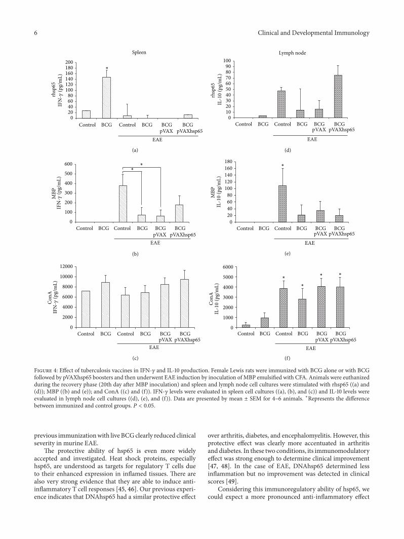
3.3. Production of IFN-𝛾 and IL-10. The highest IFN-𝛾 levels,in cultures stimulated with rhsp65, were found in the BCG

4 Clinical and Developmental Immunology
(a) (b)
(c) (d)
Figure 2: Effect of tuberculosis vaccines in brain inflammation associated with EAE. Female Lewis rats were immunized with BCG alone orwith BCG followed by pVAXhsp65 boosters and then underwent EAE induction by inoculation of MBP emulsified with CFA. Animals wereeuthanized during the recovery phase (20th day after MBP injection) and the brains were removed and further stained by haematoxylin andeosin. Normal control (a), EAE control (b), rats immunized with BCG before EAE (c), and rats immunized with BCG/pVAXhsp65 beforeEAE induction (d).
Table 1: Morphometric analysis of perivascular inflammatory infil-trate in the brain and lumbar spinal cord samples from rats immu-nized with BCG alone or associated with pVAXhsp65 before EAEinduction.
Brain𝑛 = 4
Lumbarspinal cord𝑛 = 4
(𝜇m2 of mononuclear infiltrate/mm2 of organ section)†
ControlEAE 3.55 ± 0.72 6.56 ± 2.72
BCG EAE 1.17 ± 0.38∗
7.38 ± 2.53
BCGpVAXhsp65EAE
1.50 ± 0.71∗
4.46 ± 0.79
∗𝑃 < 0.05 versus control EAE.†Morphometric analysis was done in 5-micron thick sections after haema-toxylin and eosin stain using a Nikon Microphot-FXA optical microscopeconnected to a computer and employing the KS300 software.
immunized group. In all experimental groups with EAE,independently of a preceding immunization, IFN-𝛾 levelswere very low, that is, similar to the normal control group(Figure 4(a)). As can be observed in Figure 4(d), IL-10 levelsin rhsp65 activated cultures presented a very distinct pattern:the highest levels were found in EAE and BCG/pVAXhsp65
EAE groups. BCG group presented detectable but very lowIL-10 levels.
The production of these two cytokines, in cultures stim-ulated with MBP, followed the same pattern (Figures 4(b)and 4(e)). In this case, the highest levels of both cytokineswere found in the EAE group. Previously immunized groups,that were later submitted to encephalomyelitis inductionpresented lower IFN-𝛾 (Figure 4(b)) and IL-10 (Figure 4(e))levels in comparison to the control EAE group. IFN-𝛾 and IL-10 were not detected in the BCG group.
IFN-𝛾 levels were similarly elevated in all groups stim-ulated with ConA (Figure 4(c)). On the other hand, eventhough there was some IL-10 production by cultures fromthe BCG group, the levels of this cytokine were significantlyhigh in all groups with encephalomyelitis, independently of aprevious immunization (Figure 4(f)).
4. Discussion
In this study, we investigated the effect of a previous immu-nization with BCG, associated or not with pVAXhsp65,on the development of EAE. This kind of investigation isrelevant because a possible connection between vaccines andautoimmunephenomena is surroundedby controversy. Someliterature reports indicate that BCG could be one of thevaccines associated with autoimmune diseases. For example,

Clinical and Developmental Immunology 5
(a) (b)
(c) (d)
Figure 3: Effect of tuberculosis vaccines in spinal cord inflammation associated with EAE. Female Lewis rats were immunized with BCGalone or with BCG followed by pVAXhsp65 boosters and then underwent EAE induction by inoculation of MBP emulsified with CFA.Animals were euthanized during the recovery phase (20th day after MBP injection) and the lumbar spinal cord was removed and furtherstained by haematoxylin and eosin. Normal control (a), EAE control (b), rats immunized with BCG before EAE (c), and rats immunized withBCG/pVAXhsp65 before EAE induction (d).
oligo and polyarticular arthritis were detected in approx-imately 3% of patients with bladder carcinoma that weretreatedwith intravesicular BCG [35]. It is also being describedthatmycobacteria precipitates an SLE-like syndrome inNODmice [36].
Otherwise, themaintenance of BCG in a new prophylaxisagainst TB is highly expected because this vaccine is widelyaccepted in the developing countries, it has protective effectagainst the most severe forms of TB in children and it isalso endowed with immunomodulatory properties [37–39].In this scenario, safety aspects need to be experimentallyvalidated.
In the last years, our group analyzed a DNA vaccinecontaining the hsp65 gene from M. leprae (pVAXhsp65).The results showed that this vaccine, by itself or associatedwith BCG, is able to confer protection in different TBexperimental models [22].This genetic construction includesthe mycobacterial hsp65 gene that presents a high homologydegree with the corresponding hsp60 mammalian gene. Aspecific immune response against this bacterial protein couldcross-react with the corresponding mammalian protein and,therefore, trigger an autoimmune pathology.
In this context, the most relevant contribution of thiswork was the demonstration that the previous contact withBCG or BCG followed by pVAXhsp65 boosters did not dele-teriously affect the clinical EAE development in Lewis rats.
This was initially demonstrated by the very similar clinicalevolution of the disease in vaccinated and nonvaccinatedanimals; they equally lost weight, the average clinical scorewas the same, and acute and remission phases occurred atcomparable time periods. In addition of this absence of adetrimental effect, the previous immunization with BCG orBCG/pVAXhsp65 also determined a clear anti-inflammatoryreaction in the brain. This anti-inflammatory activity didnot reach the spinal cord; this could explain the absenceof a beneficial clinical effect over EAE clinical score. Themechanism of this differential protective effect over thebrain and the spinal cord was not investigated. However,considering that interaction of circulating leukocyteswith theendothelium of blood-brain and blood-spinal cord barriersis fundamental in inflammatory pathologies of the CNSand that they differ in many aspects, we could hypothesizethat differences in adhesion molecules, chemokines, or theirreceptors could explain this finding [40].
A similar protective ability of BCG towards autoimmu-nity was already described. Van Eden et al. in 1988 [41],demonstrated that administration of BCG antigens to ratsdetermined resistance to subsequent induction of adjuvantarthritis. A preceding contact with BCG also determinedprotection against diabetes in NOD and streptozotocin dia-betes models [42, 43]. More recently, in this same lineof research, Sewell et al. in 2003 [44], demonstrated that
6 Clinical and Developmental Immunology
600
500
400
300
200
100
0
MBP
∗
IFN
-𝛾(p
g/m
L)200180160140120100
80604020
0
∗
Control BCG Control BCG BCGpVAXhsp65
EAE
rhsp
65
BCGpVAX
IFN
-𝛾(p
g/m
L)Spleen
∗
Control BCG Control BCG BCGpVAX
BCGpVAXhsp65
EAE
Control BCG Control BCG BCGpVAX
BCGpVAXhsp65
EAE
12000
10000
8000
6000
4000
2000
0
Con
AIF
N-𝛾
(pg/
mL)
Control BCG Control BCG BCGpVAXhsp65
EAE
BCGpVAX
Control Control BCG BCGpVAXhsp65
EAE
BCGpVAX
Control Control BCG BCGpVAXhsp65
EAE
BCGpVAX
BCG
BCG
Lymph node
IL-1
0(p
g/m
L)
100908070605040302010
0
180160140120100
80604020
0
6000
5000
4000
3000
2000
1000
0
IL-1
0(p
g/m
L)IL
-10
(pg/
mL)
MBP
Con
A
∗
∗
∗∗ ∗
(a) (d)
(b) (e)
(c) (f)
rhsp
65
Figure 4: Effect of tuberculosis vaccines in IFN-𝛾 and IL-10 production. Female Lewis rats were immunized with BCG alone or with BCGfollowed by pVAXhsp65 boosters and then underwent EAE induction by inoculation of MBP emulsified with CFA. Animals were euthanizedduring the recovery phase (20th day after MBP inoculation) and spleen and lymph node cell cultures were stimulated with rhsp65 ((a) and(d)); MBP ((b) and (e)); and ConA ((c) and (f)). IFN-𝛾 levels were evaluated in spleen cell cultures ((a), (b), and (c)) and IL-10 levels wereevaluated in lymph node cell cultures ((d), (e), and (f)). Data are presented by mean ± SEM for 4–6 animals. ∗Represents the differencebetween immunized and control groups. 𝑃 < 0.05.
previous immunizationwith live BCGclearly reduced clinicalseverity in murine EAE.
The protective ability of hsp65 is even more widelyaccepted and investigated. Heat shock proteins, especiallyhsp65, are understood as targets for regulatory T cells dueto their enhanced expression in inflamed tissues. There arealso very strong evidence that they are able to induce anti-inflammatory T cell responses [45, 46]. Our previous experi-ence indicates that DNAhsp65 had a similar protective effect
over arthritis, diabetes, and encephalomyelitis. However, thisprotective effect was clearly more accentuated in arthritisand diabetes. In these two conditions, its immunomodulatoryeffect was strong enough to determine clinical improvement[47, 48]. In the case of EAE, DNAhsp65 determined lessinflammation but no improvement was detected in clinicalscores [49].
Considering this immunoregulatory ability of hsp65, wecould expect a more pronounced anti-inflammatory effect

Clinical and Developmental Immunology 7
in the prime-boost, in comparison to the BCG immunizedgroup. However, these two groups presented similar clinicalevolution.This finding is in contrast to our recent experiencewith a similar prime-boost in NOD mice. In this model,primingwith BCG followed by two boosters with pVAXhsp65prevented pancreas inflammation and clinical diabetes devel-opment [50].
The analysis of IFN-𝛾 and IL-10 levels, produced byperipheral lymphoid organs, answered some mechanisticquestions. We initially asked how BCG decreased inflamma-tion in the brain. In this sense, the most interesting findingwas the accentuated drop of IFN-𝛾 and IL-10 levels inducedby MBP, in EAE experimental groups previously vaccinated,in comparison to the EAE control group. A possible expla-nation for this finding could be the trapping of nervoustissue-specific T cells in peripheral BCG inflammatory sitesas elegantly demonstrated by Sewell et al. in 2003 [44]. Alter-natively, these autoreactive T cells could be in lower numbersdue to an apoptotic process occurring in the periphery. Thisphenomenon was clearly demonstrated by O’Connor et al.in 2005 [51]. These authors detected high levels of apop-tosis among activated CD4+ T cells in BCG experimentalinfection. Interestingly and concerning to the model usedby us, these authors also described that the high apoptoticdegree occurred simultaneously with a milder experimentalencephalomyelitis course. Even though we did not observedimprovement in clinical parameters, a significant drop inbrain inflammation was detected. This lower IFN-𝛾 andIL-10 production could also be linked to the migration ofmyelin-specific T clones to the brain, where they could exertthe detected anti-inflammatory activity. IFN-𝛾 is describedas able to shape the immune infiltration of the CNS bycontrolling chemokine expression. In addition, this cytokineaccentuates apoptosis of infiltrating encephalitogenic T cellclones [52, 53]. Additionally, the production of IL-10 by Tr1regulatory cells has been widely accepted as one of the mech-anisms responsible for MS and EAE downregulation [54].
5. Conclusion
These results indicate that immunization procedures withBCG or BCG/pVAXhsp65, as used in this investigation, didnot deleteriously affect EAE development.
Acknowledgments
The authors are grateful to Sao Paulo Research Foundation(FAPESP) Grant no. 2007/05353-8 and Conselho Nacional deDesenvolvimento Cientıfico e Tecnologico (CNPq) Grant no.473351/2004-8 that supported this study.
References
[1] D. B. Young andK.Duncan, “Prospects for new interventions inthe treatment and prevention of mycobacterial disease,”AnnualReview of Microbiology, vol. 49, pp. 641–673, 1995.
[2] J. L. Flynn and J. Chan, “Tuberculosis: latency and reactivation,”Infection and Immunity, vol. 69, no. 7, pp. 4195–4201, 2001.
[3] K. L. Dierberg and R. E. Chaisson, “Human immunodeficiencyvirus-associated tuberculosis: update on prevention and treat-ment,”Clinics in ChestMedicine, vol. 34, no. 2, pp. 217–228, 2013.
[4] E. Pontali, A. Matteelli, and G. B. Migliori, “Drug-resistanttuberculosis,” Current Opinion in Pulmary Medicine, vol. 19, no.3, pp. 266–272, 2013.
[5] P. Andersen and T. M. Doherty, “The success and failure ofBCG—implications for a novel tuberculosis vaccine,” NatureReviews Microbiology, vol. 3, no. 8, pp. 656–662, 2005.
[6] Centers for Disease Control and Prevention, “Global routinevaccination coverage, 2011,” MMWR Morbidity and MortalityWeekly Report, vol. 61, no. 43, pp. 883–885, 2012.
[7] G. A. Colditz, T. F. Brewer, C. S. Berkey et al., “Efficacy ofBCG vaccine in the prevention of tuberculosis: meta-analysisof the published literature,” Journal of the American MedicalAssociation, vol. 271, no. 9, pp. 698–702, 1994.
[8] V. H. Springett and I. Sutherland, “A re-examination of the vari-ations in the efficacy of BCG vaccination against tuberculosis inclinical trials,” Tubercle and Lung Disease, vol. 75, no. 3, pp. 227–133, 1994.
[9] M. A. Behr, M. A. Wilson, W. P. Gill et al., “Comparativegenomics of BCG vaccines by whole-genomeDNAmicroarray,”Science, vol. 284, no. 5419, pp. 1520–1523, 1999.
[10] P. E. M. Fine, “Variation in protection by BCG: implications ofand for heterologous immunity,”The Lancet, vol. 346, no. 8986,pp. 1339–1345, 1995.
[11] Y. A. W. Skeiky and J. C. Sadoff, “Advances in tuberculosisvaccine strategies,” Nature Reviews Microbiology, vol. 4, no. 6,pp. 469–476, 2006.
[12] I. M. Orme, “Beyond BCG: the potential for a more effective TBvaccine,” Molecular Medicine Today, vol. 5, no. 11, pp. 487–492,1999.
[13] D. N. McMurray, “A coordinated strategy for evaluating newvaccines for human and animal tuberculosis,” Tuberculosis, vol.81, no. 1-2, pp. 141–146, 2001.
[14] P. Andersen, “TB vaccines: progress and problems,” Trends inImmunology, vol. 22, no. 3, pp. 160–168, 2001.
[15] R. E. Tascon, M. J. Colston, S. Ragno, E. Stavropoulos, D.Gregory, and D. B. Lowrie, “Vaccination against tuberculosisby DNA injection,” Nature Medicine, vol. 2, no. 8, pp. 888–892,1996.
[16] D. B. Lowrie, C. L. Silva, M. J. Colston, S. Ragno, and R. E.Tascon, “Protection against tuberculosis by a plasmid DNAvaccine,” Vaccine, vol. 15, no. 8, pp. 834–838, 1997.
[17] A. Tanghe, S. D’Souza, V. Rosseels et al., “Improved immuno-genicity and protective efficacy of a tuberculosis DNA vaccineencoding Ag85 by protein boosting,” Infection and Immunity,vol. 69, no. 5, pp. 3041–3047, 2001.
[18] V. L. D. Bonato, V.M. F. Lima, R. E. Tascon, D. B. Lowrie, and C.L. Silva, “Identification and characterization of protective T cellsin hsp65 DNA-vaccinated and Mycobacterium tuberculosis-infected mice,” Infection and Immunity, vol. 66, no. 1, pp. 169–175, 1998.
[19] D. B. Lowrie, C. L. Silva, and R. E. Tascon, “DNA vaccinesagainst tuberculosis,” Immunology and Cell Biology, vol. 75, no.6, pp. 591–594, 1997.
[20] V. L. D. Bonato, E. D. C. Goncalves, E. G. Soares et al., “Immuneregulatory effect of pHSP65 DNA therapy in pulmonary tuber-culosis: activation of CD8+ cells, interferon-𝛾 recovery andreduction of lung injury,” Immunology, vol. 113, no. 1, pp. 130–138, 2004.

8 Clinical and Developmental Immunology
[21] A. Pelizon, D. R. Martins, S. F. G. Zorzella et al., “Geneticvaccine for tuberculosis (pVAXhsp65) primes neonate mice fora strong immune response at the adult stage,” Genetic Vaccinesand Therapy, vol. 5, article 12, 2007.
[22] L. de Paula, C. L. Silva, D. Carlos et al., “Comparison ofdifferent delivery systems of DNA vaccination for the inductionof protection against tuberculosis in mice and guinea pigs,”Genetic Vaccines andTherapy, vol. 5, article 2, 2007.
[23] E. D. C. Goncalves, V. L. Bonato, D. M. da Fonseca et al.,“Improve protective efficacy of a TB DNA-HSP65 vaccine byBCG priming,” Genetic Vaccines and Therapy, vol. 5, article 7,2007.
[24] U. Feige and W. van Eden, “Infection, autoimmunity andautoimmune disease,” EXS, vol. 77, pp. 359–373, 1996.
[25] L. Horvath, L. Cervenak,M. Oroszlan et al., “Antibodies againstdifferent epitopes of heat-shock protein 60 in children with type1 diabetes mellitus,” Immunology Letters, vol. 80, no. 3, pp. 155–162, 2002.
[26] P. Keren, J. George, A. Shaish et al., “Effect of hyperglycemia andhyperlipidemia on atherosclerosis in LDL receptor-deficientmice: establishment of a combined model and association withheat shock protein 65 immunity,” Diabetes, vol. 49, no. 6, pp.1064–1069, 2000.
[27] I. R. Cohen, “Autoimmunity to chaperonins in the pathogenesisof arthritis and diabetes,” Annual Review of Immunology, vol. 9,pp. 567–589, 1991.
[28] G. Birnbaum, “Stress proteins: their role in the normal centralnervous system and in disease states, especially multiple scle-rosis,” Springer Seminars in Immunopathology, vol. 17, no. 1, pp.107–118, 1995.
[29] I. Tsunoda, N. D. Tolley, D. J.Theil, J. L. Whitton, H. Kobayashi,and R. S. Fujinami, “Exacerbation of viral and autoimmuneanimal models for multiple sclerosis by bacterial DNA,” BrainPathology, vol. 9, no. 3, pp. 481–493, 1999.
[30] B. M. Segal, J. T. Chang, and E. M. Shevach, “CpG oligonu-cleotides are potent adjuvants for the activation of autoreactiveencephalitogenic T cells in vivo,” Journal of Immunology, vol.164, no. 11, pp. 5683–5688, 2000.
[31] T. Nakamura, J.-I. Yamamura, H. Sato, H. Kakinuma, and H.Takahashi, “Vasculitis induced by immunization with BacillusCalmette-Guerin followed by atypical mycobacterium antigen:a new mouse model for Kawasaki disease,” FEMS Immunologyand Medical Microbiology, vol. 49, no. 3, pp. 391–397, 2007.
[32] A. Dubaniewicz, “Mycobacterium tuberculosis heat shock pro-teins and autoimmunity in sarcoidosis,”Autoimmunity Reviews,vol. 9, no. 6, pp. 419–424, 2010.
[33] F. Chiuso-Minicucci, D. B. Van, S. F. G. Zorzella-Pezaventoet al., “Experimental autoimmune encephalomyelitis evolutionwas not modified by multiple infections with Strongyloidesvenezuelensis,” Parasite Immunology, vol. 33, no. 5, pp. 303–308,2011.
[34] R. S. Peres, F. Chiuso-Minicucci, L. C. da Rosa et al., “Previouscontact with Strongyloides venezuelensis contributed to pre-vent insulitis inMLD-STZ diabetes,” Experimental Parasitology,vol. 134, no. 2, pp. 183–189, 2013.
[35] M. Tischler and Y. Shoenfeld, “Tuberculose et immunite: ou ensommes-nous?” Annales de L’Institut Pasteur/ActuaLites, vol. 7,no. 2, pp. 133–136, 1996.
[36] A. G. Baxter, A. C. Horsfall, D. Healey et al., “Mycobacteriaprecipitate an SLE-like syndrome in diabetes-proneNODmice,”Immunology, vol. 83, no. 2, pp. 227–231, 1994.
[37] L. C. Rodrigues, V. K. Diwan, and J. G. Wheeler, “Protectiveeffect of BCG against tuberculousmeningitis andmiliary tuber-culosis: a meta-analysis,” International Journal of Epidemiology,vol. 22, no. 6, pp. 1154–1158, 1993.
[38] G. A. Colditz, C. S. Berkey, F. Mosteller et al., “The efficacyof bacillus Calmette-Guerin vaccination of newborns andinfants in the prevention of tuberculosis: meta-analyses of thepublished literature,” Pediatrics, vol. 96, no. 1 I, pp. 29–35, 1995.
[39] J. A. C. Sterne, L. C. Rodrigues, and I. N. Guedes, “Doesthe efficacy of BCG decline with time since vaccination?”International Journal of Tuberculosis and Lung Disease, vol. 2,no. 3, pp. 200–207, 1998.
[40] B. Engelhardt, “Molecular mechanisms involved in T cellmigration across the blood-brain barrier,” Journal of NeuralTransmission, vol. 113, no. 4, pp. 477–485, 2006.
[41] W. van Eden, J. E. R. Tholet, R. V. D. Zee et al., “Cloning of themycobacterial epitope recognized byT lymphocytes in adjuvantarthritis,” Nature, vol. 331, no. 6152, pp. 171–173, 1988.
[42] M. Harada, Y. Kishimoto, and S. Makino, “Prevention ofovert diabetes and insulitis in NOD mice by a single BCGvaccination,” Diabetes Research and Clinical Practice, vol. 8, no.2, pp. 85–89, 1990.
[43] S. H. Baik, I. B. Park, K. M. Choi et al., “BCG vaccine preventsinsulitis in low dose streptozotocin-induced diabetic mice,”Diabetes Research and Clinical Practice, vol. 46, no. 2, pp. 91–97,1999.
[44] D. L. Sewell, E. K. Reinke, D. O. Co et al., “Infection withMycobacterium bovis BCG diverts traffic of myelin oligoden-droglial glycoprotein autoantigen-specific T cells away from thecentral nervous system and ameliorates experimental autoim-mune encephalomyelitis,” Clinical and Diagnostic LaboratoryImmunology, vol. 10, no. 4, pp. 564–572, 2003.
[45] W. van Eden, R. van der Zee, and B. Prakken, “Heat-shockproteins induce T-cell regulation of chronic inflammation,”Nature Reviews Immunology, vol. 5, no. 4, pp. 318–330, 2005.
[46] W. van Eden, G. Wick, S. Albani, and I. Cohen, “Stress, heatshock proteins, and autoimmunity: how immune responses toheat shock proteins are to be used for the control of chronicinflammatory diseases,” Annals of the New York Academy ofSciences, vol. 1113, pp. 217–237, 2007.
[47] R. R. Santos Jr., A. Sartori, M. De Franco et al., “Immunomodu-lation and protection induced by DNA-hsp65 vaccination in ananimal model of arthritis,”Human GeneTherapy, vol. 16, no. 11,pp. 1338–1345, 2005.
[48] R. R. Dos Santos Jr., A. Sartori, V. L. Deperon Bonato et al.,“Immune modulation induced by tuberculosis DNA vaccineprotects non-obese diabetic mice from diabetes progression,”Clinical and Experimental Immunology, vol. 149, no. 3, pp. 570–578, 2007.
[49] S. F.G. Zorzella-Pezavento, F. Chiuso-Minicucci, T.G.D. Francaet al., “Immunization with pVAXhsp65 decreases inflam-mation and modulates immune response in experimentalencephalomyelitis,”NeuroImmunoModulation, vol. 17, no. 5, pp.287–297, 2010.
[50] L. C. da Rosa, F. Chiuso-Minicucci, S. F. G. Zorzella-Pezaventoet al., “BCG/DNAhsp65 prime-boost is protective against dia-betes in NODmice but not in the STZmodel of type 1 diabetes,”Clinical and Experimental Immunology, vol. 173, no. 3, pp. 430–437, 2013.
[51] R. A. O’Connor, S. Wittmer, and D. K. Dalton, “Infection-induced apoptosis deletes bystander CD4+ T cells: amechanism

Clinical and Developmental Immunology 9
for suppression of autoimmunity during BCG infection,” Jour-nal of Autoimmunity, vol. 24, no. 2, pp. 93–100, 2005.
[52] E. H. Tran, E. N. Prince, and T. Owens, “IFN-𝛾 shapesimmune invasion of the central nervous systemvia regulation ofchemokines,” Journal of Immunology, vol. 164, no. 5, pp. 2759–2768, 2000.
[53] R. Furlan, E. Brambilla, F. Ruffini et al., “Intrathecal delivery ofIFN-𝛾 protects C57BL/6mice from chronic-progressive experi-mental autoimmune encephalomyelitis by increasing apoptosisof central nervous system-infiltrating lymphocytes,” Journal ofImmunology, vol. 167, no. 3, pp. 1821–1829, 2001.
[54] F. Jadidi-Niaragh and A. Mirshafiey, “Regulatory T-cell asorchestra leader in immunosuppression process of multiplesclerosis,” Immunopharmacology and Immunotoxicology, vol.33, no. 3, pp. 545–567, 2011.

Submit your manuscripts athttp://www.hindawi.com
Stem CellsInternational
Hindawi Publishing Corporationhttp://www.hindawi.com Volume 2014
Hindawi Publishing Corporationhttp://www.hindawi.com Volume 2014
MEDIATORSINFLAMMATION
of
Hindawi Publishing Corporationhttp://www.hindawi.com Volume 2014
Behavioural Neurology
EndocrinologyInternational Journal of
Hindawi Publishing Corporationhttp://www.hindawi.com Volume 2014
Hindawi Publishing Corporationhttp://www.hindawi.com Volume 2014
Disease Markers
Hindawi Publishing Corporationhttp://www.hindawi.com Volume 2014
BioMed Research International
OncologyJournal of
Hindawi Publishing Corporationhttp://www.hindawi.com Volume 2014
Hindawi Publishing Corporationhttp://www.hindawi.com Volume 2014
Oxidative Medicine and Cellular Longevity
Hindawi Publishing Corporationhttp://www.hindawi.com Volume 2014
PPAR Research
The Scientific World JournalHindawi Publishing Corporation http://www.hindawi.com Volume 2014
Immunology ResearchHindawi Publishing Corporationhttp://www.hindawi.com Volume 2014
Journal of
ObesityJournal of
Hindawi Publishing Corporationhttp://www.hindawi.com Volume 2014
Hindawi Publishing Corporationhttp://www.hindawi.com Volume 2014
Computational and Mathematical Methods in Medicine
OphthalmologyJournal of
Hindawi Publishing Corporationhttp://www.hindawi.com Volume 2014
Diabetes ResearchJournal of
Hindawi Publishing Corporationhttp://www.hindawi.com Volume 2014
Hindawi Publishing Corporationhttp://www.hindawi.com Volume 2014
Research and TreatmentAIDS
Hindawi Publishing Corporationhttp://www.hindawi.com Volume 2014
Gastroenterology Research and Practice
Hindawi Publishing Corporationhttp://www.hindawi.com Volume 2014
Parkinson’s Disease
Evidence-Based Complementary and Alternative Medicine
Volume 2014Hindawi Publishing Corporationhttp://www.hindawi.com