
RESEARCH ARTICLE - Asian Pacific Journal of Cancer...
7
Asian Pacific Journal of Cancer Prevention, Vol 15, 2014 4519 DOI:http://dx.doi.org/10.7314/APJCP.2014.15.11.4519 p38 MAPK Signaling Mediates Mitochondrial Apoptosis in Cancer Cells Induced by Oleanolic Acid Asian Pac J Cancer Prev, 15 (11), 4519-4525 Introduction As a nutritional component, oleanolic acid ((3ß)-3- Hydroxyolean-12-en-28-oic acid, also known as oleanic acid) is a natural pentacyclic triterpenoid distributed in a wide range of plants (Pollier et al., 2012). Oleanolic acid (OA) is considered among the most abundant compounds with bioactivity in vegetarian food and medicinal herbs (Pollier et al., 2012). Numerous studies have verified that OA accounts for, at least part of, preventive and curative effect of many dietary and medicinal plants (Pollier et al., 2012). Its pharmacologic effect includes hepatopretection, anti-inflammation, anti-virus, anti-hyperlipidemia, anti-hyperglycemia and anti-tumor, all of which have been by in vitro or/and animal studies (Pollier et al., 2012). Remarkably, OA has no cytotoxicity to normal cells and tissues, and thus, possesses high biosafety in clinical application (Pollier et al., 2012). Considering its pleiotropic function and nontoxicity, researchers have been increasingly focused on OA and making efforts to improve its activity and drugability in recent decades (Liby 1 Institutes of Oceanology, Chinese Academy of Sciences, 2 Yellow Sea Fisheries Research Institute, Chinese Academy of Fishery Sciences, 5 Department of Molecular Biology, School of Medicine and Pharmacy, Ocean University of China, Qingdao, 3 Graduate School, University of Chinese Academy of Sciences, 4 Capital Med. University, Dept. of Pharmacology, Beijing, 6 Department of Pathophysiology, Norman Bethune College of Medicine, Jilin University, Changchun, China & Equal contributors *For correspondence: [email protected], [email protected] Abstract Oleanolic acid (OA) is a nutritional component widely distributed in various vegetables. Although it has been well recognized for decades that OA exerts certain anti-tumor activity by inducing mitochondria-dependent apoptosis, it is still unclear that what molecular signaling is responsible for this effect. In this study, we employed cancer cell lines, A549, BXPC-3, PANC-1 and U2OS to elucidate the molecular mechanisms underlying OA anti- tumor activity. We found that activation of MAPK pathways, including p-38 MAPK, JNK and ERK, was triggered by OA in both a dose and time-dependent fashion in all the tested cancer cells. Activation was accompanied by cleavage of caspases and PARP as well as cytochrome C release. SB203580 (p38 MAPK inhibitor), but not SP600125 (JNK inhibitor) and U0126 (ERK inhibitor), rescued the pro-apoptotic effect of OA on A549 and BXPC- 3 cells. OA induced p38 MAPK activation promoted mitochondrial translocation of Bax and Bim, and inhibited Bcl-2 function by enhancing their phosphorylation. OA can induce reactive oxygen species (ROS)-dependent ASK1 activation, and this event was indispensable for p38 MAPK-dependent apoptosis in cancer cells. In vivo, p38 MAPK knockdown A549 tumors proved resistant to the growth-inhibitory effect of OA. Collectively, we elucidated that activation of ROS/ASK1/p38 MAPK pathways is responsible for the apoptosis stimulated by OA in cancer cells. Our finding can contribute to a better understanding of molecular mechanisms underlying the antitumor activity of nutritional components. Keywords: Oleanolic acid - apoptosis - p38 MAPK - cancer - ROS RESEARCH ARTICLE p38 MAPK Signaling Mediates Mitochondrial Apoptosis in Cancer Cells Induced by Oleanolic Acid Jia Liu 1,3& , Ning Wu 1& , Lei-Na Ma 5 , Jia-Teng Zhong 6 , Ge Liu 1, 3 , Lan-Hong Zheng 2 *, Xiu-Kun Lin 1,4 * et al., 2007; Petronelli et al., 2009). Since that cancer-related deaths keep rising both in developed and developing countries in recent years, the anti-tumor property of OA receives more and more attention. Apoptosis induction has been well known as one of the major mechanisms underlying OA anti-tumor activity on various types of cancer cells (Juan et al., 2006; Martin et al., 2007; Zhang et al., 2007; Shyu et al., 2010; Yan et al., 2010; Feng et al., 2011b; Lucio et al., 2011; Pratheeshkumar et al., 2011; Zhou et al., 2011; Chakravarti et al., 2012; George et al., 2012; Gu et al., 2012; Struh et al., 2012; Wei et al., 2012; Wang et al., 2013). Mechanistically, OA treatment increases the ratio of proapoptotic protein, Bax, to anti-apoptotic protein, Bcl-2, in the outer mitochondrial membrane, facilitates the formation of Bax oligomer, increases the permeability of mitochondrial membrane, releases cytochrome C from inner membrane into cytosol, triggers the caspase pathway through activating Apaf-1, cleaves multiple cellular target molecules including Poly ADP ribose polymerase (PARP) and DNA, and eventually leads to cell death (Martin et
Transcript of RESEARCH ARTICLE - Asian Pacific Journal of Cancer...

Asian Pacific Journal of Cancer Prevention, Vol 15, 2014 4519
DOI:http://dx.doi.org/10.7314/APJCP.2014.15.11.4519p38 MAPK Signaling Mediates Mitochondrial Apoptosis in Cancer Cells Induced by Oleanolic Acid
Asian Pac J Cancer Prev, 15 (11), 4519-4525
Introduction As a nutritional component, oleanolic acid ((3ß)-3-Hydroxyolean-12-en-28-oic acid, also known as oleanic acid) is a natural pentacyclic triterpenoid distributed in a wide range of plants (Pollier et al., 2012). Oleanolic acid (OA) is considered among the most abundant compounds with bioactivity in vegetarian food and medicinal herbs (Pollier et al., 2012). Numerous studies have verified that OA accounts for, at least part of, preventive and curative effect of many dietary and medicinal plants (Pollier et al., 2012). Its pharmacologic effect includes hepatopretection, anti-inflammation, anti-virus, anti-hyperlipidemia, anti-hyperglycemia and anti-tumor, all of which have been by in vitro or/and animal studies (Pollier et al., 2012). Remarkably, OA has no cytotoxicity to normal cells and tissues, and thus, possesses high biosafety in clinical application (Pollier et al., 2012). Considering its pleiotropic function and nontoxicity, researchers have been increasingly focused on OA and making efforts to improve its activity and drugability in recent decades (Liby 1Institutes of Oceanology, Chinese Academy of Sciences, 2Yellow Sea Fisheries Research Institute, Chinese Academy of Fishery Sciences, 5Department of Molecular Biology, School of Medicine and Pharmacy, Ocean University of China, Qingdao, 3Graduate School, University of Chinese Academy of Sciences, 4Capital Med. University, Dept. of Pharmacology, Beijing, 6Department of Pathophysiology, Norman Bethune College of Medicine, Jilin University, Changchun, China &Equal contributors *For correspondence: [email protected], [email protected]
Abstract
Oleanolic acid (OA) is a nutritional component widely distributed in various vegetables. Although it has been well recognized for decades that OA exerts certain anti-tumor activity by inducing mitochondria-dependent apoptosis, it is still unclear that what molecular signaling is responsible for this effect. In this study, we employed cancer cell lines, A549, BXPC-3, PANC-1 and U2OS to elucidate the molecular mechanisms underlying OA anti-tumor activity. We found that activation of MAPK pathways, including p-38 MAPK, JNK and ERK, was triggered by OA in both a dose and time-dependent fashion in all the tested cancer cells. Activation was accompanied by cleavage of caspases and PARP as well as cytochrome C release. SB203580 (p38 MAPK inhibitor), but not SP600125 (JNK inhibitor) and U0126 (ERK inhibitor), rescued the pro-apoptotic effect of OA on A549 and BXPC-3 cells. OA induced p38 MAPK activation promoted mitochondrial translocation of Bax and Bim, and inhibited Bcl-2 function by enhancing their phosphorylation. OA can induce reactive oxygen species (ROS)-dependent ASK1 activation, and this event was indispensable for p38 MAPK-dependent apoptosis in cancer cells. In vivo, p38 MAPK knockdown A549 tumors proved resistant to the growth-inhibitory effect of OA. Collectively, we elucidated that activation of ROS/ASK1/p38 MAPK pathways is responsible for the apoptosis stimulated by OA in cancer cells. Our finding can contribute to a better understanding of molecular mechanisms underlying the antitumor activity of nutritional components. Keywords: Oleanolic acid - apoptosis - p38 MAPK - cancer - ROS
RESEARCH ARTICLE
p38 MAPK Signaling Mediates Mitochondrial Apoptosis in Cancer Cells Induced by Oleanolic AcidJia Liu1,3&, Ning Wu1&, Lei-Na Ma5, Jia-Teng Zhong6, Ge Liu1, 3, Lan-Hong Zheng2*, Xiu-Kun Lin1,4*
et al., 2007; Petronelli et al., 2009). Since that cancer-related deaths keep rising both in developed and developing countries in recent years, the anti-tumor property of OA receives more and more attention. Apoptosis induction has been well known as one of the major mechanisms underlying OA anti-tumor activity on various types of cancer cells (Juan et al., 2006; Martin et al., 2007; Zhang et al., 2007; Shyu et al., 2010; Yan et al., 2010; Feng et al., 2011b; Lucio et al., 2011; Pratheeshkumar et al., 2011; Zhou et al., 2011; Chakravarti et al., 2012; George et al., 2012; Gu et al., 2012; Struh et al., 2012; Wei et al., 2012; Wang et al., 2013). Mechanistically, OA treatment increases the ratio of proapoptotic protein, Bax, to anti-apoptotic protein, Bcl-2, in the outer mitochondrial membrane, facilitates the formation of Bax oligomer, increases the permeability of mitochondrial membrane, releases cytochrome C from inner membrane into cytosol, triggers the caspase pathway through activating Apaf-1, cleaves multiple cellular target molecules including Poly ADP ribose polymerase (PARP) and DNA, and eventually leads to cell death (Martin et

Jia Liu et al
Asian Pacific Journal of Cancer Prevention, Vol 15, 20144520
al., 2007; Zhang et al., 2007; Shyu et al., 2010; Yan et al., 2010; Zhou et al., 2011; Chakravarti et al., 2012; Wei et al., 2012; Wang et al., 2013). Although it is well recognized that OA induces mitochondria-mediated apoptosis in a broad range of cancer cells, the mechanism by which it initiates this pathway is largely unknown. In addition, the generation of reactive oxygen species (ROS) were frequently observed in cancer cells under OA administration (Martin et al., 2007; Chakravarti et al., 2012; Wei et al., 2012; Wang et al., 2013). This event is required for OA induction of mitochondria-mediated apoptosis (Martin et al., 2007; Wei et al., 2012). However, it is still not clear how OA-induced ROS accumulation causes the activation of apoptotic pathway in cancer cells. In the present study, we used several different cancer cell lines as models to explore the molecular mechanisms underlying ROS/mitochondria-associated apoptotic pathway. p38 MAP kinase (MAPK) plays an important role in regulating cellular response to a variety of stressors, such as UV and inflammatory cytokines (Cuadrado et al., 2010). p38 MAPK has been identified to have four isoforms, p38α, β, γ and δ. MKK3 and MKK6 phosphorylate p38 MAPK at Thr180 and Tyr182 and activate its kinase activity. In turn, p38 MAPK activates or inactivates specific downstream effectors, including transcriptional factors and cytoplasmic proteins, by phosphorylation, to accomplish short- and long-term cellular responses to stimuli (Cuadrado et al., 2010). Apoptosis is one of the major outcomes of p38 MAPK activation exposed with stressors (Cuadrado et al., 2010). p38 MAPK induces apoptotic pathways primarily by two different mechanisms, promoting the transcription of pro-apoptotic genes and directly affecting the activity of apoptosis-related Bcl-2 family proteins, such as Bax, Bim and Bcl-2 (Cuadrado et al., 2010). The object of our study is to identify the mechanism by which OA induces apoptosis in cancer cells. Briefly, we found that OA activates the activation of p-38 MAPK, which triggers the translocation of pro-apoptotic Bcl-2 family, Bax, from cytosol to the mitochondria membrane, leading to mitochondrial apoptosis in cancer cells. ROS accumulation caused by OA treatment increases leads to the activation of apoptosis signal-regulating kinase 1 (ASK1), which induces p38 MAPK phosphorylation and activation.
Materials and Methods
Cell lines A549, U2OS, BXPC3 and PANC-1 cells were all purchased from American Type Culture Collection (Manassas, VA) and cultured in the recommended media with 10% heat-inactivated fetal bovine serum (Invitrogen), 4 mM glutamine, 100 units/mL penicillin, and 100 µg/mL. All the cells were cultured in a humidified 5% CO2 atmosphere at 37°C.
Compounds Oleanolic acid (OA) was purchased from Sigma Aldrich (O5504). OA was dissolved in DMSO to make
stock solution (10 mg/mL). Stock solution was diluted into the indicated concentration when used. Inhibitors that selectively blocked p-38 MAPK (SB203580, #5633), JNK (SP600125, #8177), or MEK1/2 (U0126, #9903) were purchased from Cell Signaling Technology. Cells were pretreated with SB203580 (10 μM), SP600125 (25 μM), and U0126 (10 μM) for 1 hr prior to the stimulation with OA. Selective inhibitor of apoptosis signal-regulating kinase 1 (ASK1, MAP3K5), NQDI 1, was obtained from Tocris Bioscience (#4429). Two siRNAs targeting p38 MAPK (#6564 + #6243) and control siRNA (#6568) were purchased from Cell Signaling Technology. 100 nM p38 MAPK siRNA or control siRNA was added to the culture 36 hr before treating OA.
Subcellular fractionation Tris-buffered saline (TBS)-harvested cells were subjected to 500× g centrifugation for 5 min, followed by resuspension with isotonic mitochondrial buffer (1 mM EDTA, 210 mM mannitol, 10 mM HEPES, and 70 mM sucrose, pH 7.5) containing 1 mM DTT, 0.04% digitonin, and mixed protease inhibitors (Sigma-Aldrich, S8820). Nuclear fraction was removed by centrifuging samples at 500× g for 5 min at 4°C. To get mitochondria-enriched heavy membrane pellet, the supernatants were subsequently centrifuged at 10,000× g for 30 min at 4°C. The heavy membrane pellet (mitochondrial fraction) was resuspended with 50 μl MB buffer and the supernatant was used as cytosol fraction.
Immunoblotting assay Proteins of cultured cells were harvested with M-PER Mammalian Protein Extraction Reagent (Thermo Scientific, IL), separated using 12% SDS-PAGE and transferred onto 0.45 µm nitrocellulose membranes. The membranes were blocked with 5% fat-free dry milk in PBS and incubated with primary antibodies at 4°C. Overnight, the membrane was incubated with corresponding secondary antibodies and visualized with SuperSignal West Dura Extended Duration Substrate (Thermo Scientific, IL). The antibodies involved in our study are as follows: cleaved PARP, Cell Signaling Technology, #5625; cleaved caspase-3, Cell Signaling Technology, #9664; cleaved caspase-9, Cell Signaling Technology, #9501; cytochrome C, Cell Signaling Technology, #11940; cytochrome C oxidase subunit IV (COX IV), Cell Signaling Technology, #4850; p-Bax (Thr167), AssayBiotech, A0773; p-BimEL (Ser65), Santa Cruz, sc-101646; p-Bcl-2 (Ser87), Santa Cruz, sc-16323; p-Bcl-2 (Thr56), Cell Signaling Technology, #2875; phospho-JNK (Thr183/Tyr185), #4668; JNK, #9258; β-tubulin, #2146, Cell Signaling Technology.
Apoptotic rate determination by flow cytometry The rates of apoptotic cells were evaluated by flow cytometry analysis. Briefly, cells were fixed in cold 70% ethanol for 1 hr, and then, was incubated with 10 mg/ml RNase A at 37°C for 1 hr. The cells were stained with Propidium iodide (PI, 200 mg/ml), and was immediately subjected to flow cytometry analysis (Aria II, BD Biosciences). 1×10⁵ cells were counted for each sample.

Asian Pacific Journal of Cancer Prevention, Vol 15, 2014 4521
DOI:http://dx.doi.org/10.7314/APJCP.2014.15.11.4519p38 MAPK Signaling Mediates Mitochondrial Apoptosis in Cancer Cells Induced by Oleanolic Acid
ROS detection ROS levels were determined in the cancer cells treated with indicated concentrations of OA for indicated time using Cellular Reactive Oxygen Species Detection Assay Kit (ab113851, Abcam).
Animal experiments After 48 hr infection of lentiviral vectors (10 MOI) that express scrambled shRNA (Lv-scrambled) or p38 MAPK-specific shRNA (Lv-shp38), 5×10⁶ A549 cells were injected into the flanks of 4-6 week old male nude BALB/c mice (n=24). OA (120 mg/kg) was orally administrated every day for 4 weeks. So the mice were divided into four groups: Lv-scrambled (n=6), Lv-scrambled+OA (n=6), Lv-shp38 (n=6) and Lv-shp38+OA (n=6). The diameters of tumors were periodically determined with calipers. The volumes were calculated using the following formula. Volume (mm3) = length× (width)2/2. The expression of indicated proteins was detected in the dissected tumors after the mice were sacrificed by immunoblot assay.
Statistical analysis The experiments except immunoblot assays were performed for at least three times. All values were reported as means±SD, and compared at a given time point by
Figure 1. OA Induced the Activation of p38 MAPK and JNK Signaling Pathway in Cancer Cells. A) A549 and BXPC3 cells were treated with OA of 10, 25, 50 and 100 µg/ml for 3 hr. The expression of phosphorylated p38 MAPK (p-p38 MAPK), phosphorylated JNK (p-JNK) and phosphorylated ERK (p-ERK) were detected by immunoblotting assays. B) The expression of p-p38 MAPK, p-JNK and p-ERK was quantified with ImageJ software. The bars indicated the relative level of p-p38 MAPK, p-JNK and p-ERK normalized by p38 MAPK, JNK and ERK, respectively
A549 BXPC3
- 10 25 50 100
p-p38 MAPK
β-tubulin
p-JNK
p-ERK
OA (µg/mL)
- 10 25 50 100
ERK
p38 MAPK
JNK
Rela
tive
p-p3
8 MA
PK
/p38
MAP
K
- 10 25 50 100
A549 A B BXPC3
0246810
0510152025
- 10 25 50 100
Rela
tive
p-JN
K /J
NK
048
1216
04
812
16
- 10 25 50 100 - 10 25 50 100
A549 BXPC3
Rela
tive
p-ER
K /E
RK
- 10 25 50 100 - 10 25 50 100 0
24
6
8
0
0.4
0.8
1.2
1.6A549 BXPC3
OA (µg/mL)
OA (µg/mL)
OA (µg/mL)
Figure 2. OA induced the Activation of p38 MAPK in a Time-Dependent Manner. A549 and BXPC3 cells were treated with 100 μg/ml OA. Then, the expression levels of indicated proteins were detected by immunoblot assays, at the indicated time points
A549
p-p38 MAPK
β-tubulin
time (hrs)
p-JNK1
0.5 1 2 3 6 12
DMSO 100 µg/mL OA 0.5 1 2 3 6 12
p-ERK
BXPC3
0.5 1 2 3 6 12
DMSO 100 µg/mL OA 12
p38 MAPK
JNK1
ERK
cytochrome C
Cleaved caspase 9
Cleaved caspase 3
Cleaved PARP
Figure 3. Inactivation of p38 MAPK Signaling Suppressed the Apoptosis-Inducing Effect of OA on Cancer Cells. A) A549 cells were pretreated with DMSO (-), SB203580 (10 µM, SB), SP600125 (25 µM, SP) and U0126 (10 µM, U) in the absence and presence of OA (100 μg/ml). The apoptosis-related proteins were detected by immunoblot assays 6 hr or 12 hr later. B) Sub-G0/G1 population was assessed in A549 cells under the same treatment as (A), 12 hr after OA treatment. C) After p38 MAPK expression was silenced in A549 cells by two distinct siRNAs, apoptosis-related proteins were detected by immunoblot assays 6 hr or 12 hr after 100 μg/ml OA treatment. D) Sub-G0/G1 population was assessed in A549 cells under the same treatment as (C), 12 hr after OA treatment
Cytochrome C (cytosol)
- SB SP U - SB SP U - SB SP U inhibitors
OA (100µg/ml) - - - - + + + + + + + +
Cleaved caspase-9
Cleaved caspase-3
Cleaved PARP
β-tubulin
6h 12h 12h
Cytochrome C (cytosol)
- con 1 2 - con 1 2 - con 1 2 siRNA
OA (100µg/ml) - - - - + + + + + + + +
6h 12h 12h
Cleaved caspase-3
Cleaved PARP
β-tubulin
Cleaved caspase-9
0.16% 51.34% 13.58%
45.07% 56.31%
OA OA+SB
OA+SP OA+U
DMSO
coun
ts
DNA content
B
A
0%
15%30%
45%
60%
- - SB SP U inhibitors OA (100µg/ml) - + + + +
Sub-
G0/G
1
D
C
0.39% DMSO
60.49%
coun
ts
DNA content
63.23% OA+ shGFP
- - G 1 2 shRNA OA (100µg/ml) - + + + +
Sub-
G0/G
1
OA
OA+ shp38-1
22.96% OA+ shp38-2
23.26%
0%
15%
30%
45%
60%
75%
A549 A549
A549 A549
unpaired, two-tailed t test. Data were considered to be statistically significant when p<0.05 (*) and p<0.01 (**).
Results OA triggers the activation of p-38 MAPK and JNK signaling in a wide range of cancer cells in both time and dose dependent ways We investigated if p-38 MAPK or JNK signaling pathway was activated in OA-treated cancer cells by immunoblotting analysis of the phosphorylated form of these two kinases. After 3 hr treatment, OA (10-100 µg/mL) induced both p-38 MAPK and JNK phosphorylation in A549, U2OS, and BXPC3 cells as well as several other human cancer cell lines in a dose-dependent manner (Figure 1A, 1B and Supp Figure 1). In addition, ERK pathway was also found to be activated in most, if not all, OA-stimulated cancer cells under the same conditions (Figure 1A, 1B and Supp Figure 1). Time course analysis further confirmed the activation of p-38 MAPK, JNK and ERK signaling pathways under OA treatment (Figure 2 and Supp Figure 2). p-38 MAPK activation was detected 0.5-3 hrs after OA stimulation and peaked at 2-12 hrs after treatment, in tested cancer cells. JNK was activated only 30 mins after OA stimulation (Figure 2A and Supp Figure 2A).
p38 MAPK activation is required for apoptotic event stimulated by OA in cancer cells Subsequently, cancer cells were investigated for the role of p38 MAPK, JNK and ERK activation in OA apoptosis-inducing activity. Selective inhibitors were employed to block the activation of these MAPKs. SB203580, SP600125 and U0126 were confirmed to effectively and specifically inhibit the activation of p38 MAPK, JNK and ERK, respectively, in cancer cells with
Jia Liu et al
Asian Pacific Journal of Cancer Prevention, Vol 15, 20144522
OA treatment, evidenced by decreased phosphorylation of the above kinases or/and their downstream substrates, HSP27, c-Jun and p90RSK, respectively (Supp Figure 3A). Blocking p38 MAPK activation was found to prevent the cleavage of apoptosis-related proteins, caspase-9, caspase-3 and PARP, and the release of cytochrome c from mitochondria. Whereas, suppressions of JNK and ERK signaling had no significant inhibitory effect on the protein cleavage and cytochrome c release in OA-treated A549 and BXPC3 cells (Figure 3A and Supp Figure 3D). OA stimulation led to an increase in sub-G0/G1 population, which indicated apoptotic events taking place, in A549 cells, (Figure 3B). SB203580, but not SP600125 and U0126, reversed the increase in sub-G0/G1 population in OA-stimulated A549 cells (Figure 3B). To confirm the role of p38 MAPK activation in OA-induced apoptosis, small interfering RNAs (siRNAs) were used to knock down p38 MAPK expression in cancer cells (Supp Figure 3C). The treatment of cancer cells with the two different p38 MAPK-targeting siRNAs lead to reduced expression of cleaved fragments of the above apoptosis-related proteins (Figure 3C), as well as the percentages of sub-G0/G1 population in OA-stimulated A549 cells (Figure 3D). The above data together demonstrated that p38 MAPK inhibition protected cells against apoptosis caused by OA stimulation. Also, p38 MAPK-targeting siRNA prevented OA-induced caspase 9, 3 and PARP cleavages in BXPC3 cells (Supp Figure 3D).
OA-induced p38 MAPK activation promoted the translocation of Bax and Bim to the mitochondrial membrane Subsequent experiments were performed to investigate
the mechanisms underlying p38 MAPK mediated OA-induced apoptosis. We detected the phosphorylation of apoptosis-related proteins, Bax, Bim and Bcl-2, in OA-induced cancer cells. As shown in Figure 4A, the phosphorylations of Bax, Bim and Bcl-2 were all increased when cells was incubated with OA, in both concentration- and time-dependent fashion. OA was also found to promote mitochondrial translocation of Bax and Bim according to immunoblotting analysis of their expression levels in cytosol and mitochondrial fractions (Figure 4B). SB203580 administration prevented Bax and Bim from undergoing phosphorylation and mitochondrial translocation in OA-treated cancer cells (Figure 4C and 4D), suggesting that p38 MAPK activation is necessary for the two events.
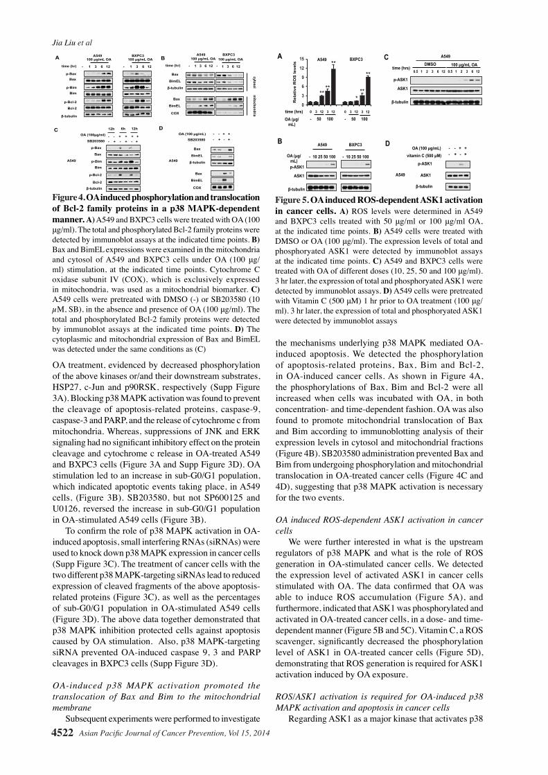
OA induced ROS-dependent ASK1 activation in cancer cells We were further interested in what is the upstream regulators of p38 MAPK and what is the role of ROS generation in OA-stimulated cancer cells. We detected the expression level of activated ASK1 in cancer cells stimulated with OA. The data confirmed that OA was able to induce ROS accumulation (Figure 5A), and furthermore, indicated that ASK1 was phosphorylated and activated in OA-treated cancer cells, in a dose- and time-dependent manner (Figure 5B and 5C). Vitamin C, a ROS scavenger, significantly decreased the phosphorylation level of ASK1 in OA-treated cancer cells (Figure 5D), demonstrating that ROS generation is required for ASK1 activation induced by OA exposure.
ROS/ASK1 activation is required for OA-induced p38 MAPK activation and apoptosis in cancer cells Regarding ASK1 as a major kinase that activates p38
Figure 4. OA induced phosphorylation and translocation of Bcl-2 family proteins in a p38 MAPK-dependent manner. A) A549 and BXPC3 cells were treated with OA (100 μg/ml). The total and phosphorylated Bcl-2 family proteins were detected by immunoblot assays at the indicated time points. B) Bax and BimEL expressions were examined in the mitochondria and cytosol of A549 and BXPC3 cells under OA (100 μg/ml) stimulation, at the indicated time points. Cytochrome C oxidase subunit IV (COX), which is exclusively expressed in mitochondria, was used as a mitochondrial biomarker. C) A549 cells were pretreated with DMSO (-) or SB203580 (10 µM, SB), in the absence and presence of OA (100 μg/ml). The total and phosphorylated Bcl-2 family proteins were detected by immunoblot assays at the indicated time points. D) The cytoplasmic and mitochondrial expression of Bax and BimEL was detected under the same conditions as (C)
β-tubulin
p-Bax
p-Bim
p-Bcl-2
Bax
Bim
Bcl-2
β-tubulin
p-Bax
p-Bim
p-Bcl-2
Bax
Bim
Bcl-2
time (hr) 1 3 6 12
100 µg/mL OA
- 1 3 6 12
100 µg/mL OA
-
Bax
BimEL
time (hr) 1 3 6 12
100 µg/mL OA
- 1 3 6 12
100 µg/mL OA
-
β-tubulin
Bax
BimEL
COX
- + - + - + SB203580
OA (100µg/ml)
6h 12h 12h
- - + + + +
A B
C D OA (100 µg/mL)
SB203580 - - + + - + - +
A549
Bax
BimEL
β-tubulin
Bax
BimEL
COX
A549 BXPC3 A549 BXPC3
A549
cytosol m
itochondria
Figure 5. OA induced ROS-dependent ASK1 activation in cancer cells. A) ROS levels were determined in A549 and BXPC3 cells treated with 50 μg/ml or 100 μg/ml OA, at the indicated time points. B) A549 cells were treated with DMSO or OA (100 μg/ml). The expression levels of total and phosphoryated ASK1 were detected by immunoblot assays at the indicated time points. C) A549 and BXPC3 cells were treated with OA of different doses (10, 25, 50 and 100 μg/ml). 3 hr later, the expression of total and phosphoryated ASK1 were detected by immunoblot assays. D) A549 cells were pretreated with Vitamin C (500 μM) 1 hr prior to OA treatment (100 μg/ml). 3 hr later, the expression of total and phosphoryated ASK1 were detected by immunoblot assays
A549 BXPC3
- 10 25 50 100
p-ASK1
β-tubulin
OA (µg/mL)
- 10 25 50 100
ASK1
time (hrs)
B
C
p-ASK1
β-tubulin
ASK1
A
OA (100 µg/mL) vitamin C (500 µM)
D - - + + - + - +
A549
p-ASK1
β-tubulin
ASK1
A549
0.5 1 2 3 6 12
DMSO 100 µg/mL OA 0.5 1 2 3 6 12
0
3
6
9
12
15
- 50 100 OA (µg/mL)
0 3 12 3 12 time (hrs) - 50 100 0 3 12 3 12
A549 BXPC3
** **
**
**
**
* Rel
ativ
e R
OS
leve
ls

Asian Pacific Journal of Cancer Prevention, Vol 15, 2014 4523
DOI:http://dx.doi.org/10.7314/APJCP.2014.15.11.4519p38 MAPK Signaling Mediates Mitochondrial Apoptosis in Cancer Cells Induced by Oleanolic Acid
MAPK, we further investigated if ROS-mediated ASK1 is responsible for the activation of p38 MAPK. Interestingly, Vitamin C also suppressed the activation of p38 MAPK in OA-treated cancer cells (Figure 6A). An ASK1 inhibitor, NQDI-1(Volynets et al., 2011), was also able to suppress OA-mediated p38 MAPK activation, evidenced by reduced level of its phosphorylation (Figure 6B). These data showed that ROS-mediated ASK activation is essential for p38 MAPK activation in OA-treated cancer cells. Furthermore, Vitamin C and NQDI 1 both suppressed the apoptotic pathways in the cancer cells with OA stimulation. The cleavage of caspase-9, caspase-3 and PARP, and the release of cytochrome c from mitochondria were partially prevented in the OA-stimulated cancer cells which were treated with Vitamin C or NQDI treatment together (Figure 6C). ROS clearance or ASK inhibition also decreased sub-G0/G1 population in the cancer cells with OA exposure (Figure 6D).
OA reduced the growth of A549 tumor xenograft in mice by inducing p38 MAPK-dependent apoptosis To further confirm the anti-tumor effect of OA in vivo, we establish a mouse model bearing A549 tumor xenograft. Prior to inoculation, A549 cells were infected with Lv-scrambled or Lv-shp38. The results indicated that OA was able to suppress the growth of A549 tumors infected with Lv-scrambled, but not Lv-shp38-transduced tumors (Figure 7A). Immunoblot assay revealed that OA induced the activation of p38 MAPK and subsequent apoptotic pathway in tumor infected with Lv-scrambled,
rather than that with p38 MAPK silenced (Figure 7B). The above data suggested that p38 MAPK activation is also required for the inhibitory effect of OA on the growth of tumors in vivo.
Discussion
p38 MAPK and JNK signaling pathways are both closely associated with the initiation of apoptotic event in a various types of cells (Cuadrado et al., 2010; Chen, 2012). Many antitumor compounds are demonstrated to induce apoptosis in cancer cells by activating p-38 MAPK and/or JNK signaling (Cuadrado et al., 2010; Chen, 2012).
In fact, p38 MAPK activation was also reported in OA-treated trophoblasts derived cell lines and vascular smooth muscle cells (Martinez-Gonzalez et al., 2008; Feng et al., 2011a; da Conceicao et al., 2012). However, OA failed to activate this signaling pathway in some other types of normal cells (Wang et al., 2010), indicating that the response of p38 MAPK pathway status to OA stimulation depend on cell types. However, it is still unknown if OA treatment induces p38 MAPK pathway in malignant cells. In this study, we demonstrated that OA induced apoptosis in tumor cells by activating p38 MAPK pathway both in vitro and in vivo. To our knowledge, this is the first time to provide evidence that OA can trigger p38 MAPK pathway in cancer cells.
JNK pathway also plays an important role in the induction of apoptotic pathways, under both physiological and pathological conditions. Its activation has been frequently detected in cancer cells treated with various anti-tumor compounds, and accounts for at least parts of their cytotoxicity. Our data showed that JNK activation also occurred in cancer cells stimulated with OA. The activation of JNK signaling occurred and peaked in a short time and prior to p38 MAPK activation (Figure 2A and Supp Figure 2A), consistent with previous studies on
Figure 6. The activation of ROS/ASK1 pathway is required for p38 MAPK activation and apoptosis induced by OA. A) A549 cells were pretreated with Vitamin C (500 μM) 1 hr prior to OA treatment (100 μg/ml). 3 hr later, the expression of total and phosphoryated p38 MAPK were detected by immunoblot assays. B) A549 cells were pretreated with NQDI (1 μM) 1 hr prior to OA treatment (100 μg/ml). 3 hr later, the expression of total and phosphoryated p38 MAPK were detected by immunoblot assays. C) A549 cells were pretreated with Vitamin C (500 μM) or NQDI (1 μM) 1 hr prior to OA treatment (100 μg/ml). 12 hr later, the expression of total and phosphoryated p38 MAPK were detected by immunoblot assays. D) Sub-G0/G1 population was assessed in A549 cells under the same treatment as (C), 12 hr after OA treatment
OA (100 µg/mL) vitamin C (500 µM)
- - - + + + - + - - + -
A549
β-tubulin
cytochrome C
Cleaved caspase 9
Cleaved caspase 3
C D
Cleaved PARP
NQDI (1 µM) - - + - - +
A B OA (100 µg/mL)
vitamin C (500 µM) - - + + - + - +
A549
OA (100 µg/mL) - - + + - + - +
A549
β-tubulin
p-p38 MAPK
p38 MAPK
β-tubulin
p-p38 MAPK
p38 MAPK
0.85% 60.10%
15.28%
OA
9.30% OA+VC DMSO
OA+N
0%
15%
30%
45%
60%
75%
- - VC N inhibitors OA (100µg/ml) - + + +
Sub
-G0/
G1
NQDI (1 µM)
Figure 7. OA reduced the growth of A549 tumor xenograft in mice by inducing p38 MAPK-dependent apoptosis. A) A549 cells were infected with Lv-scrambled and Lv-shp38, followed by subcutaneous inoculation in the flanks of nude mice. Then, the animals were orally administrated with OA (120 mg/kg). The volumes of tumors were shown as means ± SD. B) The expression of total and phosphorylated p38 MAPK, as well as cleaved forms of caspase 9, 3 and PARP was detected in the above xenografts by immunoblot assays. C) The illustration on the proapoptotic effect of OA on cancer cells by ROS/ASK1/p38 MAPK pathway was shown. The new findings were highlighted with dotted lines

Jia Liu et al
Asian Pacific Journal of Cancer Prevention, Vol 15, 20144524
some other apoptosis-inducing stimuli, such as UV (Kim et al., 2006). However, the activation of JNK pathway is not required for the proapoptotic activity of OA on cancer cells (Figure 3A, 3B and Supp Figure 3B).
p38 MAPK has been well documented to initiate mitochondrial apoptotic pathway through both enhancing transcription of pro-apoptotic genes and directly activating them. Considering that apoptosis was induced in cancer cells in a short time after OA treatment, we focused our study on if p38 MAPK phosphorylated and activated pro-apoptotic proteins in OA-stimulated cancer cells. To date, phosphorylations of Bax, Bim and Bcl-2 by p38 MAPK have been reported to facilitate the induction of apoptosis (Rosini et al., 2000; Cai et al., 2006; Capano et al., 2006; Kim et al., 2006; Markou et al., 2009). The early phosphorylation (3-6 hr after stilumation) of apoptosis-related proteins Bax, Bim and Bcl-2 was detected in cancer cells treated with OA, as well as the subsequent translation of Bax and Bim (Figure 4A and 4B). The phosphorylation of pro-apoptotic proteins, Bax (Thr167) and Bim (Ser65), lead to their translocation from cytosol to mitochondria, thereby inducing cellular apoptosis (Cai et al., 2006; Capano et al., 2006). These events mediated the proapoptotic activity of OA on cancer cells (Figure 4C and 4D). Although the enhanced expression of Bax has been demonstrated to be associated with apoptosis in cancer cells treated with OA for 24 hr (Wei et al., 2012), translocation of Bax and Bim and suppression of Bcl-2 anti-apoptotic function that were dependent on phosphorylation events was more likely to account for the quick detection of apoptosis in cancer cells treated with OA.
Apoptosis signal-regulating kinase 1 (ASK1), an upstream kinase of p38 MAPK, is thought to be essential for ROS-mediated apoptosis in a broad range of cells and is frequently found to be activated in the cells treated with anti-tumor compounds (Pan et al., 2010; Hayakawa et al., 2012; Yu et al., 2012). Then, we verified that OA induced the activation of ASK1, an upstream kinase of p38 MAPK, by elevating ROS levels (Figure 5). This event is required for the p38 MAPK-mediated proapoptotic effect of OA on cancer cells (Figure 6).
Collectively, OA stimulation elevated the levels of ROS in cancer cells. In turn, ROS activated the activation of ASK1/p38 MAPK/Bax-Bim-Bcl-2 axis and triggered the subsequent apoptotic pathway (Figure 7C). Our findings may contribute to better understanding to the mechanisms of anti-tumor activity of nutritional components.
Acknowledgements
This work was supported by National innovative drug development projects of (2014ZX-09102043-001) and 863 High Technology Project (No. 2014AA093503). The study was also supported in part by National Foundation of Natural Sci. of China (81302906, 81273550 and 41306157) and Special Scientific Research Funds for Central Non-profit Institutes, Chinese Academy of Fishery Sciences (2013B01YQ01).
ReferencesCai B, Chang SH, Becker EB, et al (2006). p38 MAP kinase
mediates apoptosis through phosphorylation of BimEL at Ser-65. J Biol Chem, 281, 25215-22.
Capano M, Crompton M (2006). Bax translocates to mitochondria of heart cells during simulated ischaemia: involvement of AMP-activated and p38 mitogen-activated protein kinases. Biochem J, 395, 57-64.
Chakravarti B, Maurya R, Siddiqui JA, et al (2012). In vitro anti-breast cancer activity of ethanolic extract of Wrightia tomentosa: role of pro-apoptotic effects of oleanolic acid and urosolic acid. J Ethnopharmacol, 142, 72-9.
Chen F (2012). JNK-induced apoptosis, compensatory growth, and cancer stem cells. Cancer Res, 72, 379-86.
Cuadrado A, Nebreda AR (2010). Mechanisms and functions of p38 MAPK signalling. Biochem J, 429, 403-17.
da Conceicao AO, de Oliveira FF, de Oliveira RA, et al (2012). Lantana macrophylla Schauer (Verbenaceae) ethanolic extract induces activation of ERK1/2 and p38 MAPKs pathway and Ca2+ imbalance in human trophoblasts derived cell lines. Food Chem Toxicol, 50, 1001-12.
Feng J, Zhang P, Chen X, He G (2011a). PI3K and ERK/Nrf2 pathways are involved in oleanolic acid-induced heme oxygenase-1 expression in rat vascular smooth muscle cells. J Cell Biochem, 112, 1524-31.
Feng L, Au-Yeung W, Xu YH, et al (2011b). Oleanolic acid from Prunella Vulgaris L. induces SPC-A-1 cell line apoptosis via regulation of Bax, Bad and Bcl-2 expression. Asian Pac J Cancer Prev, 12, 403-8.
George VC, Kumar DR, Suresh PK, Kumar RA (2012). Apoptosis-induced cell death due to oleanolic acid in HaCaT keratinocyte cells--a proof-of-principle approach for chemopreventive drug development. Asian Pac J Cancer Prev, 13, 2015-20.
Gu G, Barone I, Gelsomino L, et al (2012). Oldenlandia diffusa extracts exert antiproliferative and apoptotic effects on human breast cancer cells through ERalpha/Sp1-mediated p53 activation. J Cell Physiol, 227, 3363-72.
Hayakawa R, Hayakawa T, Takeda K, Ichijo H (2012). Therapeutic targets in the ASK1-dependent stress signaling pathways. Proc Jpn Acad Ser B Phys Biol Sci, 88, 434-53.
Juan ME, Wenzel U, Ruiz-Gutierrez V, et al (2006). Olive fruit extracts inhibit proliferation and induce apoptosis in HT-29 human colon cancer cells. J Nutr, 136, 2553-7.
Kim BJ, Ryu SW, Song BJ (2006). JNK- and p38 kinase-mediated phosphorylation of Bax leads to its activation and mitochondrial translocation and to apoptosis of human hepatoma HepG2 cells. J Biol Chem, 281, 21256-65.
Liby KT, Yore MM, Sporn MB (2007). Triterpenoids and rexinoids as multifunctional agents for the prevention and treatment of cancer. Nat Rev Cancer, 7, 357-69.
Lucio KA, Rocha Gda G, Moncao-Ribeiro LC, et al (2011). Oleanolic acid initiates apoptosis in non-small cell lung cancer cell lines and reduces metastasis of a B16F10 melanoma model in vivo. PLoS One, 6, 28596.
Markou T, Dowling AA, Kelly T, Lazou A (2009). Regulation of Bcl-2 phosphorylation in response to oxidative stress in cardiac myocytes. Free Radic Res, 43, 809-16.
Martin R, Carvalho-Tavares J, Ibeas E, et al (2007). Acidic triterpenes compromise growth and survival of astrocytoma cell lines by regulating reactive oxygen species accumulation. Cancer Res, 67, 3741-51.
Martinez-Gonzalez J, Rodriguez-Rodriguez R, Gonzalez-Diez M, et al (2008). Oleanolic acid induces prostacyclin release in human vascular smooth muscle cells through a cyclooxygenase-2-dependent mechanism. J Nutr, 138,

Asian Pacific Journal of Cancer Prevention, Vol 15, 2014 4525
DOI:http://dx.doi.org/10.7314/APJCP.2014.15.11.4519p38 MAPK Signaling Mediates Mitochondrial Apoptosis in Cancer Cells Induced by Oleanolic Acid
0
25.0
50.0
75.0
100.0
New
ly d
iagn
osed
with
out
trea
tmen
t
New
ly d
iagn
osed
with
tre
atm
ent
Pers
iste
nce
or r
ecur
renc
e
Rem
issi
on
Non
e
Chem
othe
rapy
Radi
othe
rapy
Conc
urre
nt c
hem
orad
iatio
n
10.3
0
12.8
30.025.0
20.310.16.3
51.7
75.051.1
30.031.354.2
46.856.3
27.625.033.130.031.3
23.738.0
31.3
0
25.0
50.0
75.0
100.0
New
ly d
iagn
osed
with
out
trea
tmen
t
New
ly d
iagn
osed
with
tre
atm
ent
Pers
iste
nce
or r
ecur
renc
e
Rem
issi
on
Non
e
Chem
othe
rapy
Radi
othe
rapy
Conc
urre
nt c
hem
orad
iatio
n
10.3
0
12.8
30.025.0
20.310.16.3
51.7
75.051.1
30.031.354.2
46.856.3
27.625.033.130.031.3
23.738.0
31.3
443-8.Pan J, Chang Q, Wang X, et al (2010). Reactive oxygen species-
activated Akt/ASK1/p38 signaling pathway in nickel compound-induced apoptosis in BEAS 2B cells. Chem Res Toxicol, 23, 568-77.
Petronelli A, Pannitteri G, Testa U (2009). Triterpenoids as new promising anticancer drugs. Anticancer Drugs, 20, 880-92.
Pollier J, Goossens A (2012). Oleanolic acid. Phytochemistry, 77, 10-5.
Pratheeshkumar P, Kuttan G (2011). Oleanolic acid induces apoptosis by modulating p53, Bax, Bcl-2 and caspase-3 gene expression and regulates the activation of transcription factors and cytokine profile in B16F. J Environ Pathol Toxicol Oncol, 30, 21-31.
Rosini P, De Chiara G, Lucibello M, et al (2000). NGF withdrawal induces apoptosis in CESS B cell line through p38 MAPK activation and Bcl-2 phosphorylation. Biochem Biophys Res Commun, 278, 753-9.
Shyu MH, Kao TC, Yen GC (2010). Oleanolic acid and ursolic acid induce apoptosis in HuH7 human hepatocellular carcinoma cells through a mitochondrial-dependent pathway and downregulation of XIAP. J Agric Food Chem, 58, 6110-8.
Struh CM, Jager S, Schempp CM, et al (2012). A novel triterpene extract from mistletoe induces rapid apoptosis in murine B16.F10 melanoma cells. Phytother Res, 26, 1507-12.
Volynets GP, Chekanov MO, Synyugin AR, et al (2011). Identification of 3H-naphtho[1,2,3-de]quinoline-2,7-diones as inhibitors of apoptosis signal-regulating kinase 1 (ASK1). J Med Chem, 54, 2680-6.
Wang X, Bai H, Zhang X, et al (2013). Inhibitory effect of oleanolic acid on hepatocellular carcinoma via ERK-p53-mediated cell cycle arrest and mitochondrial-dependent apoptosis. Carcinogenesis,
Wang X, Ye XL, Liu R, et al (2010). Antioxidant activities of oleanolic acid in vitro: possible role of Nrf2 and MAP kinases. Chem Biol Interact, 184, 328-37.
Wei J, Liu M, Liu H, et al (2012). Oleanolic acid arrests cell cycle and induces apoptosis via ROS-mediated mitochondrial depolarization and lysosomal membrane permeabilization in human pancreatic cancer cells. J Appl Toxicol, 33, 756-65.
Yan SL, Huang CY, Wu ST, Yin MC (2010). Oleanolic acid and ursolic acid induce apoptosis in four human liver cancer cell lines. Toxicol In Vitro, 24, 842-8.
Yu JS, Kim AK (2012). Platycodin D induces reactive oxygen species-mediated apoptosis signal-regulating kinase 1 activation and endoplasmic reticulum stress response in human breast cancer cells. J Med Food, 15, 691-9.
Zhang P, Li H, Chen D, et al (2007). Oleanolic acid induces apoptosis in human leukemia cells through caspase activation and poly(ADP-ribose) polymerase cleavage. Acta Biochim Biophys Sin, 39, 803-9.
Zhou R, Zhang Z, Zhao L, et al (2011). Inhibition of mTOR signaling by oleanolic acid contributes to its anti-tumor activity in osteosarcoma cells. J Orthop Res, 29, 846-52.


