Reproductive Physiology Pregnancy and Lactation Dr. Khalid Al-Regaiey.
29
Reproductive Physiology Pregnancy and Lactation Dr. Khalid Al-Regaiey
-
Upload
camron-burchfield -
Category
Documents
-
view
230 -
download
1
Transcript of Reproductive Physiology Pregnancy and Lactation Dr. Khalid Al-Regaiey.

Reproductive Physiology
Pregnancy and Lactation
Dr. Khalid Al-Regaiey

Accomplishing Fertilization
• The oocyte is viable for 12 to 24 hours
• Sperm is viable 24 to 72 hours
• For fertilization to occur, coitus must occur no more than:
• Three days before ovulation
• 24 hours after ovulation
• Sperm can reach the ampulla within 10-20 minutes of coitus

Acrosomal Reaction and Sperm Penetration
• An ovulated oocyte is encapsulated by:
• The corona radiata and zona pellucida
• Sperm binds to the zona pellucida and undergoes the acrosomal reaction
• Enzymes are released near the oocyte
• Hundreds of acrosomes release their enzymes to digest the zona pellucida

Blocks to Polyspermy
• Only one sperm is allowed to penetrate the oocyte
• Two mechanisms ensure monospermy
• Fast block to polyspermy – membrane depolarization prevents sperm from fusing with the oocyte membrane
• Slow block to polyspermy – zonal inhibiting proteins (ZIPs):
• Destroy sperm receptors
• Cause sperm already bound to receptors to detach

Acrosomal Reaction and Sperm Penetration

Union of Male & Female Chromosomes
• Sperm capacitation
• Sperm motility and vaginal, cervical, uterine, and oviduct contractions
• Egg contact• Penetration
• Nuclear fusion• (Zygote)

Transfer of Fertilized Ovum
• 3-5 days after fertilization, fertilized ovum (blastocyst) is transported to the uterus
• This is aided by fluid current in the tube, action of the ciliated epithelium, and possibly contractions of the fallopian tube
• Blastocyst with about 100 cells reaches the uterus

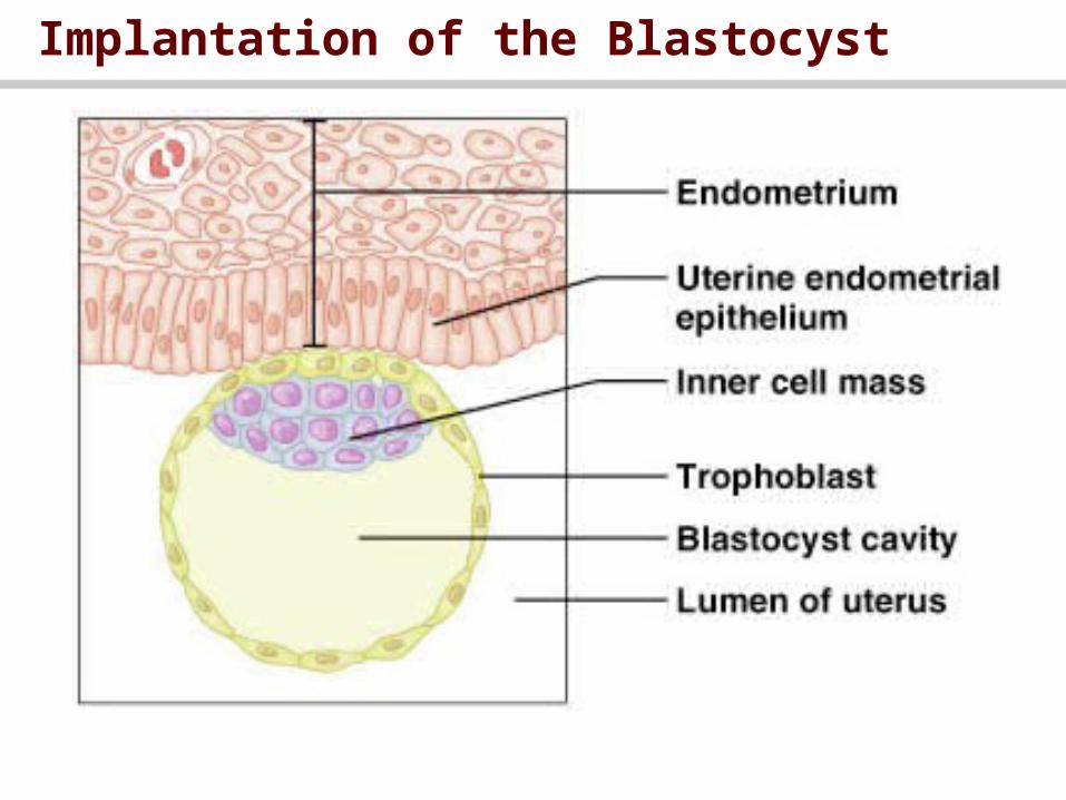
Implantation of the Blastocyst
• After reaching uterus, blastocyst stays another 1-3 days before implantation
• Blastocyst gets nutrition from uterine milk
• Trophoblast cells secrete enzymes that digest the adjacent cells of endometrium
• After implantation, trophoblast cells and other adjacent cells proliferate rapidly to form placenta and other membranes of pregnancy

Cell Division & Implantation
Implantation of the Blastocyst

Placenta
• The major function of placenta is to provide diffusion of gases, foodstuffs, and waste
• Placenta starts providing nutrition after the 16th day after fertilization

Placenta and Further Embroynic Development

Diffusion of O2 and CO2
• The same process as pulmonary membranes
• Simple diffusion
• Fetal hemoglobin has more affinity to O2
• Fetal hemoglubin (Hb) concentration is 50% higher than maternal Hb

Foodstuff and waste products Diffusion
• Simple and facilitated diffusion

Hormonal Factors in Pregnancy
• Placenta forms large quantities of: 1. human chorionic gonadotropin (hCG), 2. estrogen, 3. progesterone, 4. human chorionic somatomammotropin

human chorionic gonadotropin (hCG)
• Secreted by trophoblast 8-9 days after ovulation
• Responsible for “maternal recognition of pregnancy”
• The same structure and function of LH
• Maintains corpus luteum (CL)
• Promotes estrogen and progesterone secretion from CL (CL is important in the first 3 months)
• Stimulates testosterone production by the testes of male fetus (development and decent of testes)

Estrogen from Placenta
• Placental estrogen levels are 30 times higher than normal E production
• Not synthesized de novo, it is converted from androgenic steroids from the mother and fetus adrenal glands
• Functions of E during pregnancy include:
1. Enlargement of the uterus, 2. growth of breasts,
3. Enlargement of female external genitalia
4. Relaxes the pelvic ligaments

Progesterone From Placenta
• Placenta secrets high quantities of P
• Its functions include:
1. Development of decidual cells in the uterine endometrium
2. Decreases the contractility of the uterus
3. Development of fetus even before implantation by increasing the production of nutrients by fallopian tubes and uterus
4. Acts along with estrogen to prepare mother’s breast for lactation

Human Chorionic Somatomammotropin
• Prolactin like effect (human palcental lactogen)
• Decreases maternal insulin sensitivity and enhances fat mobilization (making more glucose available to the fetus)

Hormonal Changes During Pregnancy

Other hormonal factors
• Pitutary ( ACTH, TSH, prolactin)
• Corticsteroids: increased gluco- and mineralocorticoids
• Thyroid increased
• Parathyroid increased (more calcium available)
• relaxin

Parturition • Means birth of the baby
• Toward the end of pregnancy, uterus becomes progressively more excitable
• Estrogen:Progesterone ratio:- progesterone inhibits contractility while estrogen stimulates.
• Oxytocin: increases contractions
• Fetal hormones: oxytocin, adrenal gland, prostaglandin
• Mechanical stretch of uterine muscles increases contractility
• Stretch of the cervix also stimulate uterine contractions

Onset of labor:
• Braxton Hicks contractions: increase toward the end of pregnancy
• Positive feedback: stretch of the cervix by fetal head increases contractility
• Cervical stretching also cause oxytocin release
• Strong uterine contraction and pain from the birth canal cause neurogenic reflexes from spinal cord that induce intense abdominal muscle contractions

Parturition: Initiation of Labor

Labor

The Stages of Labor

Lactation: Producing and Releasing Milk
• Estrogen: growth of ductile system
• Progesterone: development of lobule-alveolar system
• Both E and P inhibit milk production
• Prolactin stimulate milk production
• (other roles in fertility)
• Sucking stimulus • Oxytocin • "Milk let-down" reflex

Lactation: Producing and Releasing Milk

The Milk Let-Down Reflex