Reproductive, Developmental, and Neurobehavioral Effects of Methylmercury in Fishes
16
This article was downloaded by: [Moskow State Univ Bibliote] On: 28 January 2014, At: 11:15 Publisher: Taylor & Francis Informa Ltd Registered in England and Wales Registered Number: 1072954 Registered office: Mortimer House, 37-41 Mortimer Street, London W1T 3JH, UK Journal of Environmental Science and Health, Part C: Environmental Carcinogenesis and Ecotoxicology Reviews Publication details, including instructions for authors and subscription information: http://www.tandfonline.com/loi/lesc20 Reproductive, Developmental, and Neurobehavioral Effects of Methylmercury in Fishes JUDITH S. WEIS a a Department of Biological Sciences , Rutgers University , Newark, New Jersey, USA Published online: 30 Nov 2009. To cite this article: JUDITH S. WEIS (2009) Reproductive, Developmental, and Neurobehavioral Effects of Methylmercury in Fishes, Journal of Environmental Science and Health, Part C: Environmental Carcinogenesis and Ecotoxicology Reviews, 27:4, 212-225, DOI: 10.1080/10590500903310088 To link to this article: http://dx.doi.org/10.1080/10590500903310088 PLEASE SCROLL DOWN FOR ARTICLE Taylor & Francis makes every effort to ensure the accuracy of all the information (the “Content”) contained in the publications on our platform. However, Taylor & Francis, our agents, and our licensors make no representations or warranties whatsoever as to the accuracy, completeness, or suitability for any purpose of the Content. Any opinions and views expressed in this publication are the opinions and views of the authors, and are not the views of or endorsed by Taylor & Francis. The accuracy of the Content should not be relied upon and should be independently verified with primary sources of information. Taylor and Francis shall not be liable for any losses, actions, claims, proceedings, demands, costs, expenses, damages, and other liabilities whatsoever or howsoever caused arising directly or indirectly in connection with, in relation to or arising out of the use of the Content. This article may be used for research, teaching, and private study purposes. Any substantial or systematic reproduction, redistribution, reselling, loan, sub-licensing, systematic supply, or distribution in any form to anyone is expressly forbidden. Terms &
Transcript of Reproductive, Developmental, and Neurobehavioral Effects of Methylmercury in Fishes
This article was downloaded by: [Moskow State Univ Bibliote]On: 28 January 2014, At: 11:15Publisher: Taylor & FrancisInforma Ltd Registered in England and Wales Registered Number: 1072954 Registeredoffice: Mortimer House, 37-41 Mortimer Street, London W1T 3JH, UK
Journal of Environmental Scienceand Health, Part C: EnvironmentalCarcinogenesis and EcotoxicologyReviewsPublication details, including instructions for authors andsubscription information:http://www.tandfonline.com/loi/lesc20
Reproductive, Developmental,and Neurobehavioral Effects ofMethylmercury in FishesJUDITH S. WEIS aa Department of Biological Sciences , Rutgers University , Newark,New Jersey, USAPublished online: 30 Nov 2009.
To cite this article: JUDITH S. WEIS (2009) Reproductive, Developmental, and Neurobehavioral Effectsof Methylmercury in Fishes, Journal of Environmental Science and Health, Part C: EnvironmentalCarcinogenesis and Ecotoxicology Reviews, 27:4, 212-225, DOI: 10.1080/10590500903310088
To link to this article: http://dx.doi.org/10.1080/10590500903310088
PLEASE SCROLL DOWN FOR ARTICLE
Taylor & Francis makes every effort to ensure the accuracy of all the information (the“Content”) contained in the publications on our platform. However, Taylor & Francis,our agents, and our licensors make no representations or warranties whatsoever as tothe accuracy, completeness, or suitability for any purpose of the Content. Any opinionsand views expressed in this publication are the opinions and views of the authors,and are not the views of or endorsed by Taylor & Francis. The accuracy of the Contentshould not be relied upon and should be independently verified with primary sourcesof information. Taylor and Francis shall not be liable for any losses, actions, claims,proceedings, demands, costs, expenses, damages, and other liabilities whatsoever orhowsoever caused arising directly or indirectly in connection with, in relation to or arisingout of the use of the Content.
This article may be used for research, teaching, and private study purposes. Anysubstantial or systematic reproduction, redistribution, reselling, loan, sub-licensing,systematic supply, or distribution in any form to anyone is expressly forbidden. Terms &

Conditions of access and use can be found at http://www.tandfonline.com/page/terms-and-conditions
Dow
nloa
ded
by [
Mos
kow
Sta
te U
niv
Bib
liote
] at
11:
15 2
8 Ja
nuar
y 20
14

Journal of Environmental Science and Health Part C, 27:212–225, 2009Copyright C© Taylor & Francis Group, LLCISSN: 1059-0501 (Print); 1532-4095 (Online)DOI: 10.1080/10590500903310088
Reproductive, Developmental,and Neurobehavioral Effectsof Methylmercury in Fishes
Judith S. WeisDepartment of Biological Sciences, Rutgers University, Newark, New Jersey, USA
In the decades since the Minamata tragedy in Japan, there has been a considerablebody of research performed on effects of methylmercury in fishes. The studies have re-vealed that some of the most sensitive responses seen in fishes are reminiscent of thesymptoms experienced by the Minamata victims. This article reviews the literature,with a focus on mercury’s effects on fish reproduction (hormone levels, gametogenesis,fertilization success), embryonic development (morphological abnormalities, rate), thedevelopment of behavior, and neurobehavioral effects in adults. Both experimental ex-posures and epidemiological approaches are included. There have been many studiesdemonstrating delayed effects of mercury exposure in that exposures during one lifehistory stage can produce effects much later during different life history stages. Forexample, exposure of maturing gametes can result in abnormal embryos, even thoughthe embryos were not themselves exposed to the toxicant. Exposures during sensitiveembryonic periods can produce long-lasting effects that can be seen in adult stages. Theexistence of these manifold delayed effects renders the practice of short-term toxicitytesting particularly unhelpful for understanding the effects of this (and other) toxicants.
Key Words: mercury; fish; behavior; embryo; larva
INTRODUCTION
The Minamata tragedy in Japan was perhaps the first instance of fish causingmercury poisoning in people. The human victims exhibited neurotoxic, neu-robehavioral, and developmental effects, including numbness in the hands andfeet, muscle weakness, narrowing of the field of vision, and damage to hearing
Received July 8, 2009; accepted August 6, 2009.This paper was developed from a presentation given at the symposium on “CommonEffects Endpoints for Persistent Toxic Substances in Human and Ecological Epidemiol-ogy” at the 2007 Society of Environmental Toxicology and Chemistry (SETAC) annualmeeting.Address correspondence to Judith S. Weis, Department of Biological Sciences, RutgersUniversity, Newark, NJ 07102. E-mail: [email protected]
212
Dow
nloa
ded
by [
Mos
kow
Sta
te U
niv
Bib
liote
] at
11:
15 2
8 Ja
nuar
y 20
14
Methylmercury and Fish 213
and speech. These symptoms frequently deteriorated and were followed by con-vulsions, paralysis, coma, and eventually death (1). The disease was first dis-covered in the city of Minamata in1956. Prior to the onset of human disease,there was evidence of strange behavior of cats in the area, which had been seento have convulsions, go mad, and die. Locals called it the “cat dancing disease”due to their bizarre movements.
The staple food of victims was fish and shellfish from Minamata Bay. The“dancing cats” that ate fish scraps had symptoms similar to those now dis-covered in humans. Wastewater from Chisso Corporation’s plant that was dis-charged into the bay contained many metals, including mercury in concentra-tions sufficiently high to bring about serious environmental problems. Sincethe symptoms resembled those of organic mercury poisoning, the investiga-tion centered on mercury. High concentrations of mercury were detected infish, shellfish, and sediments from the bay, with the highest concentrationsaround the factory’s wastewater discharge. Hair samples from the victims andthe general Minamata population had very high mercury concentrations, withthe levels in patients much higher than the other residents, whose levels weremuch greater than people living elsewhere.
Minamata disease was caused by the release of inorganic and methylmer-cury (MeHg) in wastewater from the chemical factory. The mercury bioaccumu-lated in shellfish and fish in Minamata Bay, which, when eaten by the people,resulted in mercury poisoning (1). In aquatic environments inorganic mercurycan be converted to the more toxic form, MeHg by bacteria in the sediments.MeHg readily accumulates in organisms and biomagnifies in food webs. Nearlyall the mercury in fish is MeHg (2, 3), and mercury is responsible for the ma-jority of all fish consumption advisories in the United States (4).
It is likely that the fish in Minamata Bay, in addition to being vectors ofhuman disease, also experienced neurological and developmental effects them-selves, although they were not studied at the time. Since then, there have beennumerous studies investigating effects of mercury on fish, and some of the mostsensitive responses are reproductive, developmental, and neurobehavioral. Wewill review some of these in this paper. It is emphasized that many of the re-sponses can be delayed and not appear until long after the initial exposure.This argues against short-term bioassays, which cannot detect these delayedeffects.
REPRODUCTION: GONADAL DEVELOPMENT, GAMETOGENESIS,REPRODUCTIVE HORMONES
A number of investigators have exposed fish to Hg and examined gonadal de-velopment and gametogenesis. Decreased reproductive success was seen inboth male and female adult rainbow trout chronically exposed to 0.2 µg/l Hg(5). When adult female catfish (Clarias batrachus) were exposed to 0.05 or
Dow
nloa
ded
by [
Mos
kow
Sta
te U
niv
Bib
liote
] at
11:
15 2
8 Ja
nuar
y 20
14

214 J. S. Weis
0.04 µg/l mercuric chloride or MeHg for 90 days, ovarian recrudescence wasinhibited and the gonadosomatic index (GSI) was reduced. Oocytes of exper-imental fish were non-vitellogenic, suggesting that Hg impairs vitellogenesis(6). When male guppies (Poecilia reticulata) were exposed to 1.8 µg/l MeHgvia the water, atrophy of the testis with inflammation and absence of maturesperm in the efferent ducts were noted (7).
Hammerschmidt et al. (8) fed juvenile fathead minnows diets with MeHgat 0.88 (low), 4.11 (medium), and 8.46 µg Hg g−1 (high). At maturity, malesand females were paired and allowed to reproduce (phase 2). Some fish werefed diets different from phase 1. The spawning success of pairs fed same dietwas 75% for controls and 46%, 50%, and 36% for the low-, medium-, andhigh-MeHg groups, respectively. The spawning success of pairs fed Hg dur-ing phase 1 and control diets during phase 2 was no better. Exposure to MeHgduring phase 2 alone did not reduce spawning success. They also found thatMeHg delayed spawning, correlated with the concentration in the fish. TheGSI and reproductive effort of females was inversely correlated with Hg accu-mulation. Friedmann et al. (9) similarly found that dietary mercury (1 µg/g)significantly impaired both growth and gonadal development in juvenile malewalleyes. Treated fish had reduced length, weight, and GSI. Furthermore,testicular atrophy was observed in fish fed the Hg-tainted food but not incontrols.
Drevnick and Sandheinrich (10) fed juvenile fathead minnows MeHgdiets: 0.06 (control), 0.87 (low), and 3.93 (medium) µg Hg g−1 dry weight.MeHg reduced testosterone in males and estradiol in females and inhibitedgonadal development of females: The GSI of females fed medium diet was40% less than those fed control or low diets. Spawning success was 32% forcontrols, 12% for fish on the low Hg diet, and 0% for medium diet. Pairs on thelow Hg diet needed 5 days longer to spawn than controls.
An epidemiological approach was taken by Friedmann et al. (11), who ex-amined reproductive health of wild largemouth bass (Micropterus salmoides)from three lakes with different levels of mercury. The only parameter that wassignificantly correlated with mercury levels in muscle was serum concentra-tion of 11-ketotestosterone.
FERTILIZATION
A particular characteristic of the teleost egg is the chorion, the outer protec-tive membrane that is initially synthesized in the ovary. A canal called themicropyle forms a hole in the membrane, through which sperm must pass inorder to fertilize the eggs. Once sperm have contacted the egg, a cortical re-action occurs in which vesicles in the outer layer of the egg cell release ma-terial that blocks the micropyle and causes elevation of the membrane and
Dow
nloa
ded
by [
Mos
kow
Sta
te U
niv
Bib
liote
] at
11:
15 2
8 Ja
nuar
y 20
14
Methylmercury and Fish 215
increasing its ability to act as a barrier (12, 13). There have been relativelyfew studies of the impact of contaminants on gametes prior to fertilization.
McIntyre (14) found that methylmercury exposure reduced steelhead trout(Salmo gairdneri, now Oncorhynchus mykiss) sperm viability, while Billardand Roubaud (15) found similar effects with mercuric chloride. Birge et al. (5)exposed S. gairdneri males to <1 µg/l mercuric chloride for 4 months; eggsfertilized with sperm from these males had substantially reduced fertilizationand hatch. Eggs from similarly exposed females fertilized with control spermhad poor fertilization and hatch but also had 27% defective larvae among thosethat did hatch, an example of delayed effects. Exposure of mummichog (Fun-dulus heteroclitus) sperm to 10 µg/l MeHg or mercuric chloride for as littleas one minute reduced fertilization success, while a similar exposure of eggshad no effect on fertilization or subsequent development (16, 17). Reductionsin sperm motility were seen in treated groups. Sperm from fish from a pollutedsite were also studied. Piles Creek (PC) in Linden, New Jersey, is a tributary ofthe Arthur Kill, which separates Staten Island from New Jersey and connectsNewark Bay in the north to Raritan Bay in the south. It is surrounded by in-dustrial sites, a sewage treatment plant, and a major highway. Oil spills in theArthur Kill have also been a source of organic contaminants. Elevated levels oforganic contaminants and metals, including mercury, are found in sedimentsand biota. Sperm from PC fish, exposed to 0.01 or 0.05 mg/L MeHg prior to in-semination, had no reduction in fertilization and no difference in motility fromcontrols. Therefore fish from the chronically polluted site had sperm that weremore tolerant.
Higher concentrations of either HgCl2 or MeHg and longer exposure ofunfertilized eggs did result in reduced fertilization and production of abnor-malities in embryos that had subsequently been raised in clean water (18, 19).Thus Hg incorporated prior to fertilization can subsequently produce embry-onic malformations in eggs that were successfully fertilized, another exampleof delayed effects. Eggs from PC fish were more tolerant than eggs from thereference population.
Inorganic and organic mercury had different mechanisms by which theyinhibited fertilization: MeHg triggered a cortical reaction, preventing spermfrom entering the micropyle, while HgCl2 caused a swelling of the lip of themicropyle, reducing its diameter and possibly impeding sperm from swimmingthrough (20).
EMBRYONIC DEVELOPMENT
Latif et al. (21), in an epidemiological study, found reduced hatching success inembryos from different lakes, which was proportional to the concentration ofMeHg in the water.
Dow
nloa
ded
by [
Mos
kow
Sta
te U
niv
Bib
liote
] at
11:
15 2
8 Ja
nuar
y 20
14

216 J. S. Weis
TeratogenesisFish embryos can be exposed to developmental toxicants during oogene-
sis by exposed females and during the brief period between shedding of ga-metes and fertilization. Some studies have shown that mercury incorporatedinto the egg during oogenesis can produce malformations in the embryos thatsubsequently develop from these eggs. McKim (22) found evidence that mer-cury transferred from the female into developing oocytes produced subsequentmalformations in the embryos. However, in most experimental studies, em-bryos have been exposed to mercury after fertilization. Chemicals can affectmorphogenetic movements such as gastrulation, tissue interactions such asinduction, growth, and degeneration or cell death, are an inherent part of em-bryonic development. All of these mechanisms can result in abnormalities inthe developing embryos (23, 24).
Fish embryos tend to become abnormal in certain ways, regardless of theparticular chemical they are exposed to (23). The most sensitive systems arethe developing skeletal system, circulatory system, and optic system. Anothercommon response is a general retardation of development. A decrease in devel-opmental rate can allow teratogenic chemicals to work for a longer time duringsensitive developmental periods (critical stages) and thus produce more severeanomalies.
Mercury Effects on Fish EmbryosA number of investigations have demonstrated abnormalities in optical de-
velopment of Hg-exposed fish embryos. Dial (25) observed disorganized retinas,abnormal pigment distribution, and invasive blood sinuses in eyes of medakaembryos exposed to 80 ug/l MeHg. Weis and Weis (26) noted cyclopia and inter-mediate conditions in which eye rudiments converge in embryos treated with50 µg/l MeHg. The mechanism underlying the fusion of the optic vesicles is in-adequate development of the forebrain, which then permits the two rudimentsto approach each other in the anterior midline of the embryo. The critical pe-riod for development of this anomaly was gastrulation, which is the time ofinduction of the forebrain and well before the actual formation of the opticcups, so this is not essentially a defect in optical development, but rather oneof craniofacial development.
Defects of the cardiac system, including thin atrial and ventricular walls, afailure of the heart tube to differentiate or to bend, hemostasis, and pericardialswelling have been observed in fish embryos including F. heteroclitus (26, 27)and O. latipes (25, 28). These effects can be produced by inorganic or MeHg.Other common defects are axial malformations, ranging from slight bending ofthe skeletal axis to the extreme condition of no axis formation at all. Flexuresand stunting were observed in F. heteroclitus (26, 27), rainbow trout (5), andmedakas (25). Birge et al. (5) noted partial or incomplete twinning and rigid
Dow
nloa
ded
by [
Mos
kow
Sta
te U
niv
Bib
liote
] at
11:
15 2
8 Ja
nuar
y 20
14

Methylmercury and Fish 217
coiling in treated rainbow trout embryos. Samson and Shenker (29) found thatMeHg-exposed zebrafish embryos (Danio rerio) developed tail flexures and fin-fold tissue abnormalities.
Most reports of teratogenic effects calculate the percent of embryos thatare abnormal vs normal. However, in comparing responses of different speciesor different populations of the same species, it is obvious that some embryosare affected to a greater degree than others, and it is possible to devise indicesto rank embryos in terms of severity of effect. Indices of craniofacial, cardiovas-cular, and skeletal malformations have been developed that can give a quan-titative estimate of the severity of the effects and allows for more precise anddetailed analysis. Using such indices, striking population differences in mum-michog embryo responses to MeHg were noted (30). Embryos from a contami-nated environment (PC described above) were much more resistant than thosefrom cleaner habitats. However, females in the cleaner reference site producedeggs with a wide range of tolerances, some females produced very susceptibleeggs and others produced resistant eggs. This great variability within the cleanpopulation, with some resistant individuals already present can be a way forthe evolution of resistance to take place very rapidly. By eliminating the frac-tion of the population that produces susceptible eggs, a resistant populationcan result in a single generation. The mechanism of increased resistance inthe PC population appeared to be more rapid development time (thus exposingembryos during critical periods for a shorter time) and reduced permeabilityof the chorion. There was a decreased mitotic index and increased percentageof abnormal mitosis in MeHg-treated embryos, and those embryos with moresevere teratogenic responses had more severe mutagenic responses (31).
DEVELOPMENT OF BEHAVIOR
Behavioral development occurs in association with the development of the ner-vous system. Many studies have been done on mammals that indicate thatembryonic exposures to various chemicals at concentrations that do not pro-duce anatomical malformations may nevertheless produce functional deficitsat later stages in life (32). The study of behavioral abnormalities resultingfrom embryonic exposures has been called “behavioral teratology” and is an-other example of delayed effects. In addition to numerous experimental studiesin laboratory animals, there is epidemiological evidence. Prenatally exposed in-dividuals in Minamata suffered from brain damage, mental retardation, and/orpsychomotor retardation (1). There was an abnormally high frequency of cere-bral palsy and other disorders in children in Minamata. The symptoms of thechildren were similar to those of adults with Minamata disease, but theirmothers did not exhibit symptoms. These children had been born after theinitial outbreak and had not eaten contaminated fish. Some of their mother’s
Dow
nloa
ded
by [
Mos
kow
Sta
te U
niv
Bib
liote
] at
11:
15 2
8 Ja
nuar
y 20
14

218 J. S. Weis
body burden of MeHg had been transferred to the fetus through the placenta,and symptoms appeared post-natally. Since Minamata, there have been manyother studies of humans exhibiting delayed neurological effects after pre-natalexposure to low levels of MeHg (33, 34) as well an numerous studies on labo-ratory animals (35, 36).
There has been a little work on comparable delayed effects in fish. Weis andWeis (37) examined behavior of mummichog larvae after embryonic exposureto levels of MeHg lower than those that produced anatomical malformations(5 and 10 µg/l). Larvae were maintained in clean water. Prey capture abilityof early larvae was impaired, but after about one week after hatching the preycapture ability was comparable to controls, showing that this was a temporaryeffect. The exposure may have caused retardation of neurological developmentthat was subsequently compensated for and therefore no long lasting effectswere produced. When many populations were investigated in an epidemiolog-ical approach, larval prey capture ability was found to be related to sedimentcontamination levels at the site (38) with significant negative correlations seenwith Hg, Pb, Zn, Cd, and PCBs. Since the levels of contaminants at a site werehighly correlated with each other, it was not possible to attribute impairedbehavior to Hg, but the most impaired population was the one from Berry’sCreek, which is a Superfund site for mercury.
Larvae that had been exposed as embryos were also more susceptible topredation by grass shrimp (Palaemonetes pugio) or adult mummichogs (39).Larvae that had been exposed to MeHg as embryos generally had increasedactivity levels, which could serve to attract a predator resulting in increasedcapture (40). Exposure of larvae alone to MeHg was relatively ineffective, butafter exposure of both embryos and larvae, effects on prey capture were greaterthan embryonic exposure alone (41). Social behavior was also impaired in lar-vae that had been exposed as embryos (42). When in groups, these larvae hada greater frequency of collisions than controls. This response also disappearedby four weeks after hatching, indicating that the effect was reversible.
In contrast, Samson et al. (43) exposed zebrafish (Danio rerio) embryosto 0, 5, 10, or 15 µg MeHg/l for varying amounts of time. Larvae were keptin clean water. Continuous embryonic exposure to 15 µg/l resulted in delayedmortality: by day 3, post-hatch (ph) activity was reduced; by day 5, post-hatchlarvae were moribund with faint heartbeat, edema, and vertebral flexures.Most embryos were dead by day 6 ph. Shorter exposures to 15 µg/l causedreduced activity and impaired prey capture; by day 4 ph, larvae showed signsof delayed mortality. Continuous exposure or exposure during the last 24 h ofdevelopment to 10 µg/l reduced activity, which did not improve. Prey capturewas impaired after continuous embryo exposure to 10 µg/l, even after four daysin clean water. Single-day embryonic exposures to10 µg/l did not affect activ-ity or prey capture of larvae, however. Thus, in contrast to the mummichogexperiments, the delayed effects seen here did not appear to be reversible.
Dow
nloa
ded
by [
Mos
kow
Sta
te U
niv
Bib
liote
] at
11:
15 2
8 Ja
nuar
y 20
14

Methylmercury and Fish 219
Alvarez et al. (44) fed Atlantic croaker fed MeHg-contaminated food atthree levels for 1 month. Fish were then induced to spawn and MeHg levelsin the eggs were measured. Behavioral performance of exposed and controllarvae was measured at different developmental stages. Behaviors analyzedincluded routine swimming speed and startle response (responsiveness, reac-tive distance, response distance, response duration, average response speed,and maximum response speed). Maternally transferred MeHg impaired thesebehaviors, which were considered survival skills.
Fjeld et al. (45) found very delayed effects. Embryos of grayling (Thymal-lus thymallus) were exposed to MeHg during the first 10 days of development.Three years later there was impaired feeding efficiencies and reduced com-petitive abilities in fish from groups that had accumulated Hg > 0.27 µg g−1.Exposed fish were 15% to 24% less efficient than controls. Controls caught2 to 6 times as many prey as exposed fish. Therefore, the embryonic expo-sures caused irreparable alterations in the brains of the fish. The concentra-tion of 0.27 µg g−1 Hg, which appeared to be a threshold for this effect, is seenin eggs of piscivorous fishes in lakes with substantial atmospheric depositionof Hg.
ADULT BEHAVIOR
There have been a number of studies on behavioral effects of adult mercuryexposures. Many of these have also focused on activity level, prey capture, andpredator avoidance—behaviors that are critical to survival and that have eco-logical relevance. Reduced feeding can affect growth rate and thereby affect thepopulation. Reduced predator avoidance ability can increase mortality ratesand affect the population. If individuals with higher concentrations of mercuryare easier for predators to capture, this will facilitate the transfer of the con-taminant to higher trophic levels. Altered predator/prey interactions can causechanges in populations of predators, prey, or both and thus affect the commu-nity.
Weis and Khan (46) found that exposure of adult mummichogs to 10 µg/lof either HgCl2 or MeHg for a week caused reductions in feeding rate. Thesame reduced feeding rate was seen in fish from a polluted environment (PC,described earlier) (47). These fish also had reduced condition. Previous workhad shown that the PC population had reduced lifespan and growth (48), andreduced feeding could be partly responsible for reduced growth. When fishfrom the clean site were maintained in aquaria with water, sediments, andfood (grass shrimp) from PC, their prey capture ability declined to that of thePC population (49), and the level of Hg in their brains increased to that ofthe PC population. When PC fish were maintained in clean water, sediments,and food, their prey capture ability increased slightly but not significantly, and
Dow
nloa
ded
by [
Mos
kow
Sta
te U
niv
Bib
liote
] at
11:
15 2
8 Ja
nuar
y 20
14

220 J. S. Weis
their brain Hg did not decrease. PC fish collected from the field had muchmore sediment and detritus in their guts and less live prey than fish from theclean site, providing field validation for the poor predatory ability observedin the laboratory. PC fish also had lower activity and were more vulnerableto predation by blue crabs (49), which can contribute to their shorter lifes-pan. The correlation of behavior with mercury does not mean that Hg is thecause of the behavioral impairment because there are many other contami-nants at the site including lead and PCBs that could contribute to behavioraldeficits.
When populations from many different sites were investigated for preycapture, their ability was found to be related to sediment and tissue levelsof contaminants (50). Since the levels of contaminants at a site were highlycorrelated with each other, effects of specific contaminants could not be de-termined. Using laboratory prey capture experiments, the number of grassshrimp captured was highest by fish from the cleanest sites. Gut contentsof field-collected fish revealed that grass shrimp made up the largest pro-portion of the diet in sites whose fish had the highest capture rates in thelaboratory.
Golden shiner (Notemigonus chrysoleucas) were fed diets with differentlevels of Hg and then exposed to a model avian predator and their responsevideotaped. Fish fed higher Hg had greater dispersal, took longer to returnto pre-exposure activity, and schools had greater areas after return to pre-exposure activity, all of which would increase their vulnerability to predation(51).
MECHANISMS UNDERLYING BEHAVIORAL IMPAIRMENT
Altered behavior may result from nervous system damage. Neurotransmittershave been found to be sensitive to contaminant exposure. Hg decreased lev-els of serotonin (5-HT) in brains of striped mullet (Mugil cephalus), whichwas associated with loss of motor control (52). Similar responses were seenin tilapia (Oreochromis mossambicus) (53). PC mummichogs had reduced sero-tonin (54), and the level of brain serotonin was directly related to the activitylevel of the fish. Exposure of mummichog larvae to MeHg produced variable al-terations in neurotransmitter levels that varied over time and were differentin the two populations both before and after exposure (55). Newly hatched PCfish, which were not behaviorally impaired, had levels of serotonin comparableto fish from the reference population, but did have higher levels of dopamineand its metabolites.
Altered behavior can also result from changes in the thyroid gland. Thy-roid hormones are essential for normal brain development, and altered thyroidhormones can alter neurobehavioral development. Altered thyroid function can
Dow
nloa
ded
by [
Mos
kow
Sta
te U
niv
Bib
liote
] at
11:
15 2
8 Ja
nuar
y 20
14
Methylmercury and Fish 221
affect activity and feeding in fish (56), and Hg exposure can alter levels ofthyroid hormones (57). Fish have individual thyroid follicles scattered through-out the branchial region rather than a cohesive gland as is found in othervertebrates. When stimulated by TSH from the pituitary gland, the epithe-lial cells surrounding the follicle increase in height and overall the folliclesenlarge. Abnormally enlarged follicles, the fish equivalent of a goiter, reflectdeficiencies in thyroid hormones, which trigger the pituitary to secrete ex-cess TSH, which stimulates the thyroid follicles. Such follicles have been seenin fish from the Great Lakes (58). PC mummichogs also have greatly ex-panded and irregularly shaped thyroid follicles and a trend of reduced lev-els of T3, which is the active form of the thyroid hormone in fish (59). How-ever, since these fish live in an environment with many other contaminants,the abnormal thyroid glands cannot be attributed to an effect of mercury inparticular.
REPRODUCTIVE BEHAVIOR
Sandheinrich and Miller (60) fed fathead minnows MeHg and examined behav-ior and hormone levels. Control fish spent 5% of their time spawning; while fishfed MeHg spent only 0.5% of their time spawning. Fish on low- and medium-MeHg diets spent 19% to 26% of their time inactive, while control fish spentonly 8% of their time inactive. No significant differences in testosterone wereseen, but testosterone was correlated with nest preparatory, courtship, andspawning behaviors in males. The authors stated that their findings suggestedthat altered behavior was, in part, responsible for suppression of reproductionin exposed fish and suggested that reproductive behavior may be more sensi-tive than steroid hormones to alteration by MeHg.
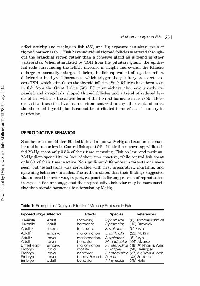
Table 1: Examples of Delayed Effects of Mercury Exposure in Fish
Exposed Stage Affected Effects Species References
Juvenile Adult spawning P. promelas (8) HammerschmidtJuvenile Adult hormones P. promelas (10) DrevnickAdult sperm fert. succ. S. gairdneri (5) BirgeAdult embryo malformation S. fontinalis (22) McKimAdult larva malformation. S. gairdneri (5) BirgeAdult larva behavior M. undulatus (44) AlvarezUnfert egg embryo malformation F. heteroclitus (18,19) Khan & WeisEmbryo larva motility O. latipes (28) HeisingerEmbryo larva behavior F. heteroclitus (37, 39) Weis & WeisEmbryo larva behav & mort. D. rerio (43) SamsonEmbryo adult behavior T. thymallus (45) Fjeld
Dow
nloa
ded
by [
Mos
kow
Sta
te U
niv
Bib
liote
] at
11:
15 2
8 Ja
nuar
y 20
14

222 J. S. Weis
CONCLUSIONS
An important take-home message from this review is that the effects of mer-cury on fish (and other organisms) are complex, multiple, and often delayed.Exposures during one phase of life can produce effects that may be manifestedlater, sometimes after years (Table 1). Similar delayed effects have been seenin other taxa, as documented by other presenters in this symposium. The exis-tence of these delayed effects means that understanding the toxicity of thiscontaminant (as well as other toxicants, as demonstrated by other presen-ters at the symposium) requires extensive study, and suggests that short-termbioassay approaches, unfortunately, are still too common, and are not partic-ularly useful. Complex, multiple effects of toxicants are only understood afterdetailed study of different life history stages.
ACKNOWLEDGEMENTS
I thank Dr. Peddrick Weis for his collaboration, insights, and analytical skillsover the years of studying mercury and fish. I appreciate the hard work ofthe many former graduate students in my laboratory who have helped to un-ravel effects of mercury on killifish and the adaptations and responses in ourlocal polluted estuaries: Margarete Heber, Swati Toppin, Abu Khan, AnwarKhan, Graeme Smith, Tong Zhou, Jennifer Samson, and Celine Santiago Bass.We appreciate funding from the NJ Sea Grant Program, NOAA, USGS WaterResources Research Institute, USEPA, and NJ DEP. I thank Tim Kubiak forinviting me to participate in the SETAC symposium.
REFERENCES
1. Clarkson TW. Mercury: major issues in environmental health. Environ. HealthPersp 1992;100:31–38.
2. Bloom NS. On the chemical form of mercury in edible fish and marine invertebratetissue. Can. J. Fish Aquat Sci 1992;49:1010–1017.
3. Wiener JG, Krabbenhoft DP, Heinz GH, Scheuhammer AM. Ecotoxicology of mer-cury. In Hoffman DJ, Rattner BA, Burton GA Cairns J, eds. Handbook of Ecotoxicology,2nd ed. Boca Raton, FL: CRC Press, Boca Raton 2003;409–463.
4. U.S. Environmental Protection Agency. 2004 National listing of fish advi-sories. Washington DC: USEPA, Office of Water Fact Sheet EPA-823-F-05-004,2005.
5. Birge WJ, Black JA, Westerman AG, Hudson JE. The effects of mercury on repro-duction of fish and amphibians. In Nriagu, J. ed. The Biogeochemistry of Mercury inthe Environment. Amsterdam: Elsevier/North Holland 1979;629–655.
6. Kirubagaran R, Joy KP. Toxic effects of mercuric chloride, methylmercuric chlorideand Emisan 6 (an organic mercurial fungicide) on ovarian recrudescence in the catfish,Clarias batrachus (L.). Bull Environ Contam Toxicol 1988;41:902–909.
Dow
nloa
ded
by [
Mos
kow
Sta
te U
niv
Bib
liote
] at
11:
15 2
8 Ja
nuar
y 20
14

Methylmercury and Fish 223
7. Wester PW. Histopathological effects of environmental pollutants B-HCH andmethyl mercury on reproductive organs in freshwater fish. Comp Biochem Physiol1991;100C:237–239.
8. Hammerschmidt CR, Sandheinrich MB, Wiener JG, Rada RG. Effects of dietarymethylmercury on reproduction in fathead minnows. Environ Sci Tech 2002;36:877–883.
9. Friedmann AS, Watzin MC, Brinck-Johnsen T, Leiter JC. Low levels of dietarymethylmercury inhibit growth and gonadal development in juvenile walleye (Stizoste-dion vitreum). Aquat Toxicol 1996;35:265–278.
10. Drevnick PE, Sandheinrich MB. Effects of dietary methylmercury on reproductiveendocrinology or fathead minnows. Environ Sci Tech 2003;37:4390–4396.
11. Friedmann AS, Costain EK, MacLatchy DL, Stansley W, Washuta EJ. Effectof mercury on general and reproductive health of largemouth bass (Micropterussalmoides) from three lakes in New Jersey. Ecotox Environ Safety 2002;52:117–122.
12. Brummet AR, Dumont JN. Initial stages of sperm penetration into the egg of Fun-dulus heteroclitus. J Exp Zool 1979;210:417–434.
13. Brummet AR, Dumont JN. Cortical vesicle breakdown in fertilized eggs of Fundu-lus heteroclitus. J Exp Zool 1981;216:63–79.
14. McIntyre JD. Toxicity of methylmercury to steelhead trout sperm. Bull EnvironContam Toxicol 1973;9:98–99.
15. Billard R, Roubaud P. The effect of metals and cyanide on fertilization in rainbowtrout (Salmo gairdneri). Water Res 1985;19:209–214.
16. Khan AT, Weis JS. Toxic effects of mercuric chloride on sperm and egg viabil-ity of two populations of mummichog, (Fundulus heteroclitus). Environ Pollut Ser A1987;48:263–273.
17. Khan AT, Weis JS. Effects of methylmercury on sperm and egg viability of two pop-ulations of killifish (Fundulus heteroclitus). Arch Environ Contam Toxicol 1987;16:499–505.
18. Khan AT, Weis JS. Effect of mercuric chloride on eggs and juvenile viability in twopopulations of killifish. Mar Poll Bull 1987;18:504–505.
19. Khan AT, Weis JS. Effect of methylmercury on egg and juvenile viability in twopopulations of killifish Fundulus heteroclitus. Environ Res 1987;44:272–278.
20. Khan AT, Weis JS. Differential effects of organic and inorganic mercury on themicropyle of the eggs of Fundulus heteroclitus. Environ Biol Fishes 1993;37:323–327.
21. Latif MA, Bodaly RA, Johnson TA, Fudge RJP. Effects of environmentally andmaternally derived methylmercury on the embryonic and larval stages of walleye (Ste-zostedion vitreum). Environ Poll 2001;111:139–148.
22. McKim J. Evaluation of tests with the early life stages of fish for predicting longterm toxicity. J Fish Res Bd Can 1977;36:1148–1154.
23. Laale HW. Teratology and early fish development. Amer Zool 1981;21:517–533.
24. Weis JS, Weis P. Effects of environmental pollutants on early fish development.Rev Aquat Sci 1989;1:45–73.
25. Dial NA. Some effects of methylmercury on development of the eye in medaka fish.Growth 1978;42:309–318.
Dow
nloa
ded
by [
Mos
kow
Sta
te U
niv
Bib
liote
] at
11:
15 2
8 Ja
nuar
y 20
14

224 J. S. Weis
26. Weis P, Weis JS. Methylmercury teratogenesis in the killifish, Fundulus heterocli-tus. Teratology 1977;16:317–326.
27. Weis JS, Weis P. The effects of heavy metals on embryonic development of thekillifish, Fundulus heteroclitus. J Fish Biol 1977;11:49–54.
28. Heisinger JF, Green W. Mercuric chloride uptake by eggs of the ricefish and re-sulting teratogenic effects. Bull Environ Contam Toxicol 1975;14:665–673.
29. Samson JC, Shenker J. 2000. The teratogenic effects of methylmercury on earlydevelopment of the zebrafish. Aquat Toxicol 48:343–353.
30. Weis JS, Weis P, Heber M, Vaidya S. Methylmercury tolerance of killifish (Fun-dulus heteroclitus) embryos from a polluted vs non-polluted environment. Mar Biol1981;65:283–287.
31. Perry D, Weis JS, Weis P. Cytogenic effects of methylmercury in embryos of thekillifish, Fundulus heteroclitus. Arch Environ Contam Toxicol. 1988;17:569–574.
32. Choi BH. Effects of prenatal methylmercury poisoning upon growth and develop-ment of fetal nervous system. In: Clarkson TW, Nordberg GF, Sager PR eds. Reproduc-tive and Developmental Toxicity of Metals. NY: Plenum Press 1983:473–495.
33. Grandjean P, Weihe P, White RF, Debes F. Cognitive performance of childrenprenatally exposed to “safe” levels of methylmercury. Environ Res 1998;77:165–172.
34. Murata K, Weihe P, Araki S, Butdz-Jorgensen E, Grandjean P. Evoked po-tentials in Faroese children prenatally exposed to methylmercury. Neurotox Teratol1999;21:471–472.
35. Burbacher TM, Rodier PM, Weiss B. Methylmercury developmental neurotoxicity:a comparison of effects in humans and animals. Neurotox Teratol 1990;12:191–202.
36. Glibert SG, Grant-Webster KS. Neurobehavioral effects of developmentalmethylmercury exposure. Environ Health Persp 1995; 103 (suppl 6):135–142.
37. Weis JS, Weis P. Effects of embryonic exposure to methylmercury on larvalprey capture ability in the mummichog, Fundulus heteroclitus. Environ Tox Chem1995;14:153–156.
38. Weis JS, Samson J, Zhou T, Skurnick J, Weis P. Evaluating prey capture by larvalmummichogs (Fundulus heteroclitus) as a potential biomarker for contaminants. MarEnviron Res 2003;55:27–38.
39. Weis JS, Weis P. Swimming performance and predator avoidance by mummichog(Fundulus heteroclitus) larvae after embryonic exposure to methylmercury. Can J FishAquat Sci 1995;52:2168–2173.
40. Zhou T, Weis JS. Swimming behavior and predator avoidance in three populationsof Fundulus heteroclitus larvae after embryonic and/or larval exposure to methylmer-cury. Aquat Toxocol 1998;43:131–148.
41. Zhou T, Scali R, Weis JS. Effects of methylmercury on ontogeny of prey captureability and growth in three populations of larval Fundulus heteroclitus. Arch EnvironContam Toxicol 2001;41:47–54.
42. Ososkov I, Weis JS. Development of social behavior in larval mummichogs afterembryonic exposure to methylmercury. Trans Amer Fish Soc 1996;125:983–987.
43. Samson JC, Goodridge R, Olubatuyi F, Weis JS. Delayed effects of embryonic expo-sure of zebrafish (Danio rerio) to methylmercury (MeHg). Aquat Toxicol 2001;51:369–376.
Dow
nloa
ded
by [
Mos
kow
Sta
te U
niv
Bib
liote
] at
11:
15 2
8 Ja
nuar
y 20
14

Methylmercury and Fish 225
44. Alvarez MC, Murphy CA, Rose KA, McCarthy ID, Fuiman LA. Maternal body bur-den of methylmercury impairs survival skills of offspring in Atlantic croaker (Microp-ogonias undulatus). Aquat Toxicol 2006;80:329–337.
45. Fjeld E, Haugen TO, Vollestad LA. Permanent impairment in the feeding behaviorof grayling (Thymallus thymallus) exposed to methylmercury during embryogenesis.Sci Total Environ 1998;213:247–254.
46. Weis JS, Khan AA. Effects of mercury on the feeding behavior of the mummichog,Fundulus heteroclitus, from a polluted habitat. Mar Environ Res 1990;30:243–249.
47. Weis JS, Khan AA. Reduction in prey capture ability and condition of mummichogsfrom a polluted habitat. Trans Amer Fish Soc 1991;120:127–129.
48. Toppin SV, Heber M, Weis JS, Weis P. 1987. Changes in reproductive biology andlife history in Fundulus heteroclitus in a polluted environment. In: Vernberg W, Cal-abrese A, Thurberg F, Vernberg J. eds. Pollution Physiology of Estuarine Organisms.Columbia: Univ. South Carolina Press, 1987;171–184.
49. Smith GM, Weis JS. Predator/prey interactions in Fundulus heteroclitus: effectsof living in a polluted environment. J Exper Mar Biol Ecol 1997;209:75–87.
50. Weis JS, Samson J, Zhou T, Skurnick J, Weis P. Prey capture ability of mummi-chogs (Fundulus heteroclitus) as a behavioral biomarker for contaminants in estuarinesystems. Can J Fish Aquat Sci 2001;58:1442–1452.
51. Webber HM, Haines TA. Mercury effects on predator avoidance behavior ofa forage fish, golden shiner (Notemigonus chrysoleucas). Environ Toxicol Chem2003;22:1556–1561.
52. Thomas P, Wofford HW, Neff, JM. Biochemical stress responses of striped mullet(Mugil cephalus L.) to fluorine analogs. Aquat Toxicol 1981;1:329–342.
53. Tsai CL, Jang TH, Wang LH. Effects of mercury on serotonin concentration in thebrain of tilapia, Oreochromis mossambicus. Neuroscience Lett 1995;184:208–211.
54. Smith GM, Khan AT, Weis JS, Weis P. Behavior and brain chemistry correlates inmummichogs (Fundulus heteroclitus) from polluted and unpolluted environments. MarEnviron Res 1995;39:329–334.
55. Zhou T, Rademacher DJ, Steinpreis RE, Weis JS. 1999. Neurotransmitter levels intwo populations of larval Fundulus heteroclitus after methylmercury exposure. CompBiochem Physiol C 1999;124:287–294.
56. Castonguay M, Cyr DG. Effects of temperature on spontaneous and thyroxine-stimulated locomotor activity of Atlantic cod. J Fish Biol 1998;53:303–315.
57. Bleau H, Daniel C, Chevalier G, van Tra H, Hontela A. Effects of acute exposureto mercury chloride and methylmercury on plasma cortisol, T3, T4, glucose and liverglycogen in rainbow trout (Oncorhynchus mykiss). Aquat Toxicol 1996;34:221–235.
58. Leatherland JF. Reflections on the thyroidology of fishes: from molecules to hu-mankind. Guelph Ichthyological Review 1994;2:1–67.
59. Zhou T, John-Alder H, Weis P, Weis JS. Thyroidal status of mummichogs (Fun-dulus heteroclitus) from a polluted vs a reference habitat. Environ Toxicol Chem1999;18:2817–2823.
60. Sandheinrich MB, Miller KM. Effects of dietary methylmercury on reproduc-tive behavior of fathead minnows (Pimephales promelas). Environ Toxicol Chem2006;25:3053–3057.
Dow
nloa
ded
by [
Mos
kow
Sta
te U
niv
Bib
liote
] at
11:
15 2
8 Ja
nuar
y 20
14