REPRODUCTIVE CHILD HEALTH CARE-(INTRANATAL) IN INDIA
53
REPRODUCTIVE CHILD HEALTH CARE IN INDIA (INTRA NATAL) DR. MAHESWARI JAIKUMAR
-
Upload
maheswari-jaikumar -
Category
Health & Medicine
-
view
361 -
download
3
Transcript of REPRODUCTIVE CHILD HEALTH CARE-(INTRANATAL) IN INDIA

REPRODUCTIVE CHILD HEALTH CARE IN INDIA (INTRA NATAL)
DR. MAHESWARI JAIKUMAR

CONTENTS.Stages of lab our.
Signs of true labour.
Management of first stage of labour.
Monitoring I stage by partogram.
Identification & management of foetal & maternal distress.
Management of II stage of labour.
Management of III stage of labour.
Episiotomy.
Perineal tears.

Normal labour & delivery room.
Early identification of abnormal delivery – labour.
Management of abnormal labour.
Malposition.
Malpresentation.
Obstructed labour.
Rapture uterus.
Post partum hemorrhage.
Retained placenta.
Shock.
Acute inversion of uterus.
Pulmonary embolism.
EUTOCIA.
Normal labour or eutocia is the orocess of expulsion per vaginum of a mature live foetus presented by vertex followed by the placenta & membranes spontaneously without any complications or delay.

STAGES OF LABOUR.
I. I stage.
II. II stage.
III. III stage.1. 12-14 hrs in primi.
2. 4-6 hrs in multipara.
FIRST STAGE : THE FIRST STAGE STARTS FROM THE ONSET OF LABOUR PAINS TILL FULL DILATION OF CERVIX.12 HRS IN PRIMI GRAVIDA & 6 HR IN MULTIPARA.

SECOND STAGE : FROM DILATION OF CERVIX TO EXPULSION OF FOETUS. IT LASTS FOR 2 HRS IN NULLIPARA & 1 HOUR IN MULTIPARA.
THIRD STAGE : THIRD STAGE OF LABOUR LASTS FOR 5 TO 10 MIN.IT SHOULD NOT EXEED 30 MIN.

SIGNS OF TRUE LABOUR.
Diagnose labour by the following signs.
Painful uterine contractions,at regular intervals,progressively increasing in duration & intensity.
Progressive cervical dilation & effacement.
Formation of bag of waters.
Presence of show.

MANAGEMENT OF FIRST SRTAGE OF LABOUR.
ENSURE,------------------
The bowel is emptied by enema.
The women is given clean gown to wear, & to be ambulant.
Posture– Ambulant, lying flat on her back may give her supine hypo tension.
Nutrition– Only clear fluids are given to prevent dehydration.Solid foods are avoided to prevent any anesthetic complication if surgery is required.
Bladder– She passes frequently as full bladder may delay the progress of labour.

MONITORING THE FIST STAGE OF LABOUR & MAINTENANCE OF PARTOGRAM
PARTOGRAPH is a tool to to monitor the progress & management of lab our.
Partograph is used to record all observations made on a women in lab our.
Its central feature is a graph where dilation of the cervix as assed by vaginal examination is plotted.
By noting the rate at which the cervix dilates the one can assess the normalcy of the labor.
There fore Partograph is a powerful tool.

ADVANTAGE OF PARTOGRAPH.
PARTOGRAPH helps to prevent prolonged & obstructed labour.
It is a clear way of recording all observations on one chart.
Helps in detecting abnormalities.
It is a potent tool to manage labour.
USAGE OF PARTOGRAPH
Observe & record in the following sequence.
Cervical dilation.
Descent of head.
Uterine contractions.
Duration shown by differential shading.
The foetal condition.
The maternal condition.

MONITOR FOETAL CONDITIONOBSERVING THE FOETAL HEART RATE IS SAFE & RELIABLE CLINICAL WAY OF KNOWING THE WELLBEING OF THE FOETUS.
A RATE < 160 beats / min (tachycardia) .
< 120 /min (bradycardia) may indicate foetal distress.
A heart beat < 100 or lower indicates very severe distress & action should be taken at once.
MONITORING THE MATERNAL CONDITION.
MONITOR THE FOLLOWING.
Pulse- half hrly.
Blood pressure- 4 hrly or as indicated frequently.
Temperature-4 hrly or frequently as indicated.
Urine volume-Encourage women to pass urine 2-4 hrly & measure I/O.
Drugs & IV fluids-chart these in appropriate coloumn
Oxytocin regime

IDENTIFICATION & MANAGEMENT OF FOETAL & METERNAL DISTRESS.
EVIDENCE OF FOETAL DISTRESS.
Persistence of tachycardia > 160/min.
Persistence bradycardia. <120/min.
Irregularity of foetal heart sounds.
Excessive foetal movements.
Passages of me conium per vaginum in cephalic presentation.

IF ANY OF THE SIGNS DO NOT IMPROVE DESPITE MANAGEMENT,
LABOUR HAS TO BE TERMINATED EITHER BY LSCS IN FIRST STAGE OR BY INSTRUMENTAL DELIVERY. ----- REFER TO FRU.

MANAGEMENT
Oxygen inhalation.
To lie in left lateral position.
Inj Ringer lactate.
Shift to FRU.
EVIDENCES OF MATERNAL DISTRESS.
THE FOLOWING ARE THE SIGNS OF MATERNAL DISTRESS.
Increased pulse rate.->100 /min.
Looks exhausted.
Rise of temp > 100.4 F.
Dehydrated tongue.
Appearance of protein & Ketones in urine.(ketostix,Rothera’s test ).

MANAGEMENT.
Start I.V 5% dextrose, followed by RL infusion.
Re assess the progress of labour.
Assess the type of uterine contraction.
Assess the foetal condition.
Manage accordingly.
MANAGEMENT OF II STAGE OF LABOUR.
DIAGNOSIS OF II STAGE.
Mother vomits or reports that she feels a need to defaecate.
Membranes spontaneously rapture.
Mother has intense urge to bear down during contraction.
PV- cervix is no longer palpable. I.e. fully dilated.

CONDUCT OF DELIVERY.
SHIFT THE MOTHER TO DELIVERY ROOM WHEN THE FOETAL HEAD IS SEEN AT VULVA IN BETWEEN CONTRACTIONS.
Monitor FHR every 5 min.
Bring the mother to the edge of the table-preferably in dorsal or semi recumbent position.
Wash perineal area with an antiseptic solution & use sterile drapers.
When the head is crowning the perineum,make a decision as to the requirement of Episiotomy.( Episiotomy are routinely ).

EPISIOTOMY- Give local infiltration With 1% xylocaine along the line of planned episiotomy cut which is usually a mediolateral one.
If the episiotomy is made at the right time,almost with the next contraction & patient’s bearing down effort, the head will deliver.
Apply counteraction anteriorly to the vertex with one hand while a towel or a pad in the other hand supports the perineum to enable a controilled delivery of the head rather than a sudden pop out.
Once the head is delivered,palpate the fetal neck to see any loop of cord wrapped around it.If the loop is loose slip it over the head posteriorly.But if it is tight, it is cut between two clamps.
.

Clear the baby’s mouth & oropharynx with mocous sucker before the body delivers.Deliver the shoulders by depressing the head posteriorly so that the lateral flexion of the body occurs.The rest of the baby automatically follows.
At the delivery of the anterior shoulder give IV methergine 0.2 mg unless there is a contraindication.
Cut the cord between clamps & hand over the baby to the paediatrician.
Note the time of the birth of the baby.
Give the baby to the mother & let the baby start sucking.
MANAGEMEMT OF THIRD STAGE OF LABOUR.
Place the left hand on lower abdomen to detect the contraction of the Ut.
Following the delivery Ut is just below the level of the umbilicus.
It ensures early detection of blood collecting inside the Ut.

SIGNS OF PLACENTAL SEPERATION.
Ut becomes hard & globular.
Ut rises just above umbilicus.
Extra vulval lengthening of the umbilical cord.
A gush of blood frequently appears.
On pushing the Ut up in the abdomen the cord does not recede back.

PLACENTA is delivered by controlled cord traction & concentration of the uterine corpus.
Cord traction should be avoided before the placenta is separated.
If methergine was not given , 0.2 mg IM should be given after the delivery of placenta.
Inspect the placenta for completeness.Feel the hard retracted Ut & if the vaginal bleeding is not excessive, proceed to inspect the vagina for evidence of any lacerations or tears.
Repair of tears & Episiotomy is done on similar lines.

EPISIOTOMYPERINEOTOMY is an incision into the perineum to enlarge the space at the outlet,thereby facilitating the birth of the child.
INDICATIONS FOR EPISIOTOMY
To preserve the integrity of the pelvic floor.
To prevent uncontrolled perineal tears.
To prevent foetal head– pre term babies,large babies,abnormal positions & in breech presentations.
PERINEAL TEARS.Many women suffer from tears of perineum .The following are the types.
First degree tear : The vaginal mucosa, the fourchette & skin of perineum are involved.
Second degree tear : The perineal muscles are torn.
Third degree tear : The tear extends through the perineal body,muscles & the annal sphinter & occasionally anterior rectal wall.
HEALING OF THE PERINEUM.
ENSURE,
General care.
Low residue diet for three days.
Laxatives to make the stool soft.
Avoid enema.
Ambulate the mother.

EQUIPMENTS FOR NORMAL DELIVERY –LABOUR ROOM
Catheters – Simple catheter & self retaining catheters.
Enema apparatus.
Sponge holding forceps.
Scissors.
Dissecting forceps.
Needle. – Round bodied, cutting.
Needle holder.
Artery forceps.
Sutures chromatic catgut- No –1 on round body & cutting needle.

Needle holder.
Artery forceps.
Sutures – Chromic catgut.No – 1 on round body.
Obstetrics forceps.
Kidney trays.
Bowls.
Rubber sheets.
Sterilized rubber gloves.
Gowns & masks.
Thermometer.
Bp apparatus.
Episiotomy tray.

Episiotomy scissors.
Small curved artery forceps.
Dissecting forceps.
Needle holder.
Sponge holding forceps.
For local infiltration.----10 ml syringe,hypodermic needle,antiseptic solution,sterile drapes & cotton swabs.
IDENTIFICATION OF ABNORMAL PROGRESS OF LABOUR.
Prolonged latent phase.
Abnormalities of the descent.

MALPRESENTATIONS.
Unanticipated breech in the II stage.
Face presentation.
Cord prolapse & Cord presentation.
Undiagnosed twins.

OBSTRUCTED LABOUR &THREATENING RUPTURE.PRIMARY MANAGEMENT.Stop syntocinon if started.
Oxygen inhalation _ Lt lat position.
Sedation.
Catheterization of the bladder.
IV dextrose saline to be given.
Antibiotics to be started.
Ref to FRU
Terminate labour & prevent rupture of Ut with a note of treatment given before referral.
Refer with blood donors.

POST PARTUM HAEMORRHAGE.
It is the loss blood in excess of 500 ml following birth of the baby or blood loss than 500 ml with detrimental effects on the mother’s condition.
TYPES.
PRIMARY HAEMORRHAGE.
SECONDARY HAEMORRHAGE.

PRIMARY HAEMORRHAGE.
When bleeding occurs with in 24 hrs following the birth of the baby.
TYPES---- CAUSE.
ATONIC.
TRAUMATIC.
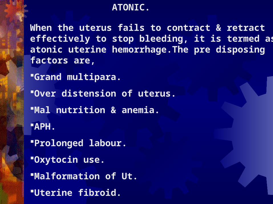
ATONIC.
When the uterus fails to contract & retract effectively to stop bleeding, it is termed as atonic uterine hemorrhage.The pre disposing factors are,
Grand multipara.
Over distension of uterus.
Mal nutrition & anemia.
APH.
Prolonged labour.
Oxytocin use.
Malformation of Ut.
Uterine fibroid.
Mis managed third stage of labour.

TRAUMATIC UTERINE BLEEDING.
Epiositomy,extesion of episiotomy incision.
Vaginal tear.
Cervical tear.
Vulval tear.( para uretheral tears ).
Cervical tears extending into Ut.
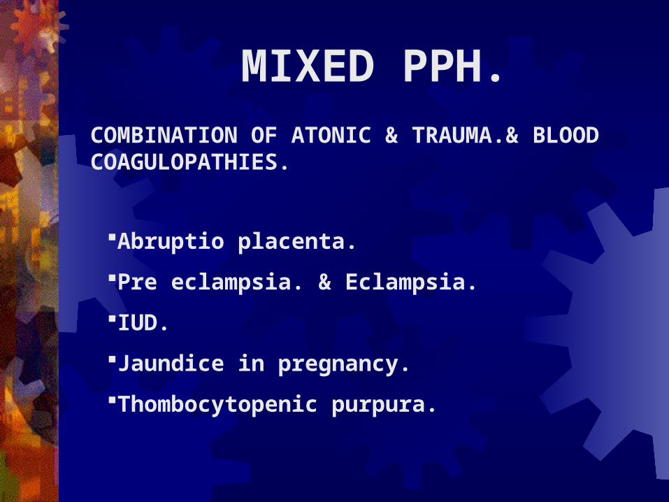
MIXED PPH.COMBINATION OF ATONIC & TRAUMA.& BLOOD COAGULOPATHIES.
Abruptio placenta.
Pre eclampsia. & Eclampsia.
IUD.
Jaundice in pregnancy.
Thombocytopenic purpura.

CILINICAL FEATURES.
Combined features. Vaginal bleeding- copious blood, or slow trickle.
Concealed haematomas (Concealed haemorrhage ).
Effect depends on pre delivery haemoglobin & blood loss.
Rapid pulse & Fall in Bp.
The UT may be flabby. (atonic.)
Well contracted Ut (Traumatic.)

MANAGEMENT.Inj. Ergometrine 0.25 mg / Methergine 0.2 mg IV.
Start IV with RL,add 20 units Synto.
Oxygen mask if hypoxia develops.
Catheterise if bladder is full.
Give bi manual uterine massage if Ut is atonic.
Explore genital tract for tear.
Repair the tear immediately.

PREVENTION OF PPH. (PRIMARY.).Improve Hb ante natally.
Screen high risk patients & refer to FRU.
Avoid kneading or fidding of the Ut & pulling of the cord in III stage.
Examine placenta & membranes. Detect missing cotyledons.
In case of induced labour-continue synto infusion o\for atleast 1 hr after delivery.
Administer Ergometrine with the delivery of anterior shoulder.
Explore vagina & Ut for trauma.
Observe the mother for 2 hrs before shifting.
Put the child to breast soon after delivery.

SECONDARY POST PARTUM HAEMORRHAGE.
OCCURS AFTER 24 HRS WITHIN 42 DAYS OF THE DELIVERY.
CAUSE.
Retained bits of placenta.
Infection.
Inversion of uterus.
Placental polyp.

MANAGEMENT.May need referral to FRU if blood loss is excessive, for exploration under anesthesia.
Inj methergine 0.2 mg IM, 8 hrly for 24 hrs.followed by T.Methergine. One tablet twice a day *3 days.
Antibiotics.
Bed rest.
Definitive treatment of the cause.

SHOCK.SHOCK IS A STATE OF COLLAPSE OF BLOOD CIRCULATION RESULTING IN CRITICAL REDUCTION OF TISSUE PERFUSION.
TYPES.
HAEMORRHAGIC SHOCK.
NON – HAEMORRHAGIC SHOCK.

HAEMORRHAGIC SHOCK.BLEEDING IS THE COMMONEST CASE OF SHOCK.HAEMORRHAGE CAUSES HYPO VOLAEMIA.
NON – HAEMORRHAGIC SHOCK.POST PARTUM COLLAPSE DUE TO,
Inversion of uterus.
Retained placenta without bleeding.
Prolonged labour – acidosis,infection.
Traumatic operative delivery.
Amniotic fluid embolism & hypofibrinogenemia.
Pulmonary embolism.

CILINICAL FEATURES.
Clinically mother may have sweating, feeling of restlessness, tachycardia,low volume pulse,low Bp,subnormal temperature,& oliguria.

MANAGEMENT.Inj Morphione sulphate 15 mg IM to relieve pain.
Continuous oxygen inhalation
Ensure patient airway.
Raise the foot end of the patient.
Fluid theraphy – Rapid infusion of glucose saline to restore blood volume.
Steroid harmones – Inj Hydrocartisone 500 – 1000 mg is use full in all kinds of shock.
Monitor vital signs for improvement.
Refer to FRU for further management.

PULMONARY EMBOLISM
CHARECTERIZED BY,
Sudden collapse.
Acute chest pain.
Air hunger.
Pleural pain.
Hemoptysis.

O/ECyanosis.
Increased RR.
Distension of jugular veins.
Pleural rub.
Death usually occurs within short time from shock,vaginal inhibition.
MANAGEMENT.Propped up position.
Oxygen inhalation.
Sedation.
Lasix 40 mg IV.
Maintenance of IV line.

THANK YOU.