Reproduction Topic 6.6 IB Biology 2 Van Roekel. BILL What are the main purposes of the reproductive...
59
Reproduction Topic 6.6 IB Biology 2 Van Roekel
-
Upload
carol-hoover -
Category
Documents
-
view
215 -
download
2
Transcript of Reproduction Topic 6.6 IB Biology 2 Van Roekel. BILL What are the main purposes of the reproductive...

Reproduction
Topic 6.6IB Biology 2Van Roekel

BILL
• What are the main purposes of the reproductive systems in the body (be appropriate)
• Produce sperm cells in male, egg cells in females• Allow for fertilization (joining of an egg cell and
sperm cell) and gestation (pregnancy and giving birth)
• ensure that offspring have genetic variety by combining half of the genetic material from one parent with half the genetic variety from another.

Statements
• 6.6.1 Draw and label diagrams of the adult male and female reproductive systems.
• 6.6.2 Outline the role of hormones in the menstrual cycle, including FSH (follicle stimulating hormone), LH (luteinizing hormone), estrogen and progesterone.
• 6.6.3 Annotate a graph showing hormone levels in the menstrual cycle, illustrating the relationship between changes in hormone levels and ovulation, menstruation and thickening of the endometrium.
• 6.6.4 List three roles of testosterone in males. • 6.6.5 Outline the process of in vitro fertilization (IVF). • 6.6.6 Discuss the ethical issues associated with IVF.
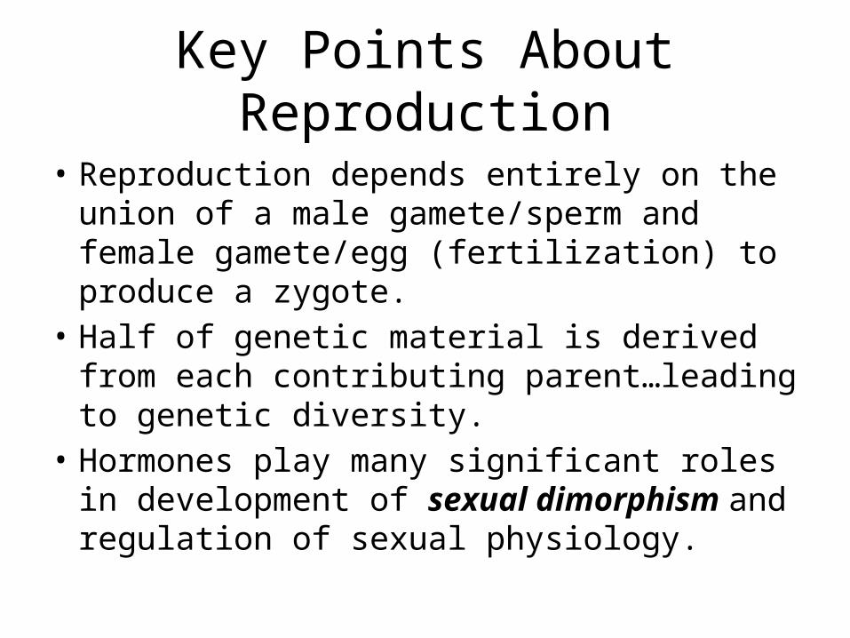
Key Points About Reproduction
• Reproduction depends entirely on the union of a male gamete/sperm and female gamete/egg (fertilization) to produce a zygote.
• Half of genetic material is derived from each contributing parent…leading to genetic diversity.
• Hormones play many significant roles in development of sexual dimorphism and regulation of sexual physiology.

6.6.1 Draw and label diagrams of the adult male and female reproductive systems. pg.185

Female pg. 186

6.6.2 Outline the role of hormones in the menstrual cycle, including FSH (follicle stimulating hormone), LH (luteinizing hormone), estrogen and progesterone.
• Points about the Menstrual Cycle– Prepares the ovaries for ovulation
and the uterus for implantaion– Lasts approximately 28 days…time
between the females release of the egg from the ovaries (known as ovulation).
– Egg must be released to the endometrium (the inner lining of the uterus.)
– The endometrium is highly vascularized which allows implantation if egg is fertilized
– If egg is not fertilized, endometrium breaksdown, which leads to menstrual bleeding
– Menstruation is a sign that pregnancy has not occurred.

Bill
• What hormones are responsible for regulating the menstrual cycle and what does each do?
• Gonatrophic Releasing Hormone – stimulates release of FSH and LH
• Follicle Stimulating Hormone – develops the oocyte and follicles, while preparing for ovulation
• Luetinizing Hormone – spikes and causes the ovulation of oocyte
• Estrogen – causes vascularization of endometrium• Progresterone – maintains vascularization of endometrium

Hormones of Menstrual Cycle
• Hypothalamus is the control center for the menstrual cycle.
• Gonadotrophin Releasing Hormone (GnRH) causes pituitary gland to secrete follicle stimulating hormone (FSH) and Leutinizing Hormone (LH) to be released into the blood stream.
• Both hormones target ovaries.

Anterior Pituitary Hormones:
• Follicle Stimulating hormone (FSH)• Stimulates the development of a primary
follicles, which surround the oocyte (egg) providing protection and nutrients.
• Develops the oocyte in the follicle and Produces follicular fluids.
• Increases the number of follicular cells which in turn produce estrogen, which causes vascularization of endometrium.


Anterior Pituitary Hormones:
• Luteinizing Hormone (LH):• high LH is associated with a resumption of meiosis in the
oocyte. Meiosis has been arrested in Prophase I since the embryonic stage. Only at the point of fertilization does meiosis complete.
• surges in mid cycle (12 days) to bring about ovulation, or release of oocyte (surrounded by follicles) from ovary.
• Some follicle cells remain in the ovaries and develop into the corpus luteum. These cells also release progesterone, which maintains the thickened vascularized endometrium


Hormonal Control of Menstrual Cycle
• As long as Progesterone is produced, it prevents break down of endometrium and implantation can still occur
• High levels of Estrogen and Progesterone are negative feedback signals w/ hypothalamus, as it will not produce GnRH, which in turn will not allow FSH and LH to affect ovaries
• Assuming no pregnancy, corpus luteum breaks down, progesterone and estrogen levels decline, endometrium breaks down causing menstruation
• Drop in estrogen and progesterone levels signals the hypothalamus to release GnRH and another menstrual cycle begins


Hormone levels during menstruation

Hormone levels during menstruation

Corpeus Luteum • The corpus luteum is essential for establishing and maintaining pregnancy in
females. The corpus luteum secretes progesterone, which is a steroid hormone responsible for the vascularization of the endometrium (its development) and maintenance, respectively.
• When egg is not fertilized• If the egg is not fertilized, the corpus luteum stops secreting progesterone and
decays (after approximately 14 days in humans). • The uterine lining breaks down without progesterone and is expelled through the
vagina (in humans and some great apes, which go through a menstrual cycle).• When egg is fertilized• If the egg is fertilized and implantation occurs, blastocyst secretes the hormone
human chorionic gonadotropin (hCG, or a similar hormone in other species).• Human chorionic gonadotropin signals the corpus luteum to continue
progesterone secretion, thereby maintaining the thick lining (endometrium) of the uterus and providing an area rich in blood vessels in which the zygote(s) can develop. From this point on, the corpus luteum is called the corpus luteum graviditatis.


6.6.4 List three roles of testosterone in males
• 1. Week 7 of embryonic development, testosterone initiates the development of male genitalia.
• 2. Around mid teens, testosterone initiates the development of secondary sexual characteristics– increase in muscle mass– increase in the length of the long bones (height)– increase in the length of the vocal cords (voice deepens)– spermatogenesis– growth of the penis and testis
• 3. Post puberty testosterone maintains the production of sperm cells and the male sex drive.

Fertilization
• Typically occurs in fallopian tubes 24-48 hours after ovulation
• Resulting zygote travels to the endometrium of the uterus, while already beginning to divide by mitosis
• Embryo implants into vascular tissue of endometrium and pregnancy begins

Problems with Fertilization
• Low sperm counts • Males w/ impotence (failure to
achieve/maintain an erection) • Females who cannot ovulate normally • Females with blocked fallopian tubes
• Reproductive technologies are able to help overcome these situations

In-vitro Fetrilization (IVF)
• Woman injected with FSH for about 10 days, causing production of many follicles, and allows for several eggs to be harvested
• Man donates his sperm into an external container• Fertilization occurs in separate culture dishes and
observations reveal which ova are fertilized and appear normal and healthy
• 2-3 healthy embryos inserted into woman’s uterus for implantation

Ethical Issues
Pro IVF• Enables couples to have
children• Embryos not healthy can be
eliminated to increase chance of implantation
• Genetic screening is possible to eliminate chance of passing on some genetic diseases
• IVF tech can advance and lead to further benefits of reproduction
Anti IVF • Embryos not implanted are frozen or
destroyed• Legal issues concerning frozen
embryos when couples split• Genetic screening could lead to
society choosing desirable traits• Bypasses natures way of decreasing
genetic frequency of reproductive/genetic problems
• Multiple births and problems associated with multiple births are more likely with IVF than natural conception

GATTACA
• https://www.youtube.com/watch?v=ZppWok6SX88
• https://www.youtube.com/watch?v=kM_dqjgyMmA

11.4 Reproduction

Assessment Statements• 11.4.1 Annotate a light micrograph of testes to show the location of interstitial cells (leydig cells), germinal epithelium cells,
developing spermatozoa, and Sertoli cells• 11.4.2 Outline the process involved in spermatogenesis within the testes, including mitosis, cell growth, meiosis, and cell
differentiation• 11.4.3 State the role of LH, FSH, and testosterone in spermatogenesis• 11.4.4 Annotate a diagram of the ovary to show the location and function of germinal epithelium, primary follicles, mature
follicle, and secondary oocyte• 11.4.5 Outline the processes involved in oogenesis within the ovary, including mitosis, cell growth, two divisions of meiosis,
unequal division of cytoplasm, and degeneration of polar bodies• 11.4.6 Draw and label the structure of a mature sperm and egg• 11.4.7 Outline the role of the epididymis, seminal vesicle, and prostate gland in the production of semen• 11.4.8 Compare the process of spermatogenesis and oogenesis, including the number of gametes and the timing of the
formation and release of gametes• 11.4.9 Describe the process of fertilization, including acrosome reaction, penetration of the egg membrane by a sperm, and
the cortical reaction• 11.4.10 Outline the role of HCG in early pregnancy • 11.4.11 Outline early embryo development up to the implantation of the blastocyst• 11.4.12 Explain how the structure and functions of the placenta, including its hormonal role in secretion of estrogen and
progesterone, maintain pregnancy• 11.4.13 State that the fetus is supported and protected by the amniotic sac and amniotic fluid• 11.4.14 State that materials are exchanged between the maternal and fetal blood in the placenta• 11.4.15 Outline the process of birth and its hormonal control, including the changes in progesterone and oxytocin levels and
positive feedback.

Spermatogenesis and the Testes
• Spermatogenesis: the production of sperm cells, which occurs within the testes
• Testes located outside the body to provide cooler temps for spermatogenesis
• Occurs within seminiferous tubules• Spermatogonia: diploid germinal epithelial cells
that will undergo mitosis (to produce more spermatogonia) or they will undergo meiosis (to produce 4 haploid spermatozoa, aka sperm cells)

Testes
• Leydig Cells: Produce testosterone and other hormones
• Germinal Epithlium Cells: spermatogonia
• Spermatozoa: sperm cells• Sertoli Cells: provide
nutrients to spermatozoa during sperm differentiation


Spermatogenesis• Spermatogonia cells contain 23 pairs of chromosome (46 total) –
undergo normal parts of cell cycle (G1, S, G2)• Meiosis occurs after DNA replication to cut number of chromosomes in
half (23 total chromosomes), making spermatozoa (1 spermatogonia 4 spermatozoa)
• Spematozoa must differentiate into fully functioning sperm cell (spermatozoon) inside seminiferous tubule, attached to sertoli cells for nutrients– Flagellum for motility– Acrosome contains enzyme necessary for fertilization
• As sperm cells develop, they move closer to lumen of seminiferous tubule
• Once development is completed, they detach from sertoli cells and carried to epididymis where it is stored

Hormones and Spermatogenesis
• Leutinizing Hormone (LH) stimulates Leydig Cells to produce testosterone
• Follicle Stimulating Hormone (FSH) and testosterone stimulate meiotic divisions of spermatogonia into spermtozoa
• Sperm cell production starts at puberty and continues throughout life, producing millions of sperm cells a day

Bill- How are sperm cells formed?
• LH and FSH stimulate production of testosterone and meiotic division of spermatogonia cells forming spermatozoa
• Spematozoa must differentiate into fully functioning sperm cell– Flagellum for motility– Acrosome contains enzyme necessary for fertilzation
• As sperm cells develop, they move closer to lumen of seminiferous tubule
• Once development is completed, they detach from sertoli cells and carried to epididymis where it is stored


Mature Sperm Cell • Flagella provides motility for swimming• Mitochondria provides ATP for energy for swimming• Head contains haploid nucleus with half number of
chromosomes• Acrosome contains hydrolytic enzymes to help with
fertilization process• Small size allows them to swim great distances


Production of Semen
• Semen: Fluid that is ejaculated during intercourse, contains sperm cells and fluids that assist in reproduction
• Epididymis: Stores sperm cells allowing them to gain motility• Vas Deferens: sperm moves from epididymis to urethra
through vas deferens• Seminal Vesicles: Add large volume of fluid (70%) that
contains high concentration of fructose, needed to provide energy to sperm cells to swim to ovum
• Prostate Gland: Adds more fluid (30%) that contains alkaline and helps sperm cells survive environment in female vagina
• Urethra: excretory tube, site of ejaculation

Oogenesis and the Ovary
• Oogenesis: the production of ovum (egg cells) through meiosis
• Produces four haploid cells, however, only one becomes an egg cell– 3 are too small to produce zygote upon
fertilization and become polar bodies (containers for divided chromosomes after meiosis)
– 1 is very large and becomes egg cell

Oogenesis before birth
• During fetal development, oogonia cells (diploid) undergo mitosis, increasing number of cells in ovary
• Oogonia cells grow into larger cells called primary oocytes (diploid), which will undergo initial steps of meiosis (through prophase 1, then pauses)
• Follicle cells undergo mitosis and a single layer surrounds primary oocytes, forming primary follicle
• Females born with half a million primary follicles

Oogenesis and Menstrual Cycle
• Each menstrual cycle, primary follicle finishes meiosis I and forms 1 secondary oocyte and polar bodies
• Follicle cells begin dividing and forming 2 rings with a fluid filled cavity around secondary oocyte
• Secondary Oocyte begins Meiosis II, but again is paused during prophase forming Graafian Follicle
• Secondary Oocyte and its inner follicle ring are released during ovulation and only completes Meiosis if fertilized by sperm cell
• * Be able to relate these steps to what happens in menstrual cycle and hormones that are used.


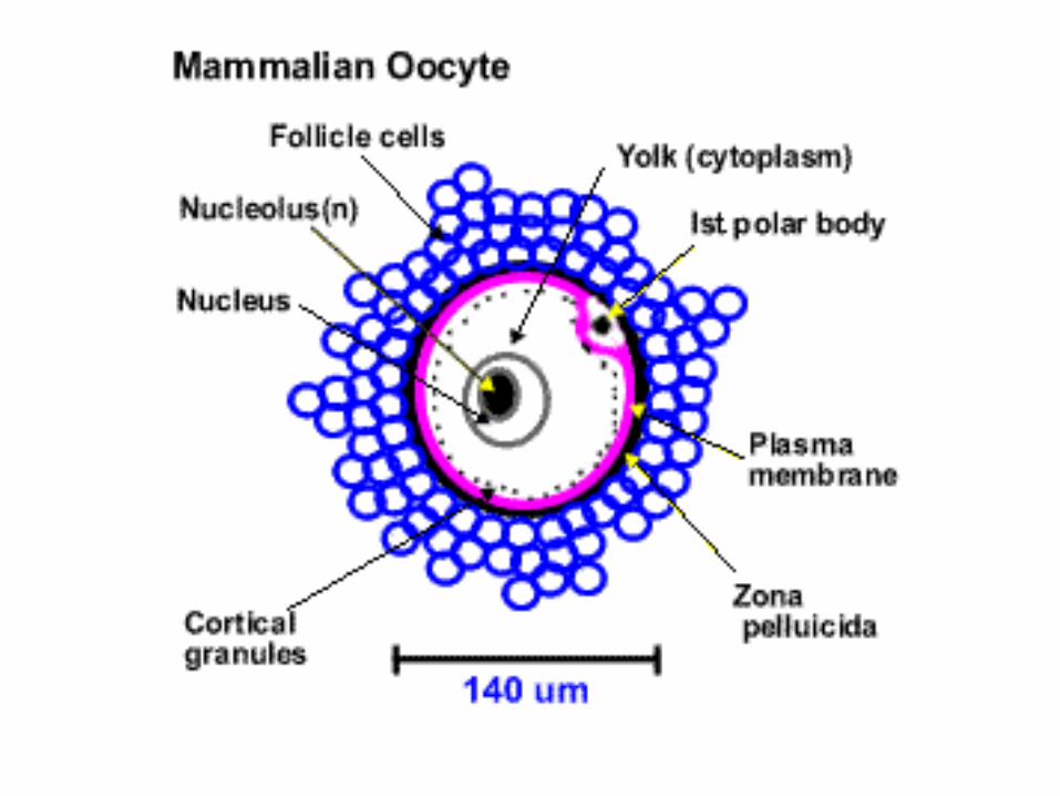
Mature Ovum • Largest cell in body by volume• Unequal distribution of cytoplasm
ensured on cell has all nutrients, cytoplasm, and organelles necessary for a new life.
• Haploid nucleus/nucleolus• Nutrients are referred to as yolk• Follicle cells, protect and nourish egg• Cortical granules ensure only one
sperm cell can fertilize egg• Zona pellucida (layer of
glycoproteins) becomes impermeable to other sperm cells upon fertilization


Spermatogenesis vs. Oogensis
• Pg. 312

Fertilization

Fertilization• The union of haploid male and female gametes to form a
diploid zygote • Typically occurs in the Fallopian Tube• Takes many sperm cells to accomplish fertilization, however
only one can fuse its membrane with the egg cell to create a zygote

Fertilization
• Many sperm cells are needed to penetrate the follicle cell layer.
• Several Sperm cells gain access to zona pellucida and release hydrolytic enzymes, enabling the cells to penetrate the zone and reach plasma membrane
• One sperm cell reaches the plasma membrane of secondary oocyte
• Plasma membranes fuse together and initiates the cortical reaction

Fertilization – Cortical Reaction
• Cortical granules fuse with cell membrane and release enzymes, making the zona pellucida impermeable to other sperm cells
• Takes place immediately after first sperm cell fuses with egg cell and ensures only one sperm cell fertilizes egg
• Egg now completes meiosis II as haploid nucleus from sperm cell enters the egg
• Resulting in a diploid zygote


Pregnancy
• Fertilization triggers mitotic division in zygote, which takes roughly 5 days to reach uterus
• Embryo consists of about 100 cells by time implantation occurs (7 days after fertilization), which appear as a ball of cells, called a blastocyst
• Blastocysts consist of:– Trophoblast – surrounding layers of cells that will help form
fetal portion of placenta– Inner Cell Mass – interior cells located towards one end of
blastocyst that will become body of embryo– Fluid filled cavity


Pregnancy - HCG
• Human Chorionic Gonadotrophin (HCG): hormone secreted by embryo that signals to corpus luteum, causing it to maintain secretions of estrogen and progesterone
• Maintains endometrium, which will form placenta after implantation

Pregnancy – Placenta
• Forms from trophoblast layer of blastocyst, and the endometrium of mother
• Two fetal blood vessels within umbilical cord carry fetal blood to placenta and exchanges materials with mother, which are brought back using a separate fetal blood vessel– Embryo passes CO2, urea, water, and hormones to mother
– Mother passes O2, nutrients, water, hormones, vitamins/minerals, alcohol/drugs (if taken), and viruses (if infected)
• At no point does blood mix, only materials are exchanged


Pregnancy - Placenta
• Placenta will also act as an endocrine organ during pregnancy, secreting estrogen and progesterone
• Functions to maintain vascularized endometrium and provide rich blood supply to embryo

Pregnancy – Amniotic Fluid
• Amniotic sac surround embryo as it continues to grow and develop
• Amniotic Fluid inside sac functions to:– Cushion embryo from blunt force applied to mother’s
abdomen– Provide environment for movement allowing exercise,
muscular and skeletal development– Thermal stability (mainly water, so has excellent
temperature stability) – General support so that the embryo does not experience
any excess pressure

Hormonal Events @ Birth
• Physiological events preparing for birth are called parturition• Progesterone levels decrease, and the hormone oxytocin is
secreted from posterior pituitary gland• Initial low levels of oxytocin are associated with first
contractions of uterus• Each uterine contraction signals to posterior lobe of pituitary
to produce more oxytocin• Higher levels of oxytocin cause more intense and frequent
contractions • This is a positive feedback mechanism and will continue and
only stops when birth occurs and uterus no longer has something to contract on.

Major Events @ Childbirth
• Major Hormone Change (discussed above)• Dilation/Opening of Cervix to 10 cm• Baby positioned head first, face down• Shoulders typically widest part to pass
through birth canal• Afterbirth is name of expelled placenta• Lactation (breast milk production) begins soon
after birth.