Report of the Tenth Meeting of the Comité ... -...
66
Report of the Tenth Meeting of the Comité Internacional para la Recuperación de la Vaquita (CIRVA) Southwest Fisheries Science Center, La Jolla, CA, December 11-12, 2017 Contents EXECUTIVE SUMMARY ......................................................................................................................................... 1 REPORT 1. Welcome .................................................................................................................................................................... 4 2. Acoustic Monitoring Program ................................................................................................................................... 4 3. Vaquita CPR Progress Report ................................................................................................................................... 6 4. Update on enforcement and regulations .................................................................................................................... 9 5. Update on Alternative Gear Development ............................................................................................................... 13 6. Socio-economic Aspects .......................................................................................................................................... 14 Annex Title Pages A List of Participants 16-17 B Agenda 18 C V01F Veterinary Report 19-29 D V02F Veterinary Report 30-63 E Report of the Express Meeting of the Comité Internacional para la Recuperación de la Vaquita 64-65 ACKNOWLEDGEMENTS Our appreciation to our funders: WWF Mexico, The Marine Mammal Commission. The views expressed in the report do not necessarily reflect those of the groups named. Also, our thanks for hosting the meeting at the Southwest Fisheries Science Center/NOAA Fisheries to Lisa Ballance and Barb Taylor. Thanks also to Brittany Hancock-Hanser and Annette Henry for her support during the meeting.
Transcript of Report of the Tenth Meeting of the Comité ... -...

Report of the Tenth Meeting of the Comité Internacional
para la Recuperación de la Vaquita (CIRVA)
Southwest Fisheries Science Center, La Jolla, CA, December 11-12, 2017
Contents
EXECUTIVE SUMMARY ......................................................................................................................................... 1
REPORT
1. Welcome .................................................................................................................................................................... 4
2. Acoustic Monitoring Program ................................................................................................................................... 4
3. Vaquita CPR Progress Report ................................................................................................................................... 6
4. Update on enforcement and regulations .................................................................................................................... 9
5. Update on Alternative Gear Development ............................................................................................................... 13
6. Socio-economic Aspects .......................................................................................................................................... 14
Annex Title Pages
A List of Participants 16-17
B Agenda 18
C V01F Veterinary Report 19-29
D V02F Veterinary Report 30-63
E Report of the Express Meeting of the Comité Internacional para la Recuperación de la
Vaquita
64-65
ACKNOWLEDGEMENTS Our appreciation to our funders: WWF Mexico, The Marine Mammal Commission. The views expressed in the report do not necessarily reflect those of the groups named. Also, our thanks for hosting the meeting at the Southwest Fisheries Science Center/NOAA Fisheries to Lisa Ballance and Barb Taylor. Thanks also to Brittany Hancock-Hanser and Annette Henry for her support during the meeting.
EXECUTIVE SUMMARY
The tenth meeting of the Comité Internacional para la Recuperación de la Vaquita (CIRVA) was held at
the Southwest Fisheries Science Center on December 11-12, 2017.
The dire status of the vaquita has worsened
Thomas et al. (2017) estimated that, as of November 2016, only approximately 30 vaquitas likely remained.
Analysis of the 2017 Acoustic Monitoring Program data showed that the decline has continued unabated.
Thus, the already desperate situation has worsened, despite existing conservation measures and current
enforcement efforts. Unless this decline can be stopped by eliminating mortality in illegal gillnets, the
vaquita will be extinct in a few years. The critical work of the Acoustic Monitoring Program must continue
in order to make possible the estimation of population trend and the evaluation of the efficacy of current
and future conservation measures.
Placing vaquitas in a temporary sanctuary is no longer an option
Given the dire situation, CIRVA previously recommended that attempts be made as a matter of urgency to
place as many vaquitas as possible into a temporary sanctuary. CIRVA recognized that the risks of capture
and captive maintenance were high, but concluded that these risks were outweighed by the very high
likelihood of human-caused mortality in the wild that would lead to extinction in a short time. During the
VaquitaCPR field effort from October 11 – November 10, 2017, two female vaquitas were captured, but
both were released after showing signs of stress. The adult female died after release, and the fate of the
smaller animal is unknown. CIRVA accepted the conclusion of experts in the VaquitaCPR team and the
Independent Review Panel that further effort to rescue vaquitas by placing them under human care should
be suspended. Despite this discouraging result, CIRVA commends SEMARNAT and its numerous partners
who made this unprecedented rescue effort possible.
High levels of illegal fishing continue
A multi-institutional program to find and remove illegal and abandoned fishing gear in the range of the
vaquita has continued. In 166 days of field work through December 8, 2017, 518 pieces of illegal,
abandoned, or derelict fishing gear were retrieved and 220 of these were active fishing gear. This shows
that illegal fishing activities, particularly the setting of large-mesh gillnets for totoaba, continue at alarming
levels within the range of the vaquita. CIRVA recommends that this important program should continue
to remove fishing gear from the range of the vaquita with focus on the area of highest risk during totoaba
spawning season.
Saving vaquitas from extinction relies on effective enforcement and continued net removal
The combination of continued decline of the vaquita population and continued retrieval of hundreds of
active gillnets constitutes strong evidence that without dramatic improvement in keeping gillnets out of the
vaquita’s habitat, Mexico will lose its largest endemic mammal. CIRVA recommends that, during the next
totoaba season (December 2017 through May 2018), Mexico establish an enhanced enforcement program
in the “exclusion zone” – the area believed to have the highest co-occurrence of vaquitas and illegal totoaba
nets (see Figure below).
Within the exclusion zone, CIRVA recommends that the Government of Mexico:
(1) prohibit all fishing and navigation;
(2) increase enforcement presence to a level which is able to respond to any report of illegal
activities within 30 minutes.
(3) increase and focus net removal efforts are within in the exclusion zone.
(4) negotiate the appropriate transit corridors to allow legal fishing to continue outside the
exclusion zone.

2
It also recommends that drones be used to monitor the areas of historical totoaba fishing and vaquita
entanglement near El Golfo de Santa Clara to prevent a geographical shift in illegal totoaba effort that could
kill vaquitas. Should evidence be brought to light of illegal fishing in this area, enforcement response will
need to adapt swiftly.
Vertex Longitude Latitude
A -114.83908 31.37629
B -114.83908 31.11555
C -114.72977 30.95786
D -114.65015 30.95786
E -114.65015 31.37629
Fig. 4. The recommended Exclusion Zone (see Item 4) is shown as a blue polygon. The exact positions of the vertices
(A-E) are shown in the small table above. The Vaquita Refuge agreed in 2005 is shown as a black broken line (the
overlap with the Exclusion Zone is hatched). The small circles show the sites where fishing gear had been recovered
(for types see legend in figure). The large circles show the raw results of acoustic monitoring between 4 June 26 and
August 26th, 2017. The size of the circle indicates sampling effort (full days) whilst the color of the circle indicates
the average acoustic detection rate (clicks/day) where black = no data, white = no detections and shade of blue
represents click numbers (see legend in figure).

3
Immediate action is needed, and CIRVA recommends that:
(1) All Mexican enforcement agencies increase their efforts on land and in water immediately
and continue this enhanced enforcement program for the duration of the period of illegal
totoaba fishing (at least until June 2018) to eliminate all setting of gillnets in the range of
the vaquita.
(2) Emergency regulations be promulgated immediately to strengthen the current gillnet ban
and enhance enforcement and prosecution by:
a. eliminating all fishing permits for transient fishermen and limiting fishing access
to only those fishermen who can demonstrate residency in the fishing villages;
b. confiscating any vessel that does not have the appropriate vessel identification,
permits, and the required vessel monitoring system;
c. requiring vessel inspection for each fishing trip at the point of departure and
landing:
d. prohibiting the sale or possession of gillnets on land and at sea within the area of
the current gillnet ban and on adjacent lands within a specified distance of the
coastline.
e. requiring that all gillnets be surrendered or confiscated and destroyed.
f. eliminating the exemptions for all gillnet fisheries, including the curvina and sierra
fisheries.
(3) Efforts to remove gillnets from vaquita habitat be continued and enhanced and the numbers
and locations of new nets recovered be published monthly.
(4) The number of inspections, interdictions, arrests, sentences, and other enforcement actions
be published monthly, together with information on observed levels of illegal activities
obtained from intelligence operations, for example from drones.
(5) Successful prosecution and subsequent penalties be sufficient to deter illegal fishing.
(6) Development of gillnet-free fisheries be enhanced and linkages to incentivize the
conversion of the fleet to gillnet-free operations be strengthened.
4
The Tenth meeting of the Comité Internacional para la Recuperación de la Vaquita (CIRVA) was held at the Southwest
Fisheries Science Center on December 11-12, 2017. CIRVA members in attendance included: Lorenzo Rojas-Bracho
(chair), Armando Jaramillo-Legorreta, Barbara Taylor, Tim Gerrodette, Peter Thomas, Andrew Read, Robert
Brownell, Greg Donovan, Frances Gulland, Nina Young, and Sarah Mesnick. CIRVA members Jay Barlow and
Randall Reeves participated remotely. The committee’s work was supported by a number of invited experts who
provided presentations and contributed to plenary discussions. Rojas-Bracho chaired the meeting and Read, Thomas,
Gerrodette, and Donovan acted as rapporteurs. Meeting participants are listed in Annex A. The agenda is given as
Annex B.
1. WELCOME
Lisa Ballance, Director of the Marine Mammal and Turtle Division, welcomed CIRVA members to the Southwest
Fisheries Science Center. Rojas-Bracho reviewed the agenda and it was adopted as amended.
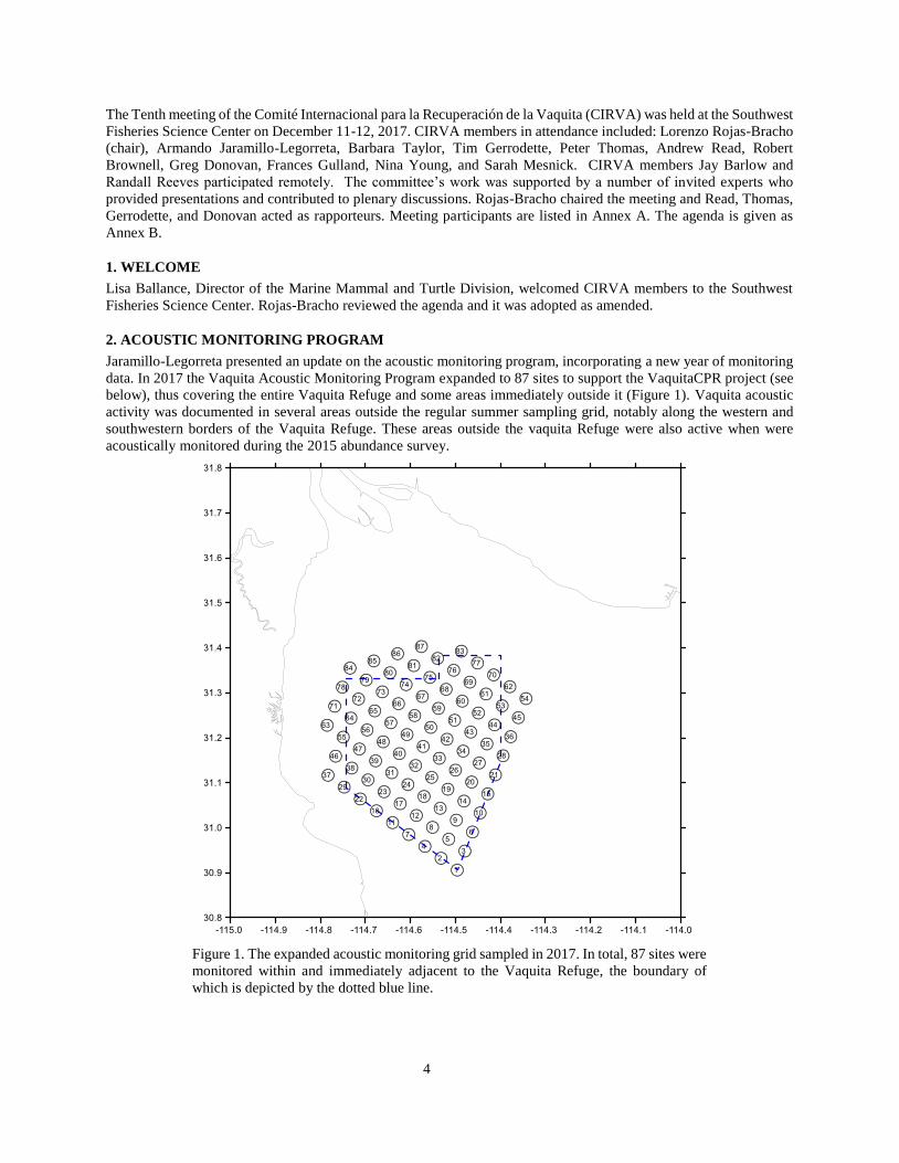
2. ACOUSTIC MONITORING PROGRAM
Jaramillo-Legorreta presented an update on the acoustic monitoring program, incorporating a new year of monitoring
data. In 2017 the Vaquita Acoustic Monitoring Program expanded to 87 sites to support the VaquitaCPR project (see
below), thus covering the entire Vaquita Refuge and some areas immediately outside it (Figure 1). Vaquita acoustic
activity was documented in several areas outside the regular summer sampling grid, notably along the western and
southwestern borders of the Vaquita Refuge. These areas outside the vaquita Refuge were also active when were
acoustically monitored during the 2015 abundance survey.
Figure 1. The expanded acoustic monitoring grid sampled in 2017. In total, 87 sites were
monitored within and immediately adjacent to the Vaquita Refuge, the boundary of
which is depicted by the dotted blue line.

5
The vaquita population trend was modeled using acoustic detections from the regular 46-site sampling grid, using the
same methods developed to analyze the 2011-2016 dataset (see Jaramillo-Legorreta et al. 2017 and Thomas et al.
2017). Specifically, both the geostatistical and post-stratification mixture models were used to estimate annual rates
of change in acoustic detection rates.
The raw acoustic detection rates (average clicks/day/site) were approximately an order of magnitude lower in 2017
than at the start of the monitoring program in 2011 (Figure 2). Assuming that the difference in click rate between 2016
and 2017 represents a change in population size (Thomas et al. 2017), then the vaquita population decline continued
unabated from 2016 to 2017.
Figure 2. Estimated vaquita click rates (clicks per day) predicted for the 46 sampling sites from
the geospatial model. Values in the legend are posterior medians (note log scale). The size of the
circles indicates the number of sampling days each year.
Jaramillo-Legorreta then presented several options for the 2018 acoustic monitoring program developed in response
to a request from SEMARNAT. These alternatives included a program that would monitor the extended 87-site
sampling grid year-round, as well as smaller grids monitored during various periods. All of these options included the
regular summer sampling program used to monitor population trend.
CIRVA strongly recommends that the regular 46-site grid be sampled as in previous years to provide an annual
empirical estimate of population trend. There was considerable discussion regarding the objectives, logistical
challenges, and merits of the other options, without agreement on a preferred alternative. CIRVA members agreed to
defer a decision on these alternatives until the 2018 enforcement program had been reviewed, but ultimately did not
have time to discuss these options fully. CIRVA will need to continue its discussion of the acoustic sampling program
between meetings, especially in light of its recommendation for an exclusion zone (see item 4 of this report).

6
3. VAQUITA CPR PROGRESS REPORT
CIRVA received several reports on the effort to capture vaquitas, as recommended by CIRVA 9. This field effort,
called Vaquita Conservation, Protection, and Recovery, or VaquitaCPR (see https://www.vaquitacpr.org/), occurred
between 12 October to 10 November 2017.
Vaquita CPR visual effort
The visual team employed three vessels (hereafter called sighting vessels): the 135’ Maria Cleofas, a converted Bering
Sea crabbing ship, and two sport-fishing boats (Wanderlust and Odissea) with flying-bridge viewing platforms. The
ship carried two pairs of 25-power binoculars (big eyes) and a full-time data recorder to record positions of vessels
and vaquitas. Observers on the two smaller sighting vessels used hand-held binoculars. All sighting vessels had
experienced vaquita observers.
The strategy to find vaquitas was to use the acoustic data (see below) to plan daily tracklines, typically searching from
south to north to avoid sun glare. A special computer program was created and used on the Maria Cleofas to track
sightings and catch vessels using an Automatic Identification System (AIS). In search mode, the two smaller sighting
vessels were positioned ahead of the Maria Cleofas (at clock positions of 2 and 10 o’clock). Search speed was
approximately 6 knots and surveys were conducted only in calm seas (winds less than 7 knots which produce only
tiny waves so as not to obscure surfacing vaquitas). When vaquitas were sighted, the sighting vessels formed a triangle
with the goal of keeping the vaquitas between them. The catch boats were then directed towards the animals by
observers aboard the sighting vessel that had vaquitas in sight.
Survey effort was limited by exceptionally windy conditions in October (Table 1). From October 12 to November 4,
only five full days of effort were possible; vaquitas were seen on four of those days. In addition, there were seven
partial days of effort, with vaquitas seen on three days. In total, 36 confirmed visual detections of vaquitas were made.
Sightings involved one to three vaquitas, with an average group size of about two. The number of vaquitas remaining
cannot be inferred from these data because in nearly all cases, the animals could not be individually identified and
therefore some individuals were likely seen multiple times.
Table 1.
Summary data on VaquitaCPR field effort. Note that a ‘Yes’ in a cell indicates the occurrence of at least one activity on that day – e.g. more than
a single vaquita was sighted on almost all occasions.
Date Comments
Full Field
Day
Partial
Field Day
Vaquitas
Seen
Net
Deployed
Vaquita
Captured
Oct 12, 2017 Begin capture efforts Yes
Oct 13, 2017 Capture efforts Yes Yes
Oct 14, 2017 Capture efforts Yes
Oct 15 - 16 2017 Conditions too bad for field work
Oct 17, 2017 Capture efforts Yes Yes
Oct 18, 2017 Capture efforts Yes Yes Yes Yes
Oct 19, 2017 Capture efforts Yes Yes Yes
Oct 20 -25, 2017 Conditions too bad for field work
Oct 26, 2017 Capture efforts Yes Yes
Oct 27, 2017 Capture efforts Yes
Oct 28, 2017 Conditions too bad for field work
Oct 29, 2017 Capture efforts Yes
Oct 30, 2017 Capture efforts Yes Yes
Oct 31, 2017 Capture efforts Yes
Nov 01, 2017 Capture efforts Yes Yes
Nov 02-03, 2017 Conditions too bad for field work
Nov 04, 2017 End capture efforts Yes Yes Yes Yes
Nov 05, 2017 Begin dedicated photo-ID Yes Yes
Nov 06, 2017 Photo-ID Yes Yes
Nov 07-09, 2017 Conditions too bad for field work
Nov 10, 2017 End field efforts Yes Yes
TOTALS 7 9 11 3 2
VaquitaCPR acoustic effort
The objective of the acoustic effort was to identify locations with a high probability of vaquitas being present, allowing
the visual detection and capture teams to focus their daily search efforts.

7
Preparatory work started in June by monitoring an 87 C-pod sampling grid designed to provide detailed insight into
the distribution patterns of vaquitas. Based on this work, which finished in September, a grid of 36 sites was designed
to facilitate daily deployment, retrieval, and analysis of the acoustic detectors. Previous experience in the fall showed
that vaquitas shifted their distribution toward the northeastern corner of the Refuge as the season progressed. Toward
the end of October, after several days with no vaquita acoustic detections within the existing sampling grid, the grid
was expanded to include eight more sites in the northeastern part of the Refuge.
Daily reports consisted of maps showing acoustic detection rates at each sampling site, using the metric of acoustic
encounters, which are nearly analogous to sightings. Periodic reports were prepared to document longer-term patterns,
such as the relationship between distribution of visual sightings and acoustic activity, or the distribution of acoustic
activity and time of day. Another report provided insight on the ability of acoustic sampling data to predict locations
where the probability of finding vaquitas some hours later would be high. This acoustic information proved invaluable
for directing the visual search team to locations where vaquitas were detected.
As expected from previous studies, acoustic activity was relatively high at certain sites, although activity was also
present in surrounding areas. Three locations of “primary” vaquita occurrence were identified. In descending order of
relative importance, these were the western boundary of the Refuge, the southern portion of the Refuge, and the
northeastern region of the Refuge.
VaquitaCPR catch efforts
Capture efforts involved an international team of experts, including researchers experienced in the capture and
handling of harbor porpoises, animal care professionals, and veterinarians. This team was distributed across three
small (~8 m) vessels. Once vaquitas were located by the visual survey team, floating gillnets (256-512 m long and 9-
18 m deep) were deployed ahead of or around the animals. As necessary, the net boats were used to herd the vaquitas
toward the nets. Once in the nets, vaquitas were able to surface easily, facilitating efforts to remove them. The
vaquitas and key personnel were then transferred to other vessels to transport them to the floating pen or shore-based
facility. Two vaquitas were successfully captured. The first vaquita, caught on October 18, was an immature female
(V01F). The second, captured on 4 November, was an adult female (V02F).
VaquitaCPR photo-identification
Over the course of capture operations, it became apparent that it was possible to obtain photographs of individually
distinctive vaquitas during field operations. Distinctive dorsal fin notches and shapes have been used previously to
identify individual vaquitas (e.g., Jefferson et al. 2009). In an attempt to refine abundance estimates and learn about
vaquita ranging patterns, team members engaged in dedicated photographic identification efforts on all workable field
days after capture operations were suspended on November 4. Experienced photographers with appropriate telephoto
lenses were distributed across three small boats and the search vessels Wanderlust and Odissea to obtain high-
resolution dorsal fin images. Upon initial sighting by observers on the primary search vessel Maria Cleofas, the
closest smaller vessels attempted to approach for photographs. Poor weather (vessel operations were possible on only
three days) and the elusive nature of vaquitas limited the number of photographs collected during these efforts. Over
the entire project, 192 images from seven photographers were examined. Seven different individuals were
documented, including the two captured vaquitas. Three fins were very similar to those documented by Jefferson et
al. (2009), but photographic quality was insufficient to confirm matches. Photographs of another distinctive individual
from 2011 were also examined, but this animal was not photographed in 2017.
Vaquita housing
Centro de Atención a la Vaquita: Two pools were ready to receive animals at a shore-based facility at the onset of
field operations. Following admittance of V01F into one of the pools on 18 October 18th, additional modifications
were made to enhance their suitability for vaquitas. To offer the team more options during attempts at animal
acclimation, another style of pool was added to the facility in late October. However, no additional animals were
introduced to the facility during field operations.
El Nido Sea-Pen Facility: The 9-meter and 6-meter diameter sea-pens were complete on 17 October and ready to
receive animals in time for the first attempt at housing, which occurred on 18 October. Following the attempt to house
V01F, animal husbandry staff made additional modifications to the sea-pens to improve the net texture that could
come into contact with the animals. These changes were made rapidly and the facility was ready to receive additional
animals on the next catch day. Following the admittance and subsequent release of V02F, no further modifications
were made and no additional animals were introduced to the facility.

8
VaquitaCPR care effort
V01F: An immature female vaquita was caught on October 18. It was in good condition, but the veterinary and animal
care team determined that the animal was not acclimating to the vaquita care center pool or to the El Nido sea-pen
facility, so the decision was made to release the animal. Prior to release, a blood sample and a skin sample were
collected for cell culture and genetic sequencing. For full details, see V01F Veterinary Report at Annex C.
V02: On November 4, an adult female (V02F) was captured. It was also considered to be in good condition for
transport to the El Nido sea-pen. However, after some promise of learning to adapt to the facility, the animal stopped
swimming and went limp and an emergency release was initiated. The release was unsuccessful and the vaquita was
quickly recaptured for administration of emergency care. Following three hours of emergency response, the animal
went into cardiac arrest and did not respond to resuscitation attempts. A necropsy was performed and, tissues were
collected for histopathology, cell culture, gamete rescue, and genetic sequencing. Gametes were successfully rescued
by collaborators at SeaWorld. Live cells have been cultured and subsequently frozen by collaborators at the San Diego
Zoo. For full details, see V02F Veterinary Report at Annex D.
Genetics, tissue culture and gamete rescue
Oliver Ryder and Marlys Houck, both of the San Diego Zoo Institute for Conservation Research, joined the meeting
via telephone. They received biopsies from the two captured vaquitas and had successfully grown cells from both
individuals. From the young female, cells are growing but have not reached the desired number for the institute’s
freezing protocol, although the investigators are cautiously optimistic that this will occur. From the adult female,
multiple samples were received from necropsy. Houck and her team initiated approximately 40 cell culture flasks, an
unprecedented effort designed to maximize the potential supply of viable cells for cryobanking and research. To date,
seven cell cultures have been frozen. Because of the multiple tissues from which these cell cultures were established,
significant resources for producing and annotating a state-of-the-art reference genome assembly for the vaquita
becomes feasible.1
High-quality samples for cell culture are available only from females, so information about the Y-chromosome
morphology is lacking. For this reason, it is not possible to produce induced pluripotent stem cells capable of
producing spermatozoa.
Ryder and Houck expressed deep appreciation to all those involved in the collection and transfer of the samples to the
laboratories at the San Diego Zoo Institute for Conservation Research, including exportation from Mexico and
importation into the United States.
Phil Morin updated CIRVA on ongoing genetic analyses funded by the National Marine Fisheries Service and The
Marine Mammal Center. DNA samples from 22 vaquitas from the SWFSC Marine Mammal and Sea Turtle Research
(MMASTR) collection have been used for full mitogenome and shotgun genome sequencing. The data are still being
processed, but Morin summarized results for the mitochondrial genome from 22 samples collected between 1985 and
2017. Previous research on the mtDNA control region (322bp, Rosel et al. 1999) showed no variation in 43 samples
collected between 1985 and 1993. Complete mitogenomes (16,370 bp) of 14 of those individuals yielded 8 different
haplotypes. Mitogenomes of 2 samples collected in 2004 and 4 samples collected in 2016-2017 all had unique
haplotypes, and differed from the samples collected between 1985 and 1993 that were previously sequenced. All the
haplotypes are very similar, with only 23 variable positions across the whole 16,370bp of the mitogenomes. This
suggests long-term small population size. The data analyzed thus far are not consistent with a loss of genetic diversity,
although the nuclear results may provide further details including: (i) identifying variation in genes that are important
for vaquita survival (e.g., immune system), (ii) determining whether low diversity is normal for vaquitas (i.e., has
prevailed for thousands or millions of years) or a result of recent population decline, and (iii) establishing baseline
variability for future monitoring.
1 On 19 December 2017 the Conservation Genetics cryogenetics team provided an update, reporting that cell cultures from both female vaquitas
captured as part of the VaquitaCPR project had been successfully frozen and thawed with high viability scores. Additional cells are being grown
for use in whole genome sequencing, assembly, and annotation of the vaquita genome. As brought up at the CIVRA meeting, Y-chromosome data
are lacking because no samples have been collected from a male vaquita.

9
The SWFSC and the Vertebrate Genome Lab at The Rockefeller University are collaborating to generate the fully
sequenced and annotated genome from the cultured cells of the adult female vaquita. Funding is being provided by
the office of the NMFS Chief Scientist, Cisco Werner. This sequencing will resolve full-chromosome genome
organization and annotate all genes to allow comparison to other cetacean and mammalian genomes and identification
of genes uniquely adaptive for the vaquita.
VaquitaCPR Next steps: conclusions, agreement on goals,and recommendations
In addition to the review of VaquitaCPR at this meeting, a short CIRVA meeting (CIRVA Express 3) was held by
teleconference on November 16, 2017 (Annex E). The objectives of that earlier meeting were to review the
VaquitaCPR capture effort, which had just concluded, and provide immediate advice to the Government of Mexico
on the critical next steps that Mexico should undertake for vaquita conservation. The recommendations of CIRVA
Express 3 are reiterated below in section 4 of this report.
At its previous meetings, CIRVA recognized that the risks of capture and captive maintenance of vaquitas were high,
but concluded that these risks were outweighed by the very high likelihood of mortality in illegal gillnets that would
lead to extinction of the species in a short time. As reflected in this report, during the VaquitaCPR field effort two
female vaquitas were captured, but both were released after showing signs of stress. The adult female died after
release, and the fate of the smaller animal is unknown. CIRVA accepts the conclusion of experts in the VaquitaCPR
team and the Independent Review Panel that further efforts to rescue vaquitas by placing them under human care
should be suspended. Despite this discouraging result, CIRVA commends SEMARNAT and its numerous partners
who made this unprecedented rescue effort possible.
CIRVA further stresses that the strong on-the-water presence during the VaquitaCPR capture effort appeared to
discourage illegal fishing. Moreover, the local, national, and international collaborations forged during VaquitaCPR
raised awareness of the urgent need for forceful action to conserve vaquitas. The effort as a whole also reinforced the
strong commitment within Mexico and internationally to do everything possible to prevent the extinction of the
vaquita. CIRVA recommends that this commitment be maintained and expanded to include further monitoring,
continued removal of gillnets, and enhanced enforcement. CIRVA commends the outreach effort that significantly
raised the profile of vaquita conservation globally and recommends that such outreach be maintained through regular
updates on vaquita status, particularly during the upcoming totoaba season.
4. UPDATE ON ENFORCEMENT AND REGULATIONS
Net extraction program Table 2
Summary data on net extraction program from 10 October 2016 to 8 December 2017
NET EXTRACTION
Phase of
project
Effective
work days Start date Finish date Nets retrieved Active
Inactive
(ghost nets)
Tons of
nets
Bags of
nets
PHASE I* 21 10 Oct 2016 15 Dec 2016 105 9.3
OP.
MILAGRO III**
72 16 Dec 2016 10 Apr 2017 201 157 41
47.75 143
PHASE II* 33 11 Apr 2017 16 Aug 2017 94 33 61
PHASE III* 40 21 Sep 2017 08 Dec 2017 118 30 88 48
TOTAL 166 518 57.05 191
*Systematic effort with all the vessels and small boats of collaborators of the program.
**Effort only done with Sea Shepherd Conservation Society vessels. This effort was targeted also for surveillance of illegal fishing activity.
Gustavo Cárdenas (INECC) presented a summary of net removals over three phases of net extraction since October
2016 (Table 2). Pangas are used to locate nets and 2 large ships remove them. In 140 days of effort in 2017, 396 illegal
nets were extracted, with a total weight of 48 tons. Eighty-eight percent of the gears extracted were intended for
illegal totoaba fishing (gillnets and longlines). The retrieved nets have been rendered inoperable, safely packed in 191
silo bags, and put into containers; they will be recycled into “ecofriendly” products. Effort to locate nets was

10
systematic, but most nets were located between the western border of the Refuge and the coast. A little over half of
the nets were judged to be active. Most of the gear recovered consisted of totoaba gillnets; other gear included gillnets
set for shrimp and finfish longlines. The active totoaba nets had been set a little further offshore than indicated by the
prior Sea Shepherd net removal program. Cárdenas discussed three options for net removal during the upcoming
totoaba season in 2018. CIRVA agreed that these efforts should be coordinated with increased enforcement in a
focused area west of the Refuge along the coast north of San Felipe (see recommendations below).
In addition to the systematic net removal program designed to cleanse the area of both active and inactive (ghost) nets,
Sea Shepherd’s Operation Milagro targets illegal fishing (often identified by radar at night) and removes active nets
soon after they are set. Locations of these nets are shown in Figure 3 below.
Figure 3. Map showing Zone A (green-left), Zone
B (blue-center), Zone C (red-right) and South Zone
(yellow-down). The blue rings correspond to the inactive fishing nets (ghost nets) and the red rings
correspond to the active ones found from Sep-
December, 2017.
The map does not include the ones found from
January to April 2017 by Sea Shepherd.
Enforcement
Capt. Carlos Guerra summarized recent enforcement efforts by the Mexican Navy (SEMAR). Enhanced enforcement
began in 2015 and has continued since then. A permanent naval station was established in San Felipe in 2017, both
for maritime emergency response (SAR) and to enhance the Government’s capability to take immediate and effective
action against illegal activities. On average, over 700 individuals, two large ships, numerous small boats, as well as
airplanes, helicopters, and drones are engaged in the enforcement effort. Capt. Guerra emphasized that SEMAR is
transparent about its actions, statistics on inspections, and enforcement results. Also, SEMAR works cooperatively
with as many NGOs as possible. Boat registration and gear inspection occurs regularly at both arrival and departure
points. In general, Capt. Guerra said that the level of enforcement is now greater and more coordinated than at any
time in the past. There are only five legal entry/exit points, which makes it possible for enforcement to identify illegal
activities more readily. There are expected to be more and faster boats in the area west of the refuge in 2018,
allowing for a more rapid response. Drones are expected to be used to aid surveillance of this area. CIRVA thanked
the Mexican Navy for its enforcement efforts and expressed support for the gear inspection and net removal efforts.

11
CIRVA notes, however, that the net removal program has demonstrated that new gillnets are still routinely set in
vaquita habitat. Enforcement thus far has failed to prevent illegal fishing and the survival of the vaquita depends on a
gillnet-free habitat. Therefore, as stated in the report of CIRVA Express 3 (see above), immediate action is needed to
improve the situation, and CIRVA recommends that:
1. All Mexican enforcement agencies increase their enforcement efforts on land and in water
immediately and continue this enhanced enforcement program for the duration of the period of
illegal totoaba fishing (at least until June 2018) to eliminate all setting of gillnets in the range of
the vaquita.
2. Emergency regulations be promulgated immediately to strengthen the current gillnet ban and
enhance enforcement and prosecution by:
a. eliminating all fishing permits for transient fishermen and limiting fishing access to only those
fishermen who can demonstrate residency in the fishing villages;
b. confiscating any vessel that does not have the appropriate vessel identification, permits, and the
required vessel monitoring system;
c. requiring vessel inspection for each fishing trip at the point of departure and landing:
d. prohibiting the sale or possession of gillnets on land and at sea within the area of the current
gillnet ban and on adjacent lands within a specified distance of the coastline.
e. requiring that all gillnets be surrendered or confiscated and destroyed.
f. eliminating the exemptions for all gillnet fisheries, including the curvina and sierra fisheries.
3. Efforts to remove gillnets from vaquita habitat be continued and enhanced and the number and
location of new nets recovered be published monthly.
4. The number of inspections, interdictions, arrests, sentences, and other enforcement actions be published
monthly, together with information on observed levels of illegal activities obtained from intelligence
operations, for example from drones.
5. Successful prosecution and subsequent penalties be sufficient to deter illegal fishing.
6. Development of gillnet-free fisheries be enhanced and linkages to incentivize the conversion of the fleet
to gillnet-free operations be strengthened.
Enforcement during the totoaba season
Jaramillo-Legorreta presented a proposal to intensify and concentrate enforcement activities during the totoaba season
in a relatively small area for maximum effectiveness. In this concentrated area, a 24-hour presence of Navy vessels,
supplemented with drone surveillance, would allow the Navy to respond quickly to any detection or report of illegal
activity. To aid enforcement, transit through this area by pangas should be prohibited. There was discussion about
the exact boundaries of such a focused area, and general agreement that the area should be determined by the overlap
of illegal totoaba fishing effort and distribution of the vaquita. A small group met to discuss the issue in more detail.
After considering the comments of the small group, CIRVA adopted the following statement.
CIRVA supports the decision of the Government of Mexico to make the ban on gillnet fishing permanent. CIRVA
reiterates the need for enhanced enforcement throughout the area of the gillnet ban. The results of the net removal
program indicate an area of intense illegal fishing west of the Refuge where hundreds of totoaba nets have been
removed. CIRVA recommends that, during the totoaba season (December 2017 through May 2018), Mexico establish
an enhanced enforcement program within this area, hereafter called the “exclusion zone,” in which the highest co-
occurrence of vaquitas and illegal totoaba nets occurs (see Figure 4):
Within the exclusion zone, CIRVA recommends that the Government of Mexico:
(1) Prohibit all fishing and navigation;
(2) Increase enforcement presence to a level which is able to respond to any report of illegal activities
within 30 minutes.
(3) Increase and focus net removal efforts within the exclusion zone.
(4) Negotiate the appropriate transit corridors to allow legal fishing to continue outside the exclusion
zone.

12
In addition, CIRVA recommends that drones be used to monitor the areas of historical totoaba fishing and vaquita
entanglement near El Golfo de Santa Clara to prevent a shift in illegal totoaba effort that could kill vaquitas. Should
evidence be brought to light of illegal fishing in this area, enforcement response will need to adapt swiftly.
Vertex Longitude Latitude
A -114.83908 31.37629
B -114.83908 31.11555
C -114.72977 30.95786
D -114.65015 30.95786
E -114.65015 31.37629
Figure 4. The recommended exclusion zone is shown as a blue polygon. The exact positions of the vertices (A-E) are shown in the small table
above. The Vaquita Refuge agreed in 2005 is shown as a black broken line (the overlap with the Exclusion Zone is hatched). The small circles
show the sites where fishing gear had been recovered (for types see legend in figure). The large circles show the raw results of acoustic monitoring between 4 June 26 and August 26th, 2017. The size of the circle indicates sampling effort (full days) whilst the color of the circle
indicates the average acoustic detection rate (clicks/day) where black = no data, white = no detections and shade of blue represents click numbers
(see legend in figure).

13
5. UPDATE ON ALTERNATIVE GEAR DEVELOPMENT
Expert Committee for Fishing Technologies
Chris Glass presented an update of the work of the Expert Committee on Fishing Technology (ECOFT) and the state
of development and trials of alternative gear for different fisheries.
Shrimp Fishery
SMALL TRAWL. Data on catch efficiency from INAPESCA trials of small trawls from 2009 to 2016 were summarized
and used as a basis for comparative analyses. As noted in CIRVA 9, it is difficult to compare results between or within
studies as there has been little or no coordination or consistency across trials. There is certainly potential to improve
the efficiency of the prototype net, but these results confirm that the small trawl is a viable alternative for catching
shrimp when employed under appropriate conditions.
ECOFT noted that the net, as currently constructed, is too large to be towed efficiently by pangas generally used in
the Upper Gulf of California. Committee members have been working with colleagues at Memorial University of
Newfoundland in Canada to revise the net design and numerical modeling of its performance characteristics is
underway. A scale model is being constructed and its performance will be measured during flume tank testing in
Newfoundland in January. This will allow determination of the force required to tow the net and of the appropriate
net size for efficient use by pangas. A smaller scale model will be constructed and compared to the current net. The
trials will be attended by two fishermen from Mexico, INAPESCA, ECOFT committee members and other
independent scientists. The flume tank tests will be live streamed to interested parties in Mexico. Partial funding for
these activities was made available by WWF Switzerland and WWF Holland. The testing will help determine the
appropriate dimensions of the net for efficient operation in the Upper Gulf and ECOFT will make recommendations
for its effective operation.
SURIPERA. On the basis of first-hand observation, ECOFT agrees that suriperas can be used for shrimp fishing without
risk of entangling vaquitas. ECOFT strongly recommends that strictly controlled, small-scale trials take place before
full-scale implementation of this gear occurs, but INAPESCA has already purchased 600 suriperas and issued permits
for their use in San Felipe and El Golfo de Santa Clara in the current shrimp fishing season (starting in mid-December
2017). INAPESCA did not inform ECOFT of this decision. Unfortunately, these suripera nets are constructed with
monofilament nylon, the same material that is used to construct gillnets.
Finfish Fisheries
POTS. Plans are well advanced for trials expected to begin in early 2018. In September, ECOFT members and two
fishermen from San Felipe traveled to Scandinavia to participate in trials conducted by Scandinavian ECOFT members
with funding from WWF Switzerland and WWF Holland. The group visited a net manufacturer and discussed a
number of pot designs appropriate for the Upper Gulf. After a series of meetings, three designs were chosen for testing.
Thirty pots, 10 of each type, will be manufactured in Mexico to strict specifications prescribed by ECOFT and trialed
in early 2018.
SEINE NETS. ECOFT continues to recommend that small-scale seine nets have great potential for catching an array of
finfish species in the Upper Gulf. ECOFT is seeking funding to build two or three seine nets to use in experimental
trials. Some of the advantages of seine nets are that they employ short-duration sets and are slow-moving, efficient at
herding different species of fish, and are fuel-efficient.
PURSE SEINES – SMALL-SCALE. ECOFT continues to strongly support small-scale purse seines, particularly for the
curvina fishery but also for sierra and other open-water fish species. INAPESCA has conducted preliminary trials for
sierra with promising results. Purse seining has great potential, but ECOFT stresses that the seine nets must be
constructed with polyethylene twine and with mesh sizes small enough to eliminate any potential for entangling
vaquitas. As with the suripera, there is no justification for using monofilament or multi-monofilament nylon in the
Upper Gulf.
TROLLING. ECOFT continues to recommend this technique, which is an effective way to target sierra, but can also be
used for other species. Trolling does not require bait and it can be a fuel-efficient method, particularly if targeting
schools of sierra. INAPESCA has been conducting trials with promising results.
Other techniques and fisheries.
Fishermen would like to continue exploring the stow net technique, but ECOFT does not believe this is an appropriate
technique to pursue at this point.

14
There are other fisheries in the area, such as those for octopus, crab, conch snails, and clams, that do not use gillnets
and have great potential for expansion.
ECOFT summary and conclusions on work on alternative gear
No single fishing technique will support commercial opportunities year-round. However, the committee believes that
the combined use of trawls and suriperas for shrimp, pots, small-scale seine nets, and purse seines for curvina, and
trolling for finfish, provide ample opportunities for commercially viable fishing year-round. Some techniques could
be operated in tandem, providing added value to a fishing day.
Competing Interests and Lack of Coordination
Many different entities are working to protect vaquitas by developing alternatives to gillnet fisheries, but with differing
approaches to achieve that end. In general, there is an absence of coordination or oversight and the approval of and
support for certain methods is non-transparent. Many fishing trials have been and continue to be conducted, but
ECOFT remains concerned that these trials are not conducted in a systematic or transparent manner that allows robust
scientific evaluation and inspires confidence (especially among fishermen) in the development of subsequent
regulations. In addition, there is a lack of funding to conduct on-water trials or associated activities.
ECOFT made a number of recommendations for CIRVA 10 to increase support for alternative gear development, both
to protect vaquitas, and to facilitate fishing livelihoods for fishermen and their communities. ECOFT urged enhanced
support from the Mexican government and the international community to develop environmentally responsible
fishing gear that will help to alleviate the social and political tensions in the Upper Gulf of California.
Specifically, ECOFT recommended the following:
• INAPESCA must have a transparent, multi-year working plan that clearly shows activities and timelines for
developing a gillnet-free fishery for the UGC.
• All members of ECOFT including INAPESCA must consult and inform ECOFT before making new tests or
proposing new gear. In all cases ECOFT members must follow recommendations of the committee, and work
together towards the multi-year working plan.
• The Mexican government must consider gear development as a priority for saving the vaquita and provide
adequate funding to support these efforts. Funding and efforts to develop alternative gear continue to be a
minimal component of the budget for actions to protect vaquitas.
• CIRVA should help identify and engage donors from the international community to support ECOFT in
developing new fishing methods and helping fishermen make a living without harming vaquitas.
• CIRVA should strongly recommend use of Electronic Monitoring Systems (EMSs) with video in all gear-
testing and fishing operations in the Upper Gulf.
• CIRVA should recommend that CONAPESCA release fishing permits for use of the small trawl by vessels
equipped with EMSs for commercial operations. ECOFT has determined that the small trawl is a viable
alternative for fishing. Some fishermen are willing to use the small trawl for commercial fishing and they
should be permitted to do so.
CIRVA conclusion
CIRVA applauded the progress reported by ECOFT in developing and testing alternative gears, but also noted the
concerns regarding competing interests and lack of cooperation. With specific respect to protecting vaquitas from
future illegal fishing activities, CIRVA expressed concern over the use of nylon monofilament in construction of any
gear because of its entangling properties. Given the risk of entanglement posed by monofilament and the enforcement
challenges its use creates, CIRVA recommends that Mexico prohibit the use of monofilament or multi-monofilament
nylon line in the construction of alternative gear, including purse seines and suriperas. In addition, CIRVA endorses
the recommendations of ECOFT.
6. SOCIO-ECONOMIC ASPECTS
Since its inception, CIRVA has considered the need to secure the livelihoods of local communities as a key element
of its advice. Mesnick provided an update on multi-institutional efforts to apply market-based approaches to vaquita
conservation and recommendations of an expert economics panel convened in La Paz, Mexico, at the North American
Association of Fisheries Economists (NAAFE) conference in April 2017. These efforts focus on the development of

15
tools and collaboration amongst industry, NGOs, and governments to incentivize the transition to gillnet-free fisheries
and improve earnings.
The preliminary recommendations from the NAAFE expert panel identified both short- and long-term actions (initially
reported at CIRVA 9, in section 3.2 and Appendix 5). In the short term, the elimination of gillnets and effective
enforcement remain critical. The presence of lucrative illegal fishing hinders the development of sustainable fisheries,
and the continued use of gillnets undermines efforts to incentivize the transition to new gears. For the long term, the
panel emphasized the importance of strengthening fisheries management with a clear definition of access rights and
the inclusion of fishermen in a manner that makes them stewards of the resources they are exploiting, development of
alternative livelihoods, and the removal of “barriers to exit”.
Mesnick discussed the use of market instruments and command-control measures to incentivize the transition to
alternative gears. Enrique Sanjuro (who joined the meeting remotely) noted that it is not a matter of selecting one or
another, but of using these instruments effectively together. To date, efforts to engage markets have been hampered
by a lack of products (particularly shrimp) caught in the Upper Gulf without gillnets. However, with the new agreement
in June 2017 between SEMARNAT and CONAPESCA, legal fisheries may be resuming allowing the movement of
finfish and shrimp into markets in Mexico and the U.S. In tandem with efforts to develop these new fisheries, buyers
have both an opportunity and a responsibility to ensure that their purchases are not supporting illegal fishing. A year-
long study of retail seafood markets in San Diego by Oriana Poindexter and collaborators indicated that traceable,
certified shrimp products from Mexico can garner a price premium for harvesters.
A number of reports of shrimp caught with gillnets in the region, and the removal of active and inactive shrimp and
curvina gillnets by the gear removal program, highlight the critical importance of continued enforcement and a
verifiable system to distinguish fishery products obtained from organisms captured in gillnets from products obtained
using alternative gears.
Recommendations
CIRVA reiterates its previous recommendation that every effort be made to strengthen direct linkages between
fishermen using alternative (vaquita-safe) gears and seafood buyers to incentivize the conversion of the fleet to gillnet-
free operations.
CIRVA recommends that Mexico work with producers, buyers, and ECOFT to conduct rigorous cost-benefit analyses
on the new gears and to test markets for the new products, including value-added improvements such as innovations
in handling fresh seafood (maintaining the chill chain from boat to shore) and live-capture fisheries.
CIRVA recommends that Mexico work with producers and buyers to develop and implement comprehensive
tracking, chain of custody, and third-party audit or certifications for vaquita-safe products from the Upper Gulf of
California. Furthermore, this system should be in place before extensive commercial fishing recommences.
CIRVA recommends that Mexico and the U.S. work together to catalyze the development of viable alternative
livelihoods (e.g., nature tourism, wind and solar energy) for the communities of the Upper Gulf of California.
16
Annex A
List of Participants
CIRVA Members
Barlow, Jay (by Webinar)
Southwest Fisheries
Science Center-NOAA
La Jolla, CA., USA
Brownell, Robert Jr.
Southwest Fisheries
Science Center-NOAA
Monterey, CA., USA
Camacho, Victor
Universidad Autónoma de Baja California
Ensenada, BC, Mexico
Donovan, Greg
International Whaling Commission
Cambridge, UK.
Gerrodette, Tim
Southwest Fisheries
Science Center-NOAA
La Jolla, CA., USA
Gulland, Frances
US Marine Mammal Commission
Marine Mammal Center
Sausalito, CA., USA
Jaramillo Legorreta, Armando
Instituto Nacional de Ecología y Cambio Climatico
Coordinación de Investigación y Conservación
de Mamíferos Marinos
C/o CICESE. Ensenada, BC., México
Mesnick, Sarah
Southwest Fisheries
Science Center-NOAA
La Jolla, CA., USA
Moore, Jeff
Southwest Fisheries
Science Center-NOAA
La Jolla, CA., USA
Read, Andy
Duke University
Durham, NC., USA
Reeves, Randall (by Webinar)
IUCN Cetacean Specialist Group
Hudson, QC., Canada
Rojas Bracho, Lorenzo
Comisión Nacional de Áreas Naturales Protegidas/
Coordinación de Investigacion y de Conservación
de Mamiferos Marinos.
C/o CICESE. Ensenada, BC., México 14
Taylor, Barbara
Southwest Fisheries
Science Center-NOAA
La Jolla, CA., USA
Thomas, Peter
US Marine Mammal Commission
International and Policy Program Director
Bethesda, MD., USA
Urbán, Jorge
Universidad Autónoma de Baja California Sur
La Paz, BCS., Mexico
Young, Nina M
Office of International Affairs and Seafood
Inspection National Marine Fisheries Service
Silver Spring, MD, USA
Expert Attendees
Bauer, Brenda
National Marine Mammal Foundation NMMF
San Diego, CA., USA
Cardenas Hinojosa, Gustavo
Instituto Nacional de Ecología y Cambio Climático
Coordinación de Investigación y Conservación
de Mamiferos Marinos
C/o CICESE. Ensenada, BC., México
Glass, Christopher
Smart Gear Competition
University of New Hampshire/EOS, USA
García Pereda, Isaac Jonathan
Delegado Federal de la PROFEPA En Baja California
México
Guerra, Carlos
Secretaría de Marina
SEMAR. Ciudad de México, México

17
Herrera, Yann
Programa Golfo de California
World Wildlife Found-México
La Paz, BCS.México
Houck, Marlys (by Webinar)
San Diego Zoo Institute for Conservation Research
San Diego, CA., USA
Morin, Phil
Southwest Fisheries
Science Center-NOAA
La Jolla, CA., USA
Nieto Garcia, Edwyna
Instituto Nacional de Ecología y Cambio Climatico
Coordinación de Investigación y Conservación
de Mamíferos Marinos
C/o CICESE. Ensenada, BC., México
Ryder, Oliver (by Webinar)
San Diego Zoo Institute for Conservation Research
San Diego, CA., USA
Sanjurjo, Enrique
Programa Golfo de California
World Wildlife Found-México
Smith, Cynthia
National Marine Mammal Foundation NMMF
San Diego, CA., USA
Vomend, Ivonne
Secretaría de Marina,
SEMAR. Ciudad de México, México
Wells, Randall
Chicago Zoological Society
Sarasota, Fl., USA
Observers
Jefferson, Tom
Viva Vaquita
San Diego, CA., USA
Poindexter, Oriana
Southwest Fisheries
Science Center-NOAA
La Jolla, CA., USA

18
Annex B
Agenda
MONDAY 11
1. Welcome
• Introduction of participants
• Confirm chair and rapporteurs
• Review and adopt the Agenda
• Documents available for this meeting
2. Acoustic monitoring program (CIRVA members only)
• 2017 results update and lessons learned from VCPR
• 2018 sampling design and budget needs
• Discussion and recommendations
3. Update on alternative gear development and socioeconomic aspects
• Gear testing program and international experts advisory group (Chriss Glass)
• Socio-economics and international expert panel review (Sarah Mesnick)
• Discussion and recommendations
4. Update on enforcement and regulations
• Enforcement: current situation (Capitán Carlos Guerra & Jonathan García)
• Update on 2017 fishing gear removal program (Gustavo Cárdenas)
• 2018 fishing gear removal proposal (Gustavo Cárdenas, Armando, Lorenzo)
• 2018 concentrated enforcement effort proposal (Armando and Lorenzo)
• Shortcomings of the current agreement on the ban of gillnets
• Discussion and recommendations
TUESDAY 12
5. Vaquita CPR Progress Report
o Find team (Barb Taylor and Armando Jaramillo)
o Catch team (Randy Wells)
o Housing and Care (Cynthia Smith, Brenda Bauer)
o Media (Steve Walker)
o Funding (Brenda Bauer)
o Case history and Necropsy Results (Frances Gulland)
o Photo ID (Randy Wells)
o Update on genetics, tissue culture and gamete rescue (Phil Morin and Ollie Ryder)
o VCPR Report plans (Cynthia and Lorenzo)
• Next steps: conclusions, agreement on goals, and recommendations
6. For Info on UNESCO/WHS visit; CITES, ETC (Lorenzo and others)
7. Discussion and decisions re what to include in CIRVA 10 report as annexes or appendices, and discussion of
intended timeline and protocols for public release etc.
8. Footage of VCPR
9. Rapporteurs to work on CIRVA 10 Report and review

19
Annex C
V01F Veterinary Report
Frances M.D. Gulland, Niels Van Elk; Cynthia Smith
Contributors & Data Collectors: Whitney Musser, M.S.; Veronica Cendejas; Forrest Townsend, DVM; Teri Rowles, DVM, PhD; Grant Abel; Loren Fish; Ricardo Robelledo; and Brenda Bauer
Date: 18 October 2017 Age: 6-8 months estimated
Time: 10:45-16:00 Weight: 20 kg estimated
Sex: Female Length: 105.4 cm
A. PHYSICAL EXAMINATION
Skin marks: Three parallel rake marks approx. 4 cms long
across dorsum caudal to dorsal fin; three white spots on left
lower caudal abdomen; linear laceration consistent with
monofilament cut along leading edge of the base of dorsal fin,
multiple fine linear superficial cuts, partial skin thickness, over
dorsal head, melon area; ectoparasite on tip of left fore-flipper
(collected); tubercles on margin of dorsal fin (see photos)

20
1. Body condition index (1-5):
Emaciated (1) Underweight (2) X Ideal (3) Overweight (4) Obese (5)
2. Post-nuchal fat pad (1-4):
Concave (1) Spongy (2) x Firm (3) Convex (4)
3. Oral cavity: X WNL Abnormal
Teeth had erupted and were 3-4 mm above gum line, no missing teeth or wear
Gingival Hyperplasia: Yes X No Mild Moderate Severe
4. Eyes: x WNL Abnormal
5. Cardiovascular: WNL Abnormal
Rate (/min): see data sheets attached
Rhythm: x Regular Sinus Arrhythmia x No Sinus Arrhythmia Other Arrhythmia
Abnormal Sounds: Yes x No
If abnormal sounds (murmurs, etc.) or arrhythmias are observed, describe and grade: _________
6. Respiratory System: WNL Abnormal
Rate (over one minute) ___see data sheets
Abnormalities: Rales Wheezes
Blow odor: x None Normal Malodorous
7. GI Tract: WNL Abnormal
Gut sounds x Present Not Present
Gastric fluid WNL Abnormal pH ______
Feces WNL Abnormal
no feces passed_________________
8. Reproductive:
Genital slit x WNL Abnormal
Vagina/Penis WNL Abnormal
Right Mammary x WNL Abnormal
Left Mammary x WNL Abnormal
Describe abnormalities: __N/A________________________________________________________
x
x

21
B. CLINICAL SUMMARY
Subjective/Objective Observations:
Mean day air temperature 25˚C.
The vaquita was caught in a salmon gill net that was set near three animals at 10.45 am, she was the only animal caught. The animal
was observed wrapped in net, showed minimal struggling, and was breathing at the water surface until the net was lifted by catch team.
The vaquita was lifted onto the net boat at approx. 10.55 am, placed in the stretcher inside the transport box. It received 0.8 ml (4 mg)
diazepam i/m within minutes of capture (see datasheet below). Respiratory rate was over 10 per minute, there were swimming
movements of the body (vertical movements of peduncle and fluke, lifting of head).
The stretcher was then lifted over the side of the boat at 11.09 and suspended in the water beside the boat in an attempt to reduce
respiratory rate and body movements. While in sea water, the respiratory rate decreased slightly, swimming movements continued, with
HR approx. 150/minute.
The stretcher was then returned to the transport box on the boat. Due to continued swimming movements and apparent agitation, a
second dose of diazepam (4 mg) was administered i/m (see datasheets below). After a few minutes, the decision was made (Townsend,
Van Elk, Gulland, Abel, capture team on boat) to aim to get the calf into a pool as soon as possible, while the catch team continued to
attempt capture of another nearby animal, presumed the mother. This decision was based on the age of the calf (estimated 6 month old,
105 cms long, with erupted teeth, suggesting it was likely to be in weaning phase, foraging on some prey while still suckling
intermittently, and considering that young animals might adapt more readily to enclosed conditions).
During transport, (approx. 1 hour duration), the heart rate was monitored by a hand placed over the ventral thorax (Brenda Bauer),
respiratory rate monitored by watching the blow hole (Niels Van Elk). The animal was protected from the sun by a wet cloth placed on
the dorsum and dorsal fin (Grant Abel) that was kept wet by squeezing wet sponges over the animal. The water level in the transport
container was raised to attempt to allow the animal to make swimming movements more freely. This increased water level did not appear
to change behavior, heart rate or respiratory rate. Water level was then lowered again to attempt to reduce the range of the swimming
movements that were causing tips of peduncle and sides of head to contact sides of the transport box. No apparent reduction in
movements, RR or HR occurred. Sponges were placed beside the head to reduce risk of head abrasions against the box side during
transport. A cloth was stretched over the top of the box to provide shade the animal from the sun, from the flukes to mid neck area.
During transport, blood was collected.
Boat speed was altered in response to changes in respiratory rate. When breathes exceeded 12/minute, boat speed was reduced then
slowly increased again. Increases in boat speed were attempted in order to minimize transport time to the indoor pool.
The boat reached the beach at 12.19 pm.
The calf was moved in the stretcher from the boat transport box to a transport box containing water on the beach. This was carried to
the tent, where the stretcher was lifted into the pool and the animal lowered onto a sponge floating on the pool surface. The animal was
not weighed as the decision was to do this later once the animal was calmer, as it was agitated in the stretcher. People were standing in
the pool against internal structures to reduce risk of the calf colliding with hard structures.
Within seconds, the calf swam fast at the surface towards the side of the pool, apparently unaware of its surroundings. The calf never
calmed down, swam erratically around, mostly along the pool perimeter, lodging itself under the overhanging side of the pool and
needing assistance to get out from underneath the side. It swam repeatedly into the pool side, the dividing structure, netted pipes and
into people. It passed easily between one pipe and the pool side. When held and walked around the pool, it continued to attempt to swim
away from people at the surface, with an elevated respiratory rate.
There was no suckling response to a finger inserted in its mouth.
Due to continued apparent agitation, repeated collision with people and the pool sides, and elevated respiratory rate, diazepam was
administered, 3.5 mg im.
While in the indoor pool, the calf appeared agitated, with breathing rates at intake of 15 – 20 per minute decreasing to 6-7 per minute,
but on average 10 per minute throughout its stay. Cardiac rate varied from 160/ min to 130/ min, on average 150/min.
After approx. 1 hour, white foam was observed from the blow hole, (2 ml max per expiration). As this suggested development of
pulmonary edema, furosemide at 4 mg/kg and solu Medrol were given i/m (see data sheet). The exudate then disappeared and was not
observed again.
At this point, the decision was made to move the calf to the sea pen, where the sloping sides could facilitate surfacing to breathe and
less people would be needed to protect the animal from colliding with the sides.
The animal was transported to the sea pen in a stretcher placed inside a transport box partially filled with sea water. It was lowered into
the sea pen from the stretcher.
In the sea pen, the calf continued to appear agitated. Behavior was of great concern, with apparent lack of response to tactile input from
its environment. Behavior deteriorated slightly with increasing lifting of the head out of the water while in the sea pen, and continuation
of irrational swimming pattern. The calf repeatedly swam into the side of the pool, then in an upward direction up the side of the net

22
pen, until repositioned by personnel in the net pen. Initially, people redirected the calf from swimming out of the pool with its head out
of the water by gently pointing it circumferentially around the pool. It then hit the adjacent person within seconds by swimming at the
surface. People then changed their responses when the animal collided with them, to redirecting the animal to face across the diameter
of the pool, in an attempt to prolong time between collisions. After this, one dive was observed (see data sheet, and video footage from
Teri Rowles cell phone).
Due to no apparent calming of behavior, continued elevation of respiratory and heart rates, some signs of exhaustion (irregular breathes
of varying depth typical of gasping, head above the surface at intervals), and consideration of the nutritional needs of the animal that
would likely require repeated gastric tubing and handling) the decision was made to release the animal as close to the capture site where
other animals were as possible.
The calf was placed in a stretcher in a transport box on a panga and driven north-easterly to meet the transport RHIB, which was a faster
and quieter vessel, to move the animal as close to the capture site as possible. This was the site of capture. During transport in the
transport box, heart rate reduced to 50 and 90 (HR LR with a respiratory split, and respiration rate was continuously 8/min. The calf
stopped making swimming movements as it had done for the four hours previously.
Prior to release, a skin and blubber punch biopsy was collected from the right dorsal area at the level of the caudal margin of the dorsal
fin (by Cynthia Smith). Skin at the biopsy site was cleansed with alcohol, anesthetic ring block was effected using lidocaine with
epinephrine, and the biopsy collected with an 8 mm diameter punch (no bleeding observed from the biopsy site). Blood was collected
from a marginal fluke vein (23G butterfly needle) for hematology and chemistry (by Van Elk) (see results below), and archiving.
Measurements of body length and dorsal fin were taken.
Upon return to the sea the calf stayed at the surface for the 20 minutes observed until visual contact was lost. At sea, she lifted her head
regularly out of the water (as has been seen in cetaceans in respiratory distress, CNS disease, or in neonates, Van Elk, personal
observations), and swam slowly at the surface.

23

24
C. TIMELINE OF EVENTS/OBSERVERS NOTES
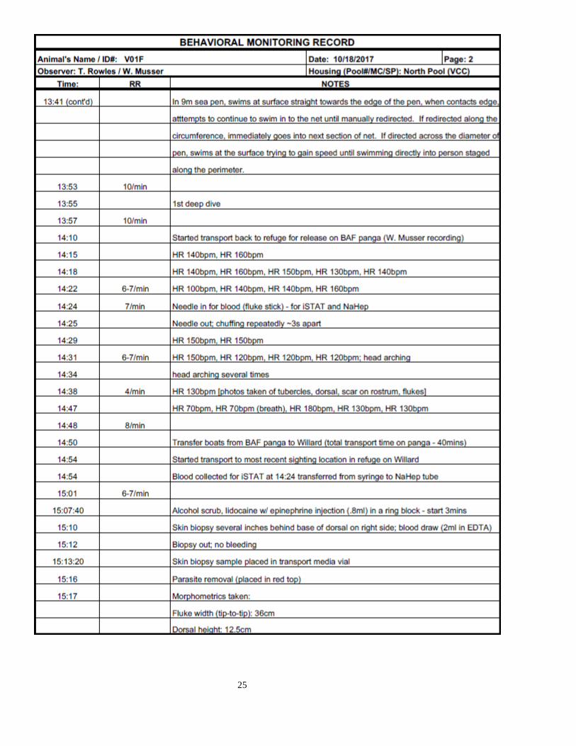
25

26
D. BLOOD RESULTS
Detailed results are given in the table below
Test type Sampling time
11.30
CBC
RBC M/μL 5.6 Hemoglobin g/dL 19.1
Hematocrit % 55.1
MCV fL 98.3 MCH pg 35.3
MCHC g/dL 35.9
RDW % 13.5 Platelet K/μL 138
MPV fL 10.1
NRBC/100 WBC - Reticulocyte % 2.26
RBC Morphology -
WBC K/μL 1.47 Neutrophils % 92
Bands % -
Lymphocytes % 5 Monocytes % 1
Eosinophils % 2
Basophils % 0

27
Chemistry Panel 14.24
Glucose mg/dL 111 BUN mg/dL 48.5
Creatinine mg/dL 1.1
Tbili mg/dL 0.6
Cholesterol mg/dL 230
Triglycerides mg/dL 101
Total Protein gm/dL 5.9 Albumin gm/dL 4.6
Globulin gm/dL 1.3
ALP U/L 583 ALT U/L 43
AST U/L 288
CPK U/L 4244 LDH U/L 1919
Calcium mg/dL 11.3
Phosphorus mg/dL 6.3 Iron mcg/dL 402
iStat
Pico Draw Time: 14.24
iStat Run Time 14.32
Na mmol/L 163
K mmol/L 4.3
CL mmol/L 127
iCa mmol/L 1.56 TCO2 mmol/L 30
Glu mg/dL 106
BUN mg/dL 45 Crea mg/dL 1.4
Hct %PCV 47
Hb (via Hct) g/dL 16 AnGap mmol/L 11
pH 7.262
pCO2 66.3 pO2 33
BE ecf 3
HCO3 29.9 TCO2 32
sO2% 53
Lac 1.28
Results of blood analyses on samples collected on final transport (approx. 4 hours post capture) submitted to SeaWorld laboratories
indicate elevated levels of creatinine kinase (CPK) (4,244 U/L), and lactate dehydrogenase (LDH) (1,919 U/L), low globulin level (1.3
g/dL), and a neutrophilia (92 %) and lymphopenia (5 %) (total wbc 1.47 x 10³ cells/µL) compared to published values for harbor
porpoises Phocoena phocoena entrapped in weirs (Koopman et al. 1995 & 1999). Muscle enzyme levels (LDH, CK) in blood are higher
than published values in Stenella chased and encircled in the Eastern Tropical pacific (St. Aubin et al. 2013). In review of CK values in
blood from cetaceans in the Netherlands, in 5,000 blood samples from captive and stranded cetaceans (harbour porpoises, and bottlenose
dolphins Tursiops truncatus), only three samples had CK levels over 2,000 (Van Elk, pers. obs.).
E. SAMPLES ARCHIVED
Skin/blubber biopsy placed in San Diego Zoo transport media with antibiotics and fungicide and stored in lab overnight at 4 C. The
sample was shaken to dislodge external debris, decanted into a second transport media vial with same antibiotics and fungicide, placed
in shipping container and taken to SWFSC for subsampling. Subsampling included separation of the blubber which was shaken and
dabbed on the culture plate to remove media and then placed into a cryovial and archived at -80 at SWFSC. The remaining skin was
scraped clean and cut into pieces.1/3 of the sample was placed in a cryovial for SWFSC-NMFS genetics and was frozen at -80 for
genetic use. The remaining 2/3 of the skin sample was again placed into fresh transport media and taken to SDZ for further processing
for cell culture.
Four aliquots of 250 µls plasma (Na heparin), two cryovials of packed red cells, one cryovial of buffy coat, one cryovial of whole blood
archived at SWFSC
Ectoparasite at NMMF in alcohol

28

29

30
Annex D
V02F Veterinary Report
Cynthia Smith, Kathleen Colegrove and Frances M.D. Gulland
Contributors & Data Collectors: Whitney Musser, M.S.; Teri Rowles, DVM, PhD; Roberto Sanchez, DVM; Jay Sweeney, DVM; Paola
Smolensky, DVM; Hendrik Nollens, DVM, PhD; Todd Robeck, DVM, PhD; Peter Thomas, PhD; Tracy Romano, PhD; Rebecca Rivera,
PhD; Sacha Stevenson; Grant Abel; Loren Fish; Ricardo Robelledo; and Brenda Bauer
Date: 4 November 2017
Sex: Female; no evidence of current pregnancy or lactation
Age: Mature; exact age to be determined from teeth sections
Weight: 41 kg
Length: 138 cm
A. PHYSICAL EXAMINATION
Skin marks: Animal had a ~1 inch 2nd degree skin abrasion
on its dorsum, caudal to the blowhole, left of midline, with
mild swelling but no associated heat or evidence of infection.
Multiple linear scars and fluke/fin notches typical of healed
previous entanglement injuries were present.

31
1. Body condition index (1-5):
Emaciated (1) Underweight (2) X Ideal (3) Overweight (4) Obese (5)
2. Post-nuchal fat pad (1-4):
Concave (1) X Spongy (2) Firm (3) Convex (4)
3. Oral cavity: WNL X Abnormal
Teeth were discolored. Some teeth missing on rostral aspect of both left and right mandibular arcades.
Gingival Hyperplasia: x Yes No Mild x Moderate Severe
Upper Left Tooth Wear: None x Few Moderate Excessive
Lower Left Tooth Wear: None Few x Moderate Excessive
Upper Right Tooth Wear: None x Few Moderate Excessive
Lower Right Tooth Wear: None Few x Moderate Excessive
Overall Tooth Loss: None x Few Many Near complete
4. Eyes: x WNL Abnormal
5. Cardiovascular: x WNL Abnormal
Rate (/minute): ~120-130; animal did not develop a sinus arrhythmia
Rhythm: Regular Sinus Arrhythmia x No Sinus Arrhythmia Other Arrhythmia
Abnormal Sounds: Yes x No
6. Respiratory System: x WNL Abnormal
Rate (/minute): ~ 6-8; clear lung sounds bilaterally
Abnormalities: x None Rales Wheezes
Blow odor: x None Normal Malodorous
Mucus: x None Mild Moderate Severe
7. GI Tract: WNL Abnormal No Data
Gut sounds Present Not Present
Gastric fluid WNL Abnormal pH ______
Feces WNL Abnormal
If abnormal, describe texture, color, odor, etc.: ________N/A_________________
8. Reproductive: x WNL Abnormal
Genital slit x WNL Abnormal
Vagina/Penis WNL Abnormal No Data
Right Mammary x WNL Abnormal
Left Mammary x WNL Abnormal
9. Skin: WNL x Abnormal

32
B. CLINICAL SUMMARY
(1) Capture event:
The weather conditions during initial sighting and capture were as follows: flat seas (sea state 1), wind < 5 knots, good visibility,
1/10 of cloud coverage, air temperature 21˚ C.
The vaquitas were first observed from the R/V Maria Cleofas at 15:45 PST at 31.140 N and 114.720 W. The Viking (net and
catch RHIB) approached the group of two similar sized animals around 1600. The first net (500 m in length) was set shortly
thereafter and then the vaquitas were driven towards the net by two other small boats (the Proline and the X-tender). Soon after
the first net was set by the Viking, the Proline also launched a net (250 m in length) in close proximity to the first net. The three
boats then worked together to herd the vaquitas towards the nets.
At 16:18, two vaquitas were entangled in a net and were visible at the surface with the net loosely around them. The Viking
and the Proline were driven quickly to the net and were at the animals within one minute of their observation (16:19). One of
the two vaquitas was loosely wrapped and escaped. The other vaquita was entangled at the float line and it was able to lift the
net to the surface when breathing. It was mainly entangled around the flippers but there was also net around the fluke and the
head. Based on the catch team’s experience, it was a typical entanglement for harbor porpoises and it took two minutes to safely
cut the net away from the vaquita and lift it out of the water and on board the Viking.
On the Viking, the animal was placed on the stretcher on top of the foam mattress (see Appendix 1). Water was poured over
the animal every 10 s and the animal did not show overt signs of stress, compared to similar sized harbor porpoise. There was
no evidence of body arching, head shaking, breath holding, or hyperventilation. The animal breathed regularly and appeared
calm. The animal was onboard the Viking for less than 10 min before it was transferred in a stretcher to the Defender (animal
transport vessel provided by Mexican Navy).
While the captured animal was being handled in the net and onboard the Viking, the Extender followed the second animal
southwards. A third net was set while the second animal was in close proximity to the X-tender, and the Proline joined in
herding the animal towards the nets.
The Viking joined the search after the first vaquita (V02F) was transferred to the Defender. The Viking attempted to herd the
second vaquita with assistance from the other catch boats and with guidance from the observer boats. The animal was followed
for about one hour and several times came very close to the net, but eventually the team lost sight of the second animal and
further searches were discontinued at 17:25.
All nets were back in the boats at 18:15.
(2) Location of capture:
Approximate location of animal capture: 31° 07.602 N 114° 42.857 W. Approximate water depth at animal capture was 15.3
meters. Both location and depth were marked at the location of Defender, which was within 1 km of the Viking.
(3) Clinical details post-capture:
The animal initially appeared clinically stable post-capture (16:21). A low-dose of diazepam (approx. 0.175 mg/kg, 7 mg total)
was administered soon after capture (16:26) as planned in an attempt to help prevent onset of capture myopathy, as well as to
provide very low dose sedation to assist with acclimation. The animal was easily and safely transferred to the Defender using
a soft stretcher. Initial vital signs were steady with a respiration rate (RR) at ~6 breaths per minute and a heart rate (HR) ranging
from 120-130 beats per minute (bpm), although no sinus arrhythmia was present and did not develop at any time during care.
After evaluating the animal and determining her clinical stability, she was placed in a stretcher in sea water over the starboard
side of the vessel during attempts to capture the second animal. She was maintained over the side of the vessel for 52 minutes
(16:43-17:35).
Diagnostic ultrasound was performed during the initial evaluation to acquire a baseline pulmonary scan (see Section E below).
Results of the first pulmonary scan at 16:50 (32 minutes post-capture) were within normal limits. However, recheck of the
lungs at 17:08 (50 minutes post-capture) showed evidence of alveolar interstitial syndrome developing in the ventral portion
of both left and right lungs fields, which was slightly worse on the right side. This finding is consistent with developing
pulmonary edema, based on distribution of affected lungs, rapid onset, and history of capture. In response, the animal received
a low dose of furosemide (diuretic) and a dose of steroid (see Section D for dosages). A recheck ultrasound after drug therapy
showed significant improvement on the left side but only partial improvement on the right. A second dose of diuretic was
administered (17:27) which resulted in resolution of the abnormal pulmonary finding when rechecked at 17:40. Although the
suspected pulmonary compromise was effectively addressed, the cause remained unknown.

33
During holding on the small vessel, the animal occasionally showed mild arching followed by recovery. Her HR was stable
throughout (typically 120 bpm) and RR rarely varied above 6-8 breaths per minute. Blood was collected for iStat analysis and
initial results showed a hyperglycemia (see Section F below)
The animal’s mammary glands were palpated and there was no evidence of lactation. Additionally, reproductive ultrasound
exams, although abbreviated, revealed no evidence of pregnancy. Her length was measured at ~136cm (later measured at
necropsy as 138cm) and weight was estimated at ~40kg (later confirmed at 41kg). Therefore, the animal was determined to be
a mature, non-lactating, non-pregnant female. When attempts to capture the second animal were unsuccessful, Rojas, Gulland,
and Smith agreed to transport the first animal to the El Nido sea-pen facility.
The animal was slowly transported on the Defender from the point of capture to El Nido, which took approximately 1 hr (1745-
1842). Animal was relatively stable during the transport, but did have a few bouts of tachypnea and some mild to moderate
arching, but would recover. Interestingly, another blood collection was attempted by experienced venipuncturists and all
attempts were unsuccessful. The decision was made to try again later, but was noted as unusual and of some concern.
The animal arrived at El Nido at 1842 and was administered a long-acting cephalosporin antibiotic injection, given the very
recent history of suspected pulmonary edema, in an effort to prevent the onset of a pulmonary infection and in hopes of being
able to maintain an initial hands-off approach. The animal entered the 9 meter diameter sea-pen at 1843 and began to swim
excitedly. She swam into the sides of the sea-pen multiple times before beginning to show evidence of learning to navigate the
area, which happened about 10 minutes after entering the pen. However, she continued to swim erratically and somewhat
dangerously. A second dose of diazepam was administered at 1849 in an attempt to provide mild sedation.
At 1857, she slowed down considerably and began to log and bob in the center of the sea-pen. The initial interpretation of her
change in behavior was that the animal was calming down and beginning to acclimate to the sea-pen. However, it became
quickly evident that she was rapidly deteriorating. At 1911, senior animal care staff (Abel and Bauer) entered the water, as she
becoming increasingly dull. At 1924, the animal became abnormally relaxed (limp) and respirations slowed dramatically.
An emergency response and emergency release was initiated at 1927. Doxapram was administered, as well as a partial dose of
flumazenil. The animal was quickly moved out of the sea-pen and onto the side of the Narvalito, which was tied up next to the
sea-pen. After confirming the animal was breathing, she was released from the side of the Narvalito, pointed straight out to sea.
The animal swam rapidly away from the Narvalito, abnormally staying on the surface, and then circled back toward the sea-
pen. She was quickly recaptured and then secured in the water by animal care staff on the outside of the Narvalito. The animal
was no longer breathing and had a faint, slow heart beat. She was given doxapram, following which there was no
cardiopulmonary response, and was then quickly transferred to the Narvalito deck for emergency care.
From 1935-2221, emergency medicine was provided to the animal, which included intubation, ventilation, oxygen
administration, chest compressions as needed, emergency medications, intravenous and subcutaneous fluid administration,
preparation of the dorsal fin for potential tagging if release became an option, and continuous monitoring. Although we were
able to restart her heart, we were gravely concerned about inadequate perfusion to her flukes and extremities and her inability
to transition from occasional spontaneous but ineffective respirations to effective spontaneous ventilation. Finally, we were
unable to revive her from a cardiac arrest at 2210.
The animal was declared dead at 2221.
(4) Location of mortality: Onboard small vessel (Narvalito) just northeast of Machorro
(5) Personnel in attendance of animal at time of death: Lorenzo Rojas-Bracho (Program Director), Cynthia Smith (General
Program Manager; Veterinarian), Frances Gulland (Lead Veterinarian), Roberto Sanchez (Veterinarian), Hendrik Nollens
(Veterinarian), Jay Sweeney (Veterinarian), Paola Smolensky (Veterinarian), Brenda Bauer (Deputy Program Manager;
Housing Project Manager), Grant Abel (Co-Program Manager, Housing & Care), Loren Fish (Animal Care Project Manager),
Ricardo Rebolledo (San Felipe Site Manager), Whitney Musser (Animal Care Technician), and Sacha Stevenson (Veterinary
Technician)
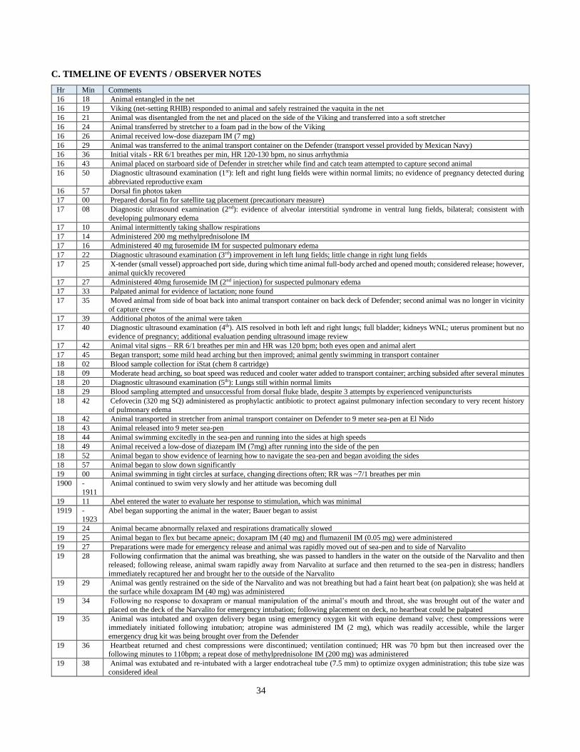
34
C. TIMELINE OF EVENTS / OBSERVER NOTES
Hr Min Comments
16 18 Animal entangled in the net
16 19 Viking (net-setting RHIB) responded to animal and safely restrained the vaquita in the net
16 21 Animal was disentangled from the net and placed on the side of the Viking and transferred into a soft stretcher
16 24 Animal transferred by stretcher to a foam pad in the bow of the Viking
16 26 Animal received low-dose diazepam IM (7 mg)
16 29 Animal was transferred to the animal transport container on the Defender (transport vessel provided by Mexican Navy)
16 36 Initial vitals - RR 6/1 breathes per min, HR 120-130 bpm, no sinus arrhythmia
16 43 Animal placed on starboard side of Defender in stretcher while find and catch team attempted to capture second animal
16 50 Diagnostic ultrasound examination (1st): left and right lung fields were within normal limits; no evidence of pregnancy detected during
abbreviated reproductive exam
16 57 Dorsal fin photos taken
17 00 Prepared dorsal fin for satellite tag placement (precautionary measure)
17 08 Diagnostic ultrasound examination (2nd): evidence of alveolar interstitial syndrome in ventral lung fields, bilateral; consistent with
developing pulmonary edema
17 10 Animal intermittently taking shallow respirations
17 14 Administered 200 mg methylprednisolone IM
17 16 Administered 40 mg furosemide IM for suspected pulmonary edema
17 22 Diagnostic ultrasound examination (3rd) improvement in left lung fields; little change in right lung fields
17 25 X-tender (small vessel) approached port side, during which time animal full-body arched and opened mouth; considered release; however,
animal quickly recovered
17 27 Administered 40mg furosemide IM (2nd injection) for suspected pulmonary edema
17 33 Palpated animal for evidence of lactation; none found
17 35 Moved animal from side of boat back into animal transport container on back deck of Defender; second animal was no longer in vicinity
of capture crew
17 39 Additional photos of the animal were taken
17 40 Diagnostic ultrasound examination (4th). AIS resolved in both left and right lungs; full bladder; kidneys WNL; uterus prominent but no
evidence of pregnancy; additional evaluation pending ultrasound image review
17 42 Animal vital signs – RR 6/1 breathes per min and HR was 120 bpm; both eyes open and animal alert
17 45 Began transport; some mild head arching but then improved; animal gently swimming in transport container
18 02 Blood sample collection for iStat (chem 8 cartridge)
18 09 Moderate head arching, so boat speed was reduced and cooler water added to transport container; arching subsided after several minutes
18 20 Diagnostic ultrasound examination (5th): Lungs still within normal limits
18 29 Blood sampling attempted and unsuccessful from dorsal fluke blade, despite 3 attempts by experienced venipuncturists
18 42 Cefovecin (320 mg SQ) administered as prophylactic antibiotic to protect against pulmonary infection secondary to very recent history
of pulmonary edema
18 42 Animal transported in stretcher from animal transport container on Defender to 9 meter sea-pen at El Nido
18 43 Animal released into 9 meter sea-pen
18 44 Animal swimming excitedly in the sea-pen and running into the sides at high speeds
18 49 Animal received a low-dose of diazepam IM (7mg) after running into the side of the pen
18 52 Animal began to show evidence of learning how to navigate the sea-pen and began avoiding the sides
18 57 Animal began to slow down significantly
19 00 Animal swimming in tight circles at surface, changing directions often; RR was ~7/1 breathes per min
1900 -
1911
Animal continued to swim very slowly and her attitude was becoming dull
19 11 Abel entered the water to evaluate her response to stimulation, which was minimal
1919 -1923
Abel began supporting the animal in the water; Bauer began to assist
19 24 Animal became abnormally relaxed and respirations dramatically slowed
19 25 Animal began to flex but became apneic; doxapram IM (40 mg) and flumazenil IM (0.05 mg) were administered
19 27 Preparations were made for emergency release and animal was rapidly moved out of sea-pen and to side of Narvalito
19 28 Following confirmation that the animal was breathing, she was passed to handlers in the water on the outside of the Narvalito and then
released; following release, animal swam rapidly away from Narvalito at surface and then returned to the sea-pen in distress; handlers
immediately recaptured her and brought her to the outside of the Narvalito
19 29 Animal was gently restrained on the side of the Narvalito and was not breathing but had a faint heart beat (on palpation); she was held at the surface while doxapram IM (40 mg) was administered
19 34 Following no response to doxapram or manual manipulation of the animal’s mouth and throat, she was brought out of the water and
placed on the deck of the Narvalito for emergency intubation; following placement on deck, no heartbeat could be palpated
19 35 Animal was intubated and oxygen delivery began using emergency oxygen kit with equine demand valve; chest compressions were immediately initiated following intubation; atropine was administered IM (2 mg), which was readily accessible, while the larger
emergency drug kit was being brought over from the Defender
19 36 Heartbeat returned and chest compressions were discontinued; ventilation continued; HR was 70 bpm but then increased over the following minutes to 110bpm; a repeat dose of methylprednisolone IM (200 mg) was administered
19 38 Animal was extubated and re-intubated with a larger endotracheal tube (7.5 mm) to optimize oxygen administration; this tube size was
considered ideal

35
19 46 Blood was sampled for iStat analysis and additional diagnostics
19 55 Biopsy punch was performed through the animal’s dorsal fin, which was placed in transport media for cell culture
20 03 Positive pupillary light reflex was confirmed
20 08 Administered repeat dose of doxapram IV (40 mg) in an attempt to stimulate spontaneously breathing
20 10 Sodium bicarbonate slowly administered IV (10 mEq)
20 11 Animal began to show signs of increased awareness, moving body and improving jaw tone, but still had no spontaneous respirations
20 12 Animal’s heart rate began to drop (70 bpm) so a repeat dose of atropine was administered IM (0.9 mg) , after which heart rate increased
20 15 Animal continued increased movement to include some jaw movement; the boat was slowly moved to Campo Uno to retrieve fluids for IV administration
20 21 HR 100-120 bpm; boat arrived at Campo Uno; additional blood collected
20 17 IV fluid (normal saline) administration began
2033 -2038
Boat was moved back into position for potential release off shore
20 41 Administered repeat dose of doxapram IV (40 mg); HR 120 bpm; another blood sample collected for iStat measurement
20 45 Rototag placed on dorsal fin; orange #72
20 47 Sodium bicarbonate administered IV (10mEq); soon after animal became more responsive and had increased jaw tone
20 51 Animal intermittently tested for spontaneous respirations; none occurred up to this time
20 53 Animal moving eyelids and eyes; followed by spontaneous swallowing; CRT still <2 sec (continuous throughout)
2058 -
2102
HR 120 bpm; animal open and closing eyes in response to light; occasional swallowing; however, still not spontaneous respirations
21 04 Animal switched from oxygen to ambient air with use of ambulatory bag; auscultation of lungs confirmed normal lung sounds bilaterally
with use of ambulatory bag
21 09 Veterinary staff concerned about progressive capture myopathy and/or seizure activity; Diazepam administered IV (7 mg)
21 10 Animal spontaneously ventilated through the endotracheal tube; animal was extubated to see if intubation was suppressing spontaneous
respiration
2110 -2113
Animal did not have control over its blowhole and was unable to effectively breathe; HR remained steady at 120 bpm
21 13 Animal was re-intubated and animal spontaneously ventilated through the tube multiple times, but then ceased spontaneous ventilation,
followed by intermittent ambient air and oxygen ventilation support
21 21 Animal had eyelid movement and regained palpebral response
21 25 Repeated administration of doxapram IV (40 mg)
21 26 More eyelid tone and animal was now squinting in response to light
21 30 Hypothermia was suspected based on rectal temperature of 34.8˚ C; positive responses continued with jaw tone and eyelid movement
21 39 Spontaneous ventilation returned and lung auscultation was within normal limits bilaterally; subcutaneous fluid administration began
(normal saline, total volume 180 cc)
2140 -
2145
Spontaneous ventilation through the endotracheal tube with minimal blowhole movement occurred, followed by occasional but ineffective
breaths through the tube and swimming body movements; intermittent oxygen and ambient air was administered as often as deemed
appropriate
21 52 Rectal temperature slightly improved at 35.0C; total volume of fluids administered at this time was 300 IV plus 120 SQ normal saline
2155 -
2205
More spontaneous breathing through endotracheal tube occurred, although only partially effective; first evidence of tongue movement
was seen
22 06 Extubated animal to test ability to effectively breathe on own, however animal was unable to gain proper control over blowhole or was
unable to effectively breathe
22 08 Animal reintubated and ambient air was administered with ambulatory bag
22 10 Animal went into cardiac arrest; immediately began chest compressions
2212 -
2215
Epinephrine administered IT (2mg) with no response, followed by epinephrine IC (4mg, then an additional 2mg) with a positive response;
HR increased from 30bpm and increased until it stabilized at 90bpm
22 16 Pupils became fixed and dilated; animal became flaccid; no corneal reflex; no other reflexes present
22 21 Animal declared dead; all resuscitation efforts were discontinued
22 29 Post-mortem blood was collected from the brachiocephalic vein; animal was then transported to Campo Uno facilities for necropsy

36
D. DIAGNOSTIC ULTRASOUND REPORT
Ultrasonographer: Cynthia Smith, Ultrasound Technicians: Whitney Musser, Sacha Stevenson
Equipment: Sonosite Edge ultrasound system with a C60x 5-2MHz curvilinear transducer; outfitted with Zeiss Cinemizer
heads-up display goggles
Data: The following ultrasound data were acquired from V02F. The first exam was performed ~32 minutes after capture. The
timestamps on the DICOM images are 3 hours ahead of the actual times images were acquired. Images were transferred to
Osirix, a medical imaging software program, to allow for an in-depth evaluation of data
Exam 1
Time/Date 1650, 4NOV2017. Duration of exam: 1 minute
Animal location Animal was examined while in a stretcher, which was immersed in sea-water on the outside of the boat (Defender)’s starboard side.
Lungs Right lung field was evaluated first (images 1 & 3), then the left lung field (images 4 & 6). No pulmonary abnormalities were
detected.
Reproductive tract No evidence of pregnancy was detected during an abbreviated reproductive exam performed from the left side of the animal. Summary No pulmonary abnormalities were detected. No evidence of pregnancy was found.
Left lung: No abnormalities Right lung: No abnormalities
Exam 2
Time/Date 1708, 4NOV2017. Duration of exam 2 minutes
Location Animal was examined while in a stretcher, which was immersed in sea-water on the outside of the boat (Defender)’s starboard side.
Lungs Right lung was evaluated first; then the left lung was evaluated. Evidence of severe alveolar interstitial syndrome (AIS) was present
in both left and right ventral lung fields. No abnormalities were detected in the dorsal or mid portions of either lung fields.
Reproductive tract No evidence of pregnancy. Prominent uterine horns detected.
Urinary tract Full bladder. No abnormalities detected.
Summary Suspect developing pulmonary edema (bilateral), based on portion of lung fields effected; rapid onset of abnormality; and recent
experience with V01F, who was suspected to have pulmonary edema following capture due to froth from the blowhole, which
responded to diuretic treatment. No evidence of pregnancy or lactation.

37
Exam 3
Time/Date 1720, 4NOV2017 Duration of exam: 2 minutes
Location Animal was examined while in a stretcher, which was immersed in sea-water on the outside of the boat (Defender)’s starboard side.
Lungs Right lung was evaluated first; then the left lung. Severe AIS was still present in the ventral portion of right lung (cine 10 & cine 11), although there was a slight decrease in the extent of lung affected. Almost complete resolution of AIS was seen in the left lung
(cine 12 & cine 13).
Summary In response to a 1 mg/kg dose of furosemide and a 5 mg/kg dose of methylprednisolone, severe AIS had almost completely resolved in the left lung and was showing slight decrease in the right lung. Due to the animal’s history and treatment, the most likely cause of
AIS was pulmonary edema.
Left lung: No abnormalities Right lung: Severe AIS in ventral portion only
Exam 4
Time/Date 1740, 4NOV2017, Duration of exam: 2 minutes
Location Animal was examined while in a stretcher, which was immersed in a porpoise transport container filled with sea-water on the back
deck of the boat (Defender).
Lungs Right lung was evaluated first; then the left lung. Severe AIS had completely resolved in both left and right lungs.
Reproductive tract The animal’s reproductive tract was evaluated from the left lateral body wall. Both left and right horns were prominent. No fluid
was detected in either left or right uterine horn. The cervices and uterine body were examined and appeared within normal limits. No evidence of lactation was detected in either left or right mammary glands, although evaluation was brief. Left ovary was briefly
visualized. There were ~2 small areas suggestive of follicular activity detected (~3mm diameter, round, anechoic) in the left ovary;
however, further evaluation in Osirix is needed to determine if these areas are consistent with follicular activity. Multiple cine loops
were obtained (cine 14-18). The right ovary was not evaluated.
Urinary tract The bladder was distended (full) and no abnormalities were detected. The left kidney appeared to be within normal limits; several
renicules were fluid-filled, which was considered normal.
Summary In response to a second dose of 1 mg/kg furosemide, severe AIS had completely resolved in both left and right lungs. No pulmonary
abnormalities were detected. There was no evidence of pregnancy or lactation. The bladder and left kidney were within normal limits.
Exam 5
Time/Date 1820, 4NOV2017, Duration of exam, 3 minutes
Location Animal was examined while in a stretcher, which was immersed in a porpoise transport container filled with sea-water on the back
deck of the boat (Defender). Lungs Left lung was evaluated first (cine 19 & 20); right lung was evaluated next (cine 22 & 23). No abnormalities were detected.
Forestomach Several fish were detected in the animal’s forestomach (cine 24 & 25). No gastric motility was detected.

38
Urinary tract Left kidney rechecked and no abnormalities were detected. Fluid-filled renicules were detected and considered within normal limits
(cine 26). The left ureter was seen exiting the caudal pole of the left kidney. Further evaluation will be performed when images are
transferred to Osirix.
Summary Exam was performed toward the end of the animal’s transport, prior to transfer to the sea-pen. Both left and right lungs were determined to be within normal limits. Evidence of normal kidney function and diuresis was detected. Fish were present in the
animal’s forestomach, although no forestomach motility was detected when evaluated.
Left lung: No abnormalities Right lung: No abnormalities
Forestomach: Fish detected in stomach
E. BLOOD RESULTS
Blood was collected during transport, during emergency response, and after death. Results following death should be
interpreted with caution, as they were collected approximately 19 minutes after cardiac arrest.
CBC results from blood collected at the end of transport and during emergency response were unremarkable. Chemistry results
from blood collected during emergency response showed a hyperglycemia, elevated creatinine kinase, and elevated LDH levels.
Creatinine kinase elevations consisted predominatly of the mm (skeletal muscle derived) and mb (cardiac muscle and
diaphragm derived) isoenzymes, rather than the bb (brain derived) isoenzyme. IStat results from blood collected at the end of
transport showed a hyperglycemia, and results during emergency response showed a hyperglygemia and acidosis, likely
metabolic. Hormonal analyses of blood collected at 20:00 revealed very high cortisol, aldosterone, epinephrine and
norepinephrine levels indicative of a severe acute stress response.
Detailed results are given in the table below:

39
Animal ID: V02F Date: 4NOV2017 Processing Lab: SeaWorld San Diego
Test type Sampling time
1805 1830 2000 2024 2229
Associated Event Transport Transport During CPR During CPR Post-Mortem
CBC
RBC M/μL - 5.44 5.68 - - Hemoglobin g/dL - 19 20.3 - -
Hematocrit % - 58.1 60.2 - -
MCV fL - 106.9 106 - - MCH pg - 35 35.7 - -
MCHC g/dL - 32.8 33.7 - -
RDW % - 13.1 12.9 - - Platelet K/μL - 137 128 - -
MPV fL - 13.9 13.7 - -
NRBC/100 WBC - 0 0 - - Reticulocyte % - 1.17 1.25 - -
RBC Morphology - N N - -
WBC K/μL - 4.95 4.88 - - Neutrophils % - 60 62 - -
Bands % - 4 2 - -
Lymphocytes % - 23 27 - - Monocytes % - 5 4 - -
Eosinophils % - 8 5 - -
Basophils % - 0 0 - -
Chemistry Panel Glucose mg/dL - - 194 198 199
BUN mg/dL - - 73.7 71.6 82.5 Creatinine mg/dL - - 1.3 1.3 2
Tbili mg/dL - - 0.2 0.2 1
Cholesterol mg/dL - - 464 468 145 Triglycerides mg/dL - - 138 134 10
Total Protein gm/dL - - 7.9 8.1 5
Albumin gm/dL - - 4.1 4.2 3.2 Globulin gm/dL - - 3.8 3.9 1.8
ALP U/L - - 75 77 22
ALT U/L - - 119 126 94 AST U/L - - 290 301 434
GGT U/L - - 32 33 30
CPK U/L - - 2219 2182 5163 CK mm 2770 5659
CK mb 165 320
CK bb 716 754 LDH U/L - - 852 862 905
Calcium mg/dL - - 10.5 10.7 10.1
Phosphorus mg/dL - - 7.3 7.5 19 Sodium mEq/L - - 164 164 164
Potassium mEq/L - - 4.6 4.6 8.9
Chloride mEq/L - - 104 106 104 Carbon Dioxide mEq/L - - 15 13 14
Iron mcg/dL - - 285 287 165
Comments - Cells appear
degenerated due to
age of specimen
None None Sample collected
post-mortem
iStat
Pico Draw Time: 1805 - 2000 - -
iStat Run Time 1807 - 2002 - -
Associated Event Transport - During CPR - -
CHEM8+
Na mmol/L 156 - 158 - -
K mmol/L 3.4 - 4 - -
CL mmol/L 115 - 116 - - iCa mmol/L 1.23 - 1.14 - -
TCO2 mmol/L 34 - 19 - -
Glu mg/dL 158 - 170 - - BUN mg/dL 61 - 100 - -
Creatinine mg/dL 1.3 - 1.4 - -
Hct %PCV 46 - 56 - - Hb (via Hct) g/dL 15.6 - 19 - -
AnGap mmol/L 11 - 28 - -

40
Hormonal Analyses
Serum progesterone 5.05 ng/mL
Cortisol 150 µg/dL
Aldosterone 236.66 pg/mL
T3 154 ng/dL
T4 14.9 µg/dL
FT4 1.75 ng/dL
Epinephrine (at 20.00) 31614.6 pg/mL
Norepinephrine (at 20.00) 1790.6 pg/mL
Dopamine (at 20.00) 126.52 pg/ml
F. GROSS NECROPSY REPORT
FIELD #: V02F NECROPSY DATE: November 4 2017
SPECIES: PHOCOENA SINUS CARCASS CONDITION CODE: 1-2 (SOME SAMPLES COLLECTED IN VIVO)
AGE CLASS: ADULT TIME: 11.00 PM
SEX: FEMALE Necropsy conducted at Campo Uno, San Felipe, within one hour of
cardiac arrest. Prior to cardiac arrest, extensive CPR efforts for 3 hours
including endo-tracheal intubation, PPV, chest compressions,
intravenous and intra-cardiac drugs.
PROSECTOR(S): F. GULLAND, C. SMITH, R.
SANCHEZ, G. ABEL
WEIGHT: 41 KG E examined, NE not examined, NA not applicable, NAD No abnormality detected
Cause of Death: Cardiac arrest.
Samples collected:
Frozen -20 C:
Teeth, liver (1 in Teflon, 1 in whirlpac), blubber (1 in Teflon, 1 whirlpac), melon fat (foil/whirlpac), acoustic fat
(foil/whirlpac), kidney (whirlpac), cerebrum (whirlpac), mesenteric lymph node (whirl pac), pulmonary lymph node (whirl
pac), skeletal muscle (whirl pac), uterus (whirl pac)
Uterus/cervix/vagina whole (Ziplock, for Mesnick SWFSC)
Stomach with contents (prey ID, SWFSC)
Urine
Dorsal fin
Skeleton (red trash bag), Soft tissues from around carcass (black trash bag)
Cell culture transport media at 4 C:
Skin plug from dorsal fin collected while animal alive, post mortem kidney, trachea, liver.
Room temp/cooled with icepacks in container:
Ovaries
DMSO:
Skin (3 vials), liver (2 vials)
Formalin:
Brain, tongue, tonsil/oral mucosa, eye (left), bronchi, lung, pulmonary LN, thyroid, skeletal muscle (neck area), diaphragm,
heart, liver, spleen, kidney, adrenal, pancreas, ileum, mesenteric LN, colon, uterus, cervix, skin (laceration on head)

41
BODY CONDITION: Average, nuchal pad slightly convex, blubber depths 1.7-0.8 cms dorsal/ventral respectively
SKIN / HUMAN INTERACTION: Multiple fine lacerations typical of monofilament cuts, and notches, on margins of dorsal fin, flippers, abrasions on
rostrum a consequence of hitting net pen wall.

42
DISCHARGES: None
UMBILICUS: Scar healed. Mammary glands: No discharge, nipples not enlarged, no milk on cut surface
BLUBBER: Pale yellow, lipid oozed from cut surface , mild bruising over blubber of left thoracic area
SKELETAL MUSCLE: No abnormalities detected
SKELETON AND JOINTS: No abnormalities detected, skeleton collected, frozen

43
EYES: NAD
ORAL CAVITY: Teeth erupted, grossly normal, 4 anterior teeth lower left missing. TEETH: Upper left 14 teeth, Upper right
14, Lower left 18, lower right 22
THORACIC CAVITY: Mild hemorrhage on intercostal muscles left flank. No free fluid in thoracic cavity
ESOPHAGUS: NAD
THYROID: Prominent, brown coloration, approx 2 cms diameter. THYMUS: ND
TRACHEA / BRONCHI / LUNGS:No fluid in airways, lungs grossly normal.
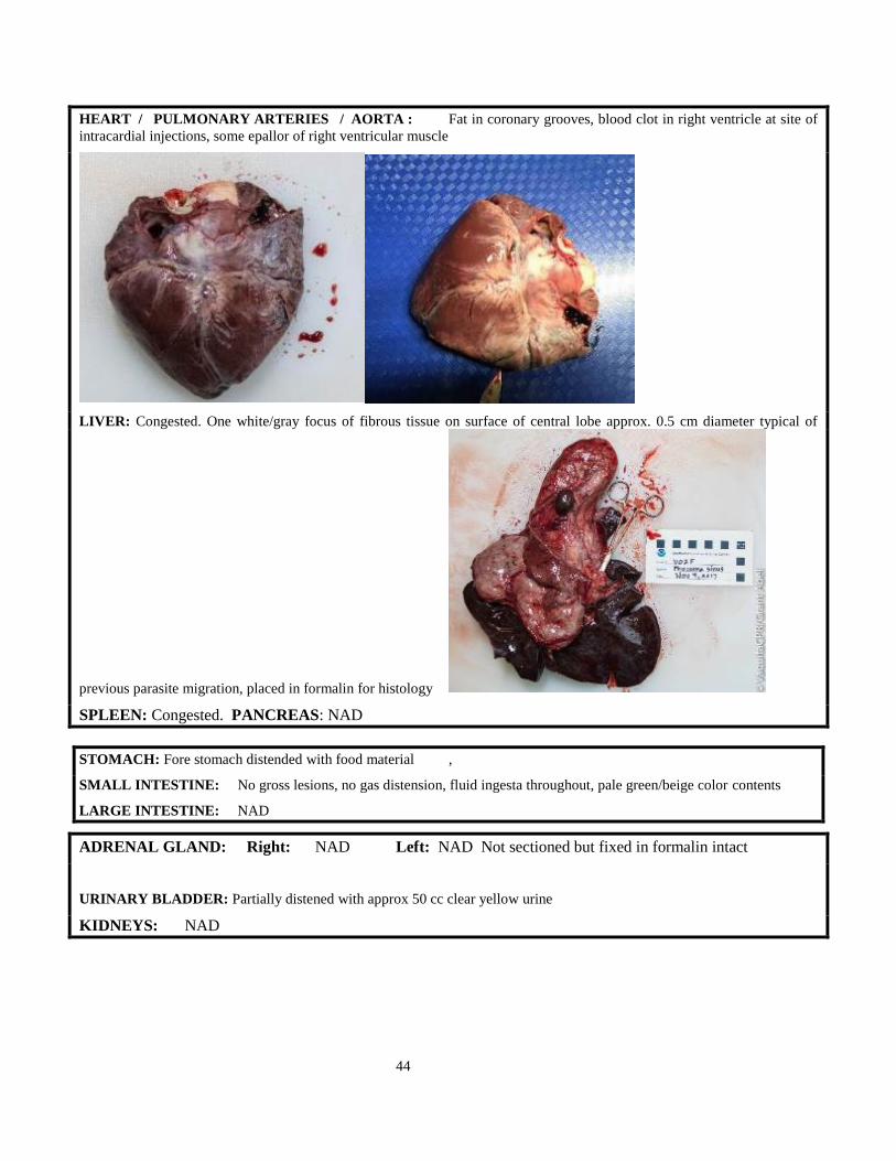
44
HEART / PULMONARY ARTERIES / AORTA : Fat in coronary grooves, blood clot in right ventricle at site of
intracardial injections, some epallor of right ventricular muscle
LIVER: Congested. One white/gray focus of fibrous tissue on surface of central lobe approx. 0.5 cm diameter typical of
previous parasite migration, placed in formalin for histology
SPLEEN: Congested. PANCREAS: NAD
STOMACH: Fore stomach distended with food material ,
SMALL INTESTINE: No gross lesions, no gas distension, fluid ingesta throughout, pale green/beige color contents
LARGE INTESTINE: NAD
ADRENAL GLAND: Right: NAD Left: NAD Not sectioned but fixed in formalin intact
URINARY BLADDER: Partially distened with approx 50 cc clear yellow urine
KIDNEYS: NAD

45
REPRODUCTIVE TRACT: Uterine horns uniform diameter through-out length, no discoloration of the endometrium
detected (only partially opened to preserve morphology for Sarah Mesnick detailed examination). Left ovary approx. 2 cms
diameter with multiple structures giving surface a knobbly appearance – detailed examination to be conducted by T. Robeck,
right ovary smaller, 1 cm diameter with one prominent CL/CA. Discrete piece of mucoid material approx. 0.5 cms diameter,
deep red color, within uterine body not attached to endometrium collected and fixed in formalin.
ovaries in bag
BRAIN: Congested blood vessels ventral surface of cerebrum, slight flatening of sulci, gyri

46
Morphometrics: Stomach (intact with food) 0.9 kg Heart 0.2 kg
1 total length………………138…cms….
2 snout to anus…………………97.5..…_cms
3 snout to genital slit……………93.5.....
4 snout to umbilicus……………..61…
5 snout to throat grooves………..…_na
6 snout to dorsal fin tip…................78
7 snout to ant dorsal fin……............55.5
8 snout to flipper…………..........…27.5
9 snout to ear……………...……….19.5
10 snout to eye…………...………...13.5_(right)
11 snout to gape………...………….9 (right)
12 snout to blowhole (s)…………....13
13 snout to melon apex………….....____nd__
20 head girth at eyes…...........…50
21 length of eye opening…..........….1.8
24 n/a
26 flipper length, anterior……………27.5
27 flipper length, posterior…………..21
28 flipper width, maximum………….10
29 length mammary slits R______2__L_1.7
30 number of mammary slits 2
31 length genital slit 10
33 fluke width………………………...42
35 fluke notch depth………………....1.5
36 dorsal fin height…………………...14.5
37 dorsal fin base length…………….....23
38 girth at eye………………………...__50_______
39 girth at axilla………………………_____68.5___
40 girth, maximum…………………...._80.5______
41 girth at anus……………………….____55.5____
45 blubber thickness, dorsal…………._____1.7___
46 blubber thickness, lateral………….______1.0___
47 blubber thickness, ventral………....______0.8__

47
G. Ovarian cryopreservation report

48

49

50

51

52
H. HISTOLOGY REPORT
Zoological pathology program: Cetacean Necropsy Report
Field ID V02F
Additional Identifier
ZPP Accession Number 17-1182
Species Phocoena sinus
Death Date and Time November 4, 2017 at 2221
Location San Felipe, Mexico
Sex Female
Age Class Adult
Necropsy Date and Time November 4, 2017 at 2300
Condition code 2
Total Length 138 cm
Weight 41 kg
Blubber Depth 1.7 cm dorsal. 0.8 cm ventral.
Body Condition Average
Histopathology Kathleen M. Colegrove DVM, PhD, Dip ACVP
Gross Necropsy: Gross necropsy and images report on file.
Tissues Received: Brain, tongue, tonsil/oral mucosa, eye (left), bronchi, lung, pulmonary LN, thyroid, skeletal muscle (neck
area), diaphragm, heart, liver, spleen, kidney, adrenal, pancreas, ileum, mesenteric LN, colon, uterus, cervix, skin (laceration
on head)
Slide 1: Cerebellum: Some neurons contain small to moderate amounts of pale yellow to tan cytoplasmic pigment (lipofuscin).
Surrounding occasional blood vessels are few histiocytes containing similar accumulations of lipofuscin.
Brain stem: Few neurons contain similar small amounts of lipofuscin.
Slide 2: Mid brain: Neurons contain similar small amounts of lipofuscin.
Slide 3: Right Hippocampus and temporal lobe: Neurons contain similar small to large amounts of lipofuscin.
Fig. 1 Slide 3. Image A. Neuronal lipofuscinosis

53
Surrounding moderate numbers of blood vessels are few histiocytes containing similar accumulations of lipofuscin. Few blood
vessels have walls segmentally mildly thickened with increased amounts of eosinophilic connective tissue (arteriosclerosis).
Fig. 2. Slide 3. Image B. Perivascular lipofuscin accumulation (arrows)
and mild arteriosclerosis
Multifocally the choroid plexus contains mildly increased amounts of fibrous connective tissue. White matter has few, widely
scattered dilated axon sheaths.
Slide 4: Left hippocampus and temporal lobe: Neurons contain similar small to large amounts of lipofuscin. Surrounding
moderate numbers of blood vessels are few histiocytes containing similar accumulations of lipofuscin. Multifocally the choroid
plexus contains mildly increased amounts of fibrous connective tissue. White matter has few, widely scattered dilated axon
sheaths.
Fig. 3. Slide 4. Image A. Normal hippocampus.
Slide 5: Cerebrum: Small blood vessels and capillaries in the cerebral cortex are prominent, congested, and lined by mildly
reactive endothelium. There is mild increased clear space surrounding vessels (possible cerebral edema). There is similar
lipofuscinosis as previously described.
Slide 6: Spinal cord: There is moderate hemorrhage along the outer dural surface (presumed post mortem artifact). Neurons
contain similar small to moderate amounts of lipofuscin.
Slide 7: Lung: Multifocally and more severely in one lung section (presumed ventral) moderate numbers of bronchioles contain
small amounts of pale eosinophilic wispy to homogenous fluid mixed with few foamy macrophages and rare neutrophils.

54
Fig. 4. Slide 7. Image A – Bronchiole with luminal proteinaceous
fluid and few responding foamy macrophages.
Rarely this fluid also contains a few strands of flattened keratinized epithelium (upper respiratory or gastric squamous cells)
and individual rectangular faintly striated skeletal myocytes (gastric contents).
Fig. 5. Slide 7. Images D and E. Bronchioles containing squamous cells (arrow) and skeletal muscle fibers (circle) indicative of aspiration of gastric fluid.
Small numbers of macrophages and neutrophils are responding this material
Multifocally to regionally small numbers of bronchioles and alveolar spaces contain small to moderate numbers of red blood
cells (hemorrhage). Focally within the parenchyma there is a single nodular accumulation of lymphocytes and smaller numbers
of central foamy macrophages. Surrounding few bronchioles are small to moderate accumulations of dark brown stippled
pigmented material (anthracosis). In one section several blood vessels are occluded by brown to light tan, partially mineralized
material.
Slide 8: Lung and bronchiole: In addition to described under slide 7, one affected medium sized bronchiole contains small
amounts of similar pale eosinophilic wispy fluid and material mixed with red blood cells and small numbers of neutrophils and
macrophages. Epithelium is segmentally thin with just the basal layer remaining. In the lumen adjacent to these regions are
small numbers of sloughed epithelial cells few of which have karyorrhectic nuclei. There are few neutrophils and karyorrhectic
debris in the directly underlying submucosa and blood vessels are congested.

55
Fig. 6. Slide 8. Image A. Erosion in a bronchus.
Slide 9: Lung: See previous description under slide 7.
Marginal lymph node: Sinuses contain mildly increased number of histiocytes and rare multinucleated giant cells that contain
moderate amounts of dark brown granular pigmented material (anthracosis).
Slide 10: Liver: Focally within the subcapsular parenchyma is a 5 mm diameter, multiloculated cystic structure lined by a single
layer of low cuboidal to flattened epithelium surrounded by abundant dense fibrous connective tissue. Adjacent to the cystic
structure are multiple simple to branching bile ductule profiles. Surrounding the dense fibrous connective tissue are small
numbers of macrophages most of which contain abundant dark brown to golden yellow pigment (presumed fluke pigment).
Fig. 7. Slide 10. Image A. Subcapsular hepatic cyst.
Throughout the remaining liver portal areas contain small numbers of similar pigment laden macrophages and moderate
numbers of Kupffer cells, especially in centrilobular areas, also contain dark brown pigment. Hepatocytes and Kupffer cells
diffusely contain small amounts of light brown cytoplasmic pigment (presumed hemosiderin). In one section moderate numbers
of centrilobular to midzonal hepatocytes contain one to several medium-sized clear distinct cytoplasmic vacuoles.

56
Slide 11: Liver: See changes under slide 10.
Thyroid gland: No significant findings (NSF).
Slide 12: Spleen: There are numerous moderately sized lymphoid follicles throughout the spleen, some of which are coalescing.
Red pulp contains small numbers of myeloid precursors, megakaryocytes, plasma cells and hemosiderin laden macrophages.
Lymph node: Sinuses contain moderate numbers of red blood cells (congestion).
Slide 13: Kidney: Multifocally small to moderate numbers of tubules contain small to occasionally moderate amounts of wispy
to globular pale eosinophilic material or bright eosinophilic slightly granular to chunky material (protein and myoglobin). This
material is positive for myoglobin via IHC. Some epithelial cells in affected tubules are slightly vacuolated and few are slightly
separated from the basement membrane slightly shrunken, have bright eosinophilic cytoplasm and pyknotic nuclei (necrosis).
Some tubules have few necrotic sloughed epithelial cells within the lumen. Few tubular epithelial cells contain small amounts
of light brown cytoplasmic pigment. There are rare sclerotic glomeruli.
Fig. 9. Slide 13. Image C. Kidney stained with anti-myoglobin
antibody. Material within tubules is positive for myoglobin
(arrows).
Fig. 8. Slide 13 Images A and B. Kidney tubules contain small amounts of myoglobin (circles) and few sloughed necrotic epithelial cells (star). There are
scattered necrotic tubular epithelial cells (arrows).

57
Slide 14: Kidney: See lesions described under slide 13.
Pancreas: NSF.
Slide 15: Small intestine: NSF
Colon: NSF
Uterus: Endometrial glands are lined by tall columnar epithelial cells with basal nuclei and clear to granular cytoplasm. Few
glands are mildly to moderate dilated or slightly branching. There are very few scattered plasma cells and rare hemosiderin
laden macrophages in the endometrium. Endometrial blood vessels have segmentally smudgy, mildly thickened eosinophilic
walls. There are also few clusters of round, dense eosinophilic connective tissue within the superficial endometrium (presumed
fibrotic small vessels secondary to previous pregnancy).
Fig. 10. Slide 15. Image A. Uterine endometrium. Small blood vessels
replaced by fibrous connective tissue (arrows) indicative of previous pregnancy. Gland epithelium exhibits changes due to progesterone
influence.
Slide 16: Uterus: See description under slide 15.
Mesenteric lymph node: Sinuses contain moderate numbers of eosinophils.
Slide 17: Internal cervix: NSF
Urinary bladder: NSF
Slide 18: Internal cervix: NSF
Vagina: Submucosa contains small numbers of scattered lymphocytes and plasma cells (within expected limits for an adult
cetacean).
Slide 19: Right and left adrenal glands: Scattered within the cortex are tight clusters of round myeloid precursors
(extramedullary hematopoiesis).
Slide 20: Tongue: Segmentally submucosa contains moderate numbers of lymphocytes and plasma cells. Deeper in the
muscular layer and submucosa are few small perivascular nodular clusters of lymphocytes.
Oral mucosa: Within the submucosa are few small perivascular nodular accumulations of lymphocytes.
Tonsil: NSF
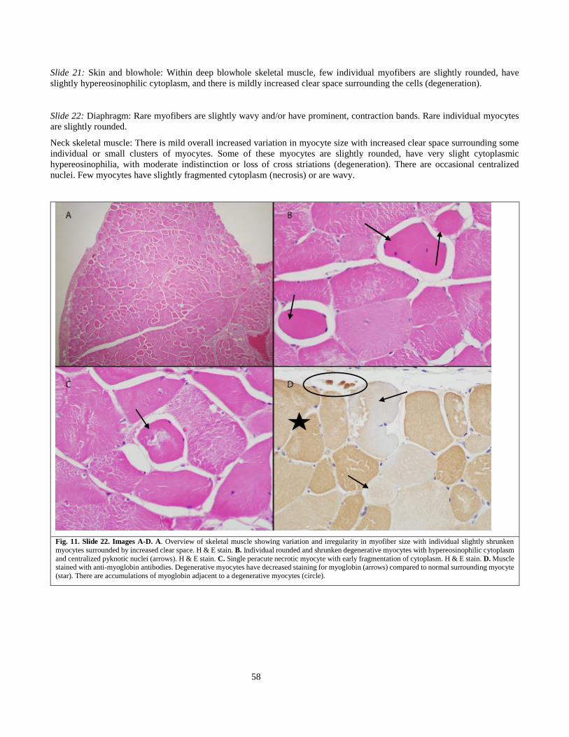
58
Slide 21: Skin and blowhole: Within deep blowhole skeletal muscle, few individual myofibers are slightly rounded, have
slightly hypereosinophilic cytoplasm, and there is mildly increased clear space surrounding the cells (degeneration).
Slide 22: Diaphragm: Rare myofibers are slightly wavy and/or have prominent, contraction bands. Rare individual myocytes
are slightly rounded.
Neck skeletal muscle: There is mild overall increased variation in myocyte size with increased clear space surrounding some
individual or small clusters of myocytes. Some of these myocytes are slightly rounded, have very slight cytoplasmic
hypereosinophilia, with moderate indistinction or loss of cross striations (degeneration). There are occasional centralized
nuclei. Few myocytes have slightly fragmented cytoplasm (necrosis) or are wavy.
Fig. 11. Slide 22. Images A-D. A. Overview of skeletal muscle showing variation and irregularity in myofiber size with individual slightly shrunken
myocytes surrounded by increased clear space. H & E stain. B. Individual rounded and shrunken degenerative myocytes with hypereosinophilic cytoplasm
and centralized pyknotic nuclei (arrows). H & E stain. C. Single peracute necrotic myocyte with early fragmentation of cytoplasm. H & E stain. D. Muscle stained with anti-myoglobin antibodies. Degenerative myocytes have decreased staining for myoglobin (arrows) compared to normal surrounding myocyte
(star). There are accumulations of myoglobin adjacent to a degenerative myocytes (circle).

59
Slide 24: Tissue from uterine lumen: Tissue is composed of clusters of endometrial glands surrounded by loose wispy, poorly
cellular connective tissue and adipocytes. Tissue is lined by endometrial surface epithelium (polyp).
Fig. 12. Slide 24. Image A. Uterine polyp comprised of clusters
of endometrial glands surrounded by loose connective tissue.
Slide 25: Skeletal muscle: Lesions are similar but less severe as noted for slide 22.
Slide 26: Heart, left ventricle: Multifocally within the myocardium moderate numbers of individual to small clusters of
myocytes have slightly hypereosinophilic cytoplasm and/or cytoplasm that is moderately to markedly vacuolated, especially in
the perinuclear areas. These cells often have pyknotic nuclei (degeneration and necrosis). There is moderate amounts of
increased clear space to faint eosinophilic wispy material surrounding myocytes (interstitial edema). Primarily along the
subepicardium small to moderate numbers of myocytes have linear cytoplasmic bands of bright eosinophilic globular to fibrillar
material with slight fragmentation of the cytoplasm (contraction band necrosis). The epicardial surface has moderate amounts
of adipose tissue that extends slightly into the myocardium.
Fig. 13. Slide 26. Images A and B. A. Overview of cardiac lesions. Multiple hypereosinophilic and vacuolated myocytes are visible. B. Higher power
image of degenerate myocytes (arrows). Myocytes are slightly rounded, have hypereosinophilic and vacuolated cytoplasm and pyknotic nuclei. There is
increased clear space and pale eosinophilic fluid surrounding myocytes indicative of myocardial interstitial edema.

60
Skin from the head at net mark: There is a focal region of epithelial loss. The exposed dermis is pale and smudgy eosinophilic
and is covered by a thick layer of red blood cells and moderate amounts of fibrillar eosinophilic material (fibrin). Along the
edge of the defect there are small numbers of neutrophils and cellular debris mixed with the fibrin.
Fig. 14. Slide 26. Image C. Focal acute skin laceration.
Slide 27: Heart, right ventricle: Lesions are similar noted in slide 26. There is prominent contraction band necrosis along the
epicardial surface. The epicardial surface contains moderate amounts of adipose tissue occasionally mixed with very small
amounts of fibrous connective tissue.
Figure 15. Slide 27. Images A and B. Heart with subepicardial contraction band necrosis (arrows).
Slide 28: Heart, right ventricle an apex: Lesions are similar to those described in slides 26 and 27. Degenerate myocytes have
loss of myoglobin via IHC.
Occasional blood vessels are surrounded by very mild increases in fibrous connective tissue and some vessels have mild
segmental areas of vessel wall thickening with increased smooth muscle or pale fibrous connective tissue (arteriosclerosis).
61
Figure 16. Slide 28. Image A. Heart stained with anti-
myoglobin antibodies. There is loss of myoglobin staining
in regions of contraction band necrosis (arrows).
Slide 29: Heart, left ventricle: Lesions are similar to those described in slides 26 and 27.
Slide 30: Heart, interventricular septum: Lesions are similar to those described in slides 26 and 27.
Slide 31: Eye: NSF.
FINAL DIAGNOSES
1. Heart: Marked, multifocal peracute to acute myocardial degeneration and subepicardial contraction band
necrosis with marked interstitial edema
2. Skeletal muscle (neck region and head): Moderate peracute to acute degeneration and necrosis
3. Kidney: Mild multifocal peracute to acute tubular degeneration and necrosis with intratubular myoglobin
(myoglobinuric nephrosis)
4. Lung: Mild multifocal acute neutrophilic and histiocytic bronchitis and bronchiolitis with aspirated gastric
contents, epithelial erosion, and minimal edema
5. Skin, head: Focal acute linear laceration
6. Liver: Focal subcapsular biliary cyst (presumed trematode associated)
7. Liver: Mild centrilobular hepatocellular vacuolation, moderate periportal trematode pigment accumulation and
mild to moderate hemosiderosis
8. Heart: Minimal multifocal perivascular fibrosis, minimal multifocal epicardial fibrosis, and mild
arteriosclerosis
9. Uterus: Mild multifocal endometrial gland dilation, minimal chronic endometritis and focal luminal
endometrial polyp
10. Spleen: Mild lymphoid hyperplasia, extramedullary hematopoiesis, and hemosiderosis
11. Lung: Minimal focal lymphocytic and histiocytic pneumonia
12. Marginal lymph node and lung: Mild to moderate anthracosis
13. Brain: Moderate to marked neuronal lipofuscinosis, mild arteriosclerosis, and few dilated axon sheaths
14. Tongue: Mild segmental chronic lymphoplasmacytic glossitis
15. Adrenal glands: Mild multifocal extramedullary hematopoiesis

62
COMMENTS:
Cardiovascular arrest was due to acute myocardial degeneration and necrosis most consistent with acute stress-induced
endogenous catecholamine release. In addition to cardiac lesions, there was evidence of acute capture or exertion-related
skeletal muscle degeneration and necrosis as well as early myoglobinuric nephrosis. These lesions may have also contributed
to clinical deterioration.
Heart lesions consisted of contraction band necrosis of subepicardial cardiac myocytes as well as small clusters of degenerative
myocytes scattered throughout the myocardium. Depletion of myoglobin was noted in degenerated cardiomyocytes via
immunohistochemistry (IHC). There was also marked interstitial edema. These lesions were highly consistent with lesions
previously reported in cetaceans and other species including humans, associated with stress and catecholamine release/exposure
(Jiang and Downing, 1990; Cowen and Curry, 2008; Herráz et al., 2013). Some degree of the degenerative lesions noted in the
heart could also have been due to the additive effects of secondary metabolic acidosis that was noted in blood work taken
during the emergency response. Though interstitial edema is also a feature of catecholamine-induced myocardial damage,
intracardiac epinephrine injections may have exasperated the edema. Since epinephrine was only given shortly prior do death
(~10 minutes prior) after cardiac arrest, it is unlikely the extensive and marked lesions noted histologically were entirely due
to this treatment, as lesions would not have had time to develop and be so prominent histologically in that very short time
period.
There was no evidence of significant underlying cardiac disease in this porpoise that would have predisposed it to acute cardiac
arrest. There was a very mild degree of epicardial fibrosis, perivascular fibrosis and arteriosclerosis noted in a few areas in the
heart. These lesions were considered very minor age-related changes not likely to be clinically significant. The pallor noted
along the right ventricle grossly may have been due to a combination of the mild degree of epicardial fibrosis, normal epicardial
fat accumulation, and interstitial edema.
Acute skeletal muscle degeneration and necrosis was also noted and identical to lesions previously associated with capture
myopathy in cetaceans (Herráz et al., 2013). Myoglobin was similarly depleted in degenerated myocytes on IHC. These lesions
appeared most severe in the muscle sampled from the neck region but was also present to a lesser degree in the diaphragm and
the muscle surrounding the blowhole. Muscle degeneration may be caused by stress, excessive muscular activity, trauma,
restraint, muscle compression or a combination of these factors.
Pulmonary edema was noted via ultrasound approximately 50 minutes post netting. Edema may be secondary to catecholamine
surge and has been shown to develop rapidly (several hours) in humans, secondary to emotional stress cardiomyopathy (Pavin
et al., 1997). As such, it is most likely that the edema was an early consequence of endogenous catecholamine release.
A very small amount of gastric material (squamous cells and fragmented skeletal muscle fibers) and fluid were noted in
bronchioles and bronchi in several sections of lung associated with damage to the airway epithelium and accompanying acute
inflammation. It is difficult to precisely pinpoint the exact age of this acute lesion. The presence of an inflammatory response
indicates that it is older than would be expected if the animal aspirated during the final emergencies procedures just prior to
death. A possible consideration is that this material was pushed into the lungs secondary to chest compressions and the
intubation procedures completed during initial emergency recitation efforts that began slightly less than 3 hours prior to the
animal being declared deceased (at around 1935). Another possibility is that aspiration occurred during the chase and netting
(~ 6 hours prior to death). The full stomach/recent food ingestion could potentially have predisposed to this small degree of
gastric content aspiration during the procedures or capture. Regardless, the amount of aspirated material and pulmonary edema
noted histologically was very mild. The amount of fluid and material in the lungs was not severe enough to have caused
significant respiratory distress and the porpoise likely would have been able to resolve this abnormality without difficulty had
it survived. The only small degree of fluid accumulation also confirms that the rapid treatment of the pulmonary edema was
successful.
Based on the histologic lesions along with the bloodwork results, it is difficult to precisely determine when the stress and
catecholamine surge would have occurred in this porpoise. Cardiac lesions can develop rapidly following a catecholamine
surge. Some pinnipeds with stress-related heart lesions died within 30 minutes of capture, though may have been in a previously
“stressed” state due to pre-existing disease (Seguel et al., 2014 and M. Seguel personnel communication). The lesions in this
porpoise were also quite severe compared to lesions previously associated with stress and capture myopathy in cetaceans (per
A. Fernández, personnel communication), which may indicate that the abnormalities started early in the capture process and
were progressive, eventually worsened by developing metabolic acidosis. Based on the elevations in CK noted approximately
3.5 hours post capture as well as the presence of myoglobin accumulation and tubular necrosis in the kidneys, skeletal muscle
abnormalities likely began early in the capture process, maybe as early as the chase and netting procedures. In experimental
studies in lab animals, myoglobin accumulation in the kidneys can begin as early as 2 hours post experimental muscle damage.

63
The degree of myoglobin accumulation and damage in the kidneys, however, is likely to be highly dependent on variation in
species size and degree of muscle damage. There was also a very mild degree of regional centrilobular hepatocellular
vacuolation noted in several of the liver sections. This change may be indicative of early hypoxic damage to the hepatocytes
secondary to the initial cardiorespiratory arrest (at ~ 1928).
There was inflammation and fibrin accumulation at the laceration lesion indicating an acute lesion. This laceration was very
mild. It was present at the initial physical exam of the animal and though acute, the injury likely occurred prior to capture.
Based on the degree of neuronal lipofuscinosis as wells as some of the other age-related changes noted in the brain and other
tissues this porpoise, this was likely an older female. It will be of interest to determine age more precisely with tooth analysis.
Though likely of older age, there was no evidence that age -related lesions were severe enough to have played a clinically
significant role in acute cardiac decompensation. All of the age-related lesions noted were extremely mild. This animal was
also in good body condition with an adequate amount of epicardial adipose tissue.
Within the uterus there was evidence of endometrial gland dilation and early hyperplasia. The structure noted in the uterine
lumen grossly was most likely an endometrial polyp. These findings are also suggestive of older age. Vascular changes in the
endometrium were consistent with previous pregnancy. There was no evidence of current pregnancy. Along with the
degenerative changes in the endometrium, there were few scattered plasma cells and hemosiderin laden macrophages. These
cells could have been present either as part of the degenerative changes in the endometrium or secondary to previous pregnancy.
Infection was deemed less likely, however, given that this is an extremely rare and endangered species Brucella sp PCR could
be ran if desired, for completeness. The appearance of the endometrial glands was consistent with progesterone influence and
corresponds to the CL that was noted in an ovary (per Robeck).
Some of the other lesions noted in this porpoise were regarded as either very minor incidental findings or mild age-related
changes. The cyst noted along the liver capsule grossly corresponded to a biliary cyst likely associated with trematode infection.
There was also a moderate amount of trematode pigment and hemosiderin accumulation in the liver, also consistent with older
age. The focal area of pneumonia was consistent with previous parasitic infection. The arteriosclerosis and axon sheath dilation
noted in the brain were also age-related changes. The degree of anthracosis noted in the lungs and thoracic lymph nodes was
somewhat surprising for a free-ranging porpoise, though may be related to increased amounts of dry dust in the environment.
The adrenal glands of this porpoise seemed subjectively large compared to other cetacean adrenal glands, though the cortex to
medulla ratio seemed normal. There was also prominent fibrous connective tissue septae in the cortex and clusters of
extramedullary hematopoiesis. It is extremely difficult to determine whether any subtle abnormalities were present in this tissue
given that we have no other vaquita to compare this animal to. Most likely these described characteristics were within the range
of normal for this species.
Although speculative, given that we rarely note these types of stress-related cardiac lesions and capture/stress-related skeletal
muscle lesions in some of the more commonly encountered stranded cetacean species, such as Tursiops sp., certain species
such as the vaquita may be particularly susceptible to catecholamine surges during perceived stressful situations despite optimal
capture conditions and procedures.
REFERENCES:
Cowan, DF and Curry, BE. 2008. Histopathology of the alarm reaction in small odontocetes. Journal of Comparative Pathology 139: 24-33.
Herráz, P, Espinosa de los Monteros, A, Fernández, A, Edwards, JF, Sacchini, S, Sierra, E. 2013. Capture myopathy in live-stranded cetaceans. The Veterinary
Journal. 196: 181-188.
Jiang, JP and Downing, SE. 1990. Catecholamine cardiomyopathy: Review and analysis of pathogenic mechanisms. Yale Journal of Biology and Medicine
63: 581-591.
Pavin D, Breton, HL, Daubert, C., 1997. Human stress cardiomyopathy mimicking acute myocardial syndrome. Heart. 78: 509-511
Seguel, M., Paredes, R., Pavés, H., Gottenker, N. 2014. Capture-induced stress cardiomyopathy in South America fur seal pups (Arctophoca australis gracilis).
Marine Mammal Science 30: 1149-1157.

64
Annex E
Report of the
Express Meeting of the Comité Internacional para la Recuperación de la Vaquita
16 November 2017
1. INTRODUCTION
A short meeting of the Comité Internacional para la Recuperación de la Vaquita (CIRVA Express 3) was held by teleconference
on November 16, 2017. The objectives of the meeting were to review recent VaquitaCPR efforts and provide advice to the
Government of Mexico on immediate next steps required to save the vaquita. Further discussion of these and other topics will
take place during the CIRVA 10 meeting in La Jolla on 11-12 December.
2. VAQUITACPR (CONSERVATION, PROTECTION AND RECOVERY)
CIRVA recognized that the risks of capture and captive maintenance were high, but concluded that these risks were outweighed
by the very high likelihood of human-caused mortality in the wild that would lead to extinction in a short time. During the
VaquitaCPR field effort from October 11 – November 10, 2017, two female vaquitas were captured, but both were released
after showing signs of stress. The adult female died after release, and fate of the smaller animal is unknown.2 CIRVA accepts
the conclusion of experts in the VaquitaCPR team and the Independent Review Panel that further effort to rescue vaquitas by
placing them under human care should be suspended. Despite this discouraging result, CIRVA commends SEMARNAT and
its numerous partners who made this unprecedented rescue effort possible.
CIRVA notes the value of the strong on-the-water presence and the local, national and international collaborations made during
VaquitaCPR that demonstrated a strong commitment to and raising awareness of vaquita conservation. CIRVA recommends
that this commitment be continued in the context of further monitoring, continued social media initiatives, and enhanced
enforcement.
3. EFFECTIVE ENFORCEMENT IS NEEDED TO AVOID THE IMMEDIATE EXTINCTION OF THE VAQUITA
CIRVA notes that captive care is not now an immediately viable alternative to save vaquita from extinction. As CIRVA has
said for many years, if vaquitas are to survive in the wild, their habitat must be free of gillnets - the results of VaquitaCPR bring
this into stark relief. The species is now on the brink of extinction. The net removal program demonstrates that new gillnets
are still routinely set in vaquita habitat. Enforcement thus far has failed to prevent illegal fishing and the survival of the vaquita
depends on a gillnet-free habitat. Immediate action is needed, so CIRVA recommends that:
(1) All Mexican enforcement agencies increase their enforcement efforts on land and in water immediately and
continue this enhanced enforcement program for the duration of the period of illegal totoaba fishing (at least
until June 2018) to eliminate all setting of gillnets in the range of the vaquita.
(2) Emergency regulations be promulgated immediately to strengthen the current gillnet ban and enhance
enforcement and prosecution by:
a. eliminating all fishing permits for transient fishermen and limiting fishing access to only those
fishermen who can demonstrate residency in the fishing villages;
b. confiscating any vessel that does not have the appropriate vessel identification, permits, and the
required vessel monitoring system;
2 Details concerning the response of these individuals to capture, handling, and confinement are given in Annexes C and D of the current report

65
c. requiring vessel inspection for each fishing trip at the point of departure and landing:
d. prohibiting the sale or possession of gillnets on land and at sea within the area of the current gillnet
ban and on adjacent lands within a specified distance of the coastline.
e. requiring that all gillnets be surrendered or confiscated and destroyed.
f. eliminating the exemptions for all gillnet fisheries, including the curvina and sierra fisheries.
(3) Efforts to remove gillnets from vaquita habitat be continued and enhanced and the number and location of
new nets recovered be published monthly.
(4) The number of inspections, interdictions, arrests, sentences, and other enforcement actions be published
monthly, together with information on observed levels of illegal activities obtained from intelligence
operations, for example from drones.
(5) Successful prosecution and subsequent penalties be sufficient to deter illegal fishing.
(6) Development of gillnet-free fisheries be enhanced and linkages to incentivize the conversion of the fleet to
gillnet-free operations be strengthened.