Report Matrix Elasticity Regulates Lamin-A,C Phosphorylation and ...
30
Transcript of Report Matrix Elasticity Regulates Lamin-A,C Phosphorylation and ...

Please cite this article in press as: Buxboim et al., Matrix Elasticity Regulates Lamin-A,C Phosphorylation and Turnover with Feed-back to Actomyosin, Current Biology (2014), http://dx.doi.org/10.1016/j.cub.2014.07.001
Matrix Elasticity Regulates L
Current Biology 24, 1–9, August 18, 2014 ª2014 Elsevier Ltd All rights reserved http://dx.doi.org/10.1016/j.cub.2014.07.001
Reportamin-A,C
Phosphorylation and Turnoverwith Feedback to Actomyosin
Amnon Buxboim,1,2,3 Joe Swift,1,3 Jerome Irianto,1
Kyle R. Spinler,1 P.C. Dave P. Dingal,1
Avathamsa Athirasala,1 Yun-Ruei C. Kao,1 Sangkyun Cho,1
Takamasa Harada,1 Jae-Won Shin,1
and Dennis E. Discher1,2,*1Molecular and Cell Biophysics Lab2Physics and AstronomyUniversity of Pennsylvania, Philadelphia, PA 19104, USA
Summary
Tissue microenvironments are characterized not only in
terms of chemical composition but also by collective proper-ties such as stiffness, which influences the contractility of a
cell, its adherent morphology, and even differentiation [1–8].The nucleoskeletal protein lamin-A,C increases with matrix
stiffness, confers nuclear mechanical properties, and influ-ences differentiation of mesenchymal stem cells (MSCs),
whereas B-type lamins remain relatively constant [9]. Herewe show in single-cell analyses that matrix stiffness couples
to myosin-II activity to promote lamin-A,C dephosphoryla-tion at Ser22, which regulates turnover, lamina physical
properties, and actomyosin expression. Lamin-A,C phos-phorylation is low in interphase versus dividing cells, and
its levels rise with states of nuclear rounding in whichmyosin-II generates little to no tension. Phosphorylated
lamin-A,C localizes to nucleoplasm, and phosphorylation
is enriched on lamin-A,C fragments and is suppressed by acyclin-dependent kinase (CDK) inhibitor. Lamin-A,C knock-
down in primary MSCs suppresses transcripts predomi-nantly among actomyosin genes, especially in the serum
response factor (SRF) pathway. Levels of myosin-IIA thusparallel levels of lamin-A,C, with phosphosite mutants
revealing a key role for phosphoregulation. In modeling thesystem as a parsimonious gene circuit, we show that ten-
sion-dependent stabilization of lamin-A,C and myosin-IIAcan suitably couple nuclear and cell morphology down-
stream of matrix mechanics.
Results and Discussion
Lamin-A,C Phosphorylation in Interphase Cells Is Favored
by Low Nuclear Stress
The inner nuclear membrane is lined by juxtaposed networksof two types of intermediate filament proteins. The main prod-ucts of the LMNA gene, lamin-A and the truncated spliceformlamin-C, have long been known to vary greatly betweendifferent tissues [10]. LMNB1 is the foundingmember of the in-termediate filament superfamily [11] that also includes LMNB2,which varies minimally in expression between different tissues[9]. The lamina interacts with numerous nuclear proteins andchromatin, as well as links across the nuclear envelope tothe cytoskeleton [12–14]. Our recent proteomics analyses of
3Co-first author
*Correspondence: [email protected]
tissue samples and cells showed that lamin-A,C (LMNA) isunique among these various factors in increasing systemati-cally with tissue stiffness [9]. We further showed with culturedcells that lamin-A,C increases with matrix stiffness andcan enhance differentiation, although the molecular basis formechanoregulation was unclear. Mass spectrometry (MS) ofbulk lysates suggested that lamin-A,C was more phosphory-lated on soft matrices than stiff matrices while B-type laminsshowed no detectable phosphorylation. All lamins are abun-dantly phosphorylated in rounded mitotic cells (uncaging thechromatin). One of the best-characterized phosphorylationsites in lamin-A,C is Ser22 [15, 16], for which there is an anti-body suitable for high-resolution cell imaging. We thereforehypothesized that pSer22 in individual interphase cells wouldbe highest in states of low cell tension.Quantitative immunofluorescence showed pSer22 in the
nuclei of every cell (Figure 1A and Figures S1A–S1C availableonline), amounting to w5%–10% of total lamin-A,C as cali-brated by synthetic peptides using MS (Figures 1B, S1D, andS1E). Intensities in each interphase nucleus were at leastseveral fold above the intensity of the secondary antibody con-trol (Figure S1B) while also being w10-fold less than those individing cells (Figures 1B, S1C, and S1D). Specificity of anti-pSer22 was confirmed with an epitope blocking phosphopep-tide that greatly decreased signal in both nondividing nucleiand dividing cell cytoplasm (Figure 1B). Lamin-A,C is thusphosphorylated at Ser22 during interphase. The A549 cellline used in these initial imaging studies possesses somekey mesenchymal characteristics [17], but similar observa-tions apply to primary human MSCs that are well known fortheir contractility-modulated adhesion [2].Mesenchymal stem cells (MSCs) were seeded from sus-
pension onto soft (0.3 kPa) or stiff (40 kPa) collagen-coatedmatrices and were then fixed and imaged at various timepoints. The fraction of lamin-A,C phosphorylated at Ser22(pSer22/LMNA) decreased concomitantly with greater nuclearspreading and total lamin-A,C, which were both promoted bystiff matrix (Figure 1C). Cells cultured on a very thin layer(3–15 mm) of soft matrix on top of glass exhibited behavior in-termediate between that of cells on soft and stiff gels (both>35 mm thick), and indeed in the case of total lamin-A,C andpSer22 per LMNA, the thin, soft gel behaved most like thethick, stiff gel. The thin, soft gel had the same chemical compo-sition as the thicker soft gel, but the proximity of the hard glasssubstrate increases the apparent stiffness ‘‘felt’’ by the cell sothat hydrogel composition seems unimportant [18, 19]. After24 hr, lamin-A,C dephosphorylation on soft gels was one-halfof that on stiff matrices, but changes in phosphorylationwere observed within hours of cell adhesion, which is similarin time scale to changes in lamin-A,C level and nuclearspreading. Rapid posttranslational changes under stresshave been reported in other mechanotransduction pathwayssuch as p130Cas when extended by cell tension to exposesites for phosphorylation [4] (opposite to the trend here). Forlamin-A,C in isolated nuclei, we have recently demonstratedstress modulation of a site-specific, nonenzymatic modifica-tion (i.e., fluorophore conjugation to a buried cysteine) [9],and so stress modulation of lamin-A,C phosphorylation could

Soft gel Stiff gel
Nuclearstress
pSer22
0 min 10 min 45 min
{0 20 40
0.0
0.5
1.0
Time in suspension (min)
pSer
22 /
LMN
A (
A.U
.)
pSer
22LM
NA
Lamin-A,CpSer22MIIA
Rigid matrix Rigid matrix
Myosin-II inhibitionDetach
Nuclearstress
Nuclearstress
A
D E
B
Lamin-A
Lamin-C
C
Soft matrix Stiff matrix
Projected nuclear area: a = π bc
h0
b0c0
hb c
Nuclearvolume: v = 4π / 3 hbc
WTsiLMNA
1 hour2 hour+ Blebb
0 1 2 3
0.5
1.0
AreaNuclear tension ~ Lamin-A,C m n
H
p < 0.001
100
200
Soft
Stiff
Nuc
lear
are
a (µ
m )
2
p < 0.001
1
2
3
LMN
A (
A.U
.)
1 12 24
Time (hours)
1 12 24
0.8
1.0
pSer
22 /
LMN
A (
A.U
.)
1 12 24
p < 0.001
Soft, thin
G
Increasing adhesion time
0 1 2 30.7
0.8
0.9
1.0
pSer
22 /
LMN
A (
A.U
.)
1.1
Soft
StiffSoft, thin
pSer
22 /
LMN
A (
A.U
.)180 200 220
1.0
1.5
2.0
Nuclear area (µm )2
p< 0.001
(x,y)
2 hours adhesion
blebb
NT
F
aa0
pSer
22LM
NA
Nor
mal
ized
freq
uenc
y 1
02% 10% 100%
Stoichiometry (pSer22 / LMNA)
Non-dividing cells Dividing cells
+ pSer22 blocking peptide2° antibody only
1° kinetics
1° kinetics 1° kinetics
Figure 1. Increased Stress on the Nucleus Suppresses Lamin-A,C Phosphorylation
(A) Lamin-A,C pSer22 is present in interphase cells. As shown in the top two rows, confocal image stacks of total and pSer22 lamin-A,C in MSCs fixed after
1–24 hr of adhesion showedwrinkled nuclei at early stages of cell adhesion that stretched and smoothedwith spreading. The third row shows pSer22/LMNA
ratios from the top two rows calculated pixel by pixel and normalized to the mean fold change. After 24 hr, a greater phospho-LMNA concentration was
observed in the nucleoplasm versus the nuclear periphery. The bottom row shows confocal cross-sections that confirmed nucleoplasmic pSer22. Scale
bars, 10 mm.
(B) Histogram of pSer22 levels in a population of A549 cells (Figures S1A–S1D) showing specificity of pSer22 immunofluorescence and stoichiometry
calibrated by MS (Figure S1E). Dividing cells showed the greatest extent of phosphorylation (Figure S1C). Preincubation with a phosphoepitope-blocking
peptide decreased immunofluorescence intensity, as did nonspecific binding to a nonphosphorylated version of the same peptide, but to a significantly
lesser extent (Figure S1D). In the absence of primary antibody (second-degree antibody only), fluorescence intensity was very low. n = 33–249 cells per
group.
(legend continued on next page)
Current Biology Vol 24 No 162
Please cite this article in press as: Buxboim et al., Matrix Elasticity Regulates Lamin-A,C Phosphorylation and Turnover with Feed-back to Actomyosin, Current Biology (2014), http://dx.doi.org/10.1016/j.cub.2014.07.001

Matrix, Lamin-A,C Phosphorylation, and Actomyosin3
Please cite this article in press as: Buxboim et al., Matrix Elasticity Regulates Lamin-A,C Phosphorylation and Turnover with Feed-back to Actomyosin, Current Biology (2014), http://dx.doi.org/10.1016/j.cub.2014.07.001
likewise depend on stress modulation of lamin-A,C’s struc-ture. However, here where we focus on the important down-stream consequences of phosphorylated lamin-A,C afterproviding further evidence of a stress-modulated mechanism.
Upon detachment from substrate into suspension, MSCsand their nuclei rapidly rounded as cytoskeletal tension wasrelaxed. This processwas accompaniedwithin tens ofminutesby lamin-A,C phosphorylation at Ser22 (Figure 1D). Cytoskel-etal tension was similarly relaxed by treatment with themyosin-II inhibitor blebbistatin (blebb), resulting in a reductionin nuclear spread area and increased pSer22/LMNA (Figures1E and S1E). Knockdown (KD) of myosin-IIA also increasedpSer22/LMNA (Figure S1F), and expression of a phospho-mimetic myosin-IIA construct, S1943D, known to suppressstress fiber assembly [20, 21] lowered the amount of lamin-A,C (Figure S1G). Matrix mechanics, cell detachment or reat-tachment, cell spreading, and myosin inhibition all collectivelyand independently support the conclusion that low nuclearstress favors lamin-A,C phosphorylation.
A nominal ‘‘nuclear tension’’ in cell spreading was estimatedfrom the product of nuclear strain and nuclear stiffness, ascalculated respectively from the fold change in projected nu-clear area (Figure 1F) and from the level of lamin-A,C, whichcontributes to stiffness (see the Supplemental Information).Replotting of the pSer22/LMNA values for matrix-dependentMSC spreading (Figure 1C) and blebb treatment (Figure 1E)versus nuclear tension collapsed all of the data onto hyperbol-ic decays, consistent with inhibition of lamin-A,C phosphoryla-tion by nuclear tension (Figures 1G and 1H).
Lamin-A Phosphomutants Moderate Nuclear
MechanosensitivityFor assessment of the functional importance and properties oflamin-A,C phosphorylation, MSCs were transfected with GFPfusions of lamin-A having either a phosphomimetic S22D or anonphosphorylatable S22A and compared to cells transfectedwith WT GFP-lamin-A. Fluorescence recovery after photo-bleaching (FRAP; Figure 2A) was used to assess the mobilefractions, f, (at 5–10 min) of the S22 variant proteins duringand after cell attachment to soft and stiff substrates. FRAPwas started 30 min after plating of MSCs, and blebb wasadded after 24 hr. Up to 30%–40% of WT GFP-lamin-A wasmobile 30 min after plating, regardless of matrix stiffness (Fig-ure S2A). However, lamin-A was progressively immobilizedwith cell adhesion, and by just 2 hr (with t z 1 hr) the solublefraction was a stable w15% on soft matrix and <5% on stiffmatrix. The higher mobility of WT GFP-lamin-A in rounded
(C) Stiff matrix enhances nuclear stress which inhibits phosphorylation of lamin
ness and adhesion time. Cells spread, with correspondingly greater projected
(40 kPa, black line) versus soft (0.3 kPa, blue line) substrate. A thin, soft matrix (0
matrix-modulated adhesion process was accompanied by an increase in the t
plots show 6SEM. n = 77–276 cells per group.
(D) Cell detachment increased pSer22/LMNA in rounding nuclei, with phospho
suspension, consistent with relaxation of nuclear stress.
(E) pSer22/LMNA increased with myosin-IIA inhibition and stress relaxation by
cells per group.
(F) Nuclear shape was modeled as an ellipsoid, with surface area assumed to
was stretched and flattened down against the substrate, subject to tangential st
with the radius, r, and the fold change in r can be estimated by the square root
strain involved in cell spreading can be estimated by εwffiffiffiffiffiffiffiffiffiffia=a0
p, where a is the
(G) Via expression of nuclear tension as a function of lamin-A,C level and nuclea
fitting for MSCs cultured on hydrogel substrates that are soft, or soft and thin
(H) Data fitting for MSCs subjected to lamin-A,C KD and blebb treatment (Figu
Error bars indicate 6SEM. See also Figure S1.
nuclei on soft matrix at both 2 hr and 24 hr, and also after inhi-bition of contractility with blebb treatment (Figure 2B), wasconsistent with higher pSer22 (Figures 1C and 1E). These find-ings are also consistent with the fact that during cell divisionthe phosphorylation of Ser22 solubilizes lamin-A,C [15].In contrast toWT lamin-A, neither S22D nor S22A exhibited a
dependence of the mobile fraction, f, on matrix stiffness (Fig-ures 2B and S2B). Phosphorylation of lamin-A thus appearedtobedownstreamofmatrix.Moreover, S22Dmobility remainedhigh 4-fold longer than theWT (tz 4 hr), regardless of stiffness.Althoughby24hr fwassimilar for S22Dand theWTonsoft gels,perhapsdue tophosphorylationof sitesother thanSer22,blebbhad no significant effect on f for S22D. Since S22A lamin-Awascompletely immobilizedafter only 2 hr of adhesion toanymatrix(FigureS2B), thephosphodynamicsofSer22seemed importantto lamin-A,C mobility. A similar increase in mobile fraction forphosphomimetic S22E lamin-A over the WT and S22A wasobserved in lung carcinoma A549 cells (Figure S2C).While ectopically expressed WT GFP-LMNA localized pre-
dominantly at the nuclear envelope, as did S22A, the phospho-mimetic S22D appeared to be more homogenous within MSCnuclei (Figure 2C). A diffuse nuclear distribution of S22D wasconsistent with confocal z stack imaging that showed nucleo-plasmic pSer22 staining in fixed MSCs (Figure 1A, bottom).Live-cell imaging showed that both S22D and S22A completelysuppressed differences in nuclear area between cells on softand stiff matrices: while the WT-expressing cells had twicethe nuclear area on stiff versus soft matrix, S22D suppressednuclear spreading on stiff matrix and S22A suppressed nuclearrounding on soft matrix (Figure 2D). The morphological andmolecular-structural characteristics of S22 phosphomutantswere also accompanied by significant changes in nuclear de-formability (Figure 2E): the relaxation times of micropipette-aspirated nuclei at fixed stress showed softening of S22Dnuclei and stiffening with S22A relative to the WT. The phos-phodynamics of S22 in lamin-A,C are thus critical in deter-mining the structural organization and mechanics of nucleiduring cell spreading.
Lamin-A,C Degradation Is Downstream of PhosphorylationDuring cell division, comprehensive phosphorylation isrequired to disassemble the lamina, but during interphase,our results suggest that cells titrate lamin-A,C levels and phos-phorylation to regulate molecular mobility and nuclear stiff-ness in proportion to cytoskeletal stress and matrix stiffness.We hypothesized that these processes were linked, withhigh pSer22 in relaxed cells favoring lamin-A,C degradation.
-A,C. Stress on nuclei in MSCs was manipulated by changing of matrix stiff-
nuclear areas, with increasing adhesion time and to a greater extent on stiff
.3 kPa, green line) showed behavior intermediate between soft and stiff. The
otal LMNA and a reduction in the fraction of pSer22/LMNA. Error bars in all
rylation (quantified by immunoblot; n = 3) increasing over tens of minutes in
blebbistatin (blebb) treatment (immunofluorescence, Figure S1E). n = 25–32
be constant during nuclear deformation. During cell spreading, the nucleus
retching (‘‘hoop strain’’) but negligible radial strain. As the hoop strain scaled
of the fold change in projected nuclear area, we concluded that the nuclear
nuclear projected area and a0 corresponds to the initial state.
r area, pSer22/LMNAdata were fit to hyperbolic decays. The plot shows data
, or stiff (adhesion time: 1, 3.5, and 24 hr; Figure 1C).
res 1E and S1E).

C D E
2 hr adhesion
2 - 24 hr adhesion
WT S22D S22A0.0
0.2
0.4
Mob
ile fr
actio
n, f
p < 0.1
Soft matrixStiff matrix
WT S22D0.0
0.1
0.2 p = 0.03
Ble
bb
.
Ble
bb
.
WT S22D S22A
0 2 40.0
0.1
0.2
1.0
Time (min)
Inte
nsity
(A
.U.)
f
WT S22A S22D
Rel
axat
ion
time
(sec
)
p = 0.01
Softmatrix
Stiffmatrix
100
200
300
400
WT
S22A
S22D
1 - 2 hr adhesion
Pro
ject
ed n
ucle
ar a
rea
(µm
)2
p = 0.01
p =
0.0
1N
S
100
1
0.01
A B
p < 0.001
Live MSCs with GFP-Lamin-A
nucleoplasmic
Figure 2. Phosphomimetic Lamin-A Constructs Show Increased Mobility and Soften the Nucleus
(A) The mobile fraction, f, of GFP-fused wild-type lamin-A and phosphomutant lamin-A constructs (S22D and S22A) was evaluated by fitting of an exponen-
tial curve to the recovering intensities of wide side-to-side bands photobleached in the nuclei of MSCs.
(B) Analysis of FRAP experiments showed that the WT construct was comparatively more mobile in MSCs cultured for 2 hr on soft (0.3 kPa) than on stiff
(40 kPa) substrate (Figure S2A). Independent of matrix stiffness, S22Dmobile fractions were higher than theWT, whereas S22A remained polymerized (Fig-
ure S2B).WT lamin-A was solubilized by blebb after 24 hr on stiff gels, but treatment of phosphomimetic S22D showed no significant change. n = 9–18 nuclei
per group.
(C) Live-cell imaging ofWT, S22D, and S22A phosphomimetic GFP-lamin-A constructs. S22D showed a diffusive nucleoplasmic distribution and low nuclear
spreading, consistent with a nucleoplasmic distribution of pSer22.
(D) S22D and S22A suppressed matrix mechanosensitivity of MSCs, whereas WT nuclei spread by >2-fold on stiff matrix (40 kPa) relative to soft matrix
(0.3 kPa). n = 5–18 nuclei per group.
(E) Relaxation times measured by micropipette aspiration showed S22D nuclei to be significantly softer than WT nuclei, and S22A nuclei were moderately
stiffer. Relaxation times were determined at the same value of creep compliance (J = 1.4 kPa21) per [22]. Error bars show the log mean6 SEM. n > 4 nuclei.
Error bars indicate 6SEM. See also Figure S2.
Current Biology Vol 24 No 164
Please cite this article in press as: Buxboim et al., Matrix Elasticity Regulates Lamin-A,C Phosphorylation and Turnover with Feed-back to Actomyosin, Current Biology (2014), http://dx.doi.org/10.1016/j.cub.2014.07.001
Full-lane immunoblots of lamin-A,C in MSCs maintained insuspension in serum-supplemented media for 10 or 45 minshowed a decrease in intact lamin-A,C with time and slightlyincreased intensity of weak lower bands (Figure 3A). Immuno-staining of the same blots for pSer22 revealed the same bandsplus many additional lower bands, with densitometry illus-trating the differences (Figures 3B, S3A, and S3B). So thatspecificity of the antibody could be confirmed, gels weresliced for MS analyses, and the lower-molecular-weight(MW) bands indeed yielded up to 28 distinct lamin-A,C-derivedpeptides (Figures 3C, 3D, and S3C). Lamin-A,C peptides werefound in each of the lower-MW gel slices from lysates of MSCsand A549 cells (Figures 3D and S3D). Immunoblotting withantibody against cleaved lamin-A also revealed an abundanceof signal at low MW with very little signal from intact protein(Figure 3E). Immunoblot analyses—as with any bulk tech-nique—could reflect distinct subpopulations of cells, butimmunofluorescence imaging demonstrated that anti-pSer22, as well as monoclonal and polyclonal antibodiesagainst anti-cleaved lamin-A, produced significant signal inevery nucleus (Figure S3F).
To assess whether degradation could be downstream ofphosphorylation, we leveraged the fact that lamin-A,C is clas-sically a target of cyclin-dependent kinases (CDKs). MSCswere treated with an inhibitor, RO3306, shown to act against
several CDKs at mM concentration [23]; treatments of %6 hrtend to minimize extraneous changes in protein levels. Indrug-treated cells, quantitative immunofluorescence revealeda w50% decrease in pSer22 and >80% decrease in immuno-staining of cleaved lamin-A,C, as assessed with two indepen-dent antibodies (Figures 3F, 3G, and S3F). Drug-treated cellsalso showed higher lamin-A,C and larger nuclear areas,consistent with a high-contractility phenotype induced byphosphoinhibition (Figure S3G). Additionally, plotting ofpSer22/LMNA as a function of nuclear tension (as in Figures1G and 1H) fit to an appropriate hyperbolic form (Figure S3H).Since the CDK inhibitor suppressed lamin-A,C phosphoryla-tion and degradation, we expected that drug-treated nucleiwould also be stiffer in micropipette aspiration; indeed, thenuclear relaxation times were w10,000-fold longer than un-treated cells (Figure 3H). Phosphorylation of lamin-A,C thusprecedes turnover and nuclear softening.
Correlated Phosphorylation of S390 and S22 Sites
in Lamin-A,CA large number of lamin-A,C phosphorylation sites have beenidentified by MS, with many occurring in mitosis [24–26]. Ourearlier MS studies identified Ser22, Ser390, Ser404, andThr424 as more phosphorylated on soft compared to stiff sub-strate [9], and recent work has associated phosphorylation of

A B C
D
E F G H
I J
Figure 3. Phosphorylation of Lamin-A,C Promotes Proteolysis and Nuclear Softening
(A) Phospho-lamin-A,C is present in both full-length and cleaved states. MSCs lysed after short (10 min) and long (45min) periods of rounding in suspension
showed numerous low-MW, fragmentation-product bands in immunoblots against lamin-A,C and, to a greater extent, against pSer22.
(B) Profiles of immunoblots showing MW ranges analyzed by MS (densitometry shown in Figures S3A and S3B).
(C) Gel slices A–D were analyzed by MS, confirming the existence of low-MW lamin-A cleavage products (peptide coverage of 25–40 kDa range is shown;
blue-to-red coloring indicates peptides detected with increased ion current). Phosphorylation sites detectable by MS are indicated in yellow.
(D) All examined bands had lamin-A fragments (see Figure S3C for sequence coverage maps).
(E) Inhibition of lamin-A,C phosphorylation suppresses cleavage. Immunoblots against lamin-A pSer22 or lamin-A cleavage product(s) are shown. The latter
antibody shows minimal detection of intact lamin-A and a high-intensity band at 40 kDa.
(F) Treatment with CDK inhibitor RO3306 reduced the extent of phosphorylation at Ser22 in MSCs.
(G) RO3306 also suppressed the formation of lamin-A,C cleavage products as determined by two independent antibodies (see Figures S3F–S3H for repre-
sentative images and analysis).
(H) Drug-induced lamin-A,C phosphoinhibition stiffened the nucleus as determined by micropipette aspiration (relaxation times determined at constant
compliance, J = 1.25 kPa21 per [22]). Error bars show the log mean 6 SEM. n > 4 nuclei.
(I) MS showed a correlated increase in phosphorylation at S22 and S390 during lamin-A overexpression in A549 cells.
(J) Phosphorylation at S22 and S390 showed a correlated decrease during lamin-A,C KD in MSCs. Error bars show6SEM. (See Figure S1D for calibration of
phosphorylation measurements by MS.)
Error bars indicate 6SEM. See also Figure S3.
Matrix, Lamin-A,C Phosphorylation, and Actomyosin5
Please cite this article in press as: Buxboim et al., Matrix Elasticity Regulates Lamin-A,C Phosphorylation and Turnover with Feed-back to Actomyosin, Current Biology (2014), http://dx.doi.org/10.1016/j.cub.2014.07.001

~ E0.7
1 10 100
0.1
1
Myosin−IIA
Lamin−A,C
Matrix elasticity, E (kPa)
Ste
ady−
stat
e le
vels
(A
.U.)
MGC systems model
~ E0.1
Gene
+
Lamin-A,C
nuclearActin
Stiff Matrix α+
Myosin-IIA
phospho-lamin-A,C
phospho-myosin-IIASoft
Matrix
turnoverTranscription factor
interaction
stress
Protein
cytopls.Actin
LMNA
MYH9
-1
0
1
-2 -1 0 1 2
mRNA LOG (KD/NT)2
)T
N/D
K(G
OLnietor
P2
No significant change (N = 296)
Sig. change in mRNA & protein(p < 0.05) (N = 137)
Sig. change and GO annotation: “actin cytoskeleton” (N = 52)
7 % of total genes & proteins0/12 SRF target genes
10 %0/12
61 %4/12
21 %8/12
1 %0/12
A
0 5 10 15 200
2
4
6
8
10
WT Lamin-AS22A Lamin-AS22E Lamin-A
).U.
A(AII-nisoy
m:9H
YM
LMNA (AU)
E
p ( < ) < 0.002
F
C
G
LMNA+MYH9+
RARG+
SRF+1.00.50.0
LMNB1LMNB2
EPB41L3EPB41
EMDSYNE2
EPB41L2EPB41L1SPTAN1
SYNE1LMNA
(KD/WT)N
T g
ene
inte
nsity
).U.
A(ytisne tni
DK
WT intensity (A.U.)
LMNA1
10
1 10
MYH9
SRF targets & cofactors:fold-change = 0.74 ± 0.05YAP1 & targets: fold-change = 1.1 ± 0.1Whole genome MKL2
MKL1MYOCD
SRFB
0 1 2 3
0.4
0.7
1.0
pSer
22 /
LMN
A (
A.U
.)
Area ~ Lamin-A,C m nNuclear tension
hyperbolic fit
GFP-Lamin-A (anti-lamin-A,C; 110 kDa)
β-actin (45 kDa)
lamin-A (75 kDa)
lamin-C (65 kDa)
GFP-Lamin-A (anti-GFP; 110 kDa)
S22A S22E
H
D
Figure 4. Lamin-A,C Level and Phosphorylation State Regulate Myosin-IIA, Factors that Can Be Combined into a Mechanosensitive Gene Circuit Model
(A) Lamin-A,C level regulates myosin-IIA through the serum response factor (SRF) pathway. Transcriptional profiling of MSCs subjected to lamin-A,C KD
is shown (n = 3) versus nontreated wild-type control (NT or WT, equivalent). SRF and cofactors (in cyan) and target genes (blue) were significantly sup-
pressed. The YAP1-regulated Hippo pathway has also been implicated in cellular mechanosensitivity [7], but it appeared to be unaffected here.
(B) Transcripts of SRF and cofactors (cyan) were suppressed with lamin-A,C KD, along with those of multiple actin-binding nuclear envelope proteins (red)
(n = 3). (See Figure S4A for Gene Ontology term analysis.)
(C) Correlation between protein and transcript changes with lamin-A,C KD in MSCs. Points represent 485 proteins quantified by MS with three or more
peptides per protein, and their associated mRNA was quantified by DNA microarray. Each point is averaged from three biological replicates, and the tran-
scripts or proteins that are significantly perturbed (p < 0.05) are counted in the four quadrants of the plot. Of genes with Gene Ontology annotation ‘‘actin
cytoskeleton,’’ (n = 52, in red), 14 are unchanged and 36 have reduced levels of protein andmRNA. Of genes classified as SRF targets (n = 12) by Olson et al.,
four are unchanged and eight—including myosin-IIA (MYH9)—have reduced levels of protein and mRNA [31].
(legend continued on next page)
Current Biology Vol 24 No 166
Please cite this article in press as: Buxboim et al., Matrix Elasticity Regulates Lamin-A,C Phosphorylation and Turnover with Feed-back to Actomyosin, Current Biology (2014), http://dx.doi.org/10.1016/j.cub.2014.07.001

Matrix, Lamin-A,C Phosphorylation, and Actomyosin7
Please cite this article in press as: Buxboim et al., Matrix Elasticity Regulates Lamin-A,C Phosphorylation and Turnover with Feed-back to Actomyosin, Current Biology (2014), http://dx.doi.org/10.1016/j.cub.2014.07.001
Ser404 with turnover of lamin-A precursor [27]. While the well-studied pSer22 modification is recognized by commercialantibodies, MS analyses of multiple samples showed thatoverexpression of lamin-A resulted in greater phosphorylationof both Ser22 and Ser390 (Figure 3I). The addition here ofexcess lamin-A, perturbing the mechanical equilibrium of thecell and conceivably relieving or disrupting tension on thenucleoskeleton, is perhaps compensated for in the cell byincreased phosphorylation and subsequent lamin turnover.Conversely, lamin-A,C KD resulted in reduced phosphoryla-tion at both sites, thus helping to maintain the integrity of thediminished lamina (Figure 3J). Furthermore, titration with syn-thetic peptides showed Ser390 to be phosphorylated 1%–5%in both A549 cells and MSCs (Figure S1E). Importantly, thesame pSer390 phoshopeptide was also detected in trypticpeptides from lysates of multiple mouse tissues (brain, mus-cle, and heart) [9], but the numerous cell types in such tissuescomplicate interpretations and strongly motivate the studiesof specific human cell types here.
Lamin-A,C Level and Phosphomutants Regulate
Myosin-IIASince the phosphomimetic mutant of lamin-A,C led to nuclearrounding and lamin-A,C interacts with nuclear actin and actin-binding proteins [28, 29] to partially regulate at least a fewcom-ponents in the serum response factor (SRF) pathway [9, 30],we hypothesized broad control of actomyosin gene expres-sion [31]. Whole-genome transcriptional profiling of LMNAKD MSCs showed repression of target genes and cofactorsof the mechanosensitive SRF pathway (Figures 4A and 4B),leading to broad suppression of the actin cytoskeleton to agreater extent than in any other pathways (Figure S4A). Prote-omic profiles showed excellent correlation with transcriptprofiles, with all significantly altered SRF targets, includingmyosin-IIA (MYH9 gene), being repressed at both the proteinand mRNA level (Figure 4C).
To investigate a functional link between lamin-A,C phos-phorylation and myosin-IIA (MYH9), we established A549 celllines with small hairpin RNA knockdown of endogenouslamin-A,C plus transduction with GFP-lamin-A constructsWT, S22E, and S22A (Figures 4D and S4B–S4D). The phospho-mimetic showed in the immunoblot a lower-MW band consis-tent with Figure 3E and phosphorylation-initiated degradation.Overexpression of lamin-A generally increased myosin-IIAabove a basal level (Figure 4E), and at higher overexpres-sion levels of the nonphosphorylatable and immobile S22Aconstruct, myosin-IIA increased to a much greater extentcompared to overexpression of phosphomimetic S22E. Ahyperbolic fit to the S22A data intercepted the y axis at a
(D) S22A and E GFP-lamin mutants expressed in cells with knockdown of endog
enous protein.
(E) Lamin-A,C phosphorylation feeds back intomyosin-IIA level. Expression of in
of endogenous lamin-A,C had minimal effect on myosin-IIA levels. In contrast
increased quantity of myosin-IIA [the x axis shows total LMNA; S22A data w
c = 4.3; R2 > 0.95); each point is averaged data from n > 20 cells; see Figures
(F) Tension-dependent phosphorylation and turnover feeds into transcriptional
model that couples both lamin-A,C and myosin-IIA levels to matrix stiffness.
(through amediator, a) and alsoMYH9 via the SRFpathway through nuclear acti
tively regulate lamin-A,C, favoring assembly and opposing degradation that o
(G) The MGC systems model predicted that lamin-A,C and myosin-IIA levels sh
substrates.
(H) MGCmodel suggests decreased lamin phosphorylation with increasing nuc
ures 1F–1H).
Error bars indicate 6SEM. See also Figure S4.
nonzero value, suggesting that a fraction ofmyosin-IIA expres-sion is independent of lamin-A,C, which is consistent with thepresence of this essential myosin in lamin-A,C knockout mice[32, 33]. Since lamin-A,C primarily regulates the actomyosincytoskeleton, depolymerization of F-actin in our studies of nu-clear stiffness (see the Experimental Procedures) as modu-lated by lamin-A,C phosphorylation (Figures 2E and 3H) seemslikely to reveal properties attributable directly to the structuralstate of lamin-A,C.
SystemsMechanobiologyGeneCircuit Couples Lamin-A,Cand Myosin-IIA Expression
While our recent work established that lamin-A,C positivelyregulated its own transcription factor [9], lamin-A,C phospho-dynamics here affect also myosin-IIA levels. Based on theseand additional experimental insights, a parsimonious modelof expression and degradation for both lamin-A,C andmyosin-IIA message and protein was formulated as a ‘‘mecha-nobiological gene circuit’’ (MGC) (Figure 4F and the Supple-mental Information) in order to assess whether a stablycoupled system could be reasonably achieved. The mostimportant and atypical aspect of the model is mechanicallyregulated degradation of these two structural proteins, whichfollows otherwise standard cooperative Michaelis-Menten-type enzyme kinetics. Transcription of both genes wasassumed to be linear in lamin-A,C protein, and linearity wasalso assumed for degradation of transcript as well as for pro-tein synthesis. Equations describing the MGC were solvednumerically, constrained by experimental observations (Fig-ures 1G and 1H and the Supplemental Information), and thenonlinear degradation terms fed back into the gene circuit togenerate stable expression states of both lamin-A,C andmyosin-IIA (Figure 4G). As matrix stiffness and cell tensionsuppressed protein phosphorylation and turnover (modeledfrom relations in the MGC in Figure 4H), steady-state levelsmonotonically increased with matrix E, consistent withcoupled mechanoregulation of lamin-A,C and myosin-IIA.With this initial model in hand, further experiments will beneeded to measure the stress-dependent rate constants forthe lamins and many other relevant proteins. Next-generation,high-throughput methods of sequencing and proteomicsseem highly appropriate [34] for these microenvironment-dependent metabolic questions.
Experimental Procedures
Additional details of the isolation and culture of primary MSCs, cell
treatments and phosphomimetic constructs, immunofluorescence and
confocal imaging, FRAP, immunoblotting, transcriptional profiling, and
enous lamin-A,C show roughly similar levels of both constructs and endog-
creasing levels of phosphomimetic GFP-S22E-lamin-A in A549 cells with KD
, expression of an nonphosphorylatable S22A construct caused a relatively
ere fit by the hyperbolic function y = abx (1 + bx)21 + c (a = 1.3; b = 5.5;
S4B–S4D for representative images and analysis of cell morphology].
regulation, as captured by amechanobiological gene circuit (MGC) systems
Lamin-A,C transcriptionally regulates LMNA via the retinoic acid pathway
n.On stiff matrix, nonphosphorylated, contraction-competentmyosins posi-
ccurs on soft matrices.
ould monotonically increase with matrix elasticity but also saturate on rigid
lear tension (consistent with the model and experimental data shown in Fig-

Current Biology Vol 24 No 168
Please cite this article in press as: Buxboim et al., Matrix Elasticity Regulates Lamin-A,C Phosphorylation and Turnover with Feed-back to Actomyosin, Current Biology (2014), http://dx.doi.org/10.1016/j.cub.2014.07.001
micropipette aspiration can be found in the Supplemental Experimental
Procedures.
Preparation of Soft and Stiff Hydrogel Substrates
The preparation of polyacrylamide gels with controlled elasticity and cova-
lent attachment to glass coverslips was described in detail in a published
methods paper [18]; the method is also summarized in the Supplemental
Experimental Procedures.
MS and Quantification of Synthetic Phosphopeptides
Quantification of proteins by label-freeMS is described in the Supplemental
Experimental Proceduresand earlier work [35]. MS response to phosphory-
lation at S22 and S390 was calibrated using synthetic versions of tryptic
peptides (SGAQASSTPLSPTR, SGAQASSTPL[pSer22]PTR, and LRLSP
SPTSQR LRL[pSer390]PSPTSQR; GenScript), which were spiked into a
tryptic cell digests (60–80 kDa MW band) (Figure S1E). The data in Figures
3C, 3D, and S3C–S3E were attained without alignment between spectra
(Elucidator, Rosetta Biosystems). The data in Figures 3I and 3Jwere aligned
against standards containing both phosphorylated and unphosphorylated
versions of the S22 and S390 tryptic peptides. Specificity of primary
lamin-A pSer22 antibody (rabbit polyclonal, Cell Signaling) was tested by
incubation overnight at 4�C in 3% BSA with the corresponding phospho-
peptide (SGAQASSTPL[pSer22]PTR) at a peptide:antibody ratio of 10:1
(Figures 1B and S1D).
Systems Mechanobiology Gene Circuit
Derivations of the equations used to describe the interactions in Figure 4F
are shown in the Supplemental Information and are solved in the steady
state to produce the data plotted in Figures 4G and 4H using code written
in Mathematica (Wolfram).
Supplemental Information
Supplemental Information includes Supplemental Experimental Procedures
and four figures and can be foundwith this article online at http://dx.doi.org/
10.1016/j.cub.2014.07.001.
Author Contributions
A.B., J.S., J.I., K.R.S., P.C.D.P.D., A.A., Y.C.K., S.C., T.H., and J.S. per-
formed experiments. A.B., J.S., J.I., and D.E.D. organized and wrote the
manuscript.
Acknowledgments
We are grateful for support from the US NIH (grants R01HL062352,
P01DK032094, R01EB007049, P30DK090969, and NCATS-8UL1TR000003),
the US National Science Foundation (grant 1200834), an American Heart
Association Grant in Aid (14GRNT20490285), the Human Frontier Science
Program, and the University of Pennsylvania’s research centers (Materials
Research Science and Engineering; Nano Science and Engineering; Nano/
Bio Interface).
Received: April 15, 2014
Revised: May 28, 2014
Accepted: July 1, 2014
Published: August 7, 2014
References
1. Discher, D.E., Janmey, P., and Wang, Y.L. (2005). Tissue cells feel
and respond to the stiffness of their substrate. Science 310, 1139–
1143.
2. Engler, A.J., Sen, S., Sweeney, H.L., and Discher, D.E. (2006). Matrix
elasticity directs stem cell lineage specification. Cell 126, 677–689.
3. Levental, K.R., Yu, H., Kass, L., Lakins, J.N., Egeblad, M., Erler, J.T.,
Fong, S.F.T., Csiszar, K., Giaccia, A., Weninger, W., et al. (2009).
Matrix crosslinking forces tumor progression by enhancing integrin
signaling. Cell 139, 891–906.
4. Sawada, Y., Tamada, M., Dubin-Thaler, B.J., Cherniavskaya, O., Sakai,
R., Tanaka, S., and Sheetz, M.P. (2006). Force sensing by mechanical
extension of the Src family kinase substrate p130Cas. Cell 127, 1015–
1026.
5. Even-Ram, S., Doyle, A.D., Conti, M.A., Matsumoto, K., Adelstein, R.S.,
and Yamada, K.M. (2007). Myosin IIA regulates cell motility and actomy-
osin-microtubule crosstalk. Nat. Cell Biol. 9, 299–309.
6. Gardel, M.L., Schneider, I.C., Aratyn-Schaus, Y., and Waterman, C.M.
(2010). Mechanical integration of actin and adhesion dynamics in cell
migration. Annu. Rev. Cell Dev. Biol. 26, 315–333.
7. Dupont, S., Morsut, L., Aragona, M., Enzo, E., Giulitti, S., Cordenonsi,
M., Zanconato, F., Le Digabel, J., Forcato, M., Bicciato, S., et al.
(2011). Role of YAP/TAZ in mechanotransduction. Nature 474, 179–183.
8. Pelham, R.J., Jr., and Wang, Yl. (1997). Cell locomotion and focal adhe-
sions are regulated by substrate flexibility. Proc. Natl. Acad. Sci. USA
94, 13661–13665.
9. Swift, J., Ivanovska, I.L., Buxboim, A., Harada, T., Dingal, P.C.D.P.,
Pinter, J., Pajerowski, J.D., Spinler, K.R., Shin, J.-W., Tewari, M., et al.
(2013). Nuclear lamin-A scales with tissue stiffness and enhances ma-
trix-directed differentiation. Science 341, 1240104.
10. Lehner, C.F., Stick, R., Eppenberger, H.M., and Nigg, E.A. (1987).
Differential expression of nuclear lamin proteins during chicken devel-
opment. J. Cell Biol. 105, 577–587.
11. Dittmer, T.A., and Misteli, T. (2011). The lamin protein family. Genome
Biol. 12, 222.
12. Schirmer, E.C., Florens, L., Guan, T.L., Yates, J.R., 3rd, and Gerace, L.
(2003). Nuclear membrane proteins with potential disease links found
by subtractive proteomics. Science 301, 1380–1382.
13. Gruenbaum, Y., Margalit, A., Goldman, R.D., Shumaker, D.K., and
Wilson, K.L. (2005). The nuclear lamina comes of age. Nat. Rev. Mol.
Cell Biol. 6, 21–31.
14. Goldman, R.D., Gruenbaum, Y., Moir, R.D., Shumaker, D.K., and Spann,
T.P. (2002). Nuclear lamins: building blocks of nuclear architecture.
Genes Dev. 16, 533–547.
15. Heald, R., and McKeon, F. (1990). Mutations of phosphorylation sites
in lamin A that prevent nuclear lamina disassembly in mitosis. Cell 61,
579–589.
16. Ward, G.E., and Kirschner, M.W. (1990). Identification of cell cycle-regu-
lated phosphorylation sites on nuclear lamin C. Cell 61, 561–577.
17. Harada, T., Swift, J., Irianto, J., Shin, J.W., Spinler, K.R., Athirasala, A.,
Diegmiller, R., Dingal, P.C., Ivanovska, I.L., and Discher, D.E. (2014).
Nuclear lamin stiffness is a barrier to 3Dmigration, but softness can limit
survival. J. Cell Biol. 204, 669–682.
18. Buxboim, A., Rajagopal, K., Brown, A.E.X., and Discher, D.E. (2010).
How deeply cells feel: methods for thin gels. J. Phys. Condens. Matter
22, 194116.
19. Buxboim, A., Ivanovska, I.L., and Discher, D.E. (2010). Matrix elasticity,
cytoskeletal forces and physics of the nucleus: how deeply do cells
‘feel’ outside and in? J. Cell Sci. 123, 297–308.
20. Vicente-Manzanares, M., Ma, X.F., Adelstein, R.S., and Horwitz, A.R.
(2009). Non-muscle myosin II takes centre stage in cell adhesion and
migration. Nat. Rev. Mol. Cell Biol. 10, 778–790.
21. Raab, M., Swift, J., Dingal, P.C.D.P., Shah, P., Shin, J.-W., and Discher,
D.E. (2012). Crawling from soft to stiff matrix polarizes the cytoskel-
eton and phosphoregulates myosin-II heavy chain. J. Cell Biol. 199,
669–683.
22. Dahl, K.N., Engler, A.J., Pajerowski, J.D., and Discher, D.E. (2005).
Power-law rheology of isolated nuclei with deformation mapping of
nuclear substructures. Biophys. J. 89, 2855–2864.
23. Vassilev, L.T., Tovar, C., Chen, S.Q., Knezevic, D., Zhao, X.L., Sun, H.M.,
Heimbrook, D.C., andChen, L. (2006). Selective small-molecule inhibitor
reveals critical mitotic functions of human CDK1. Proc. Natl. Acad. Sci.
USA 103, 10660–10665.
24. Beausoleil, S.A., Villen, J., Gerber, S.A., Rush, J., and Gygi, S.P.
(2006). A probability-based approach for high-throughput protein
phosphorylation analysis and site localization. Nat. Biotechnol. 24,
1285–1292.
25. Dephoure, N., Zhou, C., Villen, J., Beausoleil, S.A., Bakalarski, C.E.,
Elledge, S.J., and Gygi, S.P. (2008). A quantitative atlas of mitotic phos-
phorylation. Proc. Natl. Acad. Sci. USA 105, 10762–10767.
26. Olsen, J.V., Vermeulen, M., Santamaria, A., Kumar, C., Miller, M.L.,
Jensen, L.J., Gnad, F., Cox, J., Jensen, T.S., Nigg, E.A., et al. (2010).
Quantitative phosphoproteomics reveals widespread full phosphoryla-
tion site occupancy during mitosis. Sci. Signal. 3, ra3.
27. Bertacchini, J., Beretti, F., Cenni, V., Guida, M., Gibellini, F., Mediani, L.,
Marin, O., Maraldi, N.M., de Pol, A., Lattanzi, G., et al. (2013). The protein
kinase Akt/PKB regulates both prelamin A degradation and Lmna gene
expression. FASEB J. 27, 2145–2155.
Matrix, Lamin-A,C Phosphorylation, and Actomyosin9
Please cite this article in press as: Buxboim et al., Matrix Elasticity Regulates Lamin-A,C Phosphorylation and Turnover with Feed-back to Actomyosin, Current Biology (2014), http://dx.doi.org/10.1016/j.cub.2014.07.001
28. Zastrow, M.S., Vlcek, S., and Wilson, K.L. (2004). Proteins that bind
A-type lamins: integrating isolated clues. J. Cell Sci. 117, 979–987.
29. Holaska, J.M., Kowalski, A.K., and Wilson, K.L. (2004). Emerin caps the
pointed end of actin filaments: evidence for an actin cortical network at
the nuclear inner membrane. PLoS Biol. 2, E231.
30. Ho, C.Y., Jaalouk, D.E., Vartiainen, M.K., and Lammerding, J. (2013).
Lamin A/C and emerin regulate MKL1-SRF activity by modulating actin
dynamics. Nature 497, 507–511.
31. Olson, E.N., and Nordheim, A. (2010). Linking actin dynamics and gene
transcription to drive cellular motile functions. Nat. Rev. Mol. Cell Biol.
11, 353–365.
32. Sullivan, T., Escalante-Alcalde, D., Bhatt, H., Anver, M., Bhat, N.,
Nagashima, K., Stewart, C.L., and Burke, B. (1999). Loss of A-type lamin
expression compromises nuclear envelope integrity leading to
muscular dystrophy. J. Cell Biol. 147, 913–920.
33. Kubben, N., Voncken, J.W., Konings, G., van Weeghel, M., van den
Hoogenhof, M.M.G., Gijbels, M., van Erk, A., Schoonderwoerd, K., van
den Bosch, B., Dahlmans, V., et al. (2011). Post-natal myogenic and adi-
pogenic developmental: defects andmetabolic impairment upon loss of
A-type lamins. Nucleus 2, 195–207.
34. Schwanhausser, B., Busse, D., Li, N., Dittmar, G., Schuchhardt, J., Wolf,
J., Chen, W., and Selbach, M. (2011). Global quantification of mamma-
lian gene expression control. Nature 473, 337–342.
35. Swift, J., Harada, T., Buxboim, A., Shin, J.-W., Tang, H.-Y., Speicher,
D.W., and Discher, D.E. (2013). Label-free mass spectrometry exploits
dozens of detected peptides to quantify lamins in wildtype and knock-
down cells. Nucleus 4, 450–459.
Matrix, lamin-A,C phosphorylation and actomyosin: supplemental materials
Page 1 of 20!!
Supplemental Information
Matrix elasticity regulates lamin-A,C phosphorylation
and turnover with feedback to actomyosin
Amnon Buxboim, Joe Swift, Jerome Irianto, Kyle R. Spinler, P. C. Dave P. Dingal, Avathamsa Athirasala,
Yun-Ruei C. Kao, Sangkyun Cho, Takamasa Harada, Jae-Won Shin and Dennis E. Discher

Matrix, lamin-A,C phosphorylation and actomyosin: supplemental materials
Page 2 of 20!!
Supplemental figures
Figure S1.
SCsiMIIAMIIA
pSer22 {
0.4 0.7 1.0
1.0
1.5
Myosin−IIA (A.U.)
pSer
22 /
LMNA
(A.U
.) siMIIA
Scrambledcontrol (SC)
1° + 2° Ab 2° Ab
1.0
1.5
0.5
0.0
p < 0.001
LMNA
chan
nel
pSer
22ch
anne
l
pSer22 1° + 2° Ab pSer22 2° Ab only
pSer
22 /
LMNA
LMNA
pSer
22DN
A Dividingcell
20 µm
WT
WT+
blebb
10 µm
pSer22LMNA
0.0
0.5
10
9
8
7
6
-4 -3 -2 -1
Ser22 pSer22 Ser390 pSer390
(All R-squared > 0.9999)
Endogenous phosphorylationin A549s (60-80 kDa)
pSer22 ~ 1 - 2 %pSer390 ~ 5%
//
F
A B C
G
D
Log
(Ion
curre
nt in
tens
ity)
Log (Relative peptide concentration)10
10
Lamin-ALamin-C
p = 0.08
WTS1943D
Norm
alize
d int
ensit
y (A.
U.) 2
1
0Total MYH9 LMNA
H
20 µm
20 µm
No peptideSer22
pSer22
No peptideSer22
pSer222° Ab only
0.1
1
p = 0.014p = 1.3E-7
p = 3.4E-7
p = 4.1E-29
p = 1.3E-27
p = 2.2E-43
p = 5.6E-5
pSer
22 /
LMNA
(A.U
.)
p = 1.5E-5
Non-dividing cellsDividing cells
LMNA 1° + 2° Ab LMNA 1° + 2° Ab
increasedexposure time
E

Matrix, lamin-A,C phosphorylation and actomyosin: supplemental materials
Page 3 of 20!!
Figure S1.
pSer22 immunostain of all interphase nuclei is > 2° antibody (Ab) control, but << dividing cells
(see Figs. 1A, B).
(A) Immunostaining specificity to the pSer22 epitope was demonstrated in A549 cells by comparison to
secondary antibody-only staining, in conjunction with staining for lamin-A,C (LMNA) that was imaged in a
different channel. Note that the exposure time is increased in the bottom-right panel to show that non-
specific binding is not localized to the nucleus. (B) Antibody binding to pSer22 was found to be four-fold
higher than secondary-only non-specific binding. (C) Antibody specificity to the phosphorylated epitope
was confirmed by a higher pSer22 immunostaining intensity in cells undergoing mitosis.
Calibration of endogenous pSer22 using immunostaining and calibrated mass spectrometry (see
Fig. 1B).
(D) Plot showing normalized pSer22/LMNA intensity ratio in dividing and non-dividing A549 cells, with
addition of phosphorylated and non-phosphorylated Ser22 peptides, and a secondary-antibody only
control. The Ser22 peptide showed some non-specific blocking of the pSer22 antibody, but was always
significantly distinguishable from when blocking with the pSer22 peptide. (E) To evaluate pSer22/LMNA
stoichiometry in interphase cells, full length (i.e. not proteolytically degraded) lamin-A,C from an A549 cell
lysate was obtained from the 60-80 kDa region of an SDS-PAGE gel and interrogated by MS. The extent
of phosphorylation was calibrated relative to synthetic peptides: 1-2% of lamin-A,C was found to be
phosphorylated at Ser22. A similar treatment showed ~ 5% of lamin-A,C to be phosphorylated at Ser390.
Immunoblotting densitometry of the entire SDS-PAGE gel lane (i.e. inclusive of proteolytic lamin
fragments) showed that ~ 20% of total pSer22 was from intact, non-degraded lamin-A,C in MSCs (Figs.
3A, B; S3A, B). Assuming pSer22 stoichiometry of intact lamin-A,C to be 1-2% in MSCs, the fraction of
pSer22 epitopes relative to total lamin-A,C would be 5-10%. Dividing cells (< 1% of the population)
showed 10-fold higher pSer22 intensity than interphase cells (Fig. 1B), suggesting that 50-100% of lamin-
A,C was phosphorylated during mitotic breakdown of the nuclear envelope (consistent with Fig. S1C).
Effect of myosin-IIA on lamin-A,C regulation (see Fig. 1E).

Matrix, lamin-A,C phosphorylation and actomyosin: supplemental materials
Page 4 of 20!!
(F) Representative images of MSCs at 2 hours of adhesion, with and without blebbistatin (blebb)
treatment. LMNA levels were lower in blebb treated cells, but the level of pSer22 relative to LMNA was
high (quantitative image analysis is shown in Fig. 1E). (G) siRNA knockdown of myosin-IIA lead to a lower
fraction of LMNA being phosphorylated at Ser22, as determined by quantitative immunoblotting. (H) In
MSCs expressing a phosphomimetic S1943D mutant GFP-myosin-IIA construct, which suppresses
stress-fiber formation [S1, S2], total myosin-IIA levels are the same as those in cells expressing a WT
construct, but lamin-A,C is down-regulated consistent with a low contractility phenotype (n > 90 cells per
group; bars show ± SEM).

Matrix, lamin-A,C phosphorylation and actomyosin: supplemental materials
Page 5 of 20!!
Figure S2.
0.5 1 2 4 8 24 480.0
0.2
0.4
Mob
ile fr
actio
n, f WT
p = 0.002
p = 0.03
0.5 1 2 4 8 24 48
Adhesion time (hours)
p < 0.001
S22D
S22A
SoftStiffBlebb.
100 400Nuclear area (µm²)
A B
0.0
0.2
0.4
WT S22A S22E0.0
0.1
0.2
0.3
Mob
ile fr
actio
n, f
n = 13-19
p << 0.001
CMSCs A549s
Matrix, lamin-A,C phosphorylation and actomyosin: supplemental materials
Page 6 of 20!!
Figure S2.
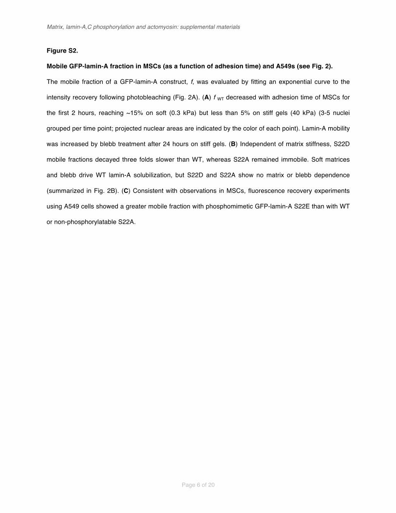
Mobile GFP-lamin-A fraction in MSCs (as a function of adhesion time) and A549s (see Fig. 2).
The mobile fraction of a GFP-lamin-A construct, f, was evaluated by fitting an exponential curve to the
intensity recovery following photobleaching (Fig. 2A). (A) f WT decreased with adhesion time of MSCs for
the first 2 hours, reaching ~15% on soft (0.3 kPa) but less than 5% on stiff gels (40 kPa) (3-5 nuclei
grouped per time point; projected nuclear areas are indicated by the color of each point). Lamin-A mobility
was increased by blebb treatment after 24 hours on stiff gels. (B) Independent of matrix stiffness, S22D
mobile fractions decayed three folds slower than WT, whereas S22A remained immobile. Soft matrices
and blebb drive WT lamin-A solubilization, but S22D and S22A show no matrix or blebb dependence
(summarized in Fig. 2B). (C) Consistent with observations in MSCs, fluorescence recovery experiments
using A549 cells showed a greater mobile fraction with phosphomimetic GFP-lamin-A S22E than with WT
or non-phosphorylatable S22A.

Matrix, lamin-A,C phosphorylation and actomyosin: supplemental materials
Page 7 of 20!!
Figure S3.
Slice-A (57 - 80 kDa); 57.8 % sequence coverage; false positive rate = 6.65 %
#pep: 30
#pep: 9
#pep: 15
60
40
20
0 60 - 110 40 - 60 < 40
Control RO3306
0
1
2
100
200
LMNA
(A.U
.)
LMNA pSer22 Cleaved (mAb) Cleaved (pAb)LMNA LMNA
Cont
rol
RO33
06Ki
nase
inhib
itor (
3.5
mM
>>
K
)
iCDK1
0.6
0.4
0.2
0.0
1.0
0.8
0.6
0.4
0.2
0.0
10 min. 45 min.
pSer
22 D
ensit
y (AU
)
Cross-section through immunoblot (AU)Cross-section through immunoblot (AU)
pSer
22 D
ensit
y (AU
)
Lamin-A
Lamin-ALamin-C
Degradation products Degradation products
pSer22 (intact lamin)Total pSer22 = 21 % pSer22 (intact lamin)
Total pSer22 = 17 %
Nucle
ar a
rea
(µm
²)
0
A B
C
E
F G
Slice-D (0 - 25 kDa);10.1% sequence coverage;false positive rate = 7.98 %1 11 21 31 41 51METPSQRRATRSGAQASSTPLSPTRITRLQEKEDLQELNDRLAVYIDRVRSLETENAGLR61 71 81 91 101 111LRITESEEVVSREVSGIKAAYEAELGDARKTLDSVAKERARLQLELSKVREEFKELKARN121 131 141 151 161 171TKKEGDLIAAQARLKDLEALLNSKEAALSTALSEKRTLEGELHDLRGQVAKLEAALGEAK181 191 201 211 221 231KQLQDEMLRRVDAENRLQTMKEELDFQKNIYSEELRETKRRHETRLVEIDNGKQREFESR241 251 261 271 281 291LADALQELRAQHEDQVEQYKKELEKTYSAKLDNARQSAERNSNLVGAAHEELQQSRIRID301 311 321 331 341 351SLSAQLSQLQKQLAAKEAKLRDLEDSLARERDTSRRLLAEKEREMAEMRARMQQQLDEYQ361 371 381 391 401 411ELLDIKLALDMEIHAYRKLLEGEEERLRLSPSPTSQRSRGRASSHSSQTQGGGSVTKKRK421 431 441 451 461 471LESTESRSSFSQHARTSGRVAVEEVDEEGKFVRLRNKSNEDQSMGNWQIKRQNGDDPLLT481 491 501 511 521 531YRFPPKFTLKAGQVVTIWAAGAGATHSPPTDLVWKAQNTWGCGNSLRTALINSTGEEVAM541 551 561 571 581 591RKLVRSVTVVEDDEDEDGDDLLHHHHGSHCSSSGDPAEYNLRSRTVLCGTCGQPADKASA601 611 621 631 641 651SGSGAQVGGPISSGSSASSVTVTRSYRSVGGSGGGSFGDNLVTRSYLLGNSSPRTQSPQN661CSIM
1 ~10
Slice-C (25 - 40 kDa);48.6 % sequence coverage;false positive rate = 7.53"%
Slice-B (40 - 57 kDa);25.5 % sequence coverage;false positive rate = 5.53 %1 11 21 31 41 51METPSQRRATRSGAQASSTPLSPTRITRLQEKEDLQELNDRLAVYIDRVRSLETENAGLR61 71 81 91 101 111LRITESEEVVSREVSGIKAAYEAELGDARKTLDSVAKERARLQLELSKVREEFKELKARN121 131 141 151 161 171TKKEGDLIAAQARLKDLEALLNSKEAALSTALSEKRTLEGELHDLRGQVAKLEAALGEAK181 191 201 211 221 231KQLQDEMLRRVDAENRLQTMKEELDFQKNIYSEELRETKRRHETRLVEIDNGKQREFESR241 251 261 271 281 291LADALQELRAQHEDQVEQYKKELEKTYSAKLDNARQSAERNSNLVGAAHEELQQSRIRID301 311 321 331 341 351SLSAQLSQLQKQLAAKEAKLRDLEDSLARERDTSRRLLAEKEREMAEMRARMQQQLDEYQ361 371 381 391 401 411ELLDIKLALDMEIHAYRKLLEGEEERLRLSPSPTSQRSRGRASSHSSQTQGGGSVTKKRK421 431 441 451 461 471LESTESRSSFSQHARTSGRVAVEEVDEEGKFVRLRNKSNEDQSMGNWQIKRQNGDDPLLT481 491 501 511 521 531YRFPPKFTLKAGQVVTIWAAGAGATHSPPTDLVWKAQNTWGCGNSLRTALINSTGEEVAM541 551 561 571 581 591RKLVRSVTVVEDDEDEDGDDLLHHHHGSHCSSSGDPAEYNLRSRTVLCGTCGQPADKASA601 611 621 631 641 651SGSGAQVGGPISSGSSASSVTVTRSYRSVGGSGGGSFGDNLVTRSYLLGNSSPRTQSPQN661CSIM
1 ~ 6
1 11 21 31 41 51METPSQRRATRSGAQASSTPLSPTRITRLQEKEDLQELNDRLAVYIDRVRSLETENAGLR61 71 81 91 101 111LRITESEEVVSREVSGIKAAYEAELGDARKTLDSVAKERARLQLELSKVREEFKELKARN121 131 141 151 161 171TKKEGDLIAAQARLKDLEALLNSKEAALSTALSEKRTLEGELHDLRGQVAKLEAALGEAK181 191 201 211 221 231KQLQDEMLRRVDAENRLQTMKEELDFQKNIYSEELRETKRRHETRLVEIDNGKQREFESR241 251 261 271 281 291LADALQELRAQHEDQVEQYKKELEKTYSAKLDNARQSAERNSNLVGAAHEELQQSRIRID301 311 321 331 341 351SLSAQLSQLQKQLAAKEAKLRDLEDSLARERDTSRRLLAEKEREMAEMRARMQQQLDEYQ361 371 381 391 401 411ELLDIKLALDMEIHAYRKLLEGEEERLRLSPSPTSQRSRGRASSHSSQTQGGGSVTKKRK421 431 441 451 461 471LESTESRSSFSQHARTSGRVAVEEVDEEGKFVRLRNKSNEDQSMGNWQIKRQNGDDPLLT481 491 501 511 521 531YRFPPKFTLKAGQVVTIWAAGAGATHSPPTDLVWKAQNTWGCGNSLRTALINSTGEEVAM541 551 561 571 581 591RKLVRSVTVVEDDEDEDGDDLLHHHHGSHCSSSGDPAEYNLRSRTVLCGTCGQPADKASA601 611 621 631 641 651SGSGAQVGGPISSGSSASSVTVTRSYRSVGGSGGGSFGDNLVTRSYLLGNSSPRTQSPQN661CSIM
1 ~10
1 11 21 31 41 51METPSQRRATRSGAQASSTPLSPTRITRLQEKEDLQELNDRLAVYIDRVRSLETENAGLR61 71 81 91 101 111LRITESEEVVSREVSGIKAAYEAELGDARKTLDSVAKERARLQLELSKVREEFKELKARN121 131 141 151 161 171TKKEGDLIAAQARLKDLEALLNSKEAALSTALSEKRTLEGELHDLRGQVAKLEAALGEAK181 191 201 211 221 231KQLQDEMLRRVDAENRLQTMKEELDFQKNIYSEELRETKRRHETRLVEIDNGKQREFESR241 251 261 271 281 291LADALQELRAQHEDQVEQYKKELEKTYSAKLDNARQSAERNSNLVGAAHEELQQSRIRID301 311 321 331 341 351SLSAQLSQLQKQLAAKEAKLRDLEDSLARERDTSRRLLAEKEREMAEMRARMQQQLDEYQ361 371 381 391 401 411ELLDIKLALDMEIHAYRKLLEGEEERLRLSPSPTSQRSRGRASSHSSQTQGGGSVTKKRK421 431 441 451 461 471LESTESRSSFSQHARTSGRVAVEEVDEEGKFVRLRNKSNEDQSMGNWQIKRQNGDDPLLT481 491 501 511 521 531YRFPPKFTLKAGQVVTIWAAGAGATHSPPTDLVWKAQNTWGCGNSLRTALINSTGEEVAM541 551 561 571 581 591RKLVRSVTVVEDDEDEDGDDLLHHHHGSHCSSSGDPAEYNLRSRTVLCGTCGQPADKASA601 611 621 631 641 651SGSGAQVGGPISSGSSASSVTVTRSYRSVGGSGGGSFGDNLVTRSYLLGNSSPRTQSPQN661CSIM
1 ~17
2.0
1.5
1.0
0.5
0.0
Ion
curre
nt(n
orm
alize
d to
cont
rol)
Slice-D(0 - 25 kDa)
Slice-C(25 - 40 kDa)
Slice-B(40 - 57 kDa)
Slice-A(57 - 80 kDa)
Control3.5 mM RO3306
p < 0.02p < 0.08
N
NC
C
P
P
P
P Ig
Ig
Head TailCoiled-coils
N
NC
C
P
P
P
P Ig
Ig
Head TailCoiled-coils
N
NC
C
P
P
P
P Ig
Ig
Head TailCoiled-coils
C
C
PP
PP Ig
Ig
Tail
N
NC
C
P
P
P
P Ig
Ig
Head TailCoiled-coils
N
N
PP
PP
Head Coiled-coils
D
Molecular weight (kDa)
% S
eq. c
over
age
A549 cells
0 10 200.0
0.5
1.0
1.5 ControlRO3306
H
MSCspS
er22
/ LM
NA (A
.U.)
AreaTension ~ Lamin-A,Cm n

Matrix, lamin-A,C phosphorylation and actomyosin: supplemental materials
Page 8 of 20!!
Figure S3.
Densitometry of lamin pSer22 western blots (see Figs. 3A, B).
(A, B) Mesenchymal stem cells (MSCs) were lysed following 10 or 45 minutes in suspension and
analyzed by immunoblotting against lamin-A,C and pSer22 (Fig. 3A). Densitometry analysis showed that
the majority of the phosphorylated lamin-A,C was fragmented below the expected molecular weight (MW;
75 kDa for lamin-A; 65 kDa for lamin-C).
Sequence coverage of lamin-A,C fragments (see Figs. 3C, D).
(C) The MSC lysate was separated by SDS-PAGE and analyzed in discrete MW ranges by MS (Fig. 3B),
confirming that lamin fragments were found at lower MW. While peptide coverage clearly extended across
all weight ranges, slice-D (< 25 kDa) showed an enrichment of tail-domain and Ig-fold peptides whereas
slice-B (40-57 kDa) showed head and coiled-coil rod domains. Relative coverage intensity (i.e. signal
strength of tryptic peptides) is indicated by the color of the sequence. Sites of phosphorylation detected by
mass spectrometry are highlighted in yellow. (D) The presence of lamin-A,C fragments was also
confirmed by MS in lower MW bands of A549 cell lysates. (E) MSCs were treated with a kinase-inhibitor
(RO3306) and compared with non-treated control cells, with analysis performed by MS (with MW bands
as shown in Fig. 3B). Changes in ion current of lamin-A,C peptides were assessed between samples.
Enrichment of intermediate MW lamin fragments in RO3306-treated cells may be indicative of a
phosphorylation event that is necessary for the progression of lamin cleavage.
Image analysis of cleaved lamin staining during inhibition of phosphorylation (see Figs. 3F, G).
(F) Representative images showing fixed MSCs, with and without kinase inhibitor RO3306 treatment, with
immunostaining against lamin-A,C, pSer22 and cleaved lamin-A with either a monoclonal or a polyclonal
antibody (data summarized in Figs. 3F, G). (G) Image analysis showed that lamin-A,C levels were ~50%
higher and the projected area of the nucleus larger in drug-treated MSCs relative to non-treated cells,
reminiscent of a highly contractile phenotype (n = 50-51 cells per group). (H) Cell-by-cell analysis of S22
phosphorylation in MSCs treated with kinase inhibitor RO3306 (Figs. 3F, G; S3F, G), plotted as a function
of nuclear tension with hyperbolic-like fit consistent with the Mechanobiological Gene Circuit (MGC) model
(Figs. 1F-H, 4E).

Matrix, lamin-A,C phosphorylation and actomyosin: supplemental materials
Page 9 of 20!!
Figure S4.
WT Lamin-A S22A Lamin-A S22E Lamin-A0
10
20
Hoec
hst D
NA st
ain (A
.U.) No significant differences
WT Lamin-A S22A Lamin-A S22E Lamin-A0
100
200
Proje
cted
nucle
ar a
rea
(µm
) No significant differences2
C D
Score Molecular function Celluar component7.1 Cytoskeletal --> Actin filament binding Cytoskeleton --> Actin
5.7 Cytoskeletal protein binding Actin cytoskeleton, non-membrane-bound organell
4.7 Focal adhesion, cell junction
3.6 Cell cortex --> Cortical cytoskeleton
3.4 Calponin-like actin-binding
2.8 Actin-binding Calponin-like actin-binding
2.6 Microtubule cytoskeleton, centrosome
2 Muscle myofibril, sarcomere
1.8 Leukocyte transendothelial migration Adherens junction
1.8 Regulation of actin cytoskeleton Focal adhesion
1
0.8
0.7
Biological process GenesActin filament bundle formation LIMA1, EZR, ACTN4, FSCN1, ACTN1,…
PPP4R2, LIMA1, TUBB2A, ARPC4,TPM2, …
LIMA1, SYT11, NEXN, PERP, TES, VCL,…
ACTB, EZR, ACTN4, LASP1, MYH9, DSTN,…
Actin filament cytoskeleton organization TAGLN, CNN2, CNN1,…
ACTN4, ACTN1, FLNA,…
PPP4R2, NEDD1, KRT18, NDEL1, CALM3,…
ANK2, ACTN1, TPM2,…
Negative regulation of cell motion ACTB, ACTN4, ACTN1, VCL,…
Calcium binding FLG, CALM3, ACTN1,…
Regulation of kinase activity SPRY2, TRIB2, DUSP6
Response to hormone stimulus PLA2G4A, MGP, GNG11
Positive regulation of apoptosis PLA2G4A, TP53INP1, PHLDA1
DOW
NUP
A
B
20 µmHoechst GFP-WT-lamin-A LMNA Myosin-IIA

Matrix, lamin-A,C phosphorylation and actomyosin: supplemental materials
Page 10 of 20!!
Figure S4.
GO-term analysis shows that lamin-A,C KD down-regulates actin cytoskeletal genes (see Figs.
4A-C).
(A) Gene ontology term analysis of the entire transcriptome shows that the cytoskeleton is broadly
affected by lamin-A,C knockdown, consistent with perturbation to the serum response factor (SRF)
pathway. The ten most significant changes, as evaluated by DAVID bioinformatics resources [S3], are
instances of down-regulation, with seven of the ten (including the top three) related to the actomyosin
cytoskeleton.
Lamin-A phosphomutations do not significantly affect chromatin or nuclear spreading in A549s
(see Figs. 4D, E).
(B) Endogenous lamin-A,C was knocked down in A549 cells in conjunction with expression of GFP-lamin-
A phosphomutant constructs (WT, S22A and S22E) in order to assess the effect on myosin-IIA.
Representative images showed the expected localization of GFP-lamin-A at the nuclear envelope.
Quantitative image analysis showed that neither Hoechst DNA staining (C) or nuclear spread area (D)
were significantly altered by lamin-A phosphomutant expression (n = 70-113 cells per group).

Matrix, lamin-A,C phosphorylation and actomyosin: supplemental materials
Page 11 of 20!!
Supplemental materials and methods
Isolation of fresh MSCs from bone marrow
Bone marrow aspirates were obtained from posterior iliac crest of anonymous human donors (University
of Pennsylvania Stem Cell Core, with Institutional Review Board approval) under the procedures and
regulations defined by the Helsinki agreement. Mono-nucleated cells (MNCs) were obtained using a Ficoll
density gradient (Ficoll-Paque PLUS, GE Healthcare) and depleted from CD34-positive cells by a micro-
bead kit (Direct CD34 Progenitor Cell Isolation Kit, Miltenyi Biotec) and screened by automated cell
separation (AutoMacs, Miltenyi Biotec) according to manufacturer’s protocols. MNCs were re-suspended
in 10% FBS (Sigma-Aldrich) and 1% penicillin/streptomycin antibiotics (P/S, GE Healthcare)
supplemented low-glucose basal medium (DMEM, Life Technologies). Typically, MNCs were seeded at
10-100k cells/cm² in standard tissue-culture plastic flasks and incubated at 37 °C and 5% CO2 humidified
conditions. Cells were thoroughly rinsed in PBS x 3 after 24 hrs to remove non-adherent cells. Fibroblastic
colony forming unit (CFU) stromal cells appeared within 3-4 days and cells expansion included medium
exchange every 4 days. Cells confluence was maintained < 80% by passaging cells and re-seeding at >
50% confluence. Expression of stromal stem cells markers (CD105, CD166, CD44, CD90) and the lack of
hematopoietic markers (CD45-RA, CD34) was verified by flow cytometry (data not shown). Differentiation
capacity towards fat and bone was verified by adipogenic and osteogenic induction media (R&D: following
manufacturer’s protocols; data not shown). Fresh human CD34-positive cells were obtained by cell
sorting from the mono-nucleated fraction of donor bone-marrow cells. Cells were cultured for seven days
with stem cells factor (SCF) and thrombopoietin (TPO) for four days.
Preparation of soft and stiff hydrogel substrates
Adapted from [S4]. Glass cover slips (thickness #1.5, Fisher Scientific) were placed in boiling ethanol for
10 min, rinsed in distilled water (DW) and immersed in RCA (DW, hydrogen peroxidase (30%, Fisher
Scientific), ammonium hydroxide (30%, Fisher Scientific) at 3:1:1 v/v) at 80 °C for 10 min and rinsed in
DW. To remove water traces, glasses were rinsed in ethanol and then in chloroform and silanized in 0.1%
allyltrichlorosilane (ATCS, Aldrich) in chloroform (Fisher Scientific) for 30 min. Silanized glasses were then
Matrix, lamin-A,C phosphorylation and actomyosin: supplemental materials
Page 12 of 20!!
rinsed in chloroform, ethanol and DW and dried under vacuum. PA gel precursors were prepared by
mixing acrylamide (AA, 40%, Sigma) and N,N’-methylenebisacrylamide (bis-AA 1.5% w/v in DW, Sigma)
in PBS (Sigma). Gelation was initiated by adding 0.1% v/v tetramethylethylenediamine (TEMED, Sigma)
and 0.1% w/v ammonium persulfate (Sigma) to gel precursor just before placing it at the center of the
silanized cover slips and covering with RCA-treated glasses. Gels were allowed to polymerize while
covalently binding the silanized glasses for 30-60 min. Non-silanized glasses were gently removed after
immersing in PBS for 1-2 hours. Nominal gel elasticity was specified by varying acrylamide and cross-
linker concentrations as calibrated by desktop rheometer. Gels were immersed in 10 mg/ml sulfo-
SANPAH (Fisher Scientific) in 50 mM, pH 8.5 HEPES and reacted under 365 nm i-line exposure for 10
min. Collagen was mixed in 0.1 M acetic acid (Fisher Scientific) at equal volume and in 50mM, pH 8.5
HEPES to reach 0.2 mg/ml final concentration. Gels were immersed with collagen while agitated
overnight at 37 °C. Prior to seeding cells, gels were UV-sterilized (cell culture hood UV light source) for
three hours. During all preparation steps, gels were maintained in a hydrated state.
siRNA and shRNA knockdown of MYH9, LMNA; myosin-IIA and kinase inhibitors
All siRNAs used in this study were purchased from Dharmacon. Cells were passaged > 24 hours prior to
transfection were and incubated with a complex of siRNA (30 nM; siLMNA: 5’-
GGUGGUGACGAUCUGGGCU-3’; siMYH9: 5’-GGCCAAACCUGCCGAAUAAAUU-3’ with complement
sequence 5’-UUUAUUCGGCAGGUUUGGCCUU-3’ or scrambled siRNA siGENOME non-targeting siRNA
#1 (Thermo Fisher Scientific)) and 1 µg/mL Lipofectamine 2000 according to the manufacturer’s
instructions for 24 hours (in low glucose DMEM with 10% FBS). shLMNA construct (Sigma,
TRCN0000061833: 5’-CGACTGGTGGAGATTGACAAT-3’) was packed into a lentiviral delivery system,
transduced into A549s by 1 µg/mL Polybrene for 72 hours and selected with 2 µg/mL puromycin. Racemic
blebbistatin (EMD) was used at 30 µM (24 hr treatment). Kinase inhibitor RO3306 (Adipogen
International) was used at 3.5 µM for no longer than 6 hours.

Matrix, lamin-A,C phosphorylation and actomyosin: supplemental materials
Page 13 of 20!!
Transfection and transductions of lamin-A, myosin-IIA and respective phosphomimetic constructs
For MSCs: a construct expressing GFP-lamin-A under the EF1-alpha promoter [S5] was packed into a
lentiviral delivery system. Cells were transduced at MOI 50 and evaluated for survival and proliferation
(data not shown). The fraction of GFP-lamin-A positive cells prior to experiment was ~ 50%. GFP-positive
cells were seeded on 6-well plates at very low density (less than one cell per 4X objective field of
view). Colonies of GFP-lamin-A expressing MSCs were isolated using a cloning cylinder (Bel-Art
Products, Pequannock, NJ), trypsinized and further expanded for experiments. Lamin-A constructs were
transfected via electroporation following manufacturer’s protocols (MSCs kit, Nucleofector; Lonza);
myosin-IIA constructs were transfected using Lipofectamine LTX with Plus reagent (Invitrogen), using 0.5
g DNA per well of a 6-well plate. For A549s: Transfections were performed with Lipofectamine LTX/Plus
reagent (Invitrogen), as above. Transfection levels were similar across all constructs (within 20-30%)
based on GFP intensities and densitometry of immunoblots (not shown). Phosphomutant constructs were
packed into a lentiviral delivery system and transduced into lamin-A knockdown A549s. Transduction
efficiencies ranged from 60-90%.
Plasmid constructs for WT GFP-myosin-IIA were obtained from Addgene. GFP-lamin-A and
S1943D GFP-myosin-IIA plasmids were generous gifts from C. H. June (University of Pennsylvania, PA),
D. M. Gilbert (State University of New York, Syracuse) and A. Bresnick (Albert Einstein College of
Medicine, Bronx, NY). GFP-Lamin-A S22A, S22D and S22E plasmids were constructed by standard site
directed mutagenesis (Stratagene).
Immunofluorescence imaging
Cells were fixed with 3.7% formaldehyde (Sigma-Aldrich) in PBS for 10 min at RT followed by PBS
washing 2X for 5 min. Blocking and primary antibody staining was performed in 1% BSA in PBS. Primary
antibody concentrations ranged between 1/300 – 1/500, depending on the stock concentration, and all
primary antibodies were incubated at RT for 2 h or overnight at 4°C. All donkey secondary antibodies
(Alexa Fluor dyes 488, 564, and 647) were stained for 1-2 hours at RT at 1:500 dilution in PBS and

Matrix, lamin-A,C phosphorylation and actomyosin: supplemental materials
Page 14 of 20!!
TRITC-phalloidin (Sigma-Aldrich) was used at a concentration of 100 ng/ml. Imaging for quantitative
immunofluorescence of lamin-A, myosin-IIA and related phosphorylation-specific antibodies was
performed using an inverted microscope (IX-71; Olympus) with either 20X (Olympus, NA-0.75) or 40X
(NA-0.60) objectives, and a cooled CCD camera (Cascade; Photometrics) and image acquisition
performed with Image Pro software (Media Cybernetics). Fixed cells were immuno- and histochemically-
stained using the following antibodies and reagents: Myosin-IIA (mouse), monoclonal HSP90AB1
(Abcam); lamin-A,C pSer22, monoclonal cleaved lamin-A,C (Cell Signaling); polyclonal myosin-IIA,
monoclonal lamin-A,C (mouse), polyclonal lamin-A,C (goat), polyclonal lamin-B1,2 (mouse), polyclonal
cleaved lamin-A,C (Santa Cruz); polyclonal myosin-IIA, Phalloidin (Sigma). Fixed cells were mounted in
mounting medium (Axell). To prevent volume distortions, samples prepared for confocal z-stacks
quantification were not mounted. Prior to each experiment, lamp intensity and field of view homogeneity
were calibrated pixel by pixel, relative to a fluorescent plastic standard. Image analysis was performed
with in-house MatLab code that included background subtraction, cell and nuclear registration and
intensity integration, morphological and statistical analyses.
Confocal microscopy
Laser scanning confocal fluorescence microcopy was carried out using the following systems: Leica
Microsystems TCS SP8, 63×/ NA1.4 oil immersion objective (Fig. 2C) and Olympus Fluoview FV1000,
150X/NA1.45 or 60X/NA1.2 oil immersion objectives (Fig. 1A). Nuclear cross-sections orthogonal to the
substrate were generated from 0.21 µm Z-stacks and constructed using Fiji [S6].
Fluorescence recovery after photobleaching (FRAP)
Confocal time-lapse imaging was acquired at 37 °C with 5% CO2 in a humidified chamber with an inverted
spinning-disk microscope (IX-81; Olympus) with a 14-bit high-resolution charge-coupled device (CCD)
camera (HQ2; Photometrics) and MetaMorph software (Molecular Devices). Time lapse images were
acquired with a 40X (Olympus, NA-0.75) and 60X water immersion lens (Olympus, NA-1.2), every 20 sec
for 5-10 min after photobleaching. Cells were imaged in phenol-red free low glucose medium (DMEM, Life

Matrix, lamin-A,C phosphorylation and actomyosin: supplemental materials
Page 15 of 20!!
Technologies) with 10% FBS and 1% penicillin/streptomycin. Image sequences were analyzed using
MatLab custom designed code to quantify fluorescence recovery kinetics. After the first day cells were
treated with 30 µM racemic blebbistatin (EMD; 24 hrs). Blebbistatin was washed out prior to FRAP in
order to prevent light-induced toxicity.
Immunoblotting
Cells were trypsinized, pelleted and stored at -20 °C until analysis. Pellets were thawed and re-
suspended in 1X LDS lysis buffer supplemented with 1% protease and 1% phosphatase inhibitors and
sonicated on ice (3 x 15 x 1 s pulses, intermediate power setting). After resting for 30 min on ice, samples
were denatured at 80 °C with 0.5% �-mercaptoethanol v/v for 10 min. Samples were loaded onto bis-Tris
4-12% gradient gels for electrophoresis (100 V x 10 min; 160 V x 55 min) and then transferred (iBlot; Life
Technologies: settings P3, 7 min) to blotting membrane. Band intensities were quantified using
Fiji/ImageJ, relative to local background levels flanking the specific bands.
Quantitative, label-free mass spectrometry
SDS-PAGE gels (NuPAGE 4-12% Bis-Tris, Invitrogen) were run at 100 V for 10 min and 160 V for 25 min.
Gel sections were washed (50% 0.2 M ammonium bicarbonate (AB) solution, 50% acetonitrile (ACN), 30
min at 37 °C), dried by lyophilization, incubated with a reducing agent (20 mM tris(2-
carboxyethyl)phosphine (TCEP) in 25 mM AB solution at pH 8.0, 15 min at 37 °C) and alkylated (40 mM
iodoacetamide (IAM) in 25 mM AB solution at pH 8.0, 30 min at 37 °C). The gel sections were dried by
lyophilization before in-gel trypsinization (20 µg/mL sequencing grade modified trypsin in buffer as
described in the manufacturer’s protocol (Promega), 18 hr at 37 °C with gentle shaking). The resulting
solutions of tryptic peptides were acidified by addition of 50% digest dilution buffer (60 mM AM solution
with 3% methanoic acid).
Peptide separations (5 µL injection volume) were performed on 15-cm PicoFrit column (75 µm
inner diameter, New Objective) packed with Magic 5 µm C18 reversed-phase resin (Michrom

Matrix, lamin-A,C phosphorylation and actomyosin: supplemental materials
Page 16 of 20!!
Bioresources) using a nanoflow high-pressure liquid chromatography system (Eksigent Technologies),
which was coupled online to a hybrid LTQ-Orbitrap XL mass spectrometer (Thermo Fisher Scientific) via
a nanoelectrospray ion source. Chromatography was performed with Solvent A (Milli-Q water with 0.1%
formic acid) and Solvent B (acetonitrile with 0.1% formic acid). Peptides were eluted at 200 nL/min for 3–
28% B over 42 min, 28–50% B over 26 min, 50–80% B over 5 min, 80% B for 4.5 min before returning to
3% B over 0.5 min. To minimize sample carryover, a fast blank gradient was run between each sample.
The LTQ-Orbitrap XL was operated in the data-dependent mode to automatically switch between full scan
MS (m/z = 350-2000 in the Orbitrap analyzer (with resolution of 60,000 at m/z 400) and the fragmentation
of the six most intense ions by collision-induced dissociation in the ion trap mass analyzer.
Raw mass spectroscopy data was processed using Elucidator (version 3.3, Rosetta Biosoftware).
The software was set up to align peaks in data from samples derived from corresponding molecular
weight regions of the 1D gels. Peptide and protein annotations were made using SEQUEST (version 28,
Thermo Fisher Scientific) with full tryptic digestion and up to 2 missed cleavage sites. Peptide masses
were selected between 800 and 4500 amu with peptide mass tolerance of 1.1 amu and fragment ion
mass tolerance of 1.0 amu. Peptides were searched against a database compiled from UniRef100
(November 2010) mouse, plus contaminants and a reverse decoy database. A deltaCn of 0.01 and mass
error limit of 20 ppm was used, resulting in a false positive rate of ~10%. In these experiments, only
proteins detected with three or more peptides were considered. The peptide database was modified to
search for alkylated cysteine residues (monoisotopic mass change, � = +57.021 Da) and oxidized
methionine (� = +15.995 Da). In proteomic profiling experiments, we also considered the acetylation of
lysine (� = +42.011 Da), methylation of lysine and arginine (� = +14.016 Da), and phosphorylation of
serine, tyrosine, threonine, histidine and aspartate (� = +79.966 Da). Ion currents of modified peptides
were summed with their parent peptide. Peptides derived from trypsin or keratin were considered to be
contaminants and were not used in subsequent calculations. When evaluating total ion current, only
signals from annotated peptides were summed. The Peptide Ratio Fingerprinting algorithm was coded for
Mathematica (version 8, Wolfram Research) and was used for all MS protein quantitation [S7].

Matrix, lamin-A,C phosphorylation and actomyosin: supplemental materials
Page 17 of 20!!
Transcriptional profiling by DNA microarrays
Total RNA was extracted from cells using Trizol and purified by RNeasy (Qiagen) with on-column DNase
digestion according to manufacturer’s protocol. Adherent cells were gently scraped in Trizol. Total RNA
was amplified and converted to cDNA using WT-Ovation Pico kit (NuGen). Fragmented and biotin-
labeled ST-cDNA was generated using WT-Ovation Exon Module (NuGen). Samples were tested with
Human Gene 1.0 ST DNA microarrays (Affymetrix), used according to the manufacturer’s instructions.
Expression data sets were analyzed by standard Robust Multi-array Averaging (RMA) methods.
Micropipette aspiration
Mesenchymal stem cells were harvested from culture, suspended in PBS + 1% BSA and treated in
suspension with 0.5 µg/mL Latrunculin-A at 37 °C for 30 min. Cells were then washed with 1% BSA and
re-suspended in 1% BSA supplemented with 0.2 µg/mL Latrunculin-A. A pulled glass micropipette was
attached to a dual-stage water manometer with reservoirs of adjustable height. Suction was applied by
syringe, and the corresponding pressure was measured by a pressure transducer (Validyne) calibrated
with a mercury U-tube manometer. Pressures for different experiments were < 10 kPa. Images were
acquired using a Nikon Eclipse TE300 inverted microscope using a 60X objective (Nikon, Plan Apo
NA1.4) and a Cascade CCD camera (Roper Scientific). Images were captured every 30 sec over 5 min of
aspiration at constant. Image were analyzed with ImageJ (NIH). Elastic stiffness was calculated according
to previous work [S8]. Specifically, creep compliance ! ! = !!! ∙
!.!∙!!∙∆! was computed per each nucleus as a
function of nuclear extension L at each time point and then fitted by ! ! = ! ∙ !!. Nuclear relaxation times
were computed with a threshold creep compliance overlapping with the dynamic range across conditions.
Systems Mechanobiology Gene Circuit (MGC) for coupled expression of lamin-A,C and myosin-IIA
Lamin-A,C (L) and myosin-IIA (M) message and protein circuitry is schematically presented in Figure 4F.
In particular, expression kinetics of both are described by coupled rate equations for the respective
transcripts (lower case) and proteins (upper case):

Matrix, lamin-A,C phosphorylation and actomyosin: supplemental materials
Page 18 of 20!!
!"!" = !! ∙ ! − !! ∙ !
Eq. 1
!"!" = !! ∙ ! − !! ∙
!!!!!!! + !!!
Eq. 2
!"!" = !! ∙! + !! ∙ ! − !! ∙! Eq. 3
!"!" = !! ∙! − !! ∙
!!!
!!!! +!!! Eq. 4
For simplicity, RNA degradation and translation are assumed linear in transcript concentration.
Lamin-A,C protein positively regulates one of its transcription factors, retinoic acid receptor (RAR), and
myosin-IIA protein positively regulates one of its transcription factors, SRF, so that each enhances its own
transcription (with rate constants !!,!! ). In addition, lamin-A,C protein also enhances myosin-IIA
transcription via the SRF pathway (rate constant !!). Our results indicate a mechanical regulation of
protein phosphorylation, and so we describe lamin-A,C and myosin-IIA protein turnover with suitable Hill
models (rate constants !!, !!). Specifically, lamin-A,C protein turnover is suppressed by myosin-generated
stress with !! = !!!, while myosin-IIA protein turnover depends on matrix elasticity E: !! = !!!!!
.
A combined coordinate to describe nuclear tension and solutions to the MGC model
We sought to estimate a dimensionless measure of nuclear tension, !, in terms of the stiffness of the
nucleus, !!"#, which depended on LMNA, and the nuclear strain, !, which depended on changes in
projected nuclear area, ∆!. In simplest form: !!=!!!!!∙!!~!LMNA!∙∆!!, where ! = 2.5 conforms to scaling
results for semi-flexible polymers [S9] as well as the enhanced nuclear viscosity with lamin-A,C [S10], and
n = 0.5 is consistent with the scaling of strain with changes in nuclear radius (Fig. 1F). When
pSer22/LMNA was plotted against this nuclear tension coordinate using data from analyses of immuno-
fluorescence images (MSCs cultured on soft vs. vs. thin/soft vs. stiff substrate, Fig. 1C; MSCs subjected
to lamin-A,C KD or blebb treatment, Figs 1E; S1F; MSCs treated with kinase inhibitor, Figs. 3F, G; S3F,
G), the data collapsed well onto a hyperbolic-like decay consistent with the MGC model (Figs. 1G, H;
S3H).

Matrix, lamin-A,C phosphorylation and actomyosin: supplemental materials
Page 19 of 20!!
Equations 1-4 of the MGC model were solved numerically at steady state (all derivatives = 0) with
free parameters adjusted to collectively obtain the best agreement with experimental data (fitting was
performed using Mathematica, Wolfram). The rate of protein turnover was modeled as being proportional
to tension per molecule. Cooperative protein turnover was !! = 10.4 and !! = 2 with the half-maximum
effect on myosin-IIA turnover occurring at !! = 5 kPa. As matrix stiffness and cell tension suppressed
protein phosphorylation and turnover, steady state levels monotonically increased with matrix E,
consistent with coupled mechano-regulation of lamin-A and myosin-IIA. Maximal agreement was
achieved with the model’s free parameters set to the following values: !! = 4.43, !! = 10,!! = 2; ! !!! =
1.4, !!! = 1.5; !!! = 0.071, !! = 2.3; !!!! = 3, !! = 10.9; !!! = 0.08, !! = 10.4, !! = 2 (Fig. 4G).

Matrix, lamin-A,C phosphorylation and actomyosin: supplemental materials
Page 20 of 20!!
Supplemental references
S1. Vicente-Manzanares, M., Ma, X.F., Adelstein, R.S., and Horwitz, A.R. (2009). Non-muscle myosin
II takes centre stage in cell adhesion and migration. Nat. Rev. Mol. Cell Biol. 10, 778-790.
S2. Raab, M., Swift, J., Dingal, P.C.D.P., Shah, P., Shin, J.-W., and Discher, D.E. (2012). Crawling
from soft to stiff matrix polarizes the cytoskeleton and phosphoregulates myosin-II heavy chain. J.
Cell Biol. 199, 669-683.
S3. Huang, D.W., Sherman, B.T., and Lempicki, R.A. (2009). Systematic and integrative analysis of
large gene lists using DAVID bioinformatics resources. Nat. Protoc. 4, 44-57.
S4. Buxboim, A., Rajagopal, K., Brown, A.E.X., and Discher, D.E. (2010). How deeply cells feel:
methods for thin gels. J. Phys.-Cond. Mat. 22.
S5. Izumi, M., Vaughan, O.A., Hutchison, C.J., and Gilbert, D.M. (2000). Head and/or CaaX domain
deletions of lamin proteins disrupt preformed lamin A and C but not lamin B structure in
mammalian cells. Mol. Biol. Cell 11, 4323-4337.
S6. Schindelin, J., Arganda-Carreras, I., Frise, E., Kaynig, V., Longair, M., Pietzsch, T., Preibisch, S.,
Rueden, C., Saalfeld, S., Schmid, B., et al. (2012). Fiji: an open-source platform for biological-
image analysis. Nat. Methods 9, 676-682.
S7. Swift, J., Harada, T., Buxboim, A., Shin, J.W., Tang, H.Y., Speicher, D.W., and Discher, D.E.
(2013). Label-free mass spectrometry exploits dozens of detected peptides to quantify lamins in
wildtype and knockdown cells. Nucleus 4, 450-459.
S8. Dahl, K.N., Engler, A.J., Pajerowski, J.D., and Discher, D.E. (2005). Power-law rheology of
isolated nuclei with deformation mapping of nuclear substructures. Biophys. J. 89, 2855-2864.
S9. Gardel, M.L., Shin, J.H., MacKintosh, F.C., Mahadevan, L., Matsudaira, P., and Weitz, D.A.
(2004). Elastic behavior of cross-linked and bundled actin networks. Science 304, 1301-1305.
S10. Swift, J., Ivanovska, I.L., Buxboim, A., Harada, T., Dingal, P.C.D.P., Pinter, J., Pajerowski, J.D.,
Spinler, K.R., Shin, J.-W., Tewari, M., et al. (2013). Nuclear lamin-A scales with tissue stiffness
and enhances matrix-directed differentiation. Science 341, 1240104.


![OPEN ACCESS viruses - Semantic Scholar...III alpha (PIKIII α) binds to and regulates th e phosphorylation status of NS 5A [14]. The phosphorylation state likely plays an important](https://static.fdocuments.us/doc/165x107/60f6ad79a1e0ef6b36059fb3/open-access-viruses-semantic-scholar-iii-alpha-pikiii-binds-to-and-regulates.jpg)