Replication of Epstein–Barr Viral...
14
Replication of Epstein –Barr Viral DNA Wolfgang Hammerschmidt 1 and Bill Sugden 2 1 Department of Gene Vectors, Helmholtz Zentrum Mu ¨nchen, German Research Center for Environmental Health, Marchioninistr. 25, D-81377 Munich, Germany 2 Department of Oncology, McArdle Laboratory for Cancer Research, University of Wisconsin-Madison, Madison, Wisconsin 53706 Correspondence: [email protected] Epstein–Barr virus (EBV) is a paradigm for human tumor viruses: it is the first virus recognized to cause cancer in people; it causes both lymphomas and carcinomas; yet these tumors arise infrequently given that most people in the world are infected with the virus. EBVis maintained extrachromosomally in infected normal and tumor cells. Eighty-four percent of these viral plasmids replicate each S phase, are licensed, require a single viral protein for their synthesis, and can use two functionally distinct origins of DNA replication, oriP, and Raji ori. Eighty- eight percent of newly synthesized plasmids are segregated faithfully to the daughter cells. Infectious viral particles are not synthesized under these conditions of latent infection. This plasmid replication is consistent with survival of EBV’s host cells. Rare cells in an infected population either spontaneouslyor following exogenous induction support EBV’s lytic cycle, which is lethal for the cell. In this case, the viral DNA replicates 100-fold or more, uses a third kind of viral origin of DNA replication, oriLyt, and many viral proteins. Here we shall describe the three modes of EBV’s replication as a function of the viral origins used and the viral and cellular proteinsthat mediate the DNA synthesis from these origins focusing, where practical, on recent advances in our understanding. oriP A single contiguous fragment of EBV DNA with a length of 1.7 kbps supports autono- mous extrachromosomal replication and main- tenance of recombinant plasmids in human cells (Yates 1996) when the Epstein–Barr nuclear protein 1 (EBNA1) is provided in trans (Yates 1996) (Fig. 1). This subgenomic EBV DNA is termed oriP for “origin of plasmid replication.” Similar to the parental EBV genome (Yates 1996), initiation of DNA replication within oriP is synchronized with the timed and strictly regulated replication of chromosomal DNA (Yates 1996; Shirakata et al. 1999), and mediated by the cellular replication machinery acting at oriP . The viral protein EBNA1 binds to oriP site-specifically and recruits the cellular DNA replication machinery (Chaudhuri et al. 2001; Dhar et al. 2001; Schepers et al. 2001; Ritzi et al. 2003). Thus, EBNA1 is essential but not sufficient for oriP’s function. In resting and pro- liferating cells, oriP confers stable extrachromo- somal maintenance of recombinant plasmids, which also relies on EBNA1 in trans independent of its role in DNA replication. An EBV genome Editors: Stephen D. Bell, Marcel Me ´chali, and Melvin L. DePamphilis Additional Perspectives on DNA Replication available at www.cshperspectives.org Copyright # 2013 Cold Spring Harbor Laboratory Press; all rights reserved; doi: 10.1101/cshperspect.a013029 Cite this article as Cold Spring Harb Perspect Biol 2013;5:a013029 1 on June 5, 2018 - Published by Cold Spring Harbor Laboratory Press http://cshperspectives.cshlp.org/ Downloaded from
Transcript of Replication of Epstein–Barr Viral...

Replication of Epstein–Barr Viral DNA
Wolfgang Hammerschmidt1 and Bill Sugden2
1Department of Gene Vectors, Helmholtz Zentrum Munchen, German Research Center for EnvironmentalHealth, Marchioninistr. 25, D-81377 Munich, Germany
2Department of Oncology, McArdle Laboratory for Cancer Research, University of Wisconsin-Madison,Madison, Wisconsin 53706
Correspondence: [email protected]
Epstein–Barr virus (EBV) is a paradigm for human tumor viruses: it is the first virus recognizedto cause cancer in people; it causes both lymphomas and carcinomas; yet these tumors ariseinfrequently given that most people in theworld are infectedwith the virus. EBVis maintainedextrachromosomally in infected normal and tumor cells. Eighty-four percent of these viralplasmids replicate each S phase, are licensed, require a single viral protein for their synthesis,and can use two functionally distinct origins of DNA replication, oriP, and Raji ori. Eighty-eight percent of newly synthesized plasmids are segregated faithfully to the daughter cells.Infectious viral particles are not synthesized under these conditions of latent infection. Thisplasmid replication is consistent with survival of EBV’s host cells. Rare cells in an infectedpopulation either spontaneouslyor following exogenous induction support EBV’s lytic cycle,which is lethal for the cell. In this case, the viral DNA replicates 100-fold or more, uses a thirdkind of viral origin of DNA replication, oriLyt, and many viral proteins. Herewe shall describethe three modes of EBV’s replication as a function of the viral origins used and the viral andcellular proteins that mediate the DNA synthesis from these origins focusing, where practical,on recent advances in our understanding.
oriP
A single contiguous fragment of EBV DNAwith a length of 1.7 kbps supports autono-
mous extrachromosomal replication and main-tenance of recombinant plasmids in human cells(Yates 1996) when the Epstein–Barr nuclearprotein 1 (EBNA1) is provided in trans (Yates1996) (Fig. 1). This subgenomic EBV DNA istermed oriP for “origin of plasmid replication.”Similar to the parental EBV genome (Yates1996), initiation of DNA replication withinoriP is synchronized with the timed and strictly
regulated replication of chromosomal DNA(Yates 1996; Shirakata et al. 1999), and mediatedby the cellular replication machinery acting atoriP. The viral protein EBNA1 binds to oriPsite-specifically and recruits the cellular DNAreplication machinery (Chaudhuri et al. 2001;Dhar et al. 2001; Schepers et al. 2001; Ritziet al. 2003). Thus, EBNA1 is essential but notsufficient for oriP’s function. In resting and pro-liferating cells, oriP confers stable extrachromo-somal maintenance of recombinant plasmids,which also relies on EBNA1 in trans independentof its role in DNA replication. An EBV genome
Editors: Stephen D. Bell, Marcel Mechali, and Melvin L. DePamphilis
Additional Perspectives on DNA Replication available at www.cshperspectives.org
Copyright # 2013 Cold Spring Harbor Laboratory Press; all rights reserved; doi: 10.1101/cshperspect.a013029
Cite this article as Cold Spring Harb Perspect Biol 2013;5:a013029
1
on June 5, 2018 - Published by Cold Spring Harbor Laboratory Press http://cshperspectives.cshlp.org/Downloaded from
with a deletion in EBNA1 has been found only tobe integrated into the cellular chromosome invirus-infected B cells, consistent with its beingunable to be maintained extrachromosomally(Humme et al. 2003).
oriP’s replicator is coincident with or locatednear the origin of DNA replication (Yates 1996)and is operationally termed the “dyad symme-try” (DS) element (Fig. 2A). About 1 kbp up-stream of DS, an array of tandem repeats 650 bpsin total size, termed the “family of repeats” (FR),provides 20 sequence motifs to which EBNA1molecules bind with high affinity. The FR ele-ment does not contribute to DNA synthesisbut is mandatory for the stable maintenanceand nuclear retention of oriP plasmids and ge-nomic EBV DNA.
FR, the Plasmid Maintenance Element
The family of repeats, termed FR, is an array of21 imperfectly conserved, 30 bp, direct repeats(Fig. 2A). EBNA1 binds as homotypic dimersto 20 sequence motifs within FR although onlyseven EBNA1-binding sites are minimally need-ed to function efficiently (Yates 1996). One es-sential role of FR is to prevent plasmid lossfrom proliferating cells. This plasmid mainte-nance/nuclear retention function appears toact by tethering the FR element via EBNA1 tocondensed mitotic chromosomes as they segre-gate during mitosis leading to comigration ofFR-carrying DNA molecules (Yates 1996; Mare-chal et al. 1999; Hung et al. 2001). This tetheringby EBNA1 is accomplished through its carboxyl
A
Id
VX
Tbc
D
G
B
K
RZ e
EL S M a O
PU
Q
F
HYW
W
WW
WW
WW
WW
WC
N
TR
s
oriP
oriLytR
oriLytL
Raji ori
EBNA1
Figure 1. A physical map of the B95-8 laboratory strain of EBV DNA of 165 kbp is shown. The letters on the innersurface of the circle denote the fragments generated by digestion of this DNAwith the BamH1 endonuclease andused to mark its transcripts (Baer et al. 1984). The primary transcript for EBNA1 is denoted by the dashed line.The white box marked TRs indicates the terminal repeats of linear virion DNA that mediate circularization of theviral DNA on infection of cells. The sites for oriP and oriLyt (L, left) are shown with the approximate location ofDNA deleted from the B95-8 strain, present in all other analyzed strains, that contains a second copy of oriLyt(R, right). Raji ori, a region that is defined only approximately maps to sequences spanning the deletion found inthe B95-8 strain.
W. Hammerschmidt and B. Sugden
2 Cite this article as Cold Spring Harb Perspect Biol 2013;5:a013029
on June 5, 2018 - Published by Cold Spring Harbor Laboratory Press http://cshperspectives.cshlp.org/Downloaded from

terminus binding site, specifically to FR and itslinking regions in its amino terminus bindingAT-rich sequences in host chromosomes (seebelow). Live-cell imaging has shown that newlysynthesized daughter molecules remain colocal-ized as pairs until anaphase when 88% segregateone-to-one to daughter cells and the remaining12% segregate randomly (Nanbo et al. 2007).The mechanism of this nonrandom segregation,which must be established in S phase, is notunderstood. FR in conjunction with EBNA1also can act as an EBNA1-dependent, transcrip-tional enhancer (Kennedy and Sugden 2003).
DS, the Origin of DNA Replication
The DS element is an EBNA1-dependent repli-cator for the EBV genome. An approximately120 bp region of oriP containing the minimalreplicator has been termed DS because it con-tains an element of dyad symmetry (DS) 65 bpin length (Fig. 2B). Bidirectional DNA synthesisinitiates at or close to DS (Yates 1996) and DSdoes support EBNA1-dependent DNA replica-tion in the absence of the FR element (Yateset al. 2000). The DS element consists of two pairsof EBNA1-binding sites (Fig. 2B). The EBNA1-
1 40 90
LR1 LR2
Maintenance Replication
b
21 bp
a
c
Rep*4 × 30 bp
Dyad symmetry (DS)Family of repeats (FR)A
B
C
20 × 30 bp
oriP
DNA binding
327
Gly-Ala-repeats DNA-binding/dimerization domainG
ly-A
rg
Gly
-Arg
UR
1
UR
2N
LS
379 459 607 641
AGGACCCTTTTACTAACCCTAATTCGATAGCATATGCTTCCCGTTGGGTAACATATGCTATTGAATTAGGGTTAGT
CTGGATAGTATATACTACTACCCGGGAAGCATATGCTACCCGTTTAGGGTTAAC
Figure 2. Shown are depictions of oriP and EBNA1. (A) oriP consists of multiple sets of binding sites for EBNA1.The family of repeats in the B95-8 strain of EBV has 20 binding sites for EBNA1, which together mediatemaintenance of oriP plasmids. DS has two pairs of binding sites and is the site or close to the site at which DNAsynthesis initiates within oriP. Rep� can substitute for DS functionally but inefficiently. It too contains a pair ofEBNA1-binding sites with the same spacing as each pair in DS. When multimerized eightfold, Rep� functions asefficiently as does DS to support initiation of DNA synthesis. (B) The sequence and spacing of EBNA1-bindingsites in DS of oriP are shown. The two sites of each pair are separated by 21 bps allowing similar contacts on oneface of helical DNA. The nonomer repeats that can bind telomere-associated proteins are underlined anddenoted a, b, and c. (C) The domains of EBNA1 from the B95-8 strain of EBVare represented and are composedof 641 residues. LR1 and LR2 are linking regions rich in Gly-, Arg residues, which when fused to protein DNA-binding domains, can link the DNAs presumably by the AT-hook activities intrinsic to LR1 and LR2. LR1 andLR2 each contain unique sequences, UR1 and UR2. UR1 is involved in transcription regulated by EBNA1; UR2does not have a known function. The Gly, Ala repeats have various activities ascribed to them but little or noeffect on EBNA1’s functions in cell culture. EBNA1 has one identified nuclear localization sequence (NLS) (Yates1996). EBNA1’s carboxy-terminal one-third comprises a dimerization and DNA-binding domain.
Replication of Epstein–Barr Viral DNA
Cite this article as Cold Spring Harb Perspect Biol 2013;5:a013029 3
on June 5, 2018 - Published by Cold Spring Harbor Laboratory Press http://cshperspectives.cshlp.org/Downloaded from

binding sites within one pair are separated by21 bp such that they are in the same helicalphase, which is essential for their acting as areplicator (Bashaw and Yates 2001). The bindingsites within FR for EBNA1 do not require thesame spatially strict arrangement for FR’s func-tions.
It has been shown that one pair of correctlyspaced EBNA1-binding sites is sufficient to re-cruit the origin recognition complex (see below)to the replicator (Julien et al. 2004) indicatingthat the correct binding of EBNA1 is a criticalparameter for origin activation. This minimalreplicator shows only a fraction of the activityof the intact DS element. Auxiliary elements arelikely to contribute to the efficiency of initiationof DNA synthesis, but their genetic dissection isstill incomplete. These auxiliary elements in-clude 14 bp repeats in which are embedded non-amer motifs (Fig. 2B) that bind some telomere-associated proteins (see below). The binding ofthese proteins increases the efficiency of DS as areplicator (Deng et al. 2002, 2003; Lindner et al.2008).
An element termed Rep� of about 300 bps,which is located downstream from DS was iden-tified to be an alternative EBNA1-dependentreplicator with reduced activity (Fig. 2A) (Kirch-maier and Sugden 1998). Although Rep� sup-ports only short-term plasmid replication, itmay contribute to the overall replication effi-ciency of oriP because multimers of Rep� sup-port long-term plasmid replication as efficientlyasdoesoriP (Wangetal.2006). InitiationofDNAreplication within an oriP replicon is likely tobe influenced by its chromatin structure. Nucle-osomes surrounding DS appear to be phasedand modified in a cell-cycle-dependent manner(Wensing et al. 2001; Avolio-Hunter and Frap-pier 2003; Zhou et al. 2005; Tempera and Lieber-man 2010). The extent to which the chromatinstructure controls the replicator or is controlledby the machinery at the replicator is not yet clear.
Proteins that Support the Functions of oriP
oriP consists largely of two stretches of bind-ing sites for EBV’s EBNA1 protein that differin their numbers, spacing, and affinities.
EBNA1 is an essential participant in oriP’s func-tions. EBNA1 can now be viewed as an assemblyof at least four functional elements that supportEBVas an extrachromosomal replicon (Fig. 2C).
The carboxy-terminal quarter of EBNA1encodes two functions essential for all trans-acting phenotypes ascribed to EBNA1. This re-gion provides a structure that forms a dimerthat binds DNA site-specifically. This struc-ture closely resembles that of the dimerizationand DNA-binding domain of the E2 protein en-coded by papillomaviruses (Hegde et al. 1992;Bochkarev et al. 1996). The dimerization andDNA-binding domain of EBNA1 alone bendsDNAs it binds site-specifically as does that ofthe E2 proteins (Hegde et al. 1992; Kim et al.2000; Bashaw and Yates 2001; Wang et al. 2006).It also acts as a dominant negative derivativefor EBNA1’s known functions such that its ex-pression in EBV-positive tumors forces the lossof the virus from cells to reveal the phenotypesprovided them by EBV (Vereide and Sugden2011).
A stretch of glycine, alanine repeats thatspans more than 200 residues in EBNA1 of theB95-8 laboratory strain of EBV has been foundto inhibit antigen processing (Levitskaya et al.1997). This same repeated element has beenfound to limit translation in cis, which mightalso underlie EBNA1’s being poorly recognizedby cytotoxic T-cells (Yin et al. 2003). These re-peats may contribute to EBNA1’s being recog-nized by CD4þ T cells following its autophagicdegradation and the display of its resulting pep-tides on MHC class II molecules (Paludan et al.2005). EBNA-1 is the only viral protein consis-tently expressed in proliferating, infected cells.Itsatypical immune recognition may be essentialfor EBV’s long-term success as a human parasite.
A third functional element of EBNA1 con-sists of a stretch of 25 amino acids termed UR1(unique region 1) positioned between a stretchof glycine, arginine and the glycine, alanine re-peats. It is essential for EBV’s transformation ofhuman B cells (Altmann et al. 2006). UR1 sup-ports EBNA1’s activation of transcription butnot replication (Wu et al. 2002; Kennedy andSugden 2003). This element contains two cys-teines, which allow two monomers of EBNA1 to
W. Hammerschmidt and B. Sugden
4 Cite this article as Cold Spring Harb Perspect Biol 2013;5:a013029
on June 5, 2018 - Published by Cold Spring Harbor Laboratory Press http://cshperspectives.cshlp.org/Downloaded from
bind Zinc coordinately. This zinc coordinationis necessary for EBNA1’s transcriptional trans-activation (Aras et al. 2009) and is reminiscentof the amino terminus of the E2 protein ofpapillomaviruses, which can form intradimerswithin one pair of monomers or interdimersbetween two pairs of monomers (Antson et al.2000).
Another functional moiety of EBNA1 con-sists of two redundant elements termed LR1 andLR2 (linking region 1 and 2). LR1 and LR2 arerich in arginine and glycine residues and wereidentified by their ability to loop regions ofDNA together to which they were bound in cis(Yates 1996) or to link such elements in trans(Yates 1996). Genetic analyses of LR1 and LR2indicate that both contribute to EBNA1’s sup-port of transcription and of replication (Mackeyand Sugden 1999). One mechanism by whichthese linking regions contribute to EBNA1’ssupport of replication of oriP has emerged.Both LR1 and LR2 have AT-hook activities de-rived from their arginine, glycine repeats, whichpromote the association of EBNA1 to AT-richstretches of DNA (Sears et al. 2004). Important-ly, fusions of the cellular protein HMGA1a,which has three AT-hooks to the dimerizationand DNA-binding domain of EBNA1 supportthe replication of oriP plasmids in human cellsstably (Hung et al. 2001; Sears et al. 2003). Fu-sions of EBNA1’s dimerization and DNA-bind-ing domain to HMG1 or EBP2, both of whichappear to lack AT-hooks, fail to support thereplication of oriP stably in human cells (Searset al. 2003, 2004). The AT-hooks of EBNA1’sLR1 and LR2 thus are important for its supportof the maintenance of oriP in proliferating cells.They likely provide EBV another, associatedfunction. The tethering of EBV plasmid DNAsto AT-rich chromosomal sites may insure thatthe viral replicators home to special sites in thenucleus where they can function when their at-tached chromosomal replicators initiate synthe-sis. A third proposed contribution of the linkingregions to replication is their binding G-richRNA and through this binding apparently re-cruiting ORC to DS bound by EBNA1 (Norseenet al. 2008). This role is supported by GST-fu-sions to LR1 and LR2 binding ORC from cell
extracts in an RNAse-susceptible manner (Nor-seen et al. 2008). This described role is perplex-ing though, because it is not easily reconciledwith the requirement for the specific spacing ofEBNA1’s DNA-binding sites needed to recruitORC (Bashaw and Yates 2001; Wang et al. 2006).In addition, Cdc6 binds EBNA1 directly in vitroin an RNAse-resistant manner and contributesto EBNA1’s recruitment of ORC to DS (Mori-yama et al. 2012).
Proteins other than EBNA1 and ORC canbind to or associate with oriP to facilitate itsfunctions. The MCM complex associates withDS in a cell cycle-dependent manner; MCM2, 3,and 7 have been detected at DS by chromatinimmunoprecipitation and to be enriched thereduring G1 and early S phases (Chaudhuri et al.2001; Ritzi et al. 2003). Additional cellular pro-teins have been found to associate with DS de-pendent on EBNA1 in cell extracts and in vivo.The three nonamer repeats (TTAGGGTTA) abut-ting EBNA1-binding sites in DS are similar totelomeric repeats (TTAGGG) and bind proteinsin vivo as measured by genomic footprinting(Fig. 2B) (Niller et al. 1995). DNA-affinity chro-matography with DS coupled to a matrix en-riches for several telomere-associated proteinsonly from EBNA1-positive cells (Deng et al.2002, 2003). These enriched proteins includeTRF1 and TRF2 (telomeric repeat binding fac-tors 1 and 2) and hRap1 (homolog of repressoractivator protein 1), which bind the nonamersdirectly (TRF1 and TRF2) or indirectly by bind-ing TRF2 (Deng et al. 2002, 2003). The rolesthese proteins have in oriP-mediated replicationare not clear, though. Derivatives of oriP withmutated nonamers that fail to bind these telo-mere-associated proteins are maintained stablyin cells at copy numbers one-half of that of wild-type oriP (Deng et al. 2003). One simple modelfor TRF2, is based on its binding ORC1 directly(Atanasiu et al. 2006). Binding of TRF2 to DSshould aid in recruiting ORC to DS. This simplemodel is consistent with analyses of Rep�. Thetwo binding sites for EBNA1 in Rep� do nothave neighboring nonamer sequences and sup-port DNA replication poorly (Wang et al. 2006).When these EBNA1-binding sites are embeddedin neutral lambda DNA and octamerized, they
Replication of Epstein–Barr Viral DNA
Cite this article as Cold Spring Harb Perspect Biol 2013;5:a013029 5
on June 5, 2018 - Published by Cold Spring Harbor Laboratory Press http://cshperspectives.cshlp.org/Downloaded from

support DNA synthesis as efficiently as doeswild-type DS (Wang et al. 2006). This result isconsistent with the finding that the overall avid-ity of EBNA1 for derivatives of DS correlateswith the efficiency of replication of those deriv-atives (Lindner et al. 2008).
Chromatin immunoprecipitation experi-ments have identified additional proteins pre-sent at oriP in cells that likelyaffect its replication.Two proteins implicated in yeast in stabilizingreplication forks, Timeless (Tim) and Timelessinteracting protein, have been detected there(Dheekollu and Lieberman 2011). InhibitingTim leads to the accumulation of linear EBVDNA in cells, consistent with these proteins sta-bilizing replication forks at oriP (Dheekollu andLieberman 2011). The deubiquitylating enzyme,USP7, binds EBNA1 in vitro and localizes to oriPin vivo (Sarkari et al. 2009). This association af-fects the levels of monoubiquitylated histoneH2B in the vicinity of oriP and is thought to af-fect transactivation of transcription by EBNA1bound to FR (Sarkari et al. 2009).
Raji ori, AN ALTERNATE LICENSEDORIGIN OF EBV
Although DS of oriP was the only origin ofplasmid DNA synthesis identified by screeningcloned fragments of EBV DNA for “ARS” activ-ity in EBV-positive cells (Yates 1996), work ofCarl Schildkraut and his colleagues has identi-fied a new class of origins that function in ex-trachromosomal EBV (Yates 1996; Norio andSchildkraut 2004). They studied replicative in-termediates of EBV DNA in Raji cells with twodimensional gel analyses and showed that DNAsynthesis in Raji EBV often originated 25 kbpaway from its oriP (Fig. 1) (Yates 1996). Multipleobservations indicate that this origin, whichwe shall refer to as “Raji ori,” is akin to chromo-somal origins consisting of “zones of replica-tion” such as that in the DHFR locus, but Rajiori functions in an extrachromosomal replicon.
Raji ori can support DNA synthesis effi-ciently and is not unique to the EBV strain inRaji cells. EBV DNA synthesis has been charac-terized in Raji cells and in the Mutu1 cell lineusing “single molecule analysis of replicated
DNA” (Norio and Schildkraut 2004). This ap-proach has revealed that DNA synthesis canoriginate throughout much or all of EBV DNAin both Raji cells and Mutu1 cells, that initiationis most common at Raji ori in Raji cells, and isless frequent but does occur in the Raji ori regionof the EBV DNA in Mutu1 cells. The frequencyof initiation within Raji ori in Raji cells does notreflect a genetic defect in the DS element of itsoriP. This DS has been isolated, sequenced, andfound not to have significant differences fromthe wild type sequence (Koons et al. 2001). DShas been deleted from a third strain of EBV, therecombinant virus introduced into an EBV-neg-ative, B-cell line, and replicative intermediatesanalyzed by two dimensional gel analysis. TheRaji ori of this recombinant strain was found tosupport the initiation of DNA synthesis effi-ciently, although other regions showed the pres-ence of bubble arcs too (Norio et al. 2000).
Raji ori shares at least two properties withsome cellular origins. First, it is licensed. TheEBV DNA in Raji cells was studied in densi-ty shift experiments and shown to incorporateBrdU at the same rate as did host chromosomalDNA showing that EBV DNA replicates semi-conservatively, once per cell cycle in these cells(Yates 1996), consistent with its being licensed.Second, Raji ori also appears to have multiplesites at which DNA synthesis can initiate (Yates1996) as has been found, for example, with thecellular DHFR locus (Kobayashi et al. 1998).
Both EBVand the host cell contribute trans-acting factors necessary for the function of Rajiori. Multiple observations indicate that EBNA1in trans in conjunction with FR in cis is essentialfor the maintenance of replicons using Raji oriin proliferating cells (Yates 1996). EBNA1 doesnot contribute directly to origin function of Rajiori, though. EBNA1 does not bind detectably toRaji ori as measured by gel shift assays using 40overlapping fragments of 600 bp that span thiszone of initiation (Wang and Sugden 2008).Given that Raji ori is a zone with multiple sitesfor the initiation of licensed DNA replication,ORC likely binds those sites but the means bywhich it does so is unknown.
One question, “Why does EBV have oriP giv-en it has Raji ori?” has been answered. The DNA
W. Hammerschmidt and B. Sugden
6 Cite this article as Cold Spring Harb Perspect Biol 2013;5:a013029
on June 5, 2018 - Published by Cold Spring Harbor Laboratory Press http://cshperspectives.cshlp.org/Downloaded from
replication mediated by DS at oriP is more effi-cient than that of Raji ori with FR in cis andEBNA1 in trans when initially introduced intoRaji cells. This more efficient replication allowsplasmids with DS plus FR to become “estab-lished” (Wang and Sugden 2008). Only 1%–10% of EBV’s plasmid replicons that are intro-duced into cells and initially replicate progress tobe maintained stably after 15 generations or so,a process termed “establishment” (Leight andSugden 2001). EBV plasmids are present in allexamined clones of cells in awide distribution ofnumbers of plasmids per cell, an equilibriumthat reflects both defects in their synthesis andpartitioning (Nanbo et al. 2007). The efficientinitial replication of replicons with DS plus FRallows them to achieve this distribution neededto be established. Replicons with Raji ori and FRfail to be established but can be maintained inRaji cellsonceestablished by virtue of having hadDS in cis (Wang and Sugden 2008). Thus, the DSreplicator is peculiarly efficient on being intro-duced into cells, a function required during virusinfection to allow EBV to be established. Onceestablished chromosomal-like replicators cantake over and sustain replication of this viral ex-trachromosomal replicon.
oriLyt
DNA replication during the lytic phase of EBV’slife cycle is uncoupled from and independentof that during its latent phase. Both the cis-act-ing elements and trans-acting factors involvedin latent and lytic viral DNA replication differ,indicative of their different mechanisms. Al-though DNA replication during the latent phaseensures the faithful duplication of 84% ofthe viral genomes in each cell cycle (Nanbo etal. 2007), viral genomes replicate independentlyof such constraints during EBV’s lytic cycleand are amplified several hundred-fold withinone to two days (Hammerschmidt and Sugden1988). The products of lytic DNA replicationare long concatemers (Bloss and Sugden1994). They become the substrate for furtherprocessing eventually yielding cleaved, pack-aged, linear genome units bound in their pre-formed capsids with polyamines (Gibson and
Roizman 1971). These “naked” viral DNAs areunmethylated, circularize in the recipient cellfollowing infection, and eventually reside inthe nucleus in which they are organized intochromatin and become methylated at cytosinesin CpG dinucleotides (Shaw et al. 1979; Fernan-dez et al. 2009; Kalla et al. 2010).
The cis-acting element, which acts as the lyt-ic origin of replication of EBV, is termed oriLyt(Yates 1996). Two copies, which are about 100kbps apart and therefore located opposite toeach other in the circular genome, are presentin all EBV strains examined with the exceptionof the B95-8 laboratory strain (Fig. 1). EBV’soriLyt shares limited sequence homology withlytic origins of members of the b- and g-her-pesvirus family but not with those of a-herpes-viruses such as herpes simplex virus type 1(HSV-1). oriLyt is characterized by a duplicated1055-bp long core element, which is virtual-ly identical in both copies of oriLyt (Fig. 3).Within this core element there are two essen-tial components separated by about 530 bps.Loosely defined auxiliary components that flankthe two essential upstream and downstreamcomponents also contribute to oriLyt’s activity(Hammerschmidt and Sugden 1988; Scheperset al. 1993b). These nonessential but auxiliarycomponents greatly enhance the activity of thecore components (Hammerschmidt and Sug-den 1988; Schepers et al. 1993b).
The products of lytic DNA replication of allherpesviruses are DNA concatemers in whichthe single genome units are arranged head totail. This arrangement has also been shown forEBV (Hammerschmidt and Sugden 1988; Sche-pers et al. 1993b) and is consistent with circu-larized monomeric DNA molecules serving astemplates for DNA replication via a rolling-cir-cle mechanism. However, a rolling-circle modealone poorly accounts for the rapid accumula-tion of progeny DNA. For example, in HSV-1-infected cells, about 30 min is needed to com-plete the replication of one viral molecule, butthis herpesviral DNA is replicated several hun-dredfold during a short period of time to yieldup to 1000 genomic copies per cell (Jacob andRoizman 1977). A pure rolling-circle mecha-nism synthesizes progeny DNA only linearly,
Replication of Epstein–Barr Viral DNA
Cite this article as Cold Spring Harb Perspect Biol 2013;5:a013029 7
on June 5, 2018 - Published by Cold Spring Harbor Laboratory Press http://cshperspectives.cshlp.org/Downloaded from

not exponentially with time, and appears inad-equate to explain the rapid amplification of her-pesviral DNA. In fact, early during EBV’s lyticcycle viral replicative DNA intermediates werefound to replicate semi-conservatively, to be am-plified exponentially, andto be covalentlyclosed,circular DNAs of parental length. These prod-ucts of the first phase of EBV’s lytic DNA syn-thesis likely provide the many circular templatesfor DNA synthesis via a rolling-circle mecha-nism needed to yield efficient DNA amplifi-cation during the second phase (Pfuller andHammerschmidt 1996). This biphasic mode ofherpesviral DNA replication is consistent withthe notion that the origin-binding proteins ofherpesviruses are needed only to initiate butnot to support continued lytic DNA synthesis(Schildgen et al. 2005).
Proteins that Support the Functions of oriLyt
All herpesviruses encode replication proteinsthat specifically interact with their origins of lyt-
ic DNA replication to provide key functions, in-cluding polymerases, helicases, primases, DNA-binding proteins, and associated factors, as well asenzymatic activities involved in nucleotide syn-thesis and phosphorylation (Challberg 1986;Challberg and Kelly 1989). Six EBV genes, whichencode essential lytic functions, have been iden-tified (Challberg 1986; Tsurumi 2001). Withthe exception of the virus-specific DNA-bind-ing proteins essential for the activation of ly-tic origins of herpesviral DNA replication, theselytic gene products share considerable prima-ry amino acid sequence and function amongherpesviruses (Table 1) (Tsurumi 2001). Thesegenes of EBV are under the control of two vi-ral transactivators, BZLF1 and BRLF1, whichthus orchestrate transcriptionally the expres-sion of EBV’s replication machinery (Feederleet al. 2000).
BZLF1, a key transcriptional regulator, isEBV’s lytic origin-binding protein (Scheperset al. 1993a, 1996). BZLF1 has been found tobe required for wild-type levels of lytic DNA
Promoter
Core
Auxiliary regionAuxiliary region
oriLyt
Promoter
BHRF1BHLF1
BZLF1 BZLF1?
R TATA
TD (~40 bp) ZRE 5 6 7
Upstreamcomponent
Downstreamcomponent
1 2 3 4 ZRETATA
Sp1ZB
P-8
9
Figure 3. oriLyt and its expanded core domain are shown. oriLyt spans about 7700 bp of the B95-8 strain of EBVincluding two genes BHLF1 and BHRF1 and their promoters. Sequences in these genes contribute to but are notessential for the function of oriLyt. The essential or core domain of oriLyt includes the promoters for andintergenic region of these genes. Two essential components (upstream and downstream) of oriLyt bind viral(BZLF1) at BZLF1-responsive elements (ZRE) and cellular factors (ZBP-89, Sp1, and likely additional unknownproteins) are required for oriLyt’s function. The BZLF1 protein also binds to promoter elements of the BHRF1gene to enhance oriLyt’s activity. This domain (ZRE sites 5, 6, and 7) can be replaced by a heterologous enhancerto support oriLyt’s function. The boxed element marked R binds the viral BRLF1 transactivator, which does notcontribute directly to EBV’s lytic DNA replication.
W. Hammerschmidt and B. Sugden
8 Cite this article as Cold Spring Harb Perspect Biol 2013;5:a013029
on June 5, 2018 - Published by Cold Spring Harbor Laboratory Press http://cshperspectives.cshlp.org/Downloaded from
replication when it is bound to four sites withinthe essential upstream component of oriLyt(Schepers et al. 1993a, 1996), although it bindsadditional sites within the oriLyt sequence (Fig.3). These four sites are an intrinsic part of theBHLF1 promoter; deletion experiments indi-cated that promoter elements in addition tothe BZFL1-binding motifs contribute to lyticDNA replication (Schepers et al. 1993b; Renne-kamp and Lieberman 2011). One of the putativefactors could be the BHLF1 transcript itself that
is expressed from this promoter (Fig. 3) (Ren-nekamp et al. 2010; Rennekamp and Lieberman2011), but efficient transcription from this pro-moter is not sufficient to support replication(Schepers et al. 1993a, 1996) because a viral mu-tant lacking all BZLF1-binding-sites withinoriLyt supported a low level of lytic replication(Feederle and Delecluse 2004).
BZLF1 may act independently of EBV’s sixlytic gene products to support the initiation ofDNA replication at oriLyt (Yates 1996). This hy-pothesis suggests that cellular proteins contrib-ute to the formation of early replicative DNAintermediates, which subsequently become thesubstrate for lytic DNA amplification. In strik-ing contrast to the origin-binding protein ofHSV-1, UL9, which is a sequence specific heli-case, BZLF1 does not contribute an intrinsic en-zymatic function to DNA synthesis.
BZLF1 contributes to the replication com-plex at oriLyt presumably in part by associatingwith some members of the complex. BZLF1 hasbeen reported to interact with the viral heli-case–primase complex (Table 1) (Liao et al.2001, 2005; El-Guindy et al. 2010) and the viralpolymerase accessory factor BMRF1 (Takagiet al. 1991; Daikoku et al. 2005; Nakayama et al.2009). BMRF1 bears structural similarities withcellular PCNA (Murayama et al. 2009; Naka-yama et al. 2010) and could potentially providean additional tethering function for the replica-tion complex (Zhang et al. 1997; Baumann et al.1999). In addition, the primase-associated fac-tor might serve a similar tethering function inconjunction with cellular, oriLyt-binding pro-teins (see below and Liao et al. 2005).
The essential downstream component oforiLyt is highly sensitive to sequence alterationswithin a stretch of about 40 bps, termed the“TD” element (Gruffat et al. 1995). TD wasfound to be the binding site for several cellularproteins (Gruffat et al. 1995). The transcriptionfactors Sp1, Sp3, and ZBP-89, have been identi-fied and shown to make essential and direct con-tributions to oriLyt’s function(s) (Gruffat et al.1995; Baumann et al. 1999). They interact withEBV’s DNA polymerase and its processivity fac-tor and likely tether viral replication proteins tooriLyt via direct protein–protein interactions at
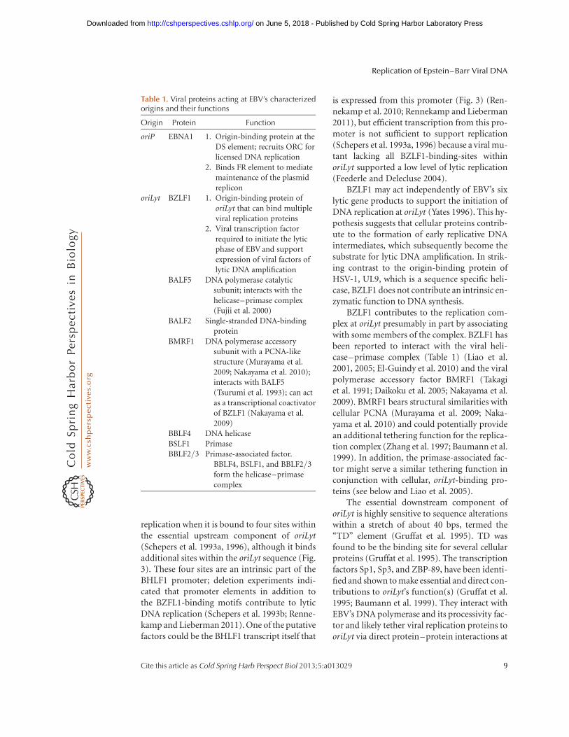
Table 1. Viral proteins acting at EBV’s characterizedorigins and their functions
Origin Protein Function
oriP EBNA1 1. Origin-binding protein at theDS element; recruits ORC forlicensed DNA replication
2. Binds FR element to mediatemaintenance of the plasmidreplicon
oriLyt BZLF1 1. Origin-binding protein oforiLyt that can bind multipleviral replication proteins
2. Viral transcription factorrequired to initiate the lyticphase of EBV and supportexpression of viral factors oflytic DNA amplification
BALF5 DNA polymerase catalyticsubunit; interacts with thehelicase–primase complex(Fujii et al. 2000)
BALF2 Single-stranded DNA-bindingprotein
BMRF1 DNA polymerase accessorysubunit with a PCNA-likestructure (Murayama et al.2009; Nakayama et al. 2010);interacts with BALF5(Tsurumi et al. 1993); can actas a transcriptional coactivatorof BZLF1 (Nakayama et al.2009)
BBLF4 DNA helicaseBSLF1 PrimaseBBLF2/3 Primase-associated factor.
BBLF4, BSLF1, and BBLF2/3form the helicase–primasecomplex
Replication of Epstein–Barr Viral DNA
Cite this article as Cold Spring Harb Perspect Biol 2013;5:a013029 9
on June 5, 2018 - Published by Cold Spring Harbor Laboratory Press http://cshperspectives.cshlp.org/Downloaded from

TD. These findings have a parallel in ZBRK1 andKAP-1, a zinc-finger DNA binding protein andits corepressor, which bind to a site locatedabout 200 bps downstream of TD (Liao et al.2005) within the previously identified oriLytenhancer region (Yates 1996) and colocalizeEBV’s helicase–primase complex to operation-ally defined replication compartments in lyti-cally induced cells. Replication compartmentsare sites to which components of the cellularhomologous recombination pathway are re-cruited (Wilkinson and Weller 2004, and ref-erences therein) together with various cis-actingelements of lytic replicons of herpesviruses.Lytic DNA replication involves both replica-tion and homologous recombination of DNA,which are two interdependent processes activeduring the lytic phase of EBV’s life cycle (Pfullerand Hammerschmidt 1996). Several cellularrecombination and DNA repair factors havebeen recently found recruited to EBV’s repli-cation compartments (Daikoku et al. 2006;Kudoh et al. 2009; Sugimoto et al. 2011) andinduction of EBV’s lytic phase induces a genu-ine DNA-damage response signal (Kudoh et al.2005; Sato et al. 2010). It now seems that oriLytis complex not only in mediating mechanis-tically distinct biphasic DNA replication butalso in using a complex repertoire of viral andcellular proteins to carry out these two modes ofreplication.
IN SUMMARY
EBV is an enormously successful human para-site, having infected more than 6.5 billion peo-ple in the world today. Its success is intimatelytied to its having evolved as an extrachromo-somal replicon, inducing the infected cell tocycle, and successfully usurping the cell’s ma-chinery to carry out its licensed DNA synthesis.It has built on a common herpesviral propertyof amplifying viral DNA during productiveinfections through dedicated viral origins ofDNA synthesis and an array of viral proteinsthat mediate this unlicensed synthesis. It carriesout these different modes of DNA replication inhuman B cells by affecting the differentiatedstate of it B-cell host.
REFERENCES
Altmann M, Pich D, Ruiss R, Wang J, Sugden B, Ham-merschmidt W. 2006. Transcriptional activation by EBVnuclear antigen 1 is essential for the expression of EBV’stransforming genes. Proc Natl Acad Sci 103: 14188–14193.
Antson AA, Burns JE, Moroz OV, Scott DJ, Sanders CM,Bronstein IB, Dodson GG, Wilson KS, Maitland NJ.2000. Structure of the intact transactivation domain ofthe human papillomavirus E2 protein. Nature 403: 805–809.
Aras S, Singh G, Johnston K, Foster T, Aiyar A. 2009. Zinccoordination is required for and regulates transcriptionactivation by Epstein–Barr nuclear antigen 1. PLoSPathog 5: e1000469.
Atanasiu C, Deng Z, Wiedmer A, Norseen J, Lieberman PM.2006. ORC binding to TRF2 stimulates OriP replication.EMBO Rep 7: 716–721.
Avolio-Hunter TM, Frappier L. 2003. EBNA1 efficiently as-sembles on chromatin containing the Epstein–Barr viruslatent origin of replication. Virology 315: 398–408.
Baer R, Bankier AT, Biggin MD, Deininger PL, Farrell PJ,Gibson TJ, Hatfull G, Hudson GS, Satchwell SC,Seguin C, et al. 1984. DNA sequence and expression ofthe B95-8 Epstein–Barr virus genome. Nature 310:207–211.
Bashaw JM, Yates JL. 2001. Replication from oriP of Ep-stein–Barr virus requires exact spacing of two bounddimers of EBNA1 which bend DNA. J Virol 75: 10603–10611.
Baumann M, Feederle R, Kremmer E, Hammerschmidt W.1999. Cellular transcription factors recruit viral replica-tion proteins to activate the Epstein–Barr virus origin oflytic DNA replication, oriLyt. EMBO J 18: 6095–6105.
Bloss TA, Sugden B. 1994. Optimal lengths for DNAs encap-sidated by Epstein–Barr virus. J Virol 68: 8217–8222.
Bochkarev A, Barwell JA, Pfuetzner RA, Bochkareva E,Frappier L, Edwards AM. 1996. Crystal structure of theDNA-binding domain of the Epstein–Barr virus origin-binding protein, EBNA1, bound to DNA. Cell 84: 791–800.
Challberg MD. 1986. A method for identifying the viralgenes required for herpesvirus DNA replication. ProcNatl Acad Sci 83: 9094–9098.
Challberg MD, Kelly TJ. 1989. Animal virus DNA replica-tion. Annu Rev Biochem 58: 671–717.
Chaudhuri B, Xu H, Todorov I, Dutta A, Yates JL. 2001.Human DNA replication initiation factors, ORC andMCM, associate with oriP of Epstein–Barr virus. ProcNatl Acad Sci 98: 10085–10089.
Daikoku T, Kudoh A, Fujita M, Sugaya Y, Isomura H,Shirata N, Tsurumi T. 2005. Architecture of replicationcompartments formed during Epstein–Barr virus lyticreplication. J Virol 79: 3409–3418.
Daikoku T, Kudoh A, Sugaya Y, Iwahori S, Shirata N,Isomura H, Tsurumi T. 2006. Postreplicative mismatchrepair factors are recruited to Epstein–Barr virus repli-cation compartments. J Biol Chem 281: 11422–11430.
Deng Z, Lezina L, Chen CJ, Shtivelband S, So W, Lieber-man PM. 2002. Telomeric proteins regulate episomal
W. Hammerschmidt and B. Sugden
10 Cite this article as Cold Spring Harb Perspect Biol 2013;5:a013029
on June 5, 2018 - Published by Cold Spring Harbor Laboratory Press http://cshperspectives.cshlp.org/Downloaded from

maintenance of Epstein–Barr virus origin of plasmidreplication. Mol Cell 9: 493–503.
Deng Z, Atanasiu C, Burg JS, Broccoli D, Lieberman PM.2003. Telomere repeat binding factors TRF1, TRF2, andhRAP1 modulate replication of Epstein–Barr virus OriP.J Virol 77: 11992–12001.
Dhar SK, Yoshida K, Machida Y, Khaira P, Chaudhuri B,Wohlschlegel JA, Leffak M, Yates J, Dutta A. 2001. Repli-cation from oriP of Epstein–Barr virus requires humanORC and is inhibited by geminin. Cell 106: 287–296.
Dheekollu J, Lieberman PM. 2011. The replisome pausingfactor Timeless is required for episomal maintenance oflatent Epstein–Barr virus. J Virol 85: 5853–5863.
El-Guindy A, Heston L, Miller G. 2010. A subset of replica-tion proteins enhances origin recognition and lytic rep-lication by the Epstein–Barr virus ZEBRA protein. PLoSPathog 6: e1001054.
Feederle R, Delecluse HJ. 2004. Low level of lytic replicationin a recombinant Epstein–Barr virus carrying an originof replication devoid of BZLF1-binding sites. J Virol 78:12082–12084.
Feederle R, Kost M, Baumann M, Janz A, Drouet E,Hammerschmidt W, Delecluse HJ. 2000. The Epstein–Barr virus lytic program is controlled by the co-operativefunctions of two transactivators. EMBO J 19: 3080–3089.
Fernandez AF, Rosales C, Lopez-Nieva P, Grana O, Bal-lestar E, Ropero S, Espada J, Melo SA, Lujambio A,Fraga MF, et al. 2009. The dynamic DNA methylomesof double-stranded DNA viruses associated with humancancer. Genome Res 19: 438–451.
Fujii K, Yokoyama N, Kiyono T, Kuzushima K, Homma M,Nishiyama Y, Fujita M, Tsurumi T. 2000. The Epstein–Barr virus pol catalytic subunit physically interacts withthe BBLF4-BSLF1-BBLF2/3 complex. J Virol 74: 2550–2557.
Gibson W, Roizman B. 1971. Compartmentalization ofspermine and spermidine in the herpes simplex virion.Proc Natl Acad Sci 68: 2818–2821.
Gruffat H, Renner O, Pich D, Hammerschmidt W. 1995.Cellular proteins bind to the downstream componentof the lytic origin of DNA replication of Epstein–Barrvirus. J Virol 69: 1878–1886.
Hammerschmidt W, Sugden B. 1988. Identification andcharacterization of oriLyt, a lytic origin of DNA replica-tion of Epstein–Barr virus. Cell 55: 427–433.
Hegde RS, Grossman SR, Laimins LA, Sigler PB. 1992. Crys-tal structure at 1.7 A of the bovine papillomavirus-1 E2DNA-binding domain bound to its DNA target. Nature359: 505–512.
Humme S, Reisbach G, Feederle R, Delecluse HJ, Bousset K,Hammerschmidt W, Schepers A. 2003. The EBV nuclearantigen 1 (EBNA1) enhances B cell immortalization sev-eral thousandfold. Proc Natl Acad Sci 100: 10989–10994.
Hung SC, Kang MS, Kieff E. 2001. Maintenance of Epstein–Barr virus (EBV) oriP-based episomes requires EBV-en-coded nuclear antigen-1 chromosome-binding domains,which can be replaced by high-mobility group-I or his-tone H1. Proc Natl Acad Sci 98: 1865–1870.
Jacob RJ, Roizman B. 1977. Anatomy of herpes simplex virusDNAVIII. Properties of the replicating DNA. J Virol 23:394–411.
Julien MD, Polonskaya Z, Hearing J. 2004. Protein and se-quence requirements for the recruitment of the humanorigin recognition complex to the latent cycle origin ofDNA replication of Epstein–Barr virus oriP. Virology326: 317–328.
Kalla M, Schmeinck A, Bergbauer M, Pich D, Hammer-schmidt W. 2010. AP-1 homolog BZLF1 of Epstein–Barr virus has two essential functions dependent on theepigenetic state of the viral genome. Proc Natl Acad Sci107: 850–855.
Kennedy G, Sugden B. 2003. EBNA-1, a bifunctional tran-scriptional activator. Mol Cell Biol 23: 6901–6908.
Kim SS, Tam JK, Wang AF, Hegde RS. 2000. The structuralbasis of DNA target discrimination by papillomavirus E2proteins. J Biol Chem 275: 31245–31254.
Kirchmaier AL, Sugden B. 1998. Rep�: A viral element thatcan partially replace the origin of plasmid DNA synthesisof Epstein–Barr virus. J Virol 72: 4657–4666.
Kobayashi T, Rein T, DePamphilis ML. 1998. Identificationof primary initiation sites for DNA replication in thehamster dihydrofolate reductase gene initiation zone.Mol Cell Biol 18: 3266–3277.
Koons MD, Van Scoy S, Hearing J. 2001. The replicator ofthe Epstein–Barr virus latent cycle origin of DNA repli-cation, oriP, is composed of multiple functional ele-ments. J Virol 75: 10582–10592.
Kudoh A, Fujita M, Zhang L, Shirata N, Daikoku T, Su-gaya Y, Isomura H, Nishiyama Y, Tsurumi T. 2005.Epstein–Barr virus lytic replication elicits ATM check-point signal transduction while providing an S-phase-like cellular environment. J Biol Chem 280: 8156–8163.
Kudoh A, Iwahori S, Sato Y, Nakayama S, Isomura H,Murata T, Tsurumi T. 2009. Homologous recombination-al repair factors are recruited and loaded onto the viralDNA genome in Epstein–Barr virus replication com-partments. J Virol 83: 6641–6651.
Leight ER, Sugden B. 2001. Establishment of an oriP repli-con is dependent upon an infrequent, epigenetic event.Mol Cell Biol 21: 4149–4161.
Levitskaya J, Sharipo A, Leonchiks A, Ciechanover A, Ma-succi MG. 1997. Inhibition of ubiquitin/proteasome-dependent protein degradation by the Gly-Ala repeatdomain of the Epstein–Barr virus nuclear antigen 1.Proc Natl Acad Sci 94: 12616–12621.
Liao G, Wu FY, Hayward SD. 2001. Interaction with theEpstein–Barr virus helicase targets Zta to DNA replica-tion compartments. J Virol 75: 8792–8802.
Liao G, Huang J, Fixman ED, Hayward SD. 2005. The Ep-stein–Barr virus replication protein BBLF2/3 providesan origin-tethering function through interaction withthe zinc finger DNA binding protein ZBRK1 and theKAP-1 corepressor. J Virol 79: 245–256.
Lindner SE, Zeller K, Schepers A, Sugden B. 2008. The af-finity of EBNA1 for its origin of DNA synthesis is a de-terminant of the origin’s replicative efficiency. J Virol 82:5693–6702.
Mackey D, Sugden B. 1999. The linking regions of EBNA1are essential for its support of replication and transcrip-tion. Mol Cell Biol 19: 3349–3359.
Marechal V, Dehee A, Chikhi-Brachet R, Piolot T, Coppey-Moisan M, Nicolas JC. 1999. Mapping EBNA-1 domains
Replication of Epstein–Barr Viral DNA
Cite this article as Cold Spring Harb Perspect Biol 2013;5:a013029 11
on June 5, 2018 - Published by Cold Spring Harbor Laboratory Press http://cshperspectives.cshlp.org/Downloaded from

involved in binding to metaphase chromosomes. J Virol73: 4385–4392.
Moriyama K, Yoshizawa-Sugata N, Obuse C, Tsurimoto T,Masai H. 2012. Epstein–Barr nuclear antigen 1 (EBNA-1)- dependent recruitment of origin recognition complex(Orc) on oriP of Epstein–Barr virus with purified pro-teins: Stimulation by Cdc6 through its direct interactionwith EBNA1. J Biol Chem 287: 23977–23994.
Murayama K, Nakayama S, Kato-Murayama M, Akasaka R,Ohbayashi N, Kamewari-Hayami Y, Terada T, Shi-rouzu M, Tsurumi T, Yokoyama S. 2009. Crystal structureof Epstein–Barr virus DNA polymerase processivity fac-tor BMRF1. J Biol Chem 284: 35896–35905.
Nakayama S, Murata T, Murayama K, Yasui Y, Sato Y,Kudoh A, Iwahori S, Isomura H, Kanda T, Tsurumi T.2009. Epstein–Barr virus polymerase processivity factorenhances BALF2 promoter transcription as a coactivatorfor the BZLF1 immediate-early protein. J Biol Chem 284:21557–21568.
Nakayama S, Murata T, Yasui Y, Murayama K, Isomura H,Kanda T, Tsurumi T. 2010. Tetrameric ring formation ofEpstein–Barr virus polymerase processivity factor is cru-cial for viral replication. J Virol 84: 12589–12598.
Nanbo A, Sugden A, Sugden B. 2007. The coupling of syn-thesis and partitioning of EBV’s plasmid replicon is re-vealed in live cells. EMBO J 26: 4252–4262.
Niller HH, Glaser G, Knuchel R, Wolf H. 1995. Nucleopro-tein complexes and DNA 50-ends at oriP of Epstein–Barrvirus. J Biol Chem 270: 12864–12868.
Norio P, Schildkraut CL. 2004. Plasticity of DNA replicationinitiation in Epstein–Barr virus episomes. PLoS Biol 2:e152.
Norio P, Schildkraut CL, Yates JL. 2000. Initiation of DNAreplication within oriP is dispensable for stable replica-tion of the latent Epstein–Barr virus chromosome afterinfection of established cell lines. J Virol 74: 8563–8574.
Norseen J, Thomae A, Sridharan V, Aiyar A, Schepers A,Lieberman PM. 2008. RNA-dependent recruitment ofthe origin recognition complex. EMBO J 27: 3024–3035.
Paludan C, Schmid D, Landthaler M, Vockerodt M, Kube D,Tuschl T, Munz C. 2005. Endogenous MHC class II pro-cessing of a viral nuclear antigen after autophagy. Science307: 593–596.
Pfuller R, Hammerschmidt W. 1996. Plasmid-like replica-tive intermediates of the Epstein–Barr virus lytic originof DNA replication. J Virol 70: 3423–3431.
Rennekamp AJ, Lieberman PM. 2011. Initiation of Epstein–Barr virus lytic replication requires transcription andthe formation of a stable RNA–DNA hybrid moleculeat OriLyt. J Virol 85: 2837–2850.
Rennekamp AJ, Wang P, Lieberman PM. 2010. Evidence forDNA hairpin recognition by Zta at the Epstein–Barrvirus origin of lytic replication. J Virol 84: 7073–7082.
Ritzi M, Tillack K, Gerhardt J, Ott E, Humme S, Kremmer E,Hammerschmidt W, Schepers A. 2003. Complex protein-DNA dynamics at the latent origin of DNA replication ofEpstein–Barr virus. J Cell Sci 116: 3971–3984.
Sarkari F, Sanchez-Alcaraz T, Wang S, Holowaty MN,Sheng Y, Frappier L. 2009. EBNA1-mediated recruit-ment of a histone H2B deubiquitylating complex to the
Epstein–Barr virus latent origin of DNA replication.PLoS Pathog 5: e1000624.
Sato Y, Shirata N, Murata T, Nakasu S, Kudoh A, Iwahori S,Nakayama S, Chiba S, Isomura H, Kanda T, et al. 2010.Transient increases in p53-responsible gene expression atearly stages of Epstein–Barr virus productive replication.Cell Cycle 9: 807–814.
Schepers A, Pich D, Hammerschmidt W. 1993a. A transcrip-tion factor with homology to the AP-1 family links RNAtranscription and DNA replication in the lytic cycle ofEpstein–Barr virus. EMBO J 12: 3921–3929.
Schepers A, Pich D, Mankertz J, Hammerschmidt W. 1993b.cis-acting elements in the lytic origin of DNA replicationof Epstein–Barr virus. J Virol 67: 4237–4245.
Schepers A, Pich D, Hammerschmidt W. 1996. Activation oforiLyt, the lytic origin of DNA replication of Epstein–Barr virus, by BZLF1. Virology 220: 367–376.
Schepers A, Ritzi M, Bousset K, Kremmer E, Yates JL,Harwood J, Diffley JF, Hammerschmidt W. 2001. Humanorigin recognition complex binds to the region of thelatent origin of DNA replication of Epstein–Barr virus.EMBO J 20: 4588–4602.
Schildgen O, Graper S, Blumel J, Matz B. 2005. Genomereplication and progeny virion production of herpessimplex virus type 1 mutants with temperature-sensitivelesions in the origin-binding protein. J Virol 79: 7273–7278.
Sears J, Kolman J, Wahl GM, Aiyar A. 2003. Metaphasechromosome tethering is necessary for the DNA synthe-sis and maintenance of oriP plasmids but is insufficientfor transcription activation by Epstein–Barr nuclear an-tigen 1. J Virol 77: 11767–11780. Erratum in J Virol 78:5531, 2004.
Sears J, Ujihara M, Wong S, Ott C, Middeldorp J, Aiyar A.2004. The amino terminus of Epstein–Barr virus (EBV)nuclear antigen 1 contains AT hooks that facilitate thereplication and partitioning of latent EBV genomes bytethering them to cellular chromosomes. J Virol 78:11487–11505.
Shaw JE, Levinger LF, Carter CW Jr. 1979. Nucleosomalstructure of Epstein–Barr virus DNA in transformedcell lines. J Virol 29: 657–665.
Shirakata M, Imadome KI, Hirai K. 1999. Requirement ofreplication licensing for the dyad symmetry element-de-pendent replication of the Epstein–Barr virus oriP mini-chromosome. Virology 263: 42–54.
Sugimoto A, Kanda T, Yamashita Y, Murata T, Saito S,Kawashima D, Isomura H, Nishiyama Y, Tsurumi T.2011. Spatiotemporally different DNA repair systemsparticipate in Epstein–Barr virus genome maturation. JVirol 85: 6127–6135.
Takagi S, Takada K, Sairenji T. 1991. Formation of intra-nuclear replication compartments of Epstein–Barr viruswith redistribution of BZLF1 and BMRF1 gene products.Virology 185: 309–315.
Tempera I, Lieberman PM. 2010. Chromatin organization ofgammaherpesvirus latent genomes. Biochim Biophys Acta1799: 236–245.
Tsurumi T. 2001. EBV replication enzymes. Curr Top Micro-biol Immunol 258: 65–87.
W. Hammerschmidt and B. Sugden
12 Cite this article as Cold Spring Harb Perspect Biol 2013;5:a013029
on June 5, 2018 - Published by Cold Spring Harbor Laboratory Press http://cshperspectives.cshlp.org/Downloaded from

Tsurumi T, Kobayashi A, Tamai K, Daikoku T, Kurachi R,Nishiyama Y. 1993. Functional expression and character-ization of the Epstein–Barr virus DNA polymerase cata-lytic subunit. J Virol 67: 4651–4658.
Vereide DT, Sugden B. 2011. Lymphomas differ in theirdependence on Epstein–Barr virus. Blood 117: 1977–1985.
Wang CY, Sugden B. 2008. Identifying a property of originsof DNA synthesis required to support plasmids stably inhuman cells. Proc Natl Acad Sci 105: 9639–9644.
Wang J, Lindner SE, Leight ER, Sugden B. 2006. Essentialelements of a licensed, mammalian plasmid origin ofDNA synthesis. Mol Cell Biol 26: 1124–1134.
Wensing B, Stuhler A, Jenkins P, Hollyoake M, Karstegl CE,Farrell PJ. 2001. Variant chromatin structure of the oriPregion of Epstein–Barr virus and regulation of EBER1expression by upstream sequences and oriP. J Virol 75:6235–6241.
Wilkinson DE, Weller SK. 2004. Recruitment of cellular re-combination and repair proteins to sites of herpes sim-plex virus type 1 DNA replication is dependent on thecomposition of viral proteins within prereplicative sitesand correlates with the induction of the DNA damageresponse. J Virol 78: 4783–4796.
Wu H, Kapoor P, Frappier L. 2002. Separation of the DNAreplication, segregation, and transcriptional activationfunctions of Epstein–Barr nuclear antigen 1. J Virol 76:2480–2490.
Yates JL. 1996. Epstein–Barr virus DNA replication. In DNAreplication in eukaryotic cells (ed. DePamphilis ML), pp.751–774. Cold Spring Harbor Laboratory Press, ColdSpring Harbor, NY.
Yates JL, Camiolo SM, Bashaw JM. 2000. The minimalreplicator of Epstein–Barr virus oriP. J Virol 74: 4512–4522.
Yin Y, Manoury B, Fahraeus R. 2003. Self-inhibition of syn-thesis and antigen presentation by Epstein–Barr virus-encoded EBNA1. Science 301: 1371–1374.
Zhang Q, Holley-Guthrie E, Ge JQ, Dorsky D, Kenney S.1997. The Epstein–Barr virus (EBV) DNA polymeraseaccessory protein, BMRF1, activates the essential down-stream component of the EBV oriLyt. Virology 230:22–34.
Zhou J, Chau CM, Deng Z, Shiekhattar R, Spindler MP,Schepers A, Lieberman PM. 2005. Cell cycle regulationof chromatin at an origin of DNA replication. EMBO J24: 1406–1417.
Replication of Epstein–Barr Viral DNA
Cite this article as Cold Spring Harb Perspect Biol 2013;5:a013029 13
on June 5, 2018 - Published by Cold Spring Harbor Laboratory Press http://cshperspectives.cshlp.org/Downloaded from

2013; doi: 10.1101/cshperspect.a013029Cold Spring Harb Perspect Biol Wolfgang Hammerschmidt and Bill Sugden
Barr Viral DNA−Replication of Epstein
Subject Collection DNA Replication
Barr Viral DNA−Replication of EpsteinWolfgang Hammerschmidt and Bill Sugden
Endoreplication
DePamphilisNorman Zielke, Bruce A. Edgar and Melvin L.
Replication Proteins and Human Disease
Nicholas ColemanAndrew P. Jackson, Ronald A. Laskey and
Replication-Fork Dynamics
Antoine M. van Oijen, et al.Karl E. Duderstadt, Rodrigo Reyes-Lamothe,
Break-Induced DNA Replication
HaberRanjith P. Anand, Susan T. Lovett and James E.
ReplicationReplication Forks at Chromosomal Origins of Helicase Activation and Establishment of
Seiji Tanaka and Hiroyuki ArakiRegulating DNA Replication in Eukarya
Khalid Siddiqui, Kin Fan On and John F.X. DiffleyPoxvirus DNA Replication
Bernard MossArchaeology of Eukaryotic DNA Replication
Kira S. Makarova and Eugene V. Koonin HelicaseThe Minichromosome Maintenance Replicative
Stephen D. Bell and Michael R. BotchanTranslesion DNA Polymerases
Myron F. Goodman and Roger WoodgateDNA Replication Origins
Alan C. Leonard and Marcel Méchali
Cancer?Human Papillomavirus Infections: Warts or
Louise T. Chow and Thomas R. BrokerBacteria, Archaea, and EukaryaPrinciples and Concepts of DNA Replication in
StillmanMichael O'Donnell, Lance Langston and Bruce
Chromatin and DNA ReplicationDavid M. MacAlpine and Geneviève Almouzni
DNA Replication TimingNicholas Rhind and David M. Gilbert
http://cshperspectives.cshlp.org/cgi/collection/ For additional articles in this collection, see
Copyright © 2013 Cold Spring Harbor Laboratory Press; all rights reserved
on June 5, 2018 - Published by Cold Spring Harbor Laboratory Press http://cshperspectives.cshlp.org/Downloaded from