Renal Physiology and Its Systemic Impact · CHAPTER 16 Renal Physiology and Its Systemic Impact 131...
11
130 CHAPTER 16 Renal Physiology and Its Systemic Impact MICHIKO SHIMADA, KAWTHER F. ALQUADAN, and A. AHSAN EJAZ INTRODUCTION The primary function of the kidneys is to maintain volume, elec- trolyte, and acid–base homeostasis in the internal milieu. Rapid deterioration in kidney function or inability to maintain adequate urine output is termed acute kidney injury (AKI). After decades of conflicting definitions, current consensus defines AKI as an abrupt (within 48 hours) reduction in kidney function, defined as an absolute increase in serum creatinine above 0.3 mg/dL (>26.4 mmol/L), a percentage increase in serum creatinine above 50% (1.5-fold from baseline) in accordance with criteria established by the Acute Kidney Injury Network (AKIN) (1). Using this criterion, the incidence of AKI in adults in a district general hospital setting in the United Kingdom was reported to be 15,325 patients/million populations per year (2) and 39.3% in the critically ill patients in Finland (3). In a meta-analysis of 154 studies from North America, Northern Europe, and East- ern Asia, from high-income countries, and from nations that spent 5% or more of the gross domestic product on total health expenditure, the incidence of AKI among hospitalized patients was 21.6% in adults and 33.7% in children by AKIN criteria (4). AKI-associated hospital mortality is high (23.9%) (4), and worsens when renal replacement therapies are also required (5). AKI is also associated with increased length of intensive care unit (ICU) and hospital stay, increase in care on discharge, and an increase in hospital readmission within 30 days (2). Knowledge of risk factors for AKI in the ICU can be help- ful in the determination of clinical outcome and contribute to more defined risk assessment and management in this cohort (Table 16.1). This chapter briefly reviews pertinent renal phys- iology, the mechanisms and outcomes of ischemic AKI, the progression of chronic kidney disease and its impact on acute critical illness, and the principles of management of AKI. Spe- cifics on the treatment of AKI and a detailed description of the various treatment modalities are to be found in later chapters of this textbook. PHYSIOLOGY OF THE KIDNEY General Anatomy The ability of the kidney to maintain the equilibrium of the corporal fluids and electrolytes depends on three essential pro- cesses: (i) filtration of the circulating blood by the glomerulus to form an ultrafiltrate; (ii) reabsorption of specific solutes from the tubular fluid to the blood; and (iii) secretion from the peritubular capillary blood system to the tubular space. The functions of the kidney are dependent on the unique anatomic arrangement of its structures. The afferent arteriole divides into several branches after entering the glomerular tuft and forms the capillary network present in the glomerulus (Fig. 16.1). The confluence of several capillaries forms the efferent arteriole which drains the blood from the glomerulus. During the ultrafiltration process, water and solutes pass through the endothelium, the glomerular basement membrane, and the slit-diaphragm between the podocytes. The determinant of the filtration of a substance is its size and charge. Substances with molecular radius of less than 2 nm are filtered freely, whereas the ionic charges of sub- stances measuring between 2 and 4 nm determine the amount of their filtration; substances with molecular radius greater than 4 nm are not filtered. The medullary region of the kidneys is characterized by low oxygen tension—10 to 15 mmHg—under normal conditions. The tubular segments located in this region—pars recta, or S3 segment of proximal tubule and medullary thick ascending limb—are characterized by active transport of Na + , which is dependent on oxidative phosphorylation for energy. The high rates of oxygen consumption associated with the precarious blood flow in the medullary region are responsible for the vulnerability of this area to ischemia. Glomerular Filtration Rate and Renal Plasma Flow The glomerular filtration rate (GFR) and renal plasma flow (RPF) are rate measurements that help to characterize the sta- tus of renal function. The total rate at which fluid is filtered into all the glomeruli constitutes the GFR; normal GFR varies between 100 and 120 mL/min/1.73 m 2 , depending on various factors including gender, age, and body weight. Changes in the GFR can result from changes in the glomerular perme- ability or capillary surface area or from changes in the net ultrafiltration. In a single glomerulus, the driving pressure for the glomerular filtration is determined by the difference of the gradient of the hydrostatic and oncotic pressures between the capillaries and the Bowman space. The rate at which plasma flows through the kidney is called RPF; renal blood flow (RBF) is the volume of blood delivered to the kidney per unit time (1 to 1.2 L/min). RBF calculations are based on RPF and hematocrit: Renal blood flow = RPF / 1 – hematocrit It is possible to measure the RPF using para-aminohippu- rate as a tracer in humans, with a normal value about 625 mL/ min, but the test is not commonly used in clinical practice due to labor intensity and cost. Autoregulatory Control of Renal Blood Flow Despite the significant variations in mean arterial pressure (MAP), RBF and GFR remain constant, a phenomenon known as autoregulation. Autoregulation is affected via changes in diameter of the afferent arterioles in response to a combina- tion of two mechanisms: • The myogenic reflex: When the renal perfusion pressure increases, the afferent arteriole constricts automatically. LWBK1580_C16_p130-140.indd 130 29/07/17 9:34 AM
Transcript of Renal Physiology and Its Systemic Impact · CHAPTER 16 Renal Physiology and Its Systemic Impact 131...

130
CHAPTER
16Renal Physiology and Its Systemic ImpactMICHIKO SHIMADA, KAWTHER F. ALQUADAN, and A. AHSAN EJAZ
IntRoductIon
The primary function of the kidneys is to maintain volume, elec-trolyte, and acid–base homeostasis in the internal milieu. Rapid deterioration in kidney function or inability to maintain adequate urine output is termed acute kidney injury (AKI). After decades of conflicting definitions, current consensus defines AKI as an abrupt (within 48 hours) reduction in kidney function, defined as an absolute increase in serum creatinine above 0.3 mg/dL (>26.4 mmol/L), a percentage increase in serum creatinine above 50% (1.5-fold from baseline) in accordance with criteria established by the Acute Kidney Injury Network (AKIN) (1). Using this criterion, the incidence of AKI in adults in a district general hospital setting in the United Kingdom was reported to be 15,325 patients/million populations per year (2) and 39.3% in the critically ill patients in Finland (3). In a meta-analysis of 154 studies from North America, Northern Europe, and East-ern Asia, from high-income countries, and from nations that spent 5% or more of the gross domestic product on total health expenditure, the incidence of AKI among hospitalized patients was 21.6% in adults and 33.7% in children by AKIN criteria (4). AKI-associated hospital mortality is high (23.9%) (4), and worsens when renal replacement therapies are also required (5). AKI is also associated with increased length of intensive care unit (ICU) and hospital stay, increase in care on discharge, and an increase in hospital readmission within 30 days (2).
Knowledge of risk factors for AKI in the ICU can be help-ful in the determination of clinical outcome and contribute to more defined risk assessment and management in this cohort (Table 16.1). This chapter briefly reviews pertinent renal phys-iology, the mechanisms and outcomes of ischemic AKI, the progression of chronic kidney disease and its impact on acute critical illness, and the principles of management of AKI. Spe-cifics on the treatment of AKI and a detailed description of the various treatment modalities are to be found in later chapters of this textbook.
PhySIology of the KIdney
General Anatomy
The ability of the kidney to maintain the equilibrium of the corporal fluids and electrolytes depends on three essential pro-cesses: (i) filtration of the circulating blood by the glomerulus to form an ultrafiltrate; (ii) reabsorption of specific solutes from the tubular fluid to the blood; and (iii) secretion from the peritubular capillary blood system to the tubular space. The functions of the kidney are dependent on the unique anatomic arrangement of its structures.
The afferent arteriole divides into several branches after entering the glomerular tuft and forms the capillary network present in the glomerulus (Fig. 16.1). The confluence of several
capillaries forms the efferent arteriole which drains the blood from the glomerulus. During the ultrafiltration process, water and solutes pass through the endothelium, the glomerular basement membrane, and the slit-diaphragm between the podocytes. The determinant of the filtration of a substance is its size and charge. Substances with molecular radius of less than 2 nm are filtered freely, whereas the ionic charges of sub-stances measuring between 2 and 4 nm determine the amount of their filtration; substances with molecular radius greater than 4 nm are not filtered.
The medullary region of the kidneys is characterized by low oxygen tension—10 to 15 mmHg—under normal conditions. The tubular segments located in this region—pars recta, or S3 segment of proximal tubule and medullary thick ascending limb—are characterized by active transport of Na+, which is dependent on oxidative phosphorylation for energy. The high rates of oxygen consumption associated with the precarious blood flow in the medullary region are responsible for the vulnerability of this area to ischemia.
Glomerular Filtration Rate and Renal Plasma Flow
The glomerular filtration rate (GFR) and renal plasma flow (RPF) are rate measurements that help to characterize the sta-tus of renal function. The total rate at which fluid is filtered into all the glomeruli constitutes the GFR; normal GFR varies between 100 and 120 mL/min/1.73 m2, depending on various factors including gender, age, and body weight. Changes in the GFR can result from changes in the glomerular perme-ability or capillary surface area or from changes in the net ultrafiltration. In a single glomerulus, the driving pressure for the glomerular filtration is determined by the difference of the gradient of the hydrostatic and oncotic pressures between the capillaries and the Bowman space.
The rate at which plasma flows through the kidney is called RPF; renal blood flow (RBF) is the volume of blood delivered to the kidney per unit time (1 to 1.2 L/min). RBF calculations are based on RPF and hematocrit:
Renal blood flow = RPF / 1 – hematocrit
It is possible to measure the RPF using para-aminohippu-rate as a tracer in humans, with a normal value about 625 mL/min, but the test is not commonly used in clinical practice due to labor intensity and cost.
Autoregulatory Control of Renal Blood Flow
Despite the significant variations in mean arterial pressure (MAP), RBF and GFR remain constant, a phenomenon known as autoregulation. Autoregulation is affected via changes in diameter of the afferent arterioles in response to a combina-tion of two mechanisms:• The myogenic reflex: When the renal perfusion pressure
increases, the afferent arteriole constricts automatically.
LWBK1580_C16_p130-140.indd 130 29/07/17 9:34 AM

CHAPTER 16 Renal Physiology and Its Systemic Impact 131
• Tubuloglomerular feedback: Situations associated with an increased delivery of sodium chloride to the macula densa result in vasoconstrictive response of the afferent arteriole. The increased uptake of chloride ions by the macula densa cells leads to adenosine triphosphate (ATP) release into the surrounding extracellular space. ATP is then converted to adenosine, which binds to adenosine A1 receptors causing afferent arteriolar vasoconstriction.
Basic Principles of Tubular Transport
The kidneys filter about 180 L of plasma per day, and all but 2 L are reabsorbed. This massive reabsorption is accomplished through several modifications of the glomerular ultrafiltrate, consisting of absorption and secretion of water and solutes before becoming the final urine. In general, three different tubular segments are involved in this process and can be rec-ognized based on the differences in the function of their cells.• The proximal tubule reabsorbs most of the filtered glucose,
amino acids, low–molecular-weight proteins, and water (approximately 65%). Other solutes, such as sodium (Na+), potassium (K+), chloride (Cl−), bicarbonate (HCO3
−), phos-phate (PO4
−), and urea are also absorbed in this nephron segment. The terminal segment of the proximal tubule—the pars recta or S3—is responsible for the secretion of numerous drugs and toxins.
TABLE 16.1 Predictors of Acute Kidney Injury in the Intensive Care Unit
Risks
All patients OR
N = 194a
Sepsis OR
N = 2,442b
CV Surgery OR
N = 43,642c
Trauma OR
N = 153d
DemographicsAge 0.93 1.1 2.82
Acute Clinical SettingHigh-risk surgery 1.51 1.98
Sepsis 3.11
High injury severity score 5.75–13.7
Emergency procedure 7.61
Cardiopulmonary bypass 2.64
Pre-existing ConditionChronic kidney disease 1.77 1.02 1.31–5.80b
Cardiac failure 1.85 1.55
Cancer 3.75 —
Prior cardiac surgery 1.93
COPD 1.26
Hypertension (SBP >160 mmHg) 1.03–1.98c
Peripheral vascular disease 1.51
Clinical FindingsElevated A-a gradient 1.04
Elevated serum bilirubin 3.6 9.7
Hypotension 3.04
Hemoperitoneum 6.80
Long bone fractures 2.36
Morbid obesity 2.1 1.11
APACHE II quartile 1.57
Elevated CVP 1.5
CV, cardiovascular; OR, odds ratio; COPD, chronic obstructive pulmonary disease; SBP, systolic blood pressure; CVP, central venous pressure.aChawla LS, Abell L, Mazhari R, et al. Identifying critically ill patients at high risk for developing acute kidney injury: a pilot study. Kidney Int. 2005;68:2274–2280.bYegenaga I, Hoste E, Van Biesen W, et al. Clinical characteristics of patients developing ARF due to sepsis/systemic inflammatory response. Am J Kidney Dis. 2004;43:817–824.cChertow GM, Lazarus JM, Christiansen CL, et al. Preoperative renal risk stratification. Circulation. 1997;95:878–884.dVivino G, Antonelli M, Moro ML, et al. Risk factors for acute kidney injury in trauma patients. Intensive Care Med. 1998;24:808–814.
Distal tubule
Proximaltubule
Loop of Henle
Inn
er M
edu
lla
Ou
ter
Med
ulla
Co
llect
ing
Du
ct
Co
rtex
Inn
erS
trip
eO
ute
rS
trip
e Proximal tubuleS3 Segment
fIguRe 16.1 Nephron, the functional unit of the kidney.
LWBK1580_C16_p130-140.indd 131 29/07/17 9:34 AM

132 SECTion 2 PHYSIOLOGY OF CRITICAL ILLNESS
• The straight portion of the proximal tubule, the thin ascend-ing and descending limbs, and the thick ascending limb con-stitute the region known as the loop of Henle. This region is responsible for the continuing reabsorption of the solutes that escaped the proximal tubules (sodium, potassium, chlo-ride, calcium, magnesium ions). It is the major area respon-sible for the ability of the kidneys to concentrate or dilute the final urine. The principal luminal transporter expressed in the thick ascending limb is the sodium-potasium-2chlo-ride cotransporter (NKCC2), which is the target of diuretics such as furosemide.
• The distal nephron is responsible for the final adjustments in the urine. Critical regulatory hormones such as vasopres-sin and aldosterone regulate the acid and potassium excre-tion and the urinary concentration at this segment. Thiazide diuretics act at the distal convoluted tubule through a thia-zide-sensitive Na+ −Cl−cotransporter on the apical membrane.
The Glomerulotubular Balance
The fact that the tubules tend to reabsorb a constant propor-tion of a glomerular filtrate rather than a constant amount is called glomerular balance. As an example, if the filtered load of Na+ were increased by 10%, total Na+ reabsorption in the tubules would also increase by 10%, keeping the final amount of Na+ in the urine stable at 100 to 250 mEq/day. In the absence of this mechanism, even small changes in the GFR would cause major changes in the final amount excreted of any solute. The mechanisms responsible for this balance are not fully under-stood, but changes in the oncotic pressure in the peritubular capillaries and in the delivery of certain solutes (glucose and amino acids) to the proximal tubule are probably involved.
Control of Effective Circulating Volume via Integrated Mechanisms
Most volume-regulatory mechanisms in the kidney use the effective circulating volume (ECV), or the degree of fullness of the vasculature, as the final target. Under normal condi-tions, the ECV varies in direct proportion to the extracellular fluid volume. As Na+ is the most abundant extracellular solute, the kidney excretion or retention of Na+ is a crucial step to control ECV. Osmoregulation is under the control of a single hormonal system, antidiuretic hormone (ADH), but volume regulation requires a complex set of redundant and overlap-ping mechanisms.
The kidneys are able to conserve water by excreting the solute load in concentrated urine in conditions of excess water loss. Similarly, in high water intake states, the urinary volume may increase to as high as 14 L/day, with an osmolality sig-nificantly lower than that of the plasma. Vasopressin or ADH regulates the water permeability in the distal nephron and is the principal hormone responsible for the determination of the urinary concentration and volume. Normally, the major stimulus to secretion of ADH is the plasma osmolality, but in situations of extracellular volume depletion, the set point to release ADH is shifted, and higher levels of this hormone are common even in hypotonic states.
The renin–angiotensin system plays a central role in the control of ECV. The afferent arteriolar cells that form part of the juxtaglomerular apparatus release renin in response to increased sympathetic nervous stimulation, reduced arterial blood pressure, or reduced delivery of sodium chloride (NaCl) to the macula densa region. Renin cleaves angiotensinogen
into angiotensin I and is then converted to angiotensin II by the angiotensin-converting enzyme. Angiotensin plays important roles in the control of blood pressure and the effective circula-tory fluid volume.• Angiotensin II has the direct effect of increasing the sodium
reabsorption in the proximal tubule (stimulation of Na+/H+ exchange).
• The aldosterone secreted by the adrenal glands in response to the angiotensin II stimulates sodium reabsorption in the distal nephron.
• Angiotensin II causes general arteriolar vasoconstriction, thereby increasing arterial pressure.Increased renal sympathetic tone enhances renal salt reab-
sorption and often decreases RBF. In addition to its direct effects on renal function, increased sympathetic outflow pro-motes the activation of the renin–angiotensin system.
ImPact of alteRed Renal PhySIology
Altered renal physiology can manifest as mild abnormalities in electrolyte and acid–base homeostasis or solute clearance to full-blown AKI that can progress to chronic kidney dis-ease. The impacts of perturbed renal physiology and proposed pathogenesis are discussed.
Consequences of Aberrant Renal Blood Flow Autoregulation
RBF is dependent on systemic blood pressure and intrarenal vascular resistance. The autoregulatory mechanisms, through changes in vascular resistance, ensure that over a wide range of perfusion pressures RBF remains stable and glomerular filtra-tion can be maintained. However, a substantial loss of renovas-cular response to neurohormonal stimuli follows ischemic AKI (6), related to increased renal vasoconstriction (7). Clinically this manifests as ischemic AKI, a syndrome that develops fol-lowing a sudden transient drop in total or regional blood flow to the kidney resulting in tissue hypoxia, tubular and vascular injury, and loss of renal structure and function (Fig. 16.2). The severe form of this entity is known as acute tubular necrosis. An aberrant RBF autoregulation is also implicated in normo-tensive ischemic AKI. Whereas normal GFR is maintained until MAP falls below 50 mmHg, in normotensive ischemic AKI RBF and GFR can decrease by as much as 50% despite the maintenance of MAP within the autoregulatory range (8), suggesting the presence of heightened renal vasoconstriction.
The paradoxical rise in renovascular resistance seen with decreasing renal perfusion in ischemic AKI is due to the loss of the usual balance of vasoconstrictors and the vasodilators required to maintain the normal tone of the renal vasculature. The downstream consequence of aberrant responses to neu-rohormonal stimuli and the persistent vasoconstriction is that it worsens renal perfusion and impairs oxygen and nutrient delivery to the areas supplied by the postglomerular vessels.
The PO2 in the outer medulla is about 10 to 15 mmHg; even a mild decrease in renal perfusion can lead to a hypoxic insult (oxidative stress) to the vulnerable medullary nephron segments. Tissue hypoxia can result in depletion of cellular ATP stores, increased intracellular calcium, and subsequent disruption of
LWBK1580_C16_p130-140.indd 132 29/07/17 9:34 AM

CHAPTER 16 Renal Physiology and Its Systemic Impact 133
actin cytoskeleton in the endothelial and vascular smooth mus-cle cells, with resultant hemodynamic impairment and tubular injury. Adenosine nucleotide metabolic products are not reused for the regeneration of ATP and are, instead, diverted through the degradatory pathways to generate xanthine and uric acid. Accumulation of adenosine and uric acid worsens vasoconstric-tion and renal perfusion via their effects on adenosine recep-tors and afferent arterioles, respectively. Cellular activation also leads to reactive oxygen species generation, phospholipase acti-vation, and membrane lipid alterations (9).
Hypoxia, with subsequent reperfusion, leads to acute inflam-matory changes. Inflammation is one of the major pathophysi-ologic pathways contributing to ischemic AKI. Ischemic injury to the vasa recta results in enhanced adherence of leukocytes to the vascular endothelial cells, sequestration of leukocytes, vascular congestion—that is, a no-flow phenomenon— cellular infiltration, production of inflammatory mediators, and generation of reactive oxygen species. Cytokines and che-mokines, released from the injured cells, attract inflammatory cells to the site of injury and potentiate the inflammatory cas-cade. A similar inflammatory response is also seen in tubular cell injury, which is also capable of producing chemokines and inflammatory mediators. The inflammatory changes are most pronounced in the outer medullary stripe, the region that is most susceptible to hypoxic insult.
Numerous stress response mechanisms are rapidly acti-vated in response to oxidative insults. Some of the pathways are preferentially linked to cell survival whereas others are proapoptotic; these pathways intersect and modulate each oth-er’s activities. Whether a particular insult leads to cell repair and survival—or death—depends on the nature and sever-ity of the insult, the balance between the proapoptotic and antiapoptotic signals, and the basal state of the cells. Ongo-ing efforts to elucidate factors that play a role in microvas-cular endothelial injury and dysfunction, the role of immune system, the expression of adhesion molecules that facilitate leukocyte– endothelial interactions, the cytokine network, the cellular response to oxidative stress, and the gene activation
patterns that regulate tissue injury and repair will result in a better understanding of the complex mechanisms involved in the pathogenesis of ischemic AKI.
Effect of Baseline Renal Function on New-Onset Acute Kidney Injury
RBF autoregulation is often impaired in the setting of chronic disease conditions such as hypertension, diabetes, and ath-erosclerosis. Additionally, endothelial dysfunction, chronic hypoxia, tubulointerstitial fibrosis, and vascular dropout may predispose the renal parenchyma to further damage. Consistent with these hypotheses, retrospective data suggests an increase in the risk of AKI from 1.95 to 40 times with increasing sever-ity of renal dysfunction—for chronic kidney disease stage 3 through stage 5 patients compared to patients with estimated GFR in the stage 1 and 2 range (10). Nevertheless, the causal relationship between renal dysfunction and risk for AKI is uncertain despite assessments based on epidemiologic and out-come data that the two entities may be interconnected (11).
Effect on Fluid Balance
It is evident from the above discussion that acute deterioration in renal physiologic functions is synonymous with the entity of AKI in most clinical situations. A major complication of AKI is decrease in urine output and, hence, inability to maintain fluid balance. AKI complicates the implementation of fluid resus-citation strategies that are often utilized to optimize systemic hemodynamics in the critically ill patients. Fluid accumula-tion can contribute to tissue edema, impaired oxygenation and metabolite diffusion, disturbed cell–cell integrity, infec-tions, delayed wound healing, and organ dysfunction. Indeed, positive fluid balance is a predictor of adverse outcomes in the critically ill patients (12–15). A 10% increase in body weight relative to baseline was reported to increase mortality and decrease the likelihood of renal recovery in critically ill patients (16). Fluid overload at dialysis initiation can double
Vasoconstriction
Imbalance
Constrictors
RAS, SNS,
ET-1, R
OS,
Cytokines,
TXA 2
Dilators
NO, PGI 2
,
Bradykinin,
EDHF
Alteredautoregulation
Tubular injuryendothelial injury
Inflammatorypathways
Oxidative stress↑ROS, ↓ATP, 1Ca2+
Hypoxia
Decreasedperfusion
Inflammatorycascade
fIguRe 16.2 Schematic representation of the mechanisms of ischemic acute kidney injury. RAS, renin–angiotensin system; SNS, sympathetic nervous system; ET-1, endothelin-1; ROS, reactive oxygen species; TXA2, thromboxane A2; NO, nitric oxide; PGI2, prostaglandin I2; EDHF, endothelium-derived hyperpolarizing factor; ATP, adenosine triphosphate; Ca2+, calcium.
LWBK1580_C16_p130-140.indd 133 29/07/17 9:34 AM

134 SECTion 2 PHYSIOLOGY OF CRITICAL ILLNESS
the adjusted odds ratio for death. In nondialyzed patients, less fluid accumulation at the peak of their serum creatinine was associated with better survival. Fluid balance can be particu-larly problematic in major surgery where a 3 to 6 kg increase in postoperative weight is not uncommon. It should be also noted that fluid overload may prevent or delay the diagnosis of AKI. Although dialysis is effective at volume removal, mortal-ity has been shown to increase in relation to the proportion of dialysis days with fluid overload (16). An intriguing question is the causal relationship between fluid balance and AKI. Data from cardiac surgery suggests that positive fluid balance devel-ops early in the intraoperative period (17) due to intravenous fluid bolus administered to treat hypotension (18). In pediatric surgery, fluid overload was associated with the development of AKI and more often preceded it than followed it (19). More-over, the predictive power of positive fluid balance to diagnose AKI has been reported to be comparable to preoperative con-ventional (serum creatinine, GFR) and postoperative 24-hour novel biomarkers (urine neutrophil gelatinase–associated lipo-calin, IL-18, and serum tumor necrosis factor-alpha [TNF-α] and monocyte chemoattractant factor-1) (15). Angiopoietin-2, a mediator of vascular permeability, was reported to positively correlate with fluid balance, pulmonary dysfunction, and death in sepsis patients (20). Despite these provocative data, the causative association between fluid balance and AKI has yet to be established.
Effect on Pulmonary Function
AKI negatively affects lung physiology significantly by alter-ing the homeostasis of fluid balance, acid–base balance, and vascular tone (21). Most patients with acute respiratory fail-ure receiving mechanical ventilation (MV) require some form of renal replacement therapy. Conversely, alterations in respi-ratory drive, mechanics, muscle function, and gas exchange are frequent consequences of uremia. The development of AKI predisposes patients to overall fluid overload, decreased plasma oncotic pressure, and leakage of fluid from pulmonary capillaries. The restrictive effects of pulmonary interstitial and alveolar edema, pleural effusion, and chest wall edema increase the work of spontaneous breathing and may con-tribute to the development of acute ventilatory failure. Addi-tionally, the metabolic acidosis present in most instances of AKI increases the demand for ventilation through compensa-tory respiratory alkalosis, further disrupting the relationship between the patient’s ventilatory needs and capabilities. Pul-monary edema and ventilation at low lung volumes can cause or worsen hypoxemia. Bidirectional kidney–lung cross-talk occurs in acute lung injury (ALI) and is deleterious for both organs. As previously mentioned, the renal medulla is sensi-tive to hypoxic injury and its presence results in activation of downstream inflammatory pathways and resultant AKI. Fur-ther, sympathetic overactivation, decreased cardiac output due to altered cardiopulmonary interaction, and release of proin-flammatory mediators in ALI can affect renal vascular tone and renal cell viability (21).
AKI can necessitate several modifications in the manage-ment of MV. Higher airway pressure is required to maintain the same level of ventilation in the presence of pulmonary edema, pleural effusion, or total-body fluid overload. Airway mucosal edema can reduce effective airway diameter, predis-posing to air trapping and intrinsic positive end-expiratory
pressure, which can reduce venous return, further compromis-ing cardiac function and increasing the risk of alveolar rup-ture. The management of ALI and acute respiratory distress syndrome (ARDS) using lung-protective ventilation strategies is made more difficult in the presence of metabolic acido-sis, which increases ventilatory drive and worsens acidemia related to permissive hypercapnia. AKI has been reported to reduce ventilator-free days in cardiac surgery patients (22).
It is estimated that 8% to 10% of lung transplant patients who develop AKI require renal replacement therapy (23). Those with AKI following lung transplant surgery are noted to have reduced 5-year survival rates (30%) compared to patients without AKI (48.8%) (24). Patients with an episode of AKI are also at increased risk for chronic kidney disease and the risk appears to be similar irrespective of whether the patients had a complete or nonrecovery of the insult (25). AKI after lung transplantation is associated with longer duration of MV, increased hospital stay, and increased early mortality (26). As in other disease states, pre-existing renal disease also adversely affects clinical outcomes in this cohort. Those with pretransplant GFR less than 50 mL/min/1.73 m2 have been shown to have a twofold increased risk for adverse outcomes compared to those with GFR above 50 mL/min/1.73 m2 (27).
Effect on Neurobehavioral Function
Executive functions may be impaired in severe AKI due to accumulation of uremic toxins that impair cellular function. Although there is credible evidence for an association between functional and cognitive impairment and hyponatremia (28), the dose-response correlation of uremia with neurobehavioral changes in AKI are not available. This is confounded by the fact that although many solutes have been implicated, very few of them were actually investigated in clinical medicine for this purpose. Plasma concentrations of urea do not correlate with mental status changes (29), but several compounds including guanidine, uric acid, indoxyl sulfate, p- cresyl sulfate, IL-1-β, IL-6, TNF-α, and PTH appear to affect the cerebrorenal inter-action (30). In dialysis patients, cognitive impairment is not uncommon and improves with dialysis therapy. In AKI, the effect of uremia, if any, is often complicated by concomitant metabolic derangements, medications, and underlying disease process. Although a trial and error approach to treatment is often utilized, causal relationship between them remains elu-sive in humans.
Effect on Drug Dosing
The pharmacokinetics of most drugs are altered in AKI; clear-ances of drugs are impaired, the half-lives are prolonged, or they accumulate in tissues and continue to exert their effects long after their administration. Some of the drugs are broken down into their metabolites with deleterious consequences; most of the drugs are not completely removable by dialysis due to their high protein binding. Many effective drug thera-pies cannot be used because of the risk of accumulation and toxicity; some drugs are removed by dialysis and require post-dialysis supplementation. However, the varying clearances of the different continuous renal replacement therapies mandate the knowledge of the clearance of the particular modality used to effectively dose a particular drug. The minimum inhibitory concentration of antibiotics to treat severe infection may not
LWBK1580_C16_p130-140.indd 134 29/07/17 9:34 AM

CHAPTER 16 Renal Physiology and Its Systemic Impact 135
be attained at their usual doses if the clearance of the drug is enhanced by renal replacement therapies and higher dosing regimens maybe required. Patients with acute or chronic kid-ney diseases, not on dialysis, also require careful consideration of drug dosing. The half-life of the drug, once-daily versus intermittent dosing regimens, bioavailability and volume of distribution between plasma and tissue compartments, acid–base dissociation constant of the drug, plasma protein binding, pharmacokinetics, and clearance are important considerations for drug dosing in these patients.
Implications of Altered Tubulointerstitial Function
Renal tubules are involved in electrolyte, acid–base, and fluid homeostasis and, therefore, alterations in their function may result in various disorders. Renal tubular dysfunction can be a consequence of medications, infections, crystalluria, rhabdo-myolysis, acute and chronic inflammatory diseases, underlying diseases, and genetic causes. Alterations in tubular function in acute or chronic tubulointerstitial nephritis, for example, can be associated with imbalances of potassium, metabolic acido-sis, and impaired urinary concentrating capacity. In critically ill patients, ischemia and non–ischemia-derived lactic acido-sis can complicate chronic acidemia with resultant decrease in myocardial contractility, shift of oxyhemoglobin curve to the right, and interference of epinephrine binding to its recep-tor (31). An underlying renal tubular acidosis—for example, acquired or due to mutations in basolateral sodium bicarbonate cotransporter, NBCe1—may worsen after with treatment with ifosfamide or other medications that are toxic to the tubules. Nephrogenic diabetes insipidus is often due to acquired tubu-lar structural damage, but may be complicated by mutations in the arginine vasopressin receptor 2 and vasopressin-sensitive water channel (aquaporin 2) gene that is associated with loss of water but normal conservation of sodium, potassium, chlo-ride, and calcium. However, inactivating mutations in genes—SLC12A1, KCNJ1, CLCNKB, CLCNKA and CLCNKB in combination, or BSND—that encode the membrane proteins of the thick ascending limb of the loop of Henle have a com-plex polyuropolydipsic syndrome with loss of water, sodium, chloride, calcium, magnesium, and potassium, and treatment is difficult (32). Disturbances in sodium and potassium bal-ance may also be due to loss—pseudohypoaldosteronism, Bartter and Gitelman syndromes—or gain of function gene mutations—Liddle syndrome (33). Careful attention to renal tubular functions is essential to avoid pitfalls in the critical care settings.
Effect of Acute Kidney Injury on the Progression to Chronic Kidney Disease
Several studies have advocated that AKI is a risk factor for progression to chronic kidney disease (CKD). In elderly patients, AKI without previous CKD, CKD without AKI, and AKI with CKD were associated with a 13-, 8.4-, and 41.2-fold increased risk for developing end-stage renal disease (ESRD) relative to those without kidney disease (34). Hematopoietic stem cell transplant patients who developed AKI within the first 100 days of stem cell transplant were reported to have an increased cumulative incidence of ESRD over time that reached 34% at 10 years (35). Additionally, dialysis requiring
ARF was independently associated with a 28-fold increase in the risk of developing stage 4 or 5 CKD and more than a twofold increased risk of death (36). It however remains controversial whether an episode of AKI is itself responsible for the long-term poor prognosis rather than the progression of the underlying renal disease. Despite the notion of intercon-nectedness of the entities, the drivers of progression to CKD may include failed differentiation and atrophy during tubular regeneration and maybe independent of the processes that ini-tiated them (37).
Clinical Outcomes Following Acute Kidney Injury
The outcome of AKI during a critical illness is of immense importance. The mean duration of inhospital AKI is 14 days. Most episodes resolve in the first month of evolution (38), and 11% of the patients require renal replacement therapy (39). However, the requirement for renal replacement therapy increases to over 70% when AKI is severe, that is, when asso-ciated with oliguria or severe azotemia (40,41). The usual ICU mortality approximates 5% without AKI, 23% with AKI, and over 60% with AKI requiring renal replacement ther-apy. Of the patients with AKI who expire, 78% do so within 2 weeks after the renal insult. The 90-day and 1-year survival of those who are discharged from the hospital are 64% and 50%, respectively (41). Interestingly, the ICU mortality of patients with ESRD is 11%, much lower than for AKI patients who do not need dialysis support (39). The increased mortality associ-ated with the acute decline in renal function is not explained simply by loss of organ function. Recent data from a propen-sity-matched cohort study has shown that dialysis was associ-ated with increased survival when initiated in patients with AKI who have a more elevated creatinine level (≥3.8 mg/dL) but was associated with increased mortality when initiated in patients with lower creatinine concentrations (42).
The recovery of renal function is influenced by many fac-tors, including pre-existing chronic illness. In one review, only 41% of the patients were reported to be in good health 3 months before entry into the ICU (43); CKD has been reported in 30% of all patients admitted to the ICU (40,41,43). Most survivors of AKI recover renal function within 2 weeks, and 65% to 94% of them have independent renal function at discharge from the hospital (40,41,44). In 43.9 months of fol-low-up of patients from the Randomised Evaluation of Nor-mal versus Augmented Levels of RRT (RENAL) study, patients with AKI had high long-term mortality (62% to 63%), but few required maintenance dialysis (5.1% to 5.8%) (5).
chRonIcally alteRed Renal PhySIology
Chronic Kidney Disease
The injured kidney can repair itself and regain its structural and functional integrity quickly if the damage is mild. How-ever with severe injury, parenchymal injury may lead to tissue fibrosis and progression to CKD. AKI increases the risk for ESRD; it has been postulated that the surviving renal tubular epithelial cells may have a role in fibrosis due to failure of dif-ferentiation and persistently high signaling activity that drives
LWBK1580_C16_p130-140.indd 135 29/07/17 9:34 AM

136 SECTion 2 PHYSIOLOGY OF CRITICAL ILLNESS
downstream events in the interstitium: inflammation, capillary rarefaction, and fibroblast proliferation (37).
Epidemiology of Chronic Kidney Disease
An increasingly elderly population with pre-existing renal dys-function is treated in the ICU. The presence of CKD on admis-sion to the ICU is associated with an increase in long-term mortality in survivors of AKI. Furthermore, recovery from AKI is often accompanied by residual renal dysfunction and the perils associated with CKD, which affects 10% of the US population (45).
In 2011, 113,136 patients in the United States started treat-ment for ESRD. The number of new cases of ESRD in people with diabetes or high blood pressure declined by about 2% in 2011 compared with 2010—the first decrease in more than 30 years; the projected ESRD incident count through 2020 is 150,722 patients, with a projected prevalent count of 784,613 (46). One reason for the discrepancy between the size of the CKD pool and the incidence of ESRD may be the premature cardiovascular death in many patients before progression to renal end stage. An independent, graded association was observed in a large, community based-population between reduced estimated GFR and the risk of death, cardiovascu-lar events, and hospitalization (47). The 5-year mortality of patients with CKD stages 2, 3, and 4 are 19.5%, 24.3%, and 45.7%, respectively; 1.1%, 1.3%, and 19.9% progress to renal replacement therapy, respectively (48). The above data underscore the impact of pre-existing organ dysfunction on the prognosis of critically ill patients.
Chronic Kidney Disease as A Final Common Pathway
The U.S. National Kidney Foundation’s Kidney Disease Out-comes Quality Initiative classification of CKD (Table 16.2) facilitates the development of appropriate management plans but does not provide information on the future risk of decline
in renal function. Once renal damage reaches a certain thresh-old, the progression of renal damage is consistent, irreversible, and independent of initial insult. The characteristic histologic findings of tubular atrophy, interstitial fibrosis, and glomeru-losclerosis in CKD of diverse causes suggest that multifacto-rial and complex interactions between numerous pathways of cellular damage by both cellular and humoral pathways con-tribute to their progression to a common final pathway. Brief overviews of the proposed mechanisms that are involved in the progression of CKD are provided in the following paragraphs.
The hyperfiltration theory emphasizes glomerular hemo-dynamic changes as the final common pathway in pro-gressive CKD (49). Accordingly, renal injury from diverse causes results in hyperfunction of the remaining glomeruli. The sustained elevations in glomerular pressures and flows favor hyperfiltration and the loss of selectivity of the per-meability of the glomerulus. The ensuing proteinuria causes tubular cell injury by “misdirected filtration”—the accumu-lation of glomerular filtrate outside of the Bowman space and into periglomerular space, creation of a cellular cover around the focus of misdirected filtration by interstitial fibroblasts, extension over the entire glomerulus leading to global sclerosis (50), luminal obstruction, and overwhelming the tubular mechanism by excessive uptake and degradation of filtered protein by the proximal tubule cells. The subse-quent extravasation of the accumulated filtered plasma pro-tein into the interstitium causes inflammatory reaction and tubulointerstitial fibrosis (51).
The complement activation theory maintains that the proteinuria-induced intraluminal activation of the terminal complement cascade, leading to the formation of C5b-9 mem-brane attack complex, is the principal mediator of chronic progressive interstitial damage and progressive renal failure in proteinuric renal disease, irrespective of primary glomerular injury. This is supported by the demonstration of increased urinary excretion of C5b-9 in nonimmunologic glomerular injury, correlation of tubulointerstitial deposition of the C5b-9 with interstitial myofibroblast accumulation and proteinuria, and the observation that, in experimental models, progressive interstitial injury was maintained only in hypocomplement-emic animals.
The chronic hypoxia theory emphasizes chronic ischemic damage in the tubulointerstitium as a final common pathway in end-stage renal injury. The extent of tubulointerstitial damage is better correlated with impaired renal function than the degree of glomerular injury. The countercurrent arrangement of the descending postglomerular vessels and ascending vasa recta ves-sels, elevated vasa recta permeability to oxygen, and high meta-bolic requirement results in a graded fall in oxygen tension from the outer cortex to the inner medulla. In extensive tubulointer-stitial injury, the loss and distortion of peritubular capillaries, increased transposition of extracellular matrix, vasoconstric-tion, glomerular damage, anemia, and oxidative stress impair oxygen supply to the corresponding regions. Hypoxia leads to apoptosis or epithelial– mesenchymal transdifferentiation and exacerbates fibrosis of the kidney and subsequent hypoxia, set-ting in motion the vicious cycle to ESRD (52).
The endothelium plays a crucial role in the maintenance of vascular tone and structure. Endothelium produces NO, a crucial mediator of vasodilation, inhibition of vasocon-strictor influences, antithrombosis, anti-inflammation, and antiproliferation. The generation of NO by nitric oxide
TABLE 16.2 Definition and Classification of Chronic Kidney Disease (CKD)
DEFINITION1. Kidney damage for ≥3 mo, as defined by structural or func-
tional abnormalities of the kidney with or without decreased GFR, manifested by either:a. Pathologic abnormalities, orb. Markers of kidney damage, including abnormalities in
the composition of the blood or urine, or abnormalities in imaging tests
2. GFR <60 mL/min/1.73 m2 for 3 mo, with or without kidney damage
CLASSIFICATION
CKD stage Description GFR (mL/min/1.73 m2)I Kidney damage with
normal/increased GFR≥90
II Kidney damage with mild decreased GFR
60–89
III Moderate decreased GFR 30–59
IV Severe decreased GFR 15–29
V Kidney failure <15 (dialysis)
GFR, glomerular filtration rate.
LWBK1580_C16_p130-140.indd 136 29/07/17 9:34 AM
CHAPTER 16 Renal Physiology and Its Systemic Impact 137
synthase is inhibited by asymmetric dimethylarginine (ADMA). Elevated plasma ADMA levels are inversely related to GFR and significantly associated with progression of CKD (53,54). Elevated ADMA in CKD is not due to impaired urinary clearance, but to increased ADMA generation, syn-thesized by protein methyltransferase, and decreased degra-dation, mainly by dimethylarginine dimethylaminohydrolase. It is speculated that uremic oxidative stress is involved in the dysregulation of protein methyltransferase and dimethylargi-nine dimethylaminohydrolase (55).
Erythrocytes represent a major antioxidant component of blood. The generation of oxidants is amplified in anemia and also enhanced in CKD due to increased oxygen consumption by the remnant hyperfunctioning nephrons. Hypoxia of the tubular cells due to decreased delivery of oxygen may be the main link between interstitial fibrosis and tubular destruction. Hypoxia stimulates the production of extracellular matrix by tubular cells and renal interstitial fibroblasts and the release of profibrotic cytokines. Treatment with erythropoietin increases RBC mass, improves RBC survival by antiapoptotic effects, and decreases oxidative stress. Whether the correction of anemia can decrease the progression of CKD remains to be seen (56).
Renal microvasculature is maintained by the balance of angiogenic growth factors—alteration of which impairs cap-illary repair, causes loss of microvasculature, and leads to a decrease in GFR—and oxygen and nutrient supply to the tubules and interstitial cells. The progressive loss of the endo-thelium results in capillary collapse and development of glo-merulosclerosis, impaired blood flow, and tubulointerstitial fibrosis. Increased expression of thrombospondin-1 and other antiangiogenic factors and decreased expression of vascular endothelial growth factor (VEGF) and other proangiogenic factors influence the renal microvasculature in progressive CKD. VEGF expression correlates with the severity of peritu-bular capillary loss and inversely correlates with the degree of tubulointerstitial inflammation; it is inhibited by macrophage-derived inflammatory cytokines (IL-10, IL-6, TNF-α) and angiotensin II, and modulated by NO—factors involved in the pathogenesis of renal injury (57).
ImPact of chRonIc KIdney dISeaSe on cRItIcal IllneSS
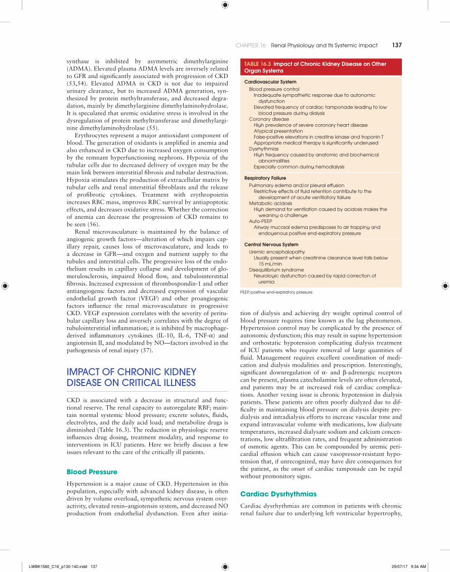
CKD is associated with a decrease in structural and func-tional reserve. The renal capacity to autoregulate RBF; main-tain normal systemic blood pressure; excrete solutes, fluids, electrolytes, and the daily acid load; and metabolize drugs is diminished (Table 16.3). The reduction in physiologic reserve influences drug dosing, treatment modality, and response to interventions in ICU patients. Here we briefly discuss a few issues relevant to the care of the critically ill patients.
Blood Pressure
Hypertension is a major cause of CKD. Hypertension in this population, especially with advanced kidney disease, is often driven by volume overload, sympathetic nervous system over-activity, elevated renin–angiotensin system, and decreased NO production from endothelial dysfunction. Even after initia-
tion of dialysis and achieving dry weight optimal control of blood pressure requires time known as the lag phenomenon. Hypertension control may be complicated by the presence of autonomic dysfunction; this may result in supine hypertension and orthostatic hypotension complicating dialysis treatment of ICU patients who require removal of large quantities of fluid. Management requires excellent coordination of medi-cation and dialysis modalities and prescription. Interestingly, significant downregulation of α- and β-adrenergic receptors can be present, plasma catecholamine levels are often elevated, and patients may be at increased risk of cardiac complica-tions. Another vexing issue is chronic hypotension in dialysis patients. These patients are often poorly dialyzed due to dif-ficulty in maintaining blood pressure on dialysis despite pre-dialysis and intradialysis efforts to increase vascular tone and expand intravascular volume with medications, low dialysate temperatures, increased dialysate sodium and calcium concen-trations, low ultrafiltration rates, and frequent administration of osmotic agents. This can be compounded by uremic peri-cardial effusion which can cause vasopressor-resistant hypo-tension that, if unrecognized, may have dire consequences for the patient, as the onset of cardiac tamponade can be rapid without premonitory signs.
Cardiac Dysrhythmias
Cardiac dysrhythmias are common in patients with chronic renal failure due to underlying left ventricular hypertrophy,
TABLE 16.3 Impact of Chronic Kidney Disease on Other Organ Systems
Cardiovascular System Blood pressure control
Inadequate sympathetic response due to autonomic dysfunction
Elevated frequency of cardiac tamponade leading to low blood pressure during dialysis
Coronary diseaseHigh prevalence of severe coronary heart diseaseAtypical presentationFalse-positive elevations in creatine kinase and troponin TAppropriate medical therapy is significantly underused
DysrhythmiasHigh frequency caused by anatomic and biochemical
abnormalitiesEspecially common during hemodialysis
Respiratory Failure Pulmonary edema and/or pleural effusion
Restrictive effects of fluid retention contribute to the development of acute ventilatory failure
Metabolic acidosisHigh demand for ventilation caused by acidosis makes the
weaning a challenge Auto-PEEP
Airway mucosal edema predisposes to air trapping and endogenous positive end-expiratory pressure
Central Nervous System Uremic encephalopathy
Usually present when creatinine clearance level falls below 15 mL/min
Disequilibrium syndromeNeurologic dysfunction caused by rapid correction of
uremia
PEEP, positive end-expiratory pressure.
LWBK1580_C16_p130-140.indd 137 29/07/17 9:34 AM

138 SECTion 2 PHYSIOLOGY OF CRITICAL ILLNESS
calcific cardiomyopathy that involves the conducting tissues, a “disturbed” metabolic milieu, and chronic tissue hypoxia. An increased incidence of cardiac dysrhythmias is seen in patients with postoperative AKI that may be exacerbated with rapid fluctuations in hemodynamics and electrolyte concentrations in those requiring dialysis support. Atrial fibrillation is common in dialysis patients and its onset is more frequent on dialysis days. In case of hemodynamic instability, synchronized cardioversion is indicated for the treatment of atrial fibrillation. The adverse effects of antiar-rhythmic medications are more common in reduced kidney function.
Chest Pain
The presence of renal dysfunction can influence the symptoms, manifestations, and progression of coronary syndromes. CKD affects outcome in patients with acute coronary syndrome and is an independent risk factor for the development of coronary artery disease and for more severe coronary heart disease; CKD is also associated with an adverse effect on prognosis from cardiovascular disease. Many dialysis patients with angina often have a fairly typical history of exercise-induced chest discomfort that is similar to those with normal renal function. However, silent myocardial ischemia is also common among patients with severe kidney disease. It has been speculated that the extremely poor prognosis among dialysis patients with an acute myocardial infarction may be due in part to a relatively increased number of atypical clinical presentations, resulting in both underdiagnosis and undertreatment. The presence of dyspnea alone due to an acute myocardial infarction in an individual scheduled to undergo a regular chronic dialysis procedure may be mistakenly attributed to volume overload. In addition, baseline abnormalities on the electrocardiogram, such as left ventricular hypertrophy, may mask characteristic changes with ischemia.
Disordered Consciousness
Uremic encephalopathy is an organic brain disorder that develops in patients with acute or chronic renal failure, usu-ally when creatinine clearance falls, and remains, below 15 mL/min. Accumulation of toxins, increases in intracellu-lar concentration of calcium in brain cells, and imbalances of neurotransmitter amino acids within the brain are thought to be responsible, although urea itself is not thought to be caus-ative. Clinical manifestations vary with worsening uremia, but prompt identification and initiation of dialysis treatment can readily reverse the symptoms. Initiation of dialysis treatment can also lead to disordered consciousness, especially when advanced states of uremia are dialyzed for excessive lengths of time during their first treatment sessions—the dialysis disequi-librium syndrome.
geneRal PRIncIPleS of management
Despite the remarkable progress achieved in understanding the pathophysiology of AKI, no specific pharmacologic agent has
yet been approved for its treatment and prevention remains the principal element in its management.
The treatment of established AKI is based on the following principles:• The preservation of RBF and optimal perfusion pressure
favorably influences the deterioration of renal function.• Correction of uremia, electrolyte, acid–base, endocrine,
hematologic and nutritional disorders, and fluid imbalance can favorably affect outcome.
• The pharmacokinetics and clearance of drugs are altered in renal failure, and the appropriate dosage adjustment requires knowledge of the pharmacokinetic parameters of the drugs and clearance characteristics of the different renal replacement techniques.
• The treatment and complications of the primary conditions (e.g., sepsis) that causes AKI may determine the outcome of AKI.
• The appropriate integration of care provided by intensivists and organ specialists can favorably affect outcome.In certain clinical situations, such as in cardiovascular
surgery, ischemic AKI is strongly associated with occult renal ischemia—associated with poor cardiac performance, fixed atherosclerotic disease of the renal arteries and/or prolonged hypoxemia, and reduced renal functional reserve. Due to the silent nature of renal ischemia, prognostic stratification using reliable surrogates can guide clinical decision making (58–61). Recently, the use of atrial natriuretic peptides has been shown to improve dialysis-free survival in thoracic aortic aneurysm surgery patients with impaired renal function. However, the use of atrial peptides in AKI remains controversial—two major clinical trials have reported unfavorable outcomes whereas two smaller clinical trials have shown favorable outcomes.
The prevalence of AKI continues to rise; however, there are indications that the mortality of patients with this disorder may be declining. This decline in mortality is not due to the effects of newer drugs in the treatment, but rather, it is due to the increased cooperation between the intensivists and subspe-cialists, which has led to a concerted approach to treatment. This has resulted in increased awareness of disease states, early initiation and higher doses of dialysis treatments, maintenance of euglycemia, and other interventions that play important roles in reversing mortality.
• Prevalence of AKI continues to increase.• Mortality of AKI, despite improvements, remains high.• Loss of autoregulation of RBF, vasoconstriction, and
subsequent downstream effects potentiate the inflam-matory cascade in AKI.
• Pre-existing organ dysfunction affects the prognosis of critically ill patients.
• Reduced kidney function as well as different renal replacement modalities influence drug dosing and response to interventions in the ICU.
• The treatment of AKI is based on the principle that the preservation of RBF and optimal perfusion pressure improves outcomes.
• AKI is a common finding in hospitalized patients.
Key Points
LWBK1580_C16_p130-140.indd 138 29/07/17 9:34 AM

CHAPTER 16 Renal Physiology and Its Systemic Impact 139
References 1. Mehta RL, Kellum JA, Shah SV, et al; Acute Kidney Injury Network. Report
of an initiative to improve outcomes in acute kidney injury. Crit Care. 2007; 11:R31.
2. Bedford M, Stevens PE, Wheeler TW, Farmer CK. What is the real impact of acute kidney injury?. BMC Nephrol. 2014;15:95.
3. Nisula S, Kaukonen KM, Vaara ST, et al; FINNAKI Study Group. Inci-dence, risk factors and 90-day mortality of patients with acute kidney injury in Finnish intensive care units: the FINNAKI study. Intensive Care Med. 2013;39:420–428.
4. Susantitaphong P, Cruz DN, Cerda J, et al; Acute Kidney Injury Advisory Group of the American Society of Nephrology. World incidence of AKI: a meta-analysis. Clin J Am Soc Nephrol. 2013;8:1482–1493.
5. Gallagher M, Cass A, Bellomo R, et al; POST-RENAL Study Investigators and the ANZICS Clinical Trials Group. Long-term survival and dialysis dependency following acute kidney injury in intensive care: extended fol-low-up of a randomized controlled trial. PLoS Med. 2014;11:e1001601.
6. Adams PL, Adams FF, Bell PD, Navar LG. Impaired renal blood flow auto-regulation in ischemic acute kidney injury. Kidney Int. 1980;18:68–76.
7. Kelleher SP, Robinette JB, Conger JD. Sympathetic nervous system in the loss of autoregulation in acute kidney injury. Am J Physiol. 1984;246: F379–F386.
8. Abuelo JG. Normotensive ischemic acute kidney injury. N Engl J Med. 2007; 357:797–805.
9. Devarajan P. Update on mechanisms of ischemic acute kidney injury. J Am Soc Nephrol. 2006;17:1503–1520.
10. Hsu CY, Ordoñez JD, Chertow GM, et al. The risk of acute kidney injury in patients with chronic kidney disease. Kidney Int. 2008;74:101–107.
11. Chawla LS, Eggers PW, Star RA, Kimmel PL. Acute kidney injury and chronic kidney disease as interconnected syndromes. N Engl J Med. 2014;371: 58–66.
12. Alsous F, Khamiees M, DeGirolamo A, et al. Negative fluid balance predicts survival in patients with septic shock: a retrospective pilot study. Chest. 2000;117:1749–1754.
13. Wiedemann HP, Wheeler AP, Bernard GR, et al; National Heart, Lung, and Blood Institute Acute Respiratory Distress Syndrome (ARDS) Clinical Tri-als Network. Comparison of two fluid-management strategies in acute lung injury. N Engl J Med. 2006;354:2564–2575.
14. Dass B, Shimada M, Kambhampati G, et al. Fluid balance as an early indica-tor of acute kidney injury in CV surgery. Clin Nephrol. 2012;77:438–444.
15. Kambhampati G, Ejaz NI, Asmar A, et al. Fluid balance and conventional and novel biomarkers of acute kidney injury in cardiovascular surgery. J Cardiovasc Surg (Torino). 2013;54:639–646.
16. Bouchard J, Soroko SB, Chertow GM, et al; Program to Improve Care in Acute Renal Disease (PICARD) Study Group. Fluid accumulation, survival and recovery of kidney function in critically ill patients with acute kidney injury. Kidney Int. 2009;76:422–427.
17. Kambhampati G, Ross EA, Alsabbagh MM, et al. Perioperative fluid bal-ance and acute kidney injury. Clin Exp Nephrol. 2012;16:730–738.
18. Parke RL, McGuinness SP, Gilder E, McCarthy LW. Intravenous fluid use after cardiac surgery: a multicentre, prospective, observational study. Crit Care Resusc. 2014;16:164–169.
19. Hassinger AB, Wald EL, Goodman DM. Early postoperative fluid overload precedes acute kidney injury and is associated with higher morbidity in pedi-atric cardiac surgery patients. Pediatr Crit Care Med. 2014;15:131–138.
20. van der Heijden M, Pickkers P, van NieuwAmerongenGP, et al. Circulat-ing angiopoietin-2 levels in the course of septic shock: relation with fluid balance, pulmonary dysfunction and mortality. Intensive Care Med. 2009; 35:1567–1574.
21. Basu RK, Wheeler DS. Kidney-lung cross-talk and acute kidney injury. Pedi-atr Nephrol. 2013;28:2239–2248.
22. Horiguchi Y, Uchiyama A, Iguchi N, et al. Perioperative fluid balance affects staging of acute kidney injury in postsurgical patients: a retrospective case-control study. J Intensive Care. 2014;2:26–32.
23. Pham PT, Slavov C, Pham PC. Acute kidney injury after liver, heart, and lung transplants: dialysis modality, predictors of renal function recovery, and impact on survival. Adv Chronic Kidney Dis. 2009;16:256–267.
24. Xue J, Wang L, Chen CM, et al. Acute kidney injury influences mortality in lung transplantation. Ren Fail. 2014;36:541–545.
25. Wehbe E, Duncan AE, Dar G, et al. Recovery from AKI and short- and long-term outcomes after lung transplantation. Clin J Am Soc Nephrol. 2013;8:19–25.
26. Rocha PN, Rocha AT, Palmer SM, et al. Acute kidney injury after lung transplantation: incidence, predictors and impact on perioperative morbid-ity and mortality. Am J Transplant. 2005;5:1469–1476.
27. Osho AA, Castleberry AW, Snyder LD, et al. Assessment of different thresh-old preoperative glomerular filtration rates as markers of outcomes in lung transplantation. Ann Thorac Surg. 2014;98:283–289.
28. Shavit L, Mikeladze I, Torem C, Slotki I. Mild hyponatremia is associated with functional and cognitive decline in chronic hemodialysis patients. Clin Nephrol. 2014;82:313–319.
29. Meyers TW, Hostetter TH. Uremia. N Engl J Med. 2007;357:1316–1325. 30. Watanabe K, Watanabe T, Nakayama M. Cerebro-renal interactions: impact
of uremic toxins on cognitive function. Neurotoxicology. 2014;44:184–193. 31. Sabatini S, Kurtzman NA. Bicarbonate therapy in severe metabolic acidosis.
J Am Soc Nephrol. 2009;20:692–695. 32. Fujiwara TM, Bichet DG. Molecular biology of hereditary diabetes insipi-
dus. J Am Soc Nephrol. 2005;16:2836–2846. 33. Bonny O, Rossier BC. Disturbances of Na/K balance: pseudohypoaldoste-
ronism revisited. J Am Soc Nephrol. 2002;13:2399–2414. 34. Ishani A, Xue JL, Himmelfarb J, et al. Acute kidney injury increases risk of
ESRD among elderly. J Am Soc Nephrol. 2009;20(1):223–228. 35. Shimoi T, Ando M, Munakata W, et al. The significant impact of acute kid-
ney injury on CKD in patients who survived over 10 years after myeloabla-tive allogeneic SCT. Bone Marrow Transplant. 2013;48:80–84.
36. Lo LJ, Go AS, Chertow GM, et al. Dialysis-requiring acute kidney injury increases the risk of progressive chronic kidney disease. Kidney Int. 2009; 76:893–899.
37. Venkatachalam MA, Griffin KA, Lan R, et al. Acute kidney injury: a springboard for progression in chronic kidney disease. Am J Physiol Renal Physiol. 2010;298:F1078–F1094.
38. Liano I, Liaño F, Pascual J; Madrid ARF Study Group. Epidemiology of acute kidney injury: a prospective, multicenter, community-based study. Kidney Int. 1996;50:811–818.
39. Clermont G, Acker CG, Angus DC, et al. Renal failure in the ICU: compari-son of the impact of acute kidney injury and end-stage renal disease on ICU outcomes. Kidney Int. 2002;62:986–996.
40. Uchino S, Kellum JA, Bellomo R, et al. Beginning and ending supportive therapy for the Kidney (BEST Kidney) Investigators. Acute kidney injury in critically ill patients: a multinational, multicenter study. JAMA. 2005; 294:813–818.
41. Hegarty J, Middleton RJ, Krebs M, et al. Severe acute kidney injury in adults: place of care, incidence and outcomes. QJM. 2005;98:661–666.
42. Wilson FP, Yang W, Machado CA, et al. Dialysis versus nondialysis in patients with AKI: a propensity-matched cohort study. Clin J Am Soc Nephrol. 2014;9:673–681.
43. Silvester W, Bellomo R, Cole L. Epidemiology, management, and outcome of severe acute kidney injury of critical illness in Australia. Crit Care Med. 2001;29:1910–1915.
44. Bagshaw SM, Laupland KB, Doig CJ, et al. Prognosis for long-term survival and renal recovery in critically ill patients with severe acute kidney injury: a population-based study. Crit Care. 2005;9:R700–R709.
45. Radhakrishnan J, Remuzzi G, Saran R, et al. Taming the chronic kidney disease epidemic: a global view of surveillance efforts. Kidney Int. 2014; 86:246–250.
46. Gilbertson DT, Liu J, Xue JL, et al. Projecting the number of patients with end-stage renal disease in the United States to the year 2015. J Am Soc Nephrol. 2005;16:3736–3741.
47. Go AS, Chertow GM, Fan D, et al. Chronic kidney disease and the risks of death, cardiovascular events, and hospitalization. N Engl J Med. 2004; 351:1296–1305.
48. Keith DS, Nichols GA, Gullion CM, et al. Longitudinal follow-up and out-comes among a population with chronic kidney disease in a large managed care organization. Arch Intern Med. 2004;164:659–663.
49. Brenner BM, Meyer TW, Hostetter TH. Dietary protein intake and the progressive nature of kidney disease: the role of hemodynamically medi-ated glomerular injury in the pathogenesis of progressive glomerular scle-rosis in aging, renal ablation, and intrinsic renal disease. N Engl J Med. 1982;307:652–659.
50. Kriz W, Elger M, Hosser H, et al. How does podocyte damage result in tubular damage? Kidney Blood Press Res. 1999;22:26–36.
51. Zandi-Nejad K, Eddy AA, Glassock RJ, et al. Why is proteinuria an ominous biomarker of progressive kidney disease? Kidney Int. 2004; 92:S76–S89.
52. Nangaku M. Chronic hypoxia and tubulointerstitial injury: a final common pathway to end-stage renal failure. J Am Soc Nephrol. 2006;17:17–25.
53. Fliser D, Kronenberg F, Kielstein JT, et al. Asymmetric dimethylarginine and progression of chronic kidney disease: the mild to moderate kidney disease study. J Am Soc Nephrol. 2005;16:2456–2461.
54. Ravani P, Tripepi G, Malberti F, et al. Asymmetrical dimethylarginine predicts progression to dialysis and death in patients with chronic kidney
LWBK1580_C16_p130-140.indd 139 29/07/17 9:34 AM

140 SECTion 2 PHYSIOLOGY OF CRITICAL ILLNESS
disease: a competing risks modeling approach. J Am Soc Nephrol. 2005;16: 2449–2455.
55. Matsuguma K, Ueda S, Yamagishi S, et al. Molecular mechanism for eleva-tion of asymmetric dimethylarginine and its role for hypertension in chronic kidney disease. J Am Soc Nephrol. 2006;17:2176–2183.
56. Rossert J, Fouqueray B, Boffa JJ. Anemia management and the delay of chronic renal failure progression. J Am Soc Nephrol. 2003;14:S173– S177.
57. Kang DH, Kanellis J, Hugo C, et al. Role of the microvascular endothelium in progressive renal disease. J Am Soc Nephrol. 2002;13:806–816.
58. Chertow GM, Lazarus JM, Christiansen CL, et al. Preoperative renal risk stratification. Circulation. 1997;95:878–884.
59. Chawla LS, Abell L, Mazhari R, et al. Identifying critically ill patients at high risk for developing acute kidney injury: a pilot study. Kidney Int. 2005; 68:2274–2280.
60. Yegenaga I, Hoste E, Van Biesen W, et al. Clinical characteristics of patients developing ARF due to sepsis/systemic inflammatory response. Am J Kidney Dis. 2004;43:817–824.
61. Vivino G, Antonelli M, Moro ML, et al. Risk factors for acute kidney injury in trauma patients. Intensive Care Med. 1998;24:808–814.
LWBK1580_C16_p130-140.indd 140 29/07/17 9:34 AM