Relationships between eye factors and lens-forming transformations in the cornea and pericorneal...
21
Relationships between Eye Factors and Lens-forming Transformations in the Cornea and Pericorneal Epidermis of Larval Xenopus laevis LUIGI BOSCO, SERGIO FILONI AND STEFAN0 CANNATA lstituto di Anatomia Comparata, Uniuersita di Roma, 00161 Roma, Italy ABSTRACT were subjected to various types of lentectomy: ner cornea; Larval Xenopus laevis at stage 56 (Nieuwkoop and Faber, '56) (1) simple lentectomy, from the pupillary space after incision of outer and in- (2) lentectomy from the dorsal region of the eye; (3) lentectomy from the dorsal region of the eye and simultaneous incision of the outer cornea; (4) lentectomy from the dorsal region of the eye and simultaneous incision of the outer and inner cornea. The results obtained show that the outer cornea underwent lens-forming transformations only when the inner cornea had been incised, thus permitting direct communication between the lentectomized eye environment and the outer cornea (Experiments I-IV). No lens regeneration occurred when the inner cornea was left intact (Experiments 11, 111). It was concluded that the factor(s) allowing the lens-forming transformations of the outer cornea is not an aspecific nutritional factor (s) but a more specific factor (s) that cannot reach the outer cornea when the inner cornea is intact. Therefore, the absence of the lens and sufficient nutrient available to the outer cornea are not enough to allow lens regeneration from the outer cornea. When lens removal was carried out through the dorsal part of the eye (Ex- periments 111-IV) the lens regenerated from the pericorneal epidermis of this region in a large number of cases. Freeman ('63) showed that the larva of Xenopus laevis has a high lens-regenerating capacity and that lens regeneration takes place at the expense of the outer cornea. This regenerating capacity disappears at metamor- phosis, i.e., at the time when the outer cornea, which is continuous with the epidermis, fuses with the inner cornea, which is continuous with the sclera. In the same work, this author showed that lens regeneration is contingent upon the presence of the eye: the outer cornea of enucleated larvae never regenerates a lens. Campbell and Jones ('68) achieved lens re- generation from outer cornea fragments culti- vated in vitro. They claim that their attempt was successful because, under their experi- mental conditions, the outer cornea had all the nutritional factors it needed to develop its latent lens-forming capacity. In fact, these workers hypothesized that the outer cornea J. EXP. ZOOL. (1979) 209: 261-282. has an intrinsic lens-forming capacity that oc- curs, in the absence of the lens, whenever suf- ficient nutritive substances are available for it. According to Campbell and Jones, one pos- sible explanation of the difference between their results and those obtained in vivo by Freeman ('63) may be ascribed to". . . the dif- ference in the availability of nutrients to the cornea in the two cases." As it involves re- moval of the aqueous humor, enucleation would thus alter cornea metabolism and in- hibit its lens-forming transformations. Waggoner ('73) implanted the outer corneas of larval Xenopus laevis into various parts of the body of host larvae (lentectomized eye, dorsal fin, subcutaneous region of the head, amputated hindlimbs) and observed, that the cornea underwent lens-forming transforma- tions only when grafted into the lentectom- ized eye or the amputated hindlimbs. He 261
-
Upload
luigi-bosco -
Category
Documents
-
view
212 -
download
0
Transcript of Relationships between eye factors and lens-forming transformations in the cornea and pericorneal...
Relationships between Eye Factors and Lens-forming Transformations in the Cornea and Pericorneal Epidermis of Larval Xenopus laevis
LUIGI BOSCO, SERGIO FILONI AND STEFAN0 CANNATA lstituto di Anatomia Comparata, Uniuersita di Roma, 00161 Roma, Italy
ABSTRACT were subjected to various types of lentectomy:
ner cornea;
Larval Xenopus laevis at stage 56 (Nieuwkoop and Faber, '56)
(1) simple lentectomy, from the pupillary space after incision of outer and in-
(2) lentectomy from the dorsal region of the eye; (3) lentectomy from the dorsal region of the eye and simultaneous incision of
the outer cornea; (4) lentectomy from the dorsal region of the eye and simultaneous incision of
the outer and inner cornea. The results obtained show that the outer cornea underwent lens-forming
transformations only when the inner cornea had been incised, thus permitting direct communication between the lentectomized eye environment and the outer cornea (Experiments I-IV). No lens regeneration occurred when the inner cornea was left intact (Experiments 11, 111). It was concluded that the factor(s) allowing the lens-forming transformations of the outer cornea is not an aspecific nutritional factor (s) but a more specific factor (s) that cannot reach the outer cornea when the inner cornea is intact. Therefore, the absence of the lens and sufficient nutrient available to the outer cornea are not enough to allow lens regeneration from the outer cornea.
When lens removal was carried out through the dorsal part of the eye (Ex- periments 111-IV) the lens regenerated from the pericorneal epidermis of this region in a large number of cases.
Freeman ('63) showed that the larva of Xenopus laevis has a high lens-regenerating capacity and that lens regeneration takes place a t the expense of the outer cornea. This regenerating capacity disappears a t metamor- phosis, i.e., at the time when the outer cornea, which is continuous with the epidermis, fuses with the inner cornea, which is continuous with the sclera. In the same work, this author showed that lens regeneration is contingent upon the presence of the eye: the outer cornea of enucleated larvae never regenerates a lens.
Campbell and Jones ('68) achieved lens re- generation from outer cornea fragments culti- vated in vitro. They claim that their attempt was successful because, under their experi- mental conditions, the outer cornea had all the nutritional factors it needed to develop its latent lens-forming capacity. In fact, these workers hypothesized that the outer cornea
J. EXP. ZOOL. (1979) 209: 261-282.
has an intrinsic lens-forming capacity that oc- curs, in the absence of the lens, whenever suf- ficient nutritive substances are available for it. According to Campbell and Jones, one pos- sible explanation of the difference between their results and those obtained in vivo by Freeman ('63) may be ascribed to". . . the dif- ference in the availability of nutrients to the cornea in the two cases." As it involves re- moval of the aqueous humor, enucleation would thus alter cornea metabolism and in- hibit its lens-forming transformations.
Waggoner ('73) implanted the outer corneas of larval Xenopus laevis into various parts of the body of host larvae (lentectomized eye, dorsal fin, subcutaneous region of the head, amputated hindlimbs) and observed, that the cornea underwent lens-forming transforma- tions only when grafted into the lentectom- ized eye or the amputated hindlimbs. He
261
262 LUIGI BOSCO, SERGIO FILONI AND STEFAN0 CANNATA
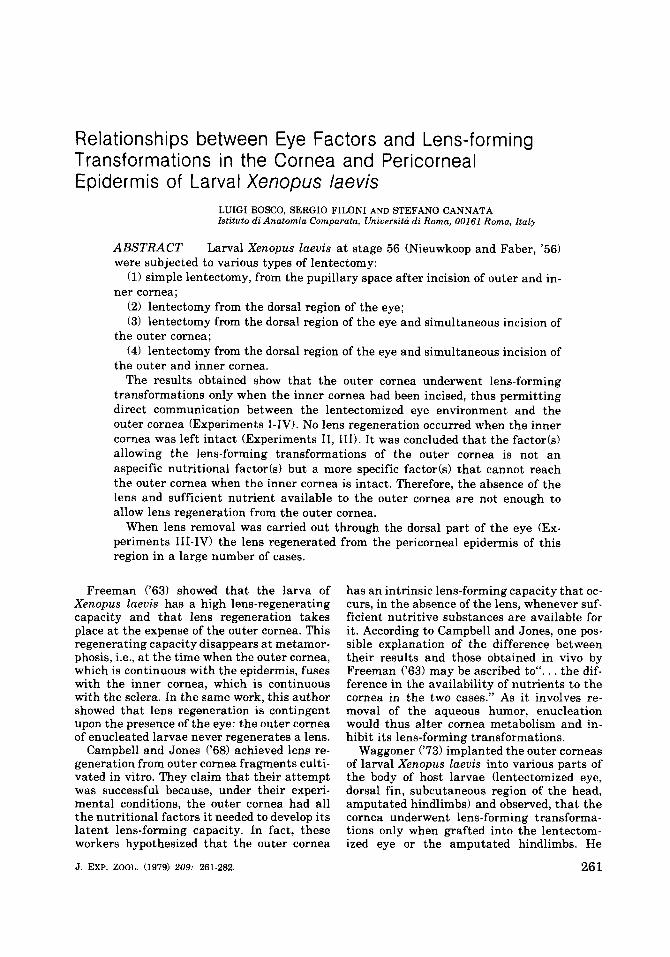
Fig. 1 Diagram of the various types of operation. A Simple lentectomy (Experiment I). B Extirpation of the lens from the dorsal region of the eye (Experiment 11). C Extirpation of the lens from the dorsal region of the eye and simultaneous incision of the outer
D Extirpation of the lens from the dorsal region of the eye and simultaneous incision of the outer cornea (Experiment 111).
and inner cornea (Experiment IV).
assumed that there was a factor(s1 in these two environments which could stimulate the cornea to transform into a lens.
In recent work (Filoni et al., '78a) on lentec- tomized Xenopus laevis larvae which were enucleated at fixed intervals after lentec- tomy, we observed that the presence of the eye is necessary not only for triggering regenera- tion but also for its later development.
The present investigation was undertaken to ascertain whether the lens-forming trans- formations observed in the outer cornea of lentectomized larval Xenopus laevis were due to a nutritional factor(s1 which normally reaches the outer cornea after passing through the inner cornea, or to a more specific factor(s1 that normally does not go as far as the outer cornea, reaching i t only after lentec- tomy. First data concerning this problem have been presented in our preliminary report (Filoni et al., '78b).
MATERIALS AND METHODS
Xenopus laevis larvae at stage 55-56 were used in all the experiments. Larvae were
reared from eggs obtained by injecting adults with Pregnyl (Organon) as described by Nieuwkoop and Faber ('56). During the opera- tion, which was effected on only one of the two eyes, the animals were immersed in full strength Holtfreter's solution and kept there for at least 24 hours after the operation. Then they were gradually transferred to dechlori- nated tap water and fed on nettle powder. Entire animals were fixed in Bouin's solution and embedded in paraffin. Seven-ten micron serial sections were cut and stained with he- matoxylin-eosin, by the Mallory- Azan method or treated by PAS reaction.
Experiment I. Simple lentectomy (fig. 1) Forty larvae were operated. An incision was
made in the outer and inner cornea in the vicinity of the pupillary space. Total lens re- moval was carried out by aspiration with a micropipette having a diameter closely ap- proximating that of the pupillary space. At the time of removal, each lens was examined under the binocular dissection microscope; lenses that were found to be deformed were
LENS REGENERATION IN LARVAL XENOPUS LAEVIS 263
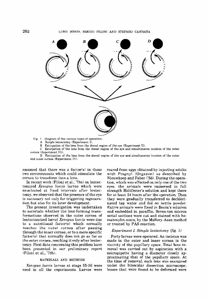
TABLE 1
Summarv o f the results of the operations for the larvae sacrificed 5, 7, 10, and I5 days after lentectomy
Lens regeneration from
Experiment Number of Number of Number of animals dead an i m a 1s Dorsal part Outer ouerations or discarded studied of the eye cornea
I 40 - 40 - 21 I1 50 6 44 18 I11 50 9 41 33 IV 50 1 49 22 12
- -
TABLE 2
Summary of lens regeneration for the animals in experiment I ~~~~ ~ ~ ~
Lens regeneration No. of stage Cases cases No. of of no
Days after
operation examined regenerates 3 4 5 regeneration
5 10 6 6 - 4 7 10 5 4 I - 5
10 10 6 - 5 1 4 15 10 4 - - 4 6
-
I In all cases, lens regeneration started from the outer cornea
discarded. This technique has been successful- ly used by us in various Anuran species (Filoni et al., '76, '77, '78a). The larvae were sacri- ficed 5, 7, 10 and 15 days after the operation (table 1).
Experiment II. Extirpation of lens from the dorsal region of the eye (fig. 1)
Eighty larvae were operated. After incising the pericorneal epidermis in the dorsal region of the eye and the eye wall in the region of the neural retina near the ora serrata, a micropip- ette was inserted into the vitreous chamber and the lens aspirated. At the time of removal, each lens was examined under the binocular dissection microscope; lenses that were found to be deformed were discarded. In order to make sure that lens removal was complete through the use of this operating technique and to follow the early histological and mor- phological transformations in the operated area, 30 larvae were fixed, in groups of 5 indi- viduals, immediately 12 hours, 1, 2, 3, and 4 days after the operation, respectively. The other 50 larvae were sacrificed 5, 7,lO and 15 days after the operation (see table 1).
Experiment III. Extirpation of lens from dorsal region of the eye
and simultaneous incision of outer cornea (fig. 1)
Fifty larvae were operated. In this experi-
ment, before lens removal was carried out as in experiment 11, the outer cornea alone was incised in the region of the pupillary space. The larvae were sacrificed 5 , 7 , 1 0 and 15 days after the operation (table 1).
Experiment IV. Extirpation of lens from dorsal region of the eye and
simultaneous incision of the outer and inner cornea (fig. 1 )
Fifty larvae were operated. In this experi- ment, before lentectomy was performed as de- scribed in experiment 11, the outer and inner cornea were incised in the region of the pupil- lary space. The larvae were sacrificed 5, 7, 10 and 15 days after the operation (table 1).
RESULTS
In the description of the results Freeman's ('63) method of staging the regenerates was used.
Experiment I. Simple lentectomy Lens regeneration was observed in 2 1 of the
40 eyes examined (table 2). Regeneration always took place starting from the outer cor- nea in the typical sequence of phases demon- strated by Freemann ('631, i.e., formation of a cell aggregate in the inner layer of the outer cornea (early stage 3: day 5 after operation), evolution of aggregate into an epithelial vesi- cle that tends to detach itself from the outer

264 LUIGI BOSCO, SERGIO FILONI AND STEFAN0 CANNATA
TABLE 3
Summary of lens regeneration for the animals in experiment I1 sacrificed 5 to 15 days after lentectomy
Lens regeneration No. of stage Cases cases No. of of no
Days after
operation examined regenerates I 3 4 5 regeneration
5 8 I 8 10 9 15 19
' In all cases lens regeneration took place in the dorsal part of the eye.
cornea (middle and late stage 3: day 5-7 after operation), gradual transformation of vesicle cells, which occupy a more distal position with respect to the cornea in the primary lens fibers (stage 4: day 10 after operation), sec- ondary fiber formation a t the expense of the equatorial zone of the lens epithelium (stage 5: day 15 after operation) (figs. 2-5).
Experiment II. Extirpation of lens from the dorsal part of the eye
Examination of the operated eyes of 30 lar- vae sacrificed immediately after the opera- tion, and in the period from 12 hours to 4 days after the operation, has shown that the operating technique we used to perform len- tectomy through the dorsal part of the eye allows total lens removal. In none of the cases examined were any fragments of the lens ob- served. In most cases, several retinal protru- sions can be observed in the dorsal part of the eye 12 hours after the operation. At this stage the epidermis begins to migrate to the oper- ated area and tends to cover the retinal pro- trusions. One day after lentectomy, the epi- dermis is already quite extensive but does not yet cover the eye completely. This occurs a t days 2-3 after the operation. At this stage, the epidermis, which was found to be thickened as early as day 1, is formed by numerous cell layers. At days 3-4 after the operation a round aggregate of epithelial cells forms in this multi-layered epidermis in the vicinity of the incision made in the eye wall for the lentec- tomy. This cell aggregate roughly resembles the one observed a t stage 2 or early stage 3 of lens regeneration from the outer cornea.
Of the other 50 larvae operated, one died during the experiment and five others were discarded because the operated eyes were much reduced in size. In 18 out of the 44 eyes, examined between day 5 and day 15 after operation, lens regeneration was in progress.
In all 18 cases the lens regenerated in the dor- sal part of the eye from which the lens had been removed (table 3).
In the five cases of regeneration examined five days after operation, the pericorneal epi- dermis, damaged during lentectomy, was found to be considerably thickened, and in its deeper portion the epithelial cells had been or- ganized into a lens vesicle a t stage 3. The eye wall was partly closed off by a proliferation of connective tissue which, in some cases, was very extensive (fig. 6). Around the edges of the wound in the retina, which had come together but not merged, numerous mitoses could be ob- served.
Of the five cases of regeneration observed seven days after operation, three were at late stage 3, one a t late stage 4 (primary fiber for- mation) (fig. 7), and one a t early stage 5 (ini- tial formation of secondary fibers). In the first four cases, the lens vesicle had penetrated the wound in the eye wall but was still extensively attached to the pericorneal epidermis by epi- thelial cells. In the fifth case, the lens was completely separate from the epidermis, lay within the retina wound, and was covered by the regenerating choroid and sclera. In the four cases of regeneration examined after ten days, the differentiation of the new lens was a t an advanced stage, i.e., the nuclei of the pri- mary fibers had disappeared and secondary fiber formation had begun at the expense of the equatorial region of the peripheral lens portion, which had become lens epithelium (stage 5).
In two cases the regenerated lens, which oc- cupied the vitreous chamber, was separated from the pericorneal epidermis, although still partially connected to the connective tissue proliferation closing the wound made in the eye wall (fig. 8).
In two further cases the new lens was situ- ated in the wound against the retina. In one

LENS REGENERATION IN LARVAL XENOPUS LAEVIS
TABLE 4
Summary of lens regeneration for the animals of the experiment I l l
265
Lens regeneration No. of stage Cases cases No. of of no
Days after
operation examined regenerates ' 3 4 5 regeneration
5 10 7 10
10 I 15 14
10 2
6 6 11
' In all cases lens regeneration took place in the dorsal part of the eye.
3The remaining two lenses were formed of an atypical mass of fibers within the thickness of the retina. T w o of these lenses were present in the same eye.
particular case it was almost completely en- closed by a mass of retinal cells. The slightly thickened pericorneal epidermis did not dis- play any connection with the lens. In only one of the four cases of regeneration examined after 15 days was the regenerated lens still connected to the pericorneal epidermis. This connection was provided by the basement membrane of the epidermis which was con- tinuous with the lens capsule. The lens itself was at stage 5, had a large volume, and ex- tended into the vitreous chamber. In the other three cases the lens, at stage 5, displayed no connection with the epidermis. It was partial- ly (2 cases) or completely (1 case) contained in the thickness of the retina in the area of the completely healed wound (figs. 9, 22).
In the remaining 26 cases, in which lens re- generation had not occurred, a smaller thick- ening of the pericorneal epidermis could be ob- served as well as a faster healing of the wound in the eye wall compared with the cases in which lens regeneration had occurred.
In none of the 44 cases examined in this ex- periment did the outer or inner cornea display any significant histological modifications.
Experiment IIZ. Extirpation of the lens from the dorsal region of the eye
and simultaneous incision of outer cornea
Four out of fifty larvae operated died during the experiment. Five eyes were discarded as being too reduced in size. Thirty three cases of lens regeneration were observed in a total of 41 eyes examined between 5 and 15 days after the operation. In all 33 cases, lens regenera- tion took place in that region of the dorsal part of the eye from which the lens had been re- moved (table 4).
At day 5 after the operation, the lens regen-
eration process was observed in nine out of the ten eyes examined.
In six eyes, the origin of the regenerating lens could clearly be traced back to the peri- corneal epidermis which had thickened con- siderably. In fact the lens vesicle, which had reached stage 3 or early stage 4, was either situated in the deep layer of the pericorneal epidermis affected by the operation, or else, after penetrating the opening made in the eye wall, had remained extensively attached to the pericorneal epidermis itself by an epithe- lial cluster (fig. 10). In one of these six eyes, two epithelial vesicles at stage 3 were present in the deep layer of the pericorneal epidermis.
In two out of the three cases of lens regen- eration, all a t stage 3, observed a t day 5 after the operation, the lens vesicle, although near a thickening in the pericorneal epidermis, was separate from it; in the remaining case, the regenerating lens was contained within the retina.
In the six cases of lens regeneration ob- served at day 7 after the operation, the territo- ry of origin of the regenerate could clearly be seen to be the pericorneal epidermis lying opposite the opening made in the dorsal wall of the eye: the regenerating lens was still con- nected to the pericorneal epidermis.
However, this connection was less extensive than that observed a t day 5 after the opera- tion as the regenerating lens had shifted inwards and now occupied the opening in the eye wall. This connection consisted either of the basement membrane of the epidermis, which was continuous with the peripheral por- tion of the lens vesicle, or of a clump of epithe- lial cells.
In four cases the lens vesicle was at late stage 3 and in two cases a t stage 4, as primary fiber formation had begun (fig. 11). In only

266 LUIGI BOSCO, SERGIO FILONI AND STEFAN0 CANNATA
TABLE 5
Summary of lens regeneration fur the animals ufthe experiment IV
Lena regeneration Lens regeneration No. of No. of stage No. of stage Cases
regenerates regenerates of no
Days after
operation cases examined Group a ’ 3 4 5 Group h 3 4 5 regeneration
1 5 i n 6 3 6 - - 5 3 5 - - I i n 1 1 - - 5 3 2 - 4 10 10 2 - 1 1 5 4 - 1 4 4
7 9 15 19 3 - - 3 7 - -
’ Comprising cases of lens regeneration from the outer cornea Compnsing cases of lens regeneration from the dorsal part of the eye In two cases two lenses were present in the same eye. one derived from the outer cornea (group a), the other from the dorsal part of the
eye (group b) ‘Two of these lenses were present in the same eye
two of the six cases of regeneration observed a t day 10 after the operation, the lens (stage 4) was connected to the pericorneal epidermis. In three cases the new lens (stage 5 ) was con- tained within the thickness of the eye wall (in the zone damaged by the operation) but was not connected to the epidermis (fig. 12). In the sixth case, a lens (at stage 5 ) was present within an aggregate of retinal cells pro- truding into the vitreous chamber (fig. 23). In only 2 out of 11 cases of regeneration exam- ined after 15 days was the regenerating lens, still at stage 3, connected to the pericorneal epidermis. In seven cases the lens was a t stage 5 . In two of these seven cases the lens was con- tained within the zone of the eye wall in which the opening had been cut. On healing, the wound had mechanically compressed the re- generating lens, which was found to be bilo- bate, one lobe protruding towards the outside and the other towards the vitreous chamber (fig. 13). In three other cases, the new lens protruded into the vitreous chamber, while in 2 cases, although having lost any correlation with the pericorneal epidermis, the lens had been left on the outside of the eye wall.
In the remaining 2 cases it was possible to observe a round mass of regenerating lens fibers within the thickness of the retina (fig. 24). In the other eight cases, in which lens re- generation had not occurred, there was a smaller thickening of the pericorneal epider- mis and a more rapid healing of the wound in the eye wall than in the cases in which lens re- generation occurred. In none of the 41 cases examined in this experiment were any histo- logical transformations observed in the outer cornea indicative of lens regeneration. Espe- cially in the first few days after operation, in some cases the outer cornea was thickened, al- though these thickenings are to be considered as due to scar tissue.
Experiment ZV. Extirpation of lens from dorsal region of eye and simultaneous
incision of outer and inner cornea Forty-nine of the 50 larvae operated were
used. One was discarded as the right eye was much reduced in size.
Lens regeneration was observed in 34 out of the 49 eyes examined between days 5 and 15. Unlike the eyes operated in Experiments I1 and 111, in which the regenerated lens formed solely in the dorsal region of the eye affected by the lentectomy operation, in this experi- ment there was a dual lens origin, i.e., from the outer cornea or from the dorsal region of the eye. In two cases two lenses were present in the same eye, one from the dorsal part of the eye, the other from the cornea (see table 5 ) .
Regenerates were divided into two groups according to their origin:
Group a: comprising cases of lens- regeneration from the outer cornea
This type of regeneration was observed in 12 cases examined between days 5 and 15 after the operation. In all cases, the histological transformations of the outer cornea and the histological evolution of the new lens vesicle corresponded exactly, under the same time and fixation conditions, t o those of the regen- eration cases in Experiment I. Except for one lens belonging to an eye fixed after 15 days, all the other lenses were connected to the outer cornea (figs. 14-17).
Group b: comprising cases of lens- regeneration from the dorsal part of the eye
This type of regeneration was observed in 22 cases examined between days 5 and 15 after the operation. Out of the five regenerating
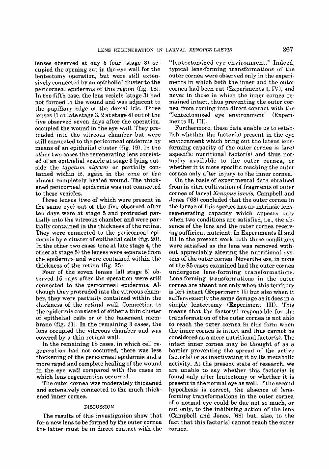
LENS REGENERATION IN LARVAL XENOPUS LAEVIS 267
lenses observed a t day 5 four (stage 3) oc- cupied the opening cut in the eye wall for the lentectomy operation, but were still exten- sively connected by an epithelial cluster to the pericorneal epidermis of this region (fig. 18). In the fifth case, the lens vesicle (stage 3) had not formed in the wound and was adjacent to the pupillary edge of the dorsal iris. Three lenses (1 a t late stage 3 , 2 a t stage 4) out of the five observed seven days after the operation, occupied the wound in the eye wall. They pro- truded into the vitreous chamber but were still connected to the pericorneal epidermis by means of an epithelial cluster (fig. 19). In the other two cases the regenerating lens consist- ed of an epithelial vesicle a t stage 3 lying out- side the tapetum nigrum or partially con- tained within it, again in the zone of the almost completely healed wound. The thick- ened pericorneal epidermis was not connected to these vesicles.
Three lenses (two of which were present in the same eye) out of the five observed after ten days were a t stage 5 and protruded par- tially into the vitreous chamber and were par- tially contained in the thickness of the retina. They were connected to the pericorneal epi- dermis by a cluster of epithelial cells (fig. 20). In the other two cases (one a t late stage 4, the other at stage 5) the lenses were separate from the epidermis and were contained within the thickness of the retina (fig. 25) .
Four of the seven lenses (all stage 5) ob- served 15 days after the operation were still connected to the pericorneal epidermis. Al- though they protruded into the vitreous cham- ber, they were partially contained within the thickness of the retinal wall. Connection to the epidermis consisted of either a thin cluster of epithelial cells or of the basement mem- brane (fig. 21). In the remaining 3 cases, the lens occupied the vitreous chamber and was covered by a thin retinal wall.
In the remaining 18 cases, in which cell re- generation had not occurred, there was less thickening of the pericorneal epidermis and a more rapid and complete healing of the wound in the eye wall compared with the cases in which lens regeneration occurred.
The outer cornea was moderately thickened and extensively connected to the much thick- ened inner cornea.
DISCUSSION
The results of this investigation show that for a new lens to be formed by the outer cornea the latter must be in direct contact with the
“lentectomized eye environment.” Indeed, typical lens-forming transformations of the outer cornea were observed only in the experi- ments in which both the inner and the outer cornea had been cut (Experiments I, IV), and never in those in which the inner cornea re- mained intact, thus preventing the outer cor- nea from coming into direct contact with the “lentectomized eye environment” (Experi- ments 11, 111).
Furthermore, these data enable us to estab- lish whether the factor(s1 present in the eye environment which bring out the latent lens- forming capacity of the outer cornea is (are) aspecific nutritional factor(s1 and thus nor- mally available to the outer cornea, or whether i t is more specific reaching the outer cornea only after injury to the inner cornea.
On the basis of experimental data obtained from in vitro cultivation of fragments of outer cornea of larval Xenopus laevis, Campbell and Jones (‘68) concluded that the outer cornea in the larvae of this species has an intrinsic lens- regenerating capacity which appears only when two conditions are satisfied, i.e., the ab- sence of the lens and the outer cornea receiv- ing sufficient nutrient. In Experiments I1 and I11 in the present work both these conditions were satisfied as the lens was removed with- out appreciably altering the nutritional sys- tem of the outer cornea. Nevertheless, in none of the 85 cases examined had the outer cornea undergone lens-forming transformations. Lens-forming transformations in the outer cornea are absent not only when this territory is left intact (Experiment 11) but also when it suffers exactly the same damage as it does in a simple lentectomy (Experiment 111). This means that the factor(s1 responsible for the transformation of the outer cornea is not able to reach the outer cornea in this form when the inner cornea is intact and thus cannot be considered as a mere nutritional factods). The intact inner cornea may be thought of as a barrier preventing the spread of the active factor(s1 or as inactivating i t by its metabolic activity. At the present state of research, we are unable to say whether this factor(s1 is found only after lentectomy or whether it is present in the normal eye as well. If the second hypothesis is correct, the absence of lens- forming transformations in the outer cornea of a normal eye could be due not so much, or not only, to the inhibiting action of the lens (Campbell and Jones, ’68) but, also, to the fact that this factor(s1 cannot reach the outer cornea.

268 LUIGI BOSCO, SERGIO FILONI AND STEFAN0 CANNATA
The results of Experiments 11, I11 and IV show that the pericorneal epidermis of the dorsal region of the eye in larval Xenopus laevis has a high lens-forming capacity. The lens-forming capacity of the pericorneal epi- dermis of younger larvae than those used in the present work had already been demon- strated by Freeman ('631, although for a dif- ferent purpose and with a different experi- mental technique. Thus, in order to ascertain the limits of the lens-forming capacity of the outer cornea, he removed the cornea itself with or without variously-sized portions of pericorneal epidermis. Freeman observed that when the entire outer cornea was removed, in a high percentage of cases the lens regener- ated from the pericorneal epidermis which had proliferated over the eye cup.
Our results demonstrate a lens-forming transformation of the pericorneal epidermis in situ and show that lens-regeneration a t the expense of this region takes place in phases that correspond to lens regeneration from the outer cornea facing the pupillary space. How- ever, analysis of the process has revealed cer- tain differences between the two types of re- generation: the thickening of the pericorneal epidermis is usually much greater than in the outer cornea and tends to remain connected to the lens vesicle formed in the deeper portion by means of a cluster of epithelial cells which follows the migration of the regenerating lens through the cut in the eye wall towards the vitreous chamber.
Also in this type of regeneration, the epider- mis must come into direct contact with the eye environment; in the case of failure to re- generate, rapid and complete healing of the wound is observed.
Something must also be said about the small number of cases of apparent lens-regen- eration from eye territories other than the cornea or the pericorneal epidermis. A few cases of lens regeneration in the retina struc- ture might be due to the lens being caught up in the retina while migrating towards the
vitreous chamber and later losing its connec- tion with the epidermis, while other cases point to a regeneration in situ.
Also noteworthy is the one recorded case of apparent lens regeneration from the dorsal edge of the iris in which the lens-forming vesi- cle was adjacent to the iris edge.
Previous workers (Overton and Freeman, '60; Campbell, '63) had already claimed that lens regeneration from the iris or the retina was possible in Xenopus laevis, although they did not give a sufficiently clear picture of the regeneration process. The possibility of lens regeneration from eye territories other than the outer cornea or the pericorneal epidermis thus needs further detailed study in order to be confirmed.
LITERATURE CITED
Campbell, J. C. 1963 Lens regeneration from iris, retina and cornea in lentectomized eyes ofXenopus Zaeuis. Anat. Rec., 145: 214.
Campbell, J. C., and K. W. Jones 1968 The in uitro develop ment of lens fron cornea of larval Xenopus laeuis. De- velop. Biol., 17: 1-15.
Filoni, S., L. Bosco and S. Cannata 1978a Influenza dell'occhio sulla rigenerazione del cristallino in larve di Xenopus laeuis. Acta Embryol. Exper., 2: 179-195.
1978b Prima dati sperimentali sui fattozi NPCPS sazi per la trasformazione lentogena della cornea ester- na di larve di Xenopus laeuis. Acta Embryol. Exper., 3; 344-345.
Filoni, S., L. Bosco and C. Cioni 1976 I1 problema della rigenerazione del cristallino degli Anfibi Anuri negli stadi postembrionaii. Experienze di asportazione del cristallino in larve di Rana esculenta e Xenopus laeuis. Acta Embryol. Exper., 3: 319-334.
I1 problema della rigenerazione del cris- tallino degli. Anfibi Anuri negli stadi postembrionali. 11. Esperienze di asportazione del cristallino in larve di Dis- coglossus pictus. Acta Embryol. Exper., 2: 155-162.
Freeman, G. 1963 Lens regeneration from the cornea in Xenopus laeuis. J. Exp. Zool., 154: 39-65.
Nieuwkoop, P. D., and J. Faber 1956 Normal table of Xenopus laeuis (Daudin). North-Holland Publishing Company, Amsterdam.
Overton, J., and G. Freeman 1960 Lens regeneration in Xenopus Zaeuis. Anat. Rec., 137: 386.
Waggoner, P. R. 1973 Lens differentiation from the cor- nea following lens extirpation or cornea transplantation in Xenopus Zaeuis. J. Exp. Zool., 186: 97-109.
1977

PLATES

PLATE 1
EXPLANATION OF FIGURES
Figs. 2-5 Regeneration of lens after simple lentectomy (Experiment I). All cases presented in these figures were stained with Mallory-Azan’s method. All photographs were taken a t a magnification of x 350.
2 Five days after operation: In the deep layer of the outer cornea a cell aggregate has formed (stage 3).
3 S e w n days after operation: the cells of the aggregate have formed into a spheroidal structure which tends to detach itself from the other cells of the outer cornea (late stage 3).
4 Ten days after operation: the formation of the primary nucleus of the lens fibers can he seen (stage 4).
5 Fifteen days after operation: on the outside of the primary nucleus the secondary fibers have formed (stage 5).
270

LENS REGENERATION IN LARVAL XENOPUS LAEVIS Luigi Bosco, Sergio Filoni and Stefan0 Cannata
PLATE 1
271
PLATE 2
EXPLANATION OF FIGURES
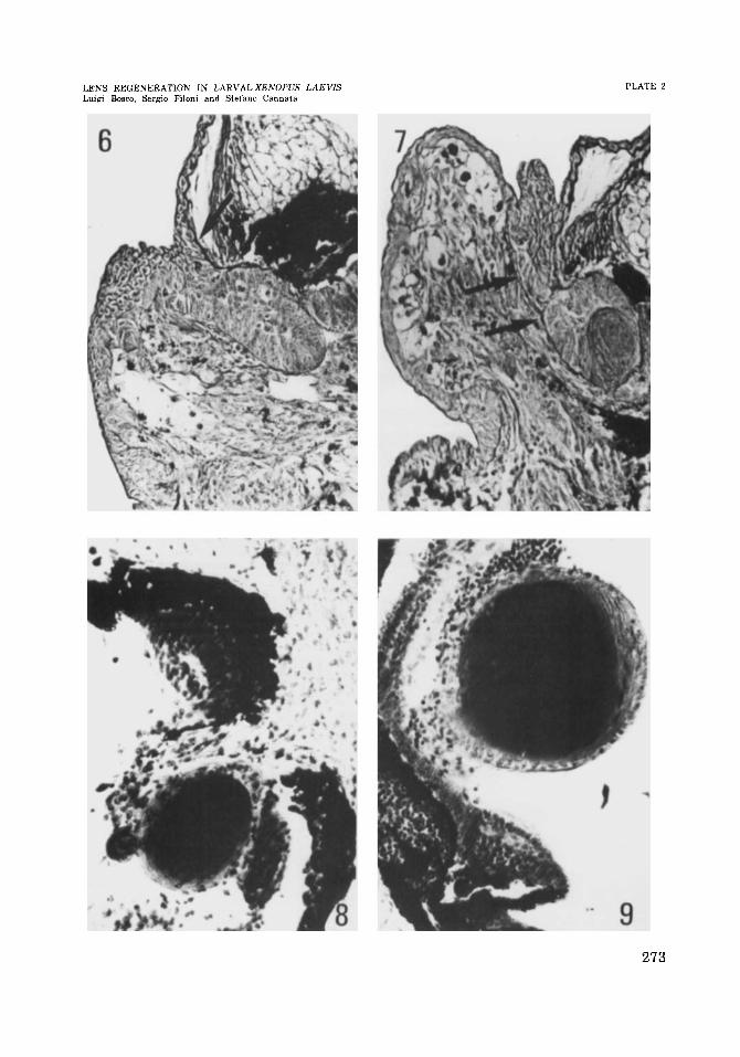
Figs. 6-9 Regeneration of lens after extirpation of lens from the dorsal region of the eye (Experiment 11). The cases presented in figure 6 and in figure 7 were stained with PAS and hematoxylin. The cases presented in figure 8 and in figure 9 were stained with Mallory-Azan’s method. All photographs (figs. 6-91 were taken a t a magnification of x 220.
6 Five days after operation: in the deep layer of the pericorneal epidermis, which has thickened considerably, a lens vesicle (at stage 3) has formed. Note the continuity of the basement membrane of the epidermis with the lens vesicle (arrow).
7 Seuen days after operation: in the context of the lens vesicle the primary nucleus of the lens fiber has formed (late stage IV). The lens vesicle is still attached to the pericorneal epidermis. Note the continuity of the basement membrane of the epider- mis with the lens vesicle (arrows).
8 Ten days after operation: the regenerated lens is a t stage 5 and partially occupies the vitreous chamber. I t is apparently connected to the proliferation of connective tissue closing the wound in the eye wall.
9 Fifteen days after operation: the lens, a t stage 5, occupies the vitreous chamber but is partially enclosed in the thickness of the retina.
272
LENS REGENERATION IN LARVAL XENOPUS LAEVIS Luigi Boeco, Sere0 Filoni and Stefan0 Cannata
PLATE 2
273

PLATE 3
EXPLANATION OF FIGURES
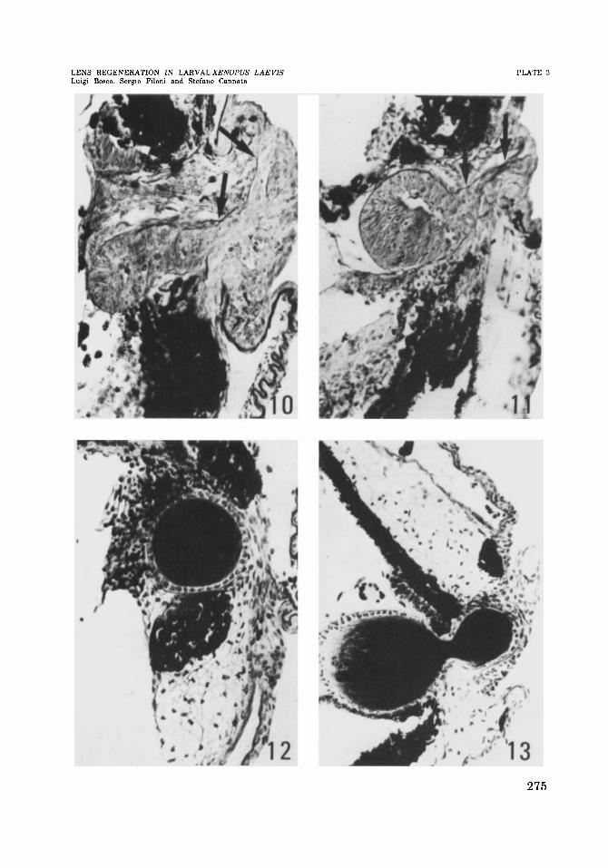
Figs. 10-13 Lens regeneration after extirpation of lens from the dorsal region of the eye and simultaneous incision of the outer cornea (Experiment 111). The cases pre- sented in figure 10 and figure 11 were stained with PAS and hematoxylin. The cases presented in figure 12 and in figure 13 were stained with Mallory-Azan’s method. All photographs (figs. 10.13) were taken a t a magnification of X 220.
10 Fiue days after operation: the lens vesicle, a t stage 3, has penetrated the wound made in the eye wall but is still extensively connected to the pericorneal epidermis. Note the continuity of the basement membrane of the epidermis with the lens vesi- cle (arrows).
11 Seven d a y s after operation: the lens vesicle, at early stage 4, partially occupies the vitreous chamber but is still connected to the pericorneal epidermis by a cluster of epithelial cells. Note the continuity of the basement membrane of the epidermis with the lens vesicle (arrows).
Ten d a y s after operation: the lens at stage 5, is contained in the thickness of the eye wall and is separate from the pericorneal epidermis.
Fifteen days after operation: the lens, a t stage 5, has a bilobate shape due to com- pression by the lips of the wound.
12
13
274
LENS REGENERATION IN LARVAL XENOPUS LAEVIS Luigi Boaco, Sergio Filoni and Stefano Cannata
PLATE 3
275

PLATE 4
EXPLANATION OF FIGURES
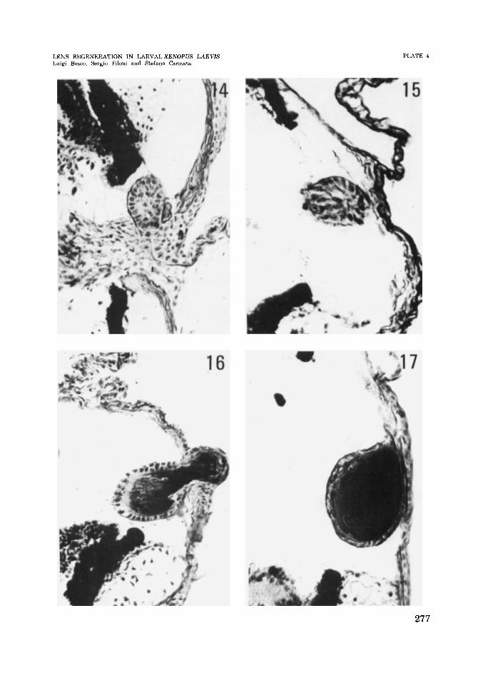
Figs. 14-17 Lens regeneration after extirpation of the lens from the dorsal region of the eye and simultaneous incision of the outer cornea and inner cornea (Experiment IV). Cases of lens regeneration from the outer cornea (group a). All cases presented in these figures were stained with Mallory-Azan’s method. All photographs (figs. 14-17) were taken a t a magnification of X 220.
14 Fiue days after operation; in the deep layer of the outer cornea a lens vesicle a t stage 3 has formed.
15 Seven days after operation: the lens vesicle, a t late stage 3, is almost completely separate from the outer cornea.
16 Ten days after operation; the newly formed lens at stage 5 has a bilobate shape and is extensively connected to the outer cornea.
17 Fifteen days after operation: the lens, a t stage 5, is still connected to the outer cornea.
276
LENS REGENERATION IN LARVAL XENOPVS LAEVIS Luigi Bosco, Sergio Filoni and Stefana Cannata
PLATE 4
277
PLATE 5
EXPLANATION OF FIGURES
Figs. 18-21 Regeneration of the lens after extirpation of lens from the dorsal region of the eye and contemporary incision of the outer cornea (Experiment IV). Cases of lens regeneration from the pericorneal epidermis (Group b). The cases presented in figure 18 and in figure 19 were stained with PAS and hematoxylin. The cases presented in figure 20 and in figure 21 were stained with Mallory-Azan’s method. All photographs (figs. 18-21) were taken a t a magnification of X 220.
18 Five days after operation: the regenerating lens, a t stage 3, occupies the wound made in the eye wall and is connected to the pericorneal epidermis (arrows).
19 Seven days after operation: the regenerating lens, a t stage 4, protrudes into the vitreous chamber but is still connected to the pericorneal epidermis by a cluster of epithelial cells. Note the continuity of the basement membrane of the epidermis with the lens vesicle (arrows).
20 Ten days after operation: the epithelial clusters connecting the pericorneal epider- mis to the regenerating lens (arrows) is partially strangulated by the closing lips of the wound in the eye wall.
21 Fifteen days after operation: the lens capsule, a t stage 5, is continuous with the basement membrane of the pericorneal epidermis (arrows).
278

LENS REGENERATION IN LARVAL XENOPUS LAEVIS Luigi Bosco, Sergio Filoni and Stefan0 Cannata
PLATE 5
279

PLATE 6
EXPLANATION OF FIGURES
Figs. 22-25 Cases of apparent lens regeneration within the retina structure. All cases presented in these figures were stained with Mallory-Azan’s method.
22 Fifteen days after extirpation of lens from the dorsal region of the eye (Experiment 11). Arrow. x 220.
23 Ten days after extirpation of the lens from the dorsal region of the eye and simul- taneous incision of the outer cornea (Experiment 111). In the context of an aggre- gate of retinal cells protruding into the vitreous chamber a regenerating lens can be seen. Arrows. x 220.
Fifteen days after extirpation of the lens from the dorsal region of the eye and si- multaneous incision of the outer cornea (Experiment 111). Arrow. X 350.
25 Ten days after extirpation of the lens from the dorsal region of the eye and simul- taneous incision of outer and inner cornea. (Experiment IV). Arrow. X 220.
24
280

LENS REGENERATION IN LARVAL XENOPUS LAEVIS LUI@ Bosco, Sergm Filoni and Stefano Cannata
PLATE 6
281