
RELATIONSHIP BETWEEN PSORIASIS AND ‘SOME ENDOCRINE
44
www.wjpps.com Vol 8, Issue 12, 2019. 1104 Eyssa et al. World Journal of Pharmacy and Pharmaceutical Sciences RELATIONSHIP BETWEEN PSORIASIS AND ‘SOME ENDOCRINE DISORDERS (HYPOTHYROIDISM, HYPERTHYROIDISM AND DIABETES MELLITUS) 1 Dr. Sabah Saleh Hussein, 2 Dr. Ibrahim Ahmed Azeez and 3 *Dr. Ahmed Abed Eyssa 1 M.B.Ch.B., 2 M.B.Ch.B., 3 M. B.Ch. B., The Council of the College of Medicine Tikrit University. CHAPTER ONE INTRODUCTION 1.1. Background Autoimmune diseases are chronic conditions initiated by the loss of immunological tolerance to self—antigens. They constitute het- erogeneous group of disorders, in which multiple alterations in the immune system result in a spectrum of syndromes that either target specific organs or affect the body systematically. [1] Recent epidemiological studies have shown a possible shift of one autoimmune disease to another or the fact that more than one au- toimmune disease may coexist in a single patient or in the same family. Numerous autoimmune diseases have been shown to co- exist frequently with thyroid autoimmune diseases. [2] Psoriasis, a common papulosquamous disease of the skin, affects about 1-3% of the population, with a peak incidence in the third decade of life. [3] The cause of psoriasis remains obscure, but a family history is found in 30% of patients and HLA- CW6 is most strongly associated with it. Despite the elucidation of numerous biochemical abnormalities affecting cyclic nucleotides, polyamines and arachidonic acid metabolism, immunological abnormalities of both humoral and cell mediate immunity and recently free radical generation abnormality, the pathogenesis of psoriasis has remained unclear; but some factors are known to be able to trigger, precipitate or aggravate the disease process, including drugs, severe sunlight and rarely metabolic disorders such as WORLD JOURNAL OF PHARMACY AND PHARMACEUTICAL SCIENCES SJIF Impact Factor 7.632 Volume 8, Issue 12, 1104-1147 Research Article ISSN 2278 – 4357 Article Received on 18 Oct. 2019, Revised on 06 Nov. 2019, Accepted on 28 Nov. 2019, DOI: 10.20959/wjpps201912-15078 *Corresponding Author Dr. Ahmed Abed Eyssa M. B.Ch. B., The Council of the College of Medicine Tikrit University.
Transcript of RELATIONSHIP BETWEEN PSORIASIS AND ‘SOME ENDOCRINE

www.wjpps.com Vol 8, Issue 12, 2019.
1104
Eyssa et al. World Journal of Pharmacy and Pharmaceutical Sciences
RELATIONSHIP BETWEEN PSORIASIS AND ‘SOME ENDOCRINE
DISORDERS (HYPOTHYROIDISM, HYPERTHYROIDISM AND
DIABETES MELLITUS)
1Dr. Sabah Saleh Hussein,
2Dr. Ibrahim Ahmed Azeez and
3*Dr. Ahmed Abed Eyssa
1M.B.Ch.B.,
2M.B.Ch.B.,
3M. B.Ch. B.,
The Council of the College of Medicine Tikrit University.
CHAPTER ONE
INTRODUCTION
1.1. Background
Autoimmune diseases are chronic conditions initiated by the loss of
immunological tolerance to self—antigens. They constitute het-
erogeneous group of disorders, in which multiple alterations in the
immune system result in a spectrum of syndromes that either target
specific organs or affect the body systematically.[1]
Recent epidemiological studies have shown a possible shift of one
autoimmune disease to another or the fact that more than one au-
toimmune disease may coexist in a single patient or in the same family. Numerous
autoimmune diseases have been shown to co- exist frequently with thyroid autoimmune
diseases.[2]
Psoriasis, a common papulosquamous disease of the skin, affects about 1-3% of the
population, with a peak incidence in the third decade of life.[3]
The cause of psoriasis remains obscure, but a family history is found in 30% of patients and
HLA- CW6 is most strongly associated with it. Despite the elucidation of numerous
biochemical abnormalities affecting cyclic nucleotides, polyamines and arachidonic acid
metabolism, immunological abnormalities of both humoral and cell mediate immunity and
recently free radical generation abnormality, the pathogenesis of psoriasis has remained
unclear; but some factors are known to be able to trigger, precipitate or aggravate the disease
process, including drugs, severe sunlight and rarely metabolic disorders such as
WORLD JOURNAL OF PHARMACY AND PHARMACEUTICAL SCIENCES
SJIF Impact Factor 7.632
Volume 8, Issue 12, 1104-1147 Research Article ISSN 2278 – 4357
Article Received on
18 Oct. 2019,
Revised on 06 Nov. 2019,
Accepted on 28 Nov. 2019,
DOI: 10.20959/wjpps201912-15078
*Corresponding Author
Dr. Ahmed Abed Eyssa
M. B.Ch. B., The Council of
the College of Medicine
Tikrit University.

www.wjpps.com Vol 8, Issue 12, 2019.
1105
Eyssa et al. World Journal of Pharmacy and Pharmaceutical Sciences
hypocalcemia of primary or secondary type.[4]
Many studies have been done on psoriasis some of which were concerned Wlth the
pathogenesis of psoriasis, and others considered psoriasis as systemic disease.
1.2. AIM OF STUDY
This study aims to investigate the association between psoriasis and thyroid disorders (hypo
and hyperthyroidism), diabetes mellitus, and family history relationship.
1.3. OBJECTIVES
From the history, identify the percentage of diabetes mellitus cases, in psoriatic patients.
Identify the family history relationship of psoriatic patients.
Investigate the psoriatic patients for thyroid function tests, T3, T4, and TSH to identify
the association between psoriasis and thyroid disorders.
Classify the psoriatic patients, according to«gender, age, types and duration of disease.
CHAPTER TWO
Literature Review
2.1. Psoriasis Definition
Psoriasis is a common skin disease characterized by thickened patches of inflamed, red skin
covered with thick silvery scales. The elbows and knees are the most common areas affected
by psoriasis. It will often appear in the same place on both sides of the body, the patches can
range in size from smaller than a dime to larger than a hand.[5]
Normally, skin cells mature and shed after about a month. In psoriasis, the cell maturation
speeds up, taking only three to four days. Because the lower layer of skin cells divides more
rapidly than normal, dead cells accumulate in thicker patches on the skin's outermost layer
(called the epidermis).[6]
2.2. Types of Psoriasis
Psoriasis appears in a variety of forms with distinct characteristics. Typically, an individual
has only one type of psoriasis at a time. Generally, one type-of psoriasis will clear and
another form of psoriasis will appear in response to a trigger.[7]
2.2.1. Plaque Psoriasis (psoriasis vulgaris)
Plaque psoriasis (psoriasis vulgaris) is the most prevalent form of the disease. About 80

www.wjpps.com Vol 8, Issue 12, 2019.
1106
Eyssa et al. World Journal of Pharmacy and Pharmaceutical Sciences
percent of those who have psoriasis have this type. It is characterized by raised, inflamed, red
lesions covered by a silvery white scale, it is typically found on the elbows, knees, scalp and
lower back.[8]
2.2.2. Guttate Psoriasis
Guttate [GUH—tate] psoriasis is a form of psoriasis that often starts in childhood or young
adulthood. The word guttate is from the Latin word meaning "drop." This form of psoriasis
appears as small, red, individual spots on the skin. Guttate lesions usually appear on the trunk
and limbs. These spots are not usually as thick as plaque lesions.[9]
Guttate psoriasis often comes on quite suddenly. A variety of conditions can bring on an
attack of guttate psoriasis, including upper respiratory infections, streptococcal throat
infections, tonsillitis, stress, injury to the skin and the administration of certain drugs
including antimalarials and beta-blockers.[10]
2.2.3. Inverse Psoriasis
Inverse psoriasis is found in the armpits, groin, under the breasts, and in other skin folds
around the genitals and the buttocks. This type of psoriasis appears as bright-red lesions that
are smooth and shiny. Inverse psoriasis is subject to irritation from rubbing and sweating
because of its location in skin folds and tender areas. It can be more troublesome in
overweight people and those with deep skin folds.[11]
2.2.4. Pustular Psoriasis
Primarily seen in adults, pustular psoriasis is characterized by white blisters of noninfectious
pus (consisting of white blood cells) surrounded by red skin.[12]
Pustular psoriasis may be localized to certain areas of the body, such as the hands and feet, or
covering most of the body. It begins with the reddening of the skin followed by formation of
pustules and scaling.[13]
Pustular psoriasis may be triggered by internal medications, irritating topical agents,
overexposure to UV light, pregnancy, systemic steroids, infections, stress and sudden
withdrawal of systemic medications or potent topical steroids.[14]
2.2.5. Erythrodermic Psoriasis
Erythrodermic [eh-REETH-ro-der—mik] psoriasis is a particularly inflammatory form of

www.wjpps.com Vol 8, Issue 12, 2019.
1107
Eyssa et al. World Journal of Pharmacy and Pharmaceutical Sciences
psoriasis that affects most of the body surface. It may occur in association with pustular
psoriasis. It is characterized by periodic, widespread, fiery redness of the skin and the
shedding of scales in sheets, rather than smaller flakes. The reddening and shedding of the
skin are often accompanied by severe itching and pain, heart rate increase, and fluctuating
body temperature.[15]
People experiencing the symptoms of erythrodermic psoriasis flare should go see a doctor
immediately. Erythrodermic psoriasis causes protein and fluid loss that can lead to severe ill
ress. The condition may also bring on infection, pneumonia and congestive heart failure.
People with severe cases of this condition often require hospitalization.[16]
Known triggers of erythrodermic psoriasis include the abrupt withdrawal of a systemic
psoriasis treatment including cortisone; allergic reaction to a drug resulting in the Koebner
response; severe sunburns; infection; and medications such as lithium, anti-malarial drugs;
and strong coal tar products.[17]
2.3. Psoriasis Theories
Psoriasis Theories are the concepts which try to explain the reasons for psoriasis appearance,
Psoriasis carmot be explained by any single theory. Each of the Psoriasis theories below is
based on the clinical observations and the results of the laboratory tests.[18]
The following psoriasis theories are presently the most reliable
2.3.1. Genetic Psoriasis Theory
Genetic psoriasis theory is based on multiple cases of family history of psoriasis. Psoriasis
can often be found in up to 6 generations of one family genetic variants affect disease
phenotype in both psoriasis and psoriasis arthritis. HLA—Cw*0602 is associated with early
onset psoriasis, higher incidence of guttate or streptococcal-induced flares, Koebner„s
phenomenon, more severe course, and is more likely to remit with pregnancy. HLA-
Cw*0602 is not associated with later onset of psoriasis, palmo-plantar psoriasis, nail or scalp
involvement and is less frequent in patients with psoriasis arthritis.[19]
In psoriasis arthritis, HLA—B27, Cw2, and DRw52 are associated with axial psoriasis
arthritis, and HLA—B38 and B39 with polyarthritis. It has also been shown that HLA—BZ7
in the presence of HLA-DR7, HLA-B39 and HLA-DQw3 in the absence of HLA-DR7 are
associated with progression of clinical damage, and that HLA-DR7, B22 alleles are

www.wjpps.com Vol 8, Issue 12, 2019.
1108
Eyssa et al. World Journal of Pharmacy and Pharmaceutical Sciences
“protective.” HLA-DRBl rheumatoid arthritis shared epitope as well as IL-4 150V
polymorphism have been shown to be associated with erosive psoriasis arthritis. Patients with
psoriasis arthtitis carrying both HLA- Cw6 and HLA-DRBI"„07 alleles have a less severe
course of arthritis.[20]
2.3.2. Autoimmune Psoriasis Theory
Autoimmune Psoriasis Theory is based on an assumption that certain antigens (foreign
substances that can stimulate an immune response in the body) may trigger an Autoimmune
Aggression in the body, and the Immune System starts attacking and tries to kill the body's
own cells„.[21]
The Immune System is a sum total of cells and organs which protect the body from foreign
invaders (i.e. infections etc.) The main organs of the Immune System include: thymus,
spleen, and lymph nodes. The main cells of the Immune System include antibodies (defense
proteins) and lymphocytes (a type of white blood cells) - NK-lymphocytes (NK-cells), B-
lymphocytes (B-cells), and T-lymphocytes (T-cells). The Immune System recognizes an
invader and gets rid of it with the aid of lymphocytes and antibodies. Normally the Immune
System can tell the difference between the body proteins itself and the foreign proteins of
bacteria or viruses. But sometimes the body's own proteins are recognized as foreign. In this
case the Immune System starts attacking and tries to kill the body's own cells. This is known
as an autoimmune agression, aut01mmune response or an autmmmune disorder.[22]
2.3.2.1. Immune Disorders and Psoriasis
According to the Immune Psoriasis Theory the immune disorders are the driving force in the
development of psoriasis. Psoriasis can be defined as general immune-dependent dermatitis.
According to the Immune Psoriasis Theory the following occurs: autoimmune aggression
directed at epidermis (the outermost layer of skin) results in the Psoriasis Appearance.
Certain antigens (foreign substances that can stimulate an immune response in the body) were
discovered in the psoriatic lesions, Whlle 1n the blood there were revealed their antibodies
(defense proteins that bind to antigens in an attempt to neutralize them) in the people with
psoriasis. These antigens and antibodies are absent in the skin and blood of people without
psoriasis. This gives grounds to consider psoriasis to be connected with the autoimmune
mechanisms.[23]

www.wjpps.com Vol 8, Issue 12, 2019.
1109
Eyssa et al. World Journal of Pharmacy and Pharmaceutical Sciences
D.U.M.Hung et a1 (1993, 1995) considered that psoriasis may "be started" by a superantigen
(antigen that binds to and activates T cells), for example by the toxins secreted by some
microbial staphylococci and streptococci.[24]
2.3.3. Metabolic Psoriasis Theory
Metabolic Psoriasis Theory is based on the data testifying about various metabolism disorders
in the people with psoriasis, changes in the DNA and RNA, the regression of psoriasis during
low-fat diet etc.[25]
Metabolism is the sum total of all the vital chemical processes that take place in a living cell
or organism. In the metabolism certain substances are broken down and converted into
energy while other necessary for life substances are synthesized.[26]
Scientists note a slower metabolism in the people with psoriasis. There is noted a reduction in
body temperature in the people with psoriasis, which also proves the slow metabolism.[27]
Carbohydrate metabolism is disrupted in 60% of people with psoriasis. Approximately 25%
of patients with psoriasis also have diabetes. Some scientists consider that carbohydrate
metabolism disorders serve as the basis for Psoriasis Appearance, while others believe these
disorders to be caused by psoriasis itself.[28]
In the people with psoriasis there are observed the fluctuations of the content of micro cells,
which participate in the oxidation-reduction processes.[29]
Y. Kamei (195 8) has revealed disorders of oxidation-reduction activity of the skin in the
people with psoriasis. E. Neumann (1957) considered the increased oxidation-reduction
activity of the epidermal cells (cells of the outer layer of the skin) to play the primary role in
the development of psoriasis.[30]
There are indications of a lowered level of oxygen in the blood in the people with psoriasis,
which negatively affects the course of psoriasis. The concentration of free radicals in the skin
of people with psoriasis exceeds their content in the skin in people without psoriasis by as
much as 3 times on the average. This proportion becomes normal closer to the clinical
recovery from psoriasis.[31]
Accelerated epidermal cell development can be confirmed by increased synthesis of glycogen

www.wjpps.com Vol 8, Issue 12, 2019.
1110
Eyssa et al. World Journal of Pharmacy and Pharmaceutical Sciences
in the skin of people with psoriasis, and especially in the psoriatic lesions. The metabolism of
vitamins with pSoriasis is also disrupted, which decreases the adaptive abilities of the body.
The content of Vitamin C is decreased in the blood, but it is increased in the skin; the content
of the Vitamins A, B6, and B12 in the blood is reduced. Various shifis are also observed with
the contents of copper, Zinc and iron.[32]
There is also found liver function disorder in the people with psoriasis. Liver fianction
disorder seems to play a specific role, but not the main role in the pathogenesis of psoriasis.
In the progressive stage of psoriasis there is frequently noted an increased function of the
thyroid and/or endocrine glands, there is noted the absence of fat and perspiration on the
psoriatic plaques and near them; it is restored when the psoriatic lesions regress. There were
discovered disorders of protein and of lipid metabolism in the people with psoriasis. The
content of lipids and cholesterol in the people with psoriasis is increased. The increase in the
lipids stimulates the overgrowth of layers of horny skin (keratosis). Good therapeutic effect
of a fat free diet proves the presence of the abnormal levels of certain fats in the people with
psoriasis. A low-calorie diet usually helps to improve the state of psoriasis.[33]
2.3.4. Hormones and Psoriasis Theory
Hormones and psoriasis theory is based on the flare-ups of psoriasis during menstruation; an
improvement in the flow of psoriasis, and even a complete recovery during pregnancy etc.
Hormones and Psoriasis seem to be tightly connected. Psoriasis is characterized by an
increased reproduction of the epidermal skin cells, which indicates the disorders of the
regulatory endocrine system.[34]
Endocrine System regulates the body organ's activity with the aid of the hormones. Hormones
are produced in the endocrine glands, endocrine system produces various hormones, for
example Steroid hormones (i.e. Glucocorticoids, Cortisone, Hydro Cortisone, Corticosterone,
Prednisolone; Sex Hormones etc.). Together with the nervous system and the immune
system, endocrine system regulates the growth and the development of an organism, sex
differentiation, reproductive functions etc.[35]
2.3.4.1. Is Psoriasis Connected with the Endocrine Glands Disorders,
In Particular - the Thyroid and Adrenal Glands Disorders Thyroid gland is an endocrine gland
that produces thyroid hormones to regulate the body's metabolism and growth. Deviations in
the thyroid gland functions are found in many people with psoriasis. Adrenal gland is an

www.wjpps.com Vol 8, Issue 12, 2019.
1111
Eyssa et al. World Journal of Pharmacy and Pharmaceutical Sciences
endocrine gland that produces the hormones adrenaline, noradrenaline, glucocorticoids (i.e.
cortisone), sex hormones and others. Adrenal gland regulates metabolism, sexual function,
water balance, heart rate, blood pressure, and stress'.[36]
In the majority of the people who had psoriasis for a long time, there is revealed a reduction
in the glucocorticoid function of the outer region of the adrenal gland - the adrenal cortex, i.e.
the production of the Steroid hormones is disrupted. In psoriasis treatment used are Topical
Steroids which cause blood vessels in the epidermis to temporary constrict and thus to
achieve an anti inflammatory effect with psoriasis. Topical Steroid hormones possess various
side-effects.[37]
There are known cases of the appearance and flare—ups of psoriasis in women at the time of
menstruation. It is also known that psoriasis may disappear during pregnancy and reappear
during breast feeding. The above clearly indicates the connection of hormonal changes with
psoriasis.[38]
However, a direct connection of any particular endocrine gland or hormone with psoriasis
was not found. The presence of endocrine disorders in people with psoriasis does not allow
asserting that these disorders are the direct cause for the appearance of psoriasis, because
similar disorders are also observed with other illnesses.[39]
2.3.5. Nervous System Psoriasis Theory
Nervous System and Psoriasis theory is based on psoriasis appearance after stress and on the
data that people with psoriasis frequently have functional disorders of the Nervous System.[40]
2.3.6. Infection and Psoriasis Theory
Infection and Psoriasis theory is based on the connection of psoriasis With a focal infection in
the tonsils, the inflammation of the nose cavities and eardrum, the inflammation of the uterine
appendages, the inflammation of the prostate gland etc.[40]
2.3.7. Virus and Psoriasis Theory
Virus and Psoriasis theory is based on an assumption that a certain virus (retrovirus) may be
an agent that provokes psoriasis.[40]
2.3.8. Toxins and Psoriasis Theory
Toxins and Psoriasis theory is based on an assumption that toxins contained in certain foods

www.wjpps.com Vol 8, Issue 12, 2019.
1112
Eyssa et al. World Journal of Pharmacy and Pharmaceutical Sciences
and medications can penetrate through the bowels and into the blood and lymph flow and
then to be excreted.[40,42]
through skin causing the appearance of psoriatic lesions.
2.3.9. Antioxidant Psoriasis Theory
Antioxidant Psoriasis theory is based on an assumption that the people with psoriasis are
better protected from the reactive oxygen as an unfortunate forms than the people without
psoriasis, which causes an inflammatory psoriatic reaction side-affect.[43]
2.4. Is PsorIaSIS an Autoimmune Disease?
AUtOIMune disorders are diseases that occur when the body r -., p oduces an inappropriate
immune response against its OWIltlssues. Sometimes the immune system will cease to
recognize one or more of the body's normal constituents as "self" and will produce
autoantibodies —antibodies that attack its own cells, tissues, and/or organs. This causes
inflammation and damage and leads to autoimmune disorders.[43]
The cause of autoimmune diseases is unknown, but it appears that there is an inherited
predisposition in many cases. In a few types of autoimmune disease (such as rheumatic
fever), avirus or infection with bacteria triggers an immune response and the antibodies or T-
cells attack normal cells because some part of their structure resembles a part of the infecting
microorganism.[44]
Autoimmune disorders fall into two general types: those that damage many organs (systemic
autoimmune diseases) and those where only a single organ or tissue is directly damaged by
the autoimmune process (localized). However, the distinctions become blurred as the effect
of localized autoimmune disorders frequently extends beyond the targeted tissues, indirectly
affecting other body organs and systems.[45]
In some cases, the antibodies may not be directed at a specific tissue or organ; for example,
antiphospholipid antibodies can react with substances (phospholipids) that are the normal
constituents of platelets and the outermost layer of cells (cell membranes), Wthh can lead to
the formation of blood clots within the blood vessels (thrombosis).[46]
Symptoms of autoimmune disorders vary by the particular disorder but many include fatigue,
dizziness, and low grade fever. Symptoms can also vary in severity over time. Acute guttate
psoriasis is often self—limiting and reflects an abnormal immune reaction to streptococcal
throat infection. Chronic plaque psoriasis, however, behaves like most autoimmune disease,

www.wjpps.com Vol 8, Issue 12, 2019.
1113
Eyssa et al. World Journal of Pharmacy and Pharmaceutical Sciences
being characterized by a chronic but fluctuating HLA-linked inflammatory process. In
contrast to other autoimmune diseases that are mostly linked to certain class II HLA alleles,
psoriasis is the only known chronic inflannnatory disease that has the strongest association
with an HLA-C allele. Thus, over 60% of psoriasis patients carry the HLA-Cw6 allele, and
the predominance of oligoclonal CD8+ T cells in lesional epidermis suggests that the
pathogenic process is driven by autoantigen, that may be presented by HLA-Cw6 in those
patients who carry this allele. The CD4+ T cells in stable lesions are also likely to be
oligoclonal, further supporting the notion that chronic psoriasis is an antlgen drlven
disease.[47]
It has been reported that patients with chronic plaque psoriasis can experience an
exacerbation after streptococcal throat infection, and we have previously postulated that
psoriasis is mediated by T cells that cross- react with epitopes which are common to
streptococcal M proteins and those keratins that are up-regulated in psoriatic lesions.[48]
Streptococcal M proteins have extensive amino acid sequence homologuey to type I keratins,
including keratins 14, 16 and 17.
Interestingly, these keratins are usually only expressed at low levels or not at all in normal
skin but are up-regulated during inflammation or trauma.[49]
T cell responses against certain amino acid sequences that streptococcal M proteins share
with these keratins have been shown to be increased in patients with active psoriasis but
absent during disease remissions.[50]
Furthermore, CLA + CD8+ T cells in HLA-Cw*0602 positive psoriasis patients with an
active disease showed IFN—y responses against the homologueous sequences present both in
keratins and M proteins while nonpsoriatic HLA—Cw*0602 positive controls only responded
to peptides from the M protein. This indicates that patients with active psoriasis have a
recirculating subset of skin homing CD8+ T cells that react specifically to keratin peptides.[51]
2.5. Thyroid Gland
The thyroid gland is one of the largest endocrine glands. The thyroid gland is found in the
neck, below the thyroid cartilage (which forms the laryntgeal prominence, or "Adam's
apple"). The isthmus (the bridge between the two lobes of the thyroid) is located inferior to
the cricoid cartilage.[52]

www.wjpps.com Vol 8, Issue 12, 2019.
1114
Eyssa et al. World Journal of Pharmacy and Pharmaceutical Sciences
The thyroid gland controls how quickly he body uses energy, makes proteins, and controls
how sensitive the body is to other hormones. It participates in these processes by producing
thyroid hormones, the principal ones being triiodothyronine (T3) and thyroxine which can
sometimes be referred to as tetraiodothyronine (T4). These hormones regulate the rate of
metabolism and affect the growth and rate of function of many other systems in the body. T3
and T4 are synthesized from oth„„b lOdlne and tyrosme. The thyrmd also produces calcitonin,
which plays a role in calcium homeostasis.[53]
Hormonal output from the thyroid is regulated by thyroid-stimulating hormone (T SH)
produced by the anterior pituitary, which itself is regulated by thyrotropin-releasing hormone
(TRH) produced by the hypothalamus. The thyroid gets its name from the Greek word for
“shield", due to the shape of the related thyroid cartilage. The most common problems of the
thyroid gland consist of an overactive thyroid gland, referred to as hyperthyroidism, and an
underactive thyroid gland, referred to as hypothyroidism.[54]
2.5.1. Thyroid Gland Physiology
The primary fimction of the thyroid is production of the hormones triiodothyronine (T3),
thyroxine (T4), and calcitonin. Up to 80% of the T4 is converted to T3 by peripheral organs
such as the liver, kidney and spleen. T3 is several times more powerful than T4, which is
largely a prohormone, perhaps four or even ten times more active.[55]
2.5.1.1. T3 and T4 Production and Action
Thyroxine (T4) is synthesised by the follicular cells from free tyrosine and on the tyrosine
residues of the protein called thyroglobulin (Tg).Iodine is captured with the "iodine trap" by
the hydrogen peroxide generated by the enzyme thyroid peroxidase (TPO) and linked to the 3'
and 5' sites of the benzene ring of the tyrosine residues on Tg, and on free tyrosme.[56]
Upon stimulation by the thyroid-stimulating hormone (TSH), the follicular cells reabsorb Tg
and cleave the iodinated tyrosines from Tg in lysosomes, forming T4 and T3 (in T3, one
iodine atom is absent compared to T4), and releasing them into the blood. Deiodinase
enzymes convert T4 to T3 „Thyroid hormone secreted from the gland is about 80-90% T4
and about 10-20% T31.[57]
Cells of the developing brain are a major target for the thyroid hormones T3 and T4. Thyroid
hormones play a particularly crucial role in brain maturation during fetal development. A
www.wjpps.com Vol 8, Issue 12, 2019.
1115
Eyssa et al. World Journal of Pharmacy and Pharmaceutical Sciences
transport protein that seems to be important for T4 transport across the blood—brain ban„ier
(OATP1C1) has been identified. A second transport protein (MCT8) is important for T3
transport across brain cell membranes.[58]
Non-genomic actions of T4 are those that are not initiated by liganding of the hormone to
intranuclear thyroid receptor. These may begin at the plasma membrane or within cytoplasm.
Plasma membrane- initiated actions begin at a receptor on the integrin alphaV beta3 that
activates ERK1/2. This binding culminates in local membrane actions on ion transport
systems such as the Na(+)/H(+) exchanger or complex cellular events including cell
proliferation. These integrins are concentrated on cells of the vasculature and on some types
of tumor cells, which in part explains the proangiogenic effects of iodothyronines and
proliferative actions of thyroid hormone on some cancers including gliomas. T4 also acts on
the mitochondrial genome via imported isoforms of nuclear thyroid receptors to affect several
mitochondrial transcription factors. Regulation of V actin polymerization by T4 is critical to
cell migration in neurons and glial cells and is important to brain development.[59]
T3 can activate phosphatidylinositol 3-kinase by a mechanism that may be cytoplasmic in
origin or may begin at integrin alpha V beta3. In the bIOOd, T4 and T3 are partially bound to
thyroxine-binding globulin (TBG), transthyretin, and albumin. Only a very small fraction of
the circulating hormone is free (unbound) - T4 003% and T3 0.3%. Only the free fraction has
hormonal activity. As with the steroid hormonesand retinoic acid, thyroid hormones cross the
cell membrane and bind to intracellular receptors (0:1, (12, 0, and [32), which act alone, in
pairs or together with the retinoid X-receptor as transcription factors to modulate DNA
transcription.[60]
2.5.1.2. T3 and T4 Regulation
The production of thyroxine and triiodothyronine is regulated by thyroid—stimulating
hormone (TSH), released by theanterior pituitary. The thyroid and thyrotropes form a
negative feedback loop: TSH production is suppressed when the T4levels are high. The TSH
production itself is modulated by thyrotropin—releasing hormone (TRH), which is produced
by the hypothalamus and secreted at an increased rate in situations such as cold exposure (to
stimulate thermogenesis). TSH production is blunted by somatostatin (SRIH), rising levels of
glucocorticoids and sex hormones (estrogen andtestosterone), and excessively high blood
iodide concentration.[61]

www.wjpps.com Vol 8, Issue 12, 2019.
1116
Eyssa et al. World Journal of Pharmacy and Pharmaceutical Sciences
An additional hormone produced by the thyroid contributes to the regulation of blood
calciumlevels. Parafollicular cells produce calcitonin in response to hypercalcemia.
Calcitonin stimulates movement of calcium into bone, in opposition to the effects of
parathyroid hormone (PTH)- However, calcitonin seems far less essential than PTH, as
calcium metabolism remains clinically normal after removal of the thyroid (thyroidectomy),
but not the parathyroids.[62]
2.5.2. Disorders of Thyroid
Thyroid disorders include hyperthyroidism (abnormally increased activity), hypothyroidism
(abnormally decreased activity) and thyroid nodules, which are generally benign thyroid
neoplasms, but may be thyroid cancers. All these disorders may give rise to goiter, that is, an
enlarged thyroid.[63]
2.5.2.1. Hyperthyroidism
Hyperthyroidism, or overactive thyroid, is the overproduction of the thyroid hormones T3
and T4, and is most commonly caused by the development of Graves' disease, an
autoimmune disease in which antibodies are produced which stimulate the thyroid to secrete
excessive quantities of thyroid hormones. The disease can result in the formation of a toxic
goiter as a result of thyroid growth in response to a lack of negative feedback mechanisms. It
presents with symptoms such as a thyroid goiter, protruding eyes (exopthalmos), palpitations,
excess sweating, diarrhea, weight loss, muscle weakness and unusual sensitivity to heat. The
appetite is often increased.[64]
Beta blockers are used to decrease symptoms of hyperthyroidism such as increased heart rate,
tremors, anxiety and heart palpitations, and anti-thyroid drugs are used to decrease the
production of thyroid hormones, in particular, in the case of Graves' disease. These
medications take several months to take full effect and have side-effects such as Skin rash or
a drop in white blood cellcount, which decreases the ability of the body to fight off infections.
These drugs involve frequent dosing (often one pill every 8 hours) and often require frequent
doctor visits and blood tests to monitor the treatment, and may sometimes lose effectiveness
over time.[65]
Due to the side-effects and inconvenience of such drug regimens, some patients choose to
undergo radioactive iodine-131 treatment. Radioactive iodine is administered in order to
destroy a portion of or the entire thyroid gland, since the radioactive iodine is selectively

www.wjpps.com Vol 8, Issue 12, 2019.
1117
Eyssa et al. World Journal of Pharmacy and Pharmaceutical Sciences
taken up by the gland and gradually destroys the cells of the gland. Alternatively, the gland
may be partially or entirely removed surgically, though iodine treatment is usually preferred
since the surgery is invasive and carries a risk of damage to the parathyroid glands or the
nerves controlling the vocal cords. If the entire thyroid gland is removed, hypothyroidism
results.[66]
2.5.2.2. Hypothyroidism
Hypothyroidism is the underproduction of the thyroid hormones T3 and T4. Hypothyroid
disorders may occur as a result of congenital thyroid abnormalities, autoimmune disorders
such asI-Iashimoto's thyroiditis, iodine deficiency (more likely in poorer countries) or the
removal of the thyroid following surgery to treat severe hyperthyroidism and/or thyroid
cancer. Typical symptoms are abnormal weight gain, tiredness, baldness, cold intolerance,
and bradycardia. Hypothyroidism is treated with hormone replacement therapy, such
aslevothyroxine, which is typically required for the rest of the patient's life. Thyroid hormone
treatment is given under the care of aphysician and may take a few weeks to become
effective.[67]
Negative feedback mechanisms result in growth of the thyroid gland when thyroid hormones
are being produced in sufficiently low quantities as a means of increasing the thyroid output;
however, where the hypothyroidism is caused by iodine insufficiency, the thyroid is unable to
produce T3 and T4 and as a result, the thyroid may continue to grow to form a non—toxic
goiter. It is termed non-toxic as it does not produce tOXiC quantities of thyroid hormones,
despite its size.
2.5.2.3. Initial Hyperthyroidism Followed by Hypothyroidism
This is the overproduction of T3 and T4 followed by the underproduction of T3 and T4.
There are two types: Hashimoto's thyroiditis and postpartum thyroiditis.[68]
Hashimoto's thyroiditis or Hashimoto's Disease is an autoimmune disorder whereby the
body's own immune system reacts with the thyroid tissues in an attempt to destroy it. At the
beginning, the gland may be overactive, and then becomes underactive as the gland is
damaged resulting in too little thyroid hormone production or hypothyroidism. Some patients
may experience "swings" in hormone levels that can progress rapidly from hyper—to-
hypothyroid (sometimes mistaken as severe moodswings, or even being bipolar, before the
proper clinical diagnosis is made). Some patients may experience these "swings" over a

www.wjpps.com Vol 8, Issue 12, 2019.
1118
Eyssa et al. World Journal of Pharmacy and Pharmaceutical Sciences
longer period of time, over days or weeks or even months. Hashimoto's is more common in
females than males, usually appearing after the age of 30, and tends to run in families
meaning it can be seen as a genetic disease. Also more common in individuals with
Hashimoto's Thyroiditis are type 1 diabetes and celiac disease.[69]
Postpartum thyroiditis occurs in some t„cmnles following the birth 01' a child. Atlcr deliver,
tho glund becomes inliumecl and the condition initially presents with overuclivity ol‟lhc
gluncl followed by undcmctivity. In some cases, the gland may recover with time and resume
its l'unetions. In others it may not. The etiology is not always known, but can sometimes be
attributed to mitohmmlnity, such as Hashimoto's Thyroiditis or Graves' Disease.[70]
2.5.2.4. Cancers
In most cases, the thyroid cancer presents as a painless mass in the neck. It is very unusual for
the thyroid cancers to present with symptoms, unless it has been neglected. One may be able
to feel a hard nodule in the neck. Diagnosis is made using a needle biopsy and various
radiological studies.[71]
2.5.2.4.1. Non-Cancerous Nodules
Many individuals may find the presence of thyroid nodules in the neck. The majority of these
thyroid nodules are benign (non cancerous). The presence of a thyroid nodule does not mean
that one has thyroid disease. Most thyroid nodules do not cause any symptoms, and most are
discovered on an incidental examination. Doctors usually perform a needle aspiration biopsy
of the thyroid to determine the status of the nodules. If the nodule is found to be non—
cancerous, no other treatment is required. If the nodule is suspicious then surgery is
recommended.[72]
2.5.2.4.2. Congenital Anomalies
A persistent thyroglossal duct or cyst is the most common clinically significant congenital
anomaly of the thyroid gland. A persistent sinus tract may remain as a vestigial remnant of
the tubular development of the thyroid gland. Parts of this tube may be obliterated, leaving
small segments to form cysts. These occur at any age and might not become evident until
adult life. Mucinous, clear secretions may collect within these cysts to form either spherical
masses or fusiform swellings, rarely larger than 2 to 3 cm in diameter. These are present in
the midline of the neck anterior to the trachea. Segments of the duct and cysts that occur high
in the neck are lined by stratified squamous epithelium, which is essentially identical to that

www.wjpps.com Vol 8, Issue 12, 2019.
1119
Eyssa et al. World Journal of Pharmacy and Pharmaceutical Sciences
covering the posterior portion of thetongue in the region of the foreamen cecum. The
anomalies that occur in the lower neck more proximal to the thyroid gland are lined by
epithelium resembling the thyroidal acinar epithelium. Characteristically, next to the lining
epithelium, there is an intense lymphocytic inflitrate. Superimposed infection may convert
these lesions into abscess cavities, and rarely, give rise to cancers.[73]
2.5.3. Other Disorders
E! Limited research shows that seasonal allergies may trigger episodes of hypo- or
hyperthyroidism. D A ectopic thyroid is an entire or parts of the thyroid located in another
part of the body than what is the usual case.[73]
2.5.3.1. Significance of Iodine
Gland can become considerably enlarged, a condition called endemic goiter. Pregnant women
on a diet that is severely deficient of iodine can give birth to infants who can present with
thyroid hormone deficiency (congenital hypothyroidism), manifesting in problems of physical
growth and development as well as brain development (a condition referred to as endemic
cretinism). In many developed countries, newborns are routinely tested for congenital
hypothyroidism as part of newborn screening. Children With congenital hypothyroidism are
treated supplementally with levothyroxine, which facilitates normal growth and
development.[74]
Thyroxine is critical to the regulation of metabolism and growth throughout the animal
kingdom. Among amphibians, for example, administering a thyroid-blocking agent such as
propylthiouracil(PTU) can prevent tadpoles from metamorphosing into frogs; in contrast,
administering thyroxine will trigger metamorphosis.[75]
Because the thyroid concentrates this element, it also concentrates the various radioactive
isotopes of iodine produced by nuclear fission. In the event of large accidental releases of
such material into the environment, the uptake of radioactive iodine isotopes by the thyroid
can, in theory, be blocked by saturating the uptake mechanism with a large surplus of non-
radioactive iodine, taken in the form of potassium iodide tablets. One consequence of the
Chernobyl disaster was an increase in thyroid cancers in children in the years followmg the
acc1dent.[76]

www.wjpps.com Vol 8, Issue 12, 2019.
1120
Eyssa et al. World Journal of Pharmacy and Pharmaceutical Sciences
The use of lodised salt is an efficient way to add iodine to the diet. It has eliminated endemic
cretinism in most developed countries, and some governments have made the iodination of
flour, cooking oil, and salt mandatory. Potassium iodide and sodium iodide are typically used
forms of supplemental iodine. As with most substances, either too much or too little can
cause problems. Recent studies on some populations are showmg that excess iodine intake
could cause an increased prevalence of autoimmune thyroid disease, resulting in permanent
hypothyroidism.[77]
2.6. Comorbid Conditions in Psoriasis
Psoriasis is newly defined as a systemic disease. Common co-morbidities associated with
psoriasis include diabetes, hypertension, and metabolic syndromes. Psoriasis can have a
significant impact on a patient's quality of life and is associated with loss of productivity,
depression, and an increased prevalence of malignancy.1 Pro—inflammatory cytokines such
as tumour necrosis factor—alpha (TNF- (1), and other factors like pro—inflammatory T-
helper type 1 cytokines that are overproduced in patients with psoriasis likely contributes to
the increased risk for development of metabolic syndrome.[78]
In terms of the other diseases associated with psoriasis, Crohn‟s disease is another condition
that is not common but its prevalence is certalnly increased in pat1entsw1th psortams.[79]
Depression or anxiety is another common problem in patients with psoriasis as is
genitourinary disease. 20 % of hospitalised patients with psoriasis have some genitourinary
complaints.[80]
Patients should adom a healthy lifestyle so as not to contribute any move to risk „lhctor„s.
'l„m„ming psoriasis and the associated co-morbid cmditious awwssivel„v from the beginning
will definitely improve the quality m„lit„e ot„the patient.[81]
2.7. Benefits of Psoriasis Therapy on Comorbid Conditions
Since many commbid conditions have inflammatory mechanisms in common with psm„iasis„
drugs tmgeting inflammation and /or suppressing the immune response ave often effective in
treating both psoriasis and related emum„bidities. A number of treatments have shown some
efficacy in hemimg both psoriasis and psoriatic arthritis including methotrexate, cyclosporine,
lefiunomide, etanercept and i11flixinmb.„Tumor necrosis factor (TNF) inhibitors etanercept
and infliximab have demonstrated halting ofjoint degradation.[82]

www.wjpps.com Vol 8, Issue 12, 2019.
1121
Eyssa et al. World Journal of Pharmacy and Pharmaceutical Sciences
2.8. 'lfi‘pe 2 diabetes. psoriasis and thiozolidinediones
Thiozolidinediones, synthetic ligands for the peroxisome proliferator— actimted mceptor —
gamma (PPAR—gamma) receptor, are insuline — sensitizing drugs licensed for use in
selected patients with type 2 diabetes mellitus. The potential therapeutic applications of the
flliozolidjnediones extend to other clinical specialties such as dermatology. Rosiglitazone and
Pioglitazone are being evaluated for the treatment of psoriasis. Type 2 diabetes and psoriasis
may coexist. prompting speculation that dual benefits might accrue for patients with both
conditions.[83]
A recent open pilot study suggest that oral pioglitazone may be benefic1a1 for moderate
chronic plague psoriasis. However, changes in antidiabetic medication must be made in the
knowledge of the cautions and contraindications to oral agents as well as the impact on
metabolic control. Further study are required before the use of thiazolidinediones for
psoriasis can be advocated.[84]
CHAPTER THREE
PATIENTS AND METHODS
3.1. Study Design
This is a cross section study designed to illustrate the relationship between psoriasis and
thyroid disorders by doing the thyroid function tests on psoriasis and control group.
3.2 Patient Selection
Sixty patients had psoriasis consulting the outpatient dermatology clinic of Tikrit General
Hospital in Tikrit city and sixty cases of normal persons (control group). Carried out from
January to august 2012.
3.3. Inclusion Criteria
1. Patient agrement
2. Age range from 16 to 65 year, depending on Tilo henseler, who dividing psoriasis into type
I and type II, which demonstrate distinct characteristics. Firstly the disease presents in
different decades of life, in type I before the age of 40 years and' later in type 11. Secondly,
contrasting frequencies of HLA alleles are found: type I patients express predominantly
HLA-Cw6,—B57, and—DR7, whereas in type 11 patients HLA-CwZ is overrepresented.
Finally, familial inheritance is found in type I but not in type II psoriasis.[85]
3. known case of psoriasis and was diagnosed by dermatologist.

www.wjpps.com Vol 8, Issue 12, 2019.
1122
Eyssa et al. World Journal of Pharmacy and Pharmaceutical Sciences
4.3. Ecxclusion Criteria
Exclusion criteria were psoriatics or controls with known thyroid impairment, those using
thyroid hormones, anti-thyroid drugs or other drugs affecting thyroid function, such as
lithium, iodine, steroids, dopamine, anticonvulsant drugs and interferon.
3.5. Clinical History
The patients' disease history recording include name, age, sex, address, occupation, site,
duration, family history, drug history, and associated endocrine diseases.
3.6. Diagnosis
The diagnosis of psoriasis is made clinically, by dermatologist.
3.7. Investigations
Investigations of thyroid function tests includes, total triiodothyronine (TT3), thyroxine (T4),
and measurement of thyroid stimulating hormone (TSH). Overt hypothyroidism was defined
as elevated TSH and low T4, subclinical hypothyroidism as elevated TSH and normal T4,
overt hyperthyroidism as low TSH and elevated T4 and subclinical hyperthyroidism as low
TSH and normal T4.[86]
3.7.1. Triiodothyronine (T3)
For quantitative measurement of total triiodothyronine (TT3) in human serum ST AIA-PACK
TT3 is used on TOSOH AIA System Analyzers.[87]
3.7.1.1. Summary and Explanation of Test
Triiodothyronine (T3) and thyroid hormone (thyroxine; T4) regulate a variety of biochemical
processes throughout the body, the majority of T3 is produced enzymatically by
monodeiodination of T4 in the peripheral tissues, rather than from the direct secretion from
the thyroid gland. Approximately one third of all T4 secreted is deiodinated to yield T3.
Sennn T3 measurement can a valuable component of a thyroid function screening panel in
diagnosing certain disorders of thyroid function in addition to conditions caused by iodide
deficiency. Assay for T3 are valuable in early detection of hyperthyroidism and for
monitoring the efficacy of treatment for thyroid disorders. A normal T3 value in the presence
of an elevated T4 level may also help to rule out hyperthyroidism.[88]
3.7.1.2. Principle of the Assay
The ST AIA-PACK TT3 is a competitive enzyme immunoassay which is performed entirely

www.wjpps.com Vol 8, Issue 12, 2019.
1123
Eyssa et al. World Journal of Pharmacy and Pharmaceutical Sciences
within the AIA-PACK test cups. Triiodothyronine, Which is displaced from its binding
protein by AN S (8-anilino-1- naphthalene sulfonic acid), and free T3 present in the test
sample compete with enzyme-labeled T3 for a limited number of binding sites on a T3.
Specific antibody immobilized on magnetic beads. The beads are washed to remove the
unbound enzyme-labeled T3 and are then incubated with a fluorogenic substrate, 4-
methylumbelliferyl phosphate (4-MUP). The amount of enzyme-labeled T 3 that binds to the
beads is inversely Proportional to the T3 concentration in the test sample. A standard curve
using a range of known standard concentration is prepared and unknown[89]
T3 concentration
are calculated usmg th1s curve.
3.7.1.3. Specimen Collection and Handiing
Serum 0r heparinized plasma is required for the assay. EDTA and citrated plasma should not
be used. 0 If using serum, a venous blood sample is collected aseptically without additives.
Store at 18-256 until a clot has formed (usually 15-45, minutes), then centrifuge to obtain the
serum specimen for assay.
If using heparinized plasma, avenous blood sample is collected aseptically with designed
additive. Centrifuge and separate plasma from the packed cells as soon as possible.
Specimens types should not be used interchangeably during serial monitoring of an individual
patient. Measured concentration may vary slightly between sample types in certain patients.
Samples may be stored at 2-86: for up t024 hours prior to analysis. If the analysis cannot be
done within 24 hours, the sample should be stored frozen at _20t': or below for up to 60 days.
Repeated freeze—thaw cycles should be avoided. Turbid serum samples or samples
containing particular matter should be centrifuged prior to testing. Prior to assay, bring frozen
samples to 18-25(': slowly and mix gently.
The sample required for analysis is ZSpL.[90]
3.7.1.4. Reference range
The interval given here was determined in serum samples from 573 apparently healthy Asian
individuals. Reference interval =0.79-1.58ng/mL (1.22-2.43nmol/L).

www.wjpps.com Vol 8, Issue 12, 2019.
1124
Eyssa et al. World Journal of Pharmacy and Pharmaceutical Sciences
3.7.1.5. Conversion Factors
T3 concentration in this application are in units of ng/Ml. conversion to SI units of nmol/l
may be made using the following equation: nmol T3/L = ng T3/mL x 1.54.[91]
3.7.2. Thyroxine (T4)
For quantitative measurement of thyroxine (T4) in human serum ST AIA-PACK T4 is used
on TOSOH AIA System Analysers.[87]
3.7.2.1. Summary and Explanation of Test
Evaluation of thyroid status is complex. The primary function of thyroid gland is the
secretion of thyroxine (T4) or triodothyroxine (T3). Abnormal secretion of T4 and/or T3 may
lead either to hyper- or hypo- thyroidism. The synthesis and release of T4 and T3 are in
response to a hypothalamic-pituitary signal, thyroid stimulating hormone (TSH), which is
released from the anterior and is principle regulator of thyroid activity. The release of TSH is
controlled by thyrotropin releasing hormone (TRH) from the hypothalamus. This combined
system regulating the release of thyroid hormone is the hypothalamic-pituitary axis. In the
circulation, T4 is 99.97% protein bound (0.03% free) while T3 is 99.7% bound (0.3% free).
Thyroxine binding globulin (TBG) is the primary binding protein. To a lesser extent,
thyroxine binding prealbumin (TBPA) and albumin can also bind T4. Only unbound (free)
forms exert the physiological action. T4 is largely converted to T 3 in peripheral tissues by
monodeiodination. Total T4 rises and falls with the TBG level in euthyroid individuals. An
erroneous interpretation of thyroid function may be obtained if a condition which changes the
TBG concentration exists. Certain drugs compete with T4 for binding to TBG, which results
in decreased levels of total T4 through the negative feedback of thyroid hormone
concentration on TSH secretion.[92]
3.7.2.2. Principle of the Assay
The ST AIA—PACK T4 is a competitive enzyme immunoassay which is perfomled entirely
within the AIA-PACK test cups. Thyroxine, WhiCh is displaced from its binding protein by
ANS (8-anilino-1-naphthalene sulfonic acid), and free T4 present in the test sample compete
with the enzyme-labeled thyroxine for a limited number of binding site on a thyroxine-
specific antibody immbolized on magnetic beads. The beads are washed to removed the
unbound enzyme labeled thyroxine and are the incubated with a fluorogenic substrate, 4-
methylumbelliferyl phosphate (4MUP). The amount of enzyme- labeled thyroxine that binds
to the heads is inversely proportional to the thyroxine concentration in the test sample. A

www.wjpps.com Vol 8, Issue 12, 2019.
1125
Eyssa et al. World Journal of Pharmacy and Pharmaceutical Sciences
standard curve using a range of known standard concentration is constructed and unknown
thyroxine concenUations are calculated using this curve.[88]
3.7.2.3. Specimen Collection and Handling
Serum or heparinized plasma is required for the assay. EDTA and citrated plasma should
not be used.
If using serum, a venous blood sample is collected aseptically without additives. Store at
18-256: until a clot has formed (usually 15-45 minutes), then centrifuge to obtain the
serum specimen for assay.
If using heparinized plasma, a venous blood sample is collected aseptically with
designated additive. Centrifuge and separate plasma from the packed cells as soon as
possible.
Specimen types should not be used interchangeably during serial monitoring of an
individual patient. Measured concentration may vary slightly between sample types in
certain patients.
Samples may be stored at 2-8c for up to 24 hours prior to analysis. If the analysis cannot
be done within 24 hours, the sample should be stored frozen at _20c or below for up to 60
days.
Repeated freeze-thaw cycles should be avoided. Turbid serum samples containing
particular matter should be centrifuged prior to testing. Prior to assay, bring frozen
samples to 18-256 slowly and mix gently.
The sample required for analysis is lOuL.[90]
3.7.2.4. Reference Range
The interval given here was determined in serum samples from 760 apparently healthy Asian
individuals. Reference interval =4.9-11.0ttg/dL ( 63.21-141.9mnol/L).[91]
3.7.2.5. Conversion Factors
Thyroxine concentration in this application are in units of pg/dl. Conversion to SI units of
nmol/L may be made using the following equation: nmol T4/L =pg T4/dL x 12.9.[92]
3.7.3. Thyroid Stimulating Hormone (TSH or thyrotropin)
For quantitative measurement of thyroid stimulating hormone (TSH) in human serum ST
AIA—PACK TSH is used on TOSOH AIA system Analysers.[87]

www.wjpps.com Vol 8, Issue 12, 2019.
1126
Eyssa et al. World Journal of Pharmacy and Pharmaceutical Sciences
3.7.3.1. Summary and Explanation of Test
Thyroid stimulating hormone is a glycoprotein hormone secreted by anterior pituitary gland.
When feedback suppression of the pituitary is reduced by a reduced production of thyroid
hormones (T4 and T3), TSH rise in an attempt to increase thyroid hormone production. This
rise occur while the patient is still a symptomatic and thus is an early and very sensitive
indication of hypothyroidism. TSH is also controlled by the hypothalamic peptide,
thyrotropin releasing hormone (TRH).
Accurate determination of serum TSH is the most useful and sensitive test for primary
hypothyroidism, where serum thyroid hormone concentration are depressed and serum TSH
concentration are significantly elevated. Serum TSH detenninations may also be used to
differentiate between pituitary (secondary) and hypothalamic (tertiary) hypothyroidisms.
Through the use of monoclonal antibody technology which provides the necessary specificity
and sensitivity, the usefulness of TSH determination in the diagnosis of hyperthyroidism
distinguished from euthyroidism has been well established.[88]
3.7.3.2. Specimen Collection and Handling
Serum or heparinized plasma is required for the assay. EDTA and citrated plasma should
not be used.
If using serum, a venous blood sample is collected aseptically without additive. Store at
18-256 until a clot has formed (usually 15-45 minutes), then centrifuge to obtain the
serum specimen for assay.
If using heparinized plasma, a venous blood sample is collected aseptically with designed
additive. Centrifuge and separate plasma from the packed cells as soon as possible.
Specimen types should not be used interchangeably during serial monitoring of an
individual patient. Measured concentration may vary slightly between sample types in
certain patients.
Samples may be stored at 2-86 for up to 24 hours prior to analysis. If the analysis cannot
be done within 24 hours. The sample should be stored frozen at _20(: or below for up to
60 days.
Repeated freeze-thaw cycles should be avoided. Turbid serum samples or samples
containing particular matter should be centrifuged prior to testing. Prior to assay, bring
frozen samples to 18-256: slowly and mix gently.
The sample required for analysis is 10011L.[89]

www.wjpps.com Vol 8, Issue 12, 2019.
1127
Eyssa et al. World Journal of Pharmacy and Pharmaceutical Sciences
The interval given here was determined in serum samples from 497 apparently healthy Asian
individuals.
3.7.3.4. Conversion Factors
TSH concentration in this application are in units of iLIU/ml. conversion to SI units of quU/L
may be made using the following equation: mIU TSH/L= pIU TSH/mL x 1.0.
3.8. Statistical Analysis
Statistical assessment was done by using the SPSS 19.0 for windows. Unpaired t-test was
used when comparing mean values between group. Chi-square test was used when comparing
differences.
CHAPTER FOUR
RESULTS
4.1. Psoriasis and Control Groups.
This study is included 60 patients with psoriasis and 60 controls. The mean age of the case
patients was 36.4 years (SD:1:11.6) and that of the controls was 33.01(SD i124). In the case
group, there were 34 (56.6%) and 26 women (43.4%). In the control group there were 30 men
(50%) and 30 women (50%).
The proportions of hypothyroidism and hyperthyroidism were increased in patients with
psoriasis as compared to the control group (table 1).
Table 1: No. and percentages of hypothyroidism and hyperthyroidism in psoriasis and
control groups.

www.wjpps.com Vol 8, Issue 12, 2019.
1128
Eyssa et al. World Journal of Pharmacy and Pharmaceutical Sciences
The number of hypothyroidisms cases were increase from 1 (1.66%) in control group to 3
(5%) in psoriasis group (significant association), also there is a significant association
between psoriasis and control group in the increase number of hyperthyroidism from
zero(0%) in control group to 2 (3.33%) in psoriasis group.
Thyroid function tests of psoriasis and control groups as follows.
T3 values mean were decreased in psoriasis group (1.727zt0.690) compared to T3 values
mean of control group (1.871i0.275), but there is no significant association as in table 2.
Table 2: T3 Values in Psoriasis and Control Groups.
T4 values mean was decreased in patient with psoriasis as compared to control group as
follows (84.5 63:20.26) and (91.78i87) respectively, which appear strong significant
association as in table 3.
Table 3: T4 Values in Psoriasis and Control Groups.
TSH level was increased in psoriasis group, comparing to control group (2.834.181), and
(2.02i0.67) respectively, showing no significant association as in table 4.

www.wjpps.com Vol 8, Issue 12, 2019.
1129
Eyssa et al. World Journal of Pharmacy and Pharmaceutical Sciences
Table 4: TSH Values in Psoriasis and Control Groups.
4.2. Psoriasis Group
In addition to thyroid function tests level, the analysis include the percentage of diabetes
mellitus in psoriasis patients, and the +ve family history of psoriasis within the family
depending on clinical history, the number of diabetes mellitus in psoriasis group was
11(18.3%), there was 12 (20%) of positive family history within the psoriasis group(figure 1).
4.2.1. Gender in Psoriasis Group
In psoriasis group the distribution of data according to gender as follows: the male number
34(56.6%), female number 26 (43.4%), the mean of age was 34.821:10.6 for male and
37.1:E12.5 for female, there was no significant difference in the number of diabetes mellitus
between male and female, and there were a significant association in the number of positive
family history between male and female 8 (66.6%) and 4 (33.4%) respectively.
There was a significant differences in the number and percentage of hypothyroidism 2 (66.6),
in male and 1 (33.4) in female, but there was no significant differences between male and
female in hyperthyroidism as shown in table 5, figure 2,3.
Table 5: Distribution of Hypothyroidism, Hyperthyroidism, Diabetes Mellitus and +ve
Family History of Psoriasis according to Gender.

www.wjpps.com Vol 8, Issue 12, 2019.
1130
Eyssa et al. World Journal of Pharmacy and Pharmaceutical Sciences
According to T3 values mean there was no significant differences between male and female
psoriasis patients instead of decrease level of T3 in females as shown in table 6.
Table 6: T3 Values in Psoriasis Group According to Gender.
There was a slightly decrease in the level of T4 among females but there was no significant
differences in T4 values mean as in table 7.
Table 7: T4 Values in Psoriasis Group According to Gender.
TSH level in males and females among psoriasis patients show decreased among females but
there was no significant differences as in table 8.
Table 8: TSH Values in Psoriasis Group According to Gender.
www.wjpps.com Vol 8, Issue 12, 2019.
1131
Eyssa et al. World Journal of Pharmacy and Pharmaceutical Sciences
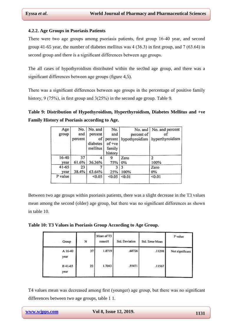
4.2.2. Age Groups in Psoriasis Patients
There were two age groups among psoriasis patients, first group 16-40 year, and second
group 41-65 year, the number of diabetes mellitus was 4 (36.3) in first group, and 7 (63.64) in
second group and there is a significant differences between age groups.
The all cases of hypothyroidism distributed within the sec0nd age group, and there was a
significant differences between age groups (figure 4,5).
There was a significant differences between age groups in the percentage of positive family
history, 9 (75%), in first group and 3(25%) in the second age group. Table 9.
Table 9: Distribution of Hypothyroidism, Hyperthyroidism, Diabetes Mellitus and +ve
Family History of Psoriasis according to Age.
Between two age groups within psoriasis patients, there was a slight decrease in the T3 values
mean among the second (older) age group, but there was no significant differences as shown
in table 10.
Table 10: T3 Values in Psoriasis Group According to Age Group.
T4 values mean was decreased among first (younger) age group, but there was no significant
differences between two age groups, table 1 1.

www.wjpps.com Vol 8, Issue 12, 2019.
1132
Eyssa et al. World Journal of Pharmacy and Pharmaceutical Sciences
Table 11: T4 Values in Psoriasis Group According to Age.
TSH values mean show no significant differences among two groups as in table 12.
Table 12: TSH Values in Psoriasis Group According to Age.
4.2.3. Type of Psoriasis in Psoriasis Patients
The psoriasis patients divided into psoriasis vulgaris group and other types groups, vulgris
type was 28 (46.6), and other types group was 32 (53.4). the mean age of psoriasis vulgaris
was 37.7521:11.37 and for other types group was 35.2121:11.94.
There “as a significant difl„erences in the number of diabetes mellitus within \„tilgaris and
other types of psoriasis 7 (63.6%) and 4(35.4) respectively. The number of positive family
history was significantly differences 9 (75%) in vulgaris group and 3 (25%) in other types
group.
Number and percentage of hypothyroidism among vulgaris group was 2 (66.6), and was
significantly differences from other types of psoriasis (163.4), there was strong significant
differences among two groups in the number and percentage of hyperthyroidism 2 (100%) in
vulgaris group and zero in other types table 13, (figure 6,7).

www.wjpps.com Vol 8, Issue 12, 2019.
1133
Eyssa et al. World Journal of Pharmacy and Pharmaceutical Sciences
Table 13: Distribution of Hypothyroidism, Hyperthyroidism, Diabetes Mellitus and +ve
Family History of Psoriasis according to Type of Psoriasis.
T3 values mean among other types of psoriasis was decreased compared to vulgaris psoriasis
type but there was no significant differences, table 14.
Table 14: T3 Values in Psoriasis Group According to Type of Psoriasis.
T4 values mean was decreased among other types group of psoriasis 80.5 and 89.2 in
psoriasis vulgaris, but this differences not significant as in table 15.
Table 15: T4 Values in Psoriasis Group According to Type of Psoriasis.
TSH values mean was decreased in psoriasis vulgaris group than other types group 2.4 and
3.2 respectively, but the differences is not significant, table 16.

www.wjpps.com Vol 8, Issue 12, 2019.
1134
Eyssa et al. World Journal of Pharmacy and Pharmaceutical Sciences
Table 16: TSH Values in Psoriasis Group According to T3pe of Psoriasis.
4.2.4. Psoriatic Groups According to Duration of Disease
Psoriasis patients were divided according to duration of psoriasis into less than 10 year
duration group (A), and more than 10 year duration group (B). In group A the number was
24(40%), group B 36(60%), there was a strong significant differences between two groups in
diabetes mellitus number, the total diabetics number were in group B(>10 year duration).
The number of positive family history was 4(33.4) in group A, and 8(66.66) in group B(>10
year duration), and there was signifith differences.
The number and percentage of hypothyroidism was significantly different among two groups
3 (100%) in group B and zero in group A.
The hyperthyroidism number and percentage was 2(100%) in group A, and zero in group B,
table 17, figure (8,9).
Table 17: Distribution of Hypothyroidism, Hyperthyroidism, Diabetes Mellitus and +ve
Family History of Psoriasis according to Duration.
T3 values mean was decreased in group A (<10 year duration), compared with group B (1.63
and 1.9) respectively, but this decreased was nbt significantly, table 18.

www.wjpps.com Vol 8, Issue 12, 2019.
1135
Eyssa et al. World Journal of Pharmacy and Pharmaceutical Sciences
Table 18: T3 Values in Psoriasis Group According to Duration of Psoriasis.
T4 values mean was decreased in group A (<10 year duration), compared w1th group B (>10
year duration), 82.04 and 86.25 respectively, that is not significantly differences, table 19.
Table 19: T4 Values in Psoriasis Group According to Duration of Psoriasis.
TSH values mean was decreased in group B compared to group A (2.62 and 3.13
respectively), but there was no significantly differences, table 20.
Table 20: TSH Values in Psoriasis Group According to Duration of Psoriasis.

www.wjpps.com Vol 8, Issue 12, 2019.
1136
Eyssa et al. World Journal of Pharmacy and Pharmaceutical Sciences
CHAPTER FIVE
DISCUSSION
The etiopathogenesis of psoriasis still remains obscure although many dvelopements and
recorded in the treatment and pathogenesis of psoriasis.[92]
Propylthiouracil, an anti -thyroid
preparation, was successfully used both in local, and systemic treatment of psoriasis.
Although the mechanism of action was unclear, it was suggested that this drug might have a
regulatory effect on the T cells in the psoriasis plague. Propylthiouracil increased the number
of total and suppressor/cytotoxic T cells and reduced activated lymphocytes in psoriatic
plaques. Other anti-thyroid agents, such as methimazole, and thiamazole have also been used
successfiilly in the treatment of psoriasis. This means that thyroid hormones may have
unknown effects on the disease.[93]
5.1. Psoriatic and Control Groups
In this study which included, 60 psoriatic patients and 60 control, the proportion of
hypothyroidism and hyperthyroidism were increased compared to the control group. There
was 3 (5%) hypothyroidism cases, and 2 (3.3%) hyperthyroidism cases, which is significantly
different from this percentage in control group (table 1), (figure 1).
According to Dr. Ray Peat and others, low thyroid function is associated with many skin
problems, including psoriasis. When thyroid function is low, prolactin increases, and Dr. Peat
associates excess prolactin with psoriasis. Why? Prolactin increases cell division and sebum
formation. Sunlight decreases prolactin formation whereas darkness and stress increase it.
This may be the connection between sunlight and the alleviation of psoriasis.[94]
In this study the serum T3 and T4 were decreased and TSH was increased in psoriatic
patients compared to control group, but only serum T4, was significantly reduced in psoriatic
group (table 2,3, and 4 respectively). This decrease in serum T4 level did not correlate with
gender, age of the patients, types of psoriasis, and duration of disease.
Hoath SB et al,[95]
explain that thyroid hormone receptors are expressed in human skin and
are thought to be involved in the regulation of epidermal proliferation and differentiation.
Thyroid hormones cause an increase in epidermal growth factor(EGF). EGF play an
important role in cell proliferation. In psoriasis, increased histochemical expression of EGF
receptors has been reported in the epidermis. This altered process of EGF receptor production
may be involved in the onset of psoriasis.[96]
www.wjpps.com Vol 8, Issue 12, 2019.
1137
Eyssa et al. World Journal of Pharmacy and Pharmaceutical Sciences
According to this theory we can explained the 3.3% of hyperthyroidism in psoriatic patients.
Also, another suggestion may be that increased T4 levels result from psoriasis. Increased
level of T4 in non-thyroid illness have been reported. About 80% of the extrathyroida1T3
pool is produced from T4 by monodeiodination with 5-deiodinase enzyme in peripheral
tissues. The activity of the 5-deiodinase enzyme may diminish in some patients with
nonthyroidal disease.[97]
5-deiodinase enzyme activity is regulated by proinflammatory
cytokines such as interleukin, and tumor necrosis factor(TNF).[32,98]
Cytokines like TNF are
directly involved in psoriasis . The reason for the increased T4 levels in psoriasis may be
related to the mcrease 1n some cytokmes 1n psorla31s.[36]
According to this explanation T4, must be increased in psoriatic patients, and the decrease T4
level in our study can explained as a result of the usage of anti-thyroid drugs in the treatment
of psoriasis.
In the present study, and from the history of psoriatic patient the percentage of diabetes
mellitus was 18.6 (11 from 60 psoriatic patients), Cohen A. D. et al.,[99]
improved that
diabetes mellitus percentage in psoriasis was 13.6, when they demonstrates that patients with
psoriasis have a significant association with each of the components of the metabolic
syndrome.
This different in two studies can be explain by the number of cases was taken, and the nature
of community and life style.
The high percentages of positive family history in psoriatic patients (20%, 12 from 60
psoriatic patients), can be explained by psoriatic genetic theory. It was suggested by several
studies that psoriasis clusters in families. In the study of Lomholt on the fame islands, 91%
had at least one affected first or second degree relative.
Farber and Nall[101]
found that 36% of 5.600 respondents with psoriasis had an affected
family member. Similarly, Kavli et al., could show the prevalence of psoriasis increases with
the number of relatives with the same disease.
5.2. Psoriatic Groups According to Gender
In the present study the psoriatic patients were divided according to gender, age, types and
duration of psoriasis.
www.wjpps.com Vol 8, Issue 12, 2019.
1138
Eyssa et al. World Journal of Pharmacy and Pharmaceutical Sciences
According to the gender there is a significant increased in the number of hypothyroidism
among females compared to number in males, Akhtar et a1. has reported 1.3%
hypothyroidism in males and 2.75% in female, that explain the higher percentage of
hypothyroidism in females psoriatic patients. There was no significant different in
hyperthyroidism distribution according to gender (table 5, figure 2,33) There was no
significant differences of thyroid function tests levels and number of diabetes mellitus cases
according to gender.
Pitchappan RM et a1., they found that a female psoriatics most often show a younger age of
onset than males.
Lindegard B., demonstrate that psoriasis associated diseases, found more often in female than
in male psoriatics.
Krasteva at el., Also established a higher incidence of diabetes in psoriasis patients, but in
contrast to the findings of Lindegard, no gender relationship was observed in regard to
diabetes and urticaria.
5.3. Psoriatic Groups According to Age
In the present study the psoriatic patients according to age was divided into younger and
older groups (table 9, figure 4,5), there was a significant increase in the number of
hyperthyroidism among younger group, which can be explained by Hoath SB, and Safer JD
et al. about the role of thyroid hormones in the etiopathogenesis of psoriasis.[95,96]
Also
younger group of psoriasis demonstrates a significant association with positive family history,
which agreed by Farbar and Nall.
The older group of psoriasis (more than 40 year), there was a significant association between
age and hypothyroidism, which may explained by the theory of long period treatment of
psoriasis with anti-thyroid drugs. The number of diabetes mellitus was significantly increased
in older psoriatic patients can be explained as a result of increase the percent of type 2
diabetes mellitus in older age group(m), there is a recognized association between diabetes
and thyroid disease, which has long been reported, Feely and others studied the relation
between diabetes mellitus and thyroid dysfunction.
There were no significant correlation in T3, T4, and TSH levels according to age groups of
psoriasis.

www.wjpps.com Vol 8, Issue 12, 2019.
1139
Eyssa et al. World Journal of Pharmacy and Pharmaceutical Sciences
5.4. Psoriatic Groups According to Psoriasis Type
In the present study the psoriatic patients dividing into psoriasis vulgaris group and other
types group of psoriasis(table13, figure 6,7), there were a significant increased in diabetes
mellitus distribution among psoriatic vulgaris group of psoriasis, this can be explained by
Nini et al., they observed an increase in I-ILA-B27 in minimal psoriasis, in A2 in guttate
psoriasis and in B41inverted psoriasis, therefore psoriasis comorbidity appear according to
these factors.
There was a significant association between psoriasis and hypo and hyperthyroidism in
psoriasis vulgaris type, which can be explained as psoriasis vulgaris is the common type of
psoriasis, for that reason more comorbidities can appear at this type of psoriasis.
5.5. Psoriatic Groups According to Duration
According to duration, psoriatic patients was divided into two groups, less and more than 10
year duration(table l7, figure 8and 9). There was a significant correlation between psoriasis
and comorbidities (hypothyroidism, hyperthyroidism, and diabete mellitus) in group of more
than 10 year duration of psoriasis, previous studies was explain the occurrence of psoriasis
comorbidies associated directly w1th the duration of psoriasis.
CHAPTER SIX
CONCLUSION AND RECOMMENDATIONS
6.1. Conclusions
From the results of the present study, it can be concluded that:
Psoriatic patients had higher percentage of diabetes mellitus.
The study showed that, many cases of psoriasis had positive family history.
Positive correlation between psoriasis and thyroid disorders.
Triiodothyronine (T3) and thyroxine (T 4) level was decreased in psoriatic patient
compared to control group.
Risk of psoriasis comorbidities were increased in older psoriatic age groups. The duration
of psoriasis had positive correlation with psoriasis comorbidities.
Psoriasis vulgaris is a common type of psoriasis.
6.2. Recommendations
F urther studies are recommended as follows: Free and total triodothyronine (T3) thyroxine
(T4) should be measured. Thyroid antibodies (antiperoxidase antibodies and

www.wjpps.com Vol 8, Issue 12, 2019.
1140
Eyssa et al. World Journal of Pharmacy and Pharmaceutical Sciences
antithyroglobuline antibodies) should be measured in the screening for thyroid autoimmmue
diseases. Ultrasonography and fine neeble aspiration make the screening for thyroid disorders
more accurate.
CONCLUSIONS
From the results of the present study, it can be concluded that: Psoriatic patients had higher
percentage of diabetes mellitus, the study showed that, many cases of psoriasis had positive
family history of psoriasis, there was a positive correlation between psoriasis and thyroid
disorders. Triiodothyronine (T3) and thyroxine (T4) level was decreased in psoriatic patient
compared to control group. Risk of psoriasis comorbidities were increased in older psoriatic
age groups, the duration of psoriasis had positive correlation with psoriasis comorbidities.
REFERANCES
1. Chao C, Dann F. Psoriasis comorbidities. J_Dermatolog Treat, 2008; 19: 5-21.
2. Krueger G, Ellis CN. Psoriasis - recent advancesin pathogenesis and treatment. J Am
Acad Dermatol, 2005; 53: 94-100.
3. Naldi L: Epidemiology of psoriasis. Curr Drug Targets Allergy, 2004; 3: 121-12.
4. Christophers E. Comorbidities in psoriasis. Clin Dermatol, understandin its Inflamm,
2007; 25: 529.
5. Langley R, Krueger features, and quality of life. Ann Rheum Dis, in patients with
psoriasis. Br J G, Griffiths C: Psoriasis: epidemiology, clinical, 2005; 64: 8 - 23.
6. Gulliver W. Long-term prognosis Dermatol, 2008; 159: 2-9.
7. Schmitt-Egenolf M: Psoriasis therapy in real life: the need for registries. Dermatology,
2006; 213: 327-330.
8. Boehncke S, Fichtlscherer S, Salgo R, Garbaraviciene J, BCSCh' mann H9 Diehl S,
Hardt K, Thaqi D, Boehncke WH Systemic therapy of plaque- type psoriasis ameliorates
endothelial cell function: results of a prospective longitudinal pilot trial. Arch Dermatol
Res, 2011; 303: 381-388.
9. Alexander B, Pinto J, Pal GS, Kamath N, Kuruvilla M Disease concomitance in
psoriasis: a clinical study of 61 cases. Indian J Dermatol Venereol Leprol, 2001; 67: 66-
68.
10. Al-Mutairi N, Al-Farag S, Al-Mutairi A, Al-Shiltawy M Comorbidities associated with
psoriasis: an experience from the Middle East. J Dermatol, 2010; 37: 146—155.
11. Augustin M, Glaeske G, Radtke MA, Christophers E, Reich K, Schafer I Epidemiology

www.wjpps.com Vol 8, Issue 12, 2019.
1141
Eyssa et al. World Journal of Pharmacy and Pharmaceutical Sciences
and comorbidity of psoriasis in children. Br J Dermatol, 2010; 162: 633—636.
12. Boehncke WH, Boehncke S, Schon MP Managing comorbid disease inatients with
psoriasis. BMJ, 2010; 340: b5666.
13. Gelfand JM, Weinstein R, Porter SB, Neimann AL, Berlin JA, Margolis 1) Prevalence
and treatment of psoriasis in the United Kingdom: a population-based study. Arch
Dermat, 2005; 141: 1537-1541.
14. Gerdes S, Zahl VA, Knopf H, Weichenthal M, Mrowietz U Comedication related to
comorbidities: a study in 1203 hospitalized patients with severe psoriasis. Br J Dermat,
2008; 159: 1116-1123.
15. Huerta C, Rivero E, Rodriguez LA Incidence and risk factors for psoriasis in the general
population. Arch Dermat, 2007; 143: 1559-1565.
16. Rapp SR‟ Feldman SR, Exum ML, Fleischer AB Jr, Reboussin DM Psoriasis causes as
much disability as other major medical diseases. J Am Acad Dermatol, 1999; 41: 401-
407.
17. Stern RS, Nijsten T, Feldman SR, Margolis DJ, Rolstad T Psoriasis is common, carries a
substantial burden even when not extensive, and is associated with widespread treatment
dissatis- faction. J Investig Dermatol Symp Proc, 2004; 9: 136-139.
18. Rahman P, Elder JT. Genetic epidemiology of psoriasis and psoriatic arthritis. Ann
Rheum Dis, 2005; 64: 37-9.
19. Elder JT, Nair RP, Guo SW, chseler T, Christophers E, Voorhees JJ. The genetics of
psoriasis. Arch Dermatol, 1994; 130: 216-24.
20. Chandran V, Schentag CT, Brockbank JE, Pellett FJ, Shanmugarajah S, Toloza SM, et
a1. Familial aggregation of psoriatic arthritisAnn Rheum Dis, 2009; 68: 664-7.
21. Burden AD, J aved S, Bailey M, Hodgins M, Connor M, Tillman D. Genetics of
psoriasis: Paternal inheritance and a locus on chromosome 6p.J Invest Dermatol, 1998;
110: 958-60.
22. Fine RM. Psoriasis and autoimmune disease. Int J Dermatol, 1988; 27: 17-8.
23. Arican O, Aral M, Sasmaz S, Ciragil P. Serum levels of TNF-u, IFN-y, IL-6, IL-8, IL-
12, IL-l7, and IL-18 in patients with active psoriasis and correlation with disease
severity. Mediators Inflamm, 2005; 2005: 273-9.
24. Chamian F, Krueger JG: Psoriasis vulgaris: an interplay of T lymphocytes, dendritic
cells, and inf lammatory cytokines in patho- genesis. Curr Opin Rheumatol, 2004; 16:
331-337.
25. Schmitt Egenolf M: Psoriasis therapy in real life: the need for registries. Dermatology,

www.wjpps.com Vol 8, Issue 12, 2019.
1142
Eyssa et al. World Journal of Pharmacy and Pharmaceutical Sciences
2006; 213: 327-330.
26. Lopez-Candales A: Metabolic syndrome X: a comprehensive review W pathophysiology
and recommended therapy. J Med, 2001; 32: 283-300.
27. Sommer DM, Jenisch S. Suchun M, Christophers E. Weichenthal M Increased
prevalence of the mctuboilc syndrome In patients wlth moderate to severe psoriasis.
Arch Dermatol Res, 2006; 2911: 321-328.
28. Joshl R. Immunopathogencsls of psorlusls. Indian J Dermatol Venereol Leprol, 2004;
70: 10-2.
29. Ayala F, Ayala F. Clinical aspects and comorbidities of psoriasis. J Rheumatol, 2009;
83: 19-20.
30. Sommer DM, Jenisch S, Suchtm M at, Increased prevalence of the metabolic syndrome
in patients with moderate to severe psoriasis. Arch Dermatol Res, 2006; 298: 321-8.
31. Mallbris L, Akre O, Granath F, Yin L. Llndo- 101' B. Ekbom A, et a1: Increased risk for
car- diovasculur mortality in psoriasis inpatients but not in outpatients. Eur J Epidemiol,
2004; 19: 225-230.
32. Pietrzak A, Zalewska A, Chodorowska G, et, Genes and structure of selected cytokines
involved in pathogenesis of psoriasis. Folia Histochem Cytob, 2008; 46: 11-21.
33. Peters BP, Weissman FG, Gill MA. Pathophysiology and treatment of psoriasis. Am J
Health Promot, 2000; 7: 645-659.
34. Gisondi P, Tessari G, Conti A, Piaserico S, Schianchi S, Peserico A, ct al. Prevalence of
metabolic syndrome in patients with psoriasis. Br J Dermatol, 2007; 157: 68-73.
35. Traub M, Marshall K. Psoriasis pathophysiology, conventional, and alternative
approaches to treatment. Altern Med Rev, 2007: 12(4): 319-330.
36. Arican O, Bilgic K, Koo K. The effect of thyroid hormones in psoriasis vulgaris. IJDVL,
2004; 70: 353-6.
37. Karanikas E, Harsoulis F, Giouzepas I. Griveas I. Stimulation of the hypothalamic-
pituitary-adrenal axis with corticotropin releasing hormone in patients with psoriasis.
Hormone(Athens), 2007; 6: 314-320.
38. Gupta AK, Langley R,Poulin Y,Lui H,Searlcs (3, Carey W et al. Pathogenesis of
Psoriasis and Current Challenges. J Cutan Med Surg, 2004; 8: 3-7.
39. Peslyak M. Model of pathogenesis of psoriasis. Part 1. Systemic psoriatic process. e3.2
ed. Moscow, 2011; 4: 1-71.
40. Campalani E, Barker JNWN. The Clinical Genetics of Psoriasis. Curr Genomics, 2005;
5: 51-50.

www.wjpps.com Vol 8, Issue 12, 2019.
1143
Eyssa et al. World Journal of Pharmacy and Pharmaceutical Sciences
41. Bowcock AM. Psoriasis Genetics: The way forward. J Invest Dermatol, 2004; 122: 15-
17.
42. Lowes MA, Bowcock AM, Krueger JG. Pathogenesis and therapy of psoriasis. Nature,
2007; 22: 866-873.
43. Campalani E, Barker JNWN. The Clinical Genetics of Psoriasis. Curr Genomics, 2005;
6: 51-60.
44. Bowcock AM. Psoriasis Genetics: The way forward. J Invest Dermatol, 2004; 122: 15-
17.
45. AlShobaili HA, Shahzad M, Al-Marshood A, Khalil A, Settin A, Barrimah 1. Genetic
Background of Psoriasis. Int J Health Sci, 2010; 4: 23-29.
46. Giardina E, Sinibaldi C, Novelli G. Mapping the future of common diseases: lessons
from psoriasis. Front, 2007; 12: 1563-1573.
47. Arakura F, Hida S, Ichikawa E, Yajima C, Nakajima S, Saida T et a1. Genetic control
directed toward spontaneous IFN-a/IFN-B responses and downstream IFN-y expression
influences the pathogenesis of a murine psoriasis-like skin disease. J Immun, 2007; 179:
3249-3257.
48. Hfiffmcier U, Lascorz J, Traupe H, B(Shm B, Horst FS, Stander M et a1. Systematic
Linkage Disequilibrium Analysis of SLC12A8 at PSORSS Confirms a Role in
Susceptibility to Psoriasis Vulgaris. J Invest Dermatol, 2005; 125: 906-912.
49. Maddin S, Kourosh AS, Miner A, Menter A. Psoriasis as the marker of underlying
systemic disease. Skin therLett, 2008; 13(1): 1-10.
50. Zhang XJ, Yan KL, Wang ZM, Yang S, Zhang GL, Fan X et a1. Polymorphisms in
interleukin-IS gene on chromosome 4q31.2 are associated with psoriasis vulgaris in
Chinese population. J Invest Dermatol, 2007; 127: 2544-2551.
51. Davidovici BB, Sattar N, Jérg PC, Puig L, Emery P, Barker JN. Psoriasis and systemic
inflammatory diseases: potential mechanistic links between skin disease and co-morbid
conditions. J Invest Dermatol, 2010; 130: 1785-1796.
52. Davidson A, Diamond B. Autoimmune diseases. N Engl J Med, 2001; 345: 340-50.
53. The thyroid gland in Endocrinology: An Integrated Approach by Stephen Nussey and
Saffron Whitehead (2001) Published by BIOS Scientific Publishers Ltd. ISBN 1-85996-
252-1.
54. Lemalre, David (2005-05-27). "eMedicine - Thyroid anatomy... Retrieved, 2008-01-19.
55. Boron WF, Boulpaep E (2003). "Chapter 48: “synthesis of thyroid hormones”, Medical
Physiology; A Cellular And Molecular Approaoch. Elsevier/Saunders. pp. 1300. ISBN

www.wjpps.com Vol 8, Issue 12, 2019.
1144
Eyssa et al. World Journal of Pharmacy and Pharmaceutical Sciences
1-4160-2328-3.
56. EkhOIm R, Bjorkman U. "Glutathione peroxidase degrades intracellular hydrogen
peroxide and thereby inhibits intracellular protein iodination in thyroid epithelium".
Endocrinology, 1997; 138(7): 2871-2878.
57. Bianco AC, Salvatore D, Gereben B, Berry MJ, Larsen PR (2002). "Biochemistry,
cellular and molecular biology, and physiological roles of the iodothyronine
selenodeiodinases".Endocr Rev, 2002; 23(1): 38-89.
58. Jansen J, Friesema EC, Milici C, Visser TJ. "Thyroid hormone transporters in health and
disease". Thyroid, 2005; 15(8): 757—68.
59. Weetman AP, McGregor AM. Autoimmune thyroid disease: further developments in our
understanding. Endocr Rev, 1994; 15: 788-830.
60. Volpe R: Autoimmune thyroid diseases. In Diseases of the thyroid Edited by: LE B.
Totowa, Humana Press, 1997; 4: 125-154.
61. Vondra K, Vrbikova J, Dvorakova K. Thyroid gland diseases in adult patients with
diabetes mellitus. Minerva Endocrinol, 2005; 30(4): 217-36.
62. Shiva S, Behbahani AG. Autoimmune thyroid disease in children and adolescents with
type 1 diabetes mellitus in Northwest Iran. Saudi Med J, 2009; 30(5): 673-6.
63. Vaidya B, Pearce S. The emerging role of the CTLA-4 gene in autoimmune
endocrinopathies. Eur J Endocrin, 2004; 150(5): 61926.
64. Hansen D, Bennedbaek FN, Hansen LK, et a1. Thyroid function, morphology an
autoimmunityin young patients with insulin-dependent 65-diabetes mellitus. Eur J
Endocrinol, 1999; 140(6): 512-8.
65. Arican O, Bilgic K, Koc K. The effect of thyroid hormones in psoriasis vulgaris. IJDVL,
2004; 70: 353-6.
66. Hansen D, Bennedbaek FN, Haier-Madsen M, et al. A prospective study of thyroid
function, morphology and autoimmunity in young patients with type 1 diabetes. Eur J
Endocrinol, 2003; 148(2): 245-51.
67. Sheikholeslami H, Ziaee A, Darvishghaderi S, et a1.Hypothyroidism and abetes mellitus.
JQUMS, 2007; 11(3): 51-6.
68. Roquer J, Escudero D, Hermiz J, Maso 11, Cam 1‟: Hashimoto'sthyroiditis. JNeuroi,
1987; 234: 23-24.
69. Karavanaki K, Kaklcas K, i‟aschali Multiple sclerosis and ' E, at aLScrcening for
associated autoimmunity in children and adolescents with type 1 diabetes mellitus
(TlDM). Horm Res, 2009; 71(4): 201-6.

www.wjpps.com Vol 8, Issue 12, 2019.
1145
Eyssa et al. World Journal of Pharmacy and Pharmaceutical Sciences
70. Huang W, Kukes GD: Hashimoto„s thyroiditis: an organ-spe-ciiic autoimmune disease--
pathogenesis and recent devci-opmcnts. Lab Invest, 1999; 79: 1175-1180.
71. Holl RW, Bohm B, Loos U, et a1, Thyroid autoimmunity in children and adolescents
with type 1 diabetes mellitus. Effect of age, gender and HLA type. Horm Res, 1999;
52(3): 113-8.
72. Nikiforov YE, Nikiforova MN: Molecular genetics and diagnosis of thyroid cancer. Nat
Rev Endocrinol, 2011; 7: 569-580.
73. Costa AM, Herrero A, Fresno MF, Heymann J, Alvarez JA, Camcselle- Teijeiro J,
Garcia-Rostan G: BRAF mutation associated with other genetic events identifies a subset
of aggressive papillary thyroid carcinoma.Ciin Endocrinol (Oxt), 2008; 68: 618-634.
74. Steiss JO, Otten A, Graef V, Klingmtiiler V. Thyroid gland ultrasound and urinary
iodine excretion in children and adolescents with type I diabetes mellitus. Klin Padiatr,
1996; 208(6): 327-33.
75. Jemal A, Bray F, Center MM, Ferlay J, Ward E, Forman D: Global cancer statistics.CA
Cancer J Clin, 2011; 61: 69-90.
76. Smallridge RC, Marlow LA, Copland JA: Anaplastic thyroid cancer: molecular
pathogenesis and emerging therapiesEndocr Relat Cancer, 2009; 16: 17-44.
77. Weetman AP, McGregor AM. Autoimmune thyroid disease: further developments in our
understanding. Endocr Rev, 1994; 15: 788-830.
78. Vena GA, Altomare G, Ayala F, Berardesca E, Calzavara-Pinton P, Chimenti S,
Giannetti A, Girolomoni G, Lotti T, Martini P, Mazzaglia G, Peserico A, Pugiisi Guerra
A, Sini G, Cassano N, Cricelli C Incidence of psoriasis and association with
comorbidities in Italy: a 5-year observational study from a national primary care
database. Eur J Dermatol, 2010; 20: 593-598.
79. Wolf N, Quaranta M, Prescott NJ, Allen M, Smith R, Burden AD, Worthington J
Griffiths CE, Mathew CG. Barker JN, Capon F, Trembath.
80. Bartlett, Mostly MN. 'iyrlnu SK. Inhibitors: The Advantages and Disadvantages 01„ this
Novel Approach 1hr the 'i„rcntmunt oi1 l'shrittuis. Skin Therapy Lott, 20011; 13(11):
1-4.
81. Summer DM, Jonisch S. Suohnn M. Christophorn Ii. Woichcnthnl M increased
prevalence of the mettthoiio syndrome In pullunts with moderate to severe psoriasis.
Amh Dermatol Res, 2006; 2911: 321-328.
82. Warren R11. Grtmths 011M. Atinllmumnh 1hr the "l„rentnwnt 01' Psoriasis. Expert Rev
Dermatol, 2009; 4(1): 15-21.

www.wjpps.com Vol 8, Issue 12, 2019.
1146
Eyssa et al. World Journal of Pharmacy and Pharmaceutical Sciences
83. T. ilenseler. Genetics ofpsoriasis. Arch Dcrmnyol Res, 1998; 290: 463-476.
84. Severinski S. Dunno S. Sevrinskl NS, et ul. Epidemiology and clinical characteristics
oi„thyroid dyshmction in children (111d adolescents with type 1 diabetes. Coll Antropol,
2009; 33(1): 273-9.
85. Ormston. B. J., et: Thyrotropin Response to ‟i„hyretropine Releasing Hormone 1n
Ophthalmic Graves Disease: correlation: with other aspects of thyroid function, Thyroid
01„Eye Signs. Clin. Endo.. Oxford, 1973; 2: 366-376.
86. Squire C. R.. Gimlette T. M. D. Assessment 01„ Optimal L-Thyroxine Replacement
Dose by the TR” Test. Ann. Clin. Biochcm, 1982; 19: 26-27.
87. Eri„urth, E. M., Hcdncr, P., Thyroid hormone metabolism in thyroid disease as reflected
by the ratio of serum Triiodothyronine to thyroxine. J. Endocrinol. Invest, 1986; 9: 407-
412.
88. Browing, M. C. K. ct Intm- individual Variation of 'l„hyroxlnc. Triiodothyronine, and
thyrotropine in trcnrcd hypothyroid patients, 505-515.
89. Cohen AJ). et: Association between psoriasis and the metabolic syndrome. JDermatol,
2008; 216: 152-155.
90. Lomhoit G: Psoriasis: prevalence, Spontaneous Course and Genetics- A census study on
the prevalence 01" skin disease on the liaroer iiands.
91. Copenhagen. 0130, (MD, 1963. Farber E, Null M: The natural history of psoriasis in
5,600 payients. Dermatologica, 1974; 148: 1-18.
92. Kavli G, Fordc O, Amcscn 11, Stcnvoid S:Psoriusis: familial predisposition and
environmental factors. DH, 1985; 291: 999-1000.
93. Akhtar. S., A. Khan, M. M. Siddiqui and Out Nuwab, 2001. Frequencies of thyroid
problems in different age, sex and season. The Sciences, 2001; 1: 153-156.
94. Pitchappan RM. Kotccswumn A. Kukkaniah VN, Manickasundari M, Rajamm V,
Muthuveeraiakshmi P, Muhcndmn V. Brahmajothibai V HLA.
95. Bw57 and DR7 association with psoriasis vulgaris. in south indie. Tissue Antigens,
1989; 34: 133-137.
96. Lindcgard B. Diseases associated with psoriasis in a general population of 159,200
middle ~aged, urban, native Swedes. Dermatologica, 1986; 172: 298- 304.
97. Krasteva M, Mateev G, Lasarva A. On psoriasis as associated diseases, Dermatologica,
1988; 177: 21-23.
98. Feely J, Isles TE Screening for thyroid dysfunction in diabetics. Br Med J, 1979; 1:
1678.
www.wjpps.com Vol 8, Issue 12, 2019.
1147
Eyssa et al. World Journal of Pharmacy and Pharmaceutical Sciences
99. Gray RS, Irvine WJ, Clarke BF Screening for thyroid dysfunction in diabetics. Br„Med
J, 1979; 2: 1439.
100. Smithson MJ. Screening for thyroid dysfunction in a community population of diabetic
patients. Diabet Med, 1998; 15: 148-150.
101. Perros P, McCrimmon RJ, Shaw G, Frier BM Frequency of thyroid dysfunction in
diabetic patients: Value of annual screening. Diabet Med, 1995; 12: 622-627.
102. Nini G, Bianchi L, Iraci S, Campione G, Spagnuolo A, Adorno D, Papola F HLA
antigens and infantile psoriasis. Acta Derm Venereol Suppl 1989; 146: 59-62.


![Psoriasis in Children and Adolescents: Diagnosis ......Guttate psoriasis is the second most common type of psoriasis in children [21, 29]. Griffiths and Barker defined guttate psoriasis](https://static.fdocuments.us/doc/165x107/5f501dea60f5a266c60b268c/psoriasis-in-children-and-adolescents-diagnosis-guttate-psoriasis-is-the.jpg)


