Regulation of transcription of the RNA splicing factor ...gilast/PAPERS/rna_hSlu7.pdf · Shomron,...
13
10.1261/rna.492907 Access the most recent version at doi: published online Sep 5, 2007; RNA Shomron, Ohad Shaham, Andrew D. Sharrocks, Ruth Ashery-Padan and Gil Ast Moti Alberstein, Maayan Amit, Keren Vaknin, Amanda O'Donnell, Chen Farhy, Yaniv Lerenthal, Noam affects alternative splicing Sp1 and Elk-1 by hSlu7 Regulation of transcription of the RNA splicing factor P<P Published online September 5, 2007 in advance of the print journal. service Email alerting click here top right corner of the article or Receive free email alerts when new articles cite this article - sign up in the box at the Notes object identifier (DOIs) and date of initial publication. by PubMed from initial publication. Citations to Advance online articles must include the digital publication). Advance online articles are citable and establish publication priority; they are indexed appeared in the paper journal (edited, typeset versions may be posted when available prior to final Advance online articles have been peer reviewed and accepted for publication but have not yet http://www.rnajournal.org/subscriptions/ go to: RNA To subscribe to © 2007 RNA Society on September 9, 2007 www.rnajournal.org Downloaded from
Transcript of Regulation of transcription of the RNA splicing factor ...gilast/PAPERS/rna_hSlu7.pdf · Shomron,...
10.1261/rna.492907Access the most recent version at doi: published online Sep 5, 2007; RNA
Shomron, Ohad Shaham, Andrew D. Sharrocks, Ruth Ashery-Padan and Gil Ast Moti Alberstein, Maayan Amit, Keren Vaknin, Amanda O'Donnell, Chen Farhy, Yaniv Lerenthal, Noam
affects alternative splicingSp1 and Elk-1 by hSlu7Regulation of transcription of the RNA splicing factor
P<P Published online September 5, 2007 in advance of the print journal.
serviceEmail alerting
click heretop right corner of the article or Receive free email alerts when new articles cite this article - sign up in the box at the
Notes
object identifier (DOIs) and date of initial publication. by PubMed from initial publication. Citations to Advance online articles must include the digital publication). Advance online articles are citable and establish publication priority; they are indexedappeared in the paper journal (edited, typeset versions may be posted when available prior to final Advance online articles have been peer reviewed and accepted for publication but have not yet
http://www.rnajournal.org/subscriptions/ go to: RNATo subscribe to
© 2007 RNA Society
on September 9, 2007 www.rnajournal.orgDownloaded from

Regulation of transcription of the RNA splicing factor
hSlu7 by Elk-1 and Sp1 affects alternative splicing
MOTI ALBERSTEIN,1,4 MAAYAN AMIT,1,4 KEREN VAKNIN,1 AMANDA O’DONNELL,2 CHEN FARHY,1
YANIV LERENTHAL,1 NOAM SHOMRON,3 OHAD SHAHAM,1 ANDREW D. SHARROCKS,2
RUTH ASHERY-PADAN,1 and GIL AST1
1Department of Human Molecular Genetics and Biochemistry, Sackler Faculty of Medicine, Tel-Aviv University, Tel Aviv 69978, Israel2Faculty of Life Sciences, University of Manchester, Manchester M13 9PT, United Kingdom3Department of Biology, Massachusetts Institute of Technology, Cambridge, Massachusetts 02139, USA
ABSTRACT
Alternative splicing plays a major role in transcriptome diversity and plasticity, but it is largely unknown how tissue-specific andembryogenesis-specific alternative splicing is regulated. The highly conserved splicing factor Slu7 is involved in 39 splice siteselection and also regulates alternative splicing. We show that Slu7 has a unique spatial pattern of expression among human andmouse embryonic and adult tissues. We identified several functional Ets binding sites and GC-boxes in the human Slu7 (hSlu7)promoter region. The Ets and GC-box binding transcription factors, Elk-1 and Sp1, respectively, exerted opposite effects onhSlu7 transcription: Sp1 protein enhances and Elk-1 protein represses transcription in a dose-dependent manner. Sp1 proteinbound to the hSlu7 promoter in vivo, and depletion of Sp1 by RNA interference (RNAi) repressed hSlu7 expression. Elk-1 proteinbound to the hSlu7 promoter in vivo, and depletion of Elk-1 by RNAi caused an increase in the endogenous level of hSlu7mRNA. Further, depletion of either Sp1 or Elk-1 affected alternative splicing. Our results provide indications of a complextranscription regulation mechanism that controls the spatial and temporal expression of Slu7, presumably allowing regulation oftissue-specific alternative splicing events.
Keywords: Slu7; alternative splicing; spliceosome; Elk-1; Sp1; transcription
INTRODUCTION
Human and mouse possess a similar number of protein-coding genes (z24,000) (Lander et al. 2001; Waterston et al.2002). Current estimates indicate that the total numberof human proteins exceeds the number of genes. Researchis therefore needed to determine the mechanisms thatunderlie this discrepancy. Alternative splicing is known tosignificantly expand the transcriptomic potential and geneticdiversity (Graveley 2001; Ast 2004; Xing and Lee 2006).Alternative splicing varies among tissues (Hanamura et al.1998; Yeo et al. 2004; Ule et al. 2005), either as a function ofdifferent developmental stages (Wang and Grabowski 1996;Cooper 2005) or due to different physiological conditions(van der Houven van Oordt et al. 2000; Pelisch et al. 2005;
Shomron et al. 2005; Guil et al. 2006). There is also a linkbetween aberrant splicing and human diseases, includingcancer (Philips and Cooper 2000; Zhang et al. 2006).
Splicing is a highly conserved process from yeast tohuman, in which introns are removed from mRNA pre-cursor and exons are ligated to generate mature mRNA.Four short sequences direct the splicing machinery to thesplice junctions: the 59 and 39 splice sites (59ss and 39ss),the branch-site (BS) sequence, and the polypyrimidine tract;the latter two regions are located upstream of the 39ss. Thesplicing reaction consists of two consecutive catalytic stepsand is facilitated by a dynamic protein–RNA complex, knownas the spliceosome. The spliceosome is composed of fivesmall nuclear ribonucleoprotein particles (U1, U2, U4, U5,and U6 snRNPs) and more than 150 proteins (Black 2003).
The splicing factor Slu7 was originally identified in yeastas a ubiquitous protein that was found to be syntheticallylethal with U5. It is involved in the second step of splicingand is dispensable for in vitro splicing of introns with lessthan 12 nucleotides (nt) between the BS and the 39ss (Franket al. 1992; Zhang and Schwer 1997).
4These authors contributed equally to this work.Reprint requests to: Gil Ast, Department of Human Molecular Genetics
and Biochemistry, Sackler Faculty of Medicine, Tel-Aviv University, TelAviv 69978, Israel; e-mail: [email protected]; fax: +972-3-640-9900.
Article published online ahead of print. Article and publication date areat http://www.rnajournal.org/cgi/doi/10.1261/rna.492907.
RNA (2007), 13:1–12. Published by Cold Spring Harbor Laboratory Press. Copyright � 2007 RNA Society. 1
on September 9, 2007 www.rnajournal.orgDownloaded from

In vitro, the human ortholog (hSlu7) affects the fidelityof 39ss selection when an incorrect 39ss sequence is adjacentto the functionally correct site. In the absence of hSlu7 theincorrect 39ss is activated. This activation only occurs whenthe distance between the 39ss and the BS is not more thanz30 nt (Chua and Reed 1999b). Recently, hSlu7 was shownto regulate alternative splicing by a sensitive nucleo-cytoplasmicshuttling. This shuttling controls the nuclear concentrationof hSlu7 following specific physiologic stress conditions(Lev-Maor et al. 2003; Shomron et al. 2004, 2005).
Although the conservation of hSlu7 from yeast to humansuggests that it is a ubiquitous spliceosomal protein, wedemonstrated that hSlu7 is not required for cell viabilityin the examined cell lines (Shomron et al. 2005). Thisobservation raises the question of whether Slu7 is indeeda constitutively expressed protein. Here, we show that themammalian Slu7 is differentially expressed in varioustissues and cell lines and also in developing embryonictissues. We have begun to unravel the elaborate regulatorymechanism of hSlu7 transcription. A complex promoterarrangement that controls hSlu7 temporal and spatialexpression via several potential Ets-like transcription factorbinding sites (also called EBS) was identified. Some of thesesites function as positive and others as negative regulatoryelements. Also, two functional regionsrich in GC-boxes that may be recognizedby the zinc finger transcription factorSp1 were identified. The experimentsdescribed suggest that Sp1 protein ele-vates transcription of hSlu7, whereasElk-1, a member of the ETS transcrip-tion factor family, represses hSlu7 tran-scription. Both Sp1 and Elk-1 proteinsbound the hSlu7 promoter in vivo.Consistent with a repressive role, deple-tion of Elk-1 in HeLa cells induced hSlu7endogenous expression. In contrast,depletion of Sp1 repressed hSlu7 expres-sion. Silencing of Elk-1 or Sp1 proteinsaffected alternative splicing of specificexons. The expression pattern of Slu7appears to be controlled by a complexpromoter arrangement and is activatedor repressed by specific regulatory genes.Our data imply that Slu7 is a splicingfactor that regulates tissue- and embry-onic-specific alternative splicing events.
RESULTS
Slu7 is differentially expressed intissues and cell lines
We have shown previously that hSlu7 isnot required for cell survival (Shomron
et al. 2005). This observation raised the question of whetherSlu7 was expressed ubiquitously in all tissues and con-ditions. Thus, we analyzed Slu7 protein and mRNAexpression within various tissues and cell lines (Fig. 1).High levels of hSlu7 protein were detected in 293T (humankidney embryonic cells), HepG2 (liver carcinoma), andDu145 (prostate carcinoma brain metastasis) cell lines (Fig.1A, lanes 1–3). Low protein levels were detected in PC3(prostate adenocarcinoma) and HT1080 (fibrosarcoma)cells (Fig. 1A, lanes 4,5). The mRNA level of hSlu7 iscorrelated with that of the protein: high levels of both theprotein and mRNA in Du145 cells and very low levels inPC3 cells (Fig. 1A, lanes 6,7). Slu7 transcripts were alsodifferentially expressed in various healthy adult tissues inboth human and mouse (data not shown).
We then characterized mouse Slu7 (mSlu7) transcriptdistribution within complex adult tissues from mouse.Paraffin sections of adult mouse pancreas and eye (post-natal days 30 and 15) were hybridized with a specific RNAprobe against mSlu7 mRNA (see Materials and Methods).A unique pattern of expression of mSlu7 within thepancreas and neuroretina was observed. In the pancreas,mSlu7 transcripts were detected in the islets of Langerhans,but not in the acinar exocrine cells (Fig. 1B, panel 1). In the
FIGURE 1. Evidence for differential expression of the mammalian Slu7. mRNA and proteinlevels of human and mouse Slu7 (hSlu7 and mSlu7, respectively) were analyzed in adult andembryonic tissues and cell lines. (A) Protein and mRNA levels in different human cell-linelysates were analyzed by Western blot using an anti-Slu7 antibody (lanes 1–5) or RT-PCR(lanes 6,7). (B) In situ hybridization using an mSlu7 antisense probe was performed onparaffin sections of pancreas and eye from postnatal day 30 (P30) and day 15 mice (P15),respectively. The expression of mSlu7 in the adult pancreas is detected in the islets ofLangerhans (L) but not in the exocrine (E) tissue (panel B1). In the eye mSlu7 transcript wasdetected in the neuroretina (panel B2). (gcl) Ganglion cell layer; (inl) inner nuclear layer; (onl)outer nuclear layer. Scale bar, 100 mm. (C) In E12.5 embryo the expression of mSlu7 is detectedin the developing heart (panel C1) and in the epithelium of the developing lung buds (arrowheads) but not in the surrounding mesenchyme (panel C2). Regions of high mSlu7 expressionare marked by arrow heads. Scale bar, 50 mm.
Alberstein et al.
2 RNA, Vol. 13, No. 11
on September 9, 2007 www.rnajournal.orgDownloaded from

neuroretina, mSlu7 was abundant in the inner nuclear layerand the ganglion cell layer but was weakly expressed in theouter nuclear layer (Fig. 1B, panel 2) and the cornea (datanot shown). Furthermore, differential tissue expression wasobserved in mouse embryo; mSlu7 transcripts were abun-dant in the developing heart and in the epithelium of thelung buds but not in the surrounding mesenchyme (Fig.1C, panels 1 and 2, respectively). These results demonstratethat Slu7 is expressed differentially among tissues andduring development, thus implicating Slu7 as a context-dependent modulator of the splicing machinery.
Cloning and analysis of the hSlu7 promoter structure
The observation that mammalian Slu7 was expresseddifferentially led us to question its transcription regulation.A rapid amplification of 59 cDNA ends (59-RACE) analysiswas used to identify the transcription start sites (TSSs) ofthe human Slu7 gene (see Supplemental Material). Fouralternative TSSs were identified; the most abundant wasselected in 11 of the 20 clones sequenced (see Fig. 2A;Supplemental Material). The most prevalent TSS waschosen as the reference point for the reporter constructassay and was named TSS(1). It is important to note thatthe use of a different TSSs does not affect the codingsequence but rather changes the length of the 59 UTR. Wethen cloned the human Slu7 promoter from position �184upstream to position +66 base pairs (bp) downstream fromTSS(1) into firefly luciferase reporter plasmid (Fig. 2B).Comparative analysis of the Slu7 promoter region (cover-ing the first exon and 184 bp upstream) from a variety ofmammals revealed 100% conservation of the TSS(1) region(Fig. 2C, marked by an arrow), which implies that thisregion is significant, probably for recruitment of basaltranscription machinery.
We analyzed the construct for promoter activity andfound it to be transcriptionally active by more than 103-fold above the empty control pGL3 vector backgroundsignal (data not shown). Next, we searched for putativetrans-acting factor binding sites, using several computa-tional programs (see Materials and Methods). We alsoexamined the conservation level within the promoterregion using multispecies comparative analysis (Fig. 2C).Using this analysis, two types of putative transcriptionalbinding sites were identified. The first to be identified weretwo promoter regions rich in GC that contain potentialbinding sites for the transcription factor Sp1 (Fig. 2C). Wealso identified five tandem putative Ets binding sites(marked EBSa–e) with a core sequence of GGAA (Fig. 2C;notice that EBSe is in the reverse orientation). The Etsfamily of transcription factors binds to a consensussequence CCGGAA. Only EBSa and EBSc fully matchedthe conserved sequence in all mammals tested, whereasEBSd and EBSe are primate specific. The Ets family oftranscription factors consists of several members that are
expressed in a tissue- and a cell-type-specific manner(Sharrocks 2001; Hsu et al. 2004).
hSlu7 promoter activity is regulated by EBS elements
To examine the functionality of the putative EBS elements,we generated specific point mutations within the GGAAelements (the mutations are shown in Fig. 2B). 239T cellswere transfected with either the pSlu7-luc reporter con-struct or the mutant reporter constructs, and cell lysateswere tested for luciferase activity 48 h post-transfection.Activities of the mutant reporter constructs were normal-ized with respect to the wild-type (WT) pSlu7-luc reporteractivity, and results are shown as fold induction (Fig. 2D).Point mutations within the core GGAA elements revealedthat all of the five elements were required for the normaltranscriptional activity of hSlu7, and each of the elementsuniquely regulates hSlu7 transcription in 293T cells. Threeof the EBS elements, EBSa, EBSc, and EBSe, functioned aspositive regulatory elements, because mutations of theseelements resulted in 40%, 50%, and 75% reduction in WTactivity, respectively (Fig. 2D, a,c,e). The other two EBSsites, EBSb and EBSd, functioned as repressive elements;mutations in these elements resulted in >2.5- and >1.4-foldinduction in the transcriptional activity, respectively (Fig.2D, b,d). It is notable that two of the functional elements,EBSd and EBSe, are primate specific (Fig. 2C).
Double and triple mutants revealed a compensatoryrelationship between the EBS sites. For example, a pointmutation in the core GGAA of the highly conservedenhancer EBSa abolished the effect of mutations in eitherone or both of the repressors EBSb and EBSd (Fig. 2D,ab,ad,abd). Induction of the activity by mutations thatabolished the repression by EBSb or EBSd was partiallyreduced when combined with a mutation in the enhancerelement EBSc (Fig. 2D, bc,cd). Finally, deletions within thetwo potential Sp1 sites, GC-rich1 and GC-rich2, resulted inmoderate effects of +20% and �20%, respectively, on thepromoter activity (Fig. 2D, GC-1 Del and GC-2 Del).However, deletions in both regions resulted in more than40% reduction in basal activity (Fig. 2D, GC-12 Del)implying an important role of these regions in mediatingSp1 induction. The mutational analyses provide evidencefor the functionality of all five EBS elements and each of thetwo GC-rich elements in both up- and down-regulation ofthe hSlu7 promoter activity.
hSlu7 promoter activity is up-regulated by Sp1 in vivo
We next asked whether specific transcription factors canregulate hSlu7 expression in vivo. Thus, 293T cells werecotransfected with the pSlu7-luc reporter construct andwith increasing amounts (0.25, 0.5, and 0.75 mg) of a vectorexpressing Sp1 protein (pcDNA4-Sp1; Fig. 3A), a vectorexpressing Elk-1 protein (pCAG-Elk-1; Fig. 4A,B), or the
Temporal and spatial expression of hSlu7 regulation
www.rnajournal.org 3
on September 9, 2007 www.rnajournal.orgDownloaded from

FIGURE 2. (Legend on next page)
Alberstein et al.
4 RNA, Vol. 13, No. 11
on September 9, 2007 www.rnajournal.orgDownloaded from

relevant empty control vectors (pcDNA4 and pCAGGS,respectively). Cells were harvested 48 h post-transfectionand cell lysates were tested for luciferase activity (Figs. 3A,4A,B). Luciferase reporter activities were measured andnormalized to the relevant empty control vectors (WTactivities). All activities were also normalized to an internalcontrol construct to normalize for transfection efficiency(see Materials and Methods). Cotransfection of Sp1 andpSlu7-luc vectors revealed specific and dose-dependentregulation of the hSlu7 promoter activity, with z1.5-foldinduction at the highest dose (Fig. 3A, left panel). Thisinduction was also observed in two other cell lines, HT1080fibrosarcoma and U2OS osteosarcoma cells (data notshown). Deletions of either of the GC-rich elements (GC-rich1 or GC-rich2) did not abolish the Sp1-mediatedactivation of the hSlu7 promoter. However, deletions inboth regions significantly reduced Sp1-mediated inductionfrom about twofold to about 1.3-fold (Fig. 3A, right panel;GC-12 Del-luc represent deletions in both GC-rich1 andGC-rich2). This suggests that these regions have important,but compensatory, functions in Sp1-mediated activation ofhSlu7 transcription. The moderate induction by Sp1 maybe explained by the fact that the GC-rich regions serve astargets for many other transcription factors that might playa role in of hSlu7 transcription. These results highlight thecomplexities of regulation of this promoter.
We also demonstrated that Sp1 binds to hSlu7 promoterin vivo. U2OS cells were transfected with HA-Sp1 (inpcDNA4) using an empty pCDNA4 as a control, and cellswere grown for 48 h. Chromatin immunoprecipitation(ChIP) was then performed using two sets of primers. Thefirst targeted the hSlu7 promoter (�140 to +44, relative toTSS(1); see Materials and Methods) and the second hSlu7intronic sequence (Fig. 3B, left panel intron 6, +6804 to+7002). The primers were designed to detect specificchromatin fragments immunoprecipitated by either hema-gglutinin (HA) or nonspecific IgG antibodies (Fig. 3B, rightpanel). A specific and intense signal from the hSlu7 pro-moter region was detected by PCR after immunoprecipi-tation with the HA antibody but not with nonspecificantibodies (Fig. 3B, right panel, cf. upper and lower
panels). This result showed that Sp1 binds the hSlu7promoter in vivo.
Furthermore, in situ hybridization analysis revealed thatSp1 and Slu7 were expressed in partially overlapping pat-terns in the mouse eye at postnatal day 15 (P15) withinspecific layers of the retina and cornea (data not shown).Altogether our results support the hypothesis that Slu7transcription is regulated by the zinc finger transcriptionfactor Sp1.
Elk-1 binds to the hSlu7 promoter in vivo andrepresses hSlu7 expression
We then looked for a transcription factor that could serveas cellular guard that represses hSlu7 expression. We foundthat Elk-1 down-regulates hSlu7 promoter activity morethan 90% in a dose-dependent manner (Fig. 4A). AnotherEts transcription factor, ERF, which is known to have astrong transcriptional repressor activity (Sgouras et al.1995), did not affect hSlu7 promoter activity (not shown).This suggests that the repression activity mediated by Elk-1is specific within the Ets family. The repression of hSlu7transcription by Elk-1 is presumably a general effect, as thisrepression was observed in all of the investigated cell lineswith only minor differences (Fig. 4B). Also, repression wasobserved even at very low levels of transfected plasmid (50ng), implying high sensitivity of Elk-1 to hSlu7 promoter.
To validate the regulatory effect of Elk-1 on the hSlu7promoter, we examined binding of Elk-1 to the hSlu7promoter in vivo. ChIP assays were performed in HeLacells using the same set of primers described for analysisof Sp1 binding to detect specific chromatin fragments im-munoprecipitated by either Elk-1 antibody or nonspecificantibodies (Fig. 5A). A specific signal from the hSlu7promoter region was detected by PCR after immunopre-cipitation with the Elk-1 antibody but not with nonspecificantibodies (Fig. 5B). This result shows that Elk-1 proteinbinds to the hSlu7 promoter in vivo.
To further validate this result, Elk-1 expression wasreduced by treatment of HeLa cells with a small interferingRNA (siRNA) that targets Elk-1; nontargeting control
FIGURE 2. Comparative genomic and nucleotide sequence analysis of the Slu7 promoter region predicts functional Sp1 and EBS elements withinthe hSlu7 promoter. (A) Alternative transcription start sites of human Slu7 gene. Four alternative TSSs were identified using a 59-RACE analysis.Black boxes represent exons and gray lines introns. The 250-nt promoter region is marked on the left with a brace. The distribution of the 20clones is indicated near each TSS. The genuine start codon of hSlu7 protein is indicated with an arrow. (B) Schematic representation of the clonedhSlu7 promoter fused to a luciferase reporter construct. Seven putative transcription factor binding sites are indicated at the top. Arrows indicatespecific mutations at the EBS and Sp1 sites (below). Transcription start site of the most prevalent TSS [TSS(1)] was used as a reference point and isindicated with an arrow. (C) Multiple sequence alignment of the 59 region (including the first exon) of the Slu7 gene from several mammalianorganisms is shown. Alignments were generated using the ClustalW algorithm (Thompson et al. 1994). Putative Ets (GGAA core element; EBSe isin an inverted orientation) and GC-rich/Sp1 putative binding sites are marked above the alignment. (D) Functionality of the EBS elements andregulation of Slu7 promoter activity. 293T cells were cotransfected with either the wild type (wt) pSlu7-luciferase (pSlu7-luc) reporter plasmid[containing the �184 to +66 promoter region, with respect to TSS(1)] or various mutant reporter constructs containing single point mutations orcombinational point mutants in the core GGAA elements (see also Materials and Methods) or deletions in the GC-rich sites (panel B shows themutations). Relative luciferase activity was examined 48 h post-transfection and normalized to the wt promoter activity (shown by a broken line).All results are represented as the mean of at least three independent experiments (standard error bars are shown; n = 3).
Temporal and spatial expression of hSlu7 regulation
www.rnajournal.org 5
on September 9, 2007 www.rnajournal.orgDownloaded from

siRNA was used as a control (Materials and Methods). Thereduction in the cellular concentration of Elk-1 wasdetermined by Western blotting (Fig. 5C, left panel). ChIPanalysis indicated that in the absence of Elk-1 protein thehSlu7 promoter was not precipitated (Fig. 5C, right panel).To confirm that Elk-1 indeed repressed endogenous hSlu7expression, RT-PCR was used to determine the effect ofknockdown of Elk-1 on the endogenous hSlu7 mRNA,Elk-1 mRNA, and on 18S rRNA expression. Reduction inElk-1 levels caused a 3.5-fold induction of endogenoushSlu7 (Fig. 5D, left graph), while Elk-1 mRNA expression
was reduced >4-fold (Fig. 5D, middlegraph). No significant change was ob-served in 18S rRNA expression (Fig. 5D,right graph). These results support theidea that hSlu7 expression is tightlyregulated by Elk-1 and also suggests aphysiological role of Elk-1 in down-regulation of hSlu7 expression.
The effects of Sp1 and Elk-1 onhSlu7 affect alternative splicing
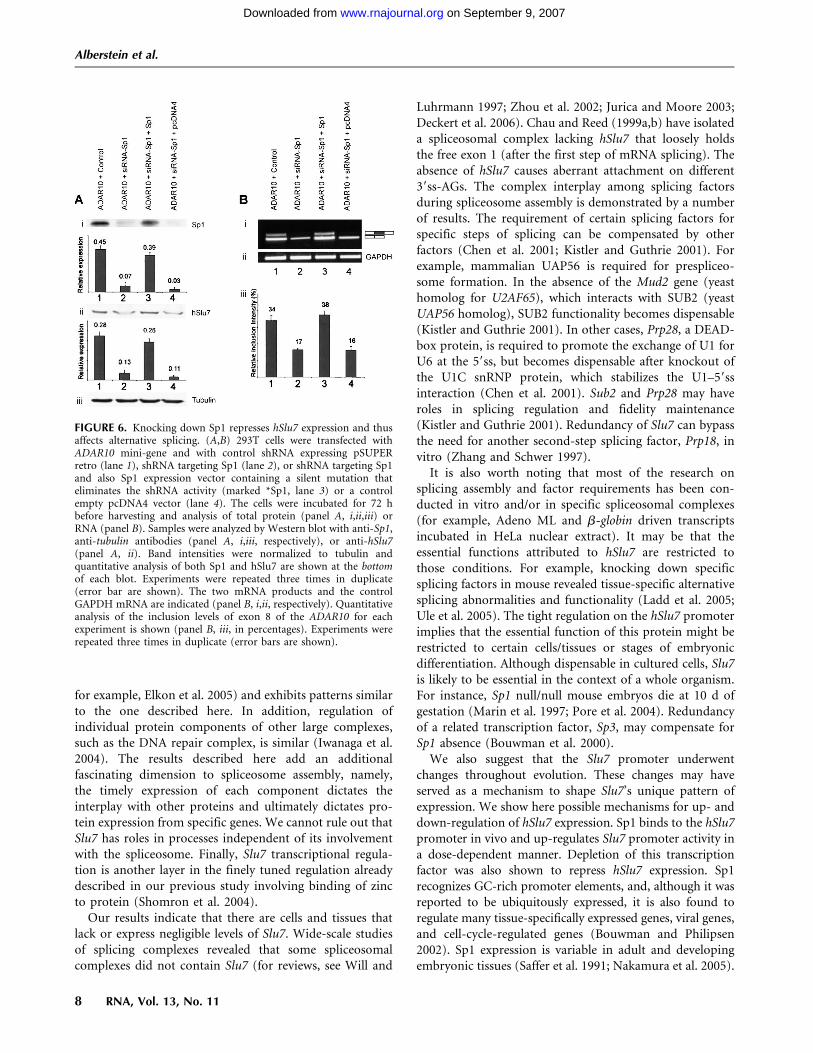
To examine if the effects of Sp1 and Elk-1 on the transcription of hSlu7 translateinto affects on alternative splicing, wereduced the cellular concentrations ofSp1 and Elk-1 using RNA interferenceand examined the effects on alternativesplicing. We previously showed that thenuclear concentration of hSlu7 affectsalternative splicing of exon 8 of theADAR10 mini-gene and of exon 4 ofDDO gene (Shomron et al. 2004, 2005).Figure 6 shows that treatment of cellswith a short hairpin RNA (shRNA) spe-cific for Sp1 gene reduced Sp1 and hSlu7protein levels significantly and alsoreduced inclusion of exon 8 of ADAR10mini-gene (Fig. 6A, panel ii; Fig. 6B,panel i; cf. lanes 1 and 2 in all panels).To ensure specificity, the shRNA-Sp1treated cells were cotransfected with Sp1cDNA containing a silent mutation thatprevents shRNA-directed degradation(see Materials and Methods) or anempty control pcDNA4 construct (Fig.6A,B lanes 3,4, respectively). The silentSp1 mutant restored both Sp1 andhSlu7 expression levels (Fig. 6A, lanes3,4) and also restore exon 8 inclusion(Fig. 6B, lanes 3,4). The siRNA treat-ment did not affect the transcription ofADAR2 (data not shown). This indi-cates that the reduction in the inclusion
of exon 8 after knocking down Sp1 did not derive from adecrease in the steady-state expression of the reporterconstruct. Also, Elk-1 siRNA treatment reduced Elk-1protein concentration and thus enhanced the inclusionlevel of exon 4 of the DDO gene (Fig. 5D,E, respectively).These results suggest that the cellular concentration of thetranscription factors that regulate hSlu7 transcription af-fects the level of exon inclusion or skipping in alternativesplicing of specific exons. We demonstrated that twodifferent transcription factors cause opposite effects ontwo different splicing events through regulation of hSlu7
FIGURE 3. Sp1 up regulates hSlu7 promoter activity with 750 ng of pcDNA4-Sp1. (A, leftpanel) Induction of hSlu7 promoter activity by Sp1. 293T cells were cotransfected with 250 ngof reporter construct (pSlu7-luc) together with 250 ng, 500 ng, or 750 ng of either theexpression plasmid pcDNA4-Sp1 or the corresponding pcDNA4 empty control vector. Thefold induction represents the fold increase in luciferase activity 48 h post-transfection relativeto that obtained following cotransfection of the reporter construct with control vector(pCDNA4). (Lower panel) Western blotting of 293T transfected cells with either anti-Sp1 oranti-actin antibodies. (Right panel) Sp1 mediated induction is significantly impaired only afterdeletion of the two GC-rich regions. Similar analysis as showed in the left panel using mutantswith deletion of GC-rich1 or -2 (GC-1 Del-luc and GC-2 Del luc, respectively) or deletions ofboth GC-rich regions (GC-12 Del-luc) (see also Fig. 2B) with 750 ng of pcDNA4-Sp1. Allluciferase activities are normalized to the wt activity (reporter and empty pcDNA4 vector).Results are represented as the mean of at least three independent experiments (standard errorsbars are shown; n = 3; P-values represent T-test for each DNA dose group; 0.046, 0.020, and0.007 for 250 ng, 500 ng, and 750 ng, respectively). (B) Sp1 binds to the Slu7 promoter in vivo.Schematic diagram of the Slu7 gene showing the location of the oligonucleotides used in thechromatin immunoprecipitation assay (left panel). U2OS cells were transfected with HA-Sp1and an empty pCDNA4 and grown for 48 h. Chromatin was precipitated using anti-HA ornormal mouse IgG antibodies and used as a template for PCR analysis using primers spanningthe promoter region of hSlu7 (right panel; upper panel) and intron 6 (right panel; lower panel)as a control (see Materials and Methods for primers). Quantitative analysis of PCR ampliconintensities is shown in the lower part of the upper gel. Input sample comprised 5% of the totalprecipitated DNA.
Alberstein et al.
6 RNA, Vol. 13, No. 11
on September 9, 2007 www.rnajournal.orgDownloaded from

expression. We cannot exclude the possibility that thecellular concentrations of Sp1 and Elk-1 also affect tran-scription of other splicing factors involved in alternativesplicing regulation of those exons. However, the splicing ofthe two genes analyzed was previously shown to be affecteddirectly by the cellular concentration of hSlu7 (Shomronet al. 2004, 2005).
DISCUSSION
Previously, it was reported that the splicing factor hSlu7,a protein involved in the fidelity of the second step ofsplicing, is not required for cell viability but that thenuclear concentration of the protein regulates alternativesplicing of certain genes (Shomron et al. 2004, 2005). Thisraised the question of whether Slu7 expression is regulatedamong tissues. In this study, we demonstrated that Slu7is differentially expressed among various tissues and celllines. Moreover, Slu7 has unique patterns of expressionwithin complex adult tissues and developing embryos,such as eye and pancreas. We also demonstrated thathSlu7 transcription is regulated by at least two transcriptionfactors, Sp1 and Elk-1, which might explain its differentialexpression.
It is intriguing that a splicing factor, one component of amegacomplex, has such a sophisticated mechanism for tran-scription regulation. Although very little is known abouttranscription regulation of other splicing factors (for exam-ple, see Romanelli et al. 2005), regulation of transcriptionof other nonsplicing proteins has been characterized (see,
FIGURE 4. Repression of hSlu7 promoter activity by Elk-1. (A) 293Tcells were cotransfected with 250 ng of reporter construct (pSlu7-luc)together with 100 ng, 250 ng, or 500 ng of either the expressionplasmid pCAG-Elk-1 or the corresponding pCAGGS empty controlvector. The fold induction represents the fold increase in luciferaseactivity 48 h post-transfection relative to that obtained followingcotransfection of the reporter construct with a control vector(pCAGGS). Western blotting of 293T transfected cells with eitheranti-Elk-1 or anti-actin antibodies are also shown (bottom panels). (B)Elk-1-mediated repression of the hSlu7 promoter occurred in severalcell lines. Cell lines (HeLa, U20S, 293T, and HT1080) were cotrans-fected with 250 ng of reporter construct (pSlu7-luc) together with250 ng of either the expression plasmid pCAG-Elk-1 or the corre-sponding pCAGGS empty control vector. Results are represented asthe mean of at least three independent experiments (standard errorsbars are shown; n = 3; P-values represent T-test for each DNA dosegroup: 0.0016, 0.00036, and 0.00031 for 100 ng, 250 ng, and 500 ng,respectively).
FIGURE 5. Elk-1 binds to the Slu7 promoter in vivo and knockingdown Elk-1 induces hSlu7 expression. (A) Schematic diagram of theSlu7 gene showing the location of the oligonucleotides used in theChIP assay. (B) Elk-1 binds to hSlu7 promoter in vivo. HeLa cells werestarved in serum-free DMEM for 48 h before harvesting. Sonicatedchromatin was immunoprecipitated with either an anti-Elk-1 anti-body or nonspecific IgG. PCR analysis of eluted DNA was performedusing oligonucleotides specific for the Slu7 promoter (top panel) orSlu7 intronic sequence (lower panel). Five percent of input DNA isshown in lane 1. The panels shown are inverted images of ethidiumbromide-stained gels. Results shown are representative of fourindependent experiments. (C) Chromatin immunoprecipitation ofElk-1 bound to the Slu7 promoter in the presence or absence of asiRNA directed against Elk-1. HeLa cells were transfected with Elk-1siRNA or a negative control siRNA 48 h before harvesting. Sonicatedchromatin was immunoprecipitated with either an anti-Elk-1 anti-body or nonspecific IgG. Right panel shows PCR analysis of elutedDNA using oligonucleotides specific for the Slu7 promoter (5% ofinput DNA is shown in lanes 1 and 2). Left panel shows a Western blotof HeLa cells treated in parallel. Results shown are representativeof two independent experiments. (D) Knocking down Elk-1 levelsinduces expression of Slu7 mRNA. HeLa cells were transfected withElk-1 siRNA for 48 h before harvesting total RNA. RT-PCR wasperformed to detect endogenous Slu7 mRNA (left graph), Elk-1mRNA (middle graph), and 18S rRNA expression (right graph).Results shown are representative of three independent experiments.(E) Knocking down Elk-1 levels results in increased inclusion ofalternatively spliced exon 4 of the DDO gene relative to cells treatedwith control siRNA. HeLa cells were transfected with Elk-1 siRNA for48 h before harvesting total RNA. Real time RT-PCR was performedto detect endogenous DDO mRNA containing alternatively splicedexon 4. Results shown are representative of three independentexperiments.
Temporal and spatial expression of hSlu7 regulation
www.rnajournal.org 7
on September 9, 2007 www.rnajournal.orgDownloaded from
for example, Elkon et al. 2005) and exhibits patterns similarto the one described here. In addition, regulation ofindividual protein components of other large complexes,such as the DNA repair complex, is similar (Iwanaga et al.2004). The results described here add an additionalfascinating dimension to spliceosome assembly, namely,the timely expression of each component dictates theinterplay with other proteins and ultimately dictates pro-tein expression from specific genes. We cannot rule out thatSlu7 has roles in processes independent of its involvementwith the spliceosome. Finally, Slu7 transcriptional regula-tion is another layer in the finely tuned regulation alreadydescribed in our previous study involving binding of zincto protein (Shomron et al. 2004).
Our results indicate that there are cells and tissues thatlack or express negligible levels of Slu7. Wide-scale studiesof splicing complexes revealed that some spliceosomalcomplexes did not contain Slu7 (for reviews, see Will and
Luhrmann 1997; Zhou et al. 2002; Jurica and Moore 2003;Deckert et al. 2006). Chau and Reed (1999a,b) have isolateda spliceosomal complex lacking hSlu7 that loosely holdsthe free exon 1 (after the first step of mRNA splicing). Theabsence of hSlu7 causes aberrant attachment on different39ss-AGs. The complex interplay among splicing factorsduring spliceosome assembly is demonstrated by a numberof results. The requirement of certain splicing factors forspecific steps of splicing can be compensated by otherfactors (Chen et al. 2001; Kistler and Guthrie 2001). Forexample, mammalian UAP56 is required for prespliceo-some formation. In the absence of the Mud2 gene (yeasthomolog for U2AF65), which interacts with SUB2 (yeastUAP56 homolog), SUB2 functionality becomes dispensable(Kistler and Guthrie 2001). In other cases, Prp28, a DEAD-box protein, is required to promote the exchange of U1 forU6 at the 59ss, but becomes dispensable after knockout ofthe U1C snRNP protein, which stabilizes the U1–59ssinteraction (Chen et al. 2001). Sub2 and Prp28 may haveroles in splicing regulation and fidelity maintenance(Kistler and Guthrie 2001). Redundancy of Slu7 can bypassthe need for another second-step splicing factor, Prp18, invitro (Zhang and Schwer 1997).
It is also worth noting that most of the research onsplicing assembly and factor requirements has been con-ducted in vitro and/or in specific spliceosomal complexes(for example, Adeno ML and b-globin driven transcriptsincubated in HeLa nuclear extract). It may be that theessential functions attributed to hSlu7 are restricted tothose conditions. For example, knocking down specificsplicing factors in mouse revealed tissue-specific alternativesplicing abnormalities and functionality (Ladd et al. 2005;Ule et al. 2005). The tight regulation on the hSlu7 promoterimplies that the essential function of this protein might berestricted to certain cells/tissues or stages of embryonicdifferentiation. Although dispensable in cultured cells, Slu7is likely to be essential in the context of a whole organism.For instance, Sp1 null/null mouse embryos die at 10 d ofgestation (Marin et al. 1997; Pore et al. 2004). Redundancyof a related transcription factor, Sp3, may compensate forSp1 absence (Bouwman et al. 2000).
We also suggest that the Slu7 promoter underwentchanges throughout evolution. These changes may haveserved as a mechanism to shape Slu7’s unique pattern ofexpression. We show here possible mechanisms for up- anddown-regulation of hSlu7 expression. Sp1 binds to the hSlu7promoter in vivo and up-regulates Slu7 promoter activity ina dose-dependent manner. Depletion of this transcriptionfactor was also shown to repress hSlu7 expression. Sp1recognizes GC-rich promoter elements, and, although it wasreported to be ubiquitously expressed, it is also found toregulate many tissue-specifically expressed genes, viral genes,and cell-cycle-regulated genes (Bouwman and Philipsen2002). Sp1 expression is variable in adult and developingembryonic tissues (Saffer et al. 1991; Nakamura et al. 2005).
FIGURE 6. Knocking down Sp1 represses hSlu7 expression and thusaffects alternative splicing. (A,B) 293T cells were transfected withADAR10 mini-gene and with control shRNA expressing pSUPERretro (lane 1), shRNA targeting Sp1 (lane 2), or shRNA targeting Sp1and also Sp1 expression vector containing a silent mutation thateliminates the shRNA activity (marked *Sp1, lane 3) or a controlempty pcDNA4 vector (lane 4). The cells were incubated for 72 hbefore harvesting and analysis of total protein (panel A, i,ii,iii) orRNA (panel B). Samples were analyzed by Western blot with anti-Sp1,anti-tubulin antibodies (panel A, i,iii, respectively), or anti-hSlu7(panel A, ii). Band intensities were normalized to tubulin andquantitative analysis of both Sp1 and hSlu7 are shown at the bottomof each blot. Experiments were repeated three times in duplicate(error bar are shown). The two mRNA products and the controlGAPDH mRNA are indicated (panel B, i,ii, respectively). Quantitativeanalysis of the inclusion levels of exon 8 of the ADAR10 for eachexperiment is shown (panel B, iii, in percentages). Experiments wererepeated three times in duplicate (error bars are shown).
Alberstein et al.
8 RNA, Vol. 13, No. 11
on September 9, 2007 www.rnajournal.orgDownloaded from

These observations imply that Sp1 might have a role insupporting hSlu7 expression in a cell-type-specific manner.
Elk-1 repressed transcription from the hSlu7 promoterand bound to the hSlu7 promoter in vivo. Moreover,siRNA-induced reduction of Elk-1 increased endogenoushSlu7 expression, suggesting that the Slu7 promoter isdown-regulated by Elk-1. Elk-1-mediated repression wasshown to be dose dependent in several cell lines. However,Elk-1-mediated repression of hSlu7 transcription was notcompletely abolished even after deletion of all five potentialElk-1 sites (data not shown). We also did not detect anypotential serum response elements (SREs) in the promoter(these elements are required for SRF-dependent Elk-1 DNAbinding). The repressive effects of Elk-1 are consistent withprevious observations that Elk-1 associates with repressivecomplexes (Yang et al. 2001; Yang and Sharrocks 2004).The exact repression mechanism remains to be elucidated.
Gene expression profiles from microarray data setsrevealed that in the PC3 prostate cell line there is anextremely low expression level of Sp1 (GEO accessionGDS1736), whereas Elk-1 is expressed. This further corrob-orates results from our Sp1 siRNA assay that showed asignificant reduction in hSlu7 expression when Sp1 expres-sion was reduced. We could not use this method to confirmthe effects of Elk-1 expression levels on hSlu7 proteinexpression because no cell lines were found in whichElk-1 expression is very low or absent. We cannot ruleout the possibility that other factors may contribute to theexpression level of Slu7.
We have shown here that the temporal and spatialexpression of the mammalian splicing factor Slu7 iselaborately regulated among tissues and during embryo-genesis. The involvement of hSlu7 in regulation of alter-native splicing of certain exons might indicate that hSlu7 isessential for tissue- and differentiation-specific alternativesplicing events.
MATERIALS AND METHODS
Isolating the 59 ends of human Slu7 mRNA
59-RACE was performed according to the circular, or concatemeric,RACE methodology (Maruyama et al. 1995) in extract from 293Tcells with a human Slu7 gene specific primer: 59-TCCTCAGAGTTAACAATCTCCTTCC-39. The first PCR primers were 59-TGCTGGAGATAACTTTGTTAGGTACAC-39 and 59-CTCATTTCTTTGGACCCCGATA-39. The second, nested PCR primers were 59-ACCATTTCAATGGCTCAGACAC-39 and 59-TGTGGCTGACATGGTTATCTGG-39. The PCR product was eluted and purifiedfollowing gel electrophoresis. After cloning into TOPO TA cloningvector (Invitrogen), 20 colonies were sequenced.
Reporter and effector constructs
The 59-flanking regions of the human Slu7 gene [�184 to + 66relative to TSS(1)] were amplified from genomic DNA and
inserted upstream of the firefly luciferase gene in the reportervector pGL3-basic (Promega) to create pSlu7-luc. Mutations wereintroduced using overlapping oligonucleotide primers containingthe desired mutation. PCR was performed using the high-fidelityDNA polymerase UltraPfu (Stratagene); then reaction productswere digested with DpnI restriction enzyme (New EnglandBiolabs) for 1 h at 37°C. A 5 mL aliquot of the reaction was usedto transform the Escherichia coli DH5a strain, and DNA frompositive colonies was extracted using a Mini-prep extraction kit(Qiagen). Primers were generated harboring the desired mutation(mutated nucleotide in bold) to abolish the GGAA core elements:
EBS_a_GG2TT_Fw 59-GCCGGAATTAGGCGAAAAGCCTTAAGTAAACATTACGAGATTGG-39;
EBS_a_GG2TT_Rv 59-CCAATCTCGTAATGTTTACTTAAGGCTTTTCGCCTAATTCCGGC-39;
EBS_b_GG2TT_Fw 59-TACCGCAGTGGCCGCCTTAATTAGGCGAAAAGCC-39;
EBS_b_ GG2TT_Rv 59-GGCTTTTCGCCTAATTAAGGCGGCCACTGCGGTA-39;
EBS_c_GG2TT_Fw 59-CATGTGCCGGTCATCCTTAAGTTACCGCAGTGGC-39;
EBS_c_GG2TT_Rv 59-GCCACTGCGGTAACTTAAGGATGACCGGCACATG-39;
EBS_d_AAA2CCT_Fw 59-CCCGGCCCCGCCAGGCCTTTAGTGCGCATGTGC-39;
EBS_d_AAA2CCT_Rv 59-GCACATGCGCACTAAAGGCCTGGCGGGGCCGGG-39;
EBS_e_CC2AA_Fw 59-GAGTTCTCGCGTTTAACACGGCGCAGGAG-39;
EBS_e_CC2AA_Rv 59-CTCCTGCGCCGTGTTAAACGCGAGAACTC-39.
The same primers were used to create all the combinationmutants. Deletion mutants were created using two phosphory-lated primers that flanked the desired regions followed by ligationof the clean PCR construct product. The pCAGGS (controlvector) and pCAG-Elk-1 constructs were kindly provided byDr. Hiroshi Kubota (Kyoto University) (Yamazaki et al. 2003).Sp1 mutated at the siRNA recognition site was cloned within themammalian expression construct pcDNA4 (data not shown).Nucleotide sequences of the reporter and effector constructs wereconfirmed by sequencing.
Cell line maintenance, transfection, and reportergene assay
HeLa, 293T, HT1080, and U2OS cells were grown on six-wellplates and maintained in Dulbecco’s modified Eagle’s medium(DMEM) with 10% fetal calf serum (FCS), 0.29 mg/mLL-glutamine, 100 U/mL penicillin, 0.1 mg/mL streptomycin, and1 U/mL nystatin at 37°C in a humidified atmosphere of 5% CO2.Cells were grown for 24 h and then transfected with 250 ng ofreporter construct together with 250 ng (or other dose asindicated) effector construct, including 20 ng of internal controlreporter vector (pRL-SV40; Promega) using FuGENE6 (Roche),as described in the manufacturer’s protocol. Cell lysates wereprepared and luciferase activities of transfected cells were deter-mined using the dual luciferase assay system (Promega), accordingto the manufacturer’s instructions, and the activity of firefly
Temporal and spatial expression of hSlu7 regulation
www.rnajournal.org 9
on September 9, 2007 www.rnajournal.orgDownloaded from

luciferase was normalized against that of the sea pansy enzyme(Renilla reniformis).
RNA isolation, RT-PCR analysis, PCR, andquantitative RT-PCR
Cells were grown in a 100-mm culture dish and were harvested48 h after transfection. Total cytoplasmic RNA was extracted usingTRI Reagent (Sigma), followed by treatment with 2 U DNase(RNase-Free; Ambion). Tissue samples from three adult micewere homogenized in TRI reagent (1 mL/30–100 mg tissue). First-strand oligo(dT)-primed cDNA synthesized with reverse tran-scriptase from avian myelobstosis virus (RT-AMV; Roche) from1000 ng of total RNA was amplified with High Fidelity Taqpolymerase (Roche) and DDO, GAPDH, mSlu7, mPrp18, andTubulin b5 primers for 30 cycles, consisting of 94°C for 30 sec,60–65°C for 45 sec, and 72°C for 1 min. The products wereseparated in 2% agarose gel and confirmed by sequencing. Forquantitative RT-PCR, total RNA was harvested using an RNeasykit (Qiagen). RNA (40 ng) was used in a one-step RT-PCRreaction using Quantitect SYBR green reagent (Qiagen) and thefollowing primers:
Slu7 forward, 59-GTGGCCAAGAACATTTGGAT-39;Slu7 reverse, 59-CATCGGCCTTCTTTCCAGTA-39;Elk-1 forward, 59-GGTGGTGAATTCAAGCTGGT-39;Elk-1 reverse, 59-ATTTGGCATGGTGGAGGTAA-39;18S forward, 59-TCAAGAACGAAAGTCGGAGGTT-39;18S reverse, 59-GGACATCTAAGGGCATCACAG-39.
Computational analyses
The hSlu7 promoter sequence was scanned for binding sites fortranscription factors using the following programs: TRANS-PLORER (http://www.developmentontheedge.com/transplorer.shtml); Genomatix (http://www.genomatix.de/); NCITE (http://www.softberry.com/berry.phtml); Signal Scan (http://bimas.dcrt.nih.gov/molbio/signal/); and TFSEARCH (http://molsun1.cbrc.aist.go.jp/research/db/TFSEARCH.html). Multiple alignment ofthe hSlu7 promoter region alignment was done using the ClustalWalgorithm (http://www.ebi.ac.uk/clustalw/).
In situ hybridization
In situ hybridization analysis was performed as described byYaron et al. (2006). For digoxigenin-labeled antisense RNAs,reverse transcription was performed on oligo(dT)-primed cDNAfrom mouse adult testis and brain tissues, and the resulting cDNAwas used as a template for PCR. Standard PCR conditions wereused with an annealing temperature of 68°C for 31 cycles. ThePCR primers were mSlu7_973_F, 59-GCTCAAACACAACTGTTTGCTTGG-39 and mSlu7_3UTR_R, 59-TAATACGACTCACTATAGGGCAGAGGACTGACGGCATGTACAT-39. For the sensenegative control mSlu7 probes, the same primers were used exceptthat the T7 promoter tag was switched from the reverse to theforward primer. The resulting 1364-bp PCR products wereanalyzed and extracted from a 1% agarose gel, and the purifiedPCR products, including a 59-T7 minimal promoter tag, wereused to create the related digoxigenin-labeled antisense in vitrousing DIG RNA labeling mix (Roche). The Sp1 probes and
primers were used as reported before (Gray et al. 2004; Nakamuraet al. 2005).
RNA interference
HeLa cells were transfected with 70 nM Elk-1 siRNA (Dharmacon)or nontargeting control siRNA (Santa Cruz Biotechnology) usingoligofectamine (Invitrogen) according to the manufacturer’s pro-tocol. HEK 293T cells were transfected with pSUPER.retro(Oligoengine) Sp1, and GFP control vectors expressing shorthairpin (shRNA) against Sp1 corresponding to cDNA position396 using fugene6 transfection reagent (Roche). For the Sp1rescue analysis cells were also cotransfected with pSUPER.retroSp1 and with either rescue construct expression Sp1 (mutated atthe siRNA recognition site) or an empty control vector (pcDNA4;Invitrogen). At 72 h post-transfection, cells were harvested forprotein and RNA preparation. Primers for the real time RT-PCRof DDO were exon3_Fw, 59-CATTCACACGCAGAAGCAGT-39,and DDO_exon4 Rv, 59-GGGTTGTAAAAGCCTGACCA-39. Pri-mers for detection of the inclusion level of exon 8 of ADAR10transcript were described before (Lev-Maor et al. 2003).
Chromatin immunoprecipitation
U2OS cells were transfected with HA-Sp1 (in pcDNA4) for theimmunoprecipitation of exogenous hemagglutinin (HA) taggedSp1 proteins and an empty pCDNA4 as a control and were grownfor 48 h. HeLa cells were untransfected (for the immunoprecip-itation of endogenous Elk-1 proteins). Cells were then treatedwith 1% formaldehyde for 10 min at room temperature beforequenching with 0.125 M glycine for 5 min. Cells were harvested inice-cold PBS with complete protease inhibitors (Roche), washedsequentially with BufferI (10 mM HEPES at pH 6.5, 0.5 mMEGTA, 10 mM EDTA, 0.25% Triton X-100) and BufferII (10 mMHEPES at pH 6.5, 0.5 mM EGTA, 1 mM EDTA, 200 mM NaCl),and then resuspended in SDS lysis buffer (50 mM Tris at pH 8.1,10 mM EDTA, 1% SDS). Lysates were sonicated on ice to yield200–800-bp DNA fragments. One quarter of a 10-cm dish wasused per IP, diluted 1/10 in IP Dilution buffer (0.01% SDS, 1.1%Triton X-100, 1.2 mM EDTA, 16.7 mM Tris at pH 8.1, 167 mMNaCl), and incubated overnight at 4°C with either 1 mg of Elk-1antibody (Santa Cruz Biotechnology) for Hela cells or anti HAF-7 (Santa Cruz Biotechnology) for U2OS cells or 1 mg non-specific IgG (Upstate Biotechnology) for both cell types. Immu-nocomplexes were precipitated by incubation for 30 min withprotein A-conjugated magnetic beads (Dynal) that had beenpreblocked by incubation with 10 mg salmon sperm DNA. Im-munoprecipitates were washed sequentially with TSEI (20 mM,Tris at pH 8.1, 2 mM EDTA, 150 mM NaCl, 1% Triton X-100,0.1% SDS), TSEII (20 mM Tris at pH 8.1, 2 mM EDTA, 500 mMNaCl, 1% Triton X-100, 0.1% SDS), BufferIII (10 mM Tris atpH 8.1, 0.25 M LiCl, 1 mM EDTA, 1% NP40, 1% DOC), andTE before eluting in 1% SDS/0.1 M NaHCO3. Cross-links werereversed by heating to 65°C overnight, then treating with pro-teinase K for 1 h at 45°C. Chromatin was cleaned using QiaQuickPCR cleanup columns (Qiagen). PCR was performed using spe-cific primers to the human Slu7 promoter (�140 to +44,relative to TSS(1); forward, 59-GCTAGAGTTCTCGCGTTTCC-39;reverse, 59-CCAAGTCCATCCGACAGAAT-39) or Slu7 intronicsequence (intron 6, +6804 to +7002 relative to TSS(1); forward,
Alberstein et al.
10 RNA, Vol. 13, No. 11
on September 9, 2007 www.rnajournal.orgDownloaded from

59-TGCAGTCAGTTTGGGAACAA-39; reverse, 59-TTCCCTGTTCCTGGACATTT-39).
Western blotting
Lysis buffer (50 mM Tris at pH 7.5, 1% NP40, 150 mM NaCl,0.1% SDS, 0.5% deoxycholic acid, protease inhibitor cocktail,and phosphatase inhibitor cocktails I and II; Sigma) was used forprotein extraction. Lysates were cold centrifuged for 30 min at14,000 rpm. Total protein concentrations were measured usingBioRad Protein Assay (Bio-Rad). Proteins were separated in 10%SDS-polyacrylamide gel electrophoresis (SDS-PAGE) and thenelectroblotted onto a Protran membrane (Schleicher and Schuell).The membranes were probed with either anti-Elk-1, anti-actinantibody (Santa Cruz Biotechnology), anti-hSlu7 (Shomron et al.2004; Abnova), or anti-Sp1 (BL938, Bethyl Laboratories), and anti-a-tubulin (B512; Sigma) followed by the appropriate secondaryantibody. Immunoblots were visualized by enhanced chemilumi-nescence (Lumi-Light Western Blotting Substrate; Roche) andexposure to X-ray film. For ChIP assay, Western blotting wasperformed using Supersignal West Dura Extended DurationSubstrate (Pierce) and primary antibodies anti-Elk-1 (Santa CruzBiotechnologies) and anti-GAPDH (Abcam). Data were visualizedusing Bio-Rad Fluor-S MultiImager and Quantity One software(Bio-Rad).
Image processing and microscopy
Acquisition of images and measurement of DNA intensity wasperformed using TINA, ImageJ, and analySIS software (SoftImaging System). Most of the results represent values obtainedfrom at least three separate experiments, and the results areaverage values. Fluorescent images were taken with a confocallaser-scanning system, consisting of an SLM 410 Zeiss confocalmicroscope with a 203 or 403 oil objective.
SUPPLEMENTAL DATA
Supplemental Material is available at http://www.tau.ac.il/zgilast/sup_mat.htm.
ACKNOWLEDGMENTS
This work was supported by a grant from the Israel ScienceFoundation (1449/04 and 40/05), MOP Germany–Israel, GIF, ICAthrough the Ber-Lehmsdorf Memorial Fund, and TAU CancerCenter. N.S. is funded in part by EURASNET. The work of R.A.-P.was supported by the Israel Science Foundation, GlaucomaResearch Foundation, and the AMN Foundation.
Received February 5, 2007; accepted July 31, 2007.
REFERENCES
Ast, G. 2004. How did alternative splicing evolve? Nat. Rev. Genet. 5:773–782.
Black, D.L. 2003. Mechanisms of alternative pre-messenger RNAsplicing. Annu. Rev. Biochem. 72: 291–336.
Bouwman, P. and Philipsen, S. 2002. Regulation of the activityof Sp1-related transcription factors. Mol. Cell. Endocrinol. 195:27–38.
Bouwman, P., Gollner, H., Elsasser, H.P., Eckhoff, G., Karis, A.,Grosveld, F., Philipsen, S., and Suske, G. 2000. Transcriptionfactor Sp3 is essential for post-natal survival and late toothdevelopment. EMBO J. 19: 655–661.
Chen, J.Y., Stands, L., Staley, J.P., Jackups Jr., R.R., Latus, L.J., andChang, T.H. 2001. Specific alterations of U1-C protein or U1 smallnuclear RNA can eliminate the requirement of Prp28p, an essentialDEAD box splicing factor. Mol. Cell 7: 227–232.
Chua, K. and Reed, R. 1999a. Human step II splicing factor hSlu7functions in restructuring the spliceosome between the catalyticsteps of splicing. Genes & Dev. 13: 841–850.
Chua, K. and Reed, R. 1999b. The RNA splicing factor hSlu7 isrequired for correct 39 splice-site choice. Nature 402: 207–210.
Cooper, T.A. 2005. Alternative splicing regulation impacts heartdevelopment. Cell 120: 1–2.
Deckert, J., Hartmuth, K., Boehringer, D., Behzadnia, N., Will, C.L.,Kastner, B., Stark, H., Urlaub, H., and Luhrmann, R. 2006. Proteincomposition and electron microscopy structure of affinity-purifiedhuman spliceosomal B complexes isolated under physiologicalconditions. Mol. Cell. Biol. 26: 5528–5543.
Elkon, R., Rashi-Elkeles, S., Lerenthal, Y., Linhart, C., Tenne, T.,Amariglio, N., Rechavi, G., Shamir, R., and Shiloh, Y. 2005.Dissection of a DNA-damage-induced transcriptional networkusing a combination of microarrays, RNA interference andcomputational promoter analysis. Genome Biol. 6: R43.
Frank, D., Patterson, B., and Guthrie, C. 1992. Synthetic lethalmutations suggest interactions between U5 small nuclear RNAand four proteins required for the second step of splicing. Mol.Cell. Biol. 12: 5197–5205.
Graveley, B.R. 2001. Alternative splicing: Increasing diversity in theproteomic world. Trends Genet. 17: 100–107.
Gray, P.A., Fu, H., Luo, P., Zhao, Q., Yu, J., Ferrari, A., Tenzen, T.,Yuk, D.I., Tsung, E.F., Cai, Z., et al. 2004. Mouse brain organi-zation revealed through direct genome-scale TF expression anal-ysis. Science 306: 2255–2257.
Guil, S., Long, J.C., and Caceres, J.F. 2006. hnRNP A1 relocalization tothe stress granules reflects a role in the stress response. Mol. Cell.Biol. 26: 5744–5758.
Hanamura, A., Caceres, J.F., Mayeda, A., Franza Jr., B.R., andKrainer, A.R. 1998. Regulated tissue-specific expression of antag-onistic pre-mRNA splicing factors. RNA 4: 430–444.
Hsu, T., Trojanowska, M., and Watson, D.K. 2004. Ets proteins inbiological control and cancer. J. Cell. Biochem. 91: 896–903.
Iwanaga, R., Komori, H., and Ohtani, K. 2004. Differential regulationof expression of the mammalian DNA repair genes by growthstimulation. Oncogene 23: 8581–8590.
Jurica, M.S. and Moore, M.J. 2003. Pre-mRNA splicing: Awash in asea of proteins. Mol. Cell 12: 5–14.
Kistler, A.L. and Guthrie, C. 2001. Deletion of MUD2, the yeasthomolog of U2AF65, can bypass the requirement for sub2, anessential spliceosomal ATPase. Genes & Dev. 15: 42–49.
Ladd, A.N., Taffet, G., Hartley, C., Kearney, D.L., and Cooper, T.A.2005. Cardiac tissue-specific repression of CELF activity disruptsalternative splicing and causes cardiomyopathy. Mol. Cell. Biol. 25:6267–6278.
Lander, E.S., Linton, L.M., Birren, B., Nusbaum, C., Zody, M.C.,Baldwin, J., Devon, K., Dewar, K., Doyle, M., FitzHugh, W., et al.2001. Initial sequencing and analysis of the human genome.Nature 409: 860–921.
Lev-Maor, G., Sorek, R., Shomron, N., and Ast, G. 2003. The birth ofan alternatively spliced exon: 39 splice-site selection in Alu exons.Science 300: 1288–1291.
Marin, M., Karis, A., Visser, P., Grosveld, F., and Philipsen, S. 1997.Transcription factor Sp1 is essential for early embryonic develop-ment but dispensable for cell growth and differentiation. Cell 89:619–628.
Maruyama, I.N., Rakow, T.L., and Maruyama, H.I. 1995. cRACE: Asimple method for identification of the 59 end of mRNAs. NucleicAcids Res. 23: 3796–3797. doi: 10.1093/nar/23.18.3796.
Temporal and spatial expression of hSlu7 regulation
www.rnajournal.org 11
on September 9, 2007 www.rnajournal.orgDownloaded from

Nakamura, H., Ueda, J., Sugar, J., and Yue, B.Y. 2005. Developmen-tally regulated expression of Sp1 in the mouse cornea. Invest.Ophthalmol. Vis. Sci. 46: 4092–4096.
Pelisch, F., Blaustein, M., Kornblihtt, A.R., and Srebrow, A. 2005.Cross-talk between signaling pathways regulates alternative splic-ing: A novel role for JNK. J. Biol. Chem. 280: 25461–25469.
Philips, A.V. and Cooper, T.A. 2000. RNA processing and humandisease. Cell. Mol. Life Sci. 57: 235–249.
Pore, N., Liu, S., Shu, H.K., Li, B., Haas-Kogan, D., Stokoe, D.,Milanini-Mongiat, J., Pages, G., O’Rourke, D.M., Bernhard, E.,et al. 2004. Sp1 is involved in Akt-mediated induction of VEGFexpression through an HIF-1-independent mechanism. Mol. Biol.Cell 15: 4841–4853.
Romanelli, M.G., Lorenzi, P., and Morandi, C. 2005. Identificationand analysis of the human neural polypyrimidine tract bindingprotein (nPTB) gene promoter region. Gene 356: 11–18.
Saffer, J.D., Jackson, S.P., and Annarella, M.B. 1991. Developmentalexpression of Sp1 in the mouse. Mol. Cell. Biol. 11: 2189–2199.
Sgouras, D.N., Athanasiou, M.A., Beal Jr., G.J., Fisher, R.J.,Blair, D.G., and Mavrothalassitis, G.J. 1995. ERF: An ETS domainprotein with strong transcriptional repressor activity, can suppressets-associated tumorigenesis and is regulated by phosphorylationduring cell cycle and mitogenic stimulation. EMBO J. 14: 4781–4793.
Sharrocks, A.D. 2001. The ETS-domain transcription factor family.Nat. Rev. Mol. Cell Biol. 2: 827–837.
Shomron, N., Reznik, M., and Ast, G. 2004. Splicing factor hSlu7contains a unique functional domain required to retain the proteinwithin the nucleus. Mol. Biol. Cell 15: 3782–3795.
Shomron, N., Alberstein, M., Reznik, M., and Ast, G. 2005. Stressalters the subcellular distribution of hSlu7 and thus modulatesalternative splicing. J. Cell Sci. 118: 1151–1159.
Thompson, J.D., Higgins, D.G., and Gibson, T.J. 1994. CLUSTAL W:Improving the sensitivity of progressive multiple sequence align-ment through sequence weighting, position-specific gap penaltiesand weight matrix choice. Nucleic Acids Res. 22: 4673–4680. doi:10.1093/nar/22.22.4673.
Ule, J., Ule, A., Spencer, J., Williams, A., Hu, J.S., Cline, M., Wang, H.,Clark, T., Fraser, C., Ruggiu, M., et al. 2005. Nova regulates brain-specific splicing to shape the synapse. Nat. Genet. 37: 844–852.
van der Houven van Oordt, W., Diaz-Meco, M.T., Lozano, J.,Krainer, A.R., Moscat, J., and Caceres, J.F. 2000. The MKK(3/6)-
p38-signaling cascade alters the subcellular distribution of hnRNPA1 and modulates alternative splicing regulation. J. Cell Biol. 149:307–316.
Wang, Z. and Grabowski, P.J. 1996. Cell- and stage-specific splicingevents resolved in specialized neurons of the rat cerebellum. RNA2: 1241–1253.
Waterston, R.H., Lindblad-Toh, K., Birney, E., Rogers, J., Abril, J.F.,Agarwal, P., Agarwala, R., Ainscough, R., Alexandersson, M.,An, P., et al. 2002. Initial sequencing and comparative analysisof the mouse genome. Nature 420: 520–562.
Will, C.L. and Luhrmann, R. 1997. Protein functions in pre-mRNAsplicing. Curr. Opin. Cell Biol. 9: 320–328.
Xing, Y. and Lee, C. 2006. Alternative splicing and RNA selectionpressure–evolutionary consequences for eukaryotic genomes. Nat.Rev. Genet. 7: 499–509.
Yamazaki, Y., Kubota, H., Nozaki, M., and Nagata, K. 2003.Transcriptional regulation of the cytosolic chaperonin theta sub-unit gene, Cctq, by Ets domain transcription factors Elk-1, Sap-1a,and Net in the absence of serum response factor. J. Biol. Chem.278: 30642–30651.
Yang, S.H. and Sharrocks, A.D. 2004. SUMO promotes HDAC-mediated transcriptional repression. Mol. Cell 13: 611–617.
Yang, S.H., Vickers, E., Brehm, A., Kouzarides, T., andSharrocks, A.D. 2001. Temporal recruitment of the mSin3A-histone deacetylase corepressor complex to the ETS domaintranscription factor Elk-1. Mol. Cell. Biol. 21: 2802–2814.
Yaron, O., Farhy, C., Marquardt, T., Applebury, M., and Ashery-Padan, R. 2006. Notch1 functions to suppress cone-photoreceptorfate specification in the developing mouse retina. Development133: 1367–1378.
Yeo, G., Holste, D., Kreiman, G., and Burge, C.B. 2004. Varia-tion in alternative splicing across human tissues. Genome Biol. 5:R74.
Zhang, X. and Schwer, B. 1997. Functional and physical interactionbetween the yeast splicing factors Slu7 and Prp18. Nucleic AcidsRes. 25: 2146–2152. doi: 10.1093/nar/25.11.2146.
Zhang, Q.S., Manche, L., Xu, R.M., and Krainer, A.R. 2006. hnRNPA1 associates with telomere ends and stimulates telomeraseactivity. RNA 12: 1116–1128.
Zhou, Z., Licklider, L.J., Gygi, S.P., and Reed, R. 2002. Comprehen-sive proteomic analysis of the human spliceosome. Nature 419:182–185.
Alberstein et al.
12 RNA, Vol. 13, No. 11
on September 9, 2007 www.rnajournal.orgDownloaded from