REGULATION OF THE IMMUNE RESPONSE IN THE …arch.apsl.edu.pl/spb/pliki/nr9/03.pdf · REGULATION OF...
22
35 Slupskie Prace Bio logiczne 9 • 2012 REGULATION OF THE IMMUNE RESPONSE IN THE CHILDREN AGED 7-11 YEARS WITH CHRONIC SENSORINEURAL HEARING LOSS REGULACJA REAKCJI IMMUNOLOGICZNEJ U DZIECI W WIEKU 7-11 LAT Z PRZEWLEKLYM NEUROSENSORYCZNYM UBYTKIEM SLUCHU Sergiu Beschasniu Elena Gasiuk Kherson State University 40 Rokiv Zhovtnya 27 Str. 73000, Kherson, Ukraine [email protected] STRESZCZENIE Obustronny neurosensoryczny ubytek słuchu jest czynnikiem wpływającym na wszystkie układy organizmu. Między innymi wpływa na układy endokrynologiczny i immunologiczny. Podwyższony poziom adrenaliny i 17-ketosteroidów świadczy o obec- ności reakcji stresowej u dzieci w wieku 7-11 lat z przewlekłym obustronnym neurosen- sorycznym ubytkiem słuchu. Podwyższony poziom tych hormonów powoduje zmiany w układzie immunologicznym. Adrenalina i 17-ketosteroidy aktywują leukocyty, po- wodując przyspieszenie ich metabolizmu i produkcję przeciwzapalnych interleukinów. Te ostatnie wpływają na limfocyty B, które aktywują się i przekształcają w komórki plazmatyczne. Interleukiny (IL-4) zwiększają liczbę limfocytów Th (helperów) typu Th2. Interleukiny wpływają na limfocyty B, które wytwarzają immonuglobuliny. We krwi dzieci z przewlekłym obustronnym sensorycznym ubytkiem słuchu wykazano podwyższony poziom immunoglobulin A i G oraz obniżony poziom immunoglobuliny M. Natomiast w ślinie badanych dzieci stwierdzono wysoki poziom przeciwciał IgG oraz niski IgA i IgM. Brak równowagi pomiędzy poziomem immunoglobulin a zmia- nami metabolicznymi leukocytów powoduje osłabienie nieswoistej odporności. Slowa kluczowe: obustronny neurosensoryczny ubytek słuchu, granulocyty, fosfata- za zasadowa, białka kationowe, mieloperoksydaza, adrenalina, 17-kortykosteroidy, interleukiny Key words: sensorineural hearing loss, granulocytes, alkaline phosphatase, cationic protein, myeloperoxidase, adrenaline, 17-korticosteroids, interleukins
Transcript of REGULATION OF THE IMMUNE RESPONSE IN THE …arch.apsl.edu.pl/spb/pliki/nr9/03.pdf · REGULATION OF...

35
S ł up s k i e P r a c e B i o l o g i c z n e 9 •••• 2012
REGULATION OF THE IMMUNE RESPONSE
IN THE CHILDREN AGED 7-11 YEARS
WITH CHRONIC SENSORINEURAL HEARING LOSS
REGULACJA REAKCJI IMMUNOLOGICZNEJ
U DZIECI W WIEKU 7-11 LAT Z PRZEWLEKŁYM
NEUROSENSORYCZNYM UBYTKIEM SŁUCHU
Sergiu Beschasniu
Elena Gasiuk
Kherson State University
40 Rokiv Zhovtnya 27 Str.
73000, Kherson, Ukraine
STRESZCZENIE
Obustronny neurosensoryczny ubytek słuchu jest czynnikiem wpływającym na
wszystkie układy organizmu. Między innymi wpływa na układy endokrynologiczny
i immunologiczny. Podwyższony poziom adrenaliny i 17-ketosteroidów świadczy o obec-
ności reakcji stresowej u dzieci w wieku 7-11 lat z przewlekłym obustronnym neurosen-
sorycznym ubytkiem słuchu. Podwyższony poziom tych hormonów powoduje zmiany
w układzie immunologicznym. Adrenalina i 17-ketosteroidy aktywują leukocyty, po-
wodując przyspieszenie ich metabolizmu i produkcję przeciwzapalnych interleukinów.
Te ostatnie wpływają na limfocyty B, które aktywują się i przekształcają w komórki
plazmatyczne. Interleukiny (IL-4) zwiększają liczbę limfocytów Th (helperów) typu
Th2. Interleukiny wpływają na limfocyty B, które wytwarzają immonuglobuliny. We
krwi dzieci z przewlekłym obustronnym sensorycznym ubytkiem słuchu wykazano
podwyższony poziom immunoglobulin A i G oraz obniżony poziom immunoglobuliny
M. Natomiast w ślinie badanych dzieci stwierdzono wysoki poziom przeciwciał IgG
oraz niski IgA i IgM. Brak równowagi pomiędzy poziomem immunoglobulin a zmia-
nami metabolicznymi leukocytów powoduje osłabienie nieswoistej odporności.
Słowa kluczowe: obustronny neurosensoryczny ubytek słuchu, granulocyty, fosfata-
za zasadowa, białka kationowe, mieloperoksydaza, adrenalina, 17-kortykosteroidy,
interleukiny
Key words: sensorineural hearing loss, granulocytes, alkaline phosphatase, cationic
protein, myeloperoxidase, adrenaline, 17-korticosteroids, interleukins
36
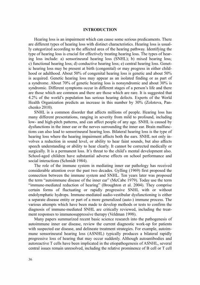
INTRODUCTION
Hearing loss is an impairment which can cause some serious predicaments. There
are different types of hearing loss with distinct characteristics. Hearing loss is usual-
ly categorized according to the affected area of the hearing pathway. Identifying the
type of hearing loss is crucial for effectively treating hearing loss. The types of hear-
ing loss include: a) sensorineural hearing loss (SNHL); b) mixed hearing loss;
c) functional hearing loss; d) conductive hearing loss; e) central hearing loss. Genet-
ic hearing loss may be present at birth (congenital) or may progress in either child-
hood or adulthood. About 50% of congenital hearing loss is genetic and about 50%
is acquired. Genetic hearing loss may appear as an isolated finding or as part of
a syndrome. About 70% of genetic hearing loss is nonsyndromic and about 30% is
syndromic. Different symptoms occur in different stages of a person’s life and there
are those which are common and there are those which are rare. It is suggested that
4.2% of the world’s population has serious hearing defects. Experts of the World
Health Organization predicts an increase in this number by 30% (Zolotova, Pan-
chenko 2010).
SNHL is a common disorder that affects millions of people. Hearing loss has
many different presentations, ranging in severity from mild to profound, including
low- and high-pitch patterns, and can affect people of any age. SNHL is caused by
dysfunctions in the inner ear or the nerves surrounding the inner ear. Brain malfunc-
tions can also lead to sensorineural hearing loss. Bilateral hearing loss is the type of
hearing loss where the hearing impairment affects both the ears. SNHL not only in-
volves a reduction in sound level, or ability to hear faint sounds, but also affects
speech understanding or ability to hear clearly. It cannot be corrected medically or
surgically. It is a permanent loss. It’s threat to the child’s mental development also.
School-aged children have substantial adverse effects on school performance and
social interactions (Schmidt 1984).
The role of the immune system in mediating inner ear pathology has received
considerable attention over the past two decades. Gylling (1969) first proposed the
connection between the immune system and SNHL. Ten years later was proposed
the term “autoimmune disease of the inner ear” (McCabe 1979). Today use the term
“immune-mediated reduction of hearing” (Broughton et al. 2004). They comprise
certain forms of fluctuating or rapidly progressive SNHL with or without
endolymphatic hydrops. Immune-mediated audio-vestibular dysfunctioning is either
a separate disease entity or part of a more generalized (auto-) immune process. The
various attempts which have been made to develop methods or tests to confirm the
diagnosis of immune-mediated SNHL are critically reviewed, including the treat-
ment responses to immunosuppressive therapy (Veldman 1998).
Many papers summarized recent basic science research into the pathogenesis of
autoimmune inner ear disease, review the current diagnostic work-up for patients
with suspected ear disease, and delineate treatment strategies. For example, autoim-
mune sensorineural hearing loss (ASNHL) typically produces a bilateral rapidly
progressive loss of hearing that may occur suddenly. Although autoantibodies and
autoreactive T cells have been implicated in the etiopathogenesis of ASNHL, several
central issues remain unresolved, including the relative prominence of B cell or T cell

37
autoimmunity in the initiation and progression of ASNHL, the identity of the puta-
tive inner ear self-antigen(s) that target ASNHL, and the development and applica-
tion of immunosuppressive therapies for preventing the progressive hearing loss
which may be profound and require cochlear implantation (Solares et al. 2003).
Patients with congenital hearing loss possess sensitized T cells, antibodies to my-
elin and neuro-specific ennolase (Elies 1983, Arnold et al. 1985, Moskalenko et al.
1989, Timen et al. 2002, 2004, Melnikov et al. 2003, Zolotova 2004, Chashcheva
2003). There are circulating antibodies to nerve growth factor in the blood. It levels
are higher than in persons lost hearing before (Zolotova, Panchenko 2010). This is
due to inadequate permeability of the hematoencephalic barrier (Reddy et al. 2005,
Tan, Shepherd 2006). In the long-term SNHL, the blood level of T-helper cells was
higher (Chashcheva 2003). There is suggesting that the progression of sensorineural
hearing loss is associated with activation and degranulation of neutrophilic granulo-
cytes in local ischemia areas due to violations of the blood-brain barrier (Malinvaud
et al. 2006). It has been considered that microglia is a mediator in these processes al-
locating a large amount of cytokines to blood (Hrebenyuk 2007). Of the myriad
proteolytic enzymes implicated in the development of disease, neutrophil elastase
has undoubtedly some of the most versatile effects. Although its key physiologic
role is in innate host defense, it can also participate in tissue remodeling and pos-
sesses secretagogue actions that are now recognized as important to local inflamma-
tory responses (Chua, Laurent 2006). Human neutrophils contain high levels of the
serine protease elastase, which is stored in azurophilic granules and is secreted in re-
sponse to inflammatory stimuli. Elastase is capable of degrading many components
of extracellular matrix and has cytotoxic effects on endothelial cells (Bless et al.
1997). An increased level of leukocyte elastase provokes the inner ear cells to apop-
tosis in patients with SNHL (Bless et al. 1997, Chua, Laurent 2006).
Immune reactivity plays a more important role in the etiopathogenesis and natu-
ral course of various inner ear disorders than was thought originally. Therefore, we
hypothesized that children with SNHL have different activity of immune system, as
well as hormonal profile compared to healthy children. The aim of the present study
was to assess the role of immune system and hormones level in the children with
SNHL. We determined an adrenaline and 17-ketosteroids levels in the daily urine
samples, phagocytic index, phagocyte number, typical reaction of granulocytes to
the alkaline phosphatase, myeloperoxidase, phospholipids’ and cationic proteins’ de-
termination, as well as immunoglobulin’s and interleukin’s levels in the blood and
saliva of the healthy children and children affected by SNHL.
MATERIALS AND METHODS
Study groups. Healthy and SNHL affected children, 7-11 years old, were ac-
quired from the special secondary school № 29 for children with diminished hearing
in Kherson (Ukraine). Comparison of data was performed with children from
schools № 13 and № 50 in Kherson. All procedures were carried out in accordance
with the guidelines and were authorized by the Commission on Ethics in Scientific
Research by the Kherson State University. The study group consisted of 60 children

38
with SNHL (27 girls, 33 boys). Diagnosis of SNHL (III-IV degree) complicated by
bilateral cochlear neuritis was made on the basis of clinical history. Sixty healthy
subjects were examined as a control group.
Samples. Blood was obtained in tubes with sodium citrate. Do not use samples that
yielded hemolysis. After rinsing the mouth saliva was collected in plastic containers
and immediately frozen. Urine was collected for 24 hours in a container of 15 g pre-
servative solution (citric acid). In the study, 20 ml. of total urine volume was used.
Hormones assay. The number of daily servings of adrenaline in the urine was
determined by calorimetric method using Folin reagent (Kucherenko et al. 1988). In
gauge test tubes on 10 ml. added to 1 ml. of urine. In one test tube added standard
solution of adrenalin (productions of pharmaceutical company “Zdorovie”, Ukraine)
with a concentration 0.04 mg. in 1 ml. After it, in every test tube added for 4 ml. of
solution the natrium of carbonate (10%) and 0.5 ml. of Folin reagent. Content of test
tube was mixed. In 5 minutes of volume test tubes led to solution the natrium of car-
bonate to 10 ml. (w = 10%) and mixed again. Photometry was conducted in cuvettes
with long by ways a 10 mm, long waves – 670 nm against control (there is standard
solution of adrenaline).
Level 17-ketosteroids were also determined by a calorimetric method using M-dini-
trobenzene (Shevryakov et al. 2003). Since the beginning of the tubes was introduced
into 20 ml. of daily urine samples, 30 ml. (w = 35%) hydrochloric acid, 0.2 ml. of for-
maldehyde (40%). The tubes are heated for 15 minutes. After hydrolysis, the tube
was cooled and the contents were transferred to a separatory watering with 10 ml. of
ethyl ether. Extraction was carried out twice for 1.5 minutes. The ether extract of 17-
-ketosteroids 3 times for 3 minutes was washed with sodium hydroxide (w = 10%).
The upper layer, which contained estrogen removed. The extract was washed with
10 ml. of distilled water. The ether extract was evaporated at 50 degrees to dryness.
To the dry residue was added 0.2 ml. of ethanol (95.5%), 0.2 ml. of M-dinitro-
benzene in ethanol, 0.2 ml. of potassium hydroxide in methanol. The mixture was left
for 1 hour in the dark. After the appearance of color, 3 ml. of ethyl alcohol (50%)
and 2 ml. of chloroform were added. The upper layer of the alcohol was removed
and 1 ml. of ethanol (95.5%). Photometry was performed in cells with a long path of
5 mm, wavelength – 530 nm versus control (0.2 ml. of M-dinitrobenzene, 0.2 ml. of
ethanol (95.5%), 0.2 ml. of potassium hydroxide in methanol).
Phagocytic assays involving S. cerevisiae. Active phagocytosis was determined
using culture S. cerevicae (Frimel 1987, Karaulov 2002). In microtube added 0.1 ml.
sodium citrate (4%), 0.2 ml. of blood, 0.1 ml. of a suspension of microorganisms S.
cerevicae. After mixing the tubes were placed in an incubator (37°C). After 30
minutes, making smears on glass slides and stained by the method of Romanovsky-
-Giemsa. Preparations looked under oil immersion lens and counted the number of
white blood cells that are involved in phagocytosis (phagocytic index) and the aver-
age number of absorbed microbial one phagocyte (phagocyte number).
Granulocyte’s metabolic state assessment. Metabolic state of granulocytes was
evaluated using the methods of cytochemistry in principle Astaldi (Astaldi, Verga
1957). To account for the results of cytochemical studies used semi-quantitative
method of assessing results on the principle Astaldi-Verga, which is based on identi-
fying the varying intensity of specific color. Depending on this, the treated cells
39
were divided into 4 groups: group-negative (-), weakly positive (+), positive (+ +),
strongly positive (+ + +). To quantify the results the 100 white blood cells, differen-
tiating them on that principle. Then the number of white blood cells with the same
color intensity was multiplied by the corresponding number of pluses that group, the
sum of these products is equivalent units. The sum of the values divided by 100 is
the average rate for the cytochemical single cell.
Alkaline phosphatase activity assay. Alkaline phosphatase activity was deter-
mined by the method of Keploy (Menshikov et al. 1987). The method is based on
the phenomenon of splitting alkaline phosphatase -α-naphthyl phosphate release -α-
naphthol, which reacts with a diazonium salt. As a result, there is a deposit of neu-
trophil cytoplasm dye purple. Dried blood spots were fixed in 10% neutral formalin
and methanol, 30 seconds. After that, the drug is applied incubation medium (10
minutes) (35 mg -α-naphthyl phosphate, 35 mg of solid blue dye RR, 35 ml. of
propanediol buffer (0.05 M, pH 9.75)), and rinsed in running water for 10 minutes.
Myeloperoxidase activity assay. The myeloperoxidase activity determined us-
ing benzidine method of Graham and Knoll (Menshikov et al. 1987). The method is
based on the principle of conversion of benzidine brown oksibenzidin. Fresh blood
smears were fixed in formalin-alcohol solution (30 seconds). After this, preparations
were washed in running water and dried. Medications filled with a mixture of
benzidine (4 mg), 6 ml. of ethanol (96%), 4 ml. of distilled water, 0.02 ml. of hydro-
gen peroxide (3%) for 5 minutes. After washed in running water and stained with
paint Romanovsky-Giemsa.
Cationic proteins level assay. The content of cationic proteins was determined
by the method of Shubich (Shubich et al.1981). The method is based on the selective
staining of cationic proteins bromophenol blue. Blood smears were fixed races are
happening sulfosalicylic acid (5%) 90 seconds. Washed with distilled water and
dried. After drying, were stained with 0.1% solution of bromophenol blue in borate
buffer (pH 8.2) for 1-2 minutes. After this, preparations were washed three times
with buffer (pH 8.2) for 3 minutes. For contrast-dried preparations were stained with
safranin. Cationic protein in the neutrophils cytoplasm detected in the form of blue
beads.
Phospholipids level assay. Phospholipid content was estimated by the method of
Goldman (Menshikov et al. 1987). The method is based on the staining of neutral fat
by Sudan-III in orange. Blood smears were fixed in formalin and alcohol (1:4). After
staining in a Sudan-III solution (ethanol, distilled water-α, naphthol, Sudan-III)
preparations were placed in ethanol (70%).
Immunoglobulins and interleukins assay. We used ELISA to determine ІgA,
ІgM, ІgG levels in serum and saliva, as well as interleukins IL-2, IL-4, IL-5, IL-10,
and IL-13. Immunoglobulin levels were determined using a set of reagents ELISA
firm “Granum” (Kharkiv, Ukraine). Blood was collected in a vacuum tube
«Vacuete» with activator folding. Indicators of immunoglobulins expressed in grams
per liter. Quantify levels of interleukin whey of blood and saliva was performed us-
ing reagent kits “ProCon ІL-2”, “ProCon ІL-4”, “ProCon ІL-5”, “ProCon ІL-10”,
“ProCon ІL-13” production “Protein Contour” (St. Petersburg, Russia).
Statistical and graphical analysis. All statistical calculation and graphical anal-
ysis of the data were performed on separate data from each individual with

40
STATISTICA 8.0 software. Results are expressed as mean ± S.E.M. Results are pre-
sented as mean and median. Significance of differences in the TBARS level, en-
zymes activities and oxygen consumption data (significance level at p<0.05) was
examined using Mann-Whitney U test. Significant of the means in the group was de-
termined using the Wilcoxon test. In addition, the relationships between data in each
group were evaluated using Spearman’s correlation analysis (Lapach et al. 2001,
Novikov, Novochadov 2005).
RESULTS AND DISCUSSION
The first step of our study was to determine the level of the daily excretion of
adrenaline and 17-ketosteroids in urea samples. It is possible to establish the pres-
ence of stress reactions in the children with SNHL. Thus, the adrenaline level was
higher (46.04 µg per day) in the children with SNHL compared to those of the con-
trol group (16.78 µg per day) (Fig. 1). The children with SNHD have higher adrena-
A B
Fig. 1. The median of adrenaline (A) and 17-ketosteroids (B) levels in the daily urine (µg per
day) in the children with SNHL (1) and in the control group (2), (p ≤ 0.05, n = 60) Ryc. 1. Mediana poziomu adrenaliny (A) i 17-kortykosteroidów (B) w moczu dobowym (mkg
na dobę) u dzieci z przewlekłym obustronnym ubytkiem słuchu (1) i w grupie kontrolnej
dzieci zdrowych (2), (p ≤ 0,05; n = 60)
line and 17-ketosteroids levels. It is suggested that these higher stress-related hor-
mones levels are rather the result than the cause of this chronic disease. The exact
impact of these hormones’ levels on the inner ear and endolymph homeostasis is yet
unknown. Evidence accumulated over the last 20 years suggests that norepinephrine
(NE) fulfills the criteria for neurotransmitter/neuromodulator in lymphoid organs.
Thus, primary and secondary lymphoid organs receive extensive sympathetic/nora-
drenergic innervation. Under stimulation, NE is released from the sympathetic nerve
terminals in these organs, and the target immune cells express adrenoreceptors.
Through stimulation of these receptors, locally released NE, or circulating catecho-

41
lamines such as epinephrine, affect lymphocyte traffic, circulation, and proliferation,
and modulate cytokine production and the functional activity of different lymphoid
cells (Elenkov et al. 2000).
Evidence accumulated over the last 20 years suggests that norepinephrine (NE)
fulfills the criteria for neurotransmitter/neuromodulator in lymphoid organs. Thus,
primary and secondary lymphoid organs receive extensive sympathetic/noradrener-
gic innervation. Under stimulation, NE is released from the sympathetic nerve ter-
minals in these organs, and the target immune cells express adrenoreceptors. Through
stimulation of these receptors, locally released NE, or circulating catecholamines
such as epinephrine, affect lymphocyte traffic, circulation, and proliferation, and
modulate cytokine production and the functional activity of different lymphoid cells
(Elenkov et al. 2000).
Numerous cells and proteins are involved in the innate immune response includ-
ing phagocytes (neutrophils and macrophages), dendritic cells, and complement pro-
teins. Phagocytes are primarily responsible for phagocytosis, the process by which
cells engulf and eliminate invading pathogens. Complement proteins function to
identify and opsonize (coat) foreign antigens, rendering them susceptible to phago-
cytosis. Phagocytic index is the average number of bacteria ingested per leukocyte
of the patient’s blood. Activity of phagocytic system in the children with SNHL also
differed. Median of phagocytic index in the group of children with SNHL was
70.8%, and was lower than in those from the control group (93.8%) (Fig. 2A).
Phagocytic number was also lower in children with SNHL (3.6%) compared with
the control group (10.4%) (Fig. 2B).
There is increasing evidence that immunomodulatory cells, especially T lympho-
cytes, might be involved in SNHL. Kassner et al. (2011) suggest an enhanced ex-
travasation of proadhesive and proinflammatory lymphocytes from the peripheral
circulation, which may contribute to SHL disease induction as well as progression
Patients with acute SNHL revealed elevated plasma levels of soluble sCD40 and
sCD40L ligand and a significantly decreased percentage (36%) of lymphocytes, es-
A B
Fig. 2. The median of phagocytic index (A) and phagocyte number (B) in the children with
SNHL (1) and the children from the control group (2) (p ≤ 0.05, n = 60)
Ryc. 2. Mediana współczynnika fagocytarnego (А) i liczby fagocytarnej (B) u dzieci z prze-
wlekłym obustronnym ubytkiem słuchu (1) i w grupie dzieci zdrowych (2) (p ≤ 0,05, n = 60)

42
pecially of T lymphocytes (28%). Additionally, in patients with acute SNHL the per-
centage of proinflammatory CD40, tumor necrosis factor alpha (TNF-α), cyclooxy-
genase-2 or CD38-positive T or B lymphocytes was significantly increased (Kassner
et al. 2011).
Metabolic activity of granulocytes also differed in the blood of the children with
SNHL. Neutrophil function relies largely on the ability of the cell to mobilize its dif-
ferent granules and vesicles to the cell surface and thereby expose and/or release ef-
fector molecules to the surrounding tissue. A range of specific markers for the clas-
sical granules is available, but finding optimal markers for the secretory vesicles and
plasma membrane has historically been more challenging. Latent and non-latent al-
kaline phosphatase activities are often used to distinguish these two light membrane
structures (Pellme et al. 2007). Boxer and Smolen (1988) have given a detailed de-
scription of the characteristics of the exocytotic responses of neutrophils to a variety
of stimuli. Secretion of the contents of the two types of granules follows different
rules. Azurophil granules are discharged only when neutrophils interact with
phagocytizable particles, whereas specific granules are discharged on interaction of
the cells with both particulate and soluble stimuli. In the latter case, specific granule
constituents are released directly into the extracellular environment, demonstrating
that these granules can function as secretory organelles (Boxer, Smolen 1988).
Polymorphonuclear neutrophils are implicated in an inflammatory response of
the host. The adhesion of neutrophils to the vessel wall and extracellular matrix pro-
teins is required for neutrophil transendothelial migration to the sites of inflamma-
tion (Bednarska et al. 2006). Neutrophil alkaline phosphatase (NAP) is used as
a marker for granulocyte maturation. NAP activity first appears in myelocytic stag-
es, and increases with granulocytic maturation (Chikkappa 1992). Alkaline phospha-
tase also takes part in the transmission of signals inside the cell (Pellme et al. 2007).
Alkaline phosphatase in the group of children with SNHL was higher compared to
A B
Fig. 3. Typical reaction to the detection of alkaline phosphatase in peripheral blood granulo-
cytes of the children with SNHL (A) and the children of the control group (B)
Ryc. 3. Reakcja typowa leukocytów na fosfatazę zasadową u dzieci z przewlekłym obustron-
nym ubytkiem słuchu (A) i w grupie dzieci zdrowych (B)
43
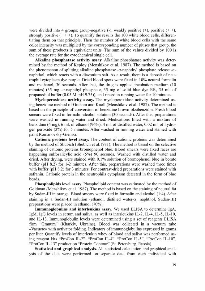
group of healthy children. Amount of the reaction products in granulocytes of chil-
dren with hearing loss of blood was very high. The nuclei of leukocytes were very
poorly painted. Dimensions of leukocytes in children with hearing loss were smaller.
Very often, the nuclei of granulocytes have been many segments (Fig. 3).
Eosinophils contain four principal cationic proteins, major basic protein (MBP),
eosinophil-derived neurotoxin (EDN), eosinophil cationic protein (ECP), and eosin-
ophil peroxidase (EPO) (Abu-Ghazaleh et al. 1992). ECP and MBP are potent
helminthotoxins while EDN is less so. Both ECP and EDN possess neurotoxic and
ribonuclease activities. The ECP nucleotide sequence shows similarity to EDN, rat
pancreatic ribonuclease, and human angiogenin; all are members of the ribonuclease
gene superfamily (Barker et al. 1989). These proteins are mainly expressed in eo-
sinophils, but that certain ones are present in basophils and neutrophils (Abu-
-Ghazaleh et al. 1992).
ECP is a heterogeneous protein with molecular weights of the variants from 16-
-24 kDa. ECP has a variety of biological activities interacting with other immune
cells and plasma proteins such as coagulation factors and proteins of the comple-
ment system. The cytotoxic activity, however, is the most conspicuous. The differ-
ent isoforms of CP seem to have different biological properties with respect to cyto-
toxicity and the effects on fibroblasts (Venge, Byström 1998). In our study, the ECP
level in leukocytes of the children with SNHL was also lower compared to healthy
children (0.9U vs. 1.25U) (Fig. 4).
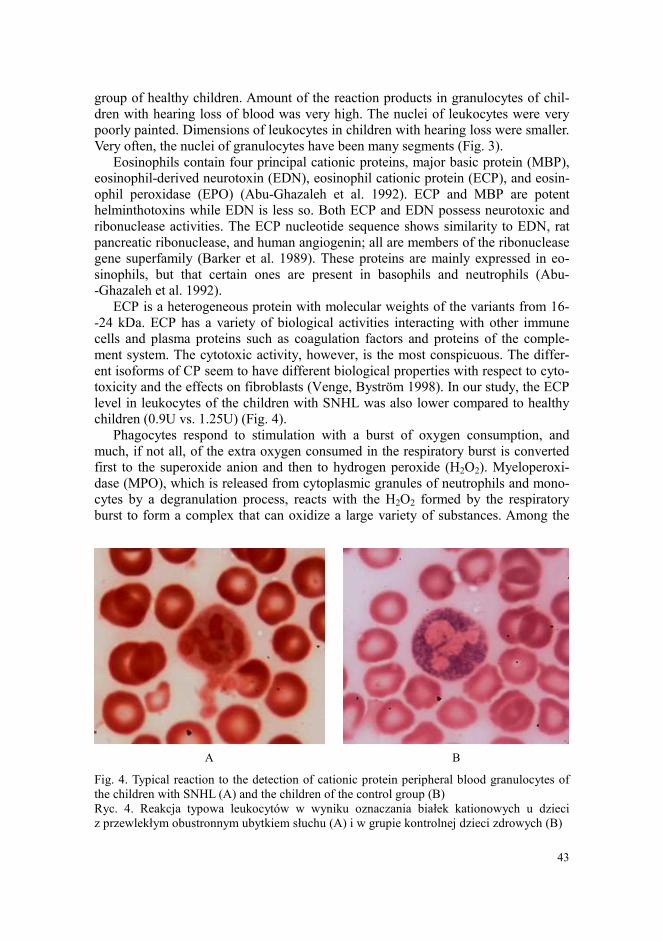
Phagocytes respond to stimulation with a burst of oxygen consumption, and
much, if not all, of the extra oxygen consumed in the respiratory burst is converted
first to the superoxide anion and then to hydrogen peroxide (H2O2). Myeloperoxi-
dase (MPO), which is released from cytoplasmic granules of neutrophils and mono-
cytes by a degranulation process, reacts with the H2O2 formed by the respiratory
burst to form a complex that can oxidize a large variety of substances. Among the
A B
Fig. 4. Typical reaction to the detection of cationic protein peripheral blood granulocytes of
the children with SNHL (A) and the children of the control group (B)
Ryc. 4. Reakcja typowa leukocytów w wyniku oznaczania białek kationowych u dzieci
z przewlekłym obustronnym ubytkiem słuchu (A) i w grupie kontrolnej dzieci zdrowych (B)
44
A B
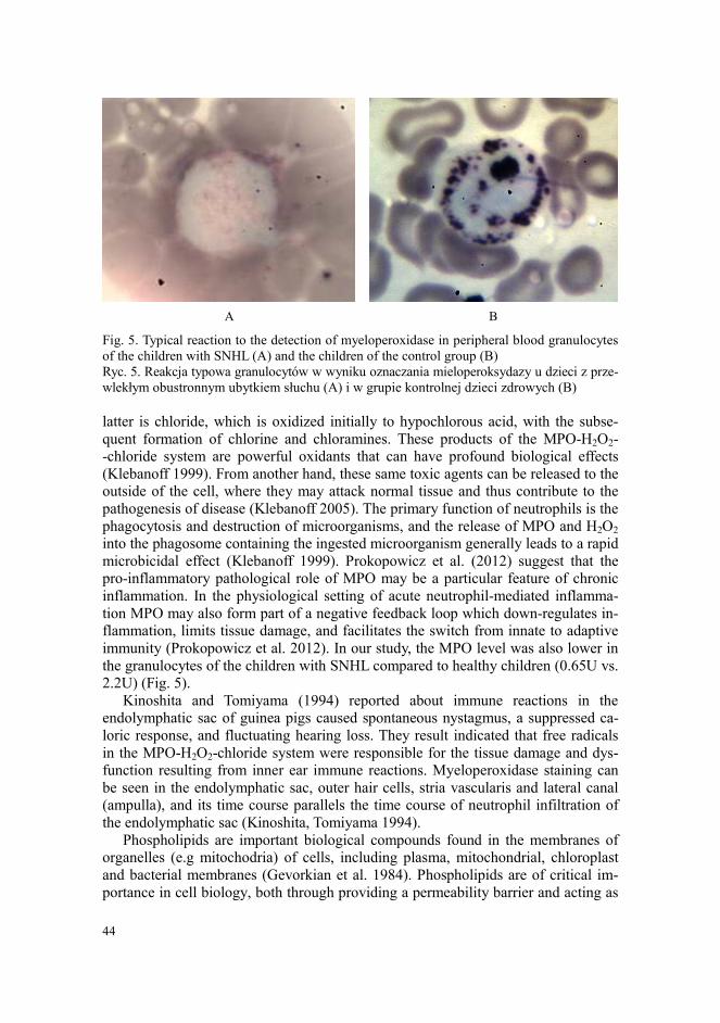
Fig. 5. Typical reaction to the detection of myeloperoxidase in peripheral blood granulocytes
of the children with SNHL (A) and the children of the control group (B)
Ryc. 5. Reakcja typowa granulocytów w wyniku oznaczania mieloperoksydazy u dzieci z prze-
wlekłym obustronnym ubytkiem słuchu (A) i w grupie kontrolnej dzieci zdrowych (B)
latter is chloride, which is oxidized initially to hypochlorous acid, with the subse-
quent formation of chlorine and chloramines. These products of the MPO-H2O2-
-chloride system are powerful oxidants that can have profound biological effects
(Klebanoff 1999). From another hand, these same toxic agents can be released to the
outside of the cell, where they may attack normal tissue and thus contribute to the
pathogenesis of disease (Klebanoff 2005). The primary function of neutrophils is the
phagocytosis and destruction of microorganisms, and the release of MPO and H2O2
into the phagosome containing the ingested microorganism generally leads to a rapid
microbicidal effect (Klebanoff 1999). Prokopowicz et al. (2012) suggest that the
pro-inflammatory pathological role of MPO may be a particular feature of chronic
inflammation. In the physiological setting of acute neutrophil-mediated inflamma-
tion MPO may also form part of a negative feedback loop which down-regulates in-
flammation, limits tissue damage, and facilitates the switch from innate to adaptive
immunity (Prokopowicz et al. 2012). In our study, the MPO level was also lower in
the granulocytes of the children with SNHL compared to healthy children (0.65U vs.
2.2U) (Fig. 5).
Kinoshita and Tomiyama (1994) reported about immune reactions in the
endolymphatic sac of guinea pigs caused spontaneous nystagmus, a suppressed ca-
loric response, and fluctuating hearing loss. They result indicated that free radicals
in the MPO-H2O2-chloride system were responsible for the tissue damage and dys-
function resulting from inner ear immune reactions. Myeloperoxidase staining can
be seen in the endolymphatic sac, outer hair cells, stria vascularis and lateral canal
(ampulla), and its time course parallels the time course of neutrophil infiltration of
the endolymphatic sac (Kinoshita, Tomiyama 1994).
Phospholipids are important biological compounds found in the membranes of
organelles (e.g mitochodria) of cells, including plasma, mitochondrial, chloroplast
and bacterial membranes (Gevorkian et al. 1984). Phospholipids are of critical im-
portance in cell biology, both through providing a permeability barrier and acting as

45
A B
Fig. 6. Typical reaction to the detection of phospholipids in peripheral blood granulocytes of
the children with SNHL (A) and the children of the control group (B)
Ryc. 6. Reakcja typowa granulocytów w wyniku oznaczania fosfolipidów u dzieci z przewle-
kłym obustronnym ubytkiem słuchu (A) i w grupie kontrolnej dzieci zdrowych (B)
substrates for synthesis of lipid mediators. O’Donnel and Murphy (2012) showed
that phospholipid oxidation by acutely activated immune cells is a controlled event,
and participate a central role in regulating membrane biology and innate immune
function during health and disease. Recently, several new families of bioactive lipids
were identified that form through the enzymatic oxidation of membrane phospholip-
ids in circulating innate immune cells and platelets. These comprise eicosanoids at-
tached to phosphatidylethanolamine and phosphatidylcholine and form within 2-5
minutes of cell activation by pathophysiologic agonists, via the coordinated action of
receptors and enzymes (O’Donnel, Murphy 2012). Our study suggest, that children
with SNHL possessed a decreased level of phospholipids (2.1 units) compared to the
control group (2.3 units) (Fig. 6, 7D).
Alkaline phosphatase (ALP) refers to a group of phosphomonoesterases that hy-
drolyze phosphate esters with optimum in vitro activity at a pH of 10, resulting in
the formation of an organic radical and inorganic phosphate. In mammals, this en-
zyme is found mainly in the liver and bones. Marked increase in serum ALP levels,
a disease known as hyperalkalinephosphatasemia, has been associated with malig-
nant biliary obstruction, primary biliary cirrhosis, primary sclerosing cholangitis,
hepatic lymphoma and sarcoidosis (Vroon, Israili 1990). In our study, the median of
alkaline phosphatase activity in the children with SNHL was 2.3 units. The healthy
children had 0.27 units (Fig. 7A).
The most prominent feature of the eosinophils are their large secondary granules,
each containing four basic proteins, the best known being the eosinophil cationic
protein (ECP). This protein has been developed as a marker for eosinophilic disease
(Bystrom et al. 2011). The ECP is a secretory ribonuclease, which is found in the
eosinophilic leukocyte and involved in the innate immune system. Its cytotoxic ac-
tivity is effective against a wide range of pathogens, suggesting a relatively non-
-specific mechanism of action (Boix et al. 2008). Elevated ECP levels are found in

46
A B
A B
Fig. 7. Medians of granulocytes’ metabolic activity in the group of the children with SNHL
(1) and the children of the control group (2) (p ≤ 0.05) (A – alkaline phosphatase, B – cationic
protein, C – myeloperoxidase, D – phospholipids)
Ryc. 7. Mediana metabolicznej aktywności granulocytów u dzieci z przewlekłym obustron-
nym ubytkiem słuchu (1) i grupy kontrolnej dzieci zdrowych (2) (р ≤ 0,05) (А – fosfataza za-
sadowa, B – białko kationowe, C – mieloperoksydaza, D – fosfolipidy)
T helper lymphocyte type 2 (atopic) diseases. ECP is a ribonuclease which has been
attributed with cytotoxic, neurotoxic, fibrosis promoting and immune-regulatory
functions. ECP regulates mucosal and immune cells and may directly act against
helminth, bacterial and viral infections (Bystrom et al. 2011). The level of CP meas-
ured in the blood of the children with SNHL was higher compared to healthy chil-
dren (Fig. 7B).
A B

47
MPO is a major protein constituent of the primary granules of vertebrate neutro-
phils. It catalyses the H2O2-mediated oxidation of halide ions to hypohalous acids,
especially HOCl. These reactive oxygen species can participate in a variety of sec-
ondary reactions, leading to modifications of amino acids and many types of biolog-
ical macromolecules (Prokopowicz et al. 2012). The granule enzyme MPO plays an
important role in neutrophil antimicrobial responses (Metzler et al. 2011). Neutro-
phils release decondensed chromatin termed neutrophil extracellular traps to trap
and kill pathogens extracellularly. Reactive oxygen species are required to initiate
this formation but the downstream molecular mechanism is unknown (Papayanno-
poulos et al. 2010). Papayannopoulos et al. (2010) show that upon activation, es-
capes from azurophilic granules and translocates to the nucleus, where it partially
degrades specific histones, promoting chromatin decondensation. Subsequently, MPO
synergizes with neutrophil elastase in driving chromatin decondensation independ-
ent of its enzymatic activity. Here we show that neutrophils’ myeloperoxidase from
children with SNHL was lower compared to the children from control group (Fig. 7C).
It is known that inflammation of the inner ear in children, sinus contents and
blood levels of IgG increased. The rate of increase in IgG correlates with disease se-
verity. IgA and IgM levels, which are responsible for the primary immune response,
almost unchanged (Veldman 1998).
It is known that interleukins regulate immune response. The ratio of anti-inflam-
matory interleukins and determines the type of immune response: cellular or humoral
immune response. The main role of IL-2 – the transformation is not specific helper
T cells in T-helper cells of the first type and the prevalence of cellular immune re-
sponses. The main role of IL-4, IL-5, IL-10, IL-13 is to direct the immune response
to “humoral” type, by activation of B lymphocytes (Broughton et al. 2004).
Antibodies are a vital part of the armamentarium of the adaptive immune system
for the fine-tuning of the recognition and response to foreign threats (Grönwall et al.
2012). Antibodies function in a variety of ways designed to eliminate the antigen
that elicited their production. Immunoglobulin A (IgA) is found in high concentra-
tions in the mucous membranes, particularly those lining the respiratory passages
and gastrointestinal tract, as well as in saliva and tears. Immunoglobulin G (IgG),
the most abundant type of antibody, is found in all body fluids and protects against
bacterial and viral infections. Immunoglobulin M (IgM), which is found mainly in
the blood and lymph fluid, is the first to be made by the body to fight a new infec-
tion. For example, only IgG and IgM antibodies have the ability to interact with and
initiate the complement cascade. Likewise, only IgG molecules can bind to the sur-
face of macrophages via Fc receptors to promote and enhance phagocytosis. IgA,
IgG, and IgM are frequently measured simultaneously. Evaluated together, they can
give doctors important information about immune system functioning, especially re-
lating to infection or autoimmune disease. IgA antibodies have potent immunomo-
dulatory properties, being able to both induce and suppress immune responses. IgA-
-mediated inhibitory function is able to inhibit several inflammatory diseases includ-
ing asthma and glomerulonephritis (Schwartz-Albiez et al. 2009). Determining the
immunoglobulin’s levels in serum and saliva are presented in Table 1. These results
show a few significant (p ≤ 0.05) differences between group of the children with
SNHL and group of healthy children (Table 1).

48
Table 1
Immunoglobulin’s level in the blood and saliva in studied children (g per L), M±m
Tabela 1
Poziom immunoglobulinów we krwi i ślinie badanych grup dzieci (g/L), M±m
Group IgA IgM IgG
Children
with SNHL
(n=60)
blood 2.5 ± 0.043 0.9 ± 0.016 21.5 ± 0.303
saliva 0.3 ± 0.008 0.5 ± 0.014 1.96 ± 0.076
Healthy children
(n=60)
blood 1.5 ± 0.025* 1.3 ± 0.015* 12.01 ± 0.21*
saliva 1.3 ± 0.014* 0.8 ± 0.018* 1.0 ± 0.021*
* Statistically significant difference between the studied groups (p≤0.05)
Decreased salivary immunoglobulin A (sIgA), a component of mucosal immuni-
ty, is associated with SNHL. We investigated significant changes in sIgA in the chil-
dren with SNHL. Salivary IgA level in the children with SNHL was lower (0.3 ± 0.008)
g/L compared to the controls (1.3±0.014) g/L. While the IgA level in the blood of
the children with SNHL was higher (2.5±0.043) g/L compared to the controls
(1.5±0.025) g/L. Our results are in agreement with reports of other authors, which
suggest that SNHL can induce immune reactions by increasing the formation of
immunoglobulins. For example, Dünne et al. (2004) evaluated a possible role of an
endured infection with Chlamydia pneumoniae or Chlamydia trachomatis as a cause
for sudden sensorineural hearing loss. For this in 60 patients with a first episode of
a SNHL and-60 sex-matched and aged-matched control, following complete oto-
neurological diagnosis blood tests for IgA, IgM and IgG with regard to Chlamydia
pneumoniae and trachomatis were evaluated. They found a statistically significant
higher prevalence of IgA positivity of Chlamydia pneumoniae in patients with sud-
den SNHL (Dünne et al. 2004).
IgG are the most common isotype of immunoglobulins and include four sub-
classes which differ from one another in the following ways: their initial amino acid
sequence, their physical and chemical properties and the different serum concentra-
tions reached with age. Every subclass has a specific biological function: the re-
sponse to proteic antigens is prevalently mediated by IgG1 and IgG3, while IgG2
mediates the response to polysaccharide antigens. Although low levels of IgG sub-
classes may be temporary, deficiencies are often associated with various diseases:
1) recidivating bacterial infections involving the respiratory and digestive tracts,
primarily sustained by capsulated or pyogenic microorganisms; 2) IgA deficiency;
3) absence of immune response following vaccination; 4) allergic or autoimmune
diseases; 5) diseases of the CNS (Cataldo, Paternostro 1990). In our study, the IgG
level in the blood of the children with SNHL was higher compared to the children
with a control group. Moreover, the IgG level in the saliva of the children with
SNHL was also higher than in the healthy children (Table 1).
Serum IgM exists as a pentamer in mammals, predominates in primary immune
responses to most antigens, is the most efficient complement fixing immunoglobulin
and comprises approximately 10% of normal human serum Ig content. IgM is also

49
expressed on the plasma mem- brane of the B lymphocytes as a monomer. It is the
B cell antigen receptor and the H chains each contain an additional hydrophobic
domain for anchoring in the membrane. Monomers of serum IgM are bound together
by disulfide bonds and a joining (J) chain (Davis, Shulman 1989). Autoantibodies of
the IgM type, on the other hand, have shown promising results in the treatment of
multiple sclerosis. These autoantibodies promote remyelination rather than modulat-
ing inflammation. Oxidation-specific epitopes, as found in atherosclerotic lesions
and on apoptotic cells, comprise one important target of natural antibodies. By rec-
ognizing these epitopes, natural antibodies neutralize proinflammatory responses
and mediate atheroprotection (Schwartz-Albiez et al. 2009). In our study, the mean
concentration of IgM was lower in the blood of children with SNHL compared with
the control group. The saliva IgM level was also lower in the children with SNHL
(Table 1).
Recent evidence is discussed that NE and epinephrine, through stimulation of the
beta(2)-adrenoreceptor-cAMP-protein kinase A pathway, inhibit the production of
type 1/proinflammatory cytokines, such as interleukin (IL-12), tumor necrosis fac-
tor-alpha, and interferon-gamma by antigen-presenting cells and T helper (Th) 1 cells,
whereas they stimulate the production of type 2/anti-inflammatory cytokines such as
IL-10 and transforming growth factor-beta. Through this mechanism, systemically,
endogenous catecholamines may cause a selective suppression of Th1 responses and
cellular immunity, and a Th2 shift toward dominance of humoral immunity. On the
other hand, in certain local responses, and under certain conditions, catecholamines
may actually boost regional immune responses, through induction of IL-1, tumor
necrosis factor-alpha, and primarily IL-8 production (Elenkov et al. 2000).
Cytokines represent the major factors involved in the communication between
T cells, macrophages and other immune cells in the course of an immune response to
antigens and infectious agents (Belardelli 1995). Much data support an essential role
for interleukin IL 2 in immune tolerance. IL-2 sometimes contributes to optimal
primary immune responses, but it is not mandatory. Emerging findings, however,
suggest an essential role for IL-2 in immune memory (Malek 2008). In our study, the
IL-2 level was lower in the children with SNHL (2.3 ± 0.05) pg/mL compared to the
controls (3.6 ± 0.03) pg/mL (Table 2).
Table 2
Interleukins level in the blood and saliva the studied groups of children (pg per mL), M ± m
Tabela 2
Poziom interleukinów we krwi i ślinie badanych grup dzieci (pg/mL), M ± m
Group IL-2 IL-4 IL-5 IL-10 IL-13
Children
with SNHL
(n=60)
blood 2.3 ± 0.05 11.02 ± 0.12 5.6 ± 0.03 8.8 ± 0.05 29.9 ± 0.66
saliva 0.04 ± 0.01 54.2 ± 0.13 3.8 ± 0.04 0.9 ± 0.05 19.4 ± 0.63
Healthy
children
(n=60)
blood 3.6 ± 0.03* 8.04 ± 0.01* 3.7 ± 0.02* 2.2 ± 0.023* 8.01± 0.01*
saliva 1.2±0.02* 0.3± 0.01* 1.03± 0.02* 0.03± 0.004* 2.3± 0.01*
* Statistically significant difference between the studied groups (p ≤ 0.05)

50
Interleukin-4 is a cytokine that regulates multiple biological functions. It can
regulate proliferation, differentiation, and apoptosis in several cell types of haema-
topoietic and non-haematopoietic origin. It has a critical role in the regulation of Th0
cell differentiation during a normal immune response, and IL-4-driven Th2 cells di-
rect host responses against parasitic infections. The IL-4 and its signalling machin-
ery have been involved in the development of immune diseases including allergy,
autoimmunity and cancer (Zamorano et al. 2003). The IL-4 level in the blood of
the children with SNHL was higher (11.02 ± 0.12) pg/mL than in the controls
(8.04 ± 0.01). The salivary IL-4 level was elevated (54.2 ± 0.13) pg/mL vs. with con-
trols (0.3 ± 0.01) pg/mL (Table 2).
Interleukin 5 (IL-5) has been shown to play an instrumental role in eosinophilic
inflammation in allergic diseases (Corren 2011). Elevated numbers of blood and tis-
sue eosinophils are present in allergic diseases and experimental evidence suggests
that eosinophils play an important pathogenic role in these conditions. Regulation of
eosinophil maturation, recruitment, and survival is under the control of a small
Fig. 8. Correlations analysis between myeloperoxidase (MP) activity and IgA level in the
blood of the children with SNHL (n = 60); IgA, IgM, IgG in the saliva (sal IgA, sal IgM, sal
IgG); interleukins (IL-4, IL-10, IL-13), interleukin-13 in saliva (sal IL-13), 17-ketosteroids
(17-KST)
Ryc. 8. Analiza korelacyjna między aktywnością mieloperoksydazy (MP) i poziomem immu-
noglobuliny IgA we krwi dzieci z przewlekłym obustronnym ubytkiem słuchu; IgA, IgM,
IgG w ślinie (sal IgA, sal IgM, sal IgG); interleukinami (IL-4, IL-10, IL-13), interleukinem-13
w ślinie (sal IL-13), 17-ketosteroidami (17-KST)

51
group of factors, including IL-5 (Corren 2012). Interleukin-10 (IL-10), first recog-
nized for its ability to inhibit activation and effector function of T cells, monocytes,
and macrophages, is a multifunctional cytokine with diverse effects on most
hemopoietic cell types (Moore et al. 2001). IL-10 is an important suppressive cyto-
kine, produced by a large number of immune cells in addition to the antigen-driven
IL-10-producing regulatory and the naturally occurring suppressor CD4+ T cells,
which is a key player in anti-inflammatory immune responses (O’Garra et al. 2004).
In addition to these activities, IL-10 regulates growth and/or differentiation of B
cells, NK cells, cytotoxic and helper T cells, mast cells, granulocytes, dendritic cells,
keratinocytes, and endothelial cells. IL-10 plays a key role in differentiation and
function of a newly appreciated type of T cell, the T regulatory cell, which may fig-
ure prominently in control of immune responses and tolerance in vivo (Moore et al.
2001). Interleukin (IL)-13 plays a major role in various inflammatory diseases in-
cluding cancer, asthma, and allergy. It mediates a variety of different effects on var-
ious cell types including B cells, monocytes, natural killer cells, endothelial cells,
and fibroblasts (Joshi et al. 2006). In our study, the children with SNHL possess
high levels of IL-5, IL-10 and IL-13, both in the blood and saliva (Table 2).
We also studied the correlations between the indicators. There was found 51 rela-
tionships between the data in the group of the healthy children, as well as 53 rela-
tionships in the group of the children with SNHL (Fig. 8). Interestingly, correlations
between the indicators in the group of the children with SNHL were distributed une-
qually. Many significant correlations was observed in the granulocyte myeloperoxi-
dase activity (n = 9) (Fig. 8).
Direct correlation was observed between the MPO activity and IgM level
(r = 0.37, p ≤ 0.05), MPO activity and IgG level (r = 0.26, p ≤ 0.05). The inverse cor-
Fig. 9. Correlation between myeloperoxidase (MPO) activity in granulocytes and 17-ketoste-
roids (17-KST) level
Ryc. 9. Analiza korelacyjna między aktywnością mieloperoksydazy (MPO) w granulocytach
i poziomem 17-ketosteroidów
52
relation between MPO activity and IgA level (r = -0.29, p ≤ 0.05), as well as MPO
activity and salivary IgA level (r = -0.42, p ≤ 0.05) was noted. The inverse correla-
tion between MPO activity and both blood IL-10 and IL-13 levels (r = -0.27,
p ≤ 0.05), and salivary IL-13 level (r = -0.29, p ≤ 0.05) also was studied. Correlation
between MPO activity and 17-ketosteroids was also inverse (r = -0.29, p ≤ 0.05)
(Fig. 9).
The brain and the immune system are the two major adaptive systems of the
body. During an immune response the brain and the immune system “talk to each
other” and this process is essential for maintaining homeostasis. Two major pathway
systems are involved in this cross-talk: the hypothalamic-pituitary-adrenal (HPA)
axis and the sympathetic nervous system (SNS). It is known that stress hormones af-
fect T-helper cells activity, and cytokine levels in the blood (Elenkov et al. 2000).
Long-term stress hormones impact causes hyperactive states in the white blood cells.
Alkaline phosphatase activity is increased in the hyperactive leukocytes. On the oth-
er hand, the myeloperoxidase level, cationic proteins and lipids level are decreases.
Active leukocytes produce increased level of IL-4, IL-5, IL-10, and IL-13. Stress
hormones and IL-4 affect the maturation of T-helper cells. “Naive” T-helper cells are
transformed into T-helper cells of the second type (Th2). Stress hormones and IL-4
suppress the production of IL-2. At the same time, IL-4, IL-5, IL-10, IL-13, and Th2
co-operate with B-lymphocytes. B-cells become hyperactive and turn into plasma
cells. B-cells produce many antibodies in the blood of the children with hearing loss.
Our results suggest that there is an imbalance between immunoglobulins’ levels in
the blood and saliva. This phenomenon in the literature named as “the effect of de-
layed stress” (McClelland et al. 1980, Evans et al. 1994, Tsujita, Morimoto 1999).
The activation of sympathetic nervous system during an immune response might be
aimed to localize the inflammatory response, through induction of neutrophil accu-
mulation and stimulation of more specific humoral immune responses, although sys-
temically it may suppress Th1 responses, and, thus protect the organism from the
detrimental effects of proinflammatory cytokines and other products of activated
macrophages (Elenkov et al. 2000).
CONCLUSIONS
Our results suggest that the children of 7-11 years old with SNHL possess in-
creased urine adrenaline and 17 ketosteroids levels. This indicates that the stress re-
action is occurs. Elevated hormones levels lead to changes in the immune system ac-
tivity. Adrenaline and 17-ketosteroids activated leukocytes. The increasing of alka-
line phosphatase activity and decreasing myeloperoxidase activity, as well as cation-
ic proteins and lipids take place in leukocytes. Activated leukocytes release anti-
inflammatory interleukins. B-lymphocytes are activated and transform into plasma
cells. These interleukins (especially IL-4) are affecting the maturation of Th2. They
also stimulate B cells to provide an immunoglobulins release. We observed elevated
blood levels of IgA and IgG, and decreased IgM level in the group of the children
with SNHL. High IgG level and decreased IgA and IgM levels were observed in sa-
liva of the children with SNHL. Impairment of immunoglobulins levels, as well as

53
metabolic changes of leukocytes leads to reduced non-specific immunity in the chil-
dren with SNHL.
REFERENCES
Abu-Ghazaleh R.I., Dunnette S.L., Loegering D.A., Checkel J.L., Kita H., Thomas L.L.,
Gleich G.J. 1992. Eosinophil granule proteins in peripheral blood granulocytes. J. Leukoc.
Biol., 52(6): 611-618.
Arnold W., Pfaltz R., Altermatt H. 1985. Evidence of serum antibodies against inner ear tis-
sues in the blood of patients with certain sensorineural hearing disoders. Acta Otolaryngol.,
99: 437-445.
Astaldi G., Verga J. 1957. The glycogen content of the cells of lymphatic leukemia. Acta
Haematol., 3: 129-136.
Barker R.L., Loegering D.A., Ten R.M., Hamann K.J., Pease L.R., Gleich G.J. 1989. Eosino-
phil cationic protein cDNA. Comparison with other toxic cationic proteins and
ribonucleases. J. Immunol., 143(3): 952-955.
Bednarska K., Klink M., Sulowska Z. 2006. Application of intracellular alkaline phosphatase
activity measurement in detection of neutrophil adherence in vitro. Mediators Inflamm.,
4: 19307-19312.
Belardelli F. 1995. Role of interferons and other cytokines in the regulation of the immune
response. Acta Pathol. Microbiol. Immunol. Scandinavica, 103(3): 161-179.
Bless N.M., Smith D., Charlton I. 1997. Protective effects of an aptamer inhibitor of neutro-
phil еlastase in lung inflammatory injury. Cum. Biol., 11: 877-880.
Boix E., Torrent M., Sánchez D., Nogués M.V. 2008. The antipathogen activities of eosino-
phil cationic protein. Curr. Pharm. Biotechnol., 9(3): 141-152.
Boxer L.A., Smolen J.E. 1988. Neutrophil granule constituents and their release in health and
disease. Hematol. Oncol. Clin. North Am., 2(1): 101-134.
Broughton S.S., Meyerhoff W.E., Cohen S.B. 2004. Immune-mediated inner ear disease: 10-year
experience. Semin. Arthritis. Rheum., 2: 544-548.
Bystrom J., Amin K., Bishop-Bailey D. 2011. Analysing the eosinophil cationic protein – a clue
to the function of the eosinophil granulocyte. Respir. Res., 12: 10.
Cataldo F., Paternostro D. 1990. IgG subclasses and their clinical significance. Minerva
Pediatr., 42(12): 509-514 (Article in Italian, Abstract in English).
Chashcheva E.G. 2003. Clinical and audiology signs of autoimmune sensorineural hearing
loss. Journal of Ear, Nose and Throat Disorders, 5: 131 (in Russian).
Chikkappa G. 1992. Control of neutrophil alkaline phosphatase synthesis by cytokines in
health and diseases. Exp. Hematol., 20: 388-390.
Chua F., Laurent G. 2006. Neutrophil elastase: mediator of extracellular matrix destruction
and accumulation. Proc. Am. Thorae. Soc., 5: 424-427.
Corren J. 2011. Anti-interleukin-5 antibody therapy in asthma and allergies. Curr. Opin. Allergy
Clin. Immunol., 11(6): 565-570.
Corren J. 2012. Inhibition of interleukin-5 for the treatment of eosinophilic diseases. Discov.
Med., (71): 305-312.
Davis A.C., Shulman M.J. 1989. IgM – molecular requirements for its assembly and function.
Immunol. Today, 10(4): 118-122, 127-128.
Dünne A.A., Wiegand A., Prinz H., Slenczka W., Werner J.A. 2004. Chlamydia pneumoniae
IgA seropositivity and sudden sensorineural hearing loss. Otolaryngol. Pol., 58(3): 427-428.
Elenkov I.J., Wilder R.L., Chrousos G.P., Vizi E.S. 2000. The sympathetic nerve – an integra-
tive interface between two supersystems: the brain and the immune system. Pharmacol.
Rev., 52(4): 595-638.

54
Elies W. 1983. Immunologische Befundе bei cochleovestibulären Störungen. Allergologie, 6:
357-361.
Evans P., Bristow M., Hucklebridge F., Clow A., Pang F.Y. 1994. Stress, arousal, cortisol and
secretory immunoglobulin A in students undergoing assessment. Br. J. Clin. Psychol., 33:
575-576.
Frimel G. 1987. Immunological methods. Medicine, Moscow (in Russian).
Gevorkian E.S., Yavronyan J.V., Panossian G.A. 1984. Comparative analysis of changes in
phospholipid composition in nuclear membranes and chromatin to the rat liver under the
influence of hydrocortisone. Abstracts of Reports Union Simposes “Structure and func-
tion of the cell nucleus”, Pushchino, Russia (in Russian).
Grönwall C., Vas J., Silverman G.J. 2012. Protective roles of natural IgM antibodies. Front
Immunol., 3: 66.
Gylling E.V. 1969. On the etiologic and pathogenesis role of allergic reactions in the genesis
lesions in the vestibular and acoustic analyzers. Journal of Ear, Nose and Throat Disor-
ders, 2: 6-12 (in Russian).
Hrebenyuk I.E. 2007. Etiopathogenic aspects of sensorineural hearing loss. Summary of the
thesis for the Doctor of Medicine degree, speciality 14.00.04 “Diseases of the ear, nose
and throat”, Moscow (in Russian).
Joshi B.H., Hogaboam C., Dover P., Husain S.R., Puri R.K. 2006. Role of interleukin-13 in
cancer, pulmonary fibrosis, and other T(H)2-type diseases. Vitam. Horm., 74: 479-504.
Karaulov A.V. 2002. Clinical Immunology and Allergology. Medical News Agency, Moscow
(in Russian).
Kassner S.S., Schöttler S., Bonaterra G.A., Stern-Sträter J., Sommer U., Hormann K., Kinschef R.,
Gössler U.R. 2011. Proinflammatory and proadhesive activation of lymphocytes and mac-
rophages in sudden sensorineural hearing loss. Audiol. Neurootol., 16(4): 254-262.
Kinoshita T., Tomiyama S. 1994. Free radicals in inner ear immune responses; immuno-
histochemical study of myeloperoxidase. Nihon Jibiinkoka Gakkai Kaiho, 97(9): 1608-
-1612 (Article in Japanese, Abstract in English).
Klebanoff S.J. 1999. Myeloperoxidase. Proc. Assoc. Am. Physicians, 111(5): 383-389.
Klebanoff S.J. 2005. Myeloperoxidase: friend and foe. J. Leukoc. Biol., 77(5): 598-625.
Kucherenko N.E., Babenyuk J.D., Vasiliev A.N. 1988. Biochemistry: The Workshop. High
School, Kiev (in Russian).
Lapach S.N., Chubenko A.V., Babich P.N. 2001. Statistical methods in medical and biological
research using Excel. MORION, Kiev (in Russian).
Malek T.R. 2008. The biology of interleukin-2. Annu. Rev. Immunol., 26: 453-479.
Malinvaud D., Avan P., Germain D.P. 2006. The cochlea in Fabry disease: a sensorineural
hearing loss model of vascular origin? Med. Intern., 7: 527-531.
McCabe B.F. 1979. Autoimmune sensorineural hearing loss. Ann. Otol. Rhinol. Laryngol.,
88: 585-589.
McClelland D.C., Floor E., Davidson R.J., Saron C. 1980. Stressed power motivation, sym-
pathetic activation, immune function, and illness. J. Human Stress, 6(2): 11-19.
Melnikov O.F., Sidorenko T.F., Zayats T.A., Timen G.E., Chashcheva E.G. 2003. Autoim-
mune reactions of humoral and cell types in the nervous tissue antigens in children with
sensorineural hearing loss. Journal of Ear, Nose and Throat Disorders, 6: 5-8 (in Russian).
Menshikov V.V., Delektorskaya L.N., Zolotnitskaya R.P. 1987. Laboratory Methods in the
Clinic. Medicine, Moscow (in Russian).
Metzler K.D., Fuchs T.A., Nauseef W.M., Reumaux D., Roesler J., Schulze I., Wahn V.,
Papayannopoulos V., Zychlinsky A. 2011. Myeloperoxidase is required for neutrophil ex-
tracellular trap formation: implications for innate immunity. Blood, 117(3): 953-959.
Moore K.W., de Waal Malefyt R., Coffman R.L., O’Garra A. 2001. Interleukin-10 and the in-
terleukin-10 receptor. Annu. Rev. Immunol., 19: 683-765.

55
Moskalenko E.P., Sizyakina L.P., Zolotov T.V. 1989. The immune status of patients with
sensorineural hearing loss. The experimental conditions in otorhinolaryngology, ophthal-
mology, neurology and neurosurgery. Rostov-on-Don (in Russian).
Novikov D.A., Novochadov V.V. 2005. Statistical methods in the medical and biological ex-
periments (typical cases). Volgograd State University, Volgograd (in Russian).
O’Donnel V.B., Murphy R.C. 2012. New families of bioactive oxidized phospholipids gener-
ated by immune cells: identification and signaling actions. Blood, 120(10): 1985-1992.
O’Garra A., Vieira P.L., Vieira P., Goldfeld A.E. 2004. IL-10-producing and naturally occur-
ring CD4+ Tregs: limiting collateral damage. J. Clin. Invest., 114(10): 1372-1378.
Papayannopoulos V., Metzler K.D., Hakkim A., Zychlinsky A. 2010. Neutrophil elastase and
myeloperoxidase regulate the formation of neutrophil extracellular traps. J. Cell Biol.,
191(3): 677-691.
Pellme S., Dahlgre N.C., Karlsson A. 2007. The two neutrophil plasma membrane markers
alkaline phosphatase and HLA class I antigen localize differently in granule-deficient
cytoplasts. An ideal plasma membrane marker in human neutrophils is still lacking.
J. Immunol. Methods, 325: 1562-1570.
Prokopowicz Z., Marcinkiewicz J., Katz D.R., Chain B.M. 2012. Neutrophil myeloperoxi-
dase: soldier and statesman. Arch. Immunol. Ther. Exp., 60(1): 43-54.
Reddy M.V., Satyanarayana V.V., Hemabindu L., Rani P.U., Reddy P.P., Lakshmi V.S. 2005.
Immunological studies in children with hearing impairment. Indian. Med. Assoc., 10:
520-521.
Schmidt R. 1984. Fundamentals of sensory physiology. Mir, Moscow (in Russian).
Schwartz-Albiez R., Monteiro R.C., Rodriguez M., Binder C.J., Shoenfeld Y. 2009. Natural
antibodies, intravenous immunoglobulin and their role in autoimmunity, cancer and in-
flammation. Clin. Exp. Immunol., 158 (Suppl. 1): 43-50.
Shevryakov M.V., Yakovenko B.V., Yavonenko O.F. 2003. Workshop on Biological Chemis-
try. University Book, Sumy (in Ukrainian).
Shubich M.G., Slavinski A.A., Vishnyakova A.P. 1981. Cationic proteins in neutrophilic leu-
kocytes in viral diseases of children. Quantitative fluorescence cytochemistry.
Laboratorna sprava, 5: 266-269 (in Russian).
Solares C.A., Hughes G.B., Tuohy V.K. 2003. Autoimmune sensorineural hearing loss: an
immunologic perspective. J. Neuroimmunol., 138(1-2): 1-7.
Tan J., Shepherd R.K. 2006. Aminoglycoside-induced degeneration of adult spiral ganglion
neurons involves differential modulation of tyrosine kinase В and p75 neurotrophin re-
ceptor signaling. Am. J. Pathol., 2: 528-543.
Timen G., Pisanko V., Mironuk B., Kobzaruk L. 2002. Complex treatment of children with
chronic sensorinevral hearing loss. The XVII-th World Congress of IFOS (Cairo): 281-
-282.
Timen G.E., Kobzaruk L.B., Smolyaninovа I.I., Mironyuk B.M., Chashcheva E.G. 2004.
Acute and chronic sensorineural hearing loss in children. Journal of Ear, Nose and Throat
Disorders, 5: 57-58 (in Russian).
Tsujita S., Morimoto K. 1999. Secretory IgA in saliva can be a useful stress marker. Environ.
Health Prev. Med., 4(1): 1-8.
Veldman J. 1998. Immune-mediated sensorineural hearing loss. Auris Nasus Larynx, 25(3):
309-317.
Venge P., Byström J. 1998. Eosinophil cationic protein (ECP). Int. J. Biochem. Cell Biol.,
30(4): 433-437.
Vroon D.H., Israili Z. 1990. Alkaline Phosphatase and Gamma Glutamyltransferase. Chapter
100. In: Clinical Methods: The History, Physical, and Laboratory Examinations. H.K. Walker,
W.D. Hall, J.W. Hurst (eds). 3rd edition. Butterworths, Boston.
56
Zamorano J., Rivas M.D., Pérez G.M. 2003. Interleukin-4: a multifunctional cytokine.
Immunología, 22(2): 215-224.
Zolotovа T. 2004. A differential approach to treatment of sensorineural hearing loss. Sum-
mary of the thesis for the Doctor of Medicine degree, speciality 14.00.04 “Diseases of the
ear, nose and throat”, Moscow (in Russian).
Zolotovа T.V., Panchenko S.N. 2010. The experimental sensorineural hearing loss Valium
genesis in animals: apoptotic cell death spiral path of the body. J. Otorhinolaryngology, 4:
29-32 (in Russian).