Regulation of the cerebral circulation: bedside assessment ...
17
REVIEW Open Access Regulation of the cerebral circulation: bedside assessment and clinical implications Joseph Donnelly 1 , Karol P. Budohoski 1 , Peter Smielewski 1 and Marek Czosnyka 1,2* Abstract Regulation of the cerebral circulation relies on the complex interplay between cardiovascular, respiratory, and neural physiology. In health, these physiologic systems act to maintain an adequate cerebral blood flow (CBF) through modulation of hydrodynamic parameters; the resistance of cerebral vessels, and the arterial, intracranial, and venous pressures. In critical illness, however, one or more of these parameters can be compromised, raising the possibility of disturbed CBF regulation and its pathophysiologic sequelae. Rigorous assessment of the cerebral circulation requires not only measuring CBF and its hydrodynamic determinants but also assessing the stability of CBF in response to changes in arterial pressure (cerebral autoregulation), the reactivity of CBF to a vasodilator (carbon dioxide reactivity, for example), and the dynamic regulation of arterial pressure (baroreceptor sensitivity). Ideally, cerebral circulation monitors in critical care should be continuous, physically robust, allow for both regional and global CBF assessment, and be conducive to application at the bedside. Regulation of the cerebral circulation is impaired not only in primary neurologic conditions that affect the vasculature such as subarachnoid haemorrhage and stroke, but also in conditions that affect the regulation of intracranial pressure (such as traumatic brain injury and hydrocephalus) or arterial blood pressure (sepsis or cardiac dysfunction). Importantly, this impairment is often associated with poor patient outcome. At present, assessment of the cerebral circulation is primarily used as a research tool to elucidate pathophysiology or prognosis. However, when combined with other physiologic signals and online analytical techniques, cerebral circulation monitoring has the appealing potential to not only prognosticate patients, but also direct critical care management. Background To function, the brain requires adequate delivery of nutrients and oxygen. A circulatory system is therefore required to maintain an optimal cerebral blood flow (CBF) for the brain’ s diverse needs. Whilst oxygen and nutrient delivery is in part dependent on the pump sup- plying it—the heart—the circulatory system has also evolved mechanisms to ensure the precise control of CBF. The cerebral vessels have the remarkable ability to rapidly adapt and react to the brain’ s chemical environ- ment, to neuronal signals, and to the pressure within the cerebral vessels. This review highlights clinically relevant aspects of cerebrovascular physiology and cerebral circulation monitoring techniques before outlining the state of current knowledge of the cerebral circulation in selected critical illnesses and highlighting promising areas for fu- ture research. Review Regulation of cerebral blood flow A haemodynamic model for the cerebral circulation has been described that allows for interrogation of the regu- lation of CBF [1, 2]. In such a model, CBF is dependent on the pressure supplied in the cerebral arteries (arterial blood pressure (ABP)), the back pressure in the cerebral venous system (usually close to intracranial pressure (ICP)), and the resistance related to the diameter of the * Correspondence: [email protected] 1 Brain Physics Laboratory, Division of Neurosurgery, Department of Clinical Neurosciences, Cambridge Biomedical Campus, University of Cambridge, Hills Road, Cambridge CB2 0QQ, UK 2 Institute of Electronic Systems, Warsaw University of Technology, ul. Nowowiejska 15/19, 00-665 Warsaw, Poland © 2016 Donnelly et al. Open Access This article is distributed under the terms of the Creative Commons Attribution 4.0 International License (http://creativecommons.org/licenses/by/4.0/), which permits unrestricted use, distribution, and reproduction in any medium, provided you give appropriate credit to the original author(s) and the source, provide a link to the Creative Commons license, and indicate if changes were made. The Creative Commons Public Domain Dedication waiver (http://creativecommons.org/publicdomain/zero/1.0/) applies to the data made available in this article, unless otherwise stated. Donnelly et al. Critical Care 2016, 18: http://ccforum.com/content/18/6/
Transcript of Regulation of the cerebral circulation: bedside assessment ...
Donnelly et al. Critical Care 2016, 18:http://ccforum.com/content/18/6/
REVIEW Open Access
Regulation of the cerebral circulation:bedside assessment and clinicalimplications
Joseph Donnelly1, Karol P. Budohoski1, Peter Smielewski1 and Marek Czosnyka1,2*Abstract
Regulation of the cerebral circulation relies on the complex interplay between cardiovascular, respiratory, and neuralphysiology. In health, these physiologic systems act to maintain an adequate cerebral blood flow (CBF) throughmodulation of hydrodynamic parameters; the resistance of cerebral vessels, and the arterial, intracranial, and venouspressures. In critical illness, however, one or more of these parameters can be compromised, raising the possibilityof disturbed CBF regulation and its pathophysiologic sequelae. Rigorous assessment of the cerebral circulationrequires not only measuring CBF and its hydrodynamic determinants but also assessing the stability of CBF inresponse to changes in arterial pressure (cerebral autoregulation), the reactivity of CBF to a vasodilator (carbondioxide reactivity, for example), and the dynamic regulation of arterial pressure (baroreceptor sensitivity). Ideally,cerebral circulation monitors in critical care should be continuous, physically robust, allow for both regional andglobal CBF assessment, and be conducive to application at the bedside. Regulation of the cerebral circulation isimpaired not only in primary neurologic conditions that affect the vasculature such as subarachnoid haemorrhageand stroke, but also in conditions that affect the regulation of intracranial pressure (such as traumatic brain injuryand hydrocephalus) or arterial blood pressure (sepsis or cardiac dysfunction). Importantly, this impairment is oftenassociated with poor patient outcome. At present, assessment of the cerebral circulation is primarily used as aresearch tool to elucidate pathophysiology or prognosis. However, when combined with other physiologic signalsand online analytical techniques, cerebral circulation monitoring has the appealing potential to not only prognosticatepatients, but also direct critical care management.
BackgroundTo function, the brain requires adequate delivery ofnutrients and oxygen. A circulatory system is thereforerequired to maintain an optimal cerebral blood flow(CBF) for the brain’s diverse needs. Whilst oxygen andnutrient delivery is in part dependent on the pump sup-plying it—the heart—the circulatory system has alsoevolved mechanisms to ensure the precise control ofCBF. The cerebral vessels have the remarkable ability torapidly adapt and react to the brain’s chemical environ-ment, to neuronal signals, and to the pressure within thecerebral vessels.
* Correspondence: [email protected] Physics Laboratory, Division of Neurosurgery, Department of ClinicalNeurosciences, Cambridge Biomedical Campus, University of Cambridge, HillsRoad, Cambridge CB2 0QQ, UK2Institute of Electronic Systems, Warsaw University of Technology, ul.Nowowiejska 15/19, 00-665 Warsaw, Poland
© 2016 Donnelly et al. Open Access This articInternational License (http://creativecommonsreproduction in any medium, provided you gthe Creative Commons license, and indicate if(http://creativecommons.org/publicdomain/ze
This review highlights clinically relevant aspects ofcerebrovascular physiology and cerebral circulationmonitoring techniques before outlining the state ofcurrent knowledge of the cerebral circulation in selectedcritical illnesses and highlighting promising areas for fu-ture research.
ReviewRegulation of cerebral blood flowA haemodynamic model for the cerebral circulation hasbeen described that allows for interrogation of the regu-lation of CBF [1, 2]. In such a model, CBF is dependenton the pressure supplied in the cerebral arteries (arterialblood pressure (ABP)), the back pressure in the cerebralvenous system (usually close to intracranial pressure(ICP)), and the resistance related to the diameter of the
le is distributed under the terms of the Creative Commons Attribution 4.0.org/licenses/by/4.0/), which permits unrestricted use, distribution, andive appropriate credit to the original author(s) and the source, provide a link tochanges were made. The Creative Commons Public Domain Dedication waiverro/1.0/) applies to the data made available in this article, unless otherwise stated.

Donnelly et al. Critical Care 2016, 18: Page 2 of 17http://ccforum.com/content/18/6/
small cerebral vessels (cerebrovascular resistance (CVR);Fig. 1). This relationship can be simplified as:
CBF ¼ ABP− ICPCVR
Thus, cardiovascular, ICP, and cerebrovascular compo-nents are all important regulators of the cerebral circula-tion. Applying this model can provide crucial insightsinto the physiologic factors that regulate cerebral perfu-sion in health and elucidate why CBF regulation is oftenimpaired in pathologic states.
The cardiovascular componentAs early as 1890, Sherrington and Roy underlinedthe importance of the ABP in the regulation of CBF:‘One of the most evident of the facts observed by usis that the blood-supply of the brain varies directlywith the blood pressure in the systemic arteries’ [3].The pressure that supplies the cerebral vessels isdependent on factors mostly outside the brain itself:the heart provides the cardiac output while the per-ipheral vessels provide the resistance, both of which
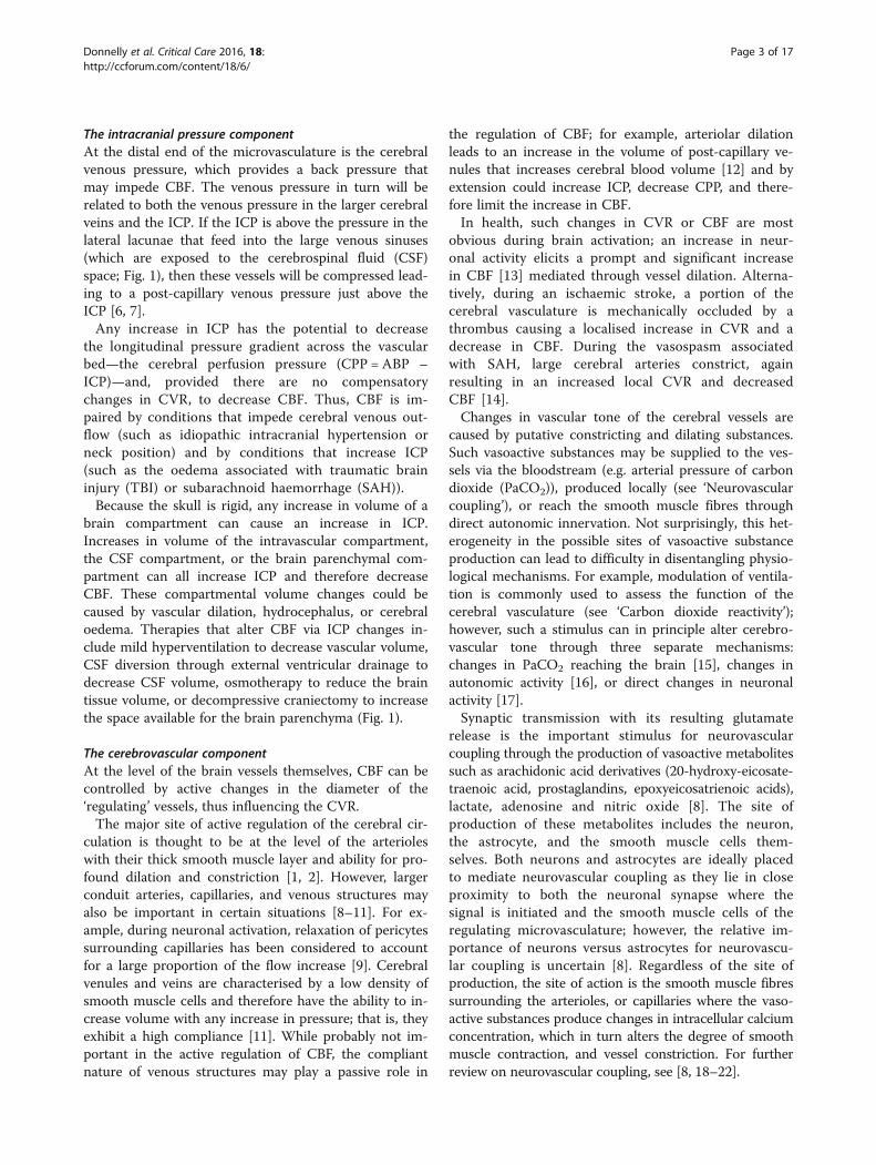
Fig. 1 Regulation of the cerebral circulation. CBF at the level of the miABP and ICP) and inversely proportional to CVR. ICP exerts its effect onvasculature where the bridging veins enter the sagittal sinus ensures thalways above ICP. CBF is modulated by the cardiovascular system in tevasopressors forms an integral part of many CBF protective strategies (is regulated at the level of the arterioles (purple) by variations in vascuischaemic stroke or vasospasm, CVR is dramatically increased, usually atthrough its coupling with cerebral venous pressure. ICP increases can bvenous), increased CSF volume or increase in parenchyma (oedema),modulate CBF do so via one (or more) of these pathways. There is tydeterminants, and influences of CBF. For example, a drop in ABP woudue to the baroreflex (HR increase in response to drop in ABP) and cdrop in ABP). ABP arterial blood pressure, CBF cerebral blood flow, CBcerebrovascular resistance, EVD external ventricular drainage, HR heart rate, ICvolume, TPR total peripheral resistance
contribute to the ABP supplying the brain. In thissense, the balance between the brain CVR and thetotal peripheral resistance determines the proportionof the cardiac output that reaches the brain. Thus,any pathological or physiological event that affectsthe heart or the vasculature as a whole has the po-tential to alter the cerebral circulation. Cardiogenicshock and arrhythmia may therefore impair CBF [4],as do conditions that affect the systemic vasculaturesuch as sepsis [5].Just as pathologies affecting ABP can affect CBF, ther-
apies to augment CBF often do so by modulating ABP.Vasopressors act to buffer ABP by constricting periph-eral vessels, while inotropes act to modulate cardiac out-put (Fig. 1). An important consideration of such anapproach is that the relationship between changes inABP and CBF is typically non-linear due to activechanges in vascular tone occurring at the level of thecerebral arterioles—a process known as cerebral auto-regulation (see later). Furthermore, modulating ABP as atherapeutic measure will not only increase blood flow tothe brain, but will also increase blood flow to any vascu-lar beds with a low vascular resistance.
crovasculature is directly proportional to CPP (difference betweenCBF through changes in CPP; compression of the venousat the bridging vein and post-capillary intravascular pressure isrms of the regulation of SV, HR, and TPR (red). Control of TPR witheven when TPR is not the primary cause of CBF disturbance). CVRlar tone in response to metabolic, neural, or myogenic inputs. Inthe level of large intracranial arteries. ICP (blue) modulates CBFe caused by increases in cerebral blood volume (arterial oror abnormal material volume (mass lesion). All therapies thatpically significant interdependence between the therapies,ld be expected to result in a drop in CBF but this is short livederebral autoregulation (decrease in vascular tone in response toV cerebral blood volume, CSF V cerebrospinal fluid volume, CVRP intracranial pressure, IIH idiopathic intracranial hypertension, SV stroke
Donnelly et al. Critical Care 2016, 18: Page 3 of 17http://ccforum.com/content/18/6/
The intracranial pressure componentAt the distal end of the microvasculature is the cerebralvenous pressure, which provides a back pressure thatmay impede CBF. The venous pressure in turn will berelated to both the venous pressure in the larger cerebralveins and the ICP. If the ICP is above the pressure in thelateral lacunae that feed into the large venous sinuses(which are exposed to the cerebrospinal fluid (CSF)space; Fig. 1), then these vessels will be compressed lead-ing to a post-capillary venous pressure just above theICP [6, 7].Any increase in ICP has the potential to decrease
the longitudinal pressure gradient across the vascularbed—the cerebral perfusion pressure (CPP = ABP –ICP)—and, provided there are no compensatorychanges in CVR, to decrease CBF. Thus, CBF is im-paired by conditions that impede cerebral venous out-flow (such as idiopathic intracranial hypertension orneck position) and by conditions that increase ICP(such as the oedema associated with traumatic braininjury (TBI) or subarachnoid haemorrhage (SAH)).Because the skull is rigid, any increase in volume of a
brain compartment can cause an increase in ICP.Increases in volume of the intravascular compartment,the CSF compartment, or the brain parenchymal com-partment can all increase ICP and therefore decreaseCBF. These compartmental volume changes could becaused by vascular dilation, hydrocephalus, or cerebraloedema. Therapies that alter CBF via ICP changes in-clude mild hyperventilation to decrease vascular volume,CSF diversion through external ventricular drainage todecrease CSF volume, osmotherapy to reduce the braintissue volume, or decompressive craniectomy to increasethe space available for the brain parenchyma (Fig. 1).
The cerebrovascular componentAt the level of the brain vessels themselves, CBF can becontrolled by active changes in the diameter of the‘regulating’ vessels, thus influencing the CVR.The major site of active regulation of the cerebral cir-
culation is thought to be at the level of the arterioleswith their thick smooth muscle layer and ability for pro-found dilation and constriction [1, 2]. However, largerconduit arteries, capillaries, and venous structures mayalso be important in certain situations [8–11]. For ex-ample, during neuronal activation, relaxation of pericytessurrounding capillaries has been considered to accountfor a large proportion of the flow increase [9]. Cerebralvenules and veins are characterised by a low density ofsmooth muscle cells and therefore have the ability to in-crease volume with any increase in pressure; that is, theyexhibit a high compliance [11]. While probably not im-portant in the active regulation of CBF, the compliantnature of venous structures may play a passive role in
the regulation of CBF; for example, arteriolar dilationleads to an increase in the volume of post-capillary ve-nules that increases cerebral blood volume [12] and byextension could increase ICP, decrease CPP, and there-fore limit the increase in CBF.In health, such changes in CVR or CBF are most
obvious during brain activation; an increase in neur-onal activity elicits a prompt and significant increasein CBF [13] mediated through vessel dilation. Alterna-tively, during an ischaemic stroke, a portion of thecerebral vasculature is mechanically occluded by athrombus causing a localised increase in CVR and adecrease in CBF. During the vasospasm associatedwith SAH, large cerebral arteries constrict, againresulting in an increased local CVR and decreasedCBF [14].Changes in vascular tone of the cerebral vessels are
caused by putative constricting and dilating substances.Such vasoactive substances may be supplied to the ves-sels via the bloodstream (e.g. arterial pressure of carbondioxide (PaCO2)), produced locally (see ‘Neurovascularcoupling’), or reach the smooth muscle fibres throughdirect autonomic innervation. Not surprisingly, this het-erogeneity in the possible sites of vasoactive substanceproduction can lead to difficulty in disentangling physio-logical mechanisms. For example, modulation of ventila-tion is commonly used to assess the function of thecerebral vasculature (see ‘Carbon dioxide reactivity’);however, such a stimulus can in principle alter cerebro-vascular tone through three separate mechanisms:changes in PaCO2 reaching the brain [15], changes inautonomic activity [16], or direct changes in neuronalactivity [17].Synaptic transmission with its resulting glutamate
release is the important stimulus for neurovascularcoupling through the production of vasoactive metabolitessuch as arachidonic acid derivatives (20-hydroxy-eicosate-traenoic acid, prostaglandins, epoxyeicosatrienoic acids),lactate, adenosine and nitric oxide [8]. The site ofproduction of these metabolites includes the neuron,the astrocyte, and the smooth muscle cells them-selves. Both neurons and astrocytes are ideally placedto mediate neurovascular coupling as they lie in closeproximity to both the neuronal synapse where thesignal is initiated and the smooth muscle cells of theregulating microvasculature; however, the relative im-portance of neurons versus astrocytes for neurovascu-lar coupling is uncertain [8]. Regardless of the site ofproduction, the site of action is the smooth muscle fibressurrounding the arterioles, or capillaries where the vaso-active substances produce changes in intracellular calciumconcentration, which in turn alters the degree of smoothmuscle contraction, and vessel constriction. For furtherreview on neurovascular coupling, see [8, 18–22].

Donnelly et al. Critical Care 2016, 18: Page 4 of 17http://ccforum.com/content/18/6/
The autonomic nervous system may also influence thevascular tone of cerebral vessels. Despite animal studiesdemonstrating a rich innervation of both the dilatingparasympathetic and constricting sympathetic fibres, theautonomic control of CBF in humans remains contro-versial [23, 24] with the divergence in opinions probablyowing to between-species variation in autonomic innerv-ation, variations in brain metabolism between experi-ments, and heterogeneous autonomic nerve distributionin the different studies [25]. Nevertheless, stimulation ofthe trigeminal ganglion in humans decreases the esti-mated CBF [26] while blockade of the stellate ganglionincreases the estimated CBF [27], highlighting a role forthe sympathetic nervous system in the regulation of thecerebral circulation in humans.In addition to the cerebrovascular, mean arterial pres-
sure, and ICP components, cardiac output has recentlybeen suggested to be an independent regulator of CBF[28]. Evidence for such a view comes from studies dem-onstrating a change in CBF after interventions thatchange cardiac output but have no effect on mean arter-ial pressure [28, 29]. An additional measure of CBFregulation could thus be assessing CBF as a fraction ofthe cardiac output. Although continuous and accuratemeasures of cardiac output are less practical than ABP,such an approach may provide additional insight into re-gional blood flow regulation in health and disease.According to the conventional model (Fig. 1), for an
increase in cardiac output to produce an increase inCBF without a change in ABP, both total peripheralresistance and CVR must decrease. As such, the auto-nomic nervous system has been speculated as the mech-anism by which changes in cardiac output may alterCBF without changes in ABP [28]; however, a metro-logical issue should also be considered. The ABP mea-sured in the examined studies (and the majority ofvascular regulation investigations) is not the ABP in thelarge cerebral arteries, but the pressure in a small per-ipheral vessel or that estimated non-invasively at the fin-ger or arm. Thus, in situations where an increase incardiac output causes an increased CBF and seeminglyunchanged ABP (estimated at the arm), it is possible thatcerebral arterial pressure actually increases. This issueneeds to be verified, probably in an animal model.Finally, the simple schema provided in Fig. 1 must be
interpreted with the knowledge of the interdependenceof variables. The cerebral circulation appears to haveseveral cerebroprotective mechanisms; for example, ifABP decreases, aortic and carotid baroreceptors willalter autonomic outflow to increase HR and thereforebuffer ABP and CBF [30]. Similarly, as proposed byLassen and elaborated upon by others, in response toa decrease in ABP, vessels will dilate in attempt tobuffer CBF [31, 32]. These important cerebroprotective
processes are known as baroreceptor sensitivity and cere-bral autoregulation.
How to assess the regulation of cerebral blood flowGiven the importance of CBF regulation in many patho-logical states, the availability of accurate and practicalassessment methodologies is crucial. Often the choice ofan appropriate measurement technique depends uponthe clinical need; a balance between availability, accur-acy, and practicality must be reached.Non-invasive monitoring techniques include trans-
cranial Doppler (TCD) and near-infrared spectroscopy(NIRS) (for a recent review, see [33, 34]). Such mo-dalities have several important advantages makingthem suitable for interrogating CBF regulation in theclinical setting (Table 1). First, both TCD and NIRSsystems are portable and non-invasive, making assess-ment feasible in the emergency room, the critical careunit, or the operating theatre. Moreover, they capturehigh-frequency and continuous data that can be com-bined with other modalities (such as ABP or end-tidalcarbon dioxide (CO2)) to give information on cerebralautoregulation and CO2 reactivity (see ‘Carbon dioxidereactivity’).Invasive cerebral perfusion methods include brain
tissue oxygen monitoring, laser Doppler flowmetry,and thermal diffusion (for review of methodologyprinciples, see [35–37]). Whilst obviously only suit-able for critically ill patients because of their inva-sive nature, these methods have the advantage ofbeing relatively robust for long-term monitoring ofthe cerebral circulation. Brain imaging techniques(computerised tomography (CT), positron emissiontomography, and magnetic resonance imaging) havethe advantage of offering a high spatial resolution ofCBF data and the ability to asses absolute CBF, butare at present not suitable for bedside monitoringbecause of size, temporal resolution, and radiationexposure [38].
Extended assessment of cerebral blood flow regulationBecause of the interdependence of the factors con-trolling CBF, it is important to measure these factors(ABP and ICP) in addition to CBF. Further, one canassess the regulation of the system by assessing theefficiency of the cardiac maintenance of ABP throughthe baroreflex sensitivity and assessing the brain vas-cular reactivity using the CBF reactivity to a vasodila-tor stimulus (CO2 reactivity), to a perfusion pressurechallenge (cerebral autoregulation), or to a burst ofneuronal activity (neurovascular coupling). Such ex-tended assessment allows for a comprehensive under-standing of the vulnerability of a patient’s cerebralcirculation.
Table 1 Clinical assessment methodologies for the cerebral circulation
Method Principle Global or localCBF assessment
Robustness Invasive Bedside Continuous Advantage Disadvantage
TCD [33] Doppler principle Global (vascularterritory)
Fair No Yes Yes High-frequencysignal
Signal easily lost.Flow velocityassessment only
NIRS [34] Absorbance ofoxygenated anddeoxygenatedhaemoglobin
Local Good No Yes Yes Easy application Uncertain intracranialcontribution to signal
PBTO2 [37] Clark electrode Local Excellent Yes Yes Yes Robust Local
LDF [36] Doppler principle Local Excellent Yes Yes Yes Assessment ofmicrocirculation
Unknown biologicalzero
Thermaldiffusion [35]
Thermal diffusion Local Excellent Yes Yes Yes Absolute CBF Frequent calibrations
Duplex neckUS [106]
Doppler principle Global Poor No Potentially No Absolute andglobal CBF
Semi-continuous
CT [107] Time-dependentattenuation ofiodine IV contrastbolus (perfusion CT)or Xe gas
Global andlocal
Excellent No Potentially No Global andregional CBF
Bulky andintermittent
PET [108] Radioactive tracersemit positronsdependent onperfusion
Global andlocal
Excellent Minimal (venousaccess)
No No Regional CBFand metabolism
Radiation, requires acyclotron
MRI [109] Perfusion-dependentdecrease in T2 signalwith gadolinium
Global andlocal
Excellent Minimal (IV access)or no for arterialspin labellingtechnique
No No Absolute,regional andglobal CBF
Time-consuming,expensive, difficultto assess criticallyill patients
CBF cerebral blood flow, CT computerised tomography, IV intravenous, LDF laser Doppler flowmetry, MRI magnetic resonance imaging, NIRS near-infraredspectroscopy, PBTO2 pressure of brain tissue oxygen, PET positron emission tomography, TCD transcranial Doppler, US ultrasound
Donnelly et al. Critical Care 2016, 18: Page 5 of 17http://ccforum.com/content/18/6/
Carbon dioxide reactivityThe cerebral vasculature is exquisitely sensitive tochanges in the PaCO2: with a decrease in pressure ofcarbon dioxide (PCO2), cerebral resistance vesselsconstrict; and with an increase in PaCO2, cerebralvessels dilate [15]. These alterations in vascular toneare probably mediated by changes in extracellularhydrogen ion concentration resulting from diffusionof PCO2 from inside the vessels. Several lines of evi-dence indicate that cerebrovascular reactivity may bea non-invasive and practical marker of cerebrovascu-lar health (see ‘Clinical applications of bedside assess-ment of CBF regulation’).The CO2 reactivity of cerebral vessels can be con-
veniently assessed at the bedside by measuring theCBF response to a decrease in PaCO2 produced byhyperventilation or to an increase in PaCO2 fromhypoventilating or adding inspired CO2 (hypercapnia).Typically, CO2 reactivity is measured as the change inCBF as a fraction of the change in PaCO2:
Cerebrovascular CO2 ¼ ΔCBF %ð ÞΔPaCO2 mmHgð Þ
An important consideration is that changes inPaCO2 may also affect ABP or ICP and thereforechanges in PaCO2 may alter CPP in addition to CVR.In the ideal monitoring scenario, therefore, one wouldmonitor CBF (perhaps using TCD), ABP (using aninvasive arterial line or non-invasive photoplethysmo-graphy device), PaCO2 (or end-tidal CO2 as a surro-gate), and in some situations ICP.Figure 2 demonstrates a CO2 reactivity test in a TBI
patient. In this case, the TCD-based flow velocity (Fv)was measured during moderate hyperventilation aimedto make the patient mildly hypocapnic. An importantconsideration easily appreciated from Fig. 1 is thatduring a CO2 reactivity test, any CO2 influence on ABPor ICP may confound interpretation.
Cerebral autoregulationWhile cerebrovascular CO2 reactivity assessment at-tempts to gain insight into vascular function from theresponse of cerebral vessels to changes in PaCO2, cere-bral autoregulation assessment attempts to gain insightinto vascular function from the response of cerebral ves-sels to changes in ABP (or in some cases CPP). In somecases, where ABP or CPP is highly variable, the cerebral

Fig. 2 CO2 reactivity after TBI. CO2 reactivity is a measure indicating how well vascular responses in the brain are preserved. Mild hyperventilation(PaCO2 challenge from 35 to 31.5 mmHg) is applied temporarily (1 h) in the patient after TBI. Right CBF velocity (FVR) in the middle cerebralartery decreased from 120 to 100 cm/s. CO2 reactivity is calculated as ΔCBF velocity (%)/Δ PaCO2 and in this case reactivity is ~ 5 %/mmHg—verygood. However, at the same time ICP decreased from 32 to 27 mmHg and blood pressure (ABP) increased from 120 to 125 mmHg. Therefore,CPP increased from 88 to 98 mmHg. The formula for cerebrovascular CO2 reactivity does not take into account the possible interaction betweenchemoregulation and autoregulation. ABP arterial blood pressure, ICP intracranial pressure
Donnelly et al. Critical Care 2016, 18: Page 6 of 17http://ccforum.com/content/18/6/
autoregulation phenomenon can be observed by plottingCBF averaged in groups of ABP or CPP (see Fig. 3). Suchdramatic swings in ABP or CPP are not always observed,however, and therefore a typical assessment of cerebralautoregulation involves inducing an ABP stimulus andmeasuring the response of CBF. In clinical scenarios,CBF is measured before and after a vasopressor is usedto augment ABP to give a point estimate of cerebralautoregulation.An alternative approach is to monitor continuously
the CBF response to natural slow variations in ABP [39].Such an approach has some important caveats: thenatural ABP variations may not be strong enough tochallenge CBF, and changes in CBF could be caused byfactors other than ABP. However, the monitoring posesno risk to the patients and has the distinct advantagethat it can assess long-term trends in cerebral autoregu-lation within a patient.The simplest methods of monitoring cerebral autoreg-
ulation assess how the slow changes of ABP occurring intime compare with the slow changes in CBF (for review,see [32]). An example of this is the mean flow index(Mx), which measures the correlation between 30 con-secutive 10-s averages of TCD mean CBF velocity andCPP [40]. Methods using the frequency spectrum of thesignals are also available. By assuming that the cerebralcirculation acts as a high-pass filter (high-frequencyfluctuations in ABP pass through to Fv unimpeded
whilst lower frequencies are dampened), transfer functionmethods assess cerebral autoregulation using the phase(shift in degrees required to align slow waves of ABP andCBF velocity), gain (dampening factor), and coherence(degree of association between ABP and Fv) [41]. NIRScan also be used for assessment of cerebral autoregulationin the time and frequency domain and is easier to apply inmany situations (less operator dependency compared withTCD). NIRS-based autoregulation indices assess the rela-tionship between CPP (or ABP) and NIRS-based cerebraloxygenation.The transient hyperaemic response test is an alterna-
tive form of cerebral autoregulation testing which in-volves assessing the increase in TCD blood flow velocityafter release of a short (5–10 s) compression of the com-mon carotid artery [42]. The degree of increase in bloodflow velocity in the seconds following release is thoughtto be a reflection of the extent of cerebral vasodilationin response to the reduced CPP during occlusion. An ex-ample of a transient hyperaemic response test is shownin Fig. 4.In some cases, cerebral autoregulation can be esti-
mated using ICP as a surrogate for cerebral bloodvolume. In this method, similarly to Mx, 30 consecutive10-s averages of ABP are correlated with ICP to yieldthe pressure reactivity index (PRx) [40]. A positivecorrelation indicates passive transmission of ABP wavesto cerebral blood volume and hence ICP, while a negative
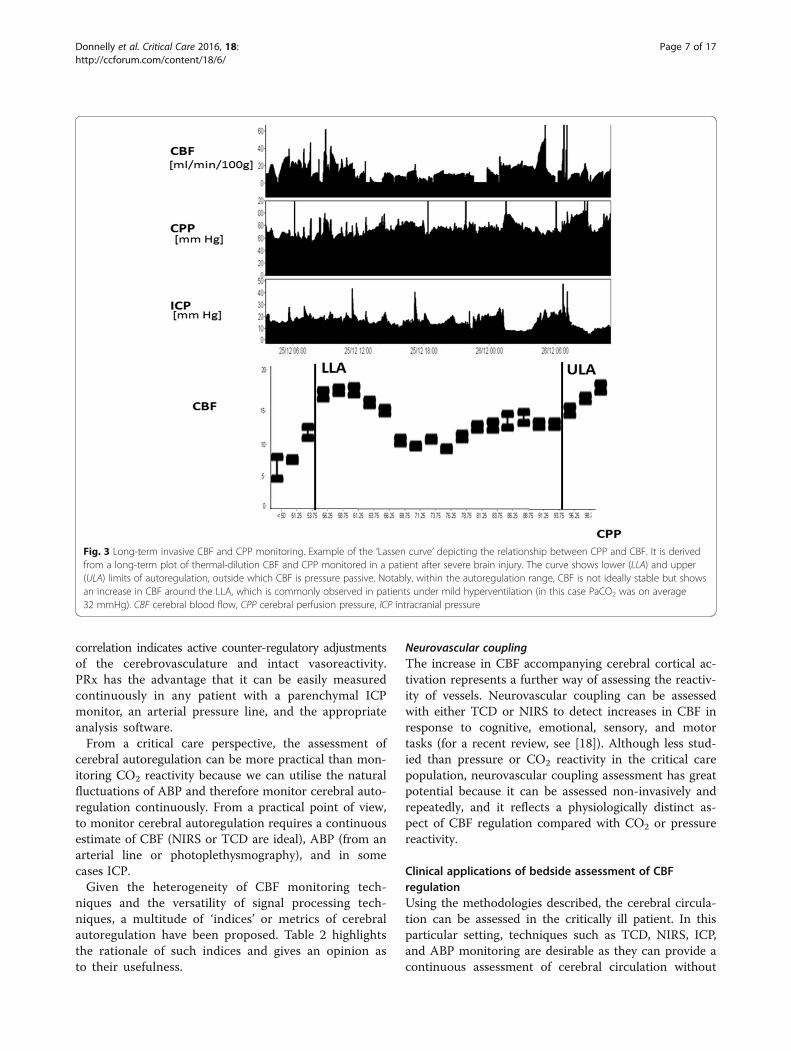
Fig. 3 Long-term invasive CBF and CPP monitoring. Example of the ‘Lassen curve’ depicting the relationship between CPP and CBF. It is derivedfrom a long-term plot of thermal-dilution CBF and CPP monitored in a patient after severe brain injury. The curve shows lower (LLA) and upper(ULA) limits of autoregulation, outside which CBF is pressure passive. Notably, within the autoregulation range, CBF is not ideally stable but showsan increase in CBF around the LLA, which is commonly observed in patients under mild hyperventilation (in this case PaCO2 was on average32 mmHg). CBF cerebral blood flow, CPP cerebral perfusion pressure, ICP intracranial pressure
Donnelly et al. Critical Care 2016, 18: Page 7 of 17http://ccforum.com/content/18/6/
correlation indicates active counter-regulatory adjustmentsof the cerebrovasculature and intact vasoreactivity.PRx has the advantage that it can be easily measuredcontinuously in any patient with a parenchymal ICPmonitor, an arterial pressure line, and the appropriateanalysis software.From a critical care perspective, the assessment of
cerebral autoregulation can be more practical than mon-itoring CO2 reactivity because we can utilise the naturalfluctuations of ABP and therefore monitor cerebral auto-regulation continuously. From a practical point of view,to monitor cerebral autoregulation requires a continuousestimate of CBF (NIRS or TCD are ideal), ABP (from anarterial line or photoplethysmography), and in somecases ICP.Given the heterogeneity of CBF monitoring tech-
niques and the versatility of signal processing tech-niques, a multitude of ‘indices’ or metrics of cerebralautoregulation have been proposed. Table 2 highlightsthe rationale of such indices and gives an opinion asto their usefulness.
Neurovascular couplingThe increase in CBF accompanying cerebral cortical ac-tivation represents a further way of assessing the reactiv-ity of vessels. Neurovascular coupling can be assessedwith either TCD or NIRS to detect increases in CBF inresponse to cognitive, emotional, sensory, and motortasks (for a recent review, see [18]). Although less stud-ied than pressure or CO2 reactivity in the critical carepopulation, neurovascular coupling assessment has greatpotential because it can be assessed non-invasively andrepeatedly, and it reflects a physiologically distinct as-pect of CBF regulation compared with CO2 or pressurereactivity.
Clinical applications of bedside assessment of CBFregulationUsing the methodologies described, the cerebral circula-tion can be assessed in the critically ill patient. In thisparticular setting, techniques such as TCD, NIRS, ICP,and ABP monitoring are desirable as they can provide acontinuous assessment of cerebral circulation without
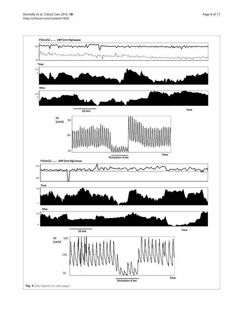
Fig. 4 (See legend on next page.)
Donnelly et al. Critical Care 2016, 18: Page 8 of 17http://ccforum.com/content/18/6/

(See figure on previous page.)Fig. 4 Cerebral perfusion monitoring in SAH. On day 3 after ictus (top 4 panels), this patient with SAH from an aneurysm of the middle cerebralartery displays a normal middle cerebral artery Fv (~60 cm/s) and intact autoregulation (TOxa and Mxa ~0 (suffix ‘a’ indicates that ABP is usedinstead of CPP)). On day 7 (bottom 4 panels) a marked increase in Fv (to 120 cm/s) can be seen, which is accompanied by an impairment inautoregulation (TOxa and Mxa close to 0). The transient hyperaemic response test also failed to show an increase in Fv after the release ofocclusion, an indicator of impaired cerebral autoregulation. ABP arterial blood pressure, Fv flow velocity, Mxa mean flow index (with ABP), TOxatotal oxygenation reactivity index (with ABP)
Table 2 Summary of autoregulation indices
Autoregulation metric Input signals Calculation Interpretation Comment
Autoregulation index (ARI) ABP, Fv Compares the CBF responseto changes in ABP with thosepredicted from a parametricmodel with 10 different‘strengths’ of autoregulation [110]
ARI = 0 absentautoregulation, ARI = 9perfect autoregulation
Moderately complex signalprocessing required
Flow index (Mx, Sx, Dx) ABP (CPP), Fv Pearson correlation betweenCPP and mean Fv (300-swindow of 10-s averages). Sxand Dx calculated with systolicand diastolic flow velocity,respectively
Impaired autoregulation =higher Mx, Dx, and Sx
Simplistic yet prognosticallyrelevant
Transfer function (phase,gain, coherence)
ABP, Fv Derived from the transfer functionof fast Fourier transform of ABPand Fv signals. Phase is the shiftrequired to align Fv and ABPsignals, gain the transmissionfrom ABP to Fv, and coherencethe statistical association betweenABP and Fv
Impaired autoregulation =low phase, high gain, highcoherence
Moderately complex signalprocessing. Some prognosticrelevance
TOx, COx, THx, HVx ABP (CPP), NIRSoxygenation
Pearson correlation between 30consecutive 10-s means of ABPand tissue oxygenation (or totalhaemoglobin for THx and HVx)
Impaired autoregulation =higher TOx, COx, THx, HVx
Correlated with TCD methodsbut allows for longer termmonitoring
TOIHRx HR, NIRS oxygenation Correlation between 30 consecutive10-s means of HR and NIRSoxygenation
?Higher TOIHRx = impairedautoregulation
Used in preterm infants.Further comparisons withstandard autoregulationindices required
Transfer function (phase,gain, coherence)
ABP, NIRS oxygenation Derived from the transfer functionof fast Fourier transform of ABPand oxygenation signals. Phase isthe shift required to alignoxygenation and ABP signals,gain the transmission from ABP toNIRS oxygenation, and coherencethe statistical association betweenABP and NIRS oxygenation
Impaired autoregulation =low phase, high gain, highcoherence
Moderately complex signalprocessing
PRx ABP, ICP Correlation between 30 consecutive10-s means of ABP and ICP
Higher PRx = impairedautoregulation
Robust measure for longmonitoring periods. Simplisticand prognostically relevant
PAx ABP, amplitude of ICP Correlation between 30 consecutive10-s means of ABP and ICP
Higher PAx = impairedautoregulation
Similar to PRx, may allowbetter estimate of pressurereactivity when the“pressure–volume”compensatory curve isflat, i.e. at low ICP
ORx CPP (ABP), PBTO2 Correlation between 30 consecutive10-s means of ABP and PBTO2
High ORx = impairedautoregulation
Further validation required
ABP arterial blood pressure, ARI autoregulatory index, CBF cerebral blood flow, COx cerebral oximetry index, CPP cerebral perfusion pressure, Dx diastolic flowindex, Fv flow velocity, HR heart rate, HVx haemoglobin volume reactivity index, ICP intracranial pressure, Mx mean flow index, ORx oxygen reactivity index,PAx pressure amplitude index, PBTO2 pressure of brain tissue oxygen, PRx pressure reactivity index, Sx systolic flow index, NIRS near-infrared spectroscopy,TCD transcranial Doppler, THx total haemoglobin reactivity index, TOIHRx total oxygenation heart rate index, TOx total oxygenation reactivity index
Donnelly et al. Critical Care 2016, 18: Page 9 of 17http://ccforum.com/content/18/6/
Donnelly et al. Critical Care 2016, 18: Page 10 of 17http://ccforum.com/content/18/6/
the need for transporting the patient. Unfortunately, val-idated ‘normal’ reference ranges are seldom available forthe cerebral circulation and interpretation must there-fore take into account relevant patient comorbidities andthe underlying physiologic milieu. In the following sec-tion we summarise the role of the cerebral circulation inTBI, SAH, stroke, sepsis, and prematurity.
Traumatic brain injuryThe pathophysiology of TBI is classically split intotwo phases, with the primary injury occurring at thetime of ictus and secondary injury occurring in thefollowing minutes, days, or even weeks. A cascade ofpathophysiologic events leads to altered cerebral andsystemic physiology that adds insult to injury; de-rangements in glucose metabolism, thermoregulation,respiration, and cerebral blood circulation all contrib-ute to neuronal injury [43].The characterisation of the cerebral circulation after
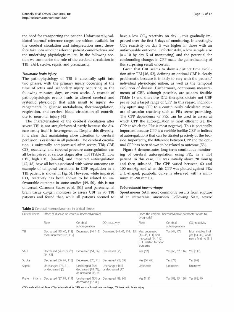
severe TBI is not straightforward partly because the dis-ease entity itself is heterogeneous. Despite this diversity,it is clear that maintaining close attention to cerebralperfusion is essential in all patients. The cerebral circula-tion is universally compromised after severe TBI; CBF,CO2 reactivity, and cerebral pressure autoregulation canall be impaired at various stages after TBI (Table 3). LowCBF, high CBF [44–46], and impaired autoregulation[47, 48] have all been associated with worse outcome (anexample of temporal variations in CBF regulation in aTBI patient is shown in Fig. 5). However, while impairedCO2 reactivity has been shown to be related to un-favourable outcome in some studies [49, 50], this is notuniversal. Carmona Suazo et al. [51] used parenchymalbrain tissue oxygen monitors to assess CBF in 90 TBIpatients and found that, while all patients seemed to
Table 3 Cerebral haemodynamics in critical illness
Critical illness Effect of disease on cerebral haemodynamics
Flow Cerebralautoregulation
CO2 reactivity
TBI Decreased [45, 46, 111]then increased [46, 112]
Decreased [44, 113] Decreased [44
SAH Decreased (vasospasm)[14, 55]
Decreased [54, 56] Decreased [5
Stroke Decreased [66, 67, 118] Decreased [70, 71] Decreased [6
Sepsis Unchanged [78, 81],or decreased [5]
Unchanged [82],decreased [78, 79],or increased [83, 84]
Unchanged [or decreased
Preterm infants Decreased [87, 89, 119] Unchanged [93] ordecreased [87, 88]
Decreased [8
CBF cerebral blood flow, CO2 carbon dioxide, SAH, subarachnoid haemorrhage; TBI,
have a low CO2 reactivity on day 1, this gradually im-proved over the first 5 days of monitoring. Interestingly,CO2 reactivity on day 5 was higher in those with anunfavourable outcome. Unfortunately, a low sample size(n = 10 by day 5 of monitoring) and the potential forconfounding changes in CPP make the generalisability ofthis surprising result uncertain.Given that CBF seems to show a distinct time evolu-
tion after TBI [46, 52], defining an optimal CBF is clearlyproblematic because it is likely to vary with the patients’individual physiologic milieu, as well as the temporalevolution of disease. Furthermore, continuous measure-ments of CBF, although possible, are seldom feasible(Table 1) and therefore ICU therapies dictate not CBFper se but a target range of CPP. In this regard, individu-ally optimising CPP to a continuously calculated meas-ure of vascular reactivity such as PRx seems promising.The CPP dependence of PRx can be used to assess atwhich CPP the autoregulation is most efficient (i.e. theCPP at which the PRx is most negative). This is potentiallyimportant because CPP is a variable (unlike CBF or indicesof autoregulation) that can be titrated precisely at the bed-side. Importantly, the difference between CPP and the opti-mal CPP has been shown to be related to outcome [53].Figure 6 demonstrates long-term continuous monitor-
ing of cerebral autoregulation using PRx in a TBIpatient. In this case, ICP was initially above 20 mmHgand then subsided. The CPP varied between 60 and100 mmHg, and when this CPP was plotted against PRxa U-shaped, parabolic curve is observed with a mini-mum at ~90 mmHg.
Subarachnoid haemorrhageSpontaneous SAH most commonly results from ruptureof an intracranial aneurysm. Following SAH, severe
Does the cerebral haemodynamic parameter relate toprognosis?
Flow Cerebralautoregulation
CO2 reactivity
, 49, 114, 115] Yes: decreased[44–46, 111] andincreased [44, 112]CBF related to pooroutcome
Yes [44, 47] Most studies findyes [44, 49], whilesome find no [51]
5] Yes [62] Yes [60, 62, 116] Yes [117]
8, 69] Yes [66, 67] Yes [71] Yes [69]
82][77]
Unknown Unknown Unknown
8, 90] Yes [119] Yes [88, 95, 120] Yes [88, 90]
traumatic brain injury
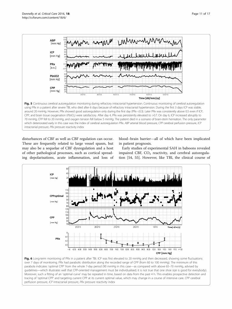
Fig. 5 Continuous cerebral autoregulation monitoring during refractory intracranial hypertension. Continuous monitoring of cerebral autoregulationusing PRx in a patient after severe TBI, who died after 6 days because of refractory intracranial hypertension. During the first 3 days ICP was stable,around 20 mmHg. However, PRx showed good autoregulation only during the first day (PRx <0.3). Later PRx was consistently above 0.5 even if ICP,CPP, and brain tissue oxygenation (PbtiO2) were satisfactory. After day 4, PRx was persistently elevated to >0.7. On day 6, ICP increased abruptly to70 mmHg, CPP fell to 20 mmHg, and oxygen tension fell below 5 mmHg. The patient died in a scenario of brain-stem herniation. The only parameterwhich deteriorated early in this case was the index of cerebral autoregulation PRx. ABP arterial blood pressure, CPP cerebral perfusion pressure, ICPintracranial pressure, PRx pressure reactivity index
Donnelly et al. Critical Care 2016, 18: Page 11 of 17http://ccforum.com/content/18/6/
disturbances of CBF as well as CBF regulation can occur.These are frequently related to large vessel spasm, butmay also be a sequelae of CBF dysregulation and a hostof other pathological processes, such as cortical spread-ing depolarisations, acute inflammation, and loss of
Fig. 6 Long-term monitoring of PRx in a patient after TBI. ICP was first elevover 7 days of monitoring. PRx had parabolic distribution along the recordparabola indicates ‘optimal CPP’ from the whole 7-day period (90 mmHg inguidelines—which illustrates well that CPP-oriented management must beMoreover, such a fitting of an ‘optimal curve’ may be repeated in time, bastracing of ‘optimal CPP’ and targeting current CPP at its current optimal vaperfusion pressure, ICP intracranial pressure, PRx pressure reactivity index
blood–brain barrier—all of which have been implicatedin patient prognosis.Early studies of experimental SAH in baboons revealed
impaired CBF, CO2 reactivity, and cerebral autoregula-tion [54, 55]. However, like TBI, the clinical course of
ated to 20 mmHg and then decreased, showing some fluctuationsed range of CPP (from 60 to 100 mmHg). The minimum of thisthis case—as compared with above 65–70 mmHg, advised byindividualised; it is not true that one shoe size is good for everybody).ed on data from the past 4 h. This enables prospective detection andlue, which may change in a course of intensive care. CPP cerebral
Donnelly et al. Critical Care 2016, 18: Page 12 of 17http://ccforum.com/content/18/6/
SAH is heterogeneous, especially with respect to CBF.Approximately 60 % of SAH cases develop vasospasmon TCD, which may be accompanied by impaired CBFand cerebral autoregulation [14, 56], and 15–30 %develop delayed ischaemic deficits [57–59]. While therelationship between vasospasm, delayed cerebral ischae-mia, and outcome can be capricious, various aspects ofcerebral haemodynamics can be useful in predicting thefuture clinical course: early impaired CO2 reactivity pre-dicts vasospasm, and impaired cerebral autoregulationpredicts delayed ischaemic deficits and poor clinical out-come [60, 61].While CBF is typically within normal limits early after
ictus, it is possible to see impaired cerebral autoregula-tion within the first 3–5 days after SAH [58, 60, 62].Furthermore, Jaeger et al. [60] demonstrated that auto-regulation can recover following the initial deterioration,a response that indicates a good prognosis. Figure 4demonstrates the time course of CBF regulation changesin a patient after SAH.Management strategies hinge on the early identifica-
tion of delayed cerebral ischaemia, followed by the insti-tution of hypertension to maintain CBF. Currently,nimodipine remains the only medication approved forprevention of delayed cerebral ischaemia. In this respect,optimisation of ABP according to cerebral autoregula-tion may be a promising avenue of research [63].
Ischaemic strokeIschaemic stroke is characterised by luminal obstructionby a blood clot. Thus, a region of the brain has abnor-mally high resistance and decreased flow (Fig. 1). Inthese patients, utmost importance is placed on promptdissolution of the clot either by thrombolysis or intravas-cular clot removal [64]. Around the central core ofinfarct is a zone of tissue with depleted, but not absent,blood flow—the ischaemic penumbra. Prompt dissol-ution of the clot can salvage this at-risk tissue.Unlike TBI, or SAH, a predisposition for ischaemic
stroke can be determined by examination of cerebrovas-cular regulation; those patients with impaired CO2 re-activity are more likely to develop an ischaemic stroke[65]. However, like TBI and SAH, ischaemic stroke is astate where careful consideration of cerebrovascularregulation in the acute phase is imperative (Table 3).In the acute phase of ischaemic stroke, those patients
with the lowest global CBF tend to have worse prognosis[66], as do those with a greater proportion of penumbralto ischaemic tissue [67]. CO2 reactivity is depressedcompared with healthy controls [68, 69] and those withlower CO2 reactivity have worse outcome [69]. Cerebralautoregulation also appears to be impaired initially,followed by further impairment over the ensuing severaldays before again improving (reviewed in [70]). In 45
ischaemic stroke patients, cerebral autoregulation im-pairment was related to both the size of infarct andfunctional outcome [71].Ongoing controversy exists regarding how best to sup-
port the cerebral circulation after efforts to break downthe intramural obstruction. While the prevention ofhypotension after ischaemic stroke seems logical, know-ledge of cerebral autoregulation has potential to helpguide the management of blood pressure. Studies ofcontinuous vascular reactivity are limited after ischaemicstroke because these patients are often managed outsidethe critical care environment without the insertion of in-vasive ABP or cerebral perfusion monitors that allow forcontinuous estimation of cerebral autoregulation. In thisregard, non-invasive perfusion assessment with NIRS andABP with finger photoplethysmography are promising.Common to large ischaemic stroke, TBI, and SAH is
the occurrence of spreading cortical depolarisations.These waves of near-complete depolarisation propagateslowly through the cortex (over a time scale of about1 min) and are followed by several minutes of markedlydepressed electrical activity [72, 73]. Their occurrence inan injured brain may decrease CBF, resulting in areas ofischaemia, and seem to lead to worse outcomes [74].Whether they are a cause or a consequence (or both) ofaltered cerebrovascular regulation needs further investi-gation with simultaneous CBF circulation and electro-cortical monitoring.
SepsisThe host response to infection—sepsis—is charac-terised by dysfunction of multiple organ systems, in-cluding the brain. This host response can haveimplications for CBF: CPP is often low, pyrexia canalter CBF, and inflammatory mediators can alter vas-cular resistance [75, 76]. Compared with the afore-mentioned diseases, the cerebral circulation in sepsisis less completely characterised.Some studies have found impaired CO2 reactivity [77],
impaired autoregulation [78–80], and decreased CBF [5]during sepsis, whilst other studies have found no signifi-cant changes in CO2 reactivity, cerebral autoregulation,or CBF [81, 82]. Interestingly, two groups have evenfound that, in the early phases of experimental sepsis inhealthy volunteers, dynamic cerebral autoregulation isactually enhanced [83, 84]. Pfister et al. [78] found thatautoregulation was impaired in those with sepsis and de-lirium, but not in those with sepsis only. These seem-ingly conflicting findings may be partially explained bythe heterogeneity of the sepsis process itself. Some septicpatients develop a hyperdynamic circulation with in-creased cardiac output and decreased ABP, while othershave both decreased cardiac output and ABP. Moreover,the physiological changes in the cerebral circulation
Donnelly et al. Critical Care 2016, 18: Page 13 of 17http://ccforum.com/content/18/6/
during sepsis probably evolve over time, thus makingcomparisons between different studies difficult.Nevertheless, brain dysfunction is one of the earliest
forms of organ dysfunction in sepsis and sepsis-induceddelirium occurs in up to 70 % of patients [76]. Charac-terising the involvement of the cerebral circulation inthe pathogenesis of sepsis-induced delirium will prob-ably require detailed haemodynamic studies with largenumbers of patients.
Preterm infantsPremature infants do not have fully functioning cerebralvessels or cardiovascular systems and therefore vitalorgan perfusion is vulnerable. Using NIRS and umbilicalartery ABP, continuous measures of cerebral autoregula-tion can be obtained.Animal studies indicate that cerebral autoregulation
starts to develop from around halfway through the ges-tational period [85]. Furthermore, even when static auto-regulation is developed, the preterm newborn probablysits close to the lower limit of autoregulation [86]. Earlyhuman investigations using Xe CT and NIRS indicatedthat CBF, CO2 reactivity, and cerebral autoregulationmay be impaired in preterm infants [87–91] (Table 3).Further, more recent human data using TCD indicatedthat cerebral autoregulation is more impaired if the babyis more premature [92]. Still other studies have indicatedthat perhaps the premature brain is able to adapt to sus-tained [93] but not dynamic [94] changes in ABP; thatis, ‘static’ autoregulation is intact, while ‘dynamic’ auto-regulation may be impaired [91].Analogous to TBI, determination of an optimal
ABP has been attempted in these preterm infantswith the finding that those who did not survive hadan ABP below their calculated optimal, whereasthose who developed periventricular haemorrhagehad an ABP above their optimal [95]. An importantconsideration when interpreting studies on cerebralhaemodynamics in infants is that, in addition to theinfluences of ABP and CO2 on CBF, arterial oxygensaturation can be highly variable, and can have pro-found influence on premature babies’ cerebral circu-lation [96].
Future directionsWith the increasing availability of bedside physiologymonitors and sophisticated online analysis software,large-scale integrated interrogations of CBF regulationare now possible. One important research theme isdeveloping robust prediction tools based on cerebralphysiologic monitoring for critically ill patients.Accurate prognosis is of obvious importance forpatients, families, and clinicians alike, but currentmethodologies have some limitations. For example,
prognostic tools in TBI use clinical, laboratory, andradiographic features on admission to predict patientoutcome [97]. However, some of the input variablesare open to interpretation (e.g. the grading of a CTscan), and prognosis should ideally be updated basedon clinical and physiological developments. In thissense, prognostic tools that update risk estimatesbased on online monitoring of CBF regulation couldfacilitate clinical decision-making.In addition to predicting outcome, incorporating
knowledge of CBF regulation into management proto-cols seems promising. Hopeful examples in TBI includestrategies that incorporate knowledge of cerebrovascularreactivity into either ICP [98] or CPP [53] management.Although still requiring further development and pro-spective assessment, similar techniques could conceiv-ably be applied to any condition where ABP or CBFregulation is impaired.Extending cerebral haemodynamic monitoring con-
cepts to other critical care pathologies is important.For example, in cardiopulmonary bypass patients,NIRS-based autoregulation has been shown to be asignificant predictor of outcome, and furthermore,as in TBI, an autoregulation-based optimal ABPseems to be prognostically important [99, 100]. Anexample of autoregulation-based optimal ABP dur-ing cardiopulmonary bypass is shown in Fig. 7. Car-diac arrest, acute shunt blockage, acute liver failure,pre-eclampsia, and malignant hypertension are allconditions that could perturb the cerebral circula-tion, and further investigation may reveal diagnostic,prognostic, or therapeutic insight.Understanding the factors that modify CBF and
vascular reactivity is also an important evolving areaof research. Although a large part of the variation incerebral autoregulation is accounted for by the levelof ABP (or CPP) itself, other systemic and localfactors may also be important. Preliminary investiga-tions indicate that common occurrences in the crit-ical care unit such as hyperglycaemia, altered renalclearance, erythrocyte transfusion, or rewarming afterhypothermia are all associated with altered cerebralpressure reactivity, underscoring the need for an in-tegrative approach to neuromonitoring [101–104].Finally, investigating and integrating additional aspects
of CBF regulation into prognostic and therapeuticapproaches is imperative. In particular, the compu-terised assessment of neurovascular coupling [18]and autonomic function (e.g. with baroreceptor sen-sitivity or heart rate variability) are non-invasive,provide unique information on the regulation ofCBF, and can be coupled with conventional measuresof CBF regulation such as cerebral autoregulationand cerebrovascular CO2 reactivity.
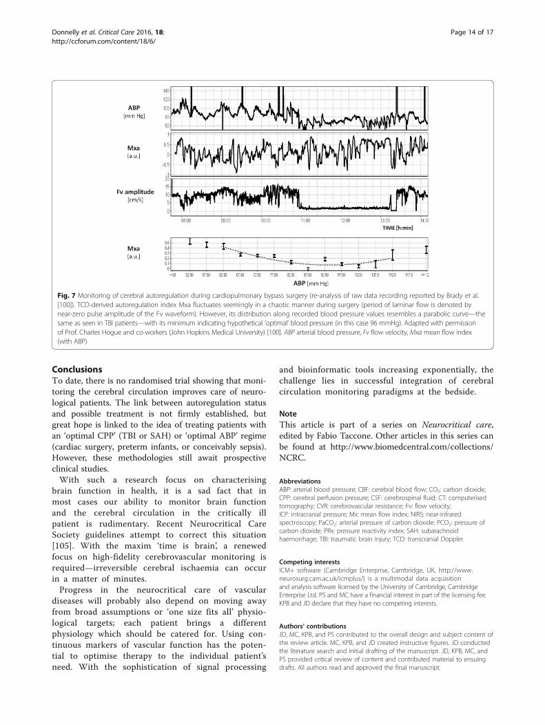
Fig. 7 Monitoring of cerebral autoregulation during cardiopulmonary bypass surgery (re-analysis of raw data recording reported by Brady et al.[100]). TCD-derived autoregulation index Mxa fluctuates seemingly in a chaotic manner during surgery (period of laminar flow is denoted bynear-zero pulse amplitude of the Fv waveform). However, its distribution along recorded blood pressure values resembles a parabolic curve—thesame as seen in TBI patients—with its minimum indicating hypothetical ‘optimal’ blood pressure (in this case 96 mmHg). Adapted with permissionof Prof. Charles Hogue and co-workers (John Hopkins Medical University) [100]. ABP arterial blood pressure, Fv flow velocity, Mxa mean flow index(with ABP)
Donnelly et al. Critical Care 2016, 18: Page 14 of 17http://ccforum.com/content/18/6/
ConclusionsTo date, there is no randomised trial showing that moni-toring the cerebral circulation improves care of neuro-logical patients. The link between autoregulation statusand possible treatment is not firmly established, butgreat hope is linked to the idea of treating patients withan ‘optimal CPP’ (TBI or SAH) or ‘optimal ABP’ regime(cardiac surgery, preterm infants, or conceivably sepsis).However, these methodologies still await prospectiveclinical studies.With such a research focus on characterising
brain function in health, it is a sad fact that inmost cases our ability to monitor brain functionand the cerebral circulation in the critically illpatient is rudimentary. Recent Neurocritical CareSociety guidelines attempt to correct this situation[105]. With the maxim ‘time is brain’, a renewedfocus on high-fidelity cerebrovascular monitoring isrequired—irreversible cerebral ischaemia can occurin a matter of minutes.Progress in the neurocritical care of vascular
diseases will probably also depend on moving awayfrom broad assumptions or ‘one size fits all’ physio-logical targets; each patient brings a differentphysiology which should be catered for. Using con-tinuous markers of vascular function has the poten-tial to optimise therapy to the individual patient’sneed. With the sophistication of signal processing
and bioinformatic tools increasing exponentially, thechallenge lies in successful integration of cerebralcirculation monitoring paradigms at the bedside.
NoteThis article is part of a series on Neurocritical care,edited by Fabio Taccone. Other articles in this series canbe found at http://www.biomedcentral.com/collections/NCRC.
AbbreviationsABP: arterial blood pressure; CBF: cerebral blood flow; CO2: carbon dioxide;CPP: cerebral perfusion pressure; CSF: cerebrospinal fluid; CT: computerisedtomography; CVR: cerebrovascular resistance; Fv: flow velocity;ICP: intracranial pressure; Mx: mean flow index; NIRS: near-infraredspectroscopy; PaCO2: arterial pressure of carbon dioxide; PCO2: pressure ofcarbon dioxide; PRx: pressure reactivity index; SAH: subarachnoidhaemorrhage; TBI: traumatic brain injury; TCD: transcranial Doppler.
Competing interestsICM+ software (Cambridge Enterprise, Cambridge, UK, http://www.neurosurg.cam.ac.uk/icmplus/) is a multimodal data acquisitionand analysis software licensed by the University of Cambridge, CambridgeEnterprise Ltd. PS and MC have a financial interest in part of the licensing fee.KPB and JD declare that they have no competing interests.
Authors’ contributionsJD, MC, KPB, and PS contributed to the overall design and subject content ofthe review article. MC, KPB, and JD created instructive figures. JD conductedthe literature search and initial drafting of the manuscript. JD, KPB, MC, andPS provided critical review of content and contributed material to ensuingdrafts. All authors read and approved the final manuscript.

Donnelly et al. Critical Care 2016, 18: Page 15 of 17http://ccforum.com/content/18/6/
AcknowledgementsJD is supported by a Woolf Fisher scholarship (New Zealand). MC is partiallysupported by the National Institute for Health Research.
References1. Ursino M, Lodi CA. A simple mathematical model of the interaction
between intracranial pressure and cerebral hemodynamics. J Appl Physiol.1997;82:1256–69.
2. Czosnyka M, Piechnik S, Richards HK, Kirkpatrick P, Smielewski P, Pickard JD.Contribution of mathematical modelling to the interpretation of bedsidetests of cerebrovascular autoregulation. J Neurol Neurosurg Psychiatry.1997;63:721–31.
3. Roy CS, Sherrington CS. On the regulation of the blood-supply of the brain.J Physiol. 1890;11:85–158.
4. Cavus E, Bein B, Dörges V, Stadlbauer K-H, Wenzel V, Steinfath M, Hanss R,Scholz J. Brain tissue oxygen pressure and cerebral metabolism in an animalmodel of cardiac arrest and cardiopulmonary resuscitation. Resuscitation.2006;71:97–106.
5. Bowton DL, Bertels NH, Prough DS, Stump DA. Cerebral blood flow isreduced in patients with sepsis syndrome. Crit Care Med. 1989;17:399–403.
6. Nakagawa Y, Tsuru M, Yada K. Site and mechanism for compression of thevenous system during experimental intracranial hypertension. J Neurosurg.1974;41:427–34.
7. Piechnik SK, Czosnyka M, Richards HK, Whitfield PC, Pickard JD. Cerebralvenous blood outflow: a theoretical model based on laboratory simulation.Neurosurgery. 2001;49:1214–22. discussion 1222–3.
8. Attwell D, Buchan AM, Charpak S, Lauritzen M, Macvicar BA, Newman EA.Glial and neuronal control of brain blood flow. Nature. 2010;468:232–43.
9. Hall CN, Reynell C, Gesslein B, Hamilton NB, Mishra A, Sutherland BA,O’Farrell FM, Buchan AM, Lauritzen M, Attwell D. Capillary pericytes regulatecerebral blood flow in health and disease. Nature. 2014;508:55–60.
10. Willie CK, Tzeng Y-C, Fisher JA, Ainslie PN. Integrative regulation of humanbrain blood flow. J Physiol. 2014;592:841–59.
11. Schaller B. Physiology of cerebral venous blood flow: from experimentaldata in animals to normal function in humans. Brain Res Rev.2004;46:243–60.
12. Lee SP, Duong TQ, Yang G, Iadecola C, Kim SG. Relative changes of cerebralarterial and venous blood volumes during increased cerebral blood flow:implications for bold fMRI. Magn Reson Med. 2001;45:791–800.
13. Fox PT, Raichle ME. Focal physiological uncoupling of cerebral blood flowand oxidative metabolism during somatosensory stimulation in humansubjects. Proc Natl Acad Sci U S A. 1986;83:1140–4.
14. Vajkoczy P, Horn P, Thome C, Munch E, Schmiedek P. Regional cerebralblood flow monitoring in the diagnosis of delayed ischemia followinganeurysmal subarachnoid hemorrhage. J Neurosurg. 2003;98:1227–34.
15. Ainslie PN, Duffin J. Integration of cerebrovascular CO2 reactivity andchemoreflex control of breathing: mechanisms of regulation,measurement, and interpretation. Am J Physiol Regul Integr CompPhysiol. 2009;296:R1473–95.
16. Somers VK, Mark AL, Zavala DC, Abboud FM. Contrasting effects of hypoxiaand hypercapnia on ventilation and sympathetic activity in humans. J ApplPhysiol. 1989;67:2101–6.
17. Zappe A, Uludaǧ K, Oeltermann A, Uǧurbil K, Logothetis N. The influence ofmoderate hypercapnia on neural activity in the anesthetized nonhumanprimate. Cereb Cortex. 2008;18:2666–73.
18. Phillips AA, Chan FHN, Mu M, Zheng Z, Krassioukov AV, Ainslie PN.Neurovascular coupling in humans: physiology, methodologicaladvances and clinical implications. J Cereb Blood Flow Metab. 2015.[Epub ahead of print].
19. Peterson EC, Wang Z, Britz G. Regulation of cerebral blood flow. Int J VascMed. 2011;2011:1–8.
20. Jackman K, Iadecola C. Neurovascular regulation in the ischemic brain.Antioxid Redox Signal. 2015;22:149–60.
21. Iadecola C, Nedergaard M. Glial regulation of the cerebral microvasculature.Nat Neurosci. 2007;10:1369–76.
22. Girouard H. Neurovascular coupling in the normal brain and inhypertension, stroke, and Alzheimer disease. J Appl Physiol.2006;100:328–35.
23. Strandgaard S, Sigurdsson ST. Point:Counterpoint: sympathetic activitydoes/does not influence cerebral blood flow. Counterpoint: sympatheticnerve activity does not influence cerebral blood flow. J Appl Physiol.2008;105:1366–7. discussion 1367–8.
24. van Lieshout JJ, Secher NH. Point:Counterpoint: sympathetic nerve activitydoes/does not influence cerebral blood flow. Point: sympathetic nerveactivity does influence cerebral blood flow. J Appl Physiol. 2008;105:1364–6.
25. Ainslie PN, Brassard P. Why is the neural control of cerebral autoregulationso controversial? F1000 Prime Rep. 2014;6:14.
26. Visocchi M, Chiappini F, Cioni B, Meglio M. Cerebral blood flow velocitiesand trigeminal ganglion stimulation. A transcranial Doppler study.Stereotact Funct Neurosurg. 1996;66:184–92.
27. Umeyama T, Kugimiya T, Ogawa T, Kandori Y, Ishizuka A, Hanaoka K. Changesin cerebral blood flow estimated after stellate ganglion block by single photonemission computed tomography. J Aut Nerv Syst. 1995;50:339–46.
28. Meng L, Hou W, Chui J, Han R, Gelb AW. Cardiac output and cerebral bloodflow: the integrated regulation of brain perfusion in adult humans.Anesthesiology. 2015;123:1198–208.
29. Ogoh S, Brothers RM, Barnes Q, Eubank WL, Hawkins MN, Purkayastha S,O-Yurvati A, Raven PB. The effect of changes in cardiac output on middlecerebral artery mean blood velocity at rest and during exercise. J Physiol.2005;569(Pt 2):697–704.
30. Lanfranchi PA, Somers VK. Arterial baroreflex function and cardiovascularvariability: interactions and implications. Am J Physiol Regul Integr CompPhysiol. 2002;283:R815–26.
31. Lassen N. Cerebral blood flow and oxygen consumption in man. PhysiolRev. 1959;39:183–238.
32. Donnelly J, Aries MJH, Czosnyka M. Further understanding of cerebralautoregulation at the bedside: possible implications for future therapy.Expert Rev Neurother. 2015;15:169–85.
33. Willie CK, Colino FL, Bailey DM, Tzeng YC, Binsted G, Jones LW, HaykowskyMJ, Bellapart J, Ogoh S, Smith KJ, Smirl JD, Day TA, Lucas SJ, Eller LK, AinsliePN. Utility of transcranial Doppler ultrasound for the integrative assessmentof cerebrovascular function. J Neurosci Methods. 2011;196:221–37.
34. Davies DJ, Su Z, Clancy MT, Lucas SJE, Dehghani H, Logan A, Belli A.Near-infrared spectroscopy in the monitoring of adult traumatic brain injury:a review. J Neurotrauma. 2015;32:933–41.
35. Vajkoczy P, Roth H, Horn P, Lucke T, Thomé C, Hubner U, Martin GT,Zappletal C, Klar E, Schilling L, Schmiedek P. Continuous monitoring ofregional cerebral blood flow: experimental and clinical validation of a novelthermal diffusion microprobe. J Neurosurg. 2000;93:265–74.
36. Rajan V, Varghese B, Van Leeuwen TG, Steenbergen W. Review ofmethodological developments in laser Doppler flowmetry. Lasers Med Sci.2009;24:269–83.
37. Rohlwink UK, Figaji AA. Methods of monitoring brain oxygenation. Child’sNerv Syst. 2010;26:453–64.
38. Rostami E, Engquist H, Enblad P. Imaging of cerebral blood flow in patientswith severe traumatic brain injury in the neurointensive care. Front Neurol.2014;5:1–9.
39. Zhang R, Zuckerman JH, Giller CA, Levine BD. Transfer function analysisof dynamic cerebral autoregulation in humans. Am J Physiol.1998;274:H233–41.
40. Czosnyka M, Smielewski P, Kirkpatrick P, Menon DK, Pickard JD. Monitoringof cerebral autoregulation in head-injured patients. Stroke. 1996;27:1829–34.
41. Panerai RB. Cerebral autoregulation: from models to clinical applications.Cardiovasc Eng. 2008;8:42–59.
42. Smielewski P, Czosnyka M, Kirkpatrick P, Pickard JD. Evaluation of thetransient hyperemic response test in head-injured patients. J Neurosurg.1997;86:773–8.
43. Menon DK. Cerebral protection in severe brain injury: physiologicaldeterminants of outcome and their optimisation. Br Med Bull. 1999;55:226–58.
44. Overgaard J, Tweed W. Cerebral circulation after head injury. 1. Cerebralblood flow and its regulation after closed head injury with emphasis onclinical correlations. J Neurosurg. 1974;41:531–41.
45. Bouma GJ, Muizelaar JP, Stringer WA, Choi SC, Fatouros P, Young HF. Ultra-earlyevaluation of regional cerebral blood flow in severely head-injured patientsusing xenon-enhanced computerized tomography. J Neurosurg. 1992;77:360–8.
46. van Santbrink H, Schouten JW, Steyerberg EW, Avezaat CJJ, Maas AI.Serial transcranial Doppler measurements in traumatic brain injury withspecial focus on the early posttraumatic period. Acta Neurochir (Wien).2002;144:1141–9.

Donnelly et al. Critical Care 2016, 18: Page 16 of 17http://ccforum.com/content/18/6/
47. Liu X, Czosnyka M, Donnelly J, Budohoski KP, Varsos GV, Nasr N, Brady KM,Reinhard M, Hutchinson PJ, Smielewski P. Comparison of frequency andtime domain methods of assessment of cerebral autoregulation intraumatic brain injury. J Cereb Blood Flow Metab. 2014;11:1–9.
48. Czosnyka M, Smielewski P, Kirkpatrick P, Laing RJ, Menon D, Pickard JD.Continuous assessment of the cerebral vasomotor reactivity in head injury.Neurosurgery. 1997;41:11–7. discussion 17–9.
49. Poon W, Ng SCP, Chan MTV, Lam JMK, Lam WWM. Cerebral blood flow(CBF)-directed management of ventilated head-injured patients. ActaNeurochir Suppl. 2005;95:9–11.
50. Schalen W, Messeter K, Nordstrom CH. Cerebral vasoreactivity and theprediction of outcome in severe traumatic brain lesions. Acta AnaesthesiolScand. 1991;35:113–22.
51. Carmona Suazo JA, Maas AI, van den Brink WA, van Santbrink H, SteyerbergEW, Avezaat CJ. CO2 reactivity and brain oxygen pressure monitoring insevere head injury. Crit Care Med. 2000;28:3268–74.
52. Martin NA, Patwardhan RV, Alexander MJ, Africk CZ, Lee JH, Shalmon E,Hovda DA, Becker DP. Characterization of cerebral hemodynamic phasesfollowing severe head trauma: hypoperfusion, hyperemia, and vasospasm.J Neurosurg. 1997;87:9–19.
53. Aries MJH, Czosnyka M, Budohoski KP, Steiner LA, Lavinio A, Kolias AG,Hutchinson PJ, Brady KM, Menon DK, Pickard JD, Smielewski P. Continuousdetermination of optimal cerebral perfusion pressure in traumatic braininjury. Crit Care Med. 2012;40:2456–63.
54. Hashi K, Meyer JS, Shinmaru S, Welch KM, Teraura T. Changes in cerebralvasomotor reactivity to CO2 and autoregulation following experimentalsubarachnoid hemorrhage. J Neurol Sci. 1972;17:15–22.
55. Mendelow AD, McCalden TA, Hattingh J, Coull A, Rosendorff C, EidelmanBH. Cerebrovascular reactivity and metabolism after subarachnoidhemorrhage in baboons. Stroke. 1981;12:58–65.
56. Soehle M, Czosnyka M, Pickard JD, Kirkpatrick PJ. Continuous assessment ofcerebral autoregulation in subarachnoid hemorrhage. Anesth Analg.2004;98:1133–9.
57. Pickard JD, Murray GD, Illingworth R, Shaw MDM, Teasdale GM, Foy PM,Humphrey PRD, Lang DA, Nelson R, Richards P, Sinar J, Bailey S, Skene A. Effectof oral nimodipine on cerebral infarction and outcome after subarachnoidhaemorrhage: British aneurysm nimodipine trial. BMJ. 1989;298:636–42.
58. Budohoski K, Czosnyka M, Smielewski P, Kasprowicz M, Helmy A, Bulters D,Pickard JD, Kirkpatrick PJ. Impairment of cerebral autoregulation predictsdelayed cerebral ischemia after subarachnoid hemorrhage: a prospectiveobservational study. Stroke. 2012;43:3230–7.
59. Kirkpatrick PJ, Turner CL, Smith C, Hutchinson PJ, Murray GD. Simvastatin inaneurysmal subarachnoid haemorrhage (STASH): a multicentre randomisedphase 3 trial. Lancet Neurol. 2014;13:666–75.
60. Jaeger M, Schuhmann MU, Soehle M, Nagel C, Meixensberger J. Continuousmonitoring of cerebrovascular autoregulation after subarachnoidhemorrhage by brain tissue oxygen pressure reactivity and its relation todelayed cerebral infarction. Stroke. 2007;38:981–6.
61. Pickard JD, Matheson M, Patterson J, Wyper D. Prediction of late ischemiccomplications after cerebral aneurysm surgery by the intraoperativemeasurement of cerebral blood flow. J Neurosurg. 1980;53:305–8.
62. Jaeger M, Soehle M, Schuhmann MU, Meixensberger J. Clinical significanceof impaired cerebrovascular autoregulation after severe aneurysmalsubarachnoid hemorrhage. Stroke. 2012;43:2097–101.
63. Bijlenga P, Czosnyka M, Budohoski KP, Smielewski P, Soehle M, Pickard JD,Kirkpatrick PJ. “Optimal cerebral perfusion pressure” in poor grade patientsafter subarachnoid hemorrhage. Neurocrit Care. 2010;13:17–23.
64. Jauch EC, Saver JL, Adams HP, Bruno A, Connors JJB, Demaerschalk BM,Khatri P, McMullan PW, Qureshi AI, Rosenfield K, Scott PA, Summers DR,Wang DZ, Wintermark M, Yonas H. Guidelines for the early management ofpatients with acute ischemic stroke: a guideline for healthcare professionalsfrom the American Heart Association/American Stroke Association. Stroke.2013;44:870–947.
65. Markus H, Cullinane M. Severely impaired cerebrovascular reactivity predictsstroke and TIA risk in patients with carotid artery stenosis and occlusion.Brain. 2001;124(Pt 3):457–67.
66. Firlik AD, Rubin G, Yonas H, Wechsler LR. Relation between cerebral bloodflow and neurologic deficit resolution in acute ischemic stroke. Neurology.1998;51:177–82.
67. Wintermark M, Reichhart M, Thiran JP, Maeder P, Chalaron M, Schnyder P,Bogousslavsky J, Meuli R. Prognostic accuracy of cerebral blood flow
measurement by perfusion computed tomography, at the time ofemergency room admission, in acute stroke patients. Ann Neurol.2002;51:417–32.
68. Cupini LM, Diomedi M, Placidi F, Silvestrini M, Giacomini P. Cerebrovascularreactivity and subcortical infarctions. Arch Neurol. 2001;58:577–81.
69. Alvarez FJ, Segura T, Castellanos M, Leira R, Blanco M, Castillo J, Davalos A,Serena J. Cerebral hemodynamic reserve and early neurologic deteriorationin acute ischemic stroke. J Cereb Blood Flow Metab. 2004;24:1267–71.
70. Aries MJH, Elting JW, De Keyser J, Kremer BPH, Vroomen PCAJ. Cerebralautoregulation in stroke a review of transcranial Doppler studies. Stroke.2010;41:2697–704.
71. Reinhard M, Rutsch S, Lambeck J, Wihler C, Czosnyka M, Weiller C, Hetzel A.Dynamic cerebral autoregulation associates with infarct size and outcomeafter ischemic stroke. Acta Neurol Scand. 2012;125:156–62.
72. Pietrobon D, Moskowitz MA. Chaos and commotion in the wake of corticalspreading depression and spreading depolarizations. Nat Rev Neurosci.2014;15:379–93.
73. Woitzik J, Hecht N, Pinczolits A, Sandow N, Major S, Winkler MKL, Weber-Carstens S, Dohmen C, Graf R, Strong AJ, Dreier JP, Vajkoczy P. Propagationof cortical spreading depolarization in the human cortex after malignantstroke. Neurology. 2013;80:1095–102.
74. Hartings JA, Bullock MR, Okonkwo DO, Murray LS, Murray GD, Fabricius M,Maas AI, Woitzik J, Sakowitz O, Mathern B, Roozenbeek B, Lingsma H, DreierJP, Puccio AM, Shutter LA, Pahl C, Strong AJ. Spreading depolarisations andoutcome after traumatic brain injury: a prospective observational study.Lancet Neurol. 2011;10:1058–64.
75. Bain AR, Nybo L, Ainslie PN. Cerebral vascular control and metabolism inheat stress. Compr Physiol. 2015;5:1345–80.
76. Burkhart CS, Siegemund M, Steiner LA. Cerebral perfusion in sepsis. CritCare. 2010;14:215.
77. Terborg C, Schummer W, Albrecht M, Reinhart K, Weiller C, Röther J.Dysfunction of vasomotor reactivity in severe sepsis and septic shock.Intensive Care Med. 2001;27:1231–4.
78. Pfister D, Siegemund M, Dell-Kuster S, Smielewski P, Rüegg S, StrebelSP, Marsch SCU, Pargger H, Steiner LA. Cerebral perfusion in sepsis-associated delirium. Crit Care. 2008;12:R63.
79. Taccone FS, Castanares-Zapatero D, Peres-Bota D, Vincent J-L, Berre’ J, MelotC. Cerebral autoregulation is influenced by carbon dioxide levels in patientswith septic shock. Neurocrit Care. 2010;12:35–42.
80. Berg RMG, Plovsing RR, Bailey DM, Holstein-Rathlou N-H, Møller K. Thedynamic cerebral autoregulatory adaptive response to noradrenaline isattenuated during systemic inflammation in humans. Clin Exp PharmacolPhysiol. 2015;42:740–6.
81. Di Giantomasso D, May CN, Bellomo R. Laboratory and animalinvestigations: vital organ blood flow during hyperdynamic sepsis. Chest.2003;124:1053–9.
82. Matta BF, Stow PJ. Sepsis-induced vasoparalysis does not involve thecerebral vasculature: indirect evidence from autoregulation and carbondioxide reactivity studies. Br J Anaesth. 1996;76:790–4.
83. Brassard P, Kim Y-S, van Lieshout J, Secher NH, Rosenmeier JB. Endotoxemiareduces cerebral perfusion but enhances dynamic cerebrovascular autoregulationat reduced arterial carbon dioxide tension. Crit Care Med. 2012;40:1873–8.
84. Berg RM, Plovsing RR, Evans KA, Christiansen CB, Bailey DM, Holstein-Rathlou N-H, Møller K. Lipopolysaccharide infusion enhances dynamiccerebral autoregulation without affecting cerebral oxygen vasoreactivity inhealthy volunteers. Crit Care. 2013;17:R238.
85. Helou S, Koehler RC, Gleason CA, Jones MD, Traystman RJ.Cerebrovascular autoregulation during fetal development in sheep.Am J Physiol. 1994;266(3 Pt 2):H1069–74.
86. Muller T, Lohle M, Schubert H, Bauer R, Wicher C, Antonow-Schlorke I,Sliwka U, Nathanielsz PW, Schwab M. Developmental changes incerebral autoregulatory capacity in the fetal sheep parietal cortex.J Physiol. 2002;539(Pt 3):957–67.
87. Lou HC, Lassen NA, Friis-Hansen B. Impaired autoregulation of cerebralblood flow in the distressed newborn infant. J Pediatr. 1979;94:118–21.
88. Pryds O, Greisen G, Lou HC, Friis-Hansen B. Vasoparalysis associatedwith brain damage in asphyxiated term infants. J Pediatr.1990;117(1 Pt 1):119–25.
89. Edwards AD, Wyatt JS, Richardson C, Delpy DT, Cope M, Reynolds EO.Cotside measurement of cerebral blood flow in ill newborn infants by nearinfrared spectroscopy. Lancet. 1988;2:770–1.

Donnelly et al. Critical Care 2016, 18: Page 17 of 17http://ccforum.com/content/18/6/
90. Müller AM, Morales C, Briner J, Baenziger O, Duc G, Bucher HU. Loss of CO2reactivity of cerebral blood flow is associated with severe brain damage inmechanically ventilated very low birth weight infants. Eur J Paediatr Neurol.1997;1:157–63.
91. Greisen G. Autoregulation of cerebral blood flow in newborn babies. EarlyHum Dev. 2005;81:423–8.
92. Rhee CJ, Fraser III CD, Kibler K, Easley RB, Andropoulos DB, Czosnyka M,Varsos GV, Smielewski P, Rusin CG, Brady KM, Kaiser JR. The ontogeny ofcerebrovascular pressure autoregulation in premature infants. J Perinatol.2014;34:926–31.
93. Tyszczuk L, Meek J, Elwell C, Wyatt JS. Cerebral blood flow is independentof mean arterial blood pressure in preterm infants undergoing intensivecare. Pediatrics. 1998;102(2 Pt 1):337–41.
94. Boylan G, Young K, Panerai R, Rennie J, Evans D. Dynamic cerebralautoregulation in sick newborn infants. Pediatr Res. 2000;48:1–8.
95. da Costa CS, Czosnyka M, Smielewski P, Mitra S, Stevenson GN, Austin T.Monitoring of cerebrovascular reactivity for determination of optimal bloodpressure in preterm infants. J Pediatr. 2015;167:86–91.
96. Tweed A, Cote J, Lou HC, Gregory G, Wade J. Impairment of cerebral blood flowautoregulation in the newborn lamb by hypoxia. Pediatr Res. 1986;20:516–9.
97. Steyerberg EW, Mushkudiani N, Perel P, Butcher I, Lu J, McHugh GS,Murray GD, Marmarou A, Roberts I, Habbema JDF, Maas AIR. Predictingoutcome after traumatic brain injury: development and internationalvalidation of prognostic scores based on admission characteristics. PLoSMed. 2008;5:e165. discussion e165.
98. Lazaridis C, DeSantis SM, Smielewski P, Menon DK, Hutchinson P, Pickard JD,Czosnyka M. Patient-specific thresholds of intracranial pressure in severetraumatic brain injury. J Neurosurg. 2014;120:893–900.
99. Ono M, Arnaoutakis GJ, Fine DM, Brady K, Easley RB, Zheng Y, Brown C,Katz NM, Grams ME, Hogue CW. Blood pressure excursions below thecerebral autoregulation threshold during cardiac surgery are associatedwith acute kidney injury. Crit Care Med. 2013;41:464–71.
100. Brady K, Joshi B, Zweifel C, Smielewski P, Czosnyka M, Easley RB, Hogue CW.Real-time continuous monitoring of cerebral blood flow autoregulationusing near-infrared spectroscopy in patients undergoing cardiopulmonarybypass. Stroke. 2010;41:1951–6.
101. Donnelly J, Czosnyka M, Sudhan N, Varsos GV, Nasr N, Jalloh I, Liu X, Dias C,Sekhon MS, Carpenter KLH, Menon DK, Hutchinson PJ, Smielewski P.Increased blood glucose is related to disturbed cerebrovascular pressurereactivity after traumatic brain injury. Neurocrit Care. 2014;22:20–5.
102. Dias C, Gaio AR, Monteiro E, Barbosa S, Cerejo A, Donnelly J, Felgueiras Ó,Smielewski P, Paiva J-A, Czosnyka M. Kidney-brain link in traumatic braininjury patients? A preliminary report. Neurocrit Care. 2015;22:192–201.
103. Lavinio A, Timofeev I, Nortje J, Outtrim J, Smielewski P, Gupta A, HutchinsonPJ, Matta BF, Pickard JD, Menon D, Czosnyka M. Cerebrovascular reactivityduring hypothermia and rewarming. Br J Anaesth. 2007;99:237–44.
104. Sekhon MS, Griesdale DE, Czosnyka M, Donnelly J, Liu X, Aries MJ, Robba C,Lavinio A, Menon DK, Smielewski P, Gupta AK. The effect of red blood celltransfusion on cerebral autoregulation in patients with severe traumaticbrain injury. Neurocrit Care. 2015;23:210–6.
105. Le Roux P, Menon DK, Citerio G, Vespa P, Bader MK, Brophy GM, DiringerMN, Stocchetti N, Videtta W, Armonda R, Badjatia N, Böesel J, Chesnut R,Chou S, Claassen J, Czosnyka M, De Georgia M, Figaji A, Fugate J, Helbok R,Horowitz D, Hutchinson P, Kumar M, McNett M, Miller C, Naidech A, Oddo M,Olson D, O’Phelan K, Provencio JJ, et al. Consensus Summary Statement of theInternational Multidisciplinary Consensus Conference on MultimodalityMonitoring in Neurocritical Care. Neurocrit Care 2014;21:1189–1209.
106. Thomas KN, Lewis NCS, Hill BG, Ainslie PN. Technical recommendations for theuse of carotid duplex ultrasound for the assessment of extracranial blood flow.Am J Physiol Regul Integr Comp Physiol. 2015;309:R707–20.
107. Miles KA, Griffiths MR. Perfusion CT: a worthwhile enhancement? Br J Radiol.2003;76:220–31.
108. Nasrallah I, Dubroff J. An overview of PET neuroimaging. Semin Nucl Med.2013;43:449–61.
109. Jahng G, Li K, Ostergaard L, Calamante F. Perfusion magnetic resonanceimaging: a comprehensive update on principles and techniques. Korean JRadiol. 2014;15:554–77.
110. Kaloostian P, Robertson C, Gopinath SP, Stippler M, King CC, Qualls C, YonasH, Nemoto EM. Outcome prediction within twelve hours after severetraumatic brain injury by quantitative cerebral blood flow. J Neurotrauma.2012;29:727–34.
111. Kelly DF, Martin NA, Kordestani R, Counelis G, Hovda DA, Bergsneider M,McBride DQ, Shalmon E, Herman D, Becker DP. Cerebral blood flow as apredictor of outcome following traumatic brain injury. J Neurosurg.1997;86:633–41.
112. Engelborghs K, Haseldonckx M, Van Reempts J, Van Rossem K, Wouters L,Borgers M, Verlooy J. Impaired autoregulation of cerebral blood flow in anexperimental model of traumatic brain injury. J Neurotrauma. 2000;17:667–77.
113. Golding EM, Robertson CS, Bryan RM. L-arginine partially restores thediminished CO2 reactivity after mild controlled cortical impact injury in theadult rat. J Cereb Blood Flow Metab. 2000;20:820–8.
114. Lee JH, Kelly DF, Oertel M, McArthur DL, Glenn TC, Vespa P, Boscardin WJ,Martin NA. Carbon dioxide reactivity, pressure autoregulation, andmetabolic suppression reactivity after head injury: a transcranial Dopplerstudy. J Neurosurg. 2001;95:222–32.
115. Budohoski KP, Czosnyka M, Smielewski P, Varsos GV, Kasprowicz M, BradyKM, Pickard JD, Kirkpatrick PJ. Cerebral autoregulation after subarachnoidhemorrhage: comparison of three methods. J Cereb Blood Flow Metab.2013;33:449–56.
116. Frontera JA, Rundek T, Schmidt JM, Claassen J, Parra A, Wartenberg KE,Temes RE, Mayer SA, Mohr JP, Marshall RS. Cerebrovascular reactivity andvasospasm after subarachnoid hemorrhage: a pilot study. Neurology.2006;66:727–9.
117. Wintermark M, Flanders AE, Velthuis B, Meuli R, van Leeuwen M, Goldsher D,Pineda C, Serena J, Vd SI, Waaijer A, Anderson J, Nesbit G, Gabriely I,Medina V, Quiles A, Pohlman S, Quist M, Schnyder P, Bogousslavsky J,Dillon WP, Pedraza S. Perfusion-CT assessment of infarct core andpenumbra: receiver operating characteristic curve analysis in 130patients suspected of acute hemispheric stroke. Stroke. 2006;37:979–85.
118. Greisen G. Cerebral blood flow in preterm infants during the first week oflife. Acta Paediatr Scand. 1986;75:43–51.
119. Mitra S, Czosnyka M, Smielewski P, O’Reilly H, Brady K, Austin T. Heart ratepassivity of cerebral tissue oxygenation is associated with predictors of pooroutcome in preterm infants. Acta Paediatr. 2014;103:e374–82.
120. Tiecks FP, Lam AM, Aaslid R, Newell DW. Comparison of static and dynamiccerebral autoregulation measurements. Stroke. 1995;26:1014–9.
doi:10.1186/s13054-016-1293-6Cite this article as: Donnelly et al.: Regulation of the cerebral circulation:bedside assessment and clinical implications. Critical Care 2016 18:.


![CEREBRAL CIRCULATION AND CEREBROSPINAL FLUID [CSF]](https://static.fdocuments.us/doc/165x107/56814ee4550346895dbc77ad/cerebral-circulation-and-cerebrospinal-fluid-csf.jpg)