
Regulation of systemic circulation. Decrease of tone of precapillary sphincters Influences of...
30
Regulation of Regulation of systemic circulation systemic circulation
-
Upload
grant-white -
Category
Documents
-
view
214 -
download
0
Transcript of Regulation of systemic circulation. Decrease of tone of precapillary sphincters Influences of...
Regulation of systemic Regulation of systemic circulationcirculation
Decrease of tone of precapillary sphincters
Decrease of tone of precapillary sphincters
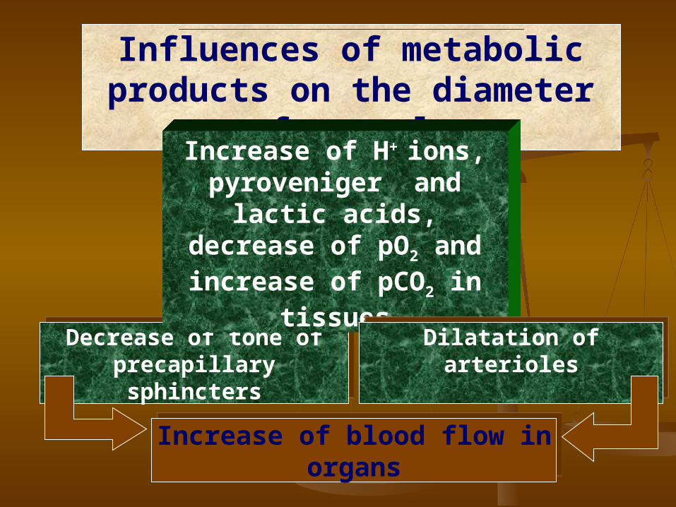
Influences of metabolic products on the diameter of vessels
Increase of Н+ ions, pyroveniger and lactic acids, decrease of pO2 and increase
of pCO2 in tissues
Dilatation of arteriolesDilatation of arterioles
Increase of blood flow in organsIncrease of blood flow in organs
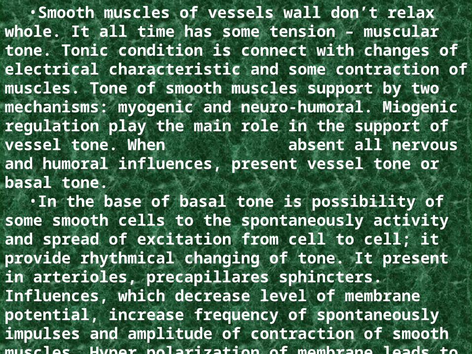
Basal tone of vessels•Smooth muscles of vessels wall don’t relax whole. It all time has
some tension – muscular tone. Tonic condition is connect with changes of electrical characteristic and some contraction of muscles. Tone of smooth muscles support by two mechanisms: myogenic and neuro-humoral. Miogenic regulation play the main role in the support of vessel tone. When absent all nervous and humoral influences, present vessel tone or basal tone.
•In the base of basal tone is possibility of some smooth cells to the spontaneously activity and spread of excitation from cell to cell; it provide rhythmical changing of tone. It present in arterioles, precapillares sphincters. Influences, which decrease level of membrane potential, increase frequency of spontaneously impulses and amplitude of contraction of smooth muscles. Hyper polarization of membrane leads to disappeared of spontaneously excitability and muscles contraction.
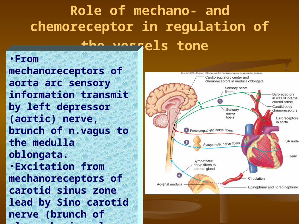
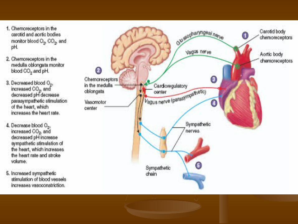
Role of mechano- and chemoreceptor in
regulation of the vessels tone •From mechanoreceptors of aorta arc sensory information transmit by left depressor (aortic) nerve, brunch of n.vagus to the medulla oblongata.•Excitation from mechanoreceptors of carotid sinus zone lead by Sino carotid nerve (brunch of glossopharingeal nerve) to the medulla oblongata.
Characteristic of afferent linkCharacteristic of afferent link
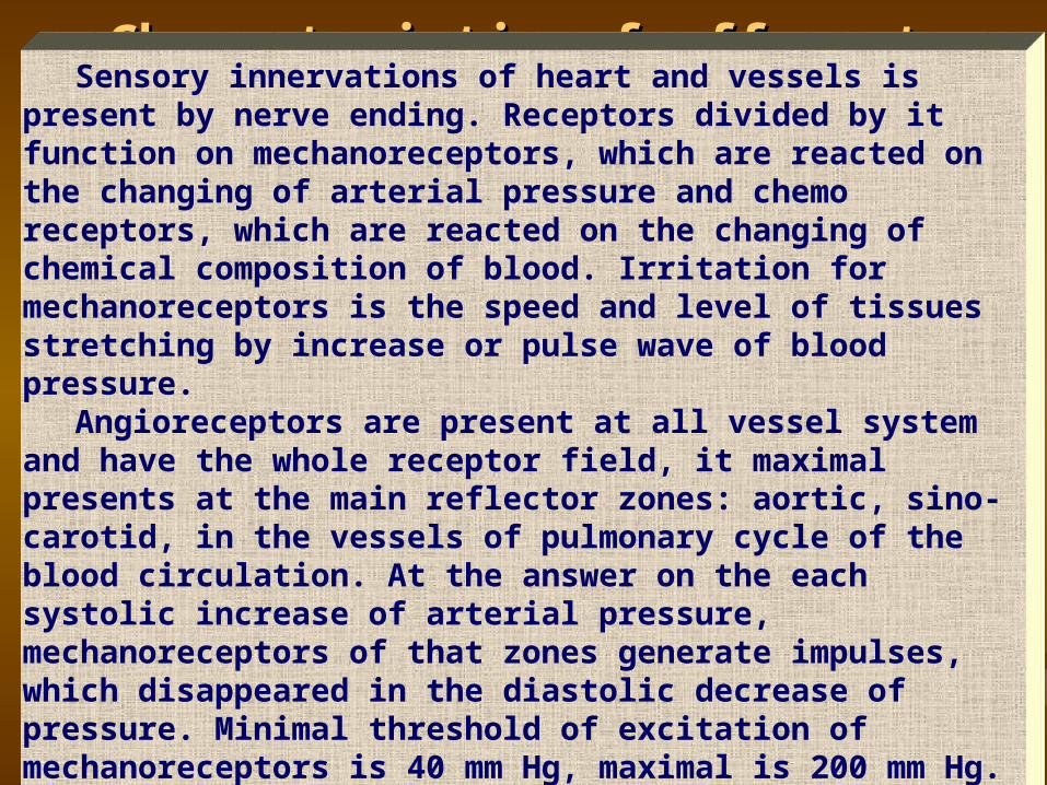
Sensory innervations of heart and vessels is present by nerve ending. Receptors divided by it function on mechanoreceptors, which are reacted on the changing of arterial pressure and chemo receptors, which are reacted on the changing of chemical composition of blood. Irritation for mechanoreceptors is the speed and level of tissues stretching by increase or pulse wave of blood pressure.
Angioreceptors are present at all vessel system and have the whole receptor field, it maximal presents at the main reflector zones: aortic, sino-carotid, in the vessels of pulmonary cycle of the blood circulation. At the answer on the each systolic increase of arterial pressure, mechanoreceptors of that zones generate impulses, which disappeared in the diastolic decrease of pressure. Minimal threshold of excitation of mechanoreceptors is 40 mm Hg, maximal is 200 mm Hg. Increase of pressure higher than that level don’t lead to addition increase of impulsation.
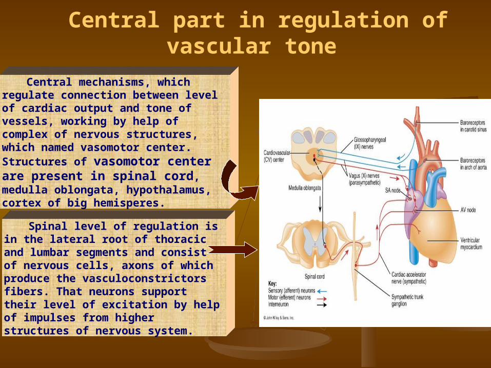
Central mechanisms, which regulate connection between level of cardiac output and tone of vessels, working by help of complex of nervous structures, which named vasomotor center. Structures of vasomotor center are present in spinal cord, medulla oblongata, hypothalamus, cortex of big hemisperes.
Spinal level of regulation is in the lateral root of thoracic and lumbar segments and consist of nervous cells, axons of which produce the vasculoconstrictors fibers. That neurons support their level of excitation by help of impulses from higher structures of nervous system.
Central part in regulation of vascular tone
Vasomotor center of medulla oblongata is the main center of regulation of blood flow. It located on the bottom of 4 ventricle, in it upper part. Vasomotor center divided on pressor and depressor zones.
Pressor zone support increase of arterial pressure. It connect with the increase of tone of resistive vessels. Also increase frequency and strength of heart contraction and as result minute volume of blood flow.
Regulatory influences of neurons of pressor zone act by help of increase of tone of sympathetic nervous system on heart and vessels.
Depressor zone support decrease of arterial pressure, heart work. It is the place of changes the impulses, which are coming from mechanoreceptors of reflector zones and cause central inhibition of tonic impulses of vasoconstrictors. Parallel the information from that zone by help of parasympathetic nerves go to heart. As result, decrease work and stroke volume of blood.
Also, depressor zone act reflector inhibition of pressor zone.
Role of brain cortexRole of brain cortex and hypothalamus and hypothalamus in regulation of blood flowin regulation of blood flow
Centers of hypothalamus give the descendent influences on the vasomotor center of medulla oblongata. In hypothalamus present depressor and pressor zones. That is why hypothalamic level give the same double reaction as bulbar center. Posterolateral part of hypothalamus cause excitation of vasomotor center. Anterior part of hypothalamus can cause mild inhibition of one.
Some zones of cortex also give the descendent influences on the vasomotor center of medulla oblongata. Motor cortex excites vasomotor center. Anterior temporal lobe, orbital areas of frontal cortex, cingulated gyrus, amygdale, septum and hippocampus can also control vasomotor center.
That influences form as a result of compare the information, which enter in higher part of nervous system from different receptor zones. It support realization of cardio-vascular component of emotions, reaction of behavior.
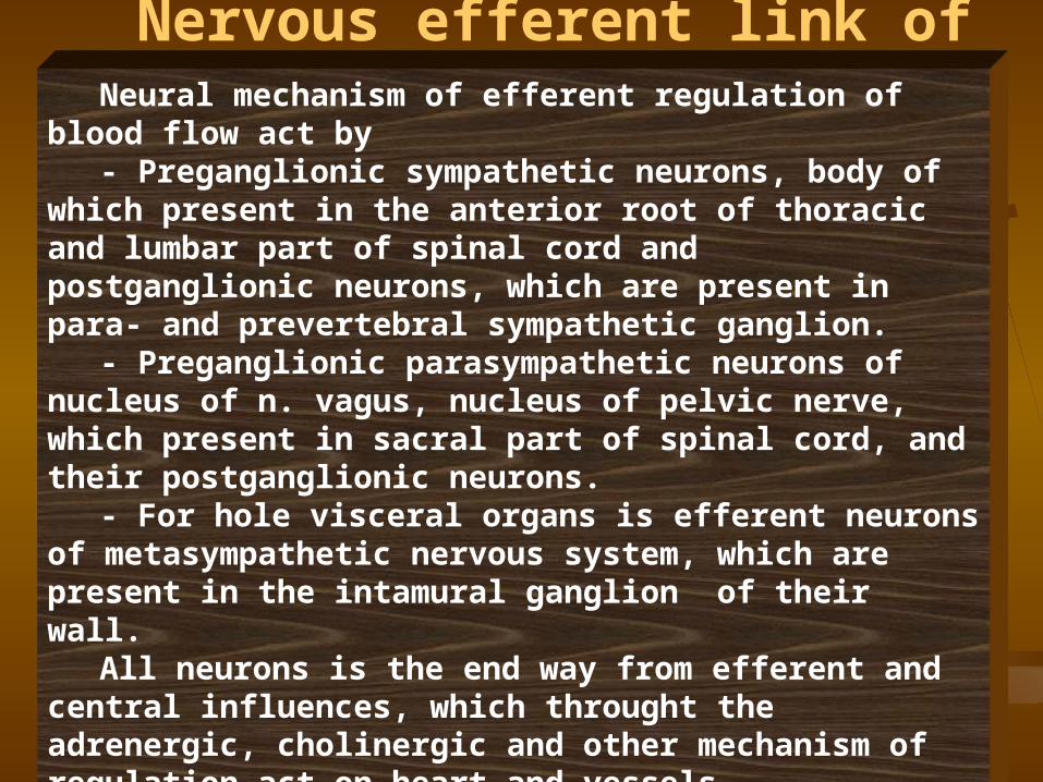
Nervous efferent link of regulation of vascular tone
Neural mechanism of efferent regulation of blood flow act by- Preganglionic sympathetic neurons, body of which present in
the anterior root of thoracic and lumbar part of spinal cord and postganglionic neurons, which are present in para- and prevertebral sympathetic ganglion.
- Preganglionic parasympathetic neurons of nucleus of n. vagus, nucleus of pelvic nerve, which present in sacral part of spinal cord, and their postganglionic neurons.
- For hole visceral organs is efferent neurons of metasympathetic nervous system, which are present in the intamural ganglion of their wall.
All neurons is the end way from efferent and central influences, which throught the adrenergic, cholinergic and other mechanism of regulation act on heart and vessels.

NorepinephrineEpinephrineEpinephrine
Action with β-adrenoreceptors of
vessel wall
Action with β-adrenoreceptors of
vessel wall
Dilation of vessels
Dilation of vessels
Spasm of vessels of skeen,
digestive organs, kidney and lungs
Spasm of vessels of skeen,
digestive organs, kidney and lungs
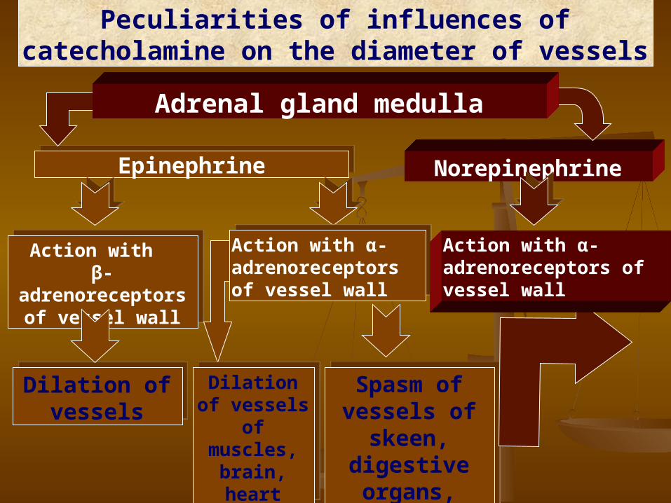
Peculiarities of influences of catecholamine on the diameter of vessels
Adrenal gland medulla
Action with α-adrenoreceptors of vessel wall
Action with α-adrenoreceptors of vessel wall
Dilation of vessels of muscles,
brain, heart
Dilation of vessels of muscles,
brain, heart
Action with α-adrenoreceptors of vessel wall

Influences of chatecholamines and vasopressin on the vessel tone
•Influences of chatecholamines from adrenal glands determined by presents of different kinds of adrenoreceptors – α and β. Connection of hormones with α–adrenoreceptors act constriction of vessel wall, with β–adrenoreceptor - relaxation.
Adrenalin connect with α– and β–adrenoreceptor, nor epinephrine with α–adrenoreceptor. Adrenalin has strong action on vessels. On artery and arterioles of skin, digestive organs, kidneys and lungs it has constrictive influences; on the vessels of skeletal muscles, brain and heart - dilatatory. On the physical load, emotional load it increase blood flow through skeletal muscles, brain and heart.
Vasopressin (antidiuretic hormone) cause spasm of artery and arterioles of organs of abdominal cavity and lungs. But vessels of brain and heart reacted on that hormone by dilatation, which help increase the nutrition of brain and heart.
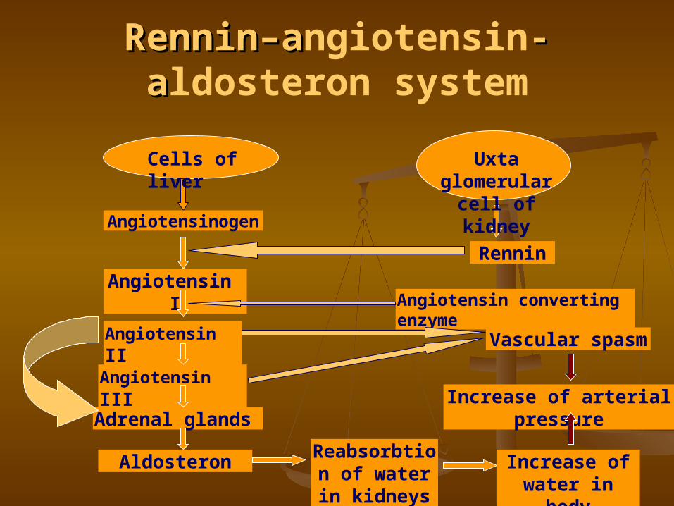
RenninRennin––aangiotensin--aaldosteron system
Angiotensinogen
Cells of liver Uxta glomerular cell of kidney
Rennin
Angiotensin І
Angiotensin ІІ
Angiotensin converting enzyme
Angiotensin ІІІ
Adrenal glands
AldosteronReabsorbtion
of water in kidneys
Increase of water in body
Vascular spasm
Increase of arterial pressure
Role of renninennin––aangiotensin--aaldosteron system in regulation of vessel tone
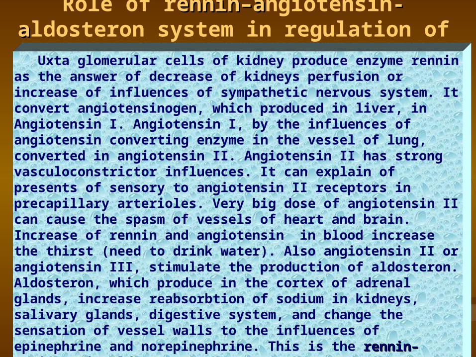
Uxta glomerular cells of kidney produce enzyme rennin as the answer of decrease of kidneys perfusion or increase of influences of sympathetic nervous system. It convert angiotensinogen, which produced in liver, in Angiotensin І. Angiotensin І, by the influences of angiotensin converting enzyme in the vessel of lung, converted in angiotensin II. Angiotensin ІІ has strong vasculoconstrictor influences. It can explain of presents of sensory to angiotensin II receptors in precapillary arterioles. Very big dose of angiotensin II can cause the spasm of vessels of heart and brain. Increase of rennin and angiotensin in blood increase the thirst (need to drink water). Also angiotensin II or angiotensin III, stimulate the production of aldosteron. Aldosteron, which produce in the cortex of adrenal glands, increase reabsorbtion of sodium in kidneys, salivary glands, digestive system, and change the sensation of vessel walls to the influences of epinephrine and norepinephrine. This is the renninrennin––angiotensinangiotensin--aldosteron systemaldosteron system ..

Change the body pose from vertical to horizontalChange the body pose from vertical to horizontal
Increase of blood flow to heartIncrease of blood flow to heart
Increase the stroke volumeIncrease the stroke volume
Increase of impulsation from mechanoreceptors of aortic arcIncrease of impulsation from mechanoreceptors of aortic arc
Activation of depressor part of vasomotor centerActivation of depressor part of vasomotor center
Inhibition of pressor part of vasomotor centerInhibition of pressor part of vasomotor center
Decrease of frequency and force of heart beat, dilation of vessels Decrease of frequency and force of heart beat, dilation of vessels
ChangesChanges of blood flowof blood flow in the clinostatic posein the clinostatic pose
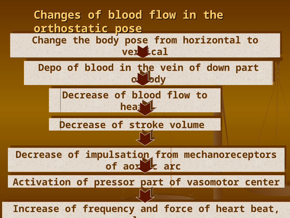
Change the body pose from horizontal to verticalChange the body pose from horizontal to vertical
Depo of blood in the vein of down part ofbodyDepo of blood in the vein of down part ofbody
Decrease of blood flow to heartDecrease of blood flow to heart
Decrease of stroke volume Decrease of stroke volume
Decrease of impulsation from mechanoreceptors of aortic arc Decrease of impulsation from mechanoreceptors of aortic arc
Activation of pressor part of vasomotor centerActivation of pressor part of vasomotor center
Increase of frequency and force of heart beat, vascular spasm Increase of frequency and force of heart beat, vascular spasm
ChangesChanges of blood flowof blood flow in the orthostatic posein the orthostatic pose
Regulation of Regulation of blood flow in blood flow in
physical physical exercisesexercises
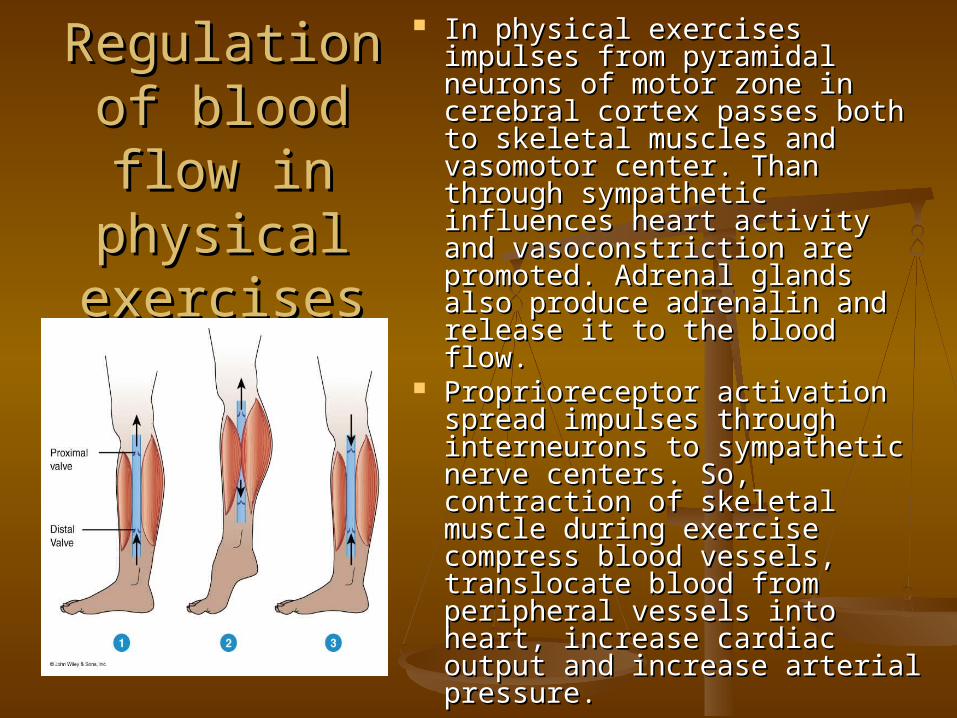
In physical exercises In physical exercises impulses from pyramidal impulses from pyramidal neurons of motor zone in neurons of motor zone in cerebral cortex passes both cerebral cortex passes both to skeletal muscles and to skeletal muscles and vasomotor center. Than vasomotor center. Than through sympathetic through sympathetic influences heart activity and influences heart activity and vasoconstriction are vasoconstriction are promoted. Adrenal glands promoted. Adrenal glands also produce adrenalin and also produce adrenalin and release it to the blood flow. release it to the blood flow.
Proprioreceptor activation Proprioreceptor activation spread impulses through spread impulses through interneurons to sympathetic interneurons to sympathetic nerve centers. So, nerve centers. So, contraction of skeletal contraction of skeletal muscle during exercise muscle during exercise compress blood vessels, compress blood vessels, translocate blood from translocate blood from peripheral vessels into heart, peripheral vessels into heart, increase cardiac output and increase cardiac output and increase arterial pressure.increase arterial pressure.
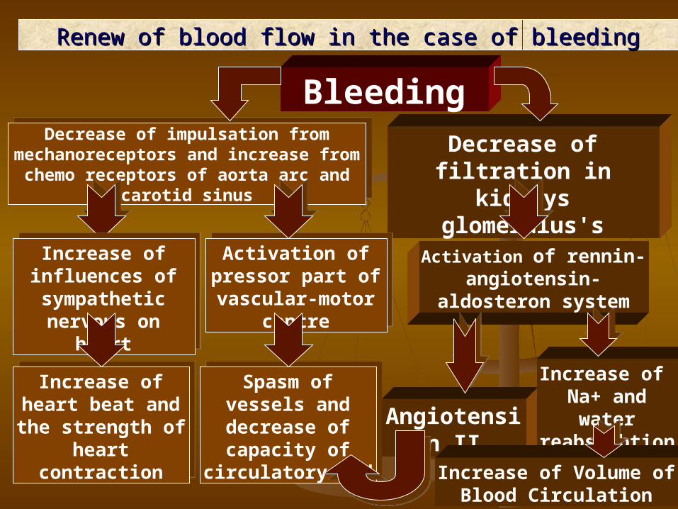
Bleeding
Decrease of filtration in kidneys glomerulus's
Decrease of impulsation from mechanoreceptors and increase from chemo receptors of aorta arc
and carotid sinus
Decrease of impulsation from mechanoreceptors and increase from chemo receptors of aorta arc
and carotid sinus
Activation of rennin-angiotensin-aldosteron
system
Activation of pressor part of vascular-
motor centre
Activation of pressor part of vascular-
motor centre
Increase of influences of sympathetic
nervous on heart
Increase of influences of sympathetic
nervous on heart
Increase of heart beat and the strength of heart contraction
Increase of heart beat and the strength of heart contraction
Spasm of vessels and decrease of capacity of circulatory bed
Spasm of vessels and decrease of capacity of circulatory bed Angiotensin ІІ
Increase of Na+ and water
reabsorbtion
Increase of Volume of Blood Circulation
Renew of blood flow in the case of bleedingRenew of blood flow in the case of bleeding
-30
-20
-10
0
10
20
30
40
50
60
70
80
90
ВС 1 хв 2 хв 3 хв 4 хв 5 хв
ЧСС САТ ДАТ
%
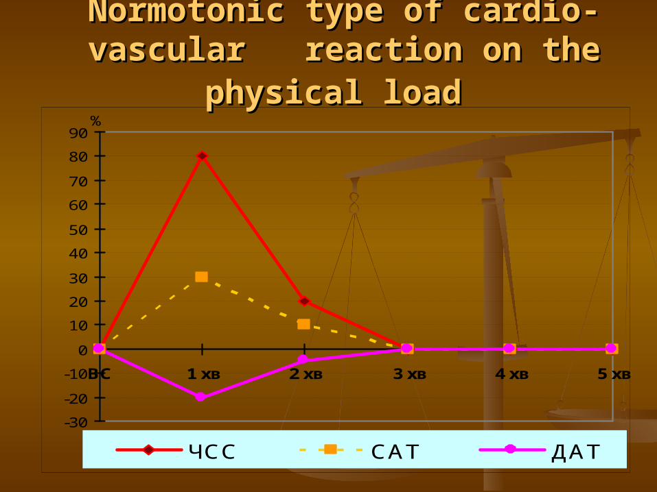
NormotonicNormotonic typetype of cardio-vascular of cardio-vascular reaction on the physical loadreaction on the physical load
InterpretationInterpretation % of increase heart beat - % of increase pulse pressure % of increase heart beat - % of increase pulse pressure
(increase systolic AP and decrease of diastolic AP)(increase systolic AP and decrease of diastolic AP)
This is rational reaction, because in the This is rational reaction, because in the case of heart beat increase also increase case of heart beat increase also increase pulse pressure and stroke volume of blood.pulse pressure and stroke volume of blood.
Increase of systolic pressure is the Increase of systolic pressure is the increase of systole of left ventricleincrease of systole of left ventricle
Decrease of diastolic pressure is Decrease of diastolic pressure is decrease of arteriole tonus, that help decrease of arteriole tonus, that help of better supply of the blood on of better supply of the blood on periphery periphery
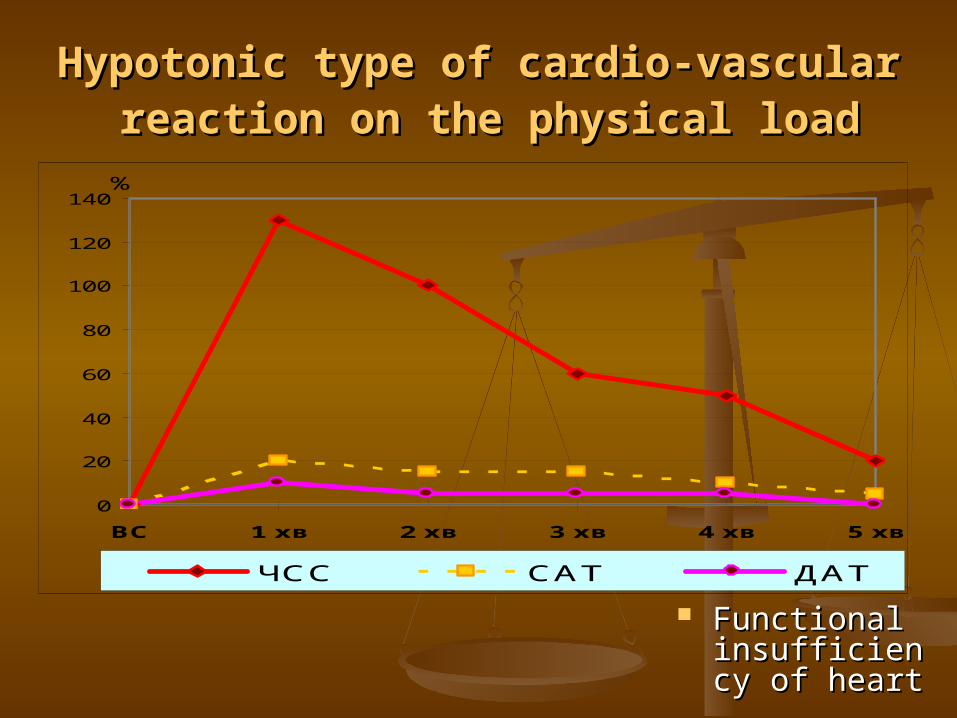
HypotonicHypotonic typetype of cardio-vascular of cardio-vascular reaction on the physical loadreaction on the physical load
0
20
40
60
80
100
120
140
ВС 1 хв 2 хв 3 хв 4 хв 5 хв
ЧСС САТ ДАТ
%
Functional Functional insufficiency insufficiency of heartof heart
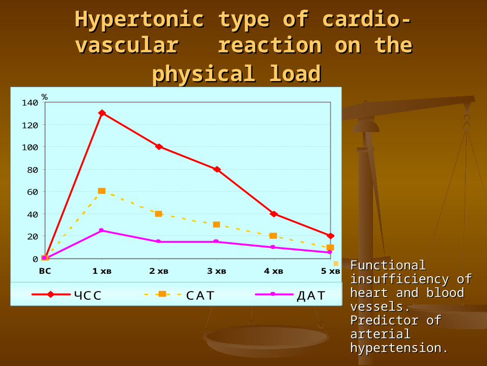
HypertonicHypertonic typetype of cardio-vascular of cardio-vascular reaction on the physical loadreaction on the physical load
0
20
40
60
80
100
120
140
ВС 1 хв 2 хв 3 хв 4 хв 5 хв
ЧСС САТ ДАТ
%
Functional Functional insufficiency of insufficiency of heart and blood heart and blood vessels. Predictor vessels. Predictor of arterial of arterial hypertension.hypertension.
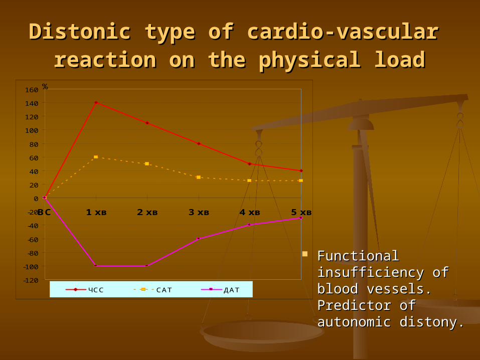
DistonicDistonic typetype of cardio-vascular of cardio-vascular reaction on the physical loadreaction on the physical load
-120
-100
-80
-60
-40
-20
0
20
40
60
80
100
120
140
160
ВС 1 хв 2 хв 3 хв 4 хв 5 хв
ЧСС САТ ДАТ
%
Functional Functional insufficiency of insufficiency of blood vessels. blood vessels. Predictor of Predictor of autonomic distony.autonomic distony.
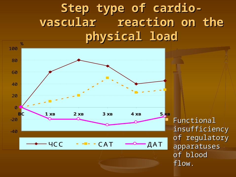
-40
-20
0
20
40
60
80
100
ВС 1 хв 2 хв 3 хв 4 хв 5 хв
ЧСС САТ ДАТ
%
StepStep typetype of cardio-vascular of cardio-vascular reaction on the physical loadreaction on the physical load
Functional Functional insufficiency insufficiency of regulatory of regulatory apparatuses apparatuses of blood flow.of blood flow.
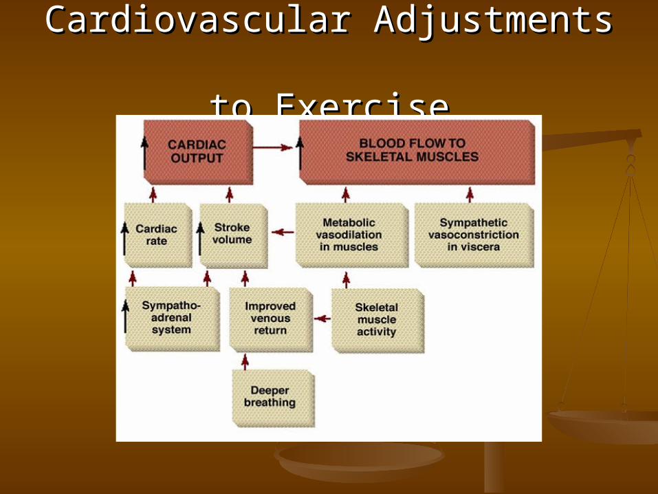
Cardiovascular Adjustments Cardiovascular Adjustments to Exerciseto Exercise

Fetal Circulation No circulation to lungs
Foramen ovale Ductus arteriosum
Circulation must go to placenta Umbilical aa., vv.
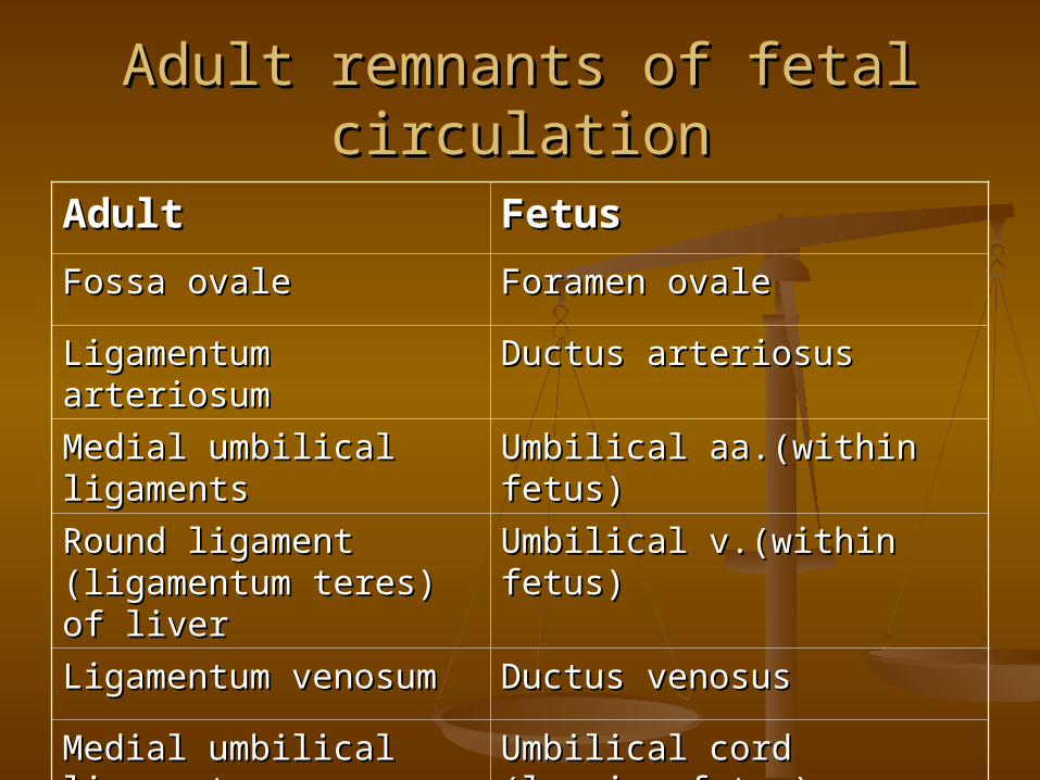
Adult remnants of fetal circulationAdult remnants of fetal circulation
AdultAdult FetusFetusFossa ovaleFossa ovale Foramen ovaleForamen ovale
Ligamentum Ligamentum arteriosumarteriosum
Ductus arteriosusDuctus arteriosus
Medial umbilical Medial umbilical ligamentsligaments
Umbilical aa.(within fetus)Umbilical aa.(within fetus)
Round ligament Round ligament (ligamentum teres) of (ligamentum teres) of liverliver
Umbilical v.(within fetus)Umbilical v.(within fetus)
Ligamentum venosumLigamentum venosum Ductus venosusDuctus venosus
Medial umbilical Medial umbilical ligamentligament
Umbilical cord (leaving Umbilical cord (leaving fetus)fetus)
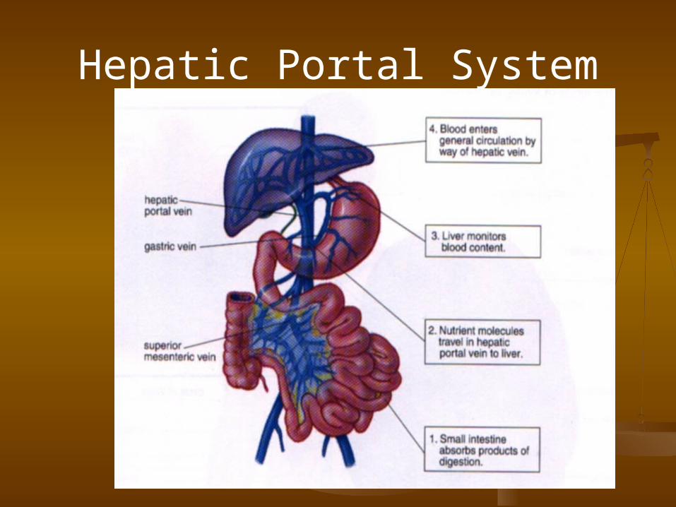
Hepatic Portal System

Thank you!Thank you!


![Measurement of Tissue Pressure in the Dental Pulp, using a ... · spectively [4-6]. Since the dental pulp resides in a low-compliance environment, an increase in blood flow, or precapillary](https://static.fdocuments.us/doc/165x107/5fa4a76a7ff1163f836ff616/measurement-of-tissue-pressure-in-the-dental-pulp-using-a-spectively-4-6.jpg)


![Effect of experimental conditions on the measurement of air ......Carson] MeasurementofAirPermeability air for of of 12 of A of of of2. — of of— Kraft) of of of of B. Papermakers'](https://static.fdocuments.us/doc/165x107/5fea82375e9c0526bf1f25cd/effect-of-experimental-conditions-on-the-measurement-of-air-carson-measurementofairpermeability.jpg)


