Regulation of Cell Cytoskeleton and Membrane Mechanics by...
12
Regulation of Cell Cytoskeleton and Membrane Mechanics by Electric Field: Role of Linker Proteins Igor Titushkin and Michael Cho* Department of Bioengineering, University of Illinois, Chicago, Illinois 60607 ABSTRACT Cellular mechanics is known to play an important role in the cell homeostasis including proliferation, motility, and differentiation. Significant variation in the mechanical properties between different cell types suggests that control of the cell metabolism is feasible through manipulation of the cell mechanical parameters using external physical stimuli. We investigated the electrocoupling mechanisms of cellular biomechanics modulation by an electrical stimulation in two mechanically distinct cell types—human mesenchymal stem cells and osteoblasts. Application of a 2 V/cm direct current electric field resulted in approximately a twofold decrease in the cell elasticity and depleted intracellular ATP. Reduction in the ATP level led to inhibition of the linker proteins that are known to physically couple the cell membrane and cytoskeleton. The membrane separation from the cytoskeleton was confirmed by up to a twofold increase in the membrane tether length that was extracted from the cell membrane after an electrical stimulation. In comparison to human mesenchymal stem cells, the membrane-cytoskeleton attachment in osteoblasts was much stronger but, in response to the same electrical stimulation, the membrane detachment from the cytoskel- eton was found to be more pronounced. The observed effects mediated by an electric field are cell type- and serum-dependent and can potentially be used for electrically assisted cell manipulation. An in-depth understanding and control of the mechanisms to regulate cell mechanics by external physical stimulus (e.g., electric field) may have great implications for stem cell-based tissue engineering and regenerative medicine. INTRODUCTION Various biological systems have been reported to respond to endogenous and exogenous electromagnetic fields (1–3). Many living tissues exhibit naturally occurring electrical activities. Examples include transepithelial potentials (~mV range) in glands and embryos (4), specific transendothelial extracellular potential gradients in blood vessels (5), electri- cal fields of strengths as large as 2 V/cm detected at wound sites (6). These observations suggest that physiologically relevant electric fields can be used as an efficient tool to control cellular and tissue homeostasis. Indeed, external electric field has been shown to induce a variety of cellular and molecular responses including microfilament reorgani- zation (7,8), cell surface receptor redistribution (9,10), changes in intracellular calcium dynamics (11–13), galvano- tropic cell migration and orientation (14,15), neuronal growth cone guidance (16), enhanced stem cell differentia- tion (17,18), and angiogenesis (19,20). Moreover, electro- therapy has been successfully used clinically for bone fracture treatment, nerve fiber repair, soft tissue regeneration, and cancer chemotherapy (21–25). Several modes of electri- cal stimulation have been tried in various in vitro and in vivo experiments, including direct current (dc), pulsed, alter- nating current electric field, and magnetically induced electrical stimulation (26,27). For consistent success in the interpretation of clinical studies, careful assessment of elec- trical stimulation characteristics such as strength, frequency, and exposure duration would be required. However, selec- tion of these electrical parameters remains mainly empirical, because biochemical and biophysical electrocoupling mech- anisms mediating cellular responses to electrical stimulation remain not fully identified. For example, a very little infor- mation is available about the effect of electric field on the cell mechanical properties. Yet, cell biomechanics has been shown to play a crucial role in many vital cellular processes including proliferation, adhesion, motility, and differentia- tion (28). Cell adapts to its biomechanical environment by adjusting its mechanical properties to match those of the surrounding tissue. Cytoskeleton is one of the most significant cellular mechanical components and provides structural stability and elasticity to the cell undergoing multiple deformations without losing its integrity (29,30). In addition, an important role of the cytoskeleton in complex intracellular signaling pathways has now been established (31). Its function as a me- chanotransducer is attributed to the cytoskeleton-associated members of numerous signaling cascades, such as Rho family GTPases (32). In this way, the cytoskeleton mediates cell response to changing biomechanical environment (e.g., substrate stiffness, cell shape and deformation, external pres- sure, shear stress) by structural rearrangement of the cyto- skeleton itself, or alterations in gene expression profiles, cell adhesion, and secretion of extracellular matrix (29,33). Another important mechanical element of the cell is its plasma membrane. Beside its function as a barrier from the outer environment, it participates in inward-outward traf- ficking, motility, and cell-cell interaction (34,35). These and many other intracellular events are regulated by the membrane surface tension, which is maintained by multiple Submitted March 5, 2008, and accepted for publication September 30, 2008. *Correspondence: [email protected] Editor: Marileen Dogterom. Ó 2009 by the Biophysical Society 0006-3495/09/01/0717/12 $2.00 doi: 10.1016/j.bpj.2008.09.035 Biophysical Journal Volume 96 January 2009 717–728 717
Transcript of Regulation of Cell Cytoskeleton and Membrane Mechanics by...

Biophysical Journal Volume 96 January 2009 717–728 717
Regulation of Cell Cytoskeleton and Membrane Mechanics by ElectricField: Role of Linker Proteins
Igor Titushkin and Michael Cho*Department of Bioengineering, University of Illinois, Chicago, Illinois 60607
ABSTRACT Cellular mechanics is known to play an important role in the cell homeostasis including proliferation, motility, anddifferentiation. Significant variation in the mechanical properties between different cell types suggests that control of the cellmetabolism is feasible through manipulation of the cell mechanical parameters using external physical stimuli. We investigatedthe electrocoupling mechanisms of cellular biomechanics modulation by an electrical stimulation in two mechanically distinct celltypes—human mesenchymal stem cells and osteoblasts. Application of a 2 V/cm direct current electric field resulted inapproximately a twofold decrease in the cell elasticity and depleted intracellular ATP. Reduction in the ATP level led to inhibitionof the linker proteins that are known to physically couple the cell membrane and cytoskeleton. The membrane separation from thecytoskeleton was confirmed by up to a twofold increase in the membrane tether length that was extracted from the cell membraneafter an electrical stimulation. In comparison to human mesenchymal stem cells, the membrane-cytoskeleton attachment inosteoblasts was much stronger but, in response to the same electrical stimulation, the membrane detachment from the cytoskel-eton was found to be more pronounced. The observed effects mediated by an electric field are cell type- and serum-dependentand can potentially be used for electrically assisted cell manipulation. An in-depth understanding and control of the mechanismsto regulate cell mechanics by external physical stimulus (e.g., electric field) may have great implications for stem cell-basedtissue engineering and regenerative medicine.
INTRODUCTION
Various biological systems have been reported to respond to
endogenous and exogenous electromagnetic fields (1–3).
Many living tissues exhibit naturally occurring electrical
activities. Examples include transepithelial potentials (~mV
range) in glands and embryos (4), specific transendothelial
extracellular potential gradients in blood vessels (5), electri-
cal fields of strengths as large as 2 V/cm detected at wound
sites (6). These observations suggest that physiologically
relevant electric fields can be used as an efficient tool to
control cellular and tissue homeostasis. Indeed, external
electric field has been shown to induce a variety of cellular
and molecular responses including microfilament reorgani-
zation (7,8), cell surface receptor redistribution (9,10),
changes in intracellular calcium dynamics (11–13), galvano-
tropic cell migration and orientation (14,15), neuronal
growth cone guidance (16), enhanced stem cell differentia-
tion (17,18), and angiogenesis (19,20). Moreover, electro-
therapy has been successfully used clinically for bone
fracture treatment, nerve fiber repair, soft tissue regeneration,
and cancer chemotherapy (21–25). Several modes of electri-
cal stimulation have been tried in various in vitro and in vivo
experiments, including direct current (dc), pulsed, alter-
nating current electric field, and magnetically induced
electrical stimulation (26,27). For consistent success in the
interpretation of clinical studies, careful assessment of elec-
trical stimulation characteristics such as strength, frequency,
and exposure duration would be required. However, selec-
Submitted March 5, 2008, and accepted for publication September 30, 2008.
*Correspondence: [email protected]
Editor: Marileen Dogterom.
� 2009 by the Biophysical Society
0006-3495/09/01/0717/12 $2.00
tion of these electrical parameters remains mainly empirical,
because biochemical and biophysical electrocoupling mech-
anisms mediating cellular responses to electrical stimulation
remain not fully identified. For example, a very little infor-
mation is available about the effect of electric field on the
cell mechanical properties. Yet, cell biomechanics has been
shown to play a crucial role in many vital cellular processes
including proliferation, adhesion, motility, and differentia-
tion (28).
Cell adapts to its biomechanical environment by adjusting
its mechanical properties to match those of the surrounding
tissue. Cytoskeleton is one of the most significant cellular
mechanical components and provides structural stability
and elasticity to the cell undergoing multiple deformations
without losing its integrity (29,30). In addition, an important
role of the cytoskeleton in complex intracellular signaling
pathways has now been established (31). Its function as a me-
chanotransducer is attributed to the cytoskeleton-associated
members of numerous signaling cascades, such as Rho
family GTPases (32). In this way, the cytoskeleton mediates
cell response to changing biomechanical environment (e.g.,
substrate stiffness, cell shape and deformation, external pres-
sure, shear stress) by structural rearrangement of the cyto-
skeleton itself, or alterations in gene expression profiles,
cell adhesion, and secretion of extracellular matrix (29,33).
Another important mechanical element of the cell is its
plasma membrane. Beside its function as a barrier from the
outer environment, it participates in inward-outward traf-
ficking, motility, and cell-cell interaction (34,35). These
and many other intracellular events are regulated by the
membrane surface tension, which is maintained by multiple
doi: 10.1016/j.bpj.2008.09.035

718 Titushkin and Cho
mechanisms including membrane reservoir and lipid mate-
rial turnover (35,36). Generally, the membrane tension is
determined by the lipid bilayer composition such as choles-
terol content (37), and the membrane interaction with the
cytoskeleton via specific biomolecular linkage systems
(38). We have shown recently that the plasma membrane
attachment to the cytoskeleton in fully differentiated osteo-
blasts is much stronger than in undifferentiated stem cells
(39). Functionally, a strong membrane-cytoskeleton adhe-
sion should be beneficial to keep the structural integrity of
osteoblasts subjected to continuous stress cycles. On the
other hand, in human mesenchymal stem cells (hMSCs)
a lower membrane tension may better facilitate endo- and
exocytosis and contributes to a higher sensitivity of these
cells to various soluble biochemical environmental stimuli.
The cells of mesodermal origin show a wide spectrum of
mechanical properties. This observation opens an exciting
perspective to regulate cell homeostasis by controlling its
biomechanics. Indeed, specific types of mechanical stimula-
tion (e.g., shear stress, cyclic stretching, compressive
loading) can be used for regulation of gene expression,
matrix components secretion, cell differentiation, and ulti-
mately engineering functional load-bearing tissues (29,40).
We have shown previously that mesenchymal stem cells
are mechanically strikingly different from fully differentiated
osteoblasts, and the mechanical properties are altered during
osteodifferentiation (41). This finding suggests that control
of cell differentiation is feasible through manipulation of
the cell mechanical properties using external physical
stimuli. Therefore, systematic investigations of the cell
mechanics modulation by external physical stimulus (e.g.,
electric field) would allow targeted regulation of cell metab-
olism through control of cell biomechanics (27). This infor-
mation will have great implications for tissue engineering
and regenerative medicine. For example, elucidation of the
electrocoupling mechanisms is expected to establish a ratio-
nale paradigm for electrically assisted differentiation of stem
cells into preselected phenotypic lineage. In this study, we
examined the effect of dc electric fields on the mechanical
properties of two model cell types: hMSCs and fully differ-
entiated human osteoblasts. We investigated the molecular
mechanisms of electrically induced mechanical changes in
the cell membrane and cytoskeleton. Careful characterization
of electrically stimulated and cell type-dependent mechan-
ical responses could lead to enhance cell differentiation
(18) and other tissue engineering applications.
MATERIALS AND METHODS
Cell culture and drug treatment
Human mesenchymal stem cells were obtained from the Tulane Center for
Gene Therapy (New Orleans, LA). Based on flow cytometry results, these
stem cells showed negative staining for CD34, CD36, CD45, and CD117
markers (all <2%), and positive staining for CD44, CD90, CD166, CD29,
CD49c, CD105, and CD147 markers (all >95%), indicating a minimal
Biophysical Journal 96(2) 717–728
heterogeneity in cell population. Normal human fetal osteoblasts (hFOB
1.19) were obtained from American Tissue Culture Collection (Manassas,
VA). Cells were grown in Dulbecco’s modified Eagle’s culture medium
with 15% fetal bovine serum, L-glutamin, and antibiotics. Two days before
experiments, cells were harvested and plated on a 24 � 30 mm glass cover-
slip. Cells were rinsed gently with Hanks’ balanced salt solution (HBSS) and
mounted on the electrical stimulation exposure chamber, AFM fluid
chamber, or sealed coverglass chamber for optical trapping. Cells between
passages 3 and 12 were used for all experiments.
For ATP depletion, cells were incubated in PBS containing the mitochon-
dria inhibitor sodium azide (10 mM) and glycolytic inhibitor 2-deoxyglu-
cose (10 mM) for 40–60 min at 37�C. Ionomycin (10 mM) was used to
equilibrate intracellular and extracellular calcium levels. All drugs were
purchased from Sigma-Aldrich (St. Louis, MO).
Electric field exposure
The chamber used to stimulate cell with an electric field has been described
earlier (9,11,14). Joule heating was minimized by incorporating a sapphire
window and a large surface/volume ratio in the chamber design. Agarose
salt bridges were used in the experiments to eliminate unwanted electrode
byproducts and to minimize pH changes. Electrical current was supplied
to the chamber by a 100 W amplifier (BOP100, Kepco, Flushing, NY)
and monitored by an oscilloscope (Tektronix, Model 2205, Beaverton,
OR). The computation of electric field strength E followed Ohm’s law,
J ¼ sE, where J is the electric current density and s is the conductivity of
the medium. Electric field application was carried out in HBSS or cell
culture medium at room temperature. Temperature rise caused by applica-
tion of a 2 V/cm field for 60 min was negligible (12).
AFM microindentation test
The live cell elasticity was measured with a Novascan atomic force micro-
scope (Novascan Technologies, Ames, IA) mounted on an inverted
TE-2000 Nikon microscope. Soft silicone nitride cantilevers (100 mm long,
Veeco, Santa Barbara, CA) were calibrated by the thermal fluctuation
method in air (42), with a typical spring constant value of 0.12 N/m. Boro-
silicate glass beads (10 mm in diameter) glued onto the cantilever served as
spherical cell indenters. Distribution of the indenting load over several-
micron area allows to remove spatial mechanical heterogeneity of fibrous
cell cytoskeleton. Individual isolated cells with normal morphology were
mechanically probed with AFM, avoiding the cell’s perinuclear region. To
obtain a force curve, the cantilever descended toward the cell at a velocity
of ~2 mm/s until a trigger force of 3 nN was reached, and then retracted.
Viscous dissipation of energy is minimal at this speed, and force measure-
ments are dominated by the elastic behavior of the cell (41). To minimize
the effect of glass substrate on the cell elasticity measurements, we used
an indentation depth up to 500 nm (~10–15% of the average cell height)
for data analysis. A total of 30–40 cells of each type and experimental condi-
tion were used, with ~15 force-distance curves acquired from each cell.
The force-distance curves were collected and analyzed according to the
Hertz model (43,44), which relates the loading force (F) with the indentation
depth (d) by
F ¼ 4
3
E
ð1� n2Þd3=2
ffiffiffi
Rp
;
where n is the cellular Poisson’s ratio, R is the radius of the spherical indenter
(5 mm), E is the local Young’s elastic modulus. The cellular Poisson’s ratio
was assumed to be 0.5, which treats the cell as an incompressible material
(44). Fitting the Hertz model to the experimental force curve with a standard
least squares minimization algorithm yielded the local apparent elastic
modulus E. The average Young’s modulus for each cell type and experi-
mental condition was calculated and subjected to t-test at the level of 0.05.

Linker Proteins and Cellular Mechanics 719
Membrane tether extraction with laser opticaltweezers
Fluorescent polystyrene beads 0.5 mm diameter with 515-nm emission
(FluoSpheres, Molecular Probes, Eugene, OR) were covalently coated
with mouse anti-CD29 antibodies and tightly bound to the cell membrane.
The beads were used as handles for a membrane tether extraction, as
described earlier (39,45). An infrared Nd:YAG laser (1064 nm, continuous
wave, 0.5 W maximum incident power at the sample; SpectraPhysics,
Mountain View, CA) with a Nikon Eclipse E-800 microscope was used
for particle optical trapping. The laser beam was focused at the cell surface
with a 100� oil immersion microscope objective (PlanApo, NA 1.4), and
this optical trap was moved in the focus plane by a system of two confocal
lenses actuated by a high-precision motorized translator. Cells, laser, and
fluorescent beads were imaged with a 16-bit charge-coupled device camera
(Photometrics, Tucson, AZ) in the bright-field and epifluorescence modes.
To extract a membrane tether from the cell, a latex bead attached to the
cell was chosen randomly and optically trapped. The bead was then dis-
placed from its equilibrium position by moving the trap away from the
cell at a constant speed of 1.5 mm/s and a constant force ~3 pN. The Stoke’s
drag in an aqueous solution and the membrane viscosity have negligible
effects on the bead dynamics at this speed. Tether growth was observed until
the bead escaped from the trap. Elastic membrane tether formation was iden-
tified by quick retraction of the bead to its original position after escape from
the trap. The total tether length was determined by tracking bead position
using the MetaMorph image processor (Molecular Devices, Downingtown,
PA). Typically, 35–40 beads from ~20 cells were analyzed for each
experimental condition and cell type. Differences in tether length were
examined using Student’s t-test. Results were deemed statistically significant
when p < 0.05.
Immunostaining and fluorescence microscopy
To explore the cytoskeleton structure and linker protein distribution of
normal and electrically stimulated cells, the samples were fixed in 3.7%
formaldehyde and permeabilized in cold (�20�C) acetone for 3 min.
Nonspecific binding sites were blocked using a 1% bovine serum albumin
solution for 30 min at room temperature. Intracellular actin filaments were
stained with rhodamine-phalloidin (5 mM) for 30 min at room temperature
(Molecular Probes). Ezrin/radixin/moesin (ERM) proteins were labeled
with rabbit ERM antibody (Cell Signaling Technology, Danvers, MA),
and secondary goat anti-rabbit antibody conjugated with AlexaFluor488
dye (Molecular Probes). Focal adhesions were labeled using mouse antibody
to vinculin (Chemicon, Temecula, CA), and secondary fluorescent goat anti-
mouse antibody.
Samples were imaged by a laser scanning confocal system (Radiance
2001MP, Bio-Rad, Hercules, CA) operating on a Nikon TE2000-S inverted
microscope with a 60� Plan Apo objective (NA 1.4), blue Argon ion (488
nm), and green HeNe (543 nm) lasers. Emission filters (515/30 nm and 600/
50 nm) were used to collect confocal images of ERM and microfilaments,
respectively.
Relative F-actin concentration was determined by fluorescent microscopy
as described elsewhere (8,46). In brief, after fixation and permeabilization
the cells were stained with rhodamine-phalloidin (5 mM) to visualize actin
and AlexaFluor488 succinimidyl ester dye (10 mM) to label total cell protein.
The rhodamine-phalloidin and succinimidyl ester images of the same region
were obtained using an E-800 Eclipse Nikon fluorescent microscope with
a 20� objective lens and a 16-bit charge-coupled device camera (Photomet-
rics). The edges of the cells on a total protein image were used to define the
mask for these cells. After background subtraction, F-actin and total protein
contents were measured by summing the total fluorescence intensity signal
on the masked images of corresponding fluorophores. The relative F-actin
content was then obtained by dividing F-actin by total protein for each
cell, and normalizing it to the corresponding control sample. Different exper-
imental groups of cells were stained using the same solutions and imaged
identically with their respective controls.
Total protein images of the cells were also used for morphological analysis
including cell surface area and form factor measurements. The form factor Fwas calculated as F ¼ 4pA/P2, where A is a cell surface area, and P is its
perimeter. Smaller F values indicate more complex cell geometry, and higher
deviation of cell shape from an ideal circle (F ¼ 1).
Bioluminescent ATP measurements
Intracellular ATP content was determined using a luciferin-luciferase assay
(Sigma, St. Louis, MO) in a SPECTRAmax Gemini XS Microplate Spectro-
fluorometer (Molecular Devices, Sunnyvale, CA). A glass coverslip with
normal or electrically exposed cells was washed with PBS and treated
with 300 ml of somatic cell ATP releasing agent. The sample was collected
into a vial and mixed with an equal volume of diluted ATP assay mix, and
amount of emitted light was measured immediately with a luminometer. In
control experiments the background chemiluminescence signal from cell
buffer was determined. In each series of experiments, ATP content was
normalized to the values from respective control samples.
Gel electrophoresis and Western blotting
The expression levels of ERM proteins were analyzed using immunoblot-
ting. Equal amounts of protein per well were diluted in Laemmli sample
buffer (BioRad, Hercules, CA), boiled for 3 min, subjected to 8% SDS-
PAGE, and transferred to PVDF membrane. The membranes were washed
with Tris-buffered saline þ Tween (TBST, 0.01 M Tris $ HCl Ph 7.4,
0.15 NaCl, and 0.1% Tween 20), blocked with 5% nonfat dry milk solution
in TBST, incubated with appropriate primary antibody, and horseradish
peroxidase-conjugated secondary antibody. Antibody recognizing total
ERM, and ezrin, radixin, and moesin phosphorylated at Thr567, Thr564,
Thr558, respectively (corresponding to the active state of these proteins)
were purchased from Cell Signaling (Beverly, MA). Antibody binding
was visualized with Opti-4CN detection kit (BioRad). Membranes were
scanned using GelDoc XR system (BioRad), and the band densiometric
measurements were carried out with BioRad Quantity One software. The
total level of ERM, i.e., the sum of the ~80 kDa band (corresponding to ezrin
and radixin) and the ~75 kDa band (corresponding to moesin) was obtained.
The phospho-ERM level was normalized to the corresponding total ERM
level in each sample, and taken relative to the untreated cell value.
RESULTS
Cell cytoskeleton elasticity measurement
To investigate changes in the cell elastic properties induced
by an electrical stimulation, we used AFM indentation tech-
nique as described earlier (41). As shown previously, the
average elastic modulus of normal hMSCs (3.2 5 1.4 kPa)
is almost a twofold higher than that of fully differentiated
osteoblasts (1.7 5 1.0 kPa; Fig. 1 A). Exposure of cells to
a 2 V/cm electric field for 60 min in HBSS resulted in
a decrease in the cell elasticity to 1.0 5 0.5 kPa in hMSCs
and to 1.1 5 0.5 kPa in osteoblasts (Fig. 1 A). Similar results
were observed after cell treatment with calcium ionophore,
which causes an influx of calcium ions into the cytoplasm
from extracellular medium. Increases in intracellular calcium
concentration in response to electrical stimulation have been
reported by multiple laboratories (2,11,12,47), and were also
detected in our experiments using the fluorescent calcium
indicator Fluo-4 (data not shown). We should note that the
cell type-dependent dynamics of elastic changes is rather
remarkable. For example, within 60 min of an electrical
Biophysical Journal 96(2) 717–728

720 Titushkin and Cho
stimulation, the hMSC elasticity decreased ~70% whereas
osteoblasts showed only a ~30% elasticity reduction under
the similar experimental conditions. Measurements of the
cell elastic moduli at intermediate time points (e.g., 30, 45
min dc field exposure) support the data on a faster elasticity
decrease in the hMSC cytoskeleton in comparison with oste-
oblasts. Interestingly, when the cells were electrically stimu-
lated in complete growth medium, only a minimal decrease
in cell stiffness was observed (Fig. 1 A). For example, an
electrical stimulation in culture medium caused higher data
dispersion and only a statistically insignificant decrease in
the osteoblast cytoskeleton elasticity. Decrease in the elastic
modulus can be partially reversed up to 60–80% of the
original value by incubating the cells in culture medium
for 60 min (data not shown), implying an important role of
serum in mediating electric field effect on the cellular
mechanics.
As reported earlier, actin is the major determinant of the
cytoskeleton elasticity rather than microtubules or interme-
diate filaments (41,48). We therefore measured relative
F-actin content in the cells after an electric stimulation. In
both cell types the amount of polymerized actin decreased
after electric field exposure or ionomycin treatment in
HBSS (Fig. 1 B). Consistent with our observation, this
decrease is less pronounced when the cells were exposed
to the electric field in the culture medium with serum. In
addition, cell viability assays showed that no less than
90% cells were viable after 60 min of electrical field stimu-
lation and/or AFM mechanical testing. A longer exposure of
cells to the electric field up to 90 min produces only a minor
further decrease in the cell elasticity, but adversely affects
cell viability. Variation of dc electric field strength in the
range 0.5–3 V/cm produced similar commensurate effects
in the cell elastic properties, which is qualitatively similar
to those reported here using a 2 V/cm field.
Electrically induced actin reorganization
Significant differences in the cellular mechanics between
hMSCs and osteoblasts are likely to result from differential
actin organization in these cells. For example, actins in oste-
oblasts are predominantly organized as thin dense filamen-
tous meshwork with focal contacts that are generally round
and smaller in size (Fig. 2 A). In contrast, stem cells show
many thick actin bundles, or stress fibers, extending
throughout the cytoplasm and terminating at focal contacts
(41) that are irregularly shaped and much larger in size
(Fig. 2 B), possibly indicative of a stronger adhesion to the
substrate, as indicated by vinculin labeling. After an electri-
cal stimulation of cells in HBSS, both actin structures were
impaired (Fig. 2, C and D). Disassembly of actin scaffolds
0
1
2
3
elas
tic m
odul
us, k
Pa
0
0.5
1
untreated, 60 min in HBSS
untreated, 60 min in
CCM
2 V/cm DC,60 min in
HBSS
2 V/cm DC, 60 min in
CCM
ionomycin,10uM, 60 min
in HBSS
Rel
ativ
e F-
actin
con
tent
hMSC osteoblast
A
B
* ** *
* ** *
*
FIGURE 1 Effect of electric field on cell cytoskeleton.
Cells were exposed to a 2 V/cm dc field for 60 min in
HBSS, and complete cell culture medium (CCM). (A)
Cell cytoskeleton elasticity determined with AFM microin-
dentation. The elastic modulus decreased significantly after
an electrical stimulation or cell treatment with calcium iono-
phore in HBSS in both hMSCs and osteoblasts. Between
400–600 force-curves were acquired for each cell type
and experimental condition. (B) Relative F-actin content
in cells exposed to the electric field. It was obtained for
each cell as a ratio of fluorescence signals from F-actin
and the total protein, and then normalized to the correspond-
ing control. The highest degree of actin depolymerization
was observed in electrically stimulated cells in the absence
of serum. Results represent the mean 5 SE. *Statistically
different from respective controls (p < 0.05).
Biophysical Journal 96(2) 717–728

Linker Proteins and Cellular Mechanics 721
seems to be responsible for decrease in the cellular elasticity.
Interestingly, we did not observe electrically induced
changes in microtubule or intermediate filament arrange-
ment. Cell treatment with ionomycin, however, caused
similar breakdown of actin cytoskeleton as did an electric
field (Fig. 2, E and F). The actin cytoskeleton reorganization
was noticeably different and appeared more complex when
cells placed in the complete culture medium with serum
were exposed to an electric field (Fig. 2, G and H). Actins
were clearly accumulated toward the cathode side and actin
depolymerization was noticeably visible at the anode side of
the cell. This anisotropic actin redistribution is typical for
FIGURE 2 Actin cytoskeleton reorganization. Fluores-
cent images of osteoblasts are in the left column and
hMSCs in the right column. Immunofluorescent images
of a thin filamentous actin meshwork (red) and vinculins
(green) showed that osteoblasts contain fewer and smaller
focal adhesions (A). In contrast, hMSCs showed thick actin
stress fibers, and multiple and large adhesion contacts (B).
Actins were partially disassembled in both osteoblast (C)
and hMSC (D) after cell exposure to a 2 V/cm field for
60 min in serum-free HBSS. Cells treatment with 10 mM
ionomycin for 40 min in HBSS produced analogous actin
dismantling effects both in osteoblast (E) and hMSC (F).
When cells were placed in cell culture medium with serum,
the electrical stimulation again caused redistribution of
actins in osteoblast (G) and stem cells (H), but in a manner
that was different than (C) and (D) without serum. Cell
incubation in the complete culture medium for 60 min at
37�C after the dc field exposure resulted in a partial
recovery of the actin structure in both osteoblasts (I) and
hMSC (J). The arrows indicate the direction of the electric
field applied.
Biophysical Journal 96(2) 717–728

722 Titushkin and Cho
serum-dependent cell galvanotaxis that has been reported
elsewhere (10,15). Indeed, we observed some electromigra-
tion of osteoblasts, but not hMSCs, toward the cathode (data
not shown). This is likely due to stronger substrate adhesion
of hMSC compared to osteoblasts as implied by abundance
and larger size focal contacts terminating multiple stress
fibers in stem cells (see Fig. 2 B). This cell type-dependent
differential cell adhesion is consistent with our previous
report that the integrins on the surface of undifferentiated
hMSCs were found to be clustered and laterally immobile
(i.e., confined or restricted) until hMSCs undergo osteodif-
ferentiation (49). Finally, the electrically mediated actin re-
modeling was reversible as shown by incubating the cells
in the complete culture medium for 60 min after an electric
field exposure (Fig. 2, I and J). The actin cytoskeleton
arrangement seems to have been restored to its original struc-
ture in both cell types. As mentioned earlier, this actin
structural repair was accompanied by a recovery of the cell
stiffness almost to its original values.
Membrane mechanical properties
Fluorescent polystyrene beads attached to the cell plasma
membrane were pulled with a laser optical trap to produce
thin lipid membrane tethers. Binding specificity of antibody-
coated probes has been verified in control experiments with
noncoated beads; ~20–40% of membrane-conjugated beads
were fluctuating on the cell surface and could be used for
tether extraction. Short exposure to the laser (~15–30 s to
pull one tether) allows to avoid laser-induced photo damage
as confirmed by repeating experiments using the same bead
with consistently reproducible results. The average tether
length in normal hMSC (10.6 5 1.1 mm) is ~2.5-fold higher
than that in osteoblasts (4.0 5 1.1 mm) (Fig. 3 A). As we
postulated earlier, this result is due to much weaker
membrane-cytoskeleton interaction in stem cells compared
to osteoblasts (39). Cell exposure to a 2 V/cm electric field re-
sulted in a twofold tether length increase in osteoblast, but had
no significant effect on the tether length in stem cells (Fig. 3
A). Similar results were achieved by intracellular ATP deple-
tion with sodium azide and 2-deoxyglucose. To test if the elec-
tric field effect on the membrane tether length is mediated by
ATP, we measured the ATP content in the cell before and after
an electrical stimulation. As expected, the intracellular ATP
level decreased in response to an electric field (Fig. 3 B).
This result is consistent with earlier findings suggesting
ATP release from the cells in response to electrical stimulation
(47,50). Unfortunately, we could not detect measurable traces
of released ATP in the electrical exposure chamber due to
a high volume of buffer/medium filling the chamber.
However, unlike osteoblasts, we could not find statistically
significant changes in the tether length in stem cells in
response to the electric field or ATP depletion. Apparently,
the current optical trapping technique sensitivity is not suffi-
cient to detect further minor electrically induced decrease in
the originally weak hMSC membrane-cytoskeleton adhesion.
Biophysical Journal 96(2) 717–728
ERM linker protein response to electricalstimulation
The membrane-cytoskeleton interaction is mediated by
special linker proteins that physically couple the membrane
with the cytoskeleton. For instance, the ERM family proteins
are found in the cytoplasm of many cell types. On activation
these proteins can bind both to polymerized actin and inte-
gral membrane proteins (51–53). For example, a dense actin
network underlying the osteoblasts plasma membrane
provides high density binding sites for the ERM proteins
that are distributed uniformly (Fig. 4 A). In contrast, in
hMSCs there are relatively few binding sites for the
ERM linkers on the sparse actin stress fibers proximal to
the plasma membrane, and the ERM proteins were found
FIGURE 3 Electrically induced changes in the plasma membrane
mechanics. (A) Membrane tether length measured with laser optical twee-
zers. Much longer tethers could be extracted from the normal hMSC
membrane than that of osteoblasts, likely due to a weaker membrane-cyto-
skeleton interaction in stem cells. The tether lengths increased in osteoblasts
after cell exposure to a 2 V/cm field or 10 mM sodium azide and 10 mM
2-deoxyglucose. At least 30 tethers were measured for each cell type and
experimental condition. (B) Intracellular ATP content determined with
bioluminescent luciferin-luciferase assay. The ATP content was normalized
to untreated samples. Results represent the mean 5 SE. *Statistically
different from respective controls (p < 0.05).

Linker Proteins and Cellular Mechanics 723
to be located primarily along stress fibers underlying the
membrane (Fig. 4 D). In response to an electrical stimulation
(Fig. 4, B and E) or ATP depletion (Fig. 4, C and F), the
amount of these linker proteins seemed to have decreased.
Moreover, there is a clear correlation between inhibition of
the ERM proteins and the membrane-cytoskeleton separa-
tion in osteoblasts. The ERM protein redistribution in
hMSCs in response to an electrical stimulation or ATP deple-
tion is much less pronounced than in osteoblasts, and the cor-
responding changes in the membrane tether length were not
significant statistically.
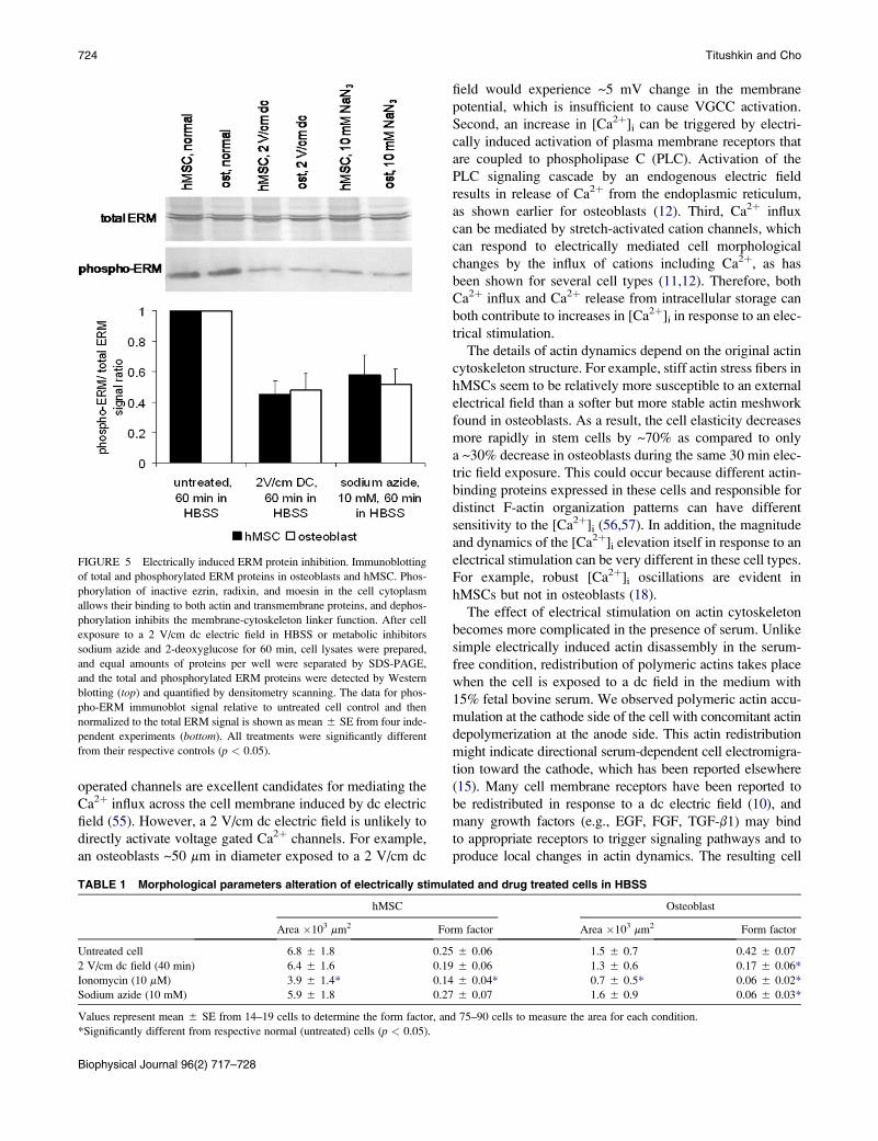
Immunoblotting of the total and phosphorylated ERM
proteins in osteoblasts and hMSCs confirmed that the rela-
tive amount of activated linker proteins decreased after cell
exposure to a dc electric field or metabolic inhibitors such
as sodium azide and 2-deoxyglucose (Fig. 5). For example,
after an electrical stimulation or sodium azide treatment,
both cell types showed a ~1.5–2-fold decrease in the immu-
noblot-detected signal corresponding to phosphorylated (i.e.,
activated) ERM linkers as compared to that measured in
untreated cells. Although an accurate quantitative phospho-
ERM concentration decrease can not be directly inferred
from immunoblotting results, this finding does suggest
a significant inhibition of the ERM proteins by an electric
field.
Altered cellular morphology
Cellular morphological properties are closely related to the
cell mechanics. For example, a stronger adhesion to the glass
substrate of hMSCs than osteoblasts is attributed to multiple
focal contacts and stress fibers in stem cells (see Fig. 2 B). As
result, hMSCs have a much higher surface area on 2D
substrate than osteoblasts (Table 1), and a more stretched
and edgy cell morphology was observed as indicated by
a lower form-factor compared to osteoblasts. Stimulation
with a 2 V/cm electric field for 40 min seemed to decrease
both the surface area and the form-factor in the two cell types
(i.e., smaller size cells and more complex spiky geometry).
Apparently, electrically induced morphological changes
are due to both actin cytoskeleton reorganization and
membrane-cytoskeleton dissociation. Indeed, ionomycin-
mediated actin depolymerization caused a significant decrease
in the cell area and the form factor in both cell types. Besides,
loosening the tight membrane-cytoskeleton attachment in
osteoblasts by ATP depletion also led to a decrease in the
form factor in these cells (Table 1). Severe changes in the
cell morphology may result in activation of stretch-activated
cation channels causing additional Ca2þ influx and further
actin reorganization.
DISCUSSION
Multiple biochemical and biophysical responses to exoge-
nous electric field have serious implications to the cell
metabolism. This study shows that the cellular biomechanics
can be modulated considerably by an externally applied elec-
tric field. The cell elasticity decreases due to substantial actin
cytoskeleton reorganization during exposure to an external
dc electric field. Direct current and low frequency alternating
current electric fields are unable to penetrate into the cell
interior due to high resistivity of the cell membrane
(membrane conductivity is ~106–108 times smaller than
that of the cytoplasm (54), excluding direct coupling to
actins. Therefore, molecular signaling pathways involved
in the regulation of cell mechanics are likely initiated at
the cell surface. Partial actin disassembly could be attributed
to an electrically induced increase in [Ca2þ]i. Changes in
[Ca2þ]i can be mediated by a variety of well-characterized
mechanisms. First, membrane depolarization can activate
voltage-gated Ca2þ channels (VGCC). These electrically
FIGURE 4 ERM protein redistribution in
response to an electric field. Osteoblasts are shown
in the upper row, and hMSCs in the lower row.
Immunofluorescently labeled ezrin, radixin, and
moesin proteins were uniformly distributed across
the osteoblast membrane, likely due to high density
ERM binding sites on the dense actin meshwork
contiguous to the membrane (A). In contrast, in
normal hMSCs ERM linkers were found localized
only along the stress fibers adjacent to the juxta-
posed membrane (D). After cell stimulation with
a 2 V/cm field for 60 min, the amount of active
(phosphorylated membrane-bound) ERM proteins
seemed to decrease in osteoblast (B) as well as in
hMSC (E). Similar effect was achieved by ATP
depletion using 10 mM sodium azide and 10 mM
2-deoxyglucose in osteoblasts (C) and hMSCs
(F). All images are 100 � 100 mm in size.
Biophysical Journal 96(2) 717–728
operated channels are excellent candidates for mediating the
Ca2þ influx across the cell membrane induced by dc electric
field (55). However, a 2 V/cm dc electric field is unlikely to
directly activate voltage gated Ca2þ channels. For example,
an osteoblasts ~50 mm in diameter exposed to a 2 V/cm dc
field would experience ~5 mV change in the membrane
potential, which is insufficient to cause VGCC activation.
Second, an increase in [Ca2þ]i can be triggered by electri-
cally induced activation of plasma membrane receptors that
are coupled to phospholipase C (PLC). Activation of the
PLC signaling cascade by an endogenous electric field
results in release of Ca2þ from the endoplasmic reticulum,
as shown earlier for osteoblasts (12). Third, Ca2þ influx
can be mediated by stretch-activated cation channels, which
can respond to electrically mediated cell morphological
changes by the influx of cations including Ca2þ, as has
been shown for several cell types (11,12). Therefore, both
Ca2þ influx and Ca2þ release from intracellular storage can
both contribute to increases in [Ca2þ]i in response to an elec-
trical stimulation.
The details of actin dynamics depend on the original actin
cytoskeleton structure. For example, stiff actin stress fibers in
hMSCs seem to be relatively more susceptible to an external
electrical field than a softer but more stable actin meshwork
found in osteoblasts. As a result, the cell elasticity decreases
more rapidly in stem cells by ~70% as compared to only
a ~30% decrease in osteoblasts during the same 30 min elec-
tric field exposure. This could occur because different actin-
binding proteins expressed in these cells and responsible for
distinct F-actin organization patterns can have different
sensitivity to the [Ca2þ]i (56,57). In addition, the magnitude
and dynamics of the [Ca2þ]i elevation itself in response to an
electrical stimulation can be very different in these cell types.
For example, robust [Ca2þ]i oscillations are evident in
hMSCs but not in osteoblasts (18).
The effect of electrical stimulation on actin cytoskeleton
becomes more complicated in the presence of serum. Unlike
simple electrically induced actin disassembly in the serum-
free condition, redistribution of polymeric actins takes place
when the cell is exposed to a dc field in the medium with
15% fetal bovine serum. We observed polymeric actin accu-
mulation at the cathode side of the cell with concomitant actin
depolymerization at the anode side. This actin redistribution
might indicate directional serum-dependent cell electromigra-
tion toward the cathode, which has been reported elsewhere
(15). Many cell membrane receptors have been reported to
be redistributed in response to a dc electric field (10), and
many growth factors (e.g., EGF, FGF, TGF-b1) may bind
to appropriate receptors to trigger signaling pathways and to
produce local changes in actin dynamics. The resulting cell
FIGURE 5 Electrically induced ERM protein inhibition. Immunoblotting
of total and phosphorylated ERM proteins in osteoblasts and hMSC. Phos-
phorylation of inactive ezrin, radixin, and moesin in the cell cytoplasm
allows their binding to both actin and transmembrane proteins, and dephos-
phorylation inhibits the membrane-cytoskeleton linker function. After cell
exposure to a 2 V/cm dc electric field in HBSS or metabolic inhibitors
sodium azide and 2-deoxyglucose for 60 min, cell lysates were prepared,
and equal amounts of proteins per well were separated by SDS-PAGE,
and the total and phosphorylated ERM proteins were detected by Western
blotting (top) and quantified by densitometry scanning. The data for phos-
pho-ERM immunoblot signal relative to untreated cell control and then
normalized to the total ERM signal is shown as mean 5 SE from four inde-
pendent experiments (bottom). All treatments were significantly different
from their respective controls (p < 0.05).
TABLE 1 Morphological parameters alteration of electrically stimulated and drug treated cells in HBSS
hMSC Osteoblast
Area �103 mm2 Form factor Area �103 mm2 Form factor
Untreated cell 6.8 5 1.8 0.25 5 0.06 1.5 5 0.7 0.42 5 0.07
2 V/cm dc field (40 min) 6.4 5 1.6 0.19 5 0.06 1.3 5 0.6 0.17 5 0.06*
Ionomycin (10 mM) 3.9 5 1.4* 0.14 5 0.04* 0.7 5 0.5* 0.06 5 0.02*
Sodium azide (10 mM) 5.9 5 1.8 0.27 5 0.07 1.6 5 0.9 0.06 5 0.03*
Values represent mean 5 SE from 14–19 cells to determine the form factor, and 75–90 cells to measure the area for each condition.
*Significantly different from respective normal (untreated) cells (p < 0.05).
Biophysical Journal 96(2) 717–728
724 Titushkin and Cho

Linker Proteins and Cellular Mechanics 725
galvanotaxis thus requires serum-derived growth factors, and
involves asymmetric actin polymerization/depolymerization
(15). We report an average decrease in the cell elasticity,
focusing on only one of several physiologically relevant
mechanisms of electrically mediated effects on actins.
Although we did not characterize quantitatively cell migration
in this study, we found that stem cells show less actin redistri-
bution (and therefore migration) activity than osteoblasts
under the similar experiment condition. This may be ex-
plained by a stronger hMSC adhesion to the substrate
compared to osteoblasts as evidenced by a higher number
and size of focal adhesions in hMSC (see Fig. 2 B). We
also found no discernible effects of electric field on either
intermediate filaments or microtubule structure. Unlike actins,
these cytoskeleton components have been shown to have only
a minor contribution to the cellular elasticity (41,48).
Electric field seems to affect the mechanical characteris-
tics of another important cell component – the plasma
membrane, which plays a crucial role in cell homeostasis:
endo- and exocytosis, signaling, cell adhesion, and motility.
The membrane mechanical performance in all these func-
tions is coordinated by its interaction with cell cytoskeleton.
Membrane is physically attached to actin cytoskeleton at
focal adhesion sites as well as by specific linker proteins
such as spectrin, ERM proteins, and myosin-I. The most
likely candidates for the membrane-cytoskeleton coupling
in eukaryotic cells are the ERM family proteins abundantly
present in the cell cytoplasm. On phosphorylation these
small (~80 kDa) molecules can bind both to polymeric actins
and integral transmembrane proteins. Inhibition of the linker
proteins by energy depletion may result in membrane sepa-
ration from the cytoskeleton and subsequent cell membrane
blebbing (58). Two cell types used in our study differ consid-
erably in the membrane-cytoskeleton interaction. Thick actin
stress fibers in stem cells provide a significant strength to the
cytoskeleton, but relatively few binding sites for the ERM
linkers. In contrast, a closely packed actin network in osteo-
blasts provides a high density binding sites for the ERM
proteins, as immunofluorescently visualized (see Fig. 4).
Osteoblasts exhibit an overall stronger mechanical coupling
between the membrane and cytoskeleton than hMSCs, as
proved by the tether extraction experiments. Indeed, when
cells are subjected to energy deprivation, and the ERM
linkers are dephosphorylated and inhibited, we observe an
increase in the tether length in osteoblasts, suggesting
membrane separation from the cytoskeleton. In contrast, in
hMSCs this treatment does not cause any further increase
in the membrane tether length primarily due to the originally
very weak membrane-cytoskeleton interaction in this cell
type. Similar effects on the membrane mechanics are
produced by application of an electric field, which seems
to induce an ATP depletion and leads to dephosphorylation
and inhibition of the ERM linker proteins. Western blotting
experiments confirm a reduced level of the ERM protein
phosphorylation in both cell types after either an electrical
stimulation or a biochemical ATP depletion. Thus, an electri-
cal stimulation results in a decrease in the intracellular ATP
level, inhibition of ERM proteins, and membrane separation
from the cytoskeleton – this effect is especially noticeable in
osteoblasts. Clearly, disruption of actin cytoskeleton itself
also results in the membrane dissociation from the cytoskel-
eton (41). However, we measured a statistically significant
tether length increase in osteoblasts after just 30 min of
an electric field exposure when only a minimal actin
rearrangement is observed. At a longer field exposure, both
actin disassembly and ERM unbinding contribute to the
membrane separation from the cytoskeleton.
The exact mechanism of ATP depletion in response to an
electric stimulation is not clear. At least two potential
mechanisms could be postulated. First, a decrease in the
intracellular ATP may be due to transiently intensive ATP
consumption by the cellular biomolecular machinery (e.g.,
transmembrane ion pumps) in response to dc field-mediated
changes in the cell metabolism. Second, electrically induced
ATP release from cells has been established and reported
(47,50). ATP can be released through exocytosis mecha-
nisms (e.g., secretory vesicles), specific ATP-transporting
systems such as anion channels, or even transient electropho-
retic membrane damage (59,60). Interestingly, the electric
field-induced ATP release can have some paracrine and au-
tocrine effects on the cells such as activation of purinergic
receptors leading to a transient [Ca2þ]i elevation. For
example, autocrine ATP signaling has been shown to play
an important role for Ca2þ homeostasis in hMSC (61). In
addition, multiple feedback loops in the electric field-
induced cell biomechanical changes are likely. Both ATP-
dependent P2X ligand-gated channels (50) and morphologi-
cally sensitive stretch-activated cation channels can
contribute to an Ca2þ influx into the cell during an electrical
stimulation. In turn, the influx of Ca2þ may interfere with
glycolysis in the cytoplasm and aerobic respiration in mito-
chondria (62). The Ca2þ-mediated actin depolymerization
further enhances the ATP depletion-driven membrane separa-
tion from the cytoskeleton. Details of the specific mecha-
nisms responsible for modulated of the cell biomechanics
by an electric field remain to be elucidated. Overall, the effect
of an electric field on the cellular mechanical properties is
a result of intricate interplay of events involving at least
two major molecular mediators—Ca2þ and ATP. Therefore,
changes in the membrane and the cytoskeleton mechanics are
concurrent during cell exposure to an electric field.
Generally, the effect of an electrical stimulation can be
regulated by changing the field strength. Outcome of cell
exposure to dc fields of strengths in the range 1–3 V/cm is
qualitatively analogous. A lower field strength requires
more time to achieve a similar modulation of the cellular
mechanical properties. The proposed model of dc field
effects on the cellular mechanics is presented schematically
in Fig. 6. It attempts to incorporate the already established
electrical effects such as cell surface receptor redistribution,
Biophysical Journal 96(2) 717–728

actin cytoskeleton disassembly mediated by intracellular
Ca2þ elevation, and what to our knowledge are new findings
from this study that the membrane-cytoskeleton separation is
caused by inhibition of the ERM linkers through intracellular
ATP depletion. The exact details of this mechanism may
vary in different cell types (e.g., degree and rate of actin reor-
ganization, original membrane-cytoskeleton adhesion
strength, Ca2þ signaling pathways), and also biochemical
environment (e.g., with or without serum).
One important conclusion of this study is that the effect of
an electrical stimulation is cell type-dependent and revers-
ible. This observation may be used to explain the synergistic
osteogenic hMSC differentiation by application of a low
intensity electrical stimulation (18). For example, as the
stress fibers seem less stable than thin microfilaments, they
may be disassembled first under an electrical exposure.
This could bring the cell elastic and structural properties
closer to those of fully differentiated osteoblasts. Electrically
induced membrane dissociation from the cytoskeleton and
subsequently a decrease in the membrane tension can then
enhance endocytosis and transmembrane trafficking of
soluble osteogenic factors. Cell recovery in the osteogenic
medium after each a short-term electrical exposure will result
in a further rearrangement of actins and ERM proteins into
the osteogenic-type pattern. In contrast, neuronal-like cells
exhibit weak actin cytoskeleton and relatively loose plasma
membrane, as indicated by tether extraction experiments
(63). Therefore, a higher strength electrical stimulation might
be required to facilitate neurogenic differentiation of hMSC,
which will maximally disrupt actin cytoskeleton and inhibit
the ERM linkers. Thus, the electrical parameters of an
FIGURE 6 Schematic for electrocoupling mechanisms of cell mechanics
modulation by electric field. External electric field induces an increase in the
cytosolic calcium concentration mediated either by Ca2þ influx through
plasma membrane or Ca2þ release from intracellular store. An elevated intra-
cellular Ca2þ level depolymerizes the F-actins and decrease the cell elas-
ticity. If present (e.g., cell electrical exposure with serum), growth factors
could bind to electrically redistributed plasma membrane receptors and
trigger a local increase in actin polymerization. Redistribution of the
membrane receptors and actins in response to an electric field may mediate
serum-dependent cell electromigration. In addition, an electrical stimulation
causes intracellular ATP depletion, for example, by ATP release, which in
turn leads to inhibition of the ERM linkers’ binding properties and their
dissociation from the membrane and actin cytoskeleton. Resultant
membrane separation from the cytoskeleton and effectively decreased
membrane tension are attributed both to electrically induced downregulation
of active ERM proteins and actin depolymerization. The exact details of
these mechanisms may vary in different cell types.
Biophysical Journal 96(2) 717–728
726
electric field may be precisely controlled for selective manip-
ulation of the mechanical properties of particular and prese-
lected cell phenotype.
Finally, in addition to potentially facilitating stem cell
differentiation into a particular lineage based on the modu-
lated biomechanics, an electrical stimulation may also be
useful for cell integration into environment with defined
mechanical properties, including control of cell distribution
patterns induced by directional electromigration. Such
a physical control of cell behaviors may have important
implications for tissue engineering by manipulating cell
differentiation, mobility, and cell incorporation into
engineered bioscaffolds, and eventual maturation of tissue
substitute. An in-depth understanding of electrocoupling
mechanisms that allows regulation of the cellular biophysical
and biochemical properties will undoubtedly lead to a more
effective development of electrotherapeutic techniques for
regenerative medicine.
This work was supported, in part, by National Institutes of Health grant
EB006067 and by a grant from the Office of Navy Research (N00014-06-
1-0100). The AFM used to measure the cellular mechanical properties
was purchased with Defense University Research Instrument Program grant
N00014-04-1-0805.
REFERENCES
1. Boscolo, P., M. Di Gioacchino, L. Di Giampaolo, A. Antonucci, and S.Di Luzio. 2007. Combined effects of electromagnetic fields onimmune and nervous responses. Int. J. Immunopathol. Pharmacol.20:59–63.
2. Mycielska, M. E., and M. B. Djamgoz. 2004. Cellular mechanisms ofdirect-current electric field effects: galvanotaxis and metastatic disease.J. Cell Sci. 117:1631–1639.
3. Song, B., M. Zhao, J. Forrester, and C. McCaig. 2004. Nerve regener-ation and wound healing are stimulated and directed by an endogenouselectrical field in vivo. J. Cell Sci. 117:4681–4690.
4. Hotary, K. B., and K. R. Robinson. 1994. Endogenous electricalcurrents and voltage gradients in Xenopus embryos and the conse-quences of their disruption. Dev. Biol. 166:789–800.
5. Revest, P. A., H. C. Jones, and N. J. Abbott. 1994. Transendothelialelectrical potential across pial vessels in anesthetized rats: a study ofion permeability and transport at the blood-brain barrier. Brain Res.652:76–82.
6. Nuccitelli, R. 2003. A role for endogenous electric fields in woundhealing. Curr. Top. Dev. Biol. 58:1–26.
7. Cho, M. R., H. S. Thatte, R. C. Lee, and D. E. Golan. 1996. Reorgani-zation of microfilament structure induced by ac electric fields. FASEB J.10:1552–1558.
8. Li, X., and J. Kolega. 2002. Effects of direct current electric fields oncell migration and actin filament distribution in bovine vascular endo-thelial cells. J. Vasc. Res. 39:391–404.
9. Cho, M. R., H. S. Thatte, R. C. Lee, and D. E. Golan. 1994. Inducedredistribution of cell surface receptors by alternating current electricfields. FASEB J. 8:771–776.
10. Zhao, M., A. Dick, J. V. Forrester, and C. D. McCaig. 1999.Electric field-directed cell motility involves up-regulated expressionand asymmetric redistribution of the epidermal growth factor receptorsand is enhanced by fibronectin and laminin. Mol. Biol. Cell. 10:1259–1276.
Titushkin and Cho

Linker Proteins and Cellular Mechanics 727
11. Cho, M. R., H. S. Thatte, M. T. Silvia, and D. E. Golan. 1999. Trans-
membrane calcium influx induced by ac electric fields. FASEB J.13:677–683.
12. Khatib, L., D. E. Golan, and M. Cho. 2004. Physiologic electrical
stimulation provokes intracellular calcium increase mediated by phos-
pholipase C activation in human osteoblasts. FASEB J. 18:1903–1905.
13. Titushkin, I. A., V. S. Rao, and M. R. Cho. 2004. Mode- and cell-type
dependent calcium responses induced by electrical stimulus. IEEETrans. Plasma Sci. IEEE Nucl. Plasma Sci. Soc. 32:1614–1619.
14. Sun, S., I. Titushkin, and M. Cho. 2006. Regulation of mesenchymal
stem cell adhesion and orientation in 3D collagen scaffold by electrical
stimulus. Bioelectrochemistry. 69:133–141.
15. Wang, E., M. Zhao, J. V. Forrester, and C. D. McCaig. 2003. Bi-direc-
tional migration of lens epithelial cells in a physiological electrical field.
Exp. Eye Res. 76:29–37.
16. McCaig, C. D., L. Sangster, and R. Stewart. 2000. Neurotrophins
enhance electric field-directed growth cone guidance and directed nerve
branching. Dev. Dyn. 217:299–308.
17. Sauer, H., G. Rahimi, J. Hescheler, and M. Wartenberg. 1999. Effects of
electrical fields on cardiomyocyte differentiation of embryonic stem
cells. J. Cell. Biochem. 75:710–723.
18. Sun, S., Y. Liu, S. Lipsky, and M. Cho. 2007. Physical manipulation of
calcium oscillations facilitates osteodifferentiation of human mesen-
chymal stem cells. FASEB J. 21:1472–1480.
19. Bai, H., C. D. McCaig, J. V. Forrester, and M. Zhao. 2004. DC electric
fields induce distinct preangiogenic responses in microvascular
and macrovascular cells. Arterioscler. Thromb. Vasc. Biol. 24:
1234–1239.
20. Zhao, M., H. Bai, E. Wang, J. V. Forrester, and C. D. McCaig. 2004.
Electrical stimulation directly induces pre-angiogenic responses in
vascular endothelial cells by signaling through VEGF receptors. J.Cell Sci. 117:397–405.
21. Ciombor, D. M., and R. K. Aaron. 2005. The role of electrical stimula-
tion in bone repair. Foot Ankle Clin. 10:579–593.
22. Gordon, G. A. 2007. Designed electromagnetic pulsed therapy: clinical
applications. J. Cell. Physiol. 212:579–582.
23. Janigro, D., C. Perju, V. Fazio, K. Hallene, G. Dini, et al. 2006. Alter-
nating current electrical stimulation enhanced chemotherapy: a novel
strategy to bypass multidrug resistance in tumor cells. BMC Cancer.6::72.
24. Markov, M. S. 2007. Expanding use of pulsed electromagnetic field
therapies. Electromagn. Biol. Med. 26:257–274.
25. Sisken, B. F., J. Walker, and M. Orgel. 1993. Prospects on clinical
applications of electrical stimulation for nerve regeneration. J. Cell.Biochem. 51:404–409.
26. Lee, R. C., D. J. Canaday, and H. Doong. 1993. A review of the
biophysical basis for the clinical application of electric fields in soft-
tissue repair. J. Burn Care Rehabil. 14:319–335.
27. McCaig, C. D., A. M. Rajnicek, B. Song, and M. Zhao. 2005. Control-
ling cell behavior electrically: current views and future potential. Phys-iol. Rev. 85:943–978.
28. Huang, H., R. D. Kamm, and R. T. Lee. 2004. Cell mechanics and me-
chanotransduction: pathways, probes, and physiology. Am. J. Physiol.Cell Physiol. 287:C1–C11.
29. Janmey, P. A., and C. A. McCulloch. 2007. Cell mechanics: integrating
cell responses to mechanical stimuli. Annu. Rev. Biomed. Eng. 9:1–34.
30. Lim, C. T., E. H. Zhou, and S. T. Quek. 2006. Mechanical models for
living cells—a review. J. Biomech. 39:195–216.
31. Ingber, D. E. 2006. Cellular mechanotransduction: putting all the pieces
together again. FASEB J. 20:811–827.
32. Orr, A. W., B. P. Helmke, B. R. Blackman, and M. A. Schwartz. 2006.
Mechanisms of mechanotransduction. Dev. Cell. 10:11–20.
33. Wang, J. H., B. P. Thampatty, J. S. Lin, and H. J. Im. 2007. Mechanor-
egulation of gene expression in fibroblasts. Gene. 391:1–15.
34. Jena, B. P. 2007. Secretion machinery at the cell plasma membrane.Curr. Opin. Struct. Biol. 17:437–443.
35. Morris, C. E., and U. Homann. 2001. Cell surface area regulation andmembrane tension. J. Membr. Biol. 179:79–102.
36. Raucher, D., and M. P. Sheetz. 1999. Characteristics of a -membrane reservoir buffering membrane tension. Biophys. J. 77:1992–2002.
37. Sun, M., N. Northup, F. Marga, T. Huber, F. J. Byfield, et al. 2007. Theeffect of cellular cholesterol on membrane-cytoskeleton adhesion.J. Cell Sci. 120:2223–2231.
38. Sheetz, M. P., J. E. Sable, and H. G. Dobereiner. 2006. Continuousmembrane-cytoskeleton adhesion requires continuous accommodationto lipid and cytoskeleton dynamics. Annu. Rev. Biophys. Biomol. Struct.35:417–434.
39. Titushkin, I., and M. Cho. 2006. Distinct membrane mechanical prop-erties of human mesenchymal stem cells determined using laser opticaltweezers. Biophys. J. 90:2582–2591.
40. Shieh, A. C., and K. A. Athanasiou. 2003. Principles of cell mechanicsfor cartilage tissue engineering. Ann. Biomed. Eng. 31:1–11.
41. Titushkin, I., and M. Cho. 2007. Modulation of cellular mechanicsduring osteogenic differentiation of human mesenchymal stem cells.Biophys. J. 93:3693–3702.
42. Hutter, J. L., and J. Bechhoefer. 1993. Calibration of atomic-forcemicroscope tips. Rev. Sci. Instrum. 64:1868–1873.
43. Reference deleted in proof.
44. Radmacher, M. 2002. Measuring the elastic properties of living cellsby the atomic force microscope. Methods Cell Biol. 68:67–90.
45. Neuman, K. C., and S. M. Block. 2004. Optical trapping. Rev. Sci.Instrum. 75:2787–2809.
46. Coates, T. D., R. G. Watts, R. Hartman, and T. H. Howard. 1992. Rela-tionship of F-actin distribution to development of polar shape in humanpolymorphonuclear neutrophils. J. Cell Biol. 117:765–774.
47. Sauer, H., R. Stanelle, J. Hescheler, and M. Wartenberg. 2002. The DCelectrical-field-induced Ca(2þ) response and growth stimulation ofmulticellular tumor spheroids are mediated by ATP release and puriner-gic receptor stimulation. J. Cell Sci. 115:3265–3273.
48. Trickey, W. R., T. P. Vail, and F. Guilak. 2004. The role of the cytoskel-eton in the viscoelastic properties of human articular chondrocytes.J. Orthop. Res. 22:131–139.
49. Chen, H., I. Titushkin, M. Stroscio, and M. Cho. 2007. Alteredmembrane dynamics of quantum dot-conjugated integrins duringosteogenic differentiation of human bone marrow derived progenitorcells. Biophys. J. 92:1399–1408.
50. Seegers, J. C., M. L. Lottering, A. M. Joubert, F. Joubert, A. Koorts,et al. 2002. A pulsed DC electric field affects P2-purinergic receptorfunctions by altering the ATP levels in in vitro and in vivo systems.Med. Hypotheses. 58:171–176.
51. Fievet, B., D. Louvard, and M. Arpin. 2007. ERM proteins inepithelial cell organization and functions. Biochim. Biophys. Acta.1773:653–660.
52. Louvet-Vallee, S. 2000. ERM proteins: from cellular architecture to cellsignaling. Biol. Cell. 92:305–316.
53. Mangeat, P., C. Roy, and M. Martin. 1999. ERM proteins in celladhesion and membrane dynamics. Trends Cell Biol. 9:187–192.
54. Poo, M. 1981. In situ electrophoresis of membrane components. Annu.Rev. Biophys. Bioeng. 10:245–276.
55. Cho, M. R. 2002. A review of electrocoupling mechanisms mediatingfacilitated wound healing. IEEE Trans. Plasma Sci. IEEE Nucl. PlasmaSci. Soc. 30:1504–1515.
56. dos Remedios, C. G., D. Chhabra, M. Kekic, I. V. Dedova, M. Tsuba-kihara, et al. 2003. Actin binding proteins: regulation of cytoskeletalmicrofilaments. Physiol. Rev. 83:433–473.
57. Paavilainen, V. O., E. Bertling, S. Falck, and P. Lappalainen. 2004.Regulation of cytoskeletal dynamics by actin-monomer-bindingproteins. Trends Cell Biol. 14:386–394.
Biophysical Journal 96(2) 717–728

58. Chen, J., and M. C. Wagner. 2001. Altered membrane-cytoskeletonlinkage and membrane blebbing in energy-depleted renal proximaltubular cells. Am. J. Physiol. Renal Physiol. 280:F619–F627.
59. Katsuragi, T., T. Tokunaga, M. Ohba, C. Sato, and T. Furukawa. 1993.Implication of ATP released from atrial, but not papillary, muscle segmentsof guinea pig by isoproterenol and forskolin. Life Sci. 53:961–967.
60. Wang, Y., R. Roman, S. D. Lidofsky, and J. G. Fitz. 1996. Autocrinesignaling through ATP release represents a novel mechanism for cellvolume regulation. Proc. Natl. Acad. Sci. USA. 93:12020–12025.
728
Biophysical Journal 96(2) 717–728
61. Kawano, S., K. Otsu, A. Kuruma, S. Shoji, E. Yanagida, et al. 2006.ATP autocrine/paracrine signaling induces calcium oscillations andNFAT activation in human mesenchymal stem cells. Cell Calcium.39:313–324.
62. Wojtczak, L. 1996. The Crabtree effect: a new look at the old problem.Acta Biochim. Pol. 43:361–368.
63. Dai, J., M. P. Sheetz, X. Wan, and C. E. Morris. 1998. Membranetension in swelling and shrinking molluscan neurons. J. Neurosci.18:6681–6692.
Titushkin and Cho