Regulation of ABCB1/PGP1-catalysed auxin transport by linker
16
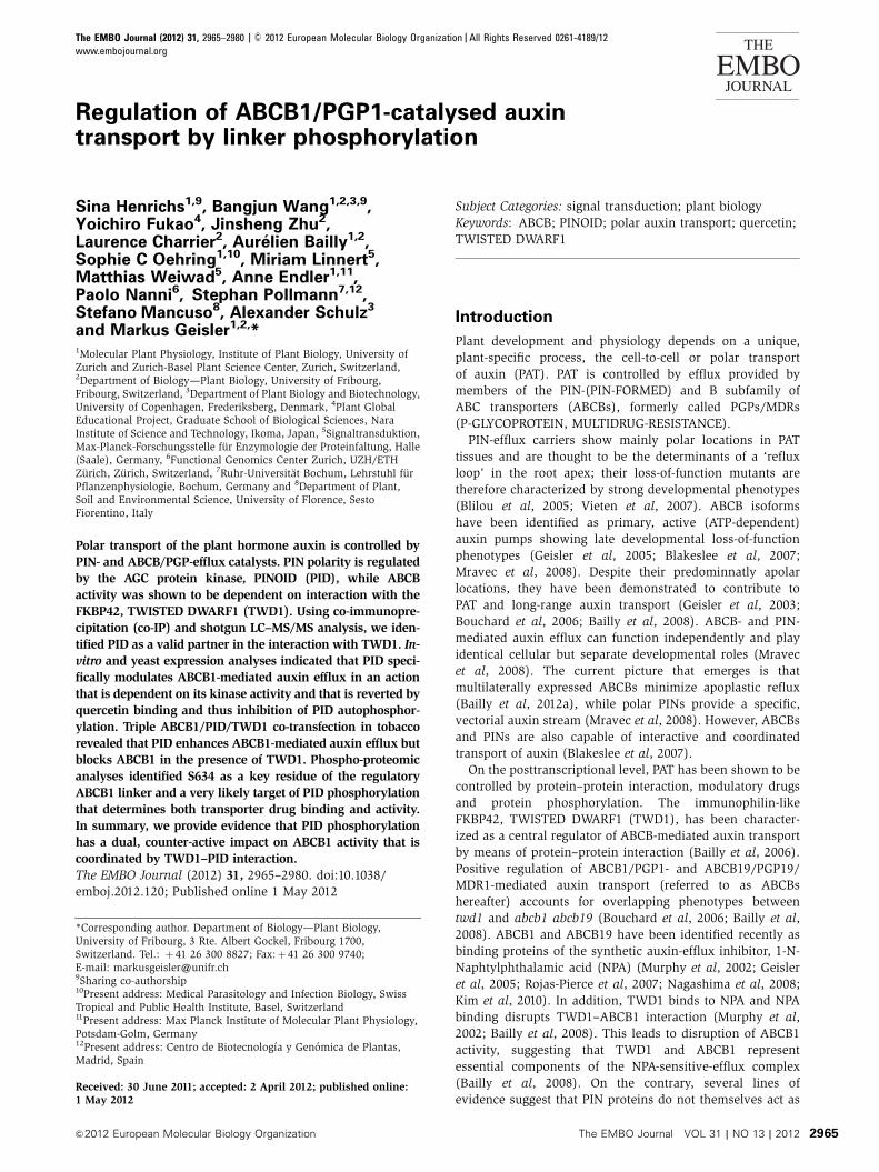
Regulation of ABCB1/PGP1-catalysed auxin transport by linker phosphorylation Sina Henrichs 1,9 , Bangjun Wang 1,2,3,9 , Yoichiro Fukao 4 , Jinsheng Zhu 2 , Laurence Charrier 2 , Aure ´ lien Bailly 1,2 , Sophie C Oehring 1,10 , Miriam Linnert 5 , Matthias Weiwad 5 , Anne Endler 1,11 , Paolo Nanni 6 , Stephan Pollmann 7,12 , Stefano Mancuso 8 , Alexander Schulz 3 and Markus Geisler 1,2, * 1 Molecular Plant Physiology, Institute of Plant Biology, University of Zurich and Zurich-Basel Plant Science Center, Zurich, Switzerland, 2 Department of Biology—Plant Biology, University of Fribourg, Fribourg, Switzerland, 3 Department of Plant Biology and Biotechnology, University of Copenhagen, Frederiksberg, Denmark, 4 Plant Global Educational Project, Graduate School of Biological Sciences, Nara Institute of Science and Technology, Ikoma, Japan, 5 Signaltransduktion, Max-Planck-Forschungsstelle fu ¨ r Enzymologie der Proteinfaltung, Halle (Saale), Germany, 6 Functional Genomics Center Zurich, UZH/ETH Zu ¨rich, Zu ¨rich, Switzerland, 7 Ruhr-Universita ¨t Bochum, Lehrstuhl fu ¨r Pflanzenphysiologie, Bochum, Germany and 8 Department of Plant, Soil and Environmental Science, University of Florence, Sesto Fiorentino, Italy Polar transport of the plant hormone auxin is controlled by PIN- and ABCB/PGP-efflux catalysts. PIN polarity is regulated by the AGC protein kinase, PINOID (PID), while ABCB activity was shown to be dependent on interaction with the FKBP42, TWISTED DWARF1 (TWD1). Using co-immunopre- cipitation (co-IP) and shotgun LC–MS/MS analysis, we iden- tified PID as a valid partner in the interaction with TWD1. In- vitro and yeast expression analyses indicated that PID speci- fically modulates ABCB1-mediated auxin efflux in an action that is dependent on its kinase activity and that is reverted by quercetin binding and thus inhibition of PID autophosphor- ylation. Triple ABCB1/PID/TWD1 co-transfection in tobacco revealed that PID enhances ABCB1-mediated auxin efflux but blocks ABCB1 in the presence of TWD1. Phospho-proteomic analyses identified S634 as a key residue of the regulatory ABCB1 linker and a very likely target of PID phosphorylation that determines both transporter drug binding and activity. In summary, we provide evidence that PID phosphorylation has a dual, counter-active impact on ABCB1 activity that is coordinated by TWD1–PID interaction. The EMBO Journal (2012) 31, 2965–2980. doi:10.1038/ emboj.2012.120; Published online 1 May 2012 Subject Categories: signal transduction; plant biology Keywords: ABCB; PINOID; polar auxin transport; quercetin; TWISTED DWARF1 Introduction Plant development and physiology depends on a unique, plant-specific process, the cell-to-cell or polar transport of auxin (PAT). PAT is controlled by efflux provided by members of the PIN-(PIN-FORMED) and B subfamily of ABC transporters (ABCBs), formerly called PGPs/MDRs (P-GLYCOPROTEIN, MULTIDRUG-RESISTANCE). PIN-efflux carriers show mainly polar locations in PAT tissues and are thought to be the determinants of a ‘reflux loop’ in the root apex; their loss-of-function mutants are therefore characterized by strong developmental phenotypes (Blilou et al, 2005; Vieten et al, 2007). ABCB isoforms have been identified as primary, active (ATP-dependent) auxin pumps showing late developmental loss-of-function phenotypes (Geisler et al, 2005; Blakeslee et al, 2007; Mravec et al, 2008). Despite their predominnatly apolar locations, they have been demonstrated to contribute to PAT and long-range auxin transport (Geisler et al, 2003; Bouchard et al, 2006; Bailly et al, 2008). ABCB- and PIN- mediated auxin efflux can function independently and play identical cellular but separate developmental roles (Mravec et al, 2008). The current picture that emerges is that multilaterally expressed ABCBs minimize apoplastic reflux (Bailly et al, 2012a), while polar PINs provide a specific, vectorial auxin stream (Mravec et al, 2008). However, ABCBs and PINs are also capable of interactive and coordinated transport of auxin (Blakeslee et al, 2007). On the posttranscriptional level, PAT has been shown to be controlled by protein–protein interaction, modulatory drugs and protein phosphorylation. The immunophilin-like FKBP42, TWISTED DWARF1 (TWD1), has been character- ized as a central regulator of ABCB-mediated auxin transport by means of protein–protein interaction (Bailly et al, 2006). Positive regulation of ABCB1/PGP1- and ABCB19/PGP19/ MDR1-mediated auxin transport (referred to as ABCBs hereafter) accounts for overlapping phenotypes between twd1 and abcb1 abcb19 (Bouchard et al, 2006; Bailly et al, 2008). ABCB1 and ABCB19 have been identified recently as binding proteins of the synthetic auxin-efflux inhibitor, 1-N- Naphtylphthalamic acid (NPA) (Murphy et al, 2002; Geisler et al, 2005; Rojas-Pierce et al, 2007; Nagashima et al, 2008; Kim et al, 2010). In addition, TWD1 binds to NPA and NPA binding disrupts TWD1–ABCB1 interaction (Murphy et al, 2002; Bailly et al, 2008). This leads to disruption of ABCB1 activity, suggesting that TWD1 and ABCB1 represent essential components of the NPA-sensitive-efflux complex (Bailly et al, 2008). On the contrary, several lines of evidence suggest that PIN proteins do not themselves act as *Corresponding author. Department of Biology—Plant Biology, University of Fribourg, 3 Rte. Albert Gockel, Fribourg 1700, Switzerland. Tel.: þ 41 26 300 8827; Fax: þ 41 26 300 9740; E-mail: [email protected] 9 Sharing co-authorship 10 Present address: Medical Parasitology and Infection Biology, Swiss Tropical and Public Health Institute, Basel, Switzerland 11 Present address: Max Planck Institute of Molecular Plant Physiology, Potsdam-Golm, Germany 12 Present address: Centro de Biotecnologı ´a y Geno ´mica de Plantas, Madrid, Spain Received: 30 June 2011; accepted: 2 April 2012; published online: 1 May 2012 The EMBO Journal (2012) 31, 2965–2980 | & 2012 European Molecular Biology Organization | All Rights Reserved 0261-4189/12 www.embojournal.org EMBO THE EMBO JOURNAL THE EMBO JOURNAL 2965 & 2012 European Molecular Biology Organization The EMBO Journal VOL 31 | NO 13 | 2012
Transcript of Regulation of ABCB1/PGP1-catalysed auxin transport by linker
Regulation of ABCB1/PGP1-catalysed auxintransport by linker phosphorylation
Sina Henrichs1,9, Bangjun Wang1,2,3,9,Yoichiro Fukao4, Jinsheng Zhu2,Laurence Charrier2, Aurelien Bailly1,2,Sophie C Oehring1,10, Miriam Linnert5,Matthias Weiwad5, Anne Endler1,11,Paolo Nanni6, Stephan Pollmann7,12,Stefano Mancuso8, Alexander Schulz3
and Markus Geisler1,2,*1Molecular Plant Physiology, Institute of Plant Biology, University ofZurich and Zurich-Basel Plant Science Center, Zurich, Switzerland,2Department of Biology—Plant Biology, University of Fribourg,Fribourg, Switzerland, 3Department of Plant Biology and Biotechnology,University of Copenhagen, Frederiksberg, Denmark, 4Plant GlobalEducational Project, Graduate School of Biological Sciences, NaraInstitute of Science and Technology, Ikoma, Japan, 5Signaltransduktion,Max-Planck-Forschungsstelle fur Enzymologie der Proteinfaltung, Halle(Saale), Germany, 6Functional Genomics Center Zurich, UZH/ETHZurich, Zurich, Switzerland, 7Ruhr-Universitat Bochum, Lehrstuhl furPflanzenphysiologie, Bochum, Germany and 8Department of Plant,Soil and Environmental Science, University of Florence, SestoFiorentino, Italy
Polar transport of the plant hormone auxin is controlled by
PIN- and ABCB/PGP-efflux catalysts. PIN polarity is regulated
by the AGC protein kinase, PINOID (PID), while ABCB
activity was shown to be dependent on interaction with the
FKBP42, TWISTED DWARF1 (TWD1). Using co-immunopre-
cipitation (co-IP) and shotgun LC–MS/MS analysis, we iden-
tified PID as a valid partner in the interaction with TWD1. In-
vitro and yeast expression analyses indicated that PID speci-
fically modulates ABCB1-mediated auxin efflux in an action
that is dependent on its kinase activity and that is reverted by
quercetin binding and thus inhibition of PID autophosphor-
ylation. Triple ABCB1/PID/TWD1 co-transfection in tobacco
revealed that PID enhances ABCB1-mediated auxin efflux but
blocks ABCB1 in the presence of TWD1. Phospho-proteomic
analyses identified S634 as a key residue of the regulatory
ABCB1 linker and a very likely target of PID phosphorylation
that determines both transporter drug binding and activity.
In summary, we provide evidence that PID phosphorylation
has a dual, counter-active impact on ABCB1 activity that is
coordinated by TWD1–PID interaction.
The EMBO Journal (2012) 31, 2965–2980. doi:10.1038/
emboj.2012.120; Published online 1 May 2012
Subject Categories: signal transduction; plant biologyKeywords: ABCB; PINOID; polar auxin transport; quercetin;
TWISTED DWARF1
Introduction
Plant development and physiology depends on a unique,
plant-specific process, the cell-to-cell or polar transport
of auxin (PAT). PAT is controlled by efflux provided by
members of the PIN-(PIN-FORMED) and B subfamily of
ABC transporters (ABCBs), formerly called PGPs/MDRs
(P-GLYCOPROTEIN, MULTIDRUG-RESISTANCE).
PIN-efflux carriers show mainly polar locations in PAT
tissues and are thought to be the determinants of a ‘reflux
loop’ in the root apex; their loss-of-function mutants are
therefore characterized by strong developmental phenotypes
(Blilou et al, 2005; Vieten et al, 2007). ABCB isoforms
have been identified as primary, active (ATP-dependent)
auxin pumps showing late developmental loss-of-function
phenotypes (Geisler et al, 2005; Blakeslee et al, 2007;
Mravec et al, 2008). Despite their predominnatly apolar
locations, they have been demonstrated to contribute to
PAT and long-range auxin transport (Geisler et al, 2003;
Bouchard et al, 2006; Bailly et al, 2008). ABCB- and PIN-
mediated auxin efflux can function independently and play
identical cellular but separate developmental roles (Mravec
et al, 2008). The current picture that emerges is that
multilaterally expressed ABCBs minimize apoplastic reflux
(Bailly et al, 2012a), while polar PINs provide a specific,
vectorial auxin stream (Mravec et al, 2008). However, ABCBs
and PINs are also capable of interactive and coordinated
transport of auxin (Blakeslee et al, 2007).
On the posttranscriptional level, PAT has been shown to be
controlled by protein–protein interaction, modulatory drugs
and protein phosphorylation. The immunophilin-like
FKBP42, TWISTED DWARF1 (TWD1), has been character-
ized as a central regulator of ABCB-mediated auxin transport
by means of protein–protein interaction (Bailly et al, 2006).
Positive regulation of ABCB1/PGP1- and ABCB19/PGP19/
MDR1-mediated auxin transport (referred to as ABCBs
hereafter) accounts for overlapping phenotypes between
twd1 and abcb1 abcb19 (Bouchard et al, 2006; Bailly et al,
2008). ABCB1 and ABCB19 have been identified recently as
binding proteins of the synthetic auxin-efflux inhibitor, 1-N-
Naphtylphthalamic acid (NPA) (Murphy et al, 2002; Geisler
et al, 2005; Rojas-Pierce et al, 2007; Nagashima et al, 2008;
Kim et al, 2010). In addition, TWD1 binds to NPA and NPA
binding disrupts TWD1–ABCB1 interaction (Murphy et al,
2002; Bailly et al, 2008). This leads to disruption of ABCB1
activity, suggesting that TWD1 and ABCB1 represent
essential components of the NPA-sensitive-efflux complex
(Bailly et al, 2008). On the contrary, several lines of
evidence suggest that PIN proteins do not themselves act as
*Corresponding author. Department of Biology—Plant Biology,University of Fribourg, 3 Rte. Albert Gockel, Fribourg 1700,Switzerland. Tel.: þ 41 26 300 8827; Fax:þ 41 26 300 9740;E-mail: [email protected] co-authorship10Present address: Medical Parasitology and Infection Biology, SwissTropical and Public Health Institute, Basel, Switzerland11Present address: Max Planck Institute of Molecular Plant Physiology,Potsdam-Golm, Germany12Present address: Centro de Biotecnologıa y Genomica de Plantas,Madrid, Spain
Received: 30 June 2011; accepted: 2 April 2012; published online:1 May 2012
The EMBO Journal (2012) 31, 2965–2980 | & 2012 European Molecular Biology Organization | All Rights Reserved 0261-4189/12
www.embojournal.org
EMBO
THE
EMBOJOURNAL
THE
EMBOJOURNAL
2965&2012 European Molecular Biology Organization The EMBO Journal VOL 31 | NO 13 | 2012
direct targets of NPA (Lomax et al, 1995; Luschnig, 2001;
Kim et al, 2010).
The serine–threonine kinase PINOID (PID) and the trimeric
serine–threonine protein phosphatase 2A (PP2A) direct the
polar targeting of PIN proteins (Friml et al, 2004;
Michniewicz et al, 2007). The current model suggests that
PID and PP2A antagonistically determine the fate of PIN
cargoes for trafficking to the appropriate membrane by
(de)phosphorylating conserved motifs of the hydrophilic
loop of PIN proteins (Kleine-Vehn et al, 2009; Dhonukshe
et al, 2010; Huang et al, 2010; Ding et al, 2011).
The regulatory A subunit, PP2AA1, called ROOTS CURL IN
NPA1 (RCN1), is a negative regulator of basipetal transport in
the root and as a consequence rcn1 roots exhibit a significant
delay in gravitropism, consistent with an increased basipetal
auxin transport (Sukumar et al, 2009; Rashotte et al, 2001).
Importantly, the rcn1 gravitropic phenotype can be rescued
by low concentrations of NPA, a concentration that is
sufficient to block gravitropism in wild-type seedlings
(Muday and DeLong, 2001). On the other hand, acropetal
auxin transport is unaffected in rcn1, but shows a dramatic
loss of NPA inhibition. Interestingly, rcn1 pin2 double-mutant
analyses indicate that elevated basipetal transport in rcn1
does not require PIN2, leading to the suggestion that an NPA-
binding protein is involved in this process (Rashotte et al,
2001).
PID belongs to the AGC family of serine/threonine kinases,
and forms—together with AGC3-4/PID2, WAG1 and WAG2—
the clade AGC3 (Galvan-Ampudia and Offringa, 2007). PID
loss- or gain-of-function changes the apical (shoot-wards) or
basal (root-wards) cellular localization of PIN proteins
influencing the direction of the auxin movement (Friml
et al, 2004). As a consequence, in the pid mutant, PIN1
localizes to the basal membrane of epidermal cells, which
in turn redirects auxin away from the meristem and prevents
the initiation of new lateral organs. This results in a pin-
shaped inflorescence (Christensen et al, 2000). On the other
hand, PID overexpression leads to a basal-to-apical switch of
PIN1, PIN2 and PIN4 in root cortex and lateral root cap cells,
and finally to a collapse of the root meristem probably due to
auxin depletion (Michniewicz et al, 2007).
WAG1WAG2 loss-of-function mutations show an auxin-
dependent root waving phenotype, and root curling is more
resistant to NPA (Santner and Watson, 2006). Recently, PID,
WAG1 and WAG2 were shown to phosphorylate PIN carriers
at a conserved TPRXS(N/S) motif in the central hydrophilic
loop leading to PIN recruitment into the apical recycling
pathway (Dhonukshe et al, 2010; Huang et al, 2010).
Moreover, disruption of PID and its three closest
homologues completely abolishes the formation of
cotyledons (Cheng et al, 2008). These findings, together
with the fact that WAG1 and WAG2 are apolar and plasma-
membrane-associated, suggested that AGC3 kinases act in the
same or in a parallel regulatory pathway of PAT (Santner and
Watson, 2006). Very recently, photoreceptor AGC4 kinase
PHOTOTROPIN1 (phot1) was shown to phosphorylate
nucleotide-binding domains (NBDs) of ABCB19 inhibiting
its efflux activity (Christie et al, 2011).
Genetic and pharmacological analyses revealed that PID
and PP2A antagonistically regulate basipetal auxin transport
and gravitropic response in the root tip (Sukumar et al, 2009).
Loss of PID activity alters the PIN2-mediated basipetal auxin
transport and impedes the gravitropic response, without
causing an obvious change in PIN2 cellular polarity. This
finding indicates that PID promotes and enhances root
gravitropism, but is not absolutely required. Furthermore,
PID appears to have a specific regulatory effect on the
basipetal transport machinery in the root, since the
acropetal transport is unaffected in pid.
All these data are widely consistent with the concept of PID
as a positive regulator of IAA efflux (Lee and Cho, 2006). The
direct effect of PID on individual transporter activities,
however, has not yet been addressed. Here, we identify and
characterize PID as a relevant partner of the TWD1/ABCB
subcomplex. Our data suggest that PID, besides its function
as a molecular switch of PIN polarity, has a direct, dual
impact on ABCB-mediated auxin-efflux activity. Transporter
regulation is reversed by binding of the protein-kinase
inhibitor, quercetin, a modulator of auxin transport.
Results
Identification of PINOID as a partner in the ABCB–TWD1
auxin-efflux complex
With the aim of identifying novel components of the auxin-
efflux complex, characterized by ABCB1 and TWD1, we
employed an IP approach followed by shotgun mass spectro-
metry analysis of TAPa-tagged TWD1 (TAPa-TWD1). Origi-
nally, we chose second-generation TAPa tagging, thus offer-
ing the possibility of IgG-BD-, 6xHIS- and 9xMYC epitope
purification, that had been optimized for the identification of
Arabidopsis protein complexes by two-step affinity purifica-
tion. However, as IP using the HIS-tag only gave poor protein
retention and the IgG-BD interfered with the MS analysis,
we employed an one-step MYC IP. As starting material, we
used total microsomes of 9-dag (days after germination)
Arabidopsis roots, thus reducing typical nonspecific contami-
nants (like ribosomal proteins, chaperons and Rubisco;
Supplementary Table S1). To gain further specificity of
TWD1-interacting proteins, we subtracted identified vector
control (35S:TAPa) proteins from pulled-down TAPa-TWD1
proteins, allowing elimination of proteins binding to the
TAPa-tag alone (Supplementary Table S1). Finally, proteins
with a score above 30 were considered as significant partners
(Figure 1A). This procedure was repeated with each of the
two independent transformants resulting in essentially the
same TWD1 partners.
Besides TWD1 as an obvious dominant pulled-down pro-
tein (protein score of 129, 13.4% coverage; Figure 1A), we
found a so-far uncharacterized polynucleotide adenylyltrans-
ferase-like protein (At3G48830) and an unknown protein
(At4G25920). In addition, two protein kinases, the histidine-
like kinase AHK5/CYTOKININ INDEPENDENT2 (At5G10720)
that regulates root elongation (Iwama et al, 2007) and the
ACG kinase PINOID (At2G34650), and the catalytic domain of
the PP2C-type protein phosphatase, AP2C1 (At2G30020),
were also identified. AP2C1 is known to be involved in
innate immunity responses by the negative regulation
of the map kinases MPK4 and MPK6 (Schweighofer et al,
2007).
PINOID (PID) is a well-known key player in polar auxin
transport regulation (Galvan-Ampudia and Offringa, 2007)
and was therefore chosen for further analysis. TWD1/ABCB1
and PID show overlapping locations, mostly in epidermal
Regulation of ABCB1 by linker phosphorylationS Henrichs et al
2966 The EMBO Journal VOL 31 | NO 13 | 2012 &2012 European Molecular Biology Organization
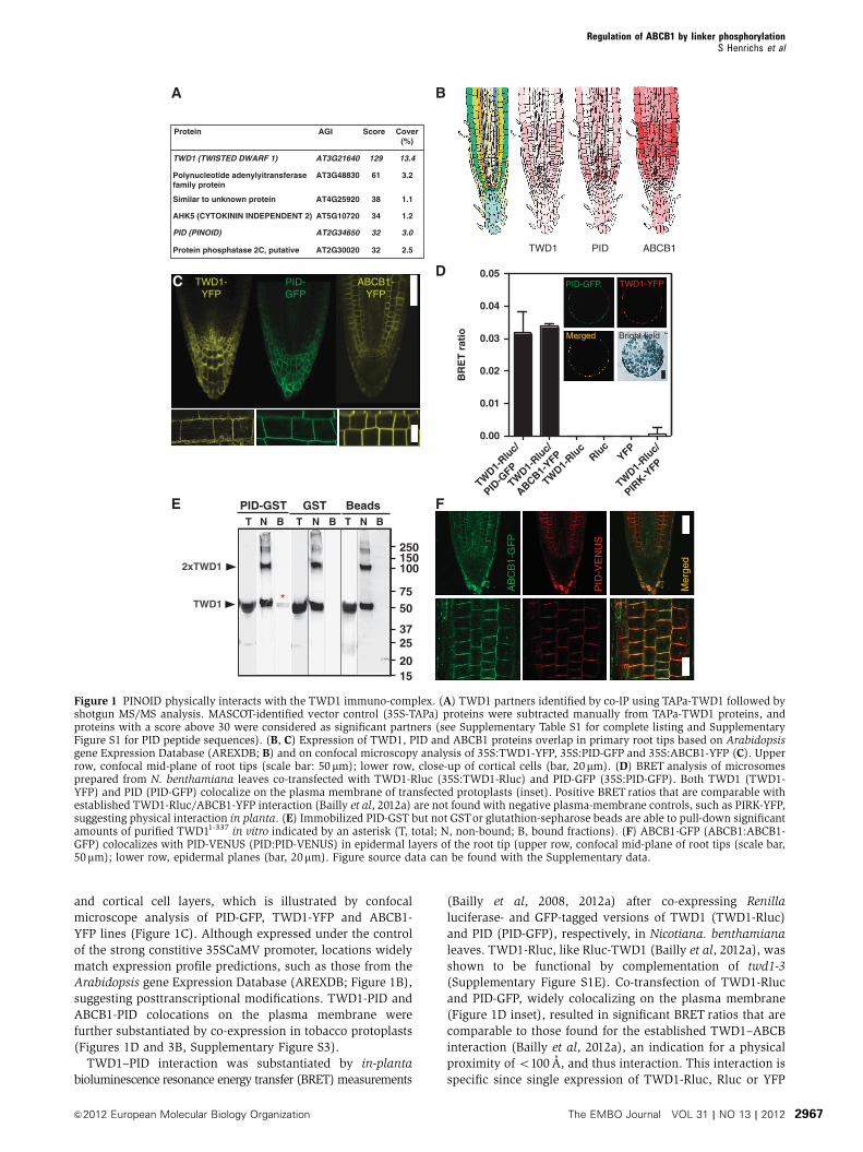
and cortical cell layers, which is illustrated by confocal
microscope analysis of PID-GFP, TWD1-YFP and ABCB1-
YFP lines (Figure 1C). Although expressed under the control
of the strong constitive 35SCaMV promoter, locations widely
match expression profile predictions, such as those from the
Arabidopsis gene Expression Database (AREXDB; Figure 1B),
suggesting posttranscriptional modifications. TWD1-PID and
ABCB1-PID colocations on the plasma membrane were
further substantiated by co-expression in tobacco protoplasts
(Figures 1D and 3B, Supplementary Figure S3).
TWD1–PID interaction was substantiated by in-planta
bioluminescence resonance energy transfer (BRET) measurements
(Bailly et al, 2008, 2012a) after co-expressing Renilla
luciferase- and GFP-tagged versions of TWD1 (TWD1-Rluc)
and PID (PID-GFP), respectively, in Nicotiana. benthamiana
leaves. TWD1-Rluc, like Rluc-TWD1 (Bailly et al, 2012a), was
shown to be functional by complementation of twd1-3
(Supplementary Figure S1E). Co-transfection of TWD1-Rluc
and PID-GFP, widely colocalizing on the plasma membrane
(Figure 1D inset), resulted in significant BRET ratios that are
comparable to those found for the established TWD1–ABCB
interaction (Bailly et al, 2012a), an indication for a physical
proximity of o100 A, and thus interaction. This interaction is
specific since single expression of TWD1-Rluc, Rluc or YFP
PID-GFP TWD1-YFP
Merged Bright field
TWD1 PID ABCB1
PID
-VE
NU
S
AB
CB
1-G
FP
Mer
ged
0.00
0.01
0.02
0.03
0.04
0.05
BR
ET
rati
o
TWD1-
Rluc/
PID-G
FP
TWD1-
Rluc/
ABCB1-YFP
TWD1-
Rluc
Rluc
YFP
TWD1-
Rluc/
PIRK-Y
FP
T N B T N TB N B
250150100
7550
3725
2015
PID-GST GST Beads
2xTWD1
TWD1 *
TWD1-YFP
PID-GFP
ABCB1-YFP
CD
BA
E F
Protein AGI Score Cover(%)
13.4129AT3G21640TWD1 (TWISTED DWARF 1)
Polynucleotide adenylyitransferasefamily protein
Protein phosphatase 2C, putative
Similar to unknown protein
AHK5 (CYTOKININ INDEPENDENT 2)
PID (PINOID)
AT3G48830
AT4G25920
AT5G10720
AT2G34650
AT2G30020
3.261
1.138
1.234
2.5
3.032
32
Figure 1 PINOID physically interacts with the TWD1 immuno-complex. (A) TWD1 partners identified by co-IP using TAPa-TWD1 followed byshotgun MS/MS analysis. MASCOT-identified vector control (35S-TAPa) proteins were subtracted manually from TAPa-TWD1 proteins, andproteins with a score above 30 were considered as significant partners (see Supplementary Table S1 for complete listing and SupplementaryFigure S1 for PID peptide sequences). (B, C) Expression of TWD1, PID and ABCB1 proteins overlap in primary root tips based on Arabidopsisgene Expression Database (AREXDB; B) and on confocal microscopy analysis of 35S:TWD1-YFP, 35S:PID-GFP and 35S:ABCB1-YFP (C). Upperrow, confocal mid-plane of root tips (scale bar: 50 mm); lower row, close-up of cortical cells (bar, 20mm). (D) BRET analysis of microsomesprepared from N. benthamiana leaves co-transfected with TWD1-Rluc (35S:TWD1-Rluc) and PID-GFP (35S:PID-GFP). Both TWD1 (TWD1-YFP) and PID (PID-GFP) colocalize on the plasma membrane of transfected protoplasts (inset). Positive BRET ratios that are comparable withestablished TWD1-Rluc/ABCB1-YFP interaction (Bailly et al, 2012a) are not found with negative plasma-membrane controls, such as PIRK-YFP,suggesting physical interaction in planta. (E) Immobilized PID-GST but not GSTor glutathion-sepharose beads are able to pull-down significantamounts of purified TWD11-337 in vitro indicated by an asterisk (T, total; N, non-bound; B, bound fractions). (F) ABCB1-GFP (ABCB1:ABCB1-GFP) colocalizes with PID-VENUS (PID:PID-VENUS) in epidermal layers of the root tip (upper row, confocal mid-plane of root tips (scale bar,50mm); lower row, epidermal planes (bar, 20mm). Figure source data can be found with the Supplementary data.
Regulation of ABCB1 by linker phosphorylationS Henrichs et al
2967&2012 European Molecular Biology Organization The EMBO Journal VOL 31 | NO 13 | 2012

alone or TWD1-Rluc in combination with the non-related,
plasma-membrane-bound protein kinase, PIRK (PIRK-YFP),
only resulted in negligible BRET ratios.
Identification of PID as a TAPa-TWD1 partner does not
necessarily imply direct physical interaction as the PID–
TWD1 interaction might also be mediated via a third
TWD1-interacting protein. Moreover, only one PID peptide
with a MASCOT score of 32 was found in the MS analysis
(Supplementary Figure S1). Therefore, in order to verify the
IP data and to test a direct mode of interaction, we performed
in vitro pull-down experiments using recombinant PID-GST
(Christensen et al, 2000) and TWD11-337 protein. TWD11-337
purified as described (Kamphausen et al, 2002) contains all
functional domains, such as the FKBD, the CaM-binding and
TPR domain, except the C-terminal hydrophobic in-plane
membrane anchor. Indeed, PID-GST, but not GST alone nor
the empty-beads control, was able to pull-down small but
significant amounts of TWD11-337 (Figure 1E).
In summary, our results demonstrate the utility of the IP
approach employed for discovering valid protein–protein
interactions of auxin transport complexes in Arabidopsis
and suggest a relevant TWD1–PID interaction in planta.
PID has a dual impact on ABCB1-mediated auxin efflux
PID defines polar PIN locations and thus the directionality of
auxin streams by direct PIN phosphorylation (Dhonukshe
et al, 2010; Huang et al, 2010). A comparable mechanism has
not so far been demonstrated for mainly nonpolar ABCBs or
for TWD1, which functions as a regulator of ABCB1-efflux
activity (Bouchard et al, 2006; Bailly et al, 2008). Therefore,
we quantified ABCB1-mediated auxin efflux in the presence
and absence of MYC-tagged PID in yeast. Surprisingly, PID
significantly reduced ABCB1, but not vector control
(background), IAA efflux by roughly 30% (Figure 2A). This
inhibition corresponds to a 70% inhibition of ABCB1-specific
efflux and is comparable to that found for ABCB1/TWD1
co-expression in yeast (Bailly et al, 2008). This inhibitory
effect, caused by PID, was specific, as it was not found with
the mutated, kinase-negative MPID (Christensen et al, 2000)
or the non-related GSK-3-like kinase BRASSINOSTEROID-
INSENSITIVE2 (BIN2), a negative regulator of brass-
inosteroid (BR) signalling and cell elongation (Vert, 2008).
The specificity of PID-induced inhibition of IAA transport was
further underlined by the fact that no effect was found for the
unspecific, diffusion control, benzoic acid (BA) (Figure 2A
lower panel).
Moreover, PID or MPID co-expression did not substantially
alter ABCB1-plasma- membrane expression or localization in
yeast (Figure 2B and C, Supplementary Figure S2A). ABCB1-
YFP localizes primarily to raft-like structures at the bound-
aries of the plasma membrane as previously shown
(Figure 2B; Bailly et al, 2008) and to the plasma membrane
as demonstrated by comparison of anti-GFP immune-positive
fractions in comparison with the plasma-membrane Hþ -
ATPase, PMA1 (Supplementary Figure S2).
To further substantiate our yeast-generated results, we
established a novel heterologous plant transport system that
allowed quantification of auxin efflux of ABCB1, PID and
TWD1 combinations from mesophyll protoplasts that were
isolated from co-transfected N. benthamiana leaves. In agree-
ment with described roles for PID on auxin-efflux activity
(Lee and Cho, 2006), PID strongly activated ABCB1-catalysed
IAA and NAA efflux. This action is specific as PID expression
alone enhanced vector control IAA and NAA efflux only
slightly, probably by activation of endogenous tobacco
ABCB-type auxin exporters. Moreover, closely related AGC3
kinase, WAG1 sharing overlapping functionality and 39%
protein sequence identity with PID (Dhonukshe et al, 2010;
Huang et al, 2010), had no significant effect on ABCB1.
In order to address the role of TWD1 in PID-mediated
activation of ABCB1, we quantified auxin efflux from
ABCB1-YFP
+ Control + PID + MPID I + BIN2
250ABCB1
ABCB1+PID
ABCB1+MPID
ABCB1+BIN
2
150ABCB1-YFP
H+-ATPase 100
A
B
C
0
50
100
BA
exp
ort (%
of
AB
CB
1 ex
po
rt)
IAA
exp
ort
*
0
Control
PID
ABCB1
ABCB1+PID
ABCB1+MPID
ABCB1+BIN
2
50
100
Figure 2 PID modulates ABCB1-mediated auxin efflux in yeast.(A) PID specifically inhibits ABCB1-mediated IAA export, while amutated, inactive PID, MPID or unrelated protein kinase, BIN2, hasno significant effect. Reduction of auxin retention (export) wascalculated as relative export of initial export where ABCB1 was setto 100% (mean±s.e.; n¼ 4–10). Significant differences (unpairedt-test with Welch’s correction, Po0.05) between –PID controls areindicated by asterisks. (B, C) Co-expression with PID, MPID or BIN2does not significantly alter location (B) and expression (C) ofABCB1-YFP as revealed by confocal microscopy (B) and westernanalysis (C). Each 20 mg of protein was subjected to PAGE andwestern analysis using anti-GFP and plasma-membrane marker,anti-PMA1 (Hþ -ATPase). ABCB1-YFP localizes primarily to raft-like structures and the plasma membrane (see SupplementaryFigure S2; Bailly et al, 2008). Bar, 2mm.
Regulation of ABCB1 by linker phosphorylationS Henrichs et al
2968 The EMBO Journal VOL 31 | NO 13 | 2012 &2012 European Molecular Biology Organization
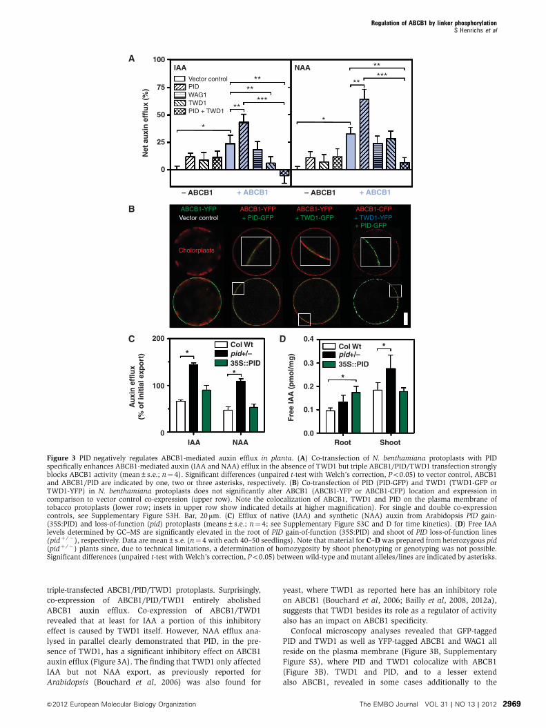
triple-transfected ABCB1/PID/TWD1 protoplasts. Surprisingly,
co-expression of ABCB1/PID/TWD1 entirely abolished
ABCB1 auxin efflux. Co-expression of ABCB1/TWD1
revealed that at least for IAA a portion of this inhibitory
effect is caused by TWD1 itself. However, NAA efflux ana-
lysed in parallel clearly demonstrated that PID, in the pre-
sence of TWD1, has a significant inhibitory effect on ABCB1
auxin efflux (Figure 3A). The finding that TWD1 only affected
IAA but not NAA export, as previously reported for
Arabidopsis (Bouchard et al, 2006) was also found for
yeast, where TWD1 as reported here has an inhibitory role
on ABCB1 (Bouchard et al, 2006; Bailly et al, 2008, 2012a),
suggests that TWD1 besides its role as a regulator of activity
also has an impact on ABCB1 specificity.
Confocal microscopy analyses revealed that GFP-tagged
PID and TWD1 as well as YFP-tagged ABCB1 and WAG1 all
reside on the plasma membrane (Figure 3B, Supplementary
Figure S3), where PID and TWD1 colocalize with ABCB1
(Figure 3B). TWD1 and PID, and to a lesser extend
also ABCB1, revealed in some cases additionally to the
100A
B
C D
IAA NAA
75Vector control
– ABCB1 – ABCB1+ ABCB1 + ABCB1
**
**
**
*
***
*****
**
*
PIDWAG1
ABCB1-YFPVector control
ABCB1-YFP
Cholorplasts
+ PID-GFPABCB1-YFP+ TWD1-GFP
ABCB1-CFP+ TWD1-YFP+ PID-GFP
TWD1PID + TWD150
Net
au
xin
eff
lux
(%)
Au
xin
eff
lux
(% o
f in
itia
l exp
ort
)
Fre
e IA
A (
pm
ol/m
g)
25
200Col Wt
IAA NAA Root Shoot
35S::PIDpid+/–
Col Wt
35S::PIDpid+/–
0.4
0.3
0.2
0.1
0.0
100
*
* *
*
0
0
Figure 3 PID negatively regulates ABCB1-mediated auxin efflux in planta. (A) Co-transfection of N. benthamiana protoplasts with PIDspecifically enhances ABCB1-mediated auxin (IAA and NAA) efflux in the absence of TWD1 but triple ABCB1/PID/TWD1 transfection stronglyblocks ABCB1 activity (mean±s.e.; n¼ 4). Significant differences (unpaired t-test with Welch’s correction, Po0.05) to vector control, ABCB1and ABCB1/PID are indicated by one, two or three asterisks, respectively. (B) Co-transfection of PID (PID-GFP) and TWD1 (TWD1-GFP orTWD1-YFP) in N. benthamiana protoplasts does not significantly alter ABCB1 (ABCB1-YFP or ABCB1-CFP) location and expression incomparison to vector control co-expression (upper row). Note the colocalization of ABCB1, TWD1 and PID on the plasma membrane oftobacco protoplasts (lower row; insets in upper row show indicated details at higher magnification). For single and double co-expressioncontrols, see Supplementary Figure S3H. Bar, 20mm. (C) Efflux of native (IAA) and synthetic (NAA) auxin from Arabidopsis PID gain-(35S:PID) and loss-of-function (pid) protoplasts (means±s.e.; n¼ 4; see Supplementary Figure S3C and D for time kinetics). (D) Free IAAlevels determined by GC–MS are significantly elevated in the root of PID gain-of-function (35S:PID) and shoot of PID loss-of-function lines(pidþ /� ), respectively. Data are mean±s.e. (n¼ 4 with each 40–50 seedlings). Note that material for C–D was prepared from heterozygous pid(pidþ /� ) plants since, due to technical limitations, a determination of homozygosity by shoot phenotyping or genotyping was not possible.Significant differences (unpaired t-test with Welch’s correction, Po0.05) between wild-type and mutant alleles/lines are indicated by asterisks.
Regulation of ABCB1 by linker phosphorylationS Henrichs et al
2969&2012 European Molecular Biology Organization The EMBO Journal VOL 31 | NO 13 | 2012
plasma-membrane signals some intracellular signals
(Figure 3, Supplementary Figure S3) that might represent
artefacts caused by the constitutive, strong overexpression
used in these assays. Importantly, and in analogy to the yeast
system, co-expression of PID and TWD1 did not alter expres-
sion of ABCB1 nor its location on the plasma membrane as
monitored microscopically (Figure 3B) and by western
detection of ABCB1-MYC when compared to the plasma-
membrane marker Hþ -ATPase, AHA2 (Supplementary
Figure S3A).
In order to address the in-planta role of PID and to clarify
its apparent dual role on ABCB1 activity, we quantified auxin
efflux from Arabidopsis PID gain- and loss-of-function alleles.
While 35S:PID mesophyll protoplasts did not show signifi-
cant differences to the wild type, auxin efflux of both native
and synthetic auxin from pid protoplasts was greatly
enhanced (Figure 3C, Supplementary Figure S3) suggesting
a negative impact on ABCB1 auxin export as found for
ABCB1/PID/TWD1 co-expression in tobacco.
In summary, these results provide evidence that PID,
dependent on the presence of TWD1, positively and nega-
tively regulates ABCB1-mediated auxin efflux in an action
that requires its kinase activity. However, based on our shoot-
derived Arabidopsis model system, PID acts as a negative
regulator in planta.
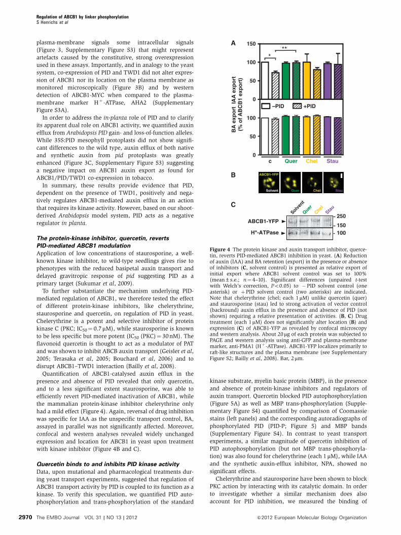
The protein-kinase inhibitor, quercetin, reverts
PID-mediated ABCB1 modulation
Application of low concentrations of staurosporine, a well-
known kinase inhibitor, to wild-type seedlings gives rise to
phenotypes with the reduced basipetal auxin transport and
delayed gravitropic response of pid suggesting PID as a
primary target (Sukumar et al, 2009).
To further substantiate the mechanism underlying PID-
mediated regulation of ABCB1, we therefore tested the effect
of different protein-kinase inhibitors, like chelerythrine,
staurosporine and quercetin, on regulation of PID in yeast.
Chelerythrine is a potent and selective inhibitor of protein
kinase C (PKC; IC50¼ 0.7 mM), while staurosporine is known
to be less specific but more potent (IC50 (PKC)¼ 30 nM). The
flavonoid quercetin is thought to act as a modulator of PAT
and was shown to inhibit ABCB auxin transport (Geisler et al,
2005; Terasaka et al, 2005; Bouchard et al, 2006) and to
disrupt ABCB1–TWD1 interaction (Bailly et al, 2008).
Quantification of ABCB1-catalysed auxin efflux in the
presence and absence of PID revealed that only quercetin,
and to a less significant extent staurosporine, was able to
efficiently revert PID-mediated inactivation of ABCB1, while
the mammalian protein-kinase inhibitor chelerythrine only
had a mild effect (Figure 4). Again, reversal of drug inhibition
was specific for IAA as the unspecific transport control, BA,
assayed in parallel was not significantly affected. Moreover,
confocal and western analyses revealed widely unchanged
expression and location for ABCB1 in yeast upon treatment
with kinase inhibitor (Figure 4B and C).
Quercetin binds to and inhibits PID kinase activity
Data, upon mutational and pharmacological treatments dur-
ing yeast transport experiments, suggested that regulation of
ABCB1 transport activity by PID is coupled to its function as a
kinase. To verify this speculation, we quantified PID auto-
phosphorylation and trans-phosphorylation of the standard
kinase substrate, myelin basic protein (MBP), in the presence
and absence of protein-kinase inhibitors and regulators of
auxin transport. Quercetin blocked PID autophosphorylation
(Figure 5A) as well as MBP trans-phosphorylation (Supple-
mentary Figure S4) quantified by comparison of Coomassie
stains (left panels) and the corresponding autoradiographs of
phosphorylated PID (PID-P; Figure 5) and MBP bands
(Supplementary Figure S4). In contrast to yeast transport
experiments, a similar magnitude of quercetin inhibition of
PID autophosphorylation (but not MBP trans-phosphoryla-
tion) was also found for chelerythrine (each 1 mM), while IAA
and the synthetic auxin-efflux inhibitor, NPA, showed no
significant effects.
Chelerythrine and staurosporine have been shown to block
PKC action by interacting with its catalytic domain. In order
to investigate whether a similar mechanism does also
account for PID inhibition, we measured the binding of
150ABCB1-YFP
H+-ATPase 100
0
50
100
150
–PID +PID
c Quer Chel Stau0
50
100
BA
exp
ort
IA
A e
xpo
rt(%
of
AB
CB
1 ex
po
rt)
A
B
C
ABCB1-YFP
Solvent Quer Chel Stau
250
***
QuerChel
Solvent
Stau
Figure 4 The protein kinase and auxin transport inhibitor, querce-tin, reverts PID-mediated ABCB1 inhibition in yeast. (A) Reductionof auxin (IAA) and BA retention (export) in the presence or absenceof inhibitors (C, solvent control) is presented as relative export ofinitial export where ABCB1 solvent control was set to 100%(mean±s.e.; n¼ 4–10). Significant differences (unpaired t-testwith Welch’s correction, Po0.05) to �PID solvent control (oneasterisk) or þPID solvent control (two asterisks) are indicated.Note that chelerythrine (chel; each 1mM) unlike quercetin (quer)and staurosporine (stau) led to strong activation of vector control(backround) auxin efflux in the presence and absence of PID (notshown) requiring a relative presentation of activities. (B, C) Drugtreatment (each 1mM) does not significantly alter location (B) andexpression (C) of ABCB1-YFP as revealed by confocal microscopyand western analysis. About 20mg of each protein was subjected toPAGE and western analysis using anti-GFP and plasma-membranemarker, anti-PMA1 (Hþ -ATPase). ABCB1-YFP localizes primarily toraft-like structures and the plasma membrane (see SupplementaryFigure S2; Bailly et al, 2008). Bar, 2mm.
Regulation of ABCB1 by linker phosphorylationS Henrichs et al
2970 The EMBO Journal VOL 31 | NO 13 | 2012 &2012 European Molecular Biology Organization

radiolabelled quercetin to purified PID-GST and GST alone.
Analysis of specific PID binding (binding to PID-GST minus
binding to GST alone) showed significant quercetin binding
(72.9±9.4 pmol/mg protein), while binding of NPA, NAA and
BA was negligible (Figure 5C). Interestingly, despite its
ineffectiveness in altering PID autophosphorylation, small
but significant amounts of IAA were also bound to PID
(20.2±4.2 pmol/mg). This phenomenon is currently under
further investigation.
Direct binding of quercetin to PID was further supported by
the fact that microsomes from PID loss-of-function alleles
(pidþ /� ) showed drastically reduced quercetin binding
(16.1±14.8% of wild-type), while gain-of-function lines
(35S:PID) showed a significantly higher signal (137.9±20.4%
of wild-type; Figure 5D). Surprisingly, interfering with PID
expression had an inverse effect on NPA binding compared to
quercetin: while NPA binding was enhanced in pidþ /�
by about a factor of 2, it was reduced to one-third in
35S:PID. This implies that PID, because it apparently does
not bind NPA itself (Figure 5C), alters NPA-binding
capacities of third-party NPA-binding proteins, such as ABCB1
or TWD1.
In summary, these data support the concept that PID is an
in vivo target of quercetin that negatively regulates PID
activity by direct drug binding.
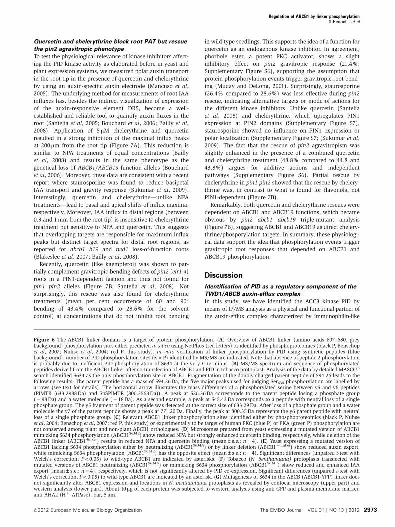
The ABCB1 linker is a target of protein phosphorylation
The non-plant ABCB linker region of about 60 amino acids
was identified to be subject of PKA (protein kinase A) or PKC
phosphorylation altering its transport and associated ATPase
activity (Chambers et al, 1994; Castro et al, 1999; Conseil
et al, 2001). Proteomics approaches predicted three clusters
of phosphorylation sites in the linker of Arabidopsis—
ABCB1, ABCB4, ABCB11 and ABCB21 (Nuhse et al, 2004)—
but the impact of these events on their activity is not entirely
clear. Interestingly, most of the phosphorylation sites
identified by in-silico prediction, experimentally or by
phospho-proteomics, are well conserved among plant or
animal orthologues, respectively, but are normally not
shared among kingdoms (Figure 6C).
In order to explore whether the Arabidopis ABCB1 linker is
indeed a target of protein-kinase phosphorylation under our
experimental conditions, we analysed ABCB1 phosphoryla-
tion by LC—ESI—MS/MS after co-transfection with PID in
tobacco leaves. The annotated MS spectra reported in
Figure 6B shows the identified serine 634 (S634), detected
previously by phosphoproteomics (Nuhse et al, 2004) as the
only phosphorylation site in ABCB1 under our experimental
settings.
In order to demonstrate that S634 identified in planta is
indeed phosphorylated by PID and not via other kinases
being themselves regulated by PID, we performed a PID
in vitro phosphorylation analysis using overlapping peptides
(peptide 1–3) covering the first half of the linker (Figure 6A).
As a control, we used a peptide covering part of the ABCB1
C-terminus that has been shown to be phosphorylated upon
early elicitor signalling (peptide 4; Benschop et al, 2007). MS
analysis revealed that neither peptide 1, 2 nor 4 but only
peptide 3 was phosphorylated by PID, leaving S633 and S634
as PID targets. S634 phosphorylation could be, however,
excluded by using peptide 5 that contained an S634A
exchange in comparison to peptide 3, suggesting that, in
combination with previous phosphoproteomics (Nuhse
et al, 2004), S634 is indeed a relevant PID phosphorylation
site.
Interestingly, the phosphorylated peptide covering S634
(NSVSSPIMTR) showed no obvious sequence homology to
the TPRXS(N/S) motif of PIN proteins recently shown to be
phosphorylated by PID (Dhonukshe et al, 2010; Huang et al,
PIDPID-P
Coommassie Radiography
C IAA NPA C Chel C IAA NPA C Chel
0 0.01 0.1 0 0.01 0.1 �M
Quercetin
PIDPID-P
A
0
100
200
300QuercetinNPA
Col Wt
pid+/
–
35S:P
ID
Sp
ecif
icd
rug
bin
din
g(%
of
wt)
0
25
50
75
100IAANAABANPAQuercetin
Sp
ecif
ic d
rug
bin
din
g(p
mo
l/�g
pro
tein
) *
*
*
C D
1
IAA
50
75
100
125
�M0.01
C
0.11
NPA Quer
1
Chel
Sig
nal
inte
nsi
ty(%
of
solv
ent
con
tro
l)
0
PID
**
B
Figure 5 Quercetin binding blocks PID kinase activity. (A, B)In vitro autophosphorylation of PID-GST is inhibited by quercetinand chelerythrine while IAA and NPA have only slight effects.Coomassie stains (left panels) of non-phosphorylated PID (PID)was used as loading control (A). Autoradiographies (right panels) ofautophosphorylated PID (PID-P), represented by the upper band inthe Coomassie stain (Christensen et al, 2000), were quantified(B) and signal intensities were plotted against solvent controls (C;lower panel; means±s.e.; n¼ 3). Significant differences (unpairedt-test with Welch’s correction, Po0.05) to solvent controls areindicated by asterisks. (C) PID-GST binds specifically quercetinand to a lesser amount as well IAA (mean±s.e.; n¼ 4).Background drug binding to column material or GST alone(background) was corrected by subtracting specific binding tocolumn-bound GST from column-bound PID-GST. (D) Microsomesprepared from PID loss- and gain-of-function lines show reducedand enhanced specific quercetin binding, respectively, butreciprocal specific NPA binding (mean±s.d.; n¼ 4). Note thatmaterial was prepared from heterozygous pid (pidþ /� ) plantssince, due to technical limitations, a determination ofhomozygosity by shoot phenotyping or genotyping was notpossible. Reported values (C, D) are the means of specificradiolabelled drug bound in the absence of cold drug (total)minus radiolabelled drug bound in the presence of cold drug(unspecific). Significant differences (unpaired t-test with Welch’scorrection, Po0.05) between GST alone (background; A) or wild-type and mutant microsomes (B) are indicated by asterisks. Figuresource data can be found with the Supplementary data.
Regulation of ABCB1 by linker phosphorylationS Henrichs et al
2971&2012 European Molecular Biology Organization The EMBO Journal VOL 31 | NO 13 | 2012
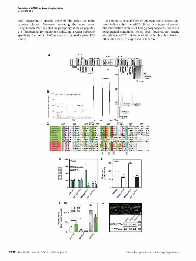
2010) suggesting a specific mode of PID action on auxin
exporter classes. Moreover, repeating the same assay
using human PKC resulted in phosphorylation of peptides
2–5 (Supplementary Figure S5) indicating a wider substrate
specificity for human PKC in comparison to the plant PID
kinase.
In summary, several lines of our own and previous ana-
lyses indicate that the ABCB1 linker is a target of protein
phosphorylation with S634 being phosphorylated under our
experimental conditions, which does, however, not strictly
exclude that ABCB1 might be additionally phosphorylated at
other sites when co-expressed in tobacco.
Peptide 20 x P
Peptide 31 x P
Peptide 10 x P
Ct (1.286)
Peptide 40 x P
A. thalianaB. campestrisS. tuberosumS. bicolorZ. maysO. sativaV. viniferaH. vulgareT. aestivumP. troglodytesC. lupus fam.R. norvegicusG. gallusH. sapiens
P PP P
P PP P PP607 617 627 637 647 667 677
680
607
Peptide 50 x P
ABCB1
Out
In
963
702
723 742 951
Nt
323
62 89 303292
100
0520 540 560 580 600 620 640 660 680 700 720 740 760 780 800 820 840 860 880
25
50
75
Rela
tive a
bundance
(%
)
m/z
A
B
C
m/z = 594.26 [NSVSpSPIMTR]2+
B1S634EB1S634AB1S634S
-YFP-YFP-YFP
250
150ABCB1-YFP
H+-ATPase 100
ABCB1
ABCB1
ABCB1
*
* * *
0
100
200
300
400
QuercetinNPA
Yeast
Net
IAA
eff
lux
(% o
f ve
cto
r co
ntr
ol)
0
50
100
150
*
* *
0
50
100
150Yeast
D E
G
**
*– PID
+ PID
Dru
g b
ind
ing
(% o
f A
BC
B1)
IAA
exp
ort
(% o
f A
BC
B1)
ABCB1
ABCB1S63
4A
ABCB1S63
4E
B1S63
4A
B1S63
4S
B1S63
4E
ABCB1–li
nker
ABCB1
ABCB1S63
4A
ABCB1S63
4E
ABCB1–li
nker
F
Regulation of ABCB1 by linker phosphorylationS Henrichs et al
2972 The EMBO Journal VOL 31 | NO 13 | 2012 &2012 European Molecular Biology Organization
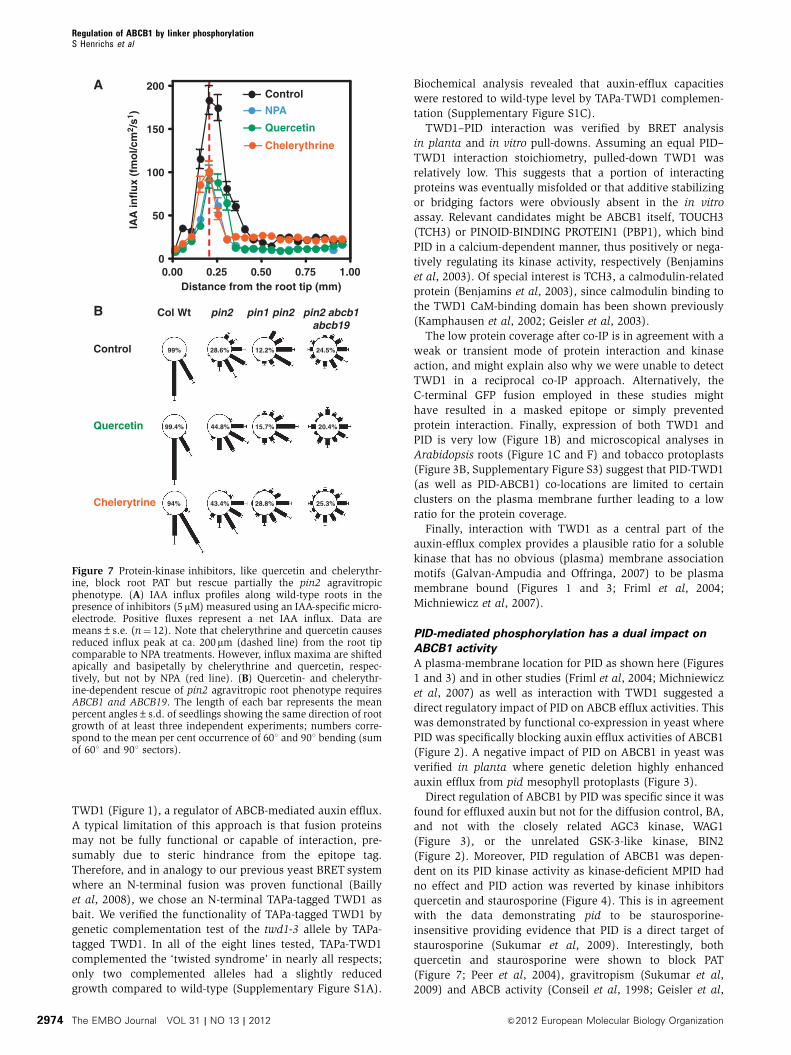
Quercetin and chelerythrine block root PAT but rescue
the pin2 agravitropic phenotype
To test the physiological relevance of kinase inhibitors affect-
ing the PID kinase activity as elaborated before in yeast and
plant expression systems, we measured polar auxin transport
in the root tip in the presence of quercetin and chelerythrine
by using an auxin-specific auxin electrode (Mancuso et al,
2005). The underlying method for measurements of root IAA
influxes has, besides the indirect visualization of expression
of the auxin-responsive element DR5, become a well-
established and reliable tool to quantify auxin fluxes in the
root (Santelia et al, 2005; Bouchard et al, 2006; Bailly et al,
2008). Application of 5mM chelerythrine and quercetin
resulted in a strong inhibition of the maximal influx peaks
at 200mm from the root tip (Figure 7A). This reduction is
similar to NPA treatments of equal concentrations (Bailly
et al, 2008) and results in the same phenotype as the
genetical loss of ABCB1/ABCB19 function alleles (Bouchard
et al, 2006). Moreover, these data are consistent with a recent
report where staurosporine was found to reduce basipetal
IAA transport and gravity response (Sukumar et al, 2009).
Interestingly, quercetin and chelerythrine—unlike NPA
treatments—lead to basal and apical shifts of influx maxima,
respectively. Moreover, IAA influx in distal regions (between
0.3 and 1 mm from the root tip) is insensitive to chelerythrine
treatment but sensitive to NPA and quercetin. This suggests
that overlapping targets are responsible for maximum influx
peaks but distinct target spectra for distal root regions, as
reported for abcb1 b19 and twd1 loss-of-function roots
(Blakeslee et al, 2007; Bailly et al, 2008).
Recently, quercetin (like kaempferol) was shown to par-
tially complement gravitropic-bending defects of pin2 (eir1-4)
roots in a PIN1-dependent fashion and thus not found for
pin1 pin2 alleles (Figure 7B; Santelia et al, 2008). Not
surprisingly, this rescue was also found for chelerythrine
treatments (mean per cent occurrence of 60 and 901
bending of 43.4% compared to 28.6% for the solvent
control) at concentrations that do not inhibit root bending
in wild-type seedlings. This supports the idea of a function for
quercetin as an endogenous kinase inhibitor. In agreement,
phorbole ester, a potent PKC activator, shows a slight
inhibitory effect on pin2 gravitropic response (21.4%;
Supplementary Figure S6), supporting the assumption that
protein phosphorylation events trigger gravitropic root bend-
ing (Muday and DeLong, 2001). Surprisingly, staurosporine
(26.4% compared to 28.6%) was less effective during pin2
rescue, indicating alternative targets or mode of actions for
the different kinase inhibitors. Unlike quercetin (Santelia
et al, 2008) and chelerythrine, which upregulates PIN1
expression at PIN2 domains (Supplementary Figure S7),
staurosporine showed no influence on PIN1 expression or
polar localization (Supplementary Figure S7; (Sukumar et al,
2009). The fact that the rescue of pin2 agravitropism was
slightly enhanced in the presence of a combined quercetin
and chelerythrine treatment (48.8% compared to 44.8 and
43.8%) argues for additive actions and independent
pathways (Supplementary Figure S6). Partial rescue by
chelerythrine in pin1 pin2 showed that the rescue by chelery-
thrine was, in contrast to what is found for flavonols, not
PIN1-dependent (Figure 7B).
Remarkably, both quercetin and chelerythrine rescues were
dependent on ABCB1 and ABCB19 functions, which became
obvious by pin2 abcb1 abcb19 triple-mutant analysis
(Figure 7B), suggesting ABCB1 and ABCB19 as direct chelery-
thrine/phosporylation targets. In summary, these physiologi-
cal data support the idea that phosphorylation events trigger
gravitropic root responses that depended on ABCB1 and
ABCB19 phosphorylation.
Discussion
Identification of PID as a regulatory component of the
TWD1/ABCB auxin-efflux complex
In this study, we have identified the AGC3 kinase PID by
means of IP/MS analysis as a physical and functional partner of
the auxin-efflux complex characterized by immunophilin-like
Figure 6 The ABCB1 linker domain is a target of protein phosphorylation. (A) Overview of ABCB1 linker (amino acids 607–680, greybackground) phosphorylation sites either predicted in silico using NetPhos (red letters) or identified by phosphoproteomics (black P, Benschopet al, 2007; Nuhse et al, 2004; red P, this study). In vitro verification of linker phosphorylation by PID using synthetic peptides (bluebackground); number of PID phosphorylation sites (X�P) identified by MS/MS are indicated. Note that absence of peptide 2 phosphorylationis probably due to inefficient PID phosphorylation of S634 at the very C-terminus. (B) MS/MS spectrum and sequence of phosphorylatedpeptides derived from the ABCB1 linker after co-transfection of ABCB1 and PID in tobacco protoplast. Analysis of the data by detailed MASCOTsearch identified S634 as the only phosphorylation site in ABCB1. Fragmentation of the doubly charged parent peptide of 594.26 leads to thefollowing results: The parent peptide has a mass of 594.26 Da; the five major peaks used for judging Ser634 phosphorylation are labelled byarrows (see text for details). The horizontal arrow illustrates the mass differences of a phosphorylated serine between y5 and y6 peptides(PIMTR (633.2988 Da) and SpSPIMTR (800.3568 Da)). A peak at 526.36 Da corresponds to the parent peptide losing a phosphate group(� 98 Da) and a water molecule (� 18 Da). As a second example, a peak at 545.43 Da corresponds to a peptide with neutral loss of a singlephosphate group. The y5 fragment of parent peptide was detected at the correct size of 633.29 Da. After loss of a phosphate group and a watermolecule the y7 of the parent peptide shows a peak at 771.20 Da. Finally, the peak at 800.35 Da represents the y6 parent peptide with neutralloss of a single phosphate group. (C) Relevant ABCB1 linker phosphorylation sites identified either by phosphoproteomics (black P, Nuhseet al, 2004; Benschop et al, 2007; red P, this study) or experimentally to be target of human PKC (blue P) or PKA (green P) phosphorylation arenot conserved among plant and non-plant ABCB1 orthologues. (D) Microsomes prepared from yeast expressing a mutated version of ABCB1mimicking S634 phosphorylation (ABCB1S634E) show reduced NPA but strongly enhanced quercetin binding, respectively, while deletion of theABCB1 linker (ABCB1-linker) results in reduced NPA and quercetin binding (mean±s.e.; n¼ 4). (E) Yeast expressing a mutated version ofABCB1 lacking S634 phosphorylation either by neutralizing (ABCB1S634A) or by linker deletion (ABCB1� linker) show reduced auxin export,while mimicking S634 phosphorylation (ABCB1S634E) has the opposite effect (mean±s.e.; n¼ 4). Significant differences (unpaired t-test withWelch’s correction, Po0.05) to wild-type ABCB1 are indicated by asterisks. (F) Tobacco (N. benthamiana) protoplasts transfected withmutated versions of ABCB1 neutralizing (ABCB1S634A) or mimicking S634 phosphorylation (ABCB1S634E) show reduced and enhanced IAAexport (mean±s.e.; n¼ 4), respectively, which is not significantly altered by PID co-expression. Significant differences (unpaired t-test withWelch’s correction, Po0.05) to wild-type ABCB1 are indicated by an asterisk. (G) Mutagenesis of S634 in the ABCB (ABCB1-YFP) linker doesnot significantly alter ABCB1 expression and locations in N. benthamiana protoplasts as revealed by confocal microscopy (upper part) andwestern analysis (lower part). About 10mg of each protein was subjected to western analysis using anti-GFP and plasma-membrane marker,anti-AHA2 (Hþ -ATPase); bar, 5mm.
Regulation of ABCB1 by linker phosphorylationS Henrichs et al
2973&2012 European Molecular Biology Organization The EMBO Journal VOL 31 | NO 13 | 2012
TWD1 (Figure 1), a regulator of ABCB-mediated auxin efflux.
A typical limitation of this approach is that fusion proteins
may not be fully functional or capable of interaction, pre-
sumably due to steric hindrance from the epitope tag.
Therefore, and in analogy to our previous yeast BRET system
where an N-terminal fusion was proven functional (Bailly
et al, 2008), we chose an N-terminal TAPa-tagged TWD1 as
bait. We verified the functionality of TAPa-tagged TWD1 by
genetic complementation test of the twd1-3 allele by TAPa-
tagged TWD1. In all of the eight lines tested, TAPa-TWD1
complemented the ‘twisted syndrome’ in nearly all respects;
only two complemented alleles had a slightly reduced
growth compared to wild-type (Supplementary Figure S1A).
Biochemical analysis revealed that auxin-efflux capacities
were restored to wild-type level by TAPa-TWD1 complemen-
tation (Supplementary Figure S1C).
TWD1–PID interaction was verified by BRET analysis
in planta and in vitro pull-downs. Assuming an equal PID–
TWD1 interaction stoichiometry, pulled-down TWD1 was
relatively low. This suggests that a portion of interacting
proteins was eventually misfolded or that additive stabilizing
or bridging factors were obviously absent in the in vitro
assay. Relevant candidates might be ABCB1 itself, TOUCH3
(TCH3) or PINOID-BINDING PROTEIN1 (PBP1), which bind
PID in a calcium-dependent manner, thus positively or nega-
tively regulating its kinase activity, respectively (Benjamins
et al, 2003). Of special interest is TCH3, a calmodulin-related
protein (Benjamins et al, 2003), since calmodulin binding to
the TWD1 CaM-binding domain has been shown previously
(Kamphausen et al, 2002; Geisler et al, 2003).
The low protein coverage after co-IP is in agreement with a
weak or transient mode of protein interaction and kinase
action, and might explain also why we were unable to detect
TWD1 in a reciprocal co-IP approach. Alternatively, the
C-terminal GFP fusion employed in these studies might
have resulted in a masked epitope or simply prevented
protein interaction. Finally, expression of both TWD1 and
PID is very low (Figure 1B) and microscopical analyses in
Arabidopsis roots (Figure 1C and F) and tobacco protoplasts
(Figure 3B, Supplementary Figure S3) suggest that PID-TWD1
(as well as PID-ABCB1) co-locations are limited to certain
clusters on the plasma membrane further leading to a low
ratio for the protein coverage.
Finally, interaction with TWD1 as a central part of the
auxin-efflux complex provides a plausible ratio for a soluble
kinase that has no obvious (plasma) membrane association
motifs (Galvan-Ampudia and Offringa, 2007) to be plasma
membrane bound (Figures 1 and 3; Friml et al, 2004;
Michniewicz et al, 2007).
PID-mediated phosphorylation has a dual impact on
ABCB1 activity
A plasma-membrane location for PID as shown here (Figures
1 and 3) and in other studies (Friml et al, 2004; Michniewicz
et al, 2007) as well as interaction with TWD1 suggested a
direct regulatory impact of PID on ABCB efflux activities. This
was demonstrated by functional co-expression in yeast where
PID was specifically blocking auxin efflux activities of ABCB1
(Figure 2). A negative impact of PID on ABCB1 in yeast was
verified in planta where genetic deletion highly enhanced
auxin efflux from pid mesophyll protoplasts (Figure 3).
Direct regulation of ABCB1 by PID was specific since it was
found for effluxed auxin but not for the diffusion control, BA,
and not with the closely related AGC3 kinase, WAG1
(Figure 3), or the unrelated GSK-3-like kinase, BIN2
(Figure 2). Moreover, PID regulation of ABCB1 was depen-
dent on its PID kinase activity as kinase-deficient MPID had
no effect and PID action was reverted by kinase inhibitors
quercetin and staurosporine (Figure 4). This is in agreement
with the data demonstrating pid to be staurosporine-
insensitive providing evidence that PID is a direct target of
staurosporine (Sukumar et al, 2009). Interestingly, both
quercetin and staurosporine were shown to block PAT
(Figure 7; Peer et al, 2004), gravitropism (Sukumar et al,
2009) and ABCB activity (Conseil et al, 1998; Geisler et al,
Col Wt pin2 pin1 pin2 pin2 abcb1abcb19
Control 99%
99.4% 44.8% 15.7% 20.4%
94% 43.4% 28.8% 25.3%
28.6% 12.2% 24.5%
Quercetin
Chelerytrine
A
B
0.00 0.25 0.50 0.75 1.000
50
100
150
200Control
NPA
Quercetin
Chelerythrine
Distance from the root tip (mm)
IAA
infl
ux
(fm
ol/c
m2 /
s1 )
Figure 7 Protein-kinase inhibitors, like quercetin and chelerythr-ine, block root PAT but rescue partially the pin2 agravitropicphenotype. (A) IAA influx profiles along wild-type roots in thepresence of inhibitors (5mM) measured using an IAA-specific micro-electrode. Positive fluxes represent a net IAA influx. Data aremeans±s.e. (n¼ 12). Note that chelerythrine and quercetin causesreduced influx peak at ca. 200 mm (dashed line) from the root tipcomparable to NPA treatments. However, influx maxima are shiftedapically and basipetally by chelerythrine and quercetin, respec-tively, but not by NPA (red line). (B) Quercetin- and chelerythr-ine-dependent rescue of pin2 agravitropic root phenotype requiresABCB1 and ABCB19. The length of each bar represents the meanpercent angles±s.d. of seedlings showing the same direction of rootgrowth of at least three independent experiments; numbers corre-spond to the mean per cent occurrence of 601 and 901 bending (sumof 601 and 901 sectors).
Regulation of ABCB1 by linker phosphorylationS Henrichs et al
2974 The EMBO Journal VOL 31 | NO 13 | 2012 &2012 European Molecular Biology Organization
2005; Terasaka et al, 2005; Blakeslee et al, 2007; Sukumar
et al, 2009).
However, use of N. benthamina protoplasts as a novel
heterologous in-planta transport system revealed that
ABCB1/PID co-expression resulted in enhanced auxin efflux
(Figure 3). These findings are in agreement with recent data
describing enhanced efflux from tobacco BY-2 cells upon PID
overexpression (Lee and Cho, 2006) but contrast at first hand
with yeast and Arabidopsis transport data (Figures 2 and 3).
This discrepancy could be solved by the finding that triple
co-expression of ABCB1/PID/TWD1 resulted in entire loss of
auxin efflux, suggesting that the presence of TWD1 defines
the positive or negative regulatory impact of PID on ABCB1.
ABCB1/PID interference in the absence of TWD1 is supported
by overlapping expression in certain tissue, such as the root
stele (Figure 1B), and plasma membrane colocalizations in
epidermal cell files (Figure 1F).
The obvious question that now arises is why PID has a
negative impact on ABCB1 in yeast in the absence of TWD1.
The most likely explanation is that ScFKBP12 is able to
functionally complement TWD1 in yeast as has been sug-
gested for TWD1 modulation of ABCB1 (Bouchard et al,
2006; Bailly et al, 2008). This is supported by the findings
that ABCB1-mediated auxin efflux from yeast is strongly
reduced in an fkbp12 strain (Bouchard et al, 2006) in
analogy to mammalian MDR3 that was shown to be
dependent on ScFKBP12 (Hemenway and Heitman, 1996).
Finally, ScFKBP12 is able to widely complement twd1 loss-
of-function alleles (unpublished data), which is slightly
surprising as ScFKBP12 lacks functional TPR and
calmodulin-binding domains as well as the C-terminal
membrane anchor.
PID, like closely related AGC3 kinases, WAG1 and WAG2,
phosphorylates the middle serines of cytoplasmic loops of
PIN proteins in three conserved TPRXS(N/S) motifs
(Michniewicz et al, 2007; Dhonukshe et al, 2010; Huang
et al, 2010). Despite the fact that PID recognition motifs in
the ABCB1 linker are distinct, three lines of evidence support
an analogous event for the ABCB1 linker: First, PID
co-expression regulates ABCB activity in an action that is
dependent on its kinase activity as shown by mutational and
pharmacological inhibition of PID kinase activity (Figures
2–4). Second, S634 of the ABCB1 linker is a target of kinase
phosphorylation as shown by MS/MS analysis of ABCB1 co-
expressed with PID in tobacco and PID in vitro peptide
phosphorylation. And, third, mutational analyses of S634
alter ABCB1 activity and NPA-binding capacity expressed in
yeast and tobacco in a manner that is in agreement with
ABCB-PID co-expression (Figure 6).
Alanine neutralization of S634 (ABCB1S634A) as well as
linker deletion strongly reduced auxin export to vector con-
trol level. On the contrary, phospho-mimicry (ABCB1S634E) of
linker phosphorylation strongly enhanced ABCB1-mediated
export in yeast and tobacco (Figure 6E and F) overcompen-
sating ABCB1/PID co-expression (Figure 6F). As shown for
yeast, mutation of the ABCB1 linker in the tobacco system
also does not alter significantly ABCB1 expression or location
(Figure 6G). However, the finding that co-expression of
mutated ABCB1S634A and ABCB1S634E with PID had no sig-
nificant influence on ABCB1 activity strongly supports
the concept that PID phosphorylates this residue in the
absence of TWD1. This would obviously require a functional
ABCB1–PID interaction, which is supported by co-locations
in Arabidopsis (Figure 1F) and tobacco (Figure 3B, Supple-
mentary Figure S3).
Our data from mutational analyses (Figure 6) are best in
agreement with a model in which PID, in the absence of
TWD1, does phosphorylate S634, resulting in ABCB1 activa-
tion (Supplementary Figure S8A). On the other hand,
negative ABCB1 regulation in the presence of TWD1 argues
together with in-planta measurements of auxin transport
(Figure 3) for a second, PID-specific ABCB1 phosphorylation
site that does not essentially need to be part of the linker. This
aspect is currently under investigation.
However, structure modelling of the Arabidopsis ABCB1 on
the inward-facing crystal structure of mouse ABCB1/PGP1
(Aller et al, 2009) illustrates that S634 is a central residue of
the linker domain connecting both NBDs (Supplementary
Figure S8B) that themselves fuel transport by ATP hydrolysis.
In order to test how the linker mechanistically might alter
ABCB functionality, we computed electrostatic surface poten-
tials in ABCB1 with and without linker (Bailly et al, 2012b).
In agreement with our transport studies (Figure 6), these
results indicate that removal of the linker significantly
ameliorates the surface potential of neighbouring transmem-
brane domains (TMDs) suggested to be responsible for sub-
strate binding and gating (Supplementary Figure S9C).
Alternatively, phosphorylation of the linker that is in direct
connection to the N-terminal nucleotide-binding fold (see
Figure 6) might also alter ATP binding to these ATP pockets.
In summary, our data support PID-mediated ABCB1 linker
phosphorylation as a novel mode of plant ABCB activity
regulation in analogy to mammalian ABCBs shown to be
phosphorylated by PKC and PKA (Chambers et al, 1994). In
analogy, plant ABCBs are also obviously regulated by
multiple (linker) phosphorylation events that result in
inverse regulatory effects (Figure 6, Supplementary Figure
S8) as found for mammalian ABCBs, that seem to be fine-
tuned via their linker phosphorylation status (Goodfellow
et al, 1996; Castro et al, 1999; Conseil et al, 2001).
The kinase and auxin transport inhibitor, quercetin,
blocks PID activity by drug binding
Inhibitor treatment of PID auto- and MBP trans-phosphoryla-
tion (Figure 5, Supplementary Figure S4), transport assays
(Figure 4) and non-invasive quantification of PAT (Figure 7)
suggested that protein-kinase inhibitors, chelerythrine, stauro-
sporine and quercetin, block PID by inhibiting its kinase
activity. These data are consistent with a recent report
suggesting PID as a primary target of staurosporine
(Sukumar et al, 2009). Staurosporine had no significant
effect on transporter locations and only mildly upregulated
ABCB19 expression (Supplementary Figure S7), indicating a
direct effect on transporter activity as shown for mammalian
ABCBs (Conseil et al, 1998; Castro et al, 1999)
Of special interest was quercetin, a well-known clinical
kinase inhibitor (Gschwendt et al, 1983) and modulator of
auxin transport (Peer and Murphy, 2007). Quercetin
efficiently blocked PID action at nM concentrations
(Figure 5) and was specifically shown to bind to recombinant
PID and in planta (Figures 5 and 7). This suggests a novel
facet of auxin transport regulation where quercetin would
block PID activity and thereby phosphorylation-dependent
(in)activation of individual transporters by direct drug
Regulation of ABCB1 by linker phosphorylationS Henrichs et al
2975&2012 European Molecular Biology Organization The EMBO Journal VOL 31 | NO 13 | 2012

binding. Interestingly, inactivation of auxin transport by
quercetin was recently also described for TWD1-dependent
ABCB1 activation by disruption of protein–protein interaction
(Bailly et al, 2008).
PID is not a direct target of NPA
Currently, PID is seen as a positive regulator of NPA-sensitive
PAT, which is based on the correlation of the following
findings: First, the pid mutant shoot phenotype can—in
analogy to the more drastic one of pin1(Palme and
Galweiler, 1999)—be widely phenocopied by NPA treatment
(Wisniewska et al, 2006). Second, pid shoots (Bennett et al,
1995) and roots (Sukumar et al, 2009) show reductions of
acropetal and basipetal PAT, respectively. And third, root
defects of 35S:PID alleles can be rescued by NPA treatment
(Christensen et al, 2000; Benjamins et al, 2001).
However, here we show that NPA has only a slight, non-
significant inhibitory effect on PID kinase activity and does
not bind to PID (Figure 5). Surprisingly, although PID itself
does not bind NPA, PID loss- or gain-of-function does ob-
viously inversely alter NPA-binding capacities of NPA-binding
proteins. As PIN proteins do obviously not bind NPA (Rojas-
Pierce et al, 2007; Kim et al, 2010), one plausible explanation
is that PID phosphorylation of the ABCB1 linker might not
only modulate ABCB1 activity but also NPA-binding
capacities. As a proof-of-concept, NPA (quercetin) binding
is significantly reduced (elevated) in yeast ABCB1S634E
microsomes (Figure 6D), mimicking ABCB1 phosphorylation.
While alanine neutralization had no significant effect, prob-
ably because yeast lacks a PID AGC3 kinase orthologue,
deletion of the linker abolished both NPA and quercetin
binding. This implies that enhanced (reduced) NPA (querce-
tin) binding to PID gain-of-function microsomes (Figure 5D)
might be a direct result of altered ABCB1 phosphorylation at
S634 by PID.
These findings, however, also suggest that the pinoid
phenotype and repression of 35S:PID defects by NPA are at
least to a certain magnitude taken over by PIN-independent
transport mechanisms, such as ABCBs. This is also supported
by additive, drastic developmental defects of pin1 pid alleles
(Furutani et al, 2007). NPA action might be therefore
mediated by closely related AGC3 kinases, like PID2 or
WAG1/WAG2, that have been shown to share the regulation
of identical NPA-sensitive PAT pathways (Santner and
Watson, 2006; Dhonukshe et al, 2010). Obviously, this role
might be shared also by other protein kinases, like co-purified
putative TWD1 interactor, AHK5, that has been localized also
to the plasma membrane (Desikan et al, 2008).
Is PID a negative or a positive regulator of auxin
transport?
Previous results from different labs have created the some-
what confusing picture that depending on the test system or
on the examined tissue, PID either functions as positive or
negative regulator of PAT. Here, we provide a molecular
rationale for these discrepancies by verifying the ABCB1
linker as a putative PID target and TWD1 as PID interactor
deciding for the regulatory impact of PID phosphorylation on
ABCB1 activity: First, as discussed above, comparison of
transport analyses obtained with heterologous yeast and
tobacco and Arabidopsis systems unambiguously suggest
that PID has a negative or positive impact on ABCB1 activity
depending on the presence or absence of TWD1 or functional
TWD1 orthologues, such as FKBP12 in yeast. We also provide
evidence that positive and negative regulation is encoded
most likely by distinct ABCB1 phosphorylation sites
(Supplementary Figure S8).
Second, and as a direct consequence of the above, depend-
ing on the plant origin of the test system and therefore
depending on its molecular environment, PID regulation
might result in positive or negative net fluxes. This is
illustrated by a negative impact of PID phosphorylation
using shoot (Figures 3 and 6) or root transport systems
(Figure 7). Although pharmacological studies obviously do
have their pitfalls, our non-invasive measurements of root
IAA fluxes in the presence of protein-kinase inhibitors sup-
port a positive PID regulation (Figure 7). As TWD1 is low but
expressed throughout the plant body (Bailly et al, 2012a), this
implies that the regulatory impact of TWD1 on ABCB1
phosphorylation by PID might be regulated by ABCB1–
TWD1 interaction (Bouchard et al, 2006; Bailly et al, 2008,
2012a) that itself is under the control of the PAT modulator,
quercetin (Bailly et al, 2008). This overall concept is in
agreement with previous data that show PID to have
specific, dose-dependent and inverse regulatory roles in the
root and shoot (Friml et al, 2004; Sukumar et al, 2009). As a
result, free IAA is elevated in 35S:PID roots and pid shoots
(Figure 3D), which is in agreement with reduced IAA levels in
the tips but enhanced signals in distal parts with emerging
lateral roots (Friml et al, 2004). These also obviously match
the findings that pid roots (unlike pid shoots) show only a
mild phenotype while the opposite holds true for 35S:PID
alleles (Michniewicz et al, 2007).
The situation is even more complicated by the fact that
PIN–ABCB interactions have been shown to be synergistic
(PIN1-ABCB1) and antagonistic (PIN2-ABCB1) on one hand
(Blakeslee et al, 2007) and that PID controls PIN polarity
(Michniewicz et al, 2007) on the other.
In summary, our data suggest that PID, besides its function
as a molecular switch of PIN polarity, has a direct impact on
auxin-efflux transporter activity. This is in principle in ana-
logy to the recently suggested model of ABCB19 regulation by
photoreceptor kinase, phot1 (Christie et al, 2011). Moreover,
also for PINs a direct regulation by D6 protein kinases has
been suggested (Zourelidou et al, 2009).
Our findings suggest an attractive scenario where TWD1
functions in recruiting PID for ABCB phosphorylation
(Supplementary Figure S8A) and as such defines the impact
of ABCB1 phosphorylation and regulation. ABCB1 activity
regulation by PID is reverted by binding of quercetin, an
inhibitor of auxin transport and protein kinases.
MS/MS and mutational analyses indicate phosphorylation
of the ABCB1 linker at S634 as a key event in ABCB1
regulation. This is of relevance as the mode of ABCB regula-
tion by TWD1 is unknown, but was initially thought to be
dependent on the TWD1 PPIase/rotamase activity (Geisler
et al, 2003; Bouchard et al, 2006; Bailly et al, 2008; Kim et al,
2010). However, all attempts to demonstrate such an
enzymatic activity for TWD1 failed until now. Alternatively,
ABCB1 linker phosphorylation might also alter ABCB1–
TWD1 interaction, which is currently under investigation.
Importantly, both modes of ABCB1 regulation, directly via
TWD1 interaction and PID phosphorylation, might also take
place in parallel or in competition, resulting in fine-tuning of
Regulation of ABCB1 by linker phosphorylationS Henrichs et al
2976 The EMBO Journal VOL 31 | NO 13 | 2012 &2012 European Molecular Biology Organization
ABCB activity as reported for mammalian ABCBs. As such,
PID would require TWD1 as a shuttle to find and dock to
ABCB targets.
Materials and methods
Construction of TAPa-TWD1 gain-of-function allelesTWD1 cDNA (At3G21640) was amplified by PCR, inserted BamHI/NotI into pENTR-3C (Invitrogen) and transferred by Gatewayrecombination into pBIN20-N-TAPa resulting in an N-terminalTAPa fusion (35S:TAPa-TWD1). pBIN20-N-TAPa was constructedby transferring the 35S:N-TAPa:Term cassette from pN-TAPa (Rubioet al, 2005) into pBIN20. Columbia wild type and twd1-3 weretransformed with 35S:TAPa-TWD1 and 35S:TAPa (vector control);positive transformants were selected by resistance to kanamycinand verified by western analysis using anti-MYC and anti-TWD1(see below). Homozygous lines were selected by progeny analysisand used for tandem affinity purification.
One-step affinity purification of TAPa-TWD1-interactingproteinsAbout 0.3 g of 9-dag Arabidopsis roots from 35S:TAPa-TWD1 orpBIN20-N-TAPa (vector control) seedlings grown vertically on ½MS plates at 16-h light (100mE) was homogenized with 0.3 ml lysisbuffer (50 mM Tris–HCl (pH 8.0), 150 mM NaCl, 1% Triton X-100)and the homogenate was centrifuged at 1000 g at 41C for 5 min. Thesupernatant was centrifuged at 8000 g at 41C for 10 min and used forIP using anti-MYC MicroBeads (Miltenyi Biotec, Germany) accord-ing to the manufacturer’s instructions. Elutes were precipitatedusing 10% TCA/acetone, washed twice with acetone, and thenpellets were dissolved in 6 M Urea/100 mM ammonium bicarbonateand digested with 0.01 mg/ml trypsin (sequence grade; Promega)and 50 mM ammonium bicarbonate at 371C for 16 h.
Shotgun mass spectrometric analysis and database searchingTrypsin-digested peptides were analysed by LC–MS/MS usingan LTQ-Orbitrap XL-HTC-PAL system (Thermo Fisher Scientific,Bremen, Germany) as described in Fukao et al (2011) with thefollowing modifications: the elution gradient was 5–45% (v/v)acetonitrile in 0.1% (v/v) formic acid over 70 min and the rangeof MS scan was m/z 450–1500.
MS/MS spectra were analysed using the MASCOT server (version2.2.) searching the TAIR8 database (The Arabidopsis InformationResource). The MASCOT search parameters were as follows: set-offthreshold at 0.05 in the expectation value cutoff, peptide toleranceat 10 p.p.m., MS/MS tolerance at ±0.8 Da, peptide charge of 2þ or3þ , trypsin as enzyme allowing up to one missed cleavage,carboxymethylation on cysteines as a fixed modification and oxida-tion on methionine as a variable modification. MASCOT-identifiedvector control proteins were subtracted manually from TAPa-TWD1co-purified proteins, and proteins with a score above 30 wereconsidered as significant partners (Figure 1 and SupplementaryFigure S1).
Interaction analysesPID-GST was expressed from pGEX4T-1-PID and purified asdescribed in Christensen et al (2000). Ca. 1mg of PID-GST boundto glutathione sepharose was incubated with a 20-fold access ofpurified TWD11-337 protein (Bailly et al, 2008) and pull-down assayswere performed as in Geisler et al (2003). Equal amounts of loadingcontrol, non-bound material and elutes were separated by PAGE,and detected using anti-TWD11075-1078.
For BRET analyses, TWD1 cDNAs were inserted by PCR intopCR8-TOPO (Invitrogen) and transferred into compatible BRETdestination vectors, pPZP-35S:attR-hRluc (AY995143; Subramanianet al, 2006) by Gateway recombination (Invitrogen). Microsomesfrom N. benthamiana leaves co-infiltrated with agrobacteria(GV3101) containing 35S:PID-GFP (pGREEN0179-PID-GFP),35S:TWD1-Rluc, 35S:PIRK-YFP or corresponding empty vectorcontrols) using standard protocols were prepared 4 days afterinfiltration (dai). BRET signals were recorded from microsomes(each ca. 10mg) in the presence of 5 mM coelenterazine (BiotiumInc.) and BRET ratios were calculated as described previously(Bailly et al, 2008, 2012a). Results are the average of 10 readingscollected every minute; values presented are averages from three
independent experiments (independent agrobacterium infiltrations)each with four replica.
Auxin transport assaysABCB1 was expressed from pNEV-PGP1/ABCB1 (Geisler et al, 2005)and pNEV-ABCB1-YFP (Bouchard et al, 2006). PID (At2g34650) orBIN2 cDNA (At4g18710) were PCR amplified from pGEX4T-1-PID(Christensen et al, 2000) or pUC8-BIN-GFP and inserted BamHI/SalIinto pRS314CUP, resulting in pRS314CUP-PID or pRS314CUP-BIN2.The inactive pRS314CUP-MPID and pNEV-ABCB1S634A/E/D wereconstructed by introducing D205A and S634A/D exchanges bysite-directed mutagenesis (QuikChange, Stratagene). Yeast IAAtransport was assayed with the unspecific BA as control inparallel and performed as in Bailly et al (2008). Relative IAA/BAexport was calculated from retained radioactivity as follows:(radioactivity in the yeast at time t¼ 10 min)� (radioactivity inthe yeast at time t¼ 0))*(100%)/(radioactivity in the yeast att¼ 0 min).
Simultaneous IAA and NAA export from Arabidopsis mesophyllprotoplasts was analysed as in Geisler et al (2005). SimultaneousIAA and NAA export and confocal microscopy analyses fromN. benthamiana mesophyll protoplasts was analysed 4 dai byagrobacterium-mediated co-transfection of combinations of35S:ABCB1-MYC, 35S:ABCB1-YFP, ABCB1:ABCB1-CFP, 35S:PID-FLAG, 35S:PID-GFP, 35S:WAG1-YFP, 35S:TWD1-GFP and35S:TWD1-YFP from pBI121-PGP1 (Sidler et al, 1998), pBIN19-ABCB1-YFP (Bailly et al, 2012a), pMOA37-ABCB1-CFP (Baillyet al, 2012a), pMOA34-PID-FLAG (see below; Dhonukshe et al,2010), pGREEN0179-WAG1-YFP-HA (Dhonukshe et al, 2010),pMDC85-TWD1-GFP (see below) and pBIN19-TWD1-YFP (Baillyet al, 2012a), respectively. pMOA34-PID-FLAG and pCAMBIA1300-TWD1-GFP were constructed by subcloning the 35S:PID-FLAG-TERM cassette from pART7-PID-FLAG (Michniewicz et al, 2007)into binary plasmid pMO34 and gateway recombination of TWD1cDNA from pDONR207-TWD1 (Bailly et al, 2012a) into pMDC85.Tobacco protoplast preparation and transport assays were identicalto the Arabidopsis protocol (Geisler et al, 2005) except that a 25%Percoll gradient was used. Relative IAA/NAA export was calculatedfrom effluxed radioactivity as follows: ((radioactivity in the mediumat time t)� (radioactivity in the medium at time t¼ 0))*(100%)/(radioactivity in the medium at t¼ 0).
Average values are presented from 6 to 8 independent experi-ments (yeast: independent transformations; protoplasts: infiltra-tions of independent agrobacterium transformants).
Phosphorylation assaysPID-GST autophosphorylation and PID-dependent phosphorylationof myelin-binding protein (MBP) was assayed as described inChristensen et al (2000). Assays were carried out in the presenceof indicated drugs or solvent control, and signal intensities ofradiolabelled PID or MBP bands were quantified after PAGE usinga Cyclone Phosphoimager and Scion Image software 1.63 (ScionCorporate). Average values are presented from two independentPID-GST preparations each with 3–8 experiments.
In vitro kinase assays each using 5-mg synthetic ABCB1 peptides(Peptide 2.0) were performed as described above each either in thepresence of 50 ng human PKCa (Sigma-Aldrich) or 1mg purifiedGST-PID (Christensen et al, 2000). Peptides were Ziptip purified(Millipore), dried and analysed by MS/MS. Sequences are asfollows: peptide 1: Q(607)EAAHETAMSNARKSSARPC, peptide 2:S(616)NARKSSARPSSARNSVSSC, peptide 3: R(629)NSVSSPIMTRNSSYGRSPC, peptide 4: T(1268)QVIGMTSGSSSRVKEDDAC andpeptide 5: R(629)NSVSAPIMTRNSSYGRSPC.
For in-planta phosphorylation analyses, 35S:ABCB1-MYC wasco-transfectedin N. benthamiana leaves with 35S:PID-FLAG asdescribed above. Total microsomes were isolated, proteins weretrypsin-digested, enriched by immobilized metal-ion affinity chro-matography (IMAC)/titanium dioxide affinity chromatography andanalysed by LC–ESI–MS/MS on a LTQ-Orbitrap XL mass spectro-meter as described recently (Endler et al, 2009). The MS/MS spectrawere acquired using both collision energy dissociation (CID) andelectron transfer dissociation (ETD) fragmentations techniques. TheMS/MS data were searched using MASCOT version 2.1.0.4. (MatrixScience, London, UK) and phosphopeptide identifications wereaccepted with a minimal MASCOT ion score of 25 and a MASCOTexpect value of p0.05. The spectra relative to phosphopeptides
Regulation of ABCB1 by linker phosphorylationS Henrichs et al
2977&2012 European Molecular Biology Organization The EMBO Journal VOL 31 | NO 13 | 2012
were further manually validated by verifying the neutral loss ofH3PO4 (98 Da).
Analysis of PID, TWD1 and ABCB1 expressionPID:PID-VENUS (Michniewicz et al, 2007) or 35S:PID-GFP(pGREEN0179-PID-GFP) were introduced into homozygousABCB1:ABCB1-GFP (Mravec et al, 2008) or Wt (all in ecotypeColumbia). 35S:TWD1-YFP was introduced into twd1-3, and35S:ABCB1-YFP was introduced into abcb1-1 (Bailly et al, 2012a),using agrobacterium-mediated transformations. Homozygous T3seedlings were grown vertically for 5 dag as described above andanalysed by confocal laser scanning microscopy (Leica TCS SP5).Sequential scans were used to record the emission of GFP (excitation488 nm, emission 498–510 nm), VENUS (excitation 514 nm, emission524–580 nm), CFP (excitation 458 nm, emission 468–500 nm) or YFP(excitation 514 nm, emission 524–550 nm). Images were electronicallycoloured and merged using Photoshop@ 10.0.1. software (AdobeSystems, Mountain View, CA). Relative transcript levels wereextracted from the Arabidopsis gene Expression Database (AREXDB).
Drug-binding studiesDrug-binding assays using Arabidopsis or yeast microsomes or PID-GST were performed by vacuum filtration as described elsewhere(Bailly et al, 2008). [3H](G)quercetin (10 Ci/mmol; 1.0 mCi/ml) wascustom-synthesized by ARC Inc. (St. Louis, USA). In short, fordetermining specific drug binding, four replicates of each 20mg ofprotein or 1mg of column-bound PID-GST (or GST alone) wereincubated with 10 nM radiolabelled drugs (30–60 Ci/mmol) in thepresence and absence of the corresponding 10 mM non-radiolabelleddrug. Reported values are the means of specific radiolabelled drugbound in the absence of cold drug (total) minus radiolabelled drugbound in the presence of cold drug (unspecific) from at least threeindependent experiments each with four replicates. Backgrounddrug binding to column material or GST alone (background) wascorrected by subtracting specific binding to column-bound GSTfrom column-bound PID-GST.
In-planta analysis of IAA contents and fluxesEndogenous free IAA was quantified from shoot and root segmentsof MeOH-extracted seedlings by using gas chromatography–massspectrometry (GC–MS) as described in Bouchard et al (2006). Dataare means of four independent lots of 30–50 seedlings each.
A platinum microelectrode was used to monitor IAA fluxes inArabidopsis roots as described previously (Santelia et al, 2005;Bouchard et al, 2006; Bailly et al, 2008; Kim et al, 2010). Formeasurements, Columbia wild-type plants were grown inhydroponic cultures and used at 5 dag. Differential currents wererecorded in the absence and presence of 5mM NPA (data taken fromBailly et al, 2008), quercetin or chelerythrine.
Quantitative analysis of root gravitropismRoot gravitropism in the dark of wild-type and pin1 (AT1G73590),pin2 (AT5G57090), abcb1/pgp1 (AT2G36910) and abcb19/pgp19(AT3G28860) mutant combinations (all ecotype Columbia (Col
Wt)) in the presence of protein-kinase inhibitors (100 nM quercetin,400 nM chelerythrine, 50 nM staurosporine and 10mM phorboleester) was performed as described previously (Santelia et al,2008). Helical wheels were plotted using PolarBar software.
Data analysisData were analysed using Prism 4.0b (GraphPad Software,San Diego, CA). The ABCB1 2D structure was created by TMRPres2Dsoftware (http://bioinformatics.biol.uoa.gr/TMRPres2D) and TMDpredictions were performed using HMMTOP (http://www.enzim.hu/hmmtop/index.php) and PRED-TMR (http://athina.biol.uoa.gr/PRED-TMR). ABCB linker alignments were conducted usingCLUSTAL X. Putative phosphorylation sites in ABCB1 were pre-dicted using NetPhos 2.0 (http://www.cbs.dtu.dk/services/NetPhos). Structure modelling was performed as in Bailly et al(2012b) and surface electrostatic potentials were computed usingthe PyMol APBS Tools2 plugin with default settings in the presenceor absence of the linker domain.
Supplementary dataSupplementary data are available at The EMBO Journal Online(http://www.embojournal.org).
Acknowledgements
We thank V Vincenzetti for the outstanding technical support,I Hwang for BIN2 cDNA, R Offringa for pART7-PID-FLAG,pGREEN0179-PID-GFP and pGREEN0179-WAG1-YFP-HA, M Heislerfor PID:PID-VENUS lines, S Christensen for pGEX4T-PID, T Kato forpBIN20-N-TAPa, A Fuglsang for anti-PMA1 and PIRK-YFP, A vonArnim for the donation of BRET vectors and help with theconstruction, A Buchala for comments on the text and E Martinoiafor continuous support and mentorship. This work was supportedby grants from the Novartis Foundation (to MG), from the Pool deRecherche of the University of Fribourg (to MG), from the DanishResearch School for Biotechnology, FOBI (to MG and AS) and fromthe Swiss National Funds (to MG).
Author contributions: SH and MG designed the research; SHperformed auxin transport, root gravitropism, PIN1,2-GFP andABCB1,19-GFP and drug-binding analyses; ML and MW purifiedTWD11-337 protein; SH, AE and PN analysed ABCB1 phosphoryla-tion; BW analysed BRET interaction and performed confocal ima-ging; SM analysed root IAA fluxes; SP analysed free auxins; JZ, AB,BW and SCO conducted site-directed mutagenesis and transportassays; AB did structure modelling and analyses; YF performed IPand MS analysis; and MG performed PID-GST pull-down, drug-binding, linker in vitro phosphorylation assays and wrote themanuscript.
Conflict of interest
The authors declare that they have no conflict of interest.
ReferencesAller SG, Yu J, Ward A, Weng Y, Chittaboina S, Zhuo R, Harrell PM,
Trinh YT, Zhang Q, Urbatsch IL, Chang G (2009) Structure ofP-glycoprotein reveals a molecular basis for poly-specific drugbinding. Science 323: 1718–1722
Bailly A, Sovero V, Geisler M (2006) The TWISTED DWARF’s ABC:how immunophilins regulate auxin transport. Plant SignalingBehav 1: 277–280
Bailly A, Sovero V, Vincenzetti V, Santelia D, Bartnik D, Koenig BW,Mancuso S, Martinoia E, Geisler M (2008) Modulationof P-glycoproteins by auxin transport inhibitors ismediated by interaction with immunophilins. J Biol Chem 283:21817–21826
Bailly A, Wang B, Zwiewka M, Vincenzetti V, Henrichs S, Sovero V,Pollmann S, Schenck D, Luthen H, Azzarello E, Mancuso S,Maeshima M, Friml J, Schulz A, Geisler M (2012a) TWISTEDDWARF1 controls cell elongation by modulating ABCB-mediatedauxin export. Plant Cell (under review)
Bailly A, Yang Y, Martinoia E, Geisler M, Murphy A (2012b) Plantlessons: understanding substrate specificity through structuralmodeling of ABCBs. Front Plant Sci 2: 108
Benjamins R, Ampudia CS, Hooykaas PJ, Offringa R (2003) PINOID-mediated signaling involves calcium-binding proteins. PlantPhysiol 132: 1623–1630
Benjamins R, Quint A, Weijers D, Hooykaas P, Offringa R (2001) ThePINOID protein kinase regulates organ development inArabidopsis by enhancing polar auxin transport. Development128: 4057–4067
Bennett S, Alvarez J, Bossinger G, Smyth D (1995) Morphogenesisin pinoid mutants of Arabidopsis thaliana. Development 128:4057–4067
Benschop JJ, Mohammed S, O’Flaherty M, Heck AJ, Slijper M,Menke FL (2007) Quantitative phosphoproteomics of earlyelicitor signaling in Arabidopsis. Mol Cell Proteomics 6:1198–1214
Regulation of ABCB1 by linker phosphorylationS Henrichs et al
2978 The EMBO Journal VOL 31 | NO 13 | 2012 &2012 European Molecular Biology Organization

Blakeslee JJ, Bandyopadhyay A, Lee OR, Mravec J,Titapiwatanakun B, Sauer M, Makam SN, Cheng Y, BouchardR, Adamec J, Geisler M, Nagashima A, Sakai T, Martinoia E, FrimlJ, Peer WA, Murphy AS (2007) Interactions among PIN-FORMEDand P-glycoprotein auxin transporters in Arabidopsis. Plant Cell19: 131–147
Blilou I, Xu J, Wildwater M, Willemsen V, Paponov I, Friml J,Heidstra R, Aida M, Palme K, Scheres B (2005) The PIN auxinefflux facilitator network controls growth and patterning inArabidopsis roots. Nature 433: 39–44
Bouchard R, Bailly A, Blakeslee JJ, Oehring SC, Vincenzetti V,Lee OR, Paponov I, Palme K, Mancuso S, Murphy AS, Schulz B,Geisler M (2006) Immunophilin-like TWISTED DWARF1 modu-lates auxin efflux activities of Arabidopsis P-glycoproteins. J BiolChem 281: 30603–30612
Castro AF, Horton JK, Vanoye CG, Altenberg GA (1999) Mechanismof inhibition of P-glycoprotein-mediated drug transport by proteinkinase C blockers. Biochem Pharmacol 58: 1723–1733
Chambers TC, Pohl J, Glass DB, Kuo JF (1994) Phosphorylation byprotein kinase C and cyclic AMP-dependent protein kinase ofsynthetic peptides derived from the linker region of humanP-glycoprotein. Biochem J 299(Part 1): 309–315
Cheng Y, Qin G, Dai X, Zhao Y (2008) NPY genes and AGC kinasesdefine two key steps in auxin-mediated organogenesis inArabidopsis. Proc Natl Acad Sci USA 105: 21017–21022
Christensen SK, Dagenais N, Chory J, Weigel D (2000) Regulationof auxin response by the protein kinase PINOID. Cell 100:469–478
Christie JM, Yang H, Richter GL, Sullivan S, Thomson CE, Lin J,Titapiwatanakun B, Ennis M, Kaiserli E, Lee OR, Adamec J, PeerWA, Murphy AS (2011) phot1 Inhibition of ABCB19 primes lateralauxin fluxes in the shoot apex required for phototropism. PLoSBiol 9: e1001076
Conseil G, Baubichon-Cortay H, Dayan G, Jault JM, Barron D, DiPietro A (1998) Flavonoids: a class of modulators with bifunc-tional interactions at vicinal ATP- and steroid-binding sites onmouse P-glycoprotein. Proc Natl Acad Sci USA 95: 9831–9836
Conseil G, Perez-Victoria JM, Jault JM, Gamarro F, Goffeau A,Hofmann J, Di Pietro A (2001) Protein kinase C effectors bindto multidrug ABC transporters and inhibit their activity.Biochemistry 40: 2564–2571
Desikan R, Horak J, Chaban C, Mira-Rodado V, Witthoft J, Elgass K,Grefen C, Cheung MK, Meixner AJ, Hooley R, Neill SJ, HancockJT, Harter K (2008) The histidine kinase AHK5 integratesendogenous and environmental signals in Arabidopsis guardcells. PLoS One 3: e2491
Dhonukshe P, Huang F, Galvan-Ampudia CS, Mahonen AP,Kleine-Vehn J, Xu J, Quint A, Prasad K, Friml J, Scheres B,Offringa R (2010) Plasma membrane-bound AGC3 kinases phos-phorylate PIN auxin carriers at TPRXS(N/S) motifs to directapical PIN recycling. Development 137: 3245–3255
Ding Z, Galvan-Ampudia CS, Demarsy E, Langowski L, Kleine-VehnJ, Fan Y, Morita MT, Tasaka M, Fankhauser C, Offringa R, Friml J(2011) Light-mediated polarization of the PIN3 auxin transporterfor the phototropic response in Arabidopsis. Nat Cell Biol 13:447–452
Endler A, Reiland S, Gerrits B, Schmidt UG, Baginsky S, Martinoia E(2009) In vivo phosphorylation sites of barley tonoplast proteinsidentified by a phosphoproteomic approach. Proteomics 9:310–321
Friml J, Yang X, Michniewicz M, Weijers D, Quint A, Tietz O,Benjamins R, Ouwerkerk PB, Ljung K, Sandberg G, HooykaasPJ, Palme K, Offringa R (2004) A PINOID-dependent binaryswitch in apical-basal PIN polar targeting directs auxin efflux.Science 306: 862–865
Fukao Y, Ferjani A, Tomioka R, Nagasaki N, Kurata R, Nishimori Y,Fujiwara M, Maeshima M (2011) iTRAQ analysis reveals mechan-isms of growth defects due to excess zinc in Arabidopsis. PlantPhysiol 155: 1893–1907
Furutani M, Kajiwara T, Kato T, Treml BS, Stockum C, Torres-RuizRA, Tasaka M (2007) The gene MACCHI-BOU 4/ENHANCER OFPINOID encodes a NPH3-like protein and reveals similaritiesbetween organogenesis and phototropism at the molecularlevel. Development 134: 3849–3859
Galvan-Ampudia CS, Offringa R (2007) Plant evolution: AGC ki-nases tell the auxin tale. Trends Plant Sci 12: 541–547
Geisler M, Blakeslee JJ, Bouchard R, Lee OR, Vincenzetti V,Bandyopadhyay A, Titapiwatanakun B, Peer WA, Bailly A,Richards EL, Ejendal KF, Smith AP, Baroux C, Grossniklaus U,Muller A, Hrycyna CA, Dudler R, Murphy AS, Martinoia E (2005)Cellular efflux of auxin catalyzed by the Arabidopsis MDR/PGPtransporter AtPGP1. Plant J 44: 179–194
Geisler M, Kolukisaoglu HU, Bouchard R, Billion K, Berger J, Saal B,Frangne N, Koncz-Kalman Z, Koncz C, Dudler R, Blakeslee JJ,Murphy AS, Martinoia E, Schulz B (2003) TWISTED DWARF1, aunique plasma membrane-anchored immunophilin-like protein,interacts with Arabidopsis multidrug resistance-like transportersAtPGP1 and AtPGP19. Mol Biol Cell 14: 4238–4249
Goodfellow HR, Sardini A, Ruetz S, Callaghan R, Gros P,McNaughton PA, Higgins CF (1996) Protein kinase C-mediatedphosphorylation does not regulate drug transport by thehuman multidrug resistance P-glycoprotein. J Biol Chem 271:13668–13674
Gschwendt M, Horn F, Kittstein W, Marks F (1983) Inhibition of thecalcium- and phospholipid-dependent protein kinase activityfrom mouse brain cytosol by quercetin. Biochem Biophys ResCommun 117: 444–447
Hemenway CS, Heitman J (1996) Immunosuppressant target pro-tein FKBP12 is required for P-glycoprotein function in yeast. J BiolChem 271: 18527–18534
Huang F, Zago MK, Abas L, van Marion A, Galvan-Ampudia CS,Offringa R (2010) Phosphorylation of conserved PIN motifsdirects Arabidopsis PIN1 polarity and auxin transport. PlantCell 22: 1129–1142
Iwama A, Yamashino T, Tanaka Y, Sakakibara H, Kakimoto T,Sato S, Kato T, Tabata S, Nagatani A, Mizuno T (2007) AHK5histidine kinase regulates root elongation through an ETR1-dependent abscisic acid and ethylene signaling pathway inArabidopsis thaliana. Plant Cell Physiol 48: 375–380
Kamphausen T, Fanghanel J, Neumann D, Schulz B, Rahfeld JU(2002) Characterization of Arabidopsis thaliana AtFKBP42 that ismembrane-bound and interacts with Hsp90. Plant J 32: 263–276
Kim JY, Henrichs S, Bailly A, Vincenzetti V, Sovero V, Mancuso S,Pollmann S, Kim D, Geisler M, Nam HG (2010) Identification ofan ABCB/P-glycoprotein-specific inhibitor of auxin transport bychemical genomics. J Biol Chem 285: 23309–23317
Kleine-Vehn J, Huang F, Naramoto S, Zhang J, Michniewicz M,Offringa R, Friml J (2009) PIN auxin efflux carrier polarity isregulated by PINOID kinase-mediated recruitment into GNOM-independent trafficking in Arabidopsis. Plant Cell 21: 3839–3849
Lee SH, Cho HT (2006) PINOID positively regulates auxin effluxin Arabidopsis root hair cells and tobacco cells. Plant Cell 18:1604–1616
Lomax TL, Muday GK, Rubery PH (1995) Auxin transport. In PlantHormones: Physiology, Biochemistry and Molecular Biology,Davies PJ (ed.) pp 509–530. Dordrecht, Netherlands: Kluwer
Luschnig C (2001) Auxin transport: why plants like to think BIG.Curr Biol 11: R831–R833
Mancuso S, Marras AM, Magnus V, Baluska F (2005) Noninvasiveand continuous recordings of auxin fluxes in intact root apex witha carbon nanotube-modified and self-referencing microelectrode.Anal Biochem 341: 344–351
Michniewicz M, Zago MK, Abas L, Weijers D, Schweighofer A,Meskiene I, Heisler MG, Ohno C, Zhang J, Huang F, Schwab R,Weigel D, Meyerowitz EM, Luschnig C, Offringa R, Friml J (2007)Antagonistic regulation of PIN phosphorylation by PP2A andPINOID directs auxin flux. Cell 130: 1044–1056
Mravec J, Kubes M, Bielach A, Gaykova V, Petrasek J, Skupa P,Chand S, Benkova E, Zazimalova E, Friml J (2008) Interaction ofPIN and PGP transport mechanisms in auxin distribution-dependent development. Development 135: 3345–3354
Muday GK, DeLong A (2001) Polar auxin transport: controllingwhere and how much. Trends Plant Sci 6: 535–542
Murphy AS, Hoogner KR, Peer WA, Taiz L (2002) Identification,purification, and molecular cloning of N-1-naphthylphthalmicacid-binding plasma membrane-associated aminopeptidasesfrom Arabidopsis. Plant Physiol 128: 935–950
Nagashima A, Suzuki G, Uehara Y, Saji K, Furukawa T, Koshiba T,Sekimoto M, Fujioka S, Kuroha T, Kojima M, Sakakibara H,Fujisawa N, Okada K, Sakai T (2008) Phytochromes andcryptochromes regulate the differential growth of Arabidopsis
Regulation of ABCB1 by linker phosphorylationS Henrichs et al
2979&2012 European Molecular Biology Organization The EMBO Journal VOL 31 | NO 13 | 2012
hypocotyls in both a PGP19-dependent and a PGP19-independentmanner. Plant J 53: 516–529
Nuhse TS, Stensballe A, Jensen ON, Peck SC (2004)Phosphoproteomics of the Arabidopsis plasma membrane and anew phosphorylation site database. Plant Cell 16: 2394–2405
Palme K, Galweiler L (1999) PIN-pointing the molecular basis ofauxin transport. Curr Opin Plant Biol 2: 375–381
Peer WA, Bandyopadhyay A, Blakeslee JJ, Makam SN, Chen RJ,Masson PH, Murphy AS (2004) Variation in expression andprotein localization of the PIN family of auxin efflux facilitatorproteins in flavonoid mutants with altered auxin transport inArabidopsis thaliana. Plant Cell 16: 1898–1911
Peer WA, Murphy AS (2007) Flavonoids and auxin transport:modulators or regulators? Trends Plant Sci 12: 556–563
Rashotte AM, DeLong A, Muday GK (2001) Genetic and chemicalreductions in protein phosphatase activity alter auxin transport,gravity response, and lateral root growth. Plant Cell 13: 1683–1697
Rojas-Pierce M, Titapiwatanakun B, Sohn EJ, Fang F, Larive CK,Blakeslee J, Cheng Y, Cutler SR, Peer WA, Murphy AS, Raikhel NV(2007) Arabidopsis P-glycoprotein19 participates in the inhibitionof gravitropism by gravacin. Chem Biol 14: 1366–1376
Rubio V, Shen Y, Saijo Y, Liu Y, Gusmaroli G, Dinesh-Kumar SP,Deng XW (2005) An alternative tandem affinity purificationstrategy applied to Arabidopsis protein complex isolation. PlantJ 41: 767–778
Santelia D, Henrichs S, Vincenzetti V, Sauer M, Bigler L, Klein M,Bailly A, Lee Y, Friml J, Geisler M, Martinoia E (2008) Flavonoidsredirect PIN-mediated polar auxin fluxes during root gravitropicresponses. J Biol Chem 283: 31218–31226
Santelia D, Vincenzetti V, Azzarello E, Bovet L, Fukao Y, Duchtig P,Mancuso S, Martinoia E, Geisler M (2005) MDR-like ABC trans-porter AtPGP4 is involved in auxin-mediated lateral root and roothair development. FEBS Lett 579: 5399–5406
Santner AA, Watson JC (2006) The WAG1 and WAG2 proteinkinases negatively regulate root waving in Arabidopsis. Plant J45: 752–764
Schweighofer A, Kazanaviciute V, Scheikl E, Teige M, Doczi R,Hirt H, Schwanninger M, Kant M, Schuurink R, Mauch F, BuchalaA, Cardinale F, Meskiene I (2007) The PP2C-type phosphataseAP2C1, which negatively regulates MPK4 and MPK6, modulatesinnate immunity, jasmonic acid, and ethylene levels inArabidopsis. Plant Cell 19: 2213–2224
Sidler M, Hassa P, Hasan S, Ringli C, Dudler R (1998) Involvementof an ABC transporter in a developmental pathwayregulating hypocotyl cell elongation in the light. Plant Cell 10:1623–1636
Subramanian C, Woo J, Cai X, Xu X, Servick S, Johnson CH,Nebenfuhr A, von Arnim AG (2006) A suite of tools andapplication notes for in vivo protein interaction assays usingbioluminescence resonance energy transfer (BRET). Plant J 48:138–152
Sukumar P, Edwards KS, Rahman A, Delong A, Muday GK (2009)PINOID kinase regulates root gravitropism through modulation ofPIN2-dependent basipetal auxin transport in Arabidopsis. PlantPhysiol 150: 722–735
Terasaka K, Blakeslee JJ, Titapiwatanakun B, Peer WA,Bandyopadhyay A, Makam SN, Lee OR, Richards EL, MurphyAS, Sato F, Yazaki K (2005) PGP4, an ATP binding cassetteP-glycoprotein, catalyzes auxin transport in Arabidopsis thalianaroots. Plant Cell 17: 2922–2939
Vert G (2008) Plant signaling: brassinosteroids, immunity andeffectors are BAK! Curr Biol 18: R963–R965
Vieten A, Sauer M, Brewer PB, Friml J (2007) Molecular and cellularaspects of auxin-transport-mediated development. Trends PlantSci 12: 160–168
Wisniewska J, Xu J, Seifertova D, Brewer PB, Ruzicka K,Blilou I, Rouquie D, Benkova E, Scheres B, Friml J (2006)Polar PIN localization directs auxin flow in plants. Science 312:883
Zourelidou M, Muller I, Willige BC, Nill C, Jikumaru Y, Li H,Schwechheimer C (2009) The polarly localized D6 PROTEINKINASE is required for efficient auxin transport in Arabidopsisthaliana. Development 136: 627–636
Regulation of ABCB1 by linker phosphorylationS Henrichs et al
2980 The EMBO Journal VOL 31 | NO 13 | 2012 &2012 European Molecular Biology Organization