Reflexes and Brain - Sinoe Medical Associationsinoemedicalassociation.org/AP/reflexesbrain.pdf ·...
75
DANIL HAMMOUDI.MD Reflexes and Brain
-
Upload
nguyenkien -
Category
Documents
-
view
222 -
download
1
Transcript of Reflexes and Brain - Sinoe Medical Associationsinoemedicalassociation.org/AP/reflexesbrain.pdf ·...

DANIL HAMMOUDI.MD
Reflexes and Brain
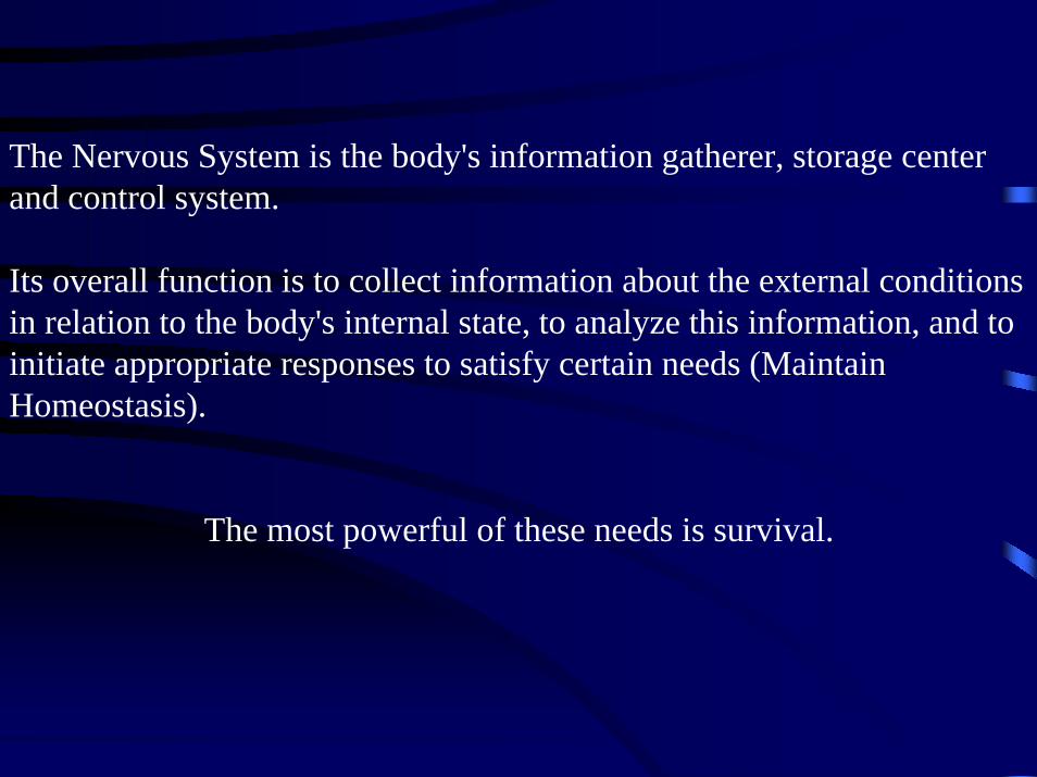
The Nervous System is the body's information gatherer, storage center and control system.
Its overall function is to collect information about the external conditions in relation to the body's internal state, to analyze this information, and to initiate appropriate responses to satisfy certain needs (Maintain Homeostasis).
The most powerful of these needs is survival.
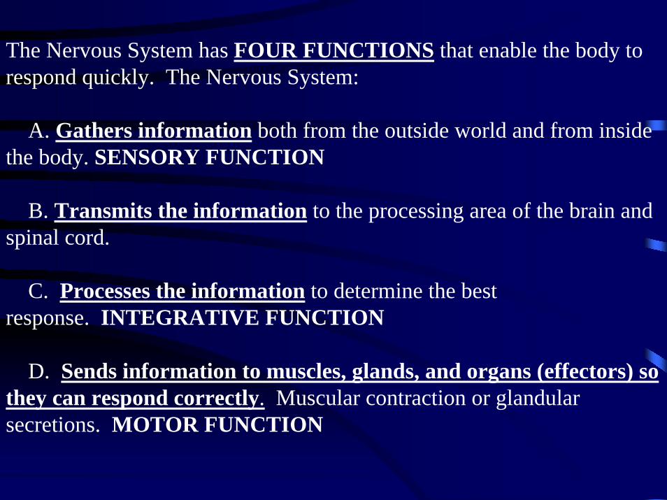
The Nervous System has FOUR FUNCTIONS that enable the body to respond quickly. The Nervous System:
A. Gathers information both from the outside world and from inside the body. SENSORY FUNCTION
B. Transmits the information to the processing area of the brain and spinal cord.
C. Processes the information to determine the best response. INTEGRATIVE FUNCTION
D. Sends information to muscles, glands, and organs (effectors) so they can respond correctly. Muscular contraction or glandular secretions. MOTOR FUNCTION

A REFLEX is the simplest response to a STIMULUS.
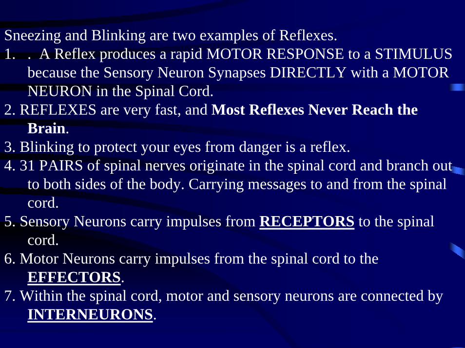
Sneezing and Blinking are two examples of Reflexes. 1. . A Reflex produces a rapid MOTOR RESPONSE to a STIMULUS
because the Sensory Neuron Synapses DIRECTLY with a MOTOR NEURON in the Spinal Cord.
2. REFLEXES are very fast, and Most Reflexes Never Reach the Brain.
3. Blinking to protect your eyes from danger is a reflex. 4. 31 PAIRS of spinal nerves originate in the spinal cord and branch out
to both sides of the body. Carrying messages to and from the spinal cord.
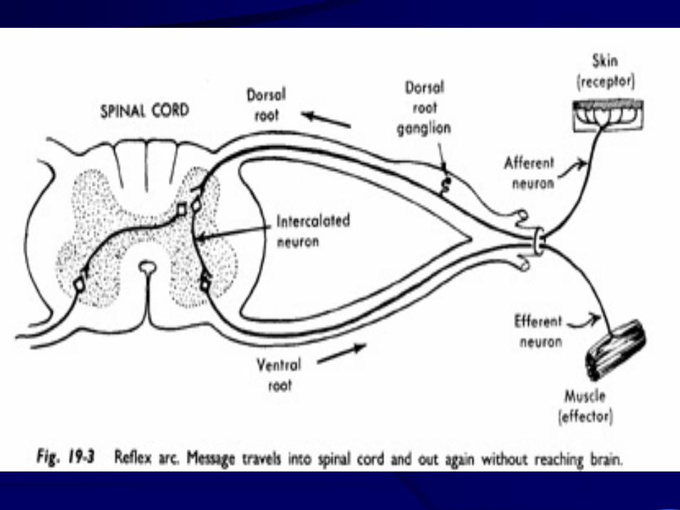
5. Sensory Neurons carry impulses from RECEPTORS to the spinal cord.
6. Motor Neurons carry impulses from the spinal cord to the EFFECTORS.
7. Within the spinal cord, motor and sensory neurons are connected by INTERNEURONS.
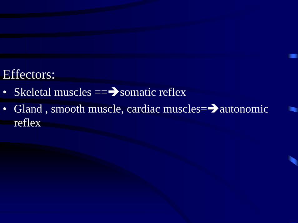
Effectors:• Skeletal muscles == somatic reflex• Gland , smooth muscle, cardiac muscles= autonomic
reflex


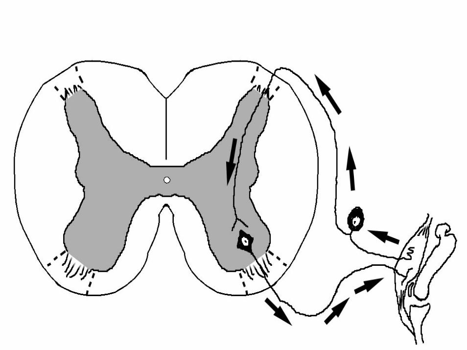
A reflex arc is the neural pathway that mediates a reflex action.


Guess these pathways do not pass through the brain, but synapse in the
spinal cord.
This characteristic allows reflex actions to occur relatively quickly by avoiding the delay of routing signals through the brain, although the brain will receive sensory input while the reflex action
occurs.

When a reflex arc consists of only two neurons (one sensory neuron and one motor neuron), it is defined as monosynaptic.
Monosynaptic refers to the presence of a single chemical synapse.
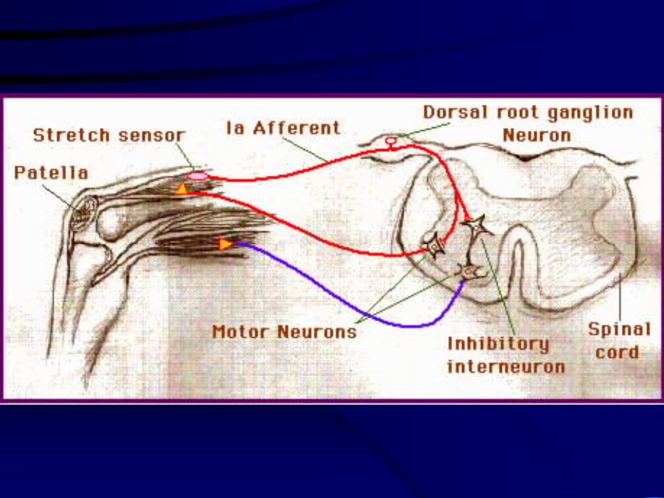
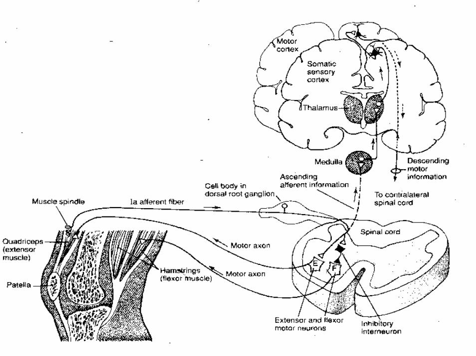
In the case of peripheral muscle reflexes (patellar reflex, achilles reflex), brief stimulation to the muscle spindle results in contraction of the agonist or effectormuscle.

in polysynaptic reflex pathways, one or moreinterneurons connect afferent (sensory) and efferent (motor) signals.
All but the most simple reflexes are polysynaptic, allowing processing or inhibition of polysynaptic reflexes within the spinal cord


The Patellar Reflex
when the patellar tendon is tapped just below the knee, the patellar reflex is initiated and the lower leg kicks forward (via contraction of the quadriceps).
The tap initiates an action potential in a specialised structure known as a muscle spindle located within the quadriceps.
This action potential travels to the spinal cord, via a sensory axon which chemically communicates (see synapse) with a motor nerve.
The result of this motor nerve activity is contraction of the quadriceps muscle, leading to extension of the lower leg at the knee.

The crossed extensor reflex is a withdrawal reflex.When the reflex occurs the flexors in the withdrawing limb contract and the extensors relax, while in the other limb the opposite occurs.An example of this is when a person steps on a nail, the leg that is stepping on the nail pulls away, while the other leg takes the weight of the whole body.
Another example of a crossed extensor reflex is when someone violently grabs your arm, the arm that is grabbed retracts towards the body while the other arm moves towards the attacker for protection.
Withdrawal reflex
The nociceptive withdrawal reflex (NWR) is a spinal reflex intended to protect the body from damaging stimuli. The classic example is when you touch something hot and withdraw your body part from the hot object. The heat stimulates temperature and pain receptors in the skin, triggering a sensory impulse that travels to the central nervous system. The sensory neuron then synapses with interneurons that connect to motor neurons. Some of these send motor impulses to the flexors to allow withdrawal; some motor neurons send inhibitory impulses to the extensors so flexion is not inhibited - this is referred to as reciprocal innervation.While all of this occurs, other interneurons relay the sensory information up to the brain so that the person becomes aware of the pain and what happened.
A nociceptor is a sensory receptor that sends signals that cause the perception of pain in response to potentially damaging stimulus.Nociceptors are the nerve endings responsible for nociception, one of the two types of persistent pain (the other, neuropathic pain, occurs when nerves in the central or peripheral nervous system are damaged). When they are activated, nociceptors can trigger a reflex.
In medicine, the pupillary reflex or pupillary light reflex is the reduction of pupil size in response to light.
It is a normal response and dependent on the function of the optic nerves and oculomotor nerves.
Pupillary constriction is sometimes used as a synonym for pupillaryreflex but something more general.
Pupillary constriction may be induced pharmacologically by parasympathomimetics and is also seen in accommodation (when the eyes focus on something close).
•Direct pupillary reflex: whether each pupil constricts with light shone into the that eye•Consensual pupillary reflex: whether each pupil constricts with light shone into the other eye

The reflex pathway consists of retinal ganglion cells, which convey information from the photoreceptors to the optic nerve which connects to the pretectal nucleus of the high midbrain. It bypasses the lateralgeniculate nucleus and the primary visual cortex. From the pretectalnucleus neurons send axons to neurons of the Edinger-Westphal nucleus whose axons run along both the left and right oculomotor nerves.Oculomotor nerve axons synapse on ciliary ganglion neurons whose axons innervate the constrictor muscle of the iris.
The brain

Brain
• Cerebrum• Diencephalon• Brain stem• Cerebellum
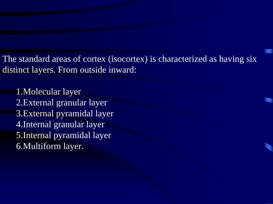
The standard areas of cortex (isocortex) is characterized as having six distinct layers. From outside inward:
1.Molecular layer2.External granular layer3.External pyramidal layer4.Internal granular layer5.Internal pyramidal layer6.Multiform layer.

•The molecular layer I contains few scattered neurons and consists mainly of extensions of apical dendrites and horizontally oriented axons, and some Cajal-Retzius and spinystellate neurons can be found.•The external granular layer II contains small pyramidal neurons and numerous stellateneurons.•The external pyramidal layer III contains predominantly small and medium sized pyramidal neurons, as well as non-pyramidal neurons with vertically-orientedintracortical axons. Layers I through III are the main target of interhemispheric corticocortical afferents, and layer III is the principal source of corticocortical efferents.•The internal granular layer IV contains different types of stellate and pyramidal neurons, and is the main target of thalamocortical afferents as well as intra-hemisphericcorticocortical afferents.•The internal pyramidal layer V contains large pyramidal neurons (as the Betz cells in the primary motor cortex), as well as interneurons, and it is the principal source of efferent for all the motor-related subcortical structures.•The multiform layer VI contains few large pyramidal neurons and many small spindle-like pyramidal and multiform neurons. The layer VI sends efferent fibers to the thalamus establishing a very precise reciprocal interconnection between the cortex and the thalamus

Cerebrum


cortex may be classified on the basis of gross topographical conventions into the following:
•Temporal Cortex•Parietal Cortex•Frontal Cortex•Occipital Cortex•Limbic Cortex•Insular Cortex
Association areas comprise three major groups:1.Parietal, temporal, and occipital lobes - all located in the posterior part of the brain - are involved in producing our perceptions resulting from what our eyes see, ears hear, and other sensory organs inform us about the position of different parts of our body and relate them to the position of other objects in the environment2.Frontal lobe - called prefrontal association complex and involved in planning actions and movement, as well as abstract thought3.Limbic association area - involved in emotion and memory.
In humans, the association areas of the left hemisphere, especially the parietal-temporal-occipital complex, are responsible for our understanding and use of language.

The cerebral cortex sends connections (efferents) and receives connections (afferents) from many subcortical structures like the thalamus and basal ganglia. Most of the sensory stimulation arrives to the cerebral cortex indirectly through different thalamic nuclei. This is the case of touch, vision and sound but not of olfactory stimulation, that arrives directly to the olfactory cortex. The largest part of the connections arriving to the cerebral cortex do not come from subcortical structures however. The main source of cortical stimulation is the cerebral cortex itself: up to 75% of the total connections.Areas that receive that particular information are called sensory areas. Parts of the cortex that receive sensory inputs from the thalamus are called primary sensory areas.
The senses of vision, audition and touch are served by the primary visual cortex, primary auditory cortex and primary somatosensory cortex. In general, the two hemispheres receive the information from the opposite sides of the body. For example the right primary somatosensory cortex receives information from the left limbs and the right visual cortex receives information from the left visual field.Other areas receive impulses from the primary sensory areas and integrate the information coming in from different types of receptors (i.e., modalities). These are often called association areas and make up a great deal of the cortex in all primates, humans included. Thus, the cortex is commonly described as comprised of the primary sensory areas, the motor areas and the association areas.

The motor areas are located in both hemispheres of the cortex. They are shaped like a pair of headphones stretching from ear to ear. The motor areas are very closely related to the control of voluntary movements, especially fine fragmented movements performed by the hand. The right half of the motor area controls the left side of your body and vice versa.Two areas of the cortex are commonly referred to as motor:
•Primary motor cortex, which executes voluntary movements•Supplementary motor areas and premotor cortex, which selectvoluntary movements.
In addition, motor functions have been described for:•Posterior Parietal Cortex, which guides voluntary movements in space•Dorsolateral Prefrontal Cortex, which decides which voluntary movements to make according to higher-order instructions, rules, and self-generated thoughts.

cerebrum
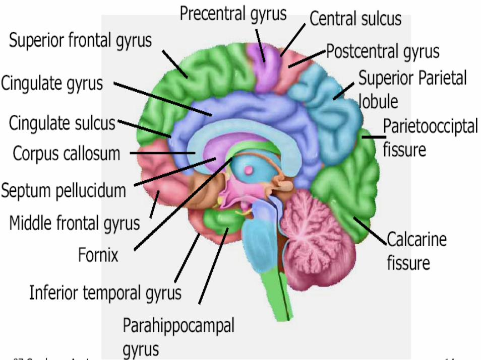
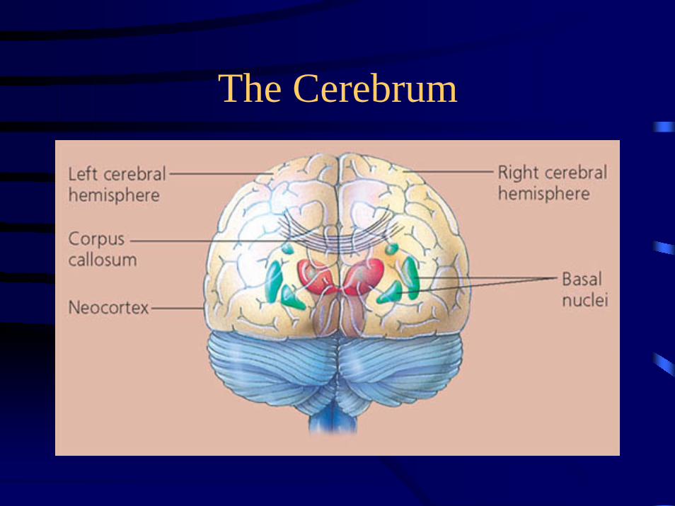
• The largest and most obvious portion of the brain is the cerebrum, which is divided by a deep longitudinal fissure into two cerebral hemispheres.
• The two hemispheres are two separate entities but are connected by an arching band of white fibers, called the corpus callosum that provides a communication pathway between the two halves.
• The cerebrum is located in the anterior portion of the forebrain. It is divided into two hemispheres that are connected by the corpus callosum.
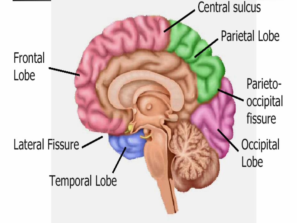
• Each cerebral hemisphere is divided into five lobes,
• four of which have the same name as the bone over them:
– the fontal lobe,– the parietal lobe, – the occipital lobe,– the temporal lobe.
• A fifth lobe, the insula or Island of Reil, lies deep within the lateral sulcus.
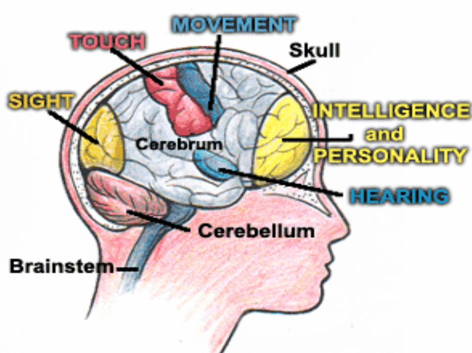
Cerebrum Function:
• Determines Intelligence • Personality • Interpretation of Sensory Impulses • Motor Function • Planning and Organization • Touch Sensation

The diencephalons is centrally located and is nearly surrounded by the cerebral hemispheres
Diencephalon• It includes the thalamus, hypothalamus, and the thalamus, hypothalamus, and
epithalamusepithalamus.
• The thalamus, about 80 percent of the diencephalons, consists of two oval masses of gray matter that serve as relay stations for sensory impulses, except for the sense of smell, going to the cerebral cortex.
• The hypothalamus is a small region below the thalamus, which plays a key role in maintaining homeostasis because it regulates many visceral activities.
• The epithalamus is the most dorsal portion of the diencephalons. This small gland is involved with the onset of puberty and rhythmic cycles in the body. It is like a biological clock.
The Epithalamus comprises •the trigonum habenulæ,• the pineal body, •and the posterior commissure.
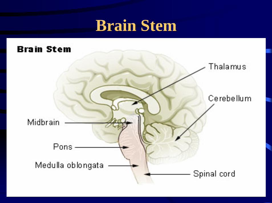
Brain Stem

The brain stem has three main parts: the medulla,pons, and midbrain. A canal runs longitudinally through these structures carrying cerebrospinal fluid.
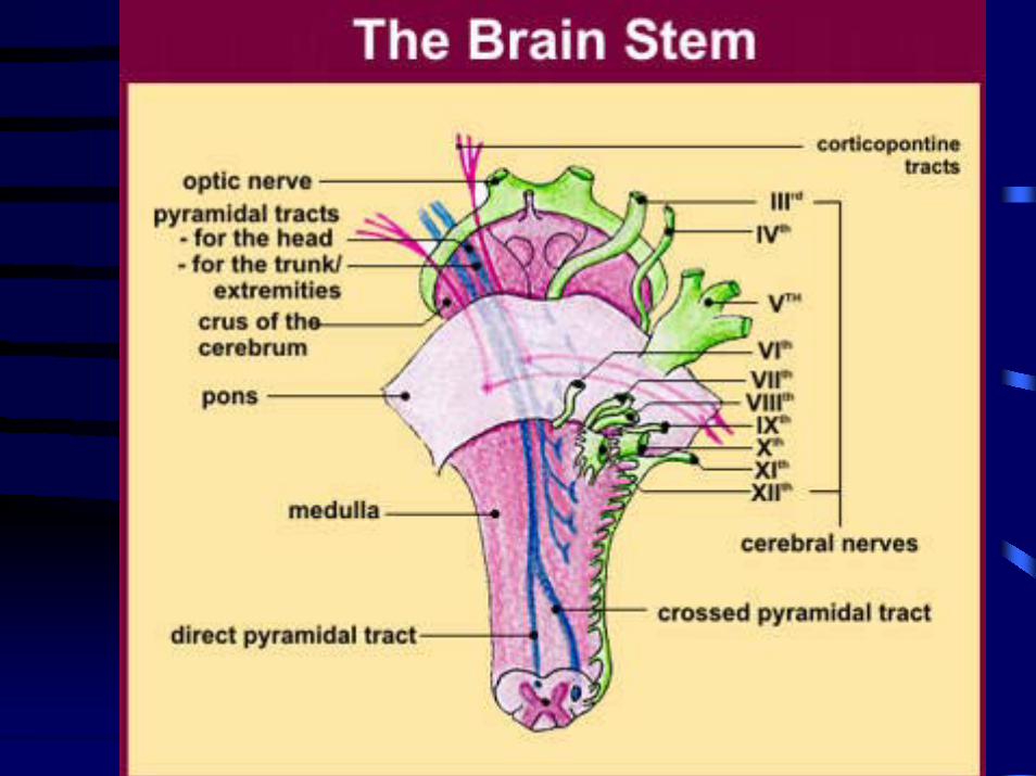


The brain stem is the region between the diencephalons and the spinal cord.
It consists of three parts: •midbrain, •pons, •and medulla oblongata.
•The midbrain is the most superior portion of the brain stem.• The pons is the bulging middle portion of the brain stem.
This region primarily consists of nerve fibers that form conduction tracts between the higher brain centers and spinal cord.
•The medulla oblongata, or simply medulla, extends inferiorly from the pons.
•It is continuous with the spinal cord at the foramen magnum.
•All the ascending (sensory) and descending (motor) nerve fibers connecting the brain and spinal cord pass through the medulla.
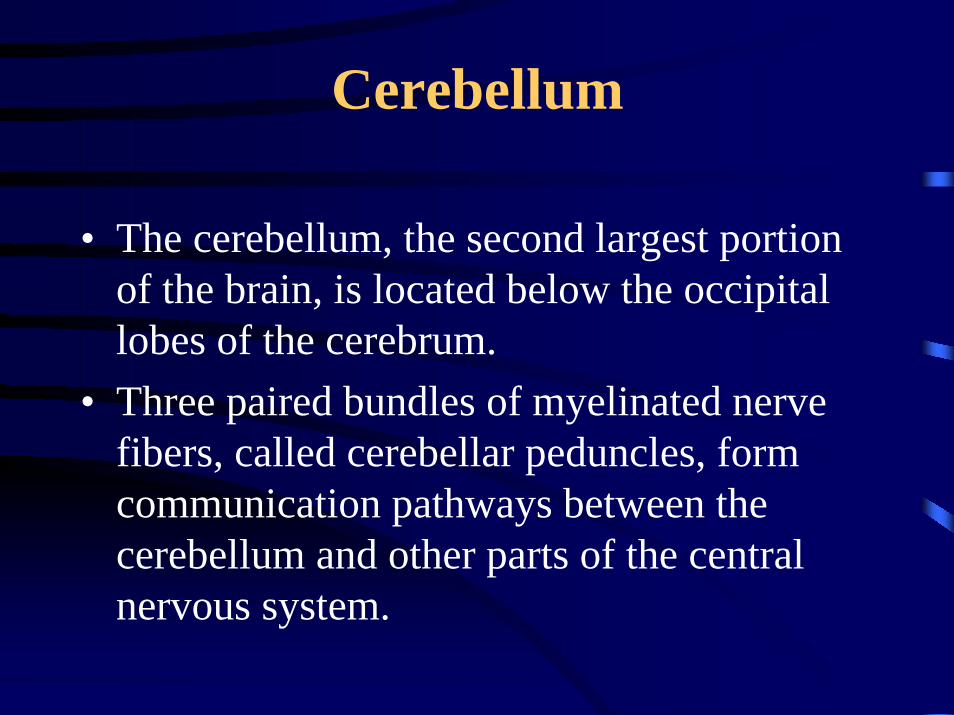
Cerebellum
• The cerebellum, the second largest portion of the brain, is located below the occipital lobes of the cerebrum.
• Three paired bundles of myelinated nerve fibers, called cerebellar peduncles, form communication pathways between the cerebellum and other parts of the central nervous system.

Ventricles and Cerebrospinal Fluid
• Ventricles and Cerebrospinal FluidA series of interconnected, fluid-filled cavities are found within the brain.
• These cavities are the ventricles of the brain, and the fluid is cerebrospinal fluid (CSF).


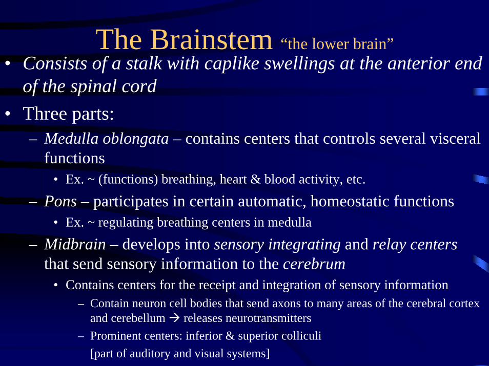
The Brainstem “the lower brain”• Consists of a stalk with caplike swellings at the anterior end
of the spinal cord• Three parts:
– Medulla oblongata – contains centers that controls several visceral functions
• Ex. ~ (functions) breathing, heart & blood activity, etc.
– Pons – participates in certain automatic, homeostatic functions• Ex. ~ regulating breathing centers in medulla
– Midbrain – develops into sensory integrating and relay centers that send sensory information to the cerebrum
• Contains centers for the receipt and integration of sensory information– Contain neuron cell bodies that send axons to many areas of the cerebral cortex
and cerebellum releases neurotransmitters– Prominent centers: inferior & superior colliculi
[part of auditory and visual systems]
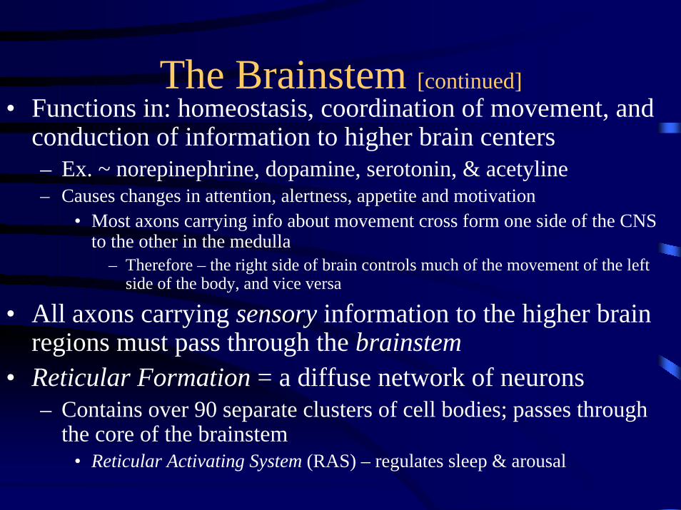
The Brainstem [continued]• Functions in: homeostasis, coordination of movement, and
conduction of information to higher brain centers– Ex. ~ norepinephrine, dopamine, serotonin, & acetyline– Causes changes in attention, alertness, appetite and motivation
• Most axons carrying info about movement cross form one side of the CNS to the other in the medulla
– Therefore – the right side of brain controls much of the movement of the left side of the body, and vice versa
• All axons carrying sensory information to the higher brain regions must pass through the brainstem
• Reticular Formation = a diffuse network of neurons– Contains over 90 separate clusters of cell bodies; passes through
the core of the brainstem• Reticular Activating System (RAS) – regulates sleep & arousal

The Cerebellum• Developed from Metencephalon• Coordination & error checking skills
– For motor, perceptual, and cognitive functions• cognitive – includes an integrated sensory awareness of the surroundings
• Learning and Remembering skills– Ex. ~ riding a bicycle
• Receives Sensory Information– Position of joints and the length of the muscles– Auditory and visual systems
• Given Input on Motor Commands– Integrates this sensory & motor information while coordinating movements &
balance• Major Ex. ~ hand-eye coordination
– If cerebellum is damaged – eyes can only follow a moving object, but they won’t stop at the same place as the object

Depending on the area and side of the cerebrum affected by the stroke, any, or all, of the following body functions may be impaired:
•movement and sensation•speech and language•eating and swallowing•vision•cognitive (thinking, reasoning, judgment and memory) ability•perception and orientation to surroundings•self-care ability•bowel and bladder control•emotional control•sexual ability
Effects of a right hemisphere stroke:The effects of a right hemisphere stroke may include the following:
•left-sided weakness (left hemiparesis) or paralysis (left hemiplegia) and sensory impairment•denial of paralysis or impairment and reduced insight into the problems created by the stroke (this concept is called "left neglect")•visual problems, including an inability to see the left visual field of each eye (homonymous hemianopsia)
•spatial problems with depth perception or directions such as up/down and front/back
•inability to localize or recognize body parts
•inability to understand maps and find objects such as clothing or toiletry items
•memory problems
•behavioral changes such as lack of concern about situations, impulsivity, inappropriateness, and depression

Effects of a left hemisphere stroke:The effects of a left hemisphere stroke may include the following:
•right-sided weakness (right hemiparesis) or paralysis (right hemiplegia) and sensory impairment
•problems with speech and understanding language (aphasia)
•visual problems, including the inability to see the right visual field of each eye (homonymous hemianopsia)
•impaired ability to do math or to organize, reason, and analyze items
•behavioral changes such as depression, cautiousness, and hesitancy
•impaired ability to read, write, and learn new information
•memory problems
What effects can be seen with a stroke in the brain stem?The brain stem is located at the very base of the brain right above the spinal cord. Many of the body's vital "life-support" functions such as heartbeat, blood pressure, and breathing are controlled by the brain stem. It also helps to control the main nerves involved with eye movement, hearing, speech, chewing, and swallowing. Some common effects of a stroke in the brain stem include problems with the following:
•breathing and heart functions•body temperature control•balance and coordination•weakness or paralysis in all four limbs•chewing, swallowing, and speaking•vision•coma
Unfortunately, death is common with brain stem strokes.
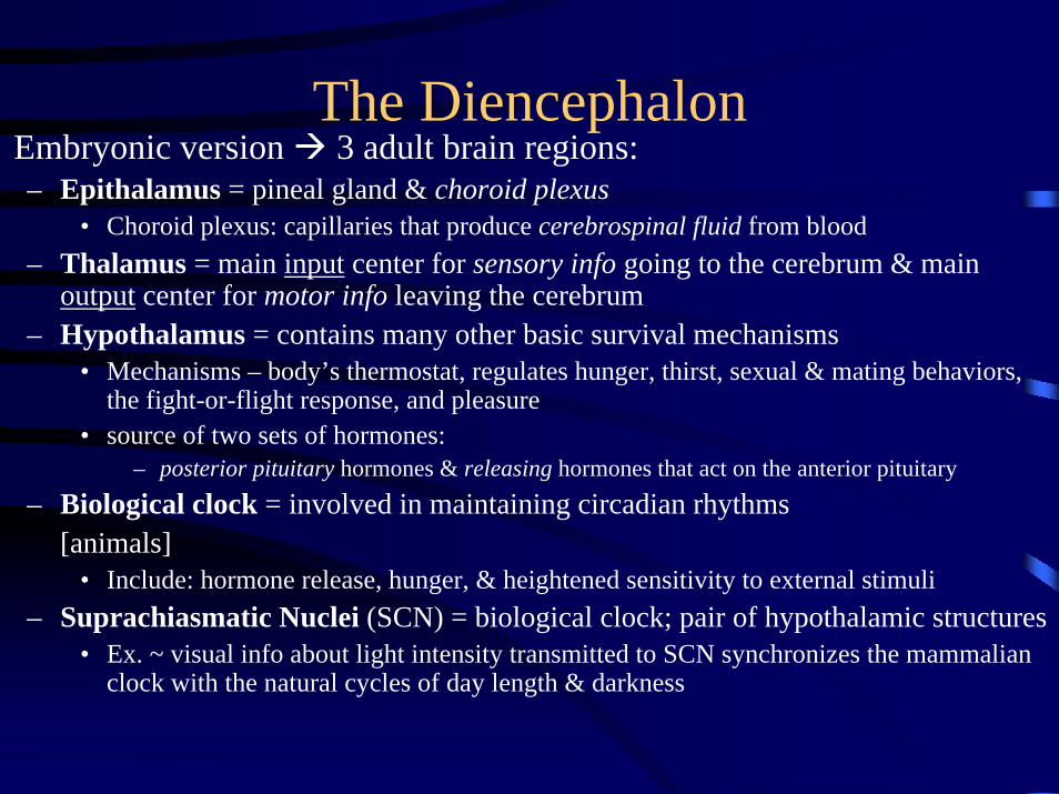
The DiencephalonEmbryonic version 3 adult brain regions:– Epithalamus = pineal gland & choroid plexus
• Choroid plexus: capillaries that produce cerebrospinal fluid from blood – Thalamus = main input center for sensory info going to the cerebrum & main
output center for motor info leaving the cerebrum– Hypothalamus = contains many other basic survival mechanisms
• Mechanisms – body’s thermostat, regulates hunger, thirst, sexual & mating behaviors, the fight-or-flight response, and pleasure
• source of two sets of hormones:– posterior pituitary hormones & releasing hormones that act on the anterior pituitary
– Biological clock = involved in maintaining circadian rhythms[animals]
• Include: hormone release, hunger, & heightened sensitivity to external stimuli– Suprachiasmatic Nuclei (SCN) = biological clock; pair of hypothalamic structures
• Ex. ~ visual info about light intensity transmitted to SCN synchronizes the mammalian clock with the natural cycles of day length & darkness

The Cerebrum• Forebrain Telencephalon cerebrum and cerebral cortex
– extends over/around much of human brain– Outgrowth of forebrain – evolved so to support olfactory reception as well as
auditory & visual processing• Cerebral Hemispheres = left & right hemisphere; consists of an outer
covering of gray matter, the cerebral cortex, internal white matter, and basal nuclei– Basal nuclei = groups of neurons located deep within the white matter
• Important centers – planning and learning movement sequences – Cerebral Cortex = sensory information analyzed, motor commands issued, and
language is generated• Mammals possess a region in it known as the neocortex
– Neocortex = forms outermost part of the mammalian cerebrum; consists of 6 parallel layers of neurons running tangential to the brain surface
– Corpus callosum = thick band of axons that enable communication between right & left cerebral cortices
» Damage to one area of the cerebrum early in development frequent causes redirection of its normal functions to other areas; Ex. ~ an entire infant’s cerebral hemisphere is removed as a treatment for severe epilepsy
The Cerebrum
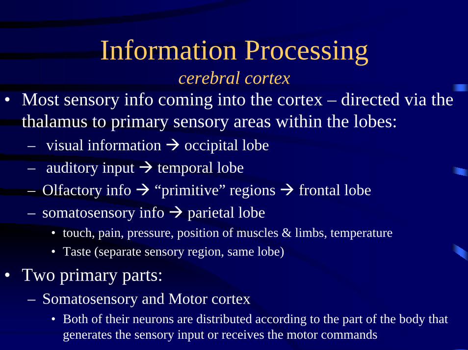
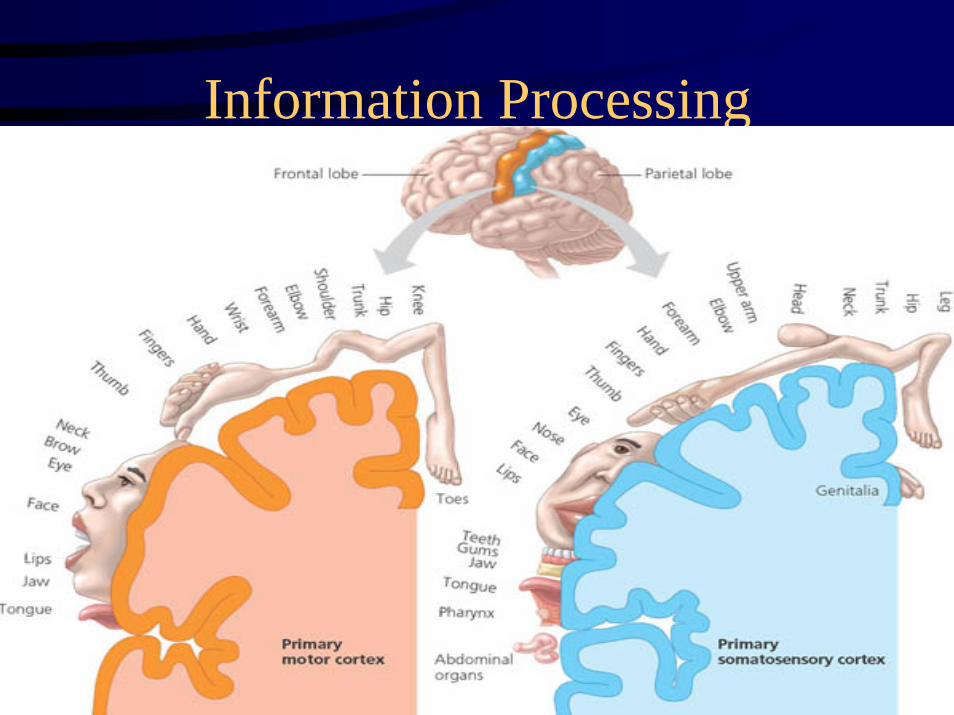
Information Processing cerebral cortex
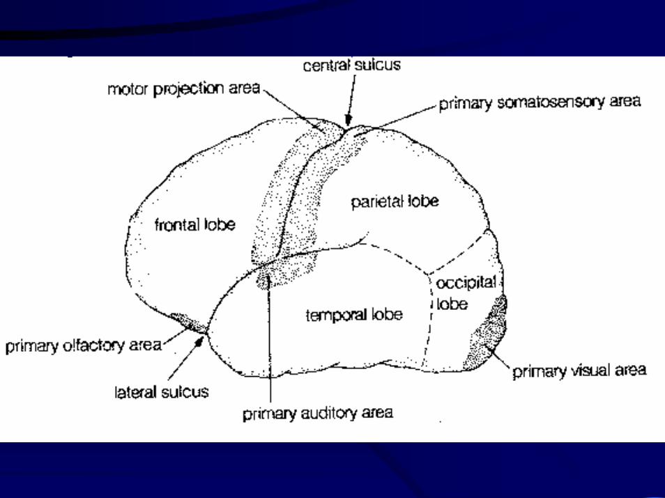
• Most sensory info coming into the cortex – directed via the thalamus to primary sensory areas within the lobes:– visual information occipital lobe– auditory input temporal lobe– Olfactory info “primitive” regions frontal lobe– somatosensory info parietal lobe
• touch, pain, pressure, position of muscles & limbs, temperature • Taste (separate sensory region, same lobe)
• Two primary parts:– Somatosensory and Motor cortex
• Both of their neurons are distributed according to the part of the body that generates the sensory input or receives the motor commands
• Primary Somatosensory Cortex – send info to nearby association areas that can process particular features in the sensory input– Ex.~ Primary visual cortex – some neurons are sensitive to bars of
light that have a certain width & orientation
• Primary Motor Cortex – helps issue commands that consist of action potentials produced by neurons[located: rear of frontal lobe, adjacent to the primary somatosensory cortex]
– Action potentials travel along axons to brainstem & spinal cord excite motor neurons excite skeletal muscle cells
Information Processing cerebral cortex
Information Processing cerebral cortex

Voluntary Movementand Cognitive Functions
• Lateralization = segregation of functions in the left and right cortex hemispheres of the brain– Left hemisphere – language, math, logical operations, & serial
processing of info sequences• Specializes in focused perception• for the detailed, speed-optimized activities & the processing of fine visual
and auditory details– Right hemisphere – pattern recognition, face recognition, spatial
relations nonverbal thinking, emotional processing in general; the simultaneous processing of many kinds of info
• Specializes in perceiving the relationship between images and the whole context in while they occur
• Emphasis on understanding and generating stress & intonation patterns of speech that convey emotional content

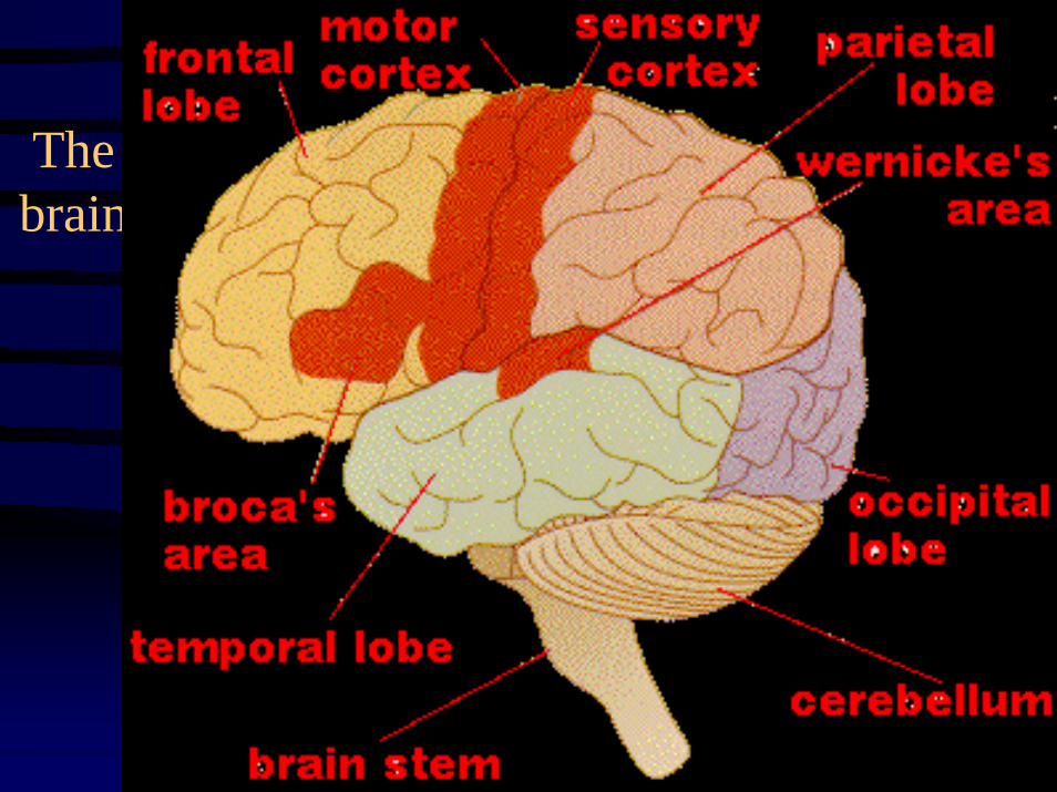
Language and speech– Broca’s area – located at left frontal lobe and is part of the primary motor cortex
that controls muscles in the face– Wernicke’s area – posterior portion of the temporal lobe that controls
comprehension and generation of speech– Frontal & temporal areas become active when meaning must be
attached to words (ex. ~ a person generates verbs to go with nouns)
• Damage in this spot abolishes the ability to comprehend speech, but left speech generation intact
Emotions– Result of a complex interplay of many regions of the brain– Limbic system = a ring of structures around the brainstem composed of three main
parts and some inner portions of the thalamus and hypothalamus• Includes three parts – amygdala, hippocampus, and olfactory bulb• Attaches “feelings” to basic, survival-related functions controlled by brainstem
– Interacts with sensory areas of the neocortex & other higher brain centers– Mediates primary emotions becomes manifested in behaviors like laughing & crying
Voluntary Movementand Cognitive Functions

• Memory and Learning– Short-term memory – the ability to hold information, anticipations, or goals for a
time and then release them if they become irrelevant– Long-term memory – the ability to hold, associate, and recall information over
one’s life– Motor skills – learned by repetition– Transfer from short-term long-term: enhanced by rehearsal (“practice makes
perfect!”), positive or negative emotional state (amygdala), and the associationof new data with data previously learned & stored in long-term memory
– Neurons make new connections (help with memorization of facts/places)– Long-term potentiation (LTP) – involves an increase in the strength of synaptic
transmission that occurs when presynaptic neurons produce a brief, high-frequency series of action potentials
• Consciousness [both broad & subjective]– fMRI – brain-imaging technique; allows for the human brain activity (in its
different states of consciousness) to be observed– Postulations: “is an emergent property of the brain” & “scanning mechanism –
repetitively sweeps across the brain, integrating widespread activity into a unified, conscious moment”
Voluntary Movementand Cognitive Functions

Physiology• Nervous system conveys high-speed electrical
signals along neurons such rapid messages control the movement of body parts in response to sudden environmental changes– Ex.1 ~ jerking hand away from a hot pan– Ex.2 ~ pupils dilating as you enter a dark room
• Overlaps with endocrine regulation– Neurosecretory cells = release hormones into
blood (contained in the hypothalamus part of the brain); produce neurohormones

Control Pathways & Feedback Loops• Receptor (sensor) – detects a stimulus and sends information
to control center– Control center = compares the incoming info to a set point (“desired”
value) and sends out a signal that directs an effector to respond– Ex. ~ a change in blood calcium level
• Positive Feedback – reinforces the stimulus and leads to an even greater response– Ex. ~ neurohormone pathway that regulates the release of milk by a
nursing mother; suckling stimulates sensory nerve cells in the nipples, which send nervous signals that eventually reach the control center (hypothalamus)
• Negative Feedback – the effector response reduces the initial stimulus response ceases– Prevents overreaction by the system and wild fluctuations in the
variable being regulated

• Baby’s Brain– http://www.pbs.org/wnet/brain/episode1/video.html
Spinal Cord • The spinal cord extends from the foramen magnum at the base of the skull to the
level of the first lumbar vertebra. • The cord is continuous with the medulla oblongata at the foramen magnum. • Like the brain, the spinal cord is surrounded by bone, meninges, and
cerebrospinal fluid. • The spinal cord is divided into 31 segments with each segment giving rise to a
pair of spinal nerves. At the distal end of the cord, many spinal nerves extend beyond the conus medullaris to form a collection that resembles a horse's tail. This is the cauda equina. In cross section, the spinal cord appears oval in shape.
• The spinal cord has two main functions:• Serving as a conduction pathway for impulses going to and from the brain.
Sensory impulses travel to the brain on ascending tracts in the cord. Motor impulses travel on descending tracts.
• Serving as a reflex center. The reflex arc is the functional unit of the nervous system. Reflexes are responses to stimuli that do not require conscious thought and consequently, they occur more quickly than reactions that require thought processes. For example, with the withdrawal reflex, the reflex action withdraws the affected part before you are aware of the pain. Many reflexes are mediated in the spinal cord without going to the higher brain centers.