References - National Kidney Foundation D Part 2.pdf · References 1. Mehrotra R, Kermah D, Budoff...
6
› Introduction › Indications for Vitamin D Replacement › Vitamin 25(OH)D Measurement › Strategies for Vitamin D Supplementation › Summary A Clinical Update on Vitamin D Deficiency and Secondary Hyperparathyroidism: Vitamin D Testing and Supplementation in CKD Stages 3-4 Part 2
Transcript of References - National Kidney Foundation D Part 2.pdf · References 1. Mehrotra R, Kermah D, Budoff...

› Introduction
› Indications for Vitamin D Replacement
› Vitamin 25(OH)D Measurement
› Strategies for Vitamin D Supplementation
› Summary
A Clinical Update on Vitamin D Deficiency and Secondary Hyperparathyroidism: Vitamin D Testing and Supplementation in CKD Stages 3-4Part 2
References
1. Mehrotra R, Kermah D, Budoff M, et al. Hypovitaminosis D in chronic kidney disease. Clin J Am Soc Nephrol. 2008;3:1144-1151.
2. Hollick MF. Vitamin D: importance in the prevention of cancers, type 1 diabetes, heart disease, and osteoporosis. Am J Clin Nutr 2004;79:362-371.
3. Giovannucci E, Liu Y, Rimm EB, et al. Prospective study of predictors of vitamin D status and cancer incidence and mortality in men. J Natl Cancer Inst 2006;98:451-459.
4. Garland CF, Garland FC, Gorham ED, et al. The role of vitamin D in cancer prevention. Am J Public Health 2006; 96:252-261.
5. Broe KE, Chen TC, Weinberg J, et al. A higher dose of vitamin D reduces the risk of falls in nursing home residents: a randomized, multiple-dose study. J Am Geriatr Soc 2007: 55:234-239.
6. Kidney Disease: Improving Global Outcomes (KDIGO). KDIGO clinical practice guideline for the diagnosis, evaluation, prevention, and treatment of chronic kidney disease-mineral and bone disorder (CKD-MBD). Kidney Int. 2009;76(suppl 113):S9-S21.
7. Uhlig K, Berns JS, Kestenbaum B, et al. KDOQI US Commentary on the 2009 KDIGO clinical practice guideline for the diagnosis, evaluation, prevention and treatment of chronic disease-mineral and bone disorder (CKD-MBD). Am J Kidney Dis; 2010; 55:773-799.
8. Quarles LD. Role of FGF23 in vitamin D and phosphate metabolism: implications in chronic kidney disease. Exp Cell Res. 2012 May 15;318:1040-1048.
9. Michaud J, Naud J, Ouimet D, et al. Reduced hepatic synthesis of calcidiol in uremia. J Am Soc Nephrol. 2010;21:1488-1497.
10. National Institutes of Health Office of Dietary Supplements. Vitamin D. https://ods.od.nih.gov/factsheets/VitaminD-HealthProfessional/. Published November 10 2014. Accessed September 2 2015.
11. Jones G. Interpreting vitamin D assay results: proceed with caution. Clin J Am Soc Nephrol. 2015;10:331-334.
12. Institute of Medicine (IOM). Dietary reference intakes for calcium and vitamin D. Washingotn, DC: The National Academies Press; 2011.
13. Melamed ML, Michos ED, Post W, Astor B. 25-hydroxyvitamin D levels and the risk of mortality in the general population. Arch Intern Med. 2008;168:1629-1637.
14. Tuohimaa P, Tenkanen L, Ahonen M, et al. Both high and low levels of blood vitamin D are associated with a higher prostate cancer risk: a longitudinal, nested case-control study in the Nordic countries. Int J Cancer. 2004;108:104-108.
15. Holick MF, Binkley NC, Bischoff-Ferrari HA, et al; Endocrine Society. Evaluation J Clin Endocrinol Metab. 2011;96:1911-1930.
16. Durup D, Jørgensen HL, Christensen J, et al. A Reverse J-Shaped Association Between Serum 25-Hydroxyvitamin D and Cardiovascular Disease Mortality: The CopD Study. J Clin Endocrinol Metab. 2015;100:2339-2346.
17. Ennis JL, Worcester EM, Coe FL, Sprague SM. Current recommended 25-hydroxyvitamin D targets for chronic kidney disease management may be too low. J Nephrol. 2016;29:63-70.
18. OPKO. OPKO diagnostics point-of-care system. Available at: http://www.opko.com/products/point-of-care-diagnostics/. Accessed September 2 2015.
19. Holick MF, Binkley NC, Bischoff-Ferrari HA, et al. Evaluation, treatment and prevention of vitamin D deficiency: An Endocrine Society Clinical Practice Guideline. J Clin Endocrin Metab. 2011;96:1911-1930.
20. Binkley N, Krueger D, Cowgill CS, et al. Assay variation confounds the diagnosis of hypovitaminosis D: a call for standardization. J Clin Endocrinol Metab. 2004;89:3152-3157.
21. Binkley N, Sempos CT; Vitamin D Standardization Program (VDSP). Standardizing vitamin D assays: the way forward. J Bone Miner Res. 2014;29:1709-1714.
22. National Institutes of Health Office of Dietary Supplements. Vitamin D Standardization Program.https://ods.od.nih.gov/Research/vdsp.aspx. Published Nov 2011. Updated Aprill 2015. Accessed August 28 2015.
23. Mann MC, Hobbs AJ, Hemmelgarn BR, et al. Effect of oral vitamin D analogs on mortality and cardiovascular outcomes among adults with chronic kidney disease: a meta-analysis. Clin Kidney J. 2015;8:41-48.
24. Nigwekar SU, Bhan I, Thadhani R. Ergocalciferol and cholecalciferol in CKD. Am J Kidney Dis. 2012 Jul;60(1):139-56.
25. Holick MF, Biancuzzo RM, Chen TC, et al. Vitamin D2 is as effective as vitamin D3 in maintaining circulating concentrations of 25-hydroxyvitamin D. J Clin Endocrinol Metab. 2008;93:677-681.
26. Armas LA, Hollis BW, Heaney RP. Vitamin D2 is much less effective than vitamin D3 in humans. J Clin Endocrinol Metab. 2004;89:5387-5391.
27. Parikh A, Chase HS, Vernocchi L, Stern L. Vitamin D resistance in chronic kidney disease (CKD). BMC Nephrology. 2014;15:47.
28. Mariani LH, White MT, Shults J, et al; CRIC Study Investigators. Increasing use of vitamin D supplementation in the chronic renal insufficiency cohort study. J Ren Nutr. 2014;24:186-193.
29. Coburn JW, Maung HM, Elangovan L, et al. Doxercalciferol safely suppresses PTH levels in patients with secondary hyperparathyroidism associated with chronic kidney disease stages 3 and 4. Am J Kidney Dis. 2004;43:877-890.
30. Coyne D, Acharya M, Qiu P, et al. Paricalcitol capsule for the treatment of secondary hyperparathyroidism in stages 3 and 4 CKD. Am J Kidney Dis. 2006;47:263-276.
31. Kalantar-Zadeh K, Kovesdy CP. Clinical outcomes with active versus nutritional vitamin D compounds in chronic kidney disease. Clin J Am Soc Nephrol. 2009;4:1529-1539.
32. Thimachai P, Supasyndh O, Chaiprasert A, Satirapoj B. Efficacy of High vs. Conventional Ergocalciferol Dose for Increasing 25-Hydroxyvitamin D and Suppressing Parathyroid Hormone Levels in Stage III-IV CKD with Vitamin D Deficiency/Insufficiency: A Randomized Controlled Trial. J Med Assoc Thai. 2015;98:643-648.
33. Kovesdy CP, Lu JL, Malakauskas SM, et al. Paricalcitol versus ergocalciferol for secondary hyperparathyroidism in CKD stages 3 and 4: a randomized controlled trial. Am J Kidney Dis. 2012;59:58-66.
34. Moe SM, Saifullah A, LaClair RE, et al. A randomized trial of cholecalciferol versus doxercalciferol for lowering parathyroid hormone in chronic kidney disease. Clin J Am Soc Nephrol. 2010;5:299-306.
35. Zisman AL, HristovaM, Ho LT, Sprague SM. Impact of ergocalciferol treatment of vitamin D deficiency on serum parathyroid hormone concentrations in chronic kidney disease. Am J Nephrol. 2007;27:36-43.
36. Al-Aly Z, Qazi RA, Gonzalez EA, et al. Changes in serum 25-hydroxyvitamin D and plasma intact PTH levels following treatment with ergocalciferol in patients with CKD. Am J Kidney Dis. 2007;50:59-68.
37. Chandra P, Binongo JN, Ziegler TR, et al. Cholecalciferol (vitamin D3) therapy and vitamin D insufficiency in patients with chronic kidney disease: a randomized controlled pilot study. Endocr Pract. 2008;14:10-17.
38. Kramer H, Berns JS, Choi MJ, et al. 25-Hydroxyvitamin D testing and supplementation in CKD: an NKF-KDOQI controversies report. Am J Kidney Dis. 2014;64:499-509.
39. Jetter A, Egli A, Dawson-Hughes B, et al. Pharmacokinetics of oral vitamin D(3) and calcifediol. Bone. 2014;59:14-19.
40. Petkovich M, Melnick J, White J, et al. Modified-release oral calcifediol corrects vitamin D insufficiency with minimal CYP24A1 upregulation. J Steroid Biochem Mol Biol. 2015;148:283-289.
41. Sprague SM, Silva AL, Al-Saghir F, et al. Modified-release calcifediol effectively controls secondary hyperparathyroidism associated with vitamin D insufficiency in chronic kidney disease. Am J Nephrol. 2014;40:535-545.
42. Vande Griend JP, McQueen RB, Linnebur SA, Vondracek SF. Prescription ergocalciferol dosing for vitamin D repletion: a retrospective evaluation. Pharmacotherapy. 2012;32:135-141.
43. Heaney RP, Recker RR, Grote J, et al. Vitamin D(3) is more potent than vitamin D(2) in humans. J Clin Endocrinol Metab. 2011;96:E447-E452.
44. Mangoo-Karim R, Da Silva Abreu J, Yanev GP, et al. Ergocalciferol versus Cholecalciferol for Nutritional Vitamin D Replacement in CKD. Nephron. 2015;130:99-104.
45. Brandenburg VM, Kruger T. Calcifediol - more than the stepchild of CKD-MBD therapy? Curr Vasc Pharmacol. 2014;12:286-93.
30 East 33rd StreetNew York, NY 10016 800.622.9010 www.kidney.org
This educational activity is supported by
DISCLAIMER Information contained in this National Kidney Foundation educational resource is based upon current data available at the time of publication. Information is intended to help clinicians become aware of new scientific findings and developments. This clinical bulletin is not intended to set out a preferred standard of care and should not be construed as one. Neither should the information be interpreted as prescribing an exclusive course of management.
© 2016 National Kidney Foundation, Inc. 02-10-7106_GBF
65

3 42
Vitamin 25(OH)D MeasurementThe major circulating form of vitamin D is 25(OH)D. Clinical practice guidelines suggest that serum total 25(OH)D be measured in patients with CKD stages 3-5D, and that repeat testing depends on baseline values and therapeutic interventions.6,7 The best approach to assess vitamin D status is measurement of serum 25(OH)D because it has a long half-life in the blood (approximately 3 weeks); however, the available assays for serum 25(OH)D are not well standardized and the definition of deficiency has not been validated.6 A new assay for 25(OH)D is under development that may allow clinicians to use a portable hardware device directly at the point-of-care to determine vitamin D status.18
Within- and between-assay variations in serum 25(OH)D measurement make it difficult to define hypovitaminosis D and cause problems when pooling study results in systematic reviews to determine dose-response and/or clinical cut points.12, 19, 20, 21 Therefore, standardization of 25(OH)D measurement in both clinical and research laboratories is needed. The Vitamin D Standardization Program (VDSP) is an international collaborative endeavor to standardize the laboratory measurement of vitamin D status. It was established by the National Institute of Health (NIH) Office of Dietary Supplements (ODS) in November 2010. The goal of VDSP is to improve the detection, evaluation, and treatment of vitamin D deficiency and insufficiency by promoting the standard measurement of serum 25(OH)D by making it accurate and comparable over time, location, and laboratory procedure.22
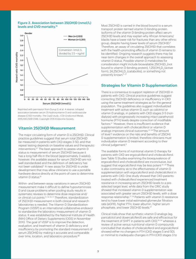
Most 25(OH)D is carried in the blood bound to a serum transport protein termed vitamin D-binding protein. Isoforms of the vitamin D-binding protein affect serum 25(OH)D levels and may explain why African Americans/blacks have a lower risk for fractures than any other ethnic group, despite having lower levels of serum 25(OH)D.11 Therefore, an assay of circulating 25(OH)D that correlates with the health-promoting effects of vitamin D remains to be identified. Ongoing research suggests there may be near-term changes in the overall approach to assessing vitamin D status. Possible vitamin D metabolites for consideration might include bioavailable 25(OH)D3 (not bound to vitamin D-binding protein), 1,25(OH)2D3 (active form), 24,25(OH)2D3 (catabolite), or something not presently known.11, 21
Strategies for Vitamin D SupplementationThere is a consensus to support repletion of 25(OH)D in patients with CKD. Clinical practice guidelines suggest correcting 25(OH)D deficiency or insufficiency in CKD using the same treatment strategies as for the general population. The guidelines also suggest individualized treatment with active vitamin D, such as calcitriol or vitamin D analogs, in patients with CKD stages 3-5 (non-dialysis) with progressively increasing intact parathyroid hormone (PTH) levels despite correction of modifiable factors.6, 7 However, there is insufficient evidence that supplementation with cholecalciferol or vitamin D analogs improves clinical outcomes.23, 24 The amount of level 1 evidence on the risks and benefits of 25(OH)D supplementation is limited, therefore clinicians must individualize vitamin D treatment according to their clinical judgement.6
The available forms of nutritional vitamin D therapy for patients with CKD are ergocalciferol and cholecalciferol. (see Table 1) Studies examining the bioequivalence of ergocalciferol and cholecalciferol are inconclusive, but suggest that ergocalciferol may be less potent.25, 26 There is also controversy as to the effectiveness of vitamin D supplementation with ergocalciferol and cholecalciferol in patients with CKD. One study showed that CKD patients treated with cholecalciferol experienced treatment resistance in increasing serum 25(OH)D levels to a pre-selected target level, while data from the CRIC study showed that increased vitamin D supplementation was associated with greater serum 25(OH)D levels, with a dose response relationship.27, 28 Patients with vitamin D resistance tend to have lower initial estimated glomerular filtration rate (eGFR), higher PTH, lower albumin, higher serum phosphate, and lower 25(OH)D levels.27
Clinical trials show that synthetic vitamin D analogs (eg. paricalcitol and doxercalciferol) are safe and efficacious for the treatment of SHPT in patients with CKD,29, 30 whereas a review of active versus nutritional vitamin D compounds concluded that studies of cholecalciferol and ergocalciferol showed either no changes in PTH (CKD stages 5 and 5D), or minimal to inadequate changes in PTH (CKD stages 3 to
Reprinted with permission from Durup D, et al. A reverse J-shaped association between serum 25-hydroxyvitamin D and cardiovascular disease (CVD) mortality: The CopD study. J Clin Endocrinol Metab. 2015;100:2339-2346. Copyright 2015 Endocrine Society.
Figure 2. Association between 25(OH)D (nmol/L) levels and CVD mortality.15
5), or changes that did not meet the targets suggested in clinical practice guidelines (CKD stage 3).31 In a study that compared conventional versus high ergocalciferol doses in CKD stages 3-4, treatment with high dose ergocalciferol resulted in a significantly greater increase of 25(OH)D levels and a significantly greater decrease in PTH levels (90.75 to 76.4 pg/ml), with no changes in serum calcium or phosphate.32
In a clinical trial comparing ergocalciferol and paricalcitol for treating SHPT in patients with CKD stages 3-4, paricalcitol showed significantly higher efficacy at decreasing PTH levels.33 After 16 weeks of therapy, serum 25(OH)D levels increased significantly in the ergocalciferol group, but not in the paricalcitol group. In a smaller, randomized clinical trial, there was no significant difference between cholecalciferol and doxercalciferol for the management of SHPT in patients with CKD stages 3-4, with PTH decreases of 10% and 27% respectively.34 The lack of statistical significance may be due to a small sample size. Serum total 25(OH)D increased from 14 to 37 ng/mL with cholecalciferol treatment, with no change in the doxercalciferol group. Additionally, cholecalciferol had a greater effect on reducing PTH levels in patients with CKD stage 3 versus stage 4. This is consistent with other studies suggesting that the ability of cholecalciferol or ergocalciferol to reduce PTH relies on residual renal function.35, 36 ,37 Therefore, supplementation with vitamin D to correct vitamin D deficiency may improve PTH levels in patients with CKD stage 3, but may be less effective in patients with CKD stages 4 and 5.38
CalcifediolA novel approach to vitamin D repletion using calcifediol (25(OH)D3) is under investigation (see Table 1). In a study of healthy females, supplementation with calcifediol was 2-3 times more effective in increasing serum 25(OH) D levels than cholecalciferol.39 Serum 25(OH)D levels of 30 ng/mL were achieved in less time with calcifediol (2 weeks) compared to cholecalciferol (9 weeks). In a trial of patients with CKD stages 3 and 4, calcifediol was provided in bolus intravenous (IV) or modified-release oral forms.40 Bolus IV calcifediol produced rapid increases in both serum 25(OH)D and calcitriol, causing surges in serum FGF23, and induction of tissue levels of CYP24A1. In contrast, modified-release oral calcifediol produced gradual increases in both serum 25(OH)D and calcitriol without inducing FGF23 or CYP24A1. The CKD patients who received modified-release oral calcifediol also had improved plasma PTH levels. A study of patients with CKD stages 2-4 with SHPT and vitamin D deficiency showed that treatment with 30, 60 or 90µg over 6 weeks of modified-release calcifediol increased serum 25(OH)D levels to >30ng/ml in more than 90% of subjects and reduced plasma PTH by >30% in more than 60% of subjects at dosages >60 µg/d.41
Table 1. Nutritional Vitamin D Supplements
Supplements Reported Results Dosing
Ergocalciferol25, 26, 42 May be equal to or less potent than cholecalciferol.
50,000-100,000 IU/week
Cholecalciferol26, 43, 44 May be equal to or > 2 times more potent in raising serum 25(OH)D levels than ergocalciferol.
2000-5000 IU/day
Calcifediol39, 41, 45 May be equal to or 2-3 times more potent in increasing serum 25(OH)D than cholecalciferol.
Modified release; 30-60 µg/day
Medication InteractionsCertain medications may interact with vitamin D supplementation. For example, corticosteroid medications such as prednisone can reduce calcium absorption and impair vitamin D metabolism. The weight loss drug orlistat and the cholesterol lowing drug cholestyramine can reduce vitamin D absorption. Phenobarbital and phenytoin increase the hepatic catabolism of vitamin D to inactive compounds and reduce calcium absorption.10
SUMMARYPatients with CKD are at high risk for vitamin D deficiency and the risk may increase with differences in patients’ ability to absorb, use, and adequately activate nutritional vitamin D. Hypovitaminosis D is difficult to define due to variation between vitamin D assays. Therefore, the VDSP is leading an international collaborative effort to standardize serum 25(OH)D measurement. Research also suggests the potential for using bioavailable 25(OH)D or other vitamin D metabolites to more accurately assess vitamin D status in the future.
The goal of vitamin D therapy in CKD is to correct vitamin D deficiency, improve abnormal mineral homeostasis, and to lower the risk and progression of SHPT. Best clinical practice supports vitamin D supplementation with either ergocalciferol or cholecalciferol for patients with CKD. Modified-release calcifediol is a novel vitamin D replacement therapy which may be safe and highly effective in treating SHPT in patients with CKD stages 3 and 4.
INTRODUCTIONSuboptimal levels of vitamin D are prevalent in patients with chronic kidney disease (CKD).1 Potential benefits of addressing vitamin D deficiency are based on findings that poor vitamin D status is associated with the pathogenesis or worsening of various diseases and conditions such as bone disease, cardiovascular disease, autoimmune disorders, malignancies, musculoskeletal weakness, and insulin resistance.2,3,4,5 Clinical practice guidelines for patients with CKD recommend supplementation with either ergocalciferol or cholecalciferol, but the optimal treatment regimen is not known.6,7 This publication focuses on the assessment of vitamin D status and the benefits and limitations of vitamin D supplementation for treating secondary hyperparathyroidism (SHPT) in stages 3-4 CKD.
Indications for Vitamin D ReplacementVitamin D deficiency is a major concern in patients with CKD. Causes of 1,25(OH)2D (calcitriol) and 25(OH)D (calcidiol, calcifediol) deficiency in the CKD population include inadequate vitamin D intake due to a low phosphate diet (limited intake of milk fortified with vitamin D), limited exposure to sunlight, loss of vitamin D-binding protein due to proteinuria, or inadequate absorption of vitamin D from the gastrointestinal (GI) tract. Additionally, as kidney disease progresses, the kidney’s lose the ability to convert 25(OH)D to active 1,25(OH)2D. The pathological consequences of loss of kidney function begin to manifest with phosphate retention, which upregulates FGF23, leading to inhibition of 1-α hydroxylase, as well as stimulating Cyp24 catabolism of 1,25(OH)2D.8 An experimental model also showed that hepatic production of calcidiol is reduced in uremia, which may lead to low levels of 1,25(OH)2D.9 The following patient groups are at high risk of vitamin D deficiency and, therefore, may require a higher dose of vitamin D supplementation:10
• Older patients – With aging, the skin cannot efficiently synthesize vitamin D due to decreases in the epidermal concentrations of the precursor 7-dehydrocholesterol. Older patients also spend less time outdoors in the sun and usually have inadequate vitamin D intake.
• Patients with limited sun exposure – Patients who are homebound, work indoors or live in northern latitudes, as well as women who wear robe-like garments and head coverings, are less likely to obtain adequate vitamin D from sunlight due to limited sun exposure. Sunscreen may also play a role in reducing vitamin D synthesis.
• Patients with dark skin–Greater amounts of melanin in the epidermal layer reduce the skin’s ability to produce vitamin D from sunlight.
• Patients with inflammatory bowel disease and other conditions that may cause malabsorption–Vitamin D is a fat soluble vitamin and its absorption relies on the GI tract’s ability to absorb dietary fat. Fat malabsorption is commonly associated with some forms of liver disease, cystic fibrosis, celiac disease, Crohn’s disease, and ulcerative colitis.
• Patients who are obese – Greater amounts of subcutaneous fat sequester more vitamin D and reduce its release into the circulation.
• Patients who have undergone gastric bypass surgery –This procedure bypasses part of the small intestine where most vitamin D is absorbed.
Differences in patients’ ability to absorb, use, and adequately activate vitamin D, as well as the risk for vitamin D toxicity, underscore the need to regularly monitor serum vitamin D levels in order to select the optimal loading and maintenance dose of vitamin D.11 There is a risk of vitamin D toxicity with long-term use of vitamin D supplementation, especially above 10,000 IU/day.12 Studies in healthy normal individuals suggest that the optimal serum 25(OH)D range for preventive health is between 20-50 ng/ml, based on endpoints such as all-cause mortality(see Figure 1), and cancer rates.(see Figure 2).11,13,14 The Endocrine Society reports that the optimal 25(OH)D level is above 30 ng/ml.15 In an observational cohort study, the Copenhagen vitamin D (CopD) study, researchers found a reverse J-shaped association between serum 25(OH)D and cardiovascular mortality, with the lowest mortality rate at 28 ng/ml (70 nmol/L).16 The authors could not determine whether this was a causal or associational finding and indicated the need for randomized clinical trials that examine the effects of 25(OH)D levels > 40 ng/ml (100 nmol/L). A cross-sectional analysis of unselected patients with stages 1-5 CKD indicated that serum 25(OH)D levels around 40-50 ng/ml may be a safe and efficacious treatment target for SHPT in CKD, but additional studies are needed to examine this idea.17
Reprinted with permission from Melmed L, et al. 25-hydroxy/vitamin D levels and the risk of mortality in the general population. Arch Intern Med.2008;168:1629-1637. Copyright 2008 American Medical Association.
Figure 1. Associations between 25(OH)D (ng/mL) levels and all-cause mortality.13
Conversion: nmol /L divided by 2.5 = ng/ml
25-OH Vitamin D level (ng/mL)
Haz
ard
Ratio
Serum 25(OH)D (nmol/L)
Haz
ard
ratio
CV
D m
orta
lity
3 42
Vitamin 25(OH)D MeasurementThe major circulating form of vitamin D is 25(OH)D. Clinical practice guidelines suggest that serum total 25(OH)D be measured in patients with CKD stages 3-5D, and that repeat testing depends on baseline values and therapeutic interventions.6,7 The best approach to assess vitamin D status is measurement of serum 25(OH)D because it has a long half-life in the blood (approximately 3 weeks); however, the available assays for serum 25(OH)D are not well standardized and the definition of deficiency has not been validated.6 A new assay for 25(OH)D is under development that may allow clinicians to use a portable hardware device directly at the point-of-care to determine vitamin D status.18
Within- and between-assay variations in serum 25(OH)D measurement make it difficult to define hypovitaminosis D and cause problems when pooling study results in systematic reviews to determine dose-response and/or clinical cut points.12, 19, 20, 21 Therefore, standardization of 25(OH)D measurement in both clinical and research laboratories is needed. The Vitamin D Standardization Program (VDSP) is an international collaborative endeavor to standardize the laboratory measurement of vitamin D status. It was established by the National Institute of Health (NIH) Office of Dietary Supplements (ODS) in November 2010. The goal of VDSP is to improve the detection, evaluation, and treatment of vitamin D deficiency and insufficiency by promoting the standard measurement of serum 25(OH)D by making it accurate and comparable over time, location, and laboratory procedure.22
Most 25(OH)D is carried in the blood bound to a serum transport protein termed vitamin D-binding protein. Isoforms of the vitamin D-binding protein affect serum 25(OH)D levels and may explain why African Americans/blacks have a lower risk for fractures than any other ethnic group, despite having lower levels of serum 25(OH)D.11 Therefore, an assay of circulating 25(OH)D that correlates with the health-promoting effects of vitamin D remains to be identified. Ongoing research suggests there may be near-term changes in the overall approach to assessing vitamin D status. Possible vitamin D metabolites for consideration might include bioavailable 25(OH)D3 (not bound to vitamin D-binding protein), 1,25(OH)2D3 (active form), 24,25(OH)2D3 (catabolite), or something not presently known.11, 21
Strategies for Vitamin D SupplementationThere is a consensus to support repletion of 25(OH)D in patients with CKD. Clinical practice guidelines suggest correcting 25(OH)D deficiency or insufficiency in CKD using the same treatment strategies as for the general population. The guidelines also suggest individualized treatment with active vitamin D, such as calcitriol or vitamin D analogs, in patients with CKD stages 3-5 (non-dialysis) with progressively increasing intact parathyroid hormone (PTH) levels despite correction of modifiable factors.6, 7 However, there is insufficient evidence that supplementation with cholecalciferol or vitamin D analogs improves clinical outcomes.23, 24 The amount of level 1 evidence on the risks and benefits of 25(OH)D supplementation is limited, therefore clinicians must individualize vitamin D treatment according to their clinical judgement.6
The available forms of nutritional vitamin D therapy for patients with CKD are ergocalciferol and cholecalciferol. (see Table 1) Studies examining the bioequivalence of ergocalciferol and cholecalciferol are inconclusive, but suggest that ergocalciferol may be less potent.25, 26 There is also controversy as to the effectiveness of vitamin D supplementation with ergocalciferol and cholecalciferol in patients with CKD. One study showed that CKD patients treated with cholecalciferol experienced treatment resistance in increasing serum 25(OH)D levels to a pre-selected target level, while data from the CRIC study showed that increased vitamin D supplementation was associated with greater serum 25(OH)D levels, with a dose response relationship.27, 28 Patients with vitamin D resistance tend to have lower initial estimated glomerular filtration rate (eGFR), higher PTH, lower albumin, higher serum phosphate, and lower 25(OH)D levels.27
Clinical trials show that synthetic vitamin D analogs (eg. paricalcitol and doxercalciferol) are safe and efficacious for the treatment of SHPT in patients with CKD,29, 30 whereas a review of active versus nutritional vitamin D compounds concluded that studies of cholecalciferol and ergocalciferol showed either no changes in PTH (CKD stages 5 and 5D), or minimal to inadequate changes in PTH (CKD stages 3 to
Reprinted with permission from Durup D, et al. A reverse J-shaped association between serum 25-hydroxyvitamin D and cardiovascular disease (CVD) mortality: The CopD study. J Clin Endocrinol Metab. 2015;100:2339-2346. Copyright 2015 Endocrine Society.
Figure 2. Association between 25(OH)D (nmol/L) levels and CVD mortality.15
5), or changes that did not meet the targets suggested in clinical practice guidelines (CKD stage 3).31 In a study that compared conventional versus high ergocalciferol doses in CKD stages 3-4, treatment with high dose ergocalciferol resulted in a significantly greater increase of 25(OH)D levels and a significantly greater decrease in PTH levels (90.75 to 76.4 pg/ml), with no changes in serum calcium or phosphate.32
In a clinical trial comparing ergocalciferol and paricalcitol for treating SHPT in patients with CKD stages 3-4, paricalcitol showed significantly higher efficacy at decreasing PTH levels.33 After 16 weeks of therapy, serum 25(OH)D levels increased significantly in the ergocalciferol group, but not in the paricalcitol group. In a smaller, randomized clinical trial, there was no significant difference between cholecalciferol and doxercalciferol for the management of SHPT in patients with CKD stages 3-4, with PTH decreases of 10% and 27% respectively.34 The lack of statistical significance may be due to a small sample size. Serum total 25(OH)D increased from 14 to 37 ng/mL with cholecalciferol treatment, with no change in the doxercalciferol group. Additionally, cholecalciferol had a greater effect on reducing PTH levels in patients with CKD stage 3 versus stage 4. This is consistent with other studies suggesting that the ability of cholecalciferol or ergocalciferol to reduce PTH relies on residual renal function.35, 36 ,37 Therefore, supplementation with vitamin D to correct vitamin D deficiency may improve PTH levels in patients with CKD stage 3, but may be less effective in patients with CKD stages 4 and 5.38
CalcifediolA novel approach to vitamin D repletion using calcifediol (25(OH)D3) is under investigation (see Table 1). In a study of healthy females, supplementation with calcifediol was 2-3 times more effective in increasing serum 25(OH) D levels than cholecalciferol.39 Serum 25(OH)D levels of 30 ng/mL were achieved in less time with calcifediol (2 weeks) compared to cholecalciferol (9 weeks). In a trial of patients with CKD stages 3 and 4, calcifediol was provided in bolus intravenous (IV) or modified-release oral forms.40 Bolus IV calcifediol produced rapid increases in both serum 25(OH)D and calcitriol, causing surges in serum FGF23, and induction of tissue levels of CYP24A1. In contrast, modified-release oral calcifediol produced gradual increases in both serum 25(OH)D and calcitriol without inducing FGF23 or CYP24A1. The CKD patients who received modified-release oral calcifediol also had improved plasma PTH levels. A study of patients with CKD stages 2-4 with SHPT and vitamin D deficiency showed that treatment with 30, 60 or 90µg over 6 weeks of modified-release calcifediol increased serum 25(OH)D levels to >30ng/ml in more than 90% of subjects and reduced plasma PTH by >30% in more than 60% of subjects at dosages >60 µg/d.41
Table 1. Nutritional Vitamin D Supplements
Supplements Reported Results Dosing
Ergocalciferol25, 26, 42 May be equal to or less potent than cholecalciferol.
50,000-100,000 IU/week
Cholecalciferol26, 43, 44 May be equal to or > 2 times more potent in raising serum 25(OH)D levels than ergocalciferol.
2000-5000 IU/day
Calcifediol39, 41, 45 May be equal to or 2-3 times more potent in increasing serum 25(OH)D than cholecalciferol.
Modified release; 30-60 µg/day
Medication InteractionsCertain medications may interact with vitamin D supplementation. For example, corticosteroid medications such as prednisone can reduce calcium absorption and impair vitamin D metabolism. The weight loss drug orlistat and the cholesterol lowing drug cholestyramine can reduce vitamin D absorption. Phenobarbital and phenytoin increase the hepatic catabolism of vitamin D to inactive compounds and reduce calcium absorption.10
SUMMARYPatients with CKD are at high risk for vitamin D deficiency and the risk may increase with differences in patients’ ability to absorb, use, and adequately activate nutritional vitamin D. Hypovitaminosis D is difficult to define due to variation between vitamin D assays. Therefore, the VDSP is leading an international collaborative effort to standardize serum 25(OH)D measurement. Research also suggests the potential for using bioavailable 25(OH)D or other vitamin D metabolites to more accurately assess vitamin D status in the future.
The goal of vitamin D therapy in CKD is to correct vitamin D deficiency, improve abnormal mineral homeostasis, and to lower the risk and progression of SHPT. Best clinical practice supports vitamin D supplementation with either ergocalciferol or cholecalciferol for patients with CKD. Modified-release calcifediol is a novel vitamin D replacement therapy which may be safe and highly effective in treating SHPT in patients with CKD stages 3 and 4.
INTRODUCTIONSuboptimal levels of vitamin D are prevalent in patients with chronic kidney disease (CKD).1 Potential benefits of addressing vitamin D deficiency are based on findings that poor vitamin D status is associated with the pathogenesis or worsening of various diseases and conditions such as bone disease, cardiovascular disease, autoimmune disorders, malignancies, musculoskeletal weakness, and insulin resistance.2,3,4,5 Clinical practice guidelines for patients with CKD recommend supplementation with either ergocalciferol or cholecalciferol, but the optimal treatment regimen is not known.6,7 This publication focuses on the assessment of vitamin D status and the benefits and limitations of vitamin D supplementation for treating secondary hyperparathyroidism (SHPT) in stages 3-4 CKD.
Indications for Vitamin D ReplacementVitamin D deficiency is a major concern in patients with CKD. Causes of 1,25(OH)2D (calcitriol) and 25(OH)D (calcidiol, calcifediol) deficiency in the CKD population include inadequate vitamin D intake due to a low phosphate diet (limited intake of milk fortified with vitamin D), limited exposure to sunlight, loss of vitamin D-binding protein due to proteinuria, or inadequate absorption of vitamin D from the gastrointestinal (GI) tract. Additionally, as kidney disease progresses, the kidney’s lose the ability to convert 25(OH)D to active 1,25(OH)2D. The pathological consequences of loss of kidney function begin to manifest with phosphate retention, which upregulates FGF23, leading to inhibition of 1-α hydroxylase, as well as stimulating Cyp24 catabolism of 1,25(OH)2D.8 An experimental model also showed that hepatic production of calcidiol is reduced in uremia, which may lead to low levels of 1,25(OH)2D.9 The following patient groups are at high risk of vitamin D deficiency and, therefore, may require a higher dose of vitamin D supplementation:10
• Older patients – With aging, the skin cannot efficiently synthesize vitamin D due to decreases in the epidermal concentrations of the precursor 7-dehydrocholesterol. Older patients also spend less time outdoors in the sun and usually have inadequate vitamin D intake.
• Patients with limited sun exposure – Patients who are homebound, work indoors or live in northern latitudes, as well as women who wear robe-like garments and head coverings, are less likely to obtain adequate vitamin D from sunlight due to limited sun exposure. Sunscreen may also play a role in reducing vitamin D synthesis.
• Patients with dark skin–Greater amounts of melanin in the epidermal layer reduce the skin’s ability to produce vitamin D from sunlight.
• Patients with inflammatory bowel disease and other conditions that may cause malabsorption–Vitamin D is a fat soluble vitamin and its absorption relies on the GI tract’s ability to absorb dietary fat. Fat malabsorption is commonly associated with some forms of liver disease, cystic fibrosis, celiac disease, Crohn’s disease, and ulcerative colitis.
• Patients who are obese – Greater amounts of subcutaneous fat sequester more vitamin D and reduce its release into the circulation.
• Patients who have undergone gastric bypass surgery –This procedure bypasses part of the small intestine where most vitamin D is absorbed.
Differences in patients’ ability to absorb, use, and adequately activate vitamin D, as well as the risk for vitamin D toxicity, underscore the need to regularly monitor serum vitamin D levels in order to select the optimal loading and maintenance dose of vitamin D.11 There is a risk of vitamin D toxicity with long-term use of vitamin D supplementation, especially above 10,000 IU/day.12 Studies in healthy normal individuals suggest that the optimal serum 25(OH)D range for preventive health is between 20-50 ng/ml, based on endpoints such as all-cause mortality(see Figure 1), and cancer rates.(see Figure 2).11,13,14 The Endocrine Society reports that the optimal 25(OH)D level is above 30 ng/ml.15 In an observational cohort study, the Copenhagen vitamin D (CopD) study, researchers found a reverse J-shaped association between serum 25(OH)D and cardiovascular mortality, with the lowest mortality rate at 28 ng/ml (70 nmol/L).16 The authors could not determine whether this was a causal or associational finding and indicated the need for randomized clinical trials that examine the effects of 25(OH)D levels > 40 ng/ml (100 nmol/L). A cross-sectional analysis of unselected patients with stages 1-5 CKD indicated that serum 25(OH)D levels around 40-50 ng/ml may be a safe and efficacious treatment target for SHPT in CKD, but additional studies are needed to examine this idea.17
Reprinted with permission from Melmed L, et al. 25-hydroxy/vitamin D levels and the risk of mortality in the general population. Arch Intern Med.2008;168:1629-1637. Copyright 2008 American Medical Association.
Figure 1. Associations between 25(OH)D (ng/mL) levels and all-cause mortality.13
Conversion: nmol /L divided by 2.5 = ng/ml
25-OH Vitamin D level (ng/mL)
Haz
ard
Ratio
Serum 25(OH)D (nmol/L)
Haz
ard
ratio
CV
D m
orta
lity
› Introduction
› Indications for Vitamin D Replacement
› Vitamin 25(OH)D Measurement
› Strategies for Vitamin D Supplementation
› Summary
A Clinical Update on Vitamin D Deficiency and Secondary Hyperparathyroidism: Vitamin D Testing and Supplementation in CKD Stages 3-4Part 2
References
1. Mehrotra R, Kermah D, Budoff M, et al. Hypovitaminosis D in chronic kidney disease. Clin J Am Soc Nephrol. 2008;3:1144-1151.
2. Hollick MF. Vitamin D: importance in the prevention of cancers, type 1 diabetes, heart disease, and osteoporosis. Am J Clin Nutr 2004;79:362-371.
3. Giovannucci E, Liu Y, Rimm EB, et al. Prospective study of predictors of vitamin D status and cancer incidence and mortality in men. J Natl Cancer Inst 2006;98:451-459.
4. Garland CF, Garland FC, Gorham ED, et al. The role of vitamin D in cancer prevention. Am J Public Health 2006; 96:252-261.
5. Broe KE, Chen TC, Weinberg J, et al. A higher dose of vitamin D reduces the risk of falls in nursing home residents: a randomized, multiple-dose study. J Am Geriatr Soc 2007: 55:234-239.
6. Kidney Disease: Improving Global Outcomes (KDIGO). KDIGO clinical practice guideline for the diagnosis, evaluation, prevention, and treatment of chronic kidney disease-mineral and bone disorder (CKD-MBD). Kidney Int. 2009;76(suppl 113):S9-S21.
7. Uhlig K, Berns JS, Kestenbaum B, et al. KDOQI US Commentary on the 2009 KDIGO clinical practice guideline for the diagnosis, evaluation, prevention and treatment of chronic disease-mineral and bone disorder (CKD-MBD). Am J Kidney Dis; 2010; 55:773-799.
8. Quarles LD. Role of FGF23 in vitamin D and phosphate metabolism: implications in chronic kidney disease. Exp Cell Res. 2012 May 15;318:1040-1048.
9. Michaud J, Naud J, Ouimet D, et al. Reduced hepatic synthesis of calcidiol in uremia. J Am Soc Nephrol. 2010;21:1488-1497.
10. National Institutes of Health Office of Dietary Supplements. Vitamin D. https://ods.od.nih.gov/factsheets/VitaminD-HealthProfessional/. Published November 10 2014. Accessed September 2 2015.
11. Jones G. Interpreting vitamin D assay results: proceed with caution. Clin J Am Soc Nephrol. 2015;10:331-334.
12. Institute of Medicine (IOM). Dietary reference intakes for calcium and vitamin D. Washingotn, DC: The National Academies Press; 2011.
13. Melamed ML, Michos ED, Post W, Astor B. 25-hydroxyvitamin D levels and the risk of mortality in the general population. Arch Intern Med. 2008;168:1629-1637.
14. Tuohimaa P, Tenkanen L, Ahonen M, et al. Both high and low levels of blood vitamin D are associated with a higher prostate cancer risk: a longitudinal, nested case-control study in the Nordic countries. Int J Cancer. 2004;108:104-108.
15. Holick MF, Binkley NC, Bischoff-Ferrari HA, et al; Endocrine Society. Evaluation J Clin Endocrinol Metab. 2011;96:1911-1930.
16. Durup D, Jørgensen HL, Christensen J, et al. A Reverse J-Shaped Association Between Serum 25-Hydroxyvitamin D and Cardiovascular Disease Mortality: The CopD Study. J Clin Endocrinol Metab. 2015;100:2339-2346.
17. Ennis JL, Worcester EM, Coe FL, Sprague SM. Current recommended 25-hydroxyvitamin D targets for chronic kidney disease management may be too low. J Nephrol. 2016;29:63-70.
18. OPKO. OPKO diagnostics point-of-care system. Available at: http://www.opko.com/products/point-of-care-diagnostics/. Accessed September 2 2015.
19. Holick MF, Binkley NC, Bischoff-Ferrari HA, et al. Evaluation, treatment and prevention of vitamin D deficiency: An Endocrine Society Clinical Practice Guideline. J Clin Endocrin Metab. 2011;96:1911-1930.
20. Binkley N, Krueger D, Cowgill CS, et al. Assay variation confounds the diagnosis of hypovitaminosis D: a call for standardization. J Clin Endocrinol Metab. 2004;89:3152-3157.
21. Binkley N, Sempos CT; Vitamin D Standardization Program (VDSP). Standardizing vitamin D assays: the way forward. J Bone Miner Res. 2014;29:1709-1714.
22. National Institutes of Health Office of Dietary Supplements. Vitamin D Standardization Program.https://ods.od.nih.gov/Research/vdsp.aspx. Published Nov 2011. Updated Aprill 2015. Accessed August 28 2015.
23. Mann MC, Hobbs AJ, Hemmelgarn BR, et al. Effect of oral vitamin D analogs on mortality and cardiovascular outcomes among adults with chronic kidney disease: a meta-analysis. Clin Kidney J. 2015;8:41-48.
24. Nigwekar SU, Bhan I, Thadhani R. Ergocalciferol and cholecalciferol in CKD. Am J Kidney Dis. 2012 Jul;60(1):139-56.
25. Holick MF, Biancuzzo RM, Chen TC, et al. Vitamin D2 is as effective as vitamin D3 in maintaining circulating concentrations of 25-hydroxyvitamin D. J Clin Endocrinol Metab. 2008;93:677-681.
26. Armas LA, Hollis BW, Heaney RP. Vitamin D2 is much less effective than vitamin D3 in humans. J Clin Endocrinol Metab. 2004;89:5387-5391.
27. Parikh A, Chase HS, Vernocchi L, Stern L. Vitamin D resistance in chronic kidney disease (CKD). BMC Nephrology. 2014;15:47.
28. Mariani LH, White MT, Shults J, et al; CRIC Study Investigators. Increasing use of vitamin D supplementation in the chronic renal insufficiency cohort study. J Ren Nutr. 2014;24:186-193.
29. Coburn JW, Maung HM, Elangovan L, et al. Doxercalciferol safely suppresses PTH levels in patients with secondary hyperparathyroidism associated with chronic kidney disease stages 3 and 4. Am J Kidney Dis. 2004;43:877-890.
30. Coyne D, Acharya M, Qiu P, et al. Paricalcitol capsule for the treatment of secondary hyperparathyroidism in stages 3 and 4 CKD. Am J Kidney Dis. 2006;47:263-276.
31. Kalantar-Zadeh K, Kovesdy CP. Clinical outcomes with active versus nutritional vitamin D compounds in chronic kidney disease. Clin J Am Soc Nephrol. 2009;4:1529-1539.
32. Thimachai P, Supasyndh O, Chaiprasert A, Satirapoj B. Efficacy of High vs. Conventional Ergocalciferol Dose for Increasing 25-Hydroxyvitamin D and Suppressing Parathyroid Hormone Levels in Stage III-IV CKD with Vitamin D Deficiency/Insufficiency: A Randomized Controlled Trial. J Med Assoc Thai. 2015;98:643-648.
33. Kovesdy CP, Lu JL, Malakauskas SM, et al. Paricalcitol versus ergocalciferol for secondary hyperparathyroidism in CKD stages 3 and 4: a randomized controlled trial. Am J Kidney Dis. 2012;59:58-66.
34. Moe SM, Saifullah A, LaClair RE, et al. A randomized trial of cholecalciferol versus doxercalciferol for lowering parathyroid hormone in chronic kidney disease. Clin J Am Soc Nephrol. 2010;5:299-306.
35. Zisman AL, HristovaM, Ho LT, Sprague SM. Impact of ergocalciferol treatment of vitamin D deficiency on serum parathyroid hormone concentrations in chronic kidney disease. Am J Nephrol. 2007;27:36-43.
36. Al-Aly Z, Qazi RA, Gonzalez EA, et al. Changes in serum 25-hydroxyvitamin D and plasma intact PTH levels following treatment with ergocalciferol in patients with CKD. Am J Kidney Dis. 2007;50:59-68.
37. Chandra P, Binongo JN, Ziegler TR, et al. Cholecalciferol (vitamin D3) therapy and vitamin D insufficiency in patients with chronic kidney disease: a randomized controlled pilot study. Endocr Pract. 2008;14:10-17.
38. Kramer H, Berns JS, Choi MJ, et al. 25-Hydroxyvitamin D testing and supplementation in CKD: an NKF-KDOQI controversies report. Am J Kidney Dis. 2014;64:499-509.
39. Jetter A, Egli A, Dawson-Hughes B, et al. Pharmacokinetics of oral vitamin D(3) and calcifediol. Bone. 2014;59:14-19.
40. Petkovich M, Melnick J, White J, et al. Modified-release oral calcifediol corrects vitamin D insufficiency with minimal CYP24A1 upregulation. J Steroid Biochem Mol Biol. 2015;148:283-289.
41. Sprague SM, Silva AL, Al-Saghir F, et al. Modified-release calcifediol effectively controls secondary hyperparathyroidism associated with vitamin D insufficiency in chronic kidney disease. Am J Nephrol. 2014;40:535-545.
42. Vande Griend JP, McQueen RB, Linnebur SA, Vondracek SF. Prescription ergocalciferol dosing for vitamin D repletion: a retrospective evaluation. Pharmacotherapy. 2012;32:135-141.
43. Heaney RP, Recker RR, Grote J, et al. Vitamin D(3) is more potent than vitamin D(2) in humans. J Clin Endocrinol Metab. 2011;96:E447-E452.
44. Mangoo-Karim R, Da Silva Abreu J, Yanev GP, et al. Ergocalciferol versus Cholecalciferol for Nutritional Vitamin D Replacement in CKD. Nephron. 2015;130:99-104.
45. Brandenburg VM, Kruger T. Calcifediol - more than the stepchild of CKD-MBD therapy? Curr Vasc Pharmacol. 2014;12:286-93.
30 East 33rd StreetNew York, NY 10016 800.622.9010 www.kidney.org
This educational activity is supported by
DISCLAIMER Information contained in this National Kidney Foundation educational resource is based upon current data available at the time of publication. Information is intended to help clinicians become aware of new scientific findings and developments. This clinical bulletin is not intended to set out a preferred standard of care and should not be construed as one. Neither should the information be interpreted as prescribing an exclusive course of management.
© 2016 National Kidney Foundation, Inc. 02-10-7106_GBF
65