Reef-dwelling Holothuroidea (Echinodermata) of the Spermonde ...
142
Reef-dwelling Holothuroidea (Echinodermata) of the Spermonde Archipelago (South-West Sulawesi, Indonesia) C. Massin Massin, C. Reef-dwelling Holothuroidea (Echinodermata) of the Spermonde Archipelago (South- West Sulawesi, Indonesia). Zool. Verh. Leiden 329, 30.xii.1999: 1-144, figs 1-114.— ISSN 0024-1652/ISBN 90-73239-74-5. Claude Massin, IRScNB, Malacology Section, 29 rue Vautier, 1000 Bruxelles, Belgium. Key words: Echinodermata; Holothuroidea; Spermonde Archipelago; Sulawesi; Indonesia; new species; taxonomy; distribution range. During a survey at the Spermonde Archipelago (22.viii-5.x.1994) 56 holothurian species were collect- ed; ten are new to the fauna of Indonesia and one is new to science: Stichopus quadrifasciatus spec. nov. Most of the species are described, figured and discussed. As far as possible, all literature records from 1970 onwards are listed and a distribution map is given for each species. Introduction Holothurians of Sulawesi (formerly Celebes), Indonesia, have not yet been the subject of a separate report. Only a few species have been recorded from the island but in papers dealing with the Indo-Pacific fauna in general (Semper, 1868; Lampert, 1885, Panning, 1941), in lists of museum collections (Ludwig, 1882; Sluiter, 1885; Rowe, 1983) and expedition reports such as those of the “Challenger” (Théel, 1886), the “Siboga” (Sluiter, 1901) and “Dr Th. Mortensen’s Pacific Expedition” (Heding, 1928). Importantly, however, Sulawesi (as Celebes) is the type locality of some very common species: Actinopyga lecanora Jaeger, 1833, A. echinites Jaeger, 1833, Bohadschia argus Jaeger, 1833, B. marmorata Jaeger, 1833, Holothuria (Halodeima) atra Jaeger, 1833, H. (Metriatyla) scabra Jaeger, 1833 and H. (Stauropora) fuscocinerea Jaeger, 1833. Sulawesi (see fig. 1) is an island extending for more than 900 km between Ujung Pandang (south-west) and Manado (north-east) with more than 4,000 km of coast, surrounded by hundreds of islands, islets, and reefs. The range of biotopes around the island includes a highly diverse fauna which differ considerably between the wide shallow-water reefs of the Spermonde Archipelago off Ujung Pandang (see fig. 2) and the narrow reefs with steep drop-offs near Butung and the Tukang Besi Islands off the SE of the island. The holothurian fauna of some reefs of the Spermonde Archi- pelago is thus certainly not representative of Sulawesi as a whole. This paper forms the first survey of holothurian species living on shallow-water reefs. Material and methods Specimens were collected by Scuba diving down to 37 m depth, and by hand col- lecting from reef flats at low tide, from 22.viii to 5.x.1994. The following sites have been sampled (see fig. 2): Gusung (3 dives), Samalona (12 dives and 1x collecting on reef flat), Kudingareng Keke (14 dives and 1x collecting on reef flat), Badi (2 dives), Barang Lompo (3 dives), Bone Tambung (2 dives), Panikiang (1 dive and 1x collecting on reef flat), Garong Kong (1x collecting on beach), Kapoposang (4 dives). Additional material was collected by Dr B.W. Hoeksema from Gusung (31.v.1994), Barang
Transcript of Reef-dwelling Holothuroidea (Echinodermata) of the Spermonde ...
Reef-dwelling Holothuroidea (Echinodermata) of the SpermondeArchipelago (South-West Sulawesi, Indonesia)
C. Massin
Massin, C. Reef-dwelling Holothuroidea (Echinodermata) of the Spermonde Archipelago (South-West Sulawesi, Indonesia).Zool. Verh. Leiden 329, 30.xii.1999: 1-144, figs 1-114.— ISSN 0024-1652/ISBN 90-73239-74-5.Claude Massin, IRScNB, Malacology Section, 29 rue Vautier, 1000 Bruxelles, Belgium.
Key words: Echinodermata; Holothuroidea; Spermonde Archipelago; Sulawesi; Indonesia; newspecies; taxonomy; distribution range.During a survey at the Spermonde Archipelago (22.viii-5.x.1994) 56 holothurian species were collect-ed; ten are new to the fauna of Indonesia and one is new to science: Stichopus quadrifasciatus spec. nov.Most of the species are described, figured and discussed. As far as possible, all literature records from1970 onwards are listed and a distribution map is given for each species.
Introduction
Holothurians of Sulawesi (formerly Celebes), Indonesia, have not yet been thesubject of a separate report. Only a few species have been recorded from the islandbut in papers dealing with the Indo-Pacific fauna in general (Semper, 1868; Lampert,1885, Panning, 1941), in lists of museum collections (Ludwig, 1882; Sluiter, 1885;Rowe, 1983) and expedition reports such as those of the “Challenger” (Théel, 1886),the “Siboga” (Sluiter, 1901) and “Dr Th. Mortensen’s Pacific Expedition” (Heding,1928). Importantly, however, Sulawesi (as Celebes) is the type locality of some verycommon species: Actinopyga lecanora Jaeger, 1833, A. echinites Jaeger, 1833, Bohadschiaargus Jaeger, 1833, B. marmorata Jaeger, 1833, Holothuria (Halodeima) atra Jaeger, 1833,H. (Metriatyla) scabra Jaeger, 1833 and H. (Stauropora) fuscocinerea Jaeger, 1833.
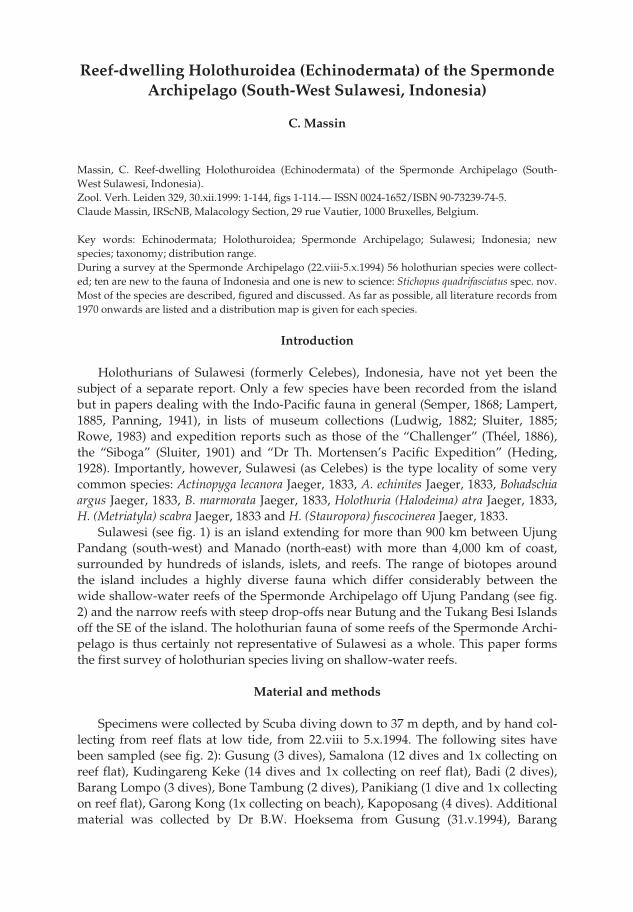
Sulawesi (see fig. 1) is an island extending for more than 900 km between UjungPandang (south-west) and Manado (north-east) with more than 4,000 km of coast,surrounded by hundreds of islands, islets, and reefs. The range of biotopes aroundthe island includes a highly diverse fauna which differ considerably between thewide shallow-water reefs of the Spermonde Archipelago off Ujung Pandang (see fig.2) and the narrow reefs with steep drop-offs near Butung and the Tukang Besi Islandsoff the SE of the island. The holothurian fauna of some reefs of the Spermonde Archi-pelago is thus certainly not representative of Sulawesi as a whole. This paper formsthe first survey of holothurian species living on shallow-water reefs.
Material and methods
Specimens were collected by Scuba diving down to 37 m depth, and by hand col-lecting from reef flats at low tide, from 22.viii to 5.x.1994. The following sites havebeen sampled (see fig. 2): Gusung (3 dives), Samalona (12 dives and 1x collecting onreef flat), Kudingareng Keke (14 dives and 1x collecting on reef flat), Badi (2 dives),Barang Lompo (3 dives), Bone Tambung (2 dives), Panikiang (1 dive and 1x collectingon reef flat), Garong Kong (1x collecting on beach), Kapoposang (4 dives). Additionalmaterial was collected by Dr B.W. Hoeksema from Gusung (31.v.1994), Barang
pp 003-144 02-01-2007 15:33 Pagina 3

Massin. Holothuroidea of the Spermonde Archipelago. Zool. Verh. Leiden 329 (1999)4
Lompo (24.xi.1995) and Bone Baku (14.v.1997). Muddy or sandy bottoms and man-grove areas at the mainland coastline were not prospected. Table 1 presents the list ofcollected species for each locality.
Specimens were anaesthetized in 4-7% magnesium chloride during 0.5 to 3haccording to their size and fixed in 10 % buffered formalin (pH 8.2-8.4). Additionally,large specimens (more than 10 cm long) were injected with 100 % formalin in thecoelomic cavity. Later on, specimens were transferred to 70 % buffered alcohol forpermanent storage. Ossicles were prepared for light microscopy by dissolving small
Fig.1. Location of the study area along the south west coast of Sulawesi.
pp 003-144 02-01-2007 15:33 Pagina 4
5Massin. Holothuroidea of the Spermonde Archipelago. Zool. Verh. Leiden 329 (1999)
pieces of body wall, tube feet, papillae and tentacles in bleach, and by rinsing themcarefully (6-8 times) with distilled water before to let them dry. Ossicles were mount-ed with Euparal under a cover slip.
The specimens were deposited in the collections of the Royal Belgian Institute ofNatural Sciences (IRSNB), Brussels, Belgium (IG.28251), the Marine laboratory of Uni-versitas Hasanuddin (Ujung Pandang) and at the National Museum of Natural Histo-ry at Leiden (RMNH). Material for comparative study was obtained from the MuséeNational d’Histoire Naturelle Paris, France (MNHN), the Zoological Museum,
Fig. 2. The Spermonde Archipelago.
pp 003-144 02-01-2007 15:33 Pagina 5

Massin. Holothuroidea of the Spermonde Archipelago. Zool. Verh. Leiden 329 (1999)6T
able
1: L
ist o
f spe
cies
col
lect
ed in
the
Sper
mon
de
Arc
hipe
lago
dur
ing
the
pres
ent s
urve
y. «
*»:
new
rec
ord
for
Ind
ones
ia; «
**»
: new
spe
cies
.
Gus
ung
Sam
alon
aK
udin
gare
ngB
adi
Bar
ang
Kap
opos
ang
Pani
kian
gG
aron
gB
one
Kek
eL
ompo
Kon
gB
aku
Act
inop
yga
leca
nora
XX
Act
inop
yga
mili
aris
XX
Boh
adsc
hia
argu
sX
XB
ohad
schi
a vi
tien
sis
XB
ohad
schi
asp
ec.
XH
olot
huri
a(A
cant
hotr
apez
a) c
olub
erX
XX
Hol
othu
ria
(Hal
odei
ma)
atr
aX
XX
Hol
othu
ria
(Hal
odei
ma)
edu
lisX
XH
olot
huri
a(L
esso
noth
uria
) haw
aien
sis
*X
XH
olot
huri
a(L
esso
noth
uria
) par
dalis
XH
olot
huri
a(M
erte
nsio
thur
ia) l
euco
spilo
taX
XH
olot
huri
a(M
etri
atyl
a) s
cabr
aX
Hol
othu
ria
(Mic
roth
ele)
nob
ilis
XX
Hol
othu
ria
(Pla
type
rona
) diff
icili
sX
Hol
othu
ria
(Pla
type
rona
) exc
elle
ns*
XH
olot
huri
a(S
empe
roth
uria
) fla
vom
acul
ata
XH
olot
huri
a(S
empe
roth
uria
) im
itan
s*
XH
olot
huri
a(S
taur
opor
a) d
iscr
epan
s*
XH
olot
huri
a(S
taur
opor
a) fu
scoc
iner
eaX
XH
olot
huri
a(S
taur
opor
a) o
livac
eaX
Hol
othu
ria
(The
elot
huri
a) tu
rris
cels
a*
XH
olot
huri
a(T
hym
iosy
cia)
hill
aX
XX
Hol
othu
ria
(Thy
mio
syci
a) im
pati
ens
XX
XX
XLa
bido
dem
asru
gosu
mX
Labi
dode
mas
sem
peri
anum
XX
Pea
rson
othu
ria
grae
ffei
XSt
icho
pus
herr
man
niX
XSt
icho
pus
noct
ivag
us*
XSt
icho
pus
quad
rifa
scia
tus
**X
XX
Stic
hopu
sva
stus
XX
pp 003-144 02-01-2007 15:33 Pagina 6

7Massin. Holothuroidea of the Spermonde Archipelago. Zool. Verh. Leiden 329 (1999)T
able
1 c
onti
nued
. Lis
t of s
peci
es c
olle
cted
in th
e Sp
erm
ond
e A
rchi
pela
go d
urin
g th
e pr
esen
t sur
vey.
«*»
: ne
w r
ecor
d fo
r In
don
esia
; «**
»: n
ew s
peci
es.
Gus
ung
Sam
alon
aK
udin
gare
ngB
adi
Bar
ang
Kap
opos
ang
Pani
kian
gG
aron
gB
one
Kek
eL
ompo
Kon
gB
aku
The
leno
ta a
nana
sX
The
leno
ta a
nax
XC
oloc
hiru
s ro
bust
usX
Ple
sioc
oloc
hiru
s au
stra
lisX
XP
lesi
ocol
ochi
rus
cf. a
ustr
alis
XN
eoth
yoni
dium
mag
num
XX
Phy
rella
trap
eza
*X
Hem
ithy
one
sem
peri
XLi
potr
apez
asp
ec.
XA
froc
ucum
is a
fric
ana
XC
lado
labe
s sc
hmel
tzii
XX
Osh
imel
la e
hren
berg
ii*
XE
uapt
a go
deffr
oyi
XO
pheo
deso
ma
gris
eaX
XP
olyp
lekt
ana
kalli
pepl
osX
Pol
yple
ktan
a ke
fers
tein
iX
XSy
napt
a m
acul
ata
XSy
napt
ula
cf. d
enti
cula
taX
Syna
ptul
acf
. lam
pert
iX
Syna
ptul
a m
edia
*X
Syna
ptul
a re
ticu
lata
XX
XSy
napt
ula
cf. r
etic
ulat
aX
XSy
napt
ula
psar
aX
XSy
napt
ula
rect
aX
XSy
napt
ula
spec
.X
Chi
rido
ta s
thul
man
niX
Num
ber
of s
peci
es2
2226
145
108
11
Num
ber
of d
ives
312
142
34
11
1
pp 003-144 02-01-2007 15:33 Pagina 7

Massin. Holothuroidea of the Spermonde Archipelago. Zool. Verh. Leiden 329 (1999)8
Kopenhagen, Denmark (ZMC), the Zoological Museum of Amsterdam, the Nether-lands (ZMA) and the IRSNB, Brussels, Belgium.
The abbreviations used for the geographical range from different parts of Aus-tralia are as follows: GBR: Great Barrier Reef; NSW: New South Wales; NT: NorthernTerritory; QLD: Queensland; WA: Western Australia.
Literature records are as complete as possible for each species, excepting the mostcommon species for which I mention only the recent records (from 1970 onwards).For the older records I refer to the comprehensive papers of Panning (1929, 1935a-d,1949), Heding & Panning (1954), Cherbonnier (1955a, 1988) or Cherbonnier & Féral(1984a, b). Papers dealing with physiology or biochimestry are not included unlessthey give a record for a new locality.
Systematic account
Order Aspidochirotida Grube, 1840Family Holothuriidae Ludwig, 1894
Genus Actinopyga Broon, 1860Actinopyga lecanora (Jaeger, 1833)
(figs 3a-j, 4, 110a)
Mülleria lecanora Jaeger, 1833: 18, pl. 2 figs 2, 2b, pl. 3 fig. 8.Holothuria (Actinopyga) lecanora; Panning, 1929 [1931]: 127, fig. 9a-c (synonymy and records before
1929).Actinopyga lecanora; Endean, 1957: 254; Levin, 1979: 19; Sloan et al., 1979: 121; Mary Bai, 1980: 7, fig. 8B;
Liao, 1980: 115; Price, 1982: 10; James, 1983: 93; Liao, 1984: 221; Price & Reid, 1985: 3; Reyes-Leonardo et al., 1985: 269, pl. 2 fig. 3a-f; Cannon & Silver, 1986: 20, textfig.; Féral & Cherbonnier,1986: 72; Cherbonnier, 1988: 20, fig. 4A-I (synonymy and records before 1979); Jangoux et al.,1989: 163; Conand, 1989: 17; Levin & Dao Tan Ho, 1989: 55; Adams, 1992: 13; Marsh et al., 1993:63; Allen & Steene, 1994: 242; Holland, 1994: 2; James, 1994: 28; James & Manikfan, 1994: 102, pl.2B; Colin & Arneson, 1995: 260, fig. 1224; Rowe & Gates, 1995: 287; Liao & A.M. Clark, 1995: 425,fig 243a-b; Sant, 1995: 27; Gosliner et al., 1996: 277, fig. 1022; Massin, 1996b: 8, fig. 2A-B; Liao,1997: 84, fig. 47a-b; Baine & Forbes, 1998: 4.
Material.— IRSNB IG.28251/126 (1 specimen), Samalona SW, 19.ix.94, 12 m depth; RMNH Ech. 6075(1 specimen), Kapoposang, 30.ix.94, 10 m depth, night dive.
Description.— Specimens 195 × 75 and 160 × 70 mm, nearly uniformly chocolatebrown with some lighter spots (fig. 110a); around the anus, the characteristic whitezone with fine brown lines (fig. 110a); ventral sole of the smaller specimen beige; inalcohol beige-brown dorsally and white ventrally. Five strong yellow anal teeth (fig.110a). Ventral sole flat, dorsal surface arched. Papillae few, scattered all over the bivi-um; tube feet restricted to the ambulacra; in each ambulacrum, tube feet long, nar-row, on 5-10 rows in a zig-zag pattern (5-6 in the lateral ambulacra and 7-10 in thecentral ambulacrum). Skin very thick (10-11 mm). Both specimens contracted andconsequently tentacles difficult to observe; specimen IRSNB IG 28251/126 with 19tentacles visible.
Calcareous ring stout with large radial pieces (fig. 3a). On their central anterior
pp 003-144 02-01-2007 15:33 Pagina 8

9Massin. Holothuroidea of the Spermonde Archipelago. Zool. Verh. Leiden 329 (1999)
tooth a slit for the insertion of the flat longitudinal muscles. One large Polian vesicle(50 mm long), swollen at the free extremity. One stone canal, very short with a largemassive L-shaped madreporic plate.
Ossicles of the body wall small rosettes, 25-35 µm long dorsally (fig. 3b) and 20-25µm long ventrally (fig. 3c). In the dorsal papillae, rosettes similar to those of the bodywall; some more massive and longer (fig. 3d); together with the rosettes, spiny plates(fig. 3e) and rods (fig. 3f). In the tube feet, very small rosettes, 8-25 µm long (fig. 3g);terminal plate 250-300 µm across. In the tentacles, massive rods (figs 3h, j), 45-450 µmlong, spiny at the extremities.
Geographic distribution (fig. 4).— Somalia, Kenya, Madagascar, Mauritius, SriLanka, Maldive Islands, India (Andaman Islands), Myanmar (Mergui Archipelago),Malaysia, Indonesia (Sumatra, Java, Sulawesi = type locality, Sumba, Salayer, Timor,Celebes Sea, Ambon, Kai Islands), Australia (WA, Timor Sea, NT, QLD), Philippines,
Fig. 3. Actinopyga lecanora (Jaeger, 1833). a: calcareous ring (IR: interradial piece; R: radial piece); b:rosettes from dorsal body wall; c: rosettes from ventral body wall; d: rosettes from dorsal papillae; e:plates from dorsal papillae; f: rods from dorsal papillae; g: tube foot rosettes ; h & j: tentacle rods.
a
R
IR
100 µm
b
c
d
f
g
h
j a
b-d, f, g
e, j
h200 µm
50 µm
5 mm
e
pp 003-144 02-01-2007 15:33 Pagina 9

Massin. Holothuroidea of the Spermonde Archipelago. Zool. Verh. Leiden 329 (1999)10
Vietnam, China, Japan (Bonin Islands, Ryukyu Islands), Papua New Guinea, SolomonIslands, Loyalty Islands, New Caledonia, Fiji.
Remarks.— Specimens of Sulawesi do not differ from those from other areas ofthe Indo-Pacific Ocean. The white anal cone is very characteristic and present trough-out the area of distribution (fig. 4), except in some juveniles (Massin, 1996b). Thecolour pattern of the body wall is variable, ranging from uniformly chocolate brownto a complicated patchwork of brown, beige and white (Allen & Steene, 1994: 242).
Actinopyga miliaris (Quoy & Gaimard, 1833)(figs 5a-d, 6, 110b)
Holothuria miliaris Quoy & Gaimard, 1833: 137.Holothuria (Actinopyga) miliaris; Panning, 1929 [1931]: 127, fig. 10a-g (synonymy and records before
1929).Actinopyga miliaris; Bell, 1887a: 523; Endean, 1956: 132; Endean, 1957: 254; James, 1969: 61; Tortonese,
1977: 275; Sloan et al., 1979: 121; Levin, 1979: 19; Mary Bai, 1980: 7, fig. 8D; Liao, 1980: 115;Humphreys, 1981: 33; Price, 1982: 10; James, 1983: 93, pl. 1C; Reyes-Leonardo, 1984a: 144, pl. 1 fig.5a-e; Cherbonnier & Féral, 1984a: 667, fig. 4A-J (synonymy and records before 1984); Liao, 1984:221; A.M. Clark, 1984: 99; Price & Reid, 1985: 3; Reyes-Leonardo et al., 1985: 269; Cannon & Silver,1986: 20, fig. 5b, text-fig.; Féral & Cherbonnier, 1986: 74, fig. 40B; Conand, 1989: 19; James, 1989:129; Chambers, 1989: 89; Adams, 1992: 13; Holland, 1994: 2; Kerr, 1994: 166, fig. 4a; James, 1994:28; James & Manikfan, 1994: 102, pl. 1D; James & James, 1994: 4, fig. 1; Sant, 1995: 27; Colin &Arneson, 1995: 260, fig. 1222; Rowe & Gates, 1995: 287; Liao & A.M. Clark, 1995, fig. 245a-c;Massin, 1996b: 12, figs 6A-F, 7A-B; Belhadjali, 1997: 3; Tsuda, 1997: 16; Liao, 1997: 87, fig. 49a-c;Rowe & Richmond, 1997: 302; Baine & Forbes, 1998: 4.
Blackfish (= Actinopyga miliaris); Anon., 1996: 13.
Material.— IRSNB IG.28251/31 (1 specimen), Panikiang, 29.viii.94, reef flat at low tide, among sea-grass-beds; RMNH Ech. 6076 (1 specimen), Kudingareng Keke S, 28.ix.94, reef flat at low tide.
Description.— Specimens 95 × 38 and 145 × 55 mm, uniformly deep brown dorsal-ly and beige-brown ventrally; in alcohol uniformly beige-white. Dorsal surface with athin mucous layer incorporating sand grains (fig. 110b). Ventral sole nearly flat, dor-sum arched. Mouth and anus terminal; mouth with 20 tentacles surrounded by a col-lar of papillae; anus surrounded by 5 strong calcareous teeth. Body slightly tapering,both anteriorly and posteriorly. Papillae numerous, long, slender, scattered all over
Fig. 4. Distribution of Actinopyga lecanora (Jaeger, 1833).
pp 003-144 02-01-2007 15:33 Pagina 10

11Massin. Holothuroidea of the Spermonde Archipelago. Zool. Verh. Leiden 329 (1999)
the bivium; tube feet along the 3 ventral ambulacra, numerous, in 4-5 zig-zag rows ineach lateral ambulacrum and 6-7 rows in the central ambulacrum. Skin 5 mm thick.
Very characteristic calcareous ring composed of radial pieces twice as broad asthe interradial ones (fig. 5a). Radial pieces with 3 anterior teeth, the central one with anotch for the insertion of the longitudinal muscle. One Polian vesicle. One very shortstone canal ending in a spherical yellow madreporic plate (fig. 5b), 1.8 mm across sit-uated against the dorsal mesentery.
In the body wall a few rosettes, 20-30 µm long (fig. 5c ), most of them located ven-trally. In the tube feet no rosettes. Dorsally tube feet with a small terminal plate, 200-240 µm across, ventrally with a larger one, 400-500 µm across. In the tentacles rods,30-300 µm long and slightly spinose at the extremities (fig. 5d).
Geographic distribution (fig. 6).— Red Sea, Kenya, Zanzibar, Mozambique, Mada-gascar, Mauritius, Seychelles (Mahé, Aldabra), Comores (Mayotte), Sri Lanka, Mal-dive Islands, India (Laccadive Islands, Gulf of Mannar, Andaman Islands, NicobarIslands), Myanmar (Mergui Archipelago), Christmas Islands, Malaysia, Indonesia(Sumatra, Sumbawa, Sulawesi, Salayer, Tukang Besi Islands, Timor, Ambon, SulaIslands, Kai Islands, Irian Jaya), Australia (GBR, QLD, NE Coast), New Caledonia,
Fig. 5. Actinopyga miliaris (Quoy & Gaimard, 1833). a: calcareous ring (IR: interradial piece; R: radialpiece); b: madreporic plate; c: rosettes from ventral body wall; d: tentacle rods.
a
R IR
5 mma
b
d
c
b
dc 50 µm
1 mm
100 µm
Fig. 6. Distribution of Actinopyga miliaris (Quoy & Gaimard, 1833).
pp 003-144 02-01-2007 15:33 Pagina 11

Massin. Holothuroidea of the Spermonde Archipelago. Zool. Verh. Leiden 329 (1999)12
China, Japan, Philippines, Mariana Islands (Saipan), Easter Caroline Islands (Kosrae),Solomon Islands (Vanikoro = type locality), Ellice Islands (Tuvalu), Vanuatu (= NewHebrides), Fiji, Tonga Islands.
Remarks.— The general aspect, calcareous ring and ossicles of the body wall,leave no doubt about the identity of the Sulawesi specimens as Actinopyga miliaris (seeCherbonnier & Féral, 1984a). However, both specimens have very few rosettes in thebody wall and none in the tube feet which is uncommon for the species. Actinopygamiliaris seems to be a rather variable species, sometimes with very special rods in thedorsal tube feet (Massin, 1996b). A comparative study of specimens from differentlocalities of its wide distributional area (fig. 6) would be useful to define its intraspe-cific variation.
Genus Bohadschia Jaeger, 1833Bohadschia argus Jaeger, 1833
(figs 7, 110c)
Bohadschia argus Jaeger, 1833: 19, pl. 2 figs 1, 1b; A.M. Clark, 1962: 100 (colour plate); A.M. Clark &Taylor, 1971: 92; Levin, 1979: 19; Liao, 1980: 115; Tan Tiu, 1981: 68, pl. 10 figs 1-2; Grosenbaugh,1981: 51; Rowe, 1983: 154; James, 1983: 93; Liao, 1984: 221; Brouns & Heijs, 1985: 175; Richard,1985: 457; Reyes-Leonardo et al., 1985: 270; Marsh, 1986: 73; Cannon & Silver, 1986: 20, figs 3b, 5c;Massin & Doumen, 1986: 188; George & George, 1987: 246, pl. 11f; Cherbonnier, 1988: 34, fig. 10A-H (synonymy and records before 1979); Mukhopadhyay, 1988: 13; Conand, 1989: 21; James, 1989:129; Chambers, 1989: 89; Zoutendijk, 1989: 2; Machida, 1989: 363;Levin & Dao Tan Ho, 1989: 55;Kalashnikov, 1989: 66; Chao & Chang, 1990: 66, figs 1, 3; Marsh et al., 1993: 63; Kerr, 1994: 163,166; Marsh, 1994a: 10; Marsh, 1994b: 57; Holland, 1994: 2; Allen & Steene, 1994: 243; James, 1994:28; James & James, 1994: 11, fig. 4; Rowe & Gates, 1995: 288; Liao & A. M. Clark, 1995: 428, fig.246a-c; Sant, 1995: 27; Colin & Arneson, 1995: 260, fig. 1225; Gosliner et al., 1996: 278, fig. 1024;Belhadjali, 1997: 3; Tsuda, 1997: 16; Liao, 1997: 89, fig. 50a-c; Baine & Forbes, 1998: 4.
Holothuria (Bohadschia) argus; Panning, 1929 [1931]: 121, fig. 2a-i (synonymy and records before 1929)Holothuria argus; Dawydoff, 1952: 117; Endean, 1953: 56; Endean, 1956: 130; Endean, 1957: 252;
Endean, 1965: 253; James, 1969: 62; Townsley & Townsley, 1972: 176.Tiger/Leopardfish (= Bohadschia argus); Anon., 1996: 13.
Material.— One specimen observed at Panikiang (15 m depth) and one at Samalona (20 m depth).
Geographic distribution (fig. 7).— Seychelles, Madagascar, Chagos Archipelago(Diego Garcia), Sri Lanka, India (Gulf of Mannar, Laccadive Islands, AndamanIslands, Nicobar Islands), Gulf of Bengal, Myanmar (Mergui Archipelago), CocosKeeling Islands, Malaysia, Indonesia (Sumatra, Sumbawa, Flores, Sulawesi = typelocality, Rotti, Timor, Ambon, Banda Islands, Irian Jaya), Malaysia (Sabah), Australia(WA, NT, QLD, GBR), Philippines, Vietnam, China, Taiwan, Japan, Mariana Islands(Guam, Saipan), Caroline Islands (Kosrae, Yap), Papua New Guinea (MadangProvince), Solomon Islands, New Caledonia, Samoa, Ellice Islands (Funafuti, Tuvalu),Fiji, Vanuatu (= New Hebrides), Tonga, Line Islands (Fanning Island), Cook Islands,Society Islands (Tahiti).
Remarks.— No specimens of Bohadschia argus were collected because the speciescan be identified at sight without any doubt. This very characteristic species (fig.110c) is normally abundant on reef flats and in shallow water. In the Spermonde
pp 003-144 02-01-2007 15:33 Pagina 12

13Massin. Holothuroidea of the Spermonde Archipelago. Zool. Verh. Leiden 329 (1999)
Archipelago it is actually uncommon and lives deeper (15-25 m). It is certainly not aprized species as trepang (Anon., 1974; Conand, 1986) even in Indonesia (Sutaman,1993) and the scarsity of the species cannot be explained by high human fishing pres-sure.
Bohadschia vitiensis (Semper, 1868)(figs 8a-k, 9)
Holothuria vitiensis Semper, 1868: 80, pl. 30 fig. 2; Domantay, 1933: 76, pl.1 fig. 2; Domantay, 1953: 119. Holothuria (Bohadschia) vitiensis; Panning, 1929 [1931]: 122, fig. 3a-k (synonymy and records before
1931); Domantay, 1935: 399.Bohadschia marmorata vitiensis; Panning, 1944: 40, fig. 11a-y; Anon., 1979: 20; Belhadjali, 1997: 3.Bohadschia vitiensis; Rowe, 1969: 130; A.M. Clark & Rowe, 1971: 176, pl. 27 fig. 5; Clastres et al., 1978:
973; Levin, 1979: 20; Mary Bai, 1980: 8, textfig. 8H; James, 1983, 93, pl. 1D; Féral & Cherbonnier,1986: 78; Cherbonnier, 1988: 42, fig. 14A-I; Conand, 1989: 21, fig. 3; Chambers, 1989: 89; Rowe &Richmond, 1997: 302.
Bohadschia spec.; Colin & Arneson, 1995: 260, fig. 1229.
Material.— IRSNB IG.28251/12 (1 specimen), Samalona S, 24.viii.94, 21 m depth; RMNH Ech. 6077 (1specimen), Samalona NW, 16.ix.94, 18 m depth.
Description.— Specimens 370 × 90 and 300 × 85 mm, brown dorsally with tubefeet yellow at the base and brown at the tip; ventrally, yellowish with a median longi-tudinal line nearly white; tube feet circled with white at their base; in alcohol coloursimilar to that of living specimens but faded. Body cylindrical with mouth ventraland anus terminal; mouth with 20 short tentacles; anus surrounded by 5 radialgroups of 4-5 papillae. Tube feet densely crowded dorsally and ventrally withoutalignment. Body wall 3-9 mm thick according to the state of contraction.
Calcareous ring stout; very large radial pieces with a deep anterior notch (fig. 8a);interradial pieces half the width of the radial ones (fig. 8a). One large Polian vesicle(1/10 of body length), ventrally located; one stone canal and very long tentacleampullae (1/5 to 1/3 of body length). Cuvierian tubules numerous and quicklyexpelled when the animal is manipulated.
Ossicles of dorsal body wall rosettes (fig. 8b) 14-27 µm long. In the ventral bodywall perforated and unperforated grains together with rosettes (fig. 8c), 13-27 µm long.
Fig. 7. Distribution of Bohadschia argus Jaeger, 1833.
pp 003-144 02-01-2007 15:33 Pagina 13

Massin. Holothuroidea of the Spermonde Archipelago. Zool. Verh. Leiden 329 (1999)14
In the ventral tube feet rods (fig. 8d), 33-140 µm long and rosettes (fig. 8e) 13-35 µmlong; end plate 450-680 µm across, made of several pieces. In the dorsal tube feetnumerous rods (fig. 8f) 60-180 µm long, branched rods (fig. 8g) 20-65 µm long androsettes (fig. 8h) 20-55 µm long; end plate 325-500 µm across, made of one piece. In thetube feet, rods, branched rods and rosettes without clear separation. In the tentaclesspiny rods (fig. 8j), 30-470 µm long, with bifurcated or bended extremities (fig. 8k.
Fig. 8. Bohadschia vitiensis (Semper, 1868). a: calcareous ring (IR: interradial piece; R: radial piece); b:rosettes from dorsal body wall; c: grains and rosettes from ventral body wall; d: rods from ventraltube feet; e: rosettes from ventral tube feet; f: rods from ventral tube feet; g: branched rods from dorsaltube feet; h: rosettes from dorsal tube feet; j: spiny rods from tentacles; k: extremities from tentaclerods.
a
R IR
100 µm
b-h
b
c
e
g
f
h
d
k
j
50 µm
1 cm
j-k
a
pp 003-144 02-01-2007 15:33 Pagina 14
15Massin. Holothuroidea of the Spermonde Archipelago. Zool. Verh. Leiden 329 (1999)
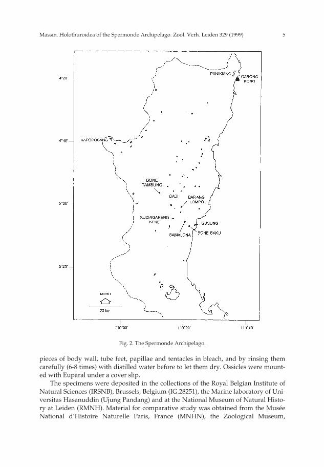
Geographic distribution (fig. 9).— Madagascar, Sri Lanka, India (Nicobar Islands),Indonesia (Java, Sulawesi, Makassar Strait, Ceram), Philippines, Japan (RyukyuIslands), Caroline Islands (Chuuk (= Truck) Atoll), Papua New Guinea (New Britain),Australia (GBR, Queensland), Vanuatu (= New Hebrides; Sfate Island), New Caledonia,Ellice Islands (Tuvalu), Fiji (Viti Levu Island = type locality), Samoa (Navigator Island).
Remarks.— The colour pattern of the specimens from Sulawesi is similar to thethat of specimens depicted by Féral & Cherbonnier (1986: pl. on p. 79) and by Colin &Arneson (1995: fig. 1229). These specimens are without darker bands or spots as illus-trated by Rowe & Doty (1977: fig. 6g, h) for Bohadschia marmorata Jaeger, 1833.
The ossicles of the dorsal and ventral body wall are similar to those illustrated byPanning (1944: fig. 11a-y) for Bohadschia marmorata vitiensis. However, I compared thematerial from Sulawesi with specimens of B. vitiensis from New Caledonia (held inthe MNHN) and identified by Cherbonnier. These specimens have unperforatedgrains only in the ventral body wall, unlike those from Sulawesi. One therefore won-ders whether we are dealing with two different species or with a single highly vari-able species?
Panning (1944), Rowe & Doty (1977), Tan Tiu (1981), Reyes-Leonardo (1984) andRowe & Gates (1995) opted for a single species whose ossicle complexity increaseswith growth. Consequently, they consider B. vitiensis, together with other species, assubspecies or synonyms of B. marmorata. Provisionally, I maintain B. vitiensis as a sep-arate species. In my view it is desirable to repeat the studies of growth series of Bohad-schia to determine the degree of intraspecific variation and to relate ossicle changeswith growth and colour pattern in order to determine the validity of conclusions ofPanning (1944), Rowe & Doty (1977) and Rowe & Gates (1995). Because of thesenomenclatorial problems some localities assigned to B. marmorata may belong to B.vitiensis, making its area of distribution wider than here presented (fig. 9).
Bohadschia spec.(fig.10a-h)
Material.— IRSNB IG.28251/ 47 (1 specimen), Samalona W, 1.ix.94, 20 m depth, on sand.
Description.— Specimen 220 × 120 mm. Colour in alcohol yellow-white; the tube
Fig. 9. Distribution of Bohadschia vitiensis (Semper, 1868).
pp 003-144 02-01-2007 15:33 Pagina 15

Massin. Holothuroidea of the Spermonde Archipelago. Zool. Verh. Leiden 329 (1999)16
feet whitish, the base circled with brown. Body cylindrical, mouth ventral, anus ter-minal; because of the state of contraction of the specimen, groups of radial papillaearound the anus not seen; body wall 2-7 mm thick; tentacles 20, short, brown.
Calcareous ring stout (fig. 10a); large radial pieces with a deep anterior notch;interradial pieces half the width of the radial ones (fig. 10a). One Polian vesicle; onelong (50 mm), straight stone canal ending in a madreporic plate narrower than canaldiameter. Tentacle ampullae very long (1/3 to 1/4 of body length). Cuvierian tubulesnumerous.
Ossicles of ventral body wall perforated grains and plump rosettes (fig. 10b) 15-33µm long, together with fine rosettes (fig. 10c) 14-27 µm long. In the dorsal body wall
Fig. 10. Bohadschia spec. a: calcareous ring (IR: interradial piece; R: radial piece); b: grains and plumprosettes from ventral body wall; c: slender rosettes from ventral body wall; d: grains from dorsal bodywall; e: rosettes from dorsal body wall; f: branched rods from ventral tube feet; g: rods and rosettesfrom dorsal tube feet ; h: tentacles rods.
a
RIR
100 µm
b-g
b
c
de
f
g
h a
h
5 mm
50 µm
pp 003-144 02-01-2007 15:33 Pagina 16

17Massin. Holothuroidea of the Spermonde Archipelago. Zool. Verh. Leiden 329 (1999)
perforated and unperforated grains (fig. 10d) 15-38 µm long and rosettes (fig. 10e) 14-31 µm long. In the ventral tube feet branched rods only (fig. 10f) 19-40 µm long; endplate 295-315 µm across. In the dorsal tube feet branched rods 15-85 µm long androsettes 15-60 µm long with numerous intermediate stages (fig. 10g) between thesetwo forms; end plate 400-550 µm across. In the tentacles rods with granulous extremi-ties, 15-160 µm long (fig. 10h).
Remarks.— This Bohadschia specimen was collected in the same locality and depthas the two specimens of B. vitiensis described above. The size and the general aspectof the three specimens is similar. Only the colour of the base of the tube feet of thepresent specimen is different (brown versus yellow-white). However, the ossicles ofits body wall and ventral tube feet are completely different. The specimen has ossiclespresenting a mixture of characters from several other species such as Bohadschiacousteaui Cherbonnier, 1954 and Bohadschia mitsoensis Cherbonnier, 1988. The absenceof unperforated grains ventrally is the only character which does not appear in otherBohadschia spp., although Massin (1996b) has described a specimen of Bohadschia sim-ilis (Semper, 1868) without unperforated grains ventrally.
The colour of the present specimen is the same as that of a specimen figured inGosliner et al. (1996: fig. 1027) and in my view misidentified as Bohadschia paradoxa(Selenka, 1867) (cf. the descriptions and illustrations in Fisher, 1907, and Panning,1929). The latter species lacks perforated and unperforated grains dorsally and thedorsal “rods” described by Fisher (1907), are quite different from those of the presentspecimen.
In conclusion, bearing in mind the high variability of some Bohadschia spp., it isnot realistic to describe the present specimen as a new species.
Genus Holothuria Linnaeus, 1767Subgenus Acanthotrapeza Rowe, 1969
Holothuria (Acanthotrapeza) coluber Semper, 1868(figs 11a-k, 12)
Holothuria coluber Semper, 1868: 90, pl. 28, pl. 30 fig. 28, pl. 34 fig. 5; Panning, 1944: 62, fig. 30a-i;Dawydoff, 1952: 117; Endean, 1956: 131; Endean, 1957: 252; Endean, 1965: 233; Baine & Forbes,1998: 4.
Holothuria (Holothuria) coluber; Panning, 1935a: 35, fig. 30a, b (synonymy and records before 1935).Holothuria (Acanthotrapeza) coluber; Rowe, 1969: 138; Levin, 1979: 20; Cherbonnier, 1980: 636, fig. 11A-
H (records before 1980); Tan Tiu, 1981: 74, pl. 16 figs. 1-3; Reyes-Leonardo, 1984a: 147, pl.2 fig.2A-M; Price & Reid, 1985: 3; Reyes-Leonardo et al., 1985: 273; Cannon & Silver, 1986: 21, figs 3d,6c; Féral & Cherbonnier, 1986: 80, fig. 40A, F; Conand, 1989: 16, 23; Chambers, 1989: 89; Marsh etal., 1993: 63; Kerr, 1994: 168, fig. 4b; Marsh, 1994a: 10; Marsh, 1994b: 57; Rowe & Gates, 1995: 290.
Material.— IRSNB IG.28251/106 (1 specimen), Kudingareng Keke NW, 15.ix.94, 5 m depth under abig coral slab; RMNH Ech. 6056 (1 specimen), Kudingareng Keke S, 29.ix.94, reef flat; IRSNBIG.28251/222 (1 specimen), Kapoposang, 30.ix.94, 1 m depth in the lagoon, night dive; RMNH Ech.6078 (1 specimen), Badi, 3.x.94, 2 m depth.
Description.— One cylindrical juvenile specimen, 17 × 7 mm; three others, widerposteriorly than anteriorly, 150 × 18-23, 240 × 20-37 and 390 × 22-30 mm. Specimensblack with white papillae dorsally and deep brown with white tube feet ventrally; in
pp 003-144 02-01-2007 15:33 Pagina 17

Massin. Holothuroidea of the Spermonde Archipelago. Zool. Verh. Leiden 329 (1999)18
Fig. 11. Holothuria (Acanthotrapeza) coluber Semper, 1868. a: calcareous ring (IR: interradial piece; R:radial piece); b: body wall tables (L=390 mm; RMNH Ech. 6078); c: body wall tables (L=17 mm; IRSNBIG.28251/222); d: body wall buttons (L=390 mm; RMNH Ech. 6078); e: body wall buttons (L=17 mm;IRSNB IG.28251/222); f: tube foot tables; g & h: tube foot buttons; j: tube foot rods; k: tentacle rods.
a
R IR
100 µm
b-f, h, k
e
b
d
c
f
g
hj
k
a
g, j
5 mm
5 0
pp 003-144 02-01-2007 15:33 Pagina 18
19Massin. Holothuroidea of the Spermonde Archipelago. Zool. Verh. Leiden 329 (1999)
alcohol light brown dorsally with white papillae, and white ventrally. For large speci-mens, papillae and tube feet very similar, numerous, and without regular arrange-ment. Juvenile with podia and papillae making a clear difference between triviumand bivium. Papillae 9-10 along the length of the body, without alignment, very large,white coloured due to dense accumulation of ossicles. Tube feet in ambulacral andinterambulacral areas, cylindrical, short. Skin rough to the touch due to the orienta-tion of tall-spired tables. Mouth ventral, anus terminal; tentacles 20.
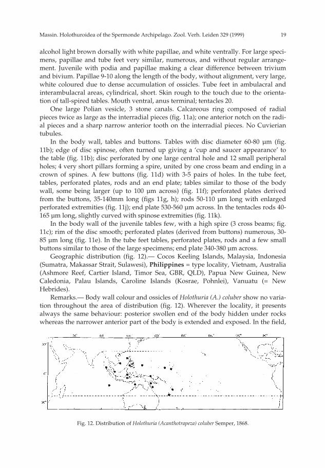
One large Polian vesicle, 3 stone canals. Calcareous ring composed of radialpieces twice as large as the interradial pieces (fig. 11a); one anterior notch on the radi-al pieces and a sharp narrow anterior tooth on the interradial pieces. No Cuvieriantubules.
In the body wall, tables and buttons. Tables with disc diameter 60-80 µm (fig.11b); edge of disc spinose, often turned up giving a ‘cup and saucer appearance’ tothe table (fig. 11b); disc perforated by one large central hole and 12 small peripheralholes; 4 very short pillars forming a spire, united by one cross beam and ending in acrown of spines. A few buttons (fig. 11d) with 3-5 pairs of holes. In the tube feet,tables, perforated plates, rods and an end plate; tables similar to those of the bodywall, some being larger (up to 100 µm across) (fig. 11f); perforated plates derivedfrom the buttons, 35-140mm long (figs 11g, h); rods 50-110 µm long with enlargedperforated extremities (fig. 11j); end plate 530-560 µm across. In the tentacles rods 40-165 µm long, slightly curved with spinose extremities (fig. 11k).
In the body wall of the juvenile tables few, with a high spire (3 cross beams; fig.11c); rim of the disc smooth; perforated plates (derived from buttons) numerous, 30-85 µm long (fig. 11e). In the tube feet tables, perforated plates, rods and a few smallbuttons similar to those of the large specimens; end plate 340-380 µm across.
Geographic distribution (fig. 12).— Cocos Keeling Islands, Malaysia, Indonesia(Sumatra, Makassar Strait, Sulawesi), Philippines = type locality, Vietnam, Australia(Ashmore Reef, Cartier Island, Timor Sea, GBR, QLD), Papua New Guinea, NewCaledonia, Palau Islands, Caroline Islands (Kosrae, Pohnlei), Vanuatu (= NewHebrides).
Remarks.— Body wall colour and ossicles of Holothuria (A.) coluber show no varia-tion throughout the area of distribution (fig. 12). Wherever the locality, it presentsalways the same behaviour: posterior swollen end of the body hidden under rockswhereas the narrower anterior part of the body is extended and exposed. In the field,
Fig. 12. Distribution of Holothuria (Acanthotrapeza) coluber Semper, 1868.
pp 003-144 02-01-2007 15:33 Pagina 19

Massin. Holothuroidea of the Spermonde Archipelago. Zool. Verh. Leiden 329 (1999)20
it is easy to distinguish it from Holothuria leucospilota, which has a similar colour pat-tern and behaviour, because of its very rough and hard skin. Holothuria (A.) colubergenerally lives on the reef flats at less than a meter deep (Endean, 1965; Cherbonnier,1980; Cannon & Silver, 1986; Conand, 1989; Chamber, 1989; Kerr, 1994; presentstudy). There are only two records from somewhat deeper water (Semper, 1868: 6-8Faden = 11-15 m; Féral & Cherbonnier, 1986: 25 m).
Subgenus Halodeima Pearson, 1914Holothuria (Halodeima) atra Jaeger, 1833
(fig. 13)
Holothuria atra Jaeger, 1833: 22; Dawydoff, 1952: 117; Endean, 1953: 56; Endean, 1956: 131; Endean,1957: 252; Bonham & Held, 1963: 305, fig. 3A; Loi & Sach, 1963: 241, pl. 2 figs B, C, pl. 4 fig. 7;Caso, 1965: 271, textfigs 17-19, pl. 5 figs 1-12, pl.7 figs 1-11; Townsley & Townsley, 1972: 176;McKnight, 1974: 45; Tortonese, 1977: 275; Tortonese, 1979: 316; Liao, 1980: 115; Lawrence, 1980:202; Kropp, 1982: 446; James, 1983: 93; Brouns & Heijs, 1985: 175; Massin & Doumen, 1986: 188;Zoutendijk, 1989: 2; Dalzell et al., 1993: 37; Allen & Steene, 1994: 243; Colin & Arneson, 1995: 260,fig. 1230; Adjeroud, 1997: 14; Baine & Forbes, 1998: 4.
Holothuria (Holothuria) atra; Panning, 1935a: 30, fig. 22a-f (synonymy and records before 1935).Holothuria (Halodeima) atra; A.M. Clark & Taylor, 1971: 91; Liao, 1975: 210, fig. 10 (1-3); Rowe & Doty,
1977: 230, figs 3d, 7a; Ebert, 1978: 183; Levin, 1979: 20; Sloan et al., 1979: 122; Tortonese, 1980: 107;Cherbonnier, 1980: 631, fig. 8A-N (synonymy and records before 1974); Mary Bai, 1980: 12,textfig. 9D; Grosenbaugh, 1981: 51; Tan Tiu, 1981: 73, pl. 15 figs 1-3, pl. 29 figs 1-2e; Humphreys,1981: 35; Price, 1982: 11; Rowe, 1983: 155; Price, 1983: 89, fig. 47a-d; Mukhopadhyay & Samanta:1983: 302, fig. 3A-E; Reyes-Leonardo, 1984a: 145, pl. 2 fig. 1a-d; Liao, 1984: 222; A.M. Clark, 1984:99; Price & Reid, 1985: 4; James, 1985 [1988]: 404; Richard, 1985: 457; Reyes-Leonardo et al., 1985:271; Marsh, 1986: 73; Cannon & Silver, 1986: 22, figs 3f, 6d; Féral & Cherbonnier, 1986: 80, fig. 40E;George & George, 1987: 246; Mukhopadhyay, 1988: 5, fig. 3a-c; Cherbonnier, 1988: 73, fig. 28A-J;Jangoux et al., 1989: 163; Conand, 1989: 23, fig. 2; Chao & Chang, 1989: 117, figs 13, 29H; James,1989: 124; Chambers, 1989: 89; Levin & Dao Tan Ho, 1989: 55; Kalashnikov, 1989: 63; Marsh et al.,1993: 64; Kerr, 1994: 168; Marsh, 1994a: 10; Marsh, 1994b: 57; Holland, 1994: 2; James & Manikfan,1994: 102; James & James, 1994: 16, fig. 10; Sant, 1995: 27; Rowe & Gates, 1995: 291; Liao & A.M.Clark, 1995: 435, fig. 251a-c; James, 1995a: 51, fig. 1E; Massin, 1996b: 18, fig. 10A-E; Gosliner et al.,1996: 279, fig. 1028; Britayev & Zamishliak, 1996: 177; Tsuda, 1997: 16; Liao, 1997: 99, fig. 55a-d;Rowe & Richmond, 1997: 304.
Lollyfish (= Holothuria atra); Anon., 1996: 13.
Material.— IRSNB IG.28251/66 (1 specimen), Samalona, 2.ix.94, reef flat at low tide; IRSNBIG.28251/201b (1 specimen) and RMNH Ech. 6057 (1 specimen), Kudingareng Keke, 28.ix.94, 1 mdepth.
Description and remarks.— Specimens 100 × 25, 120 × 35, 140 × 40 mm, uniformlyblack; in alcohol brown dorsally and lighter ventrally. External features, calcareousring and ossicles are similar to those observed for Holothuria atra from Ambon(Massin, 1996b).
Holothuria atra has been observed on Samalona, Kudingareng Keke andKapoposang. It is still abundant on the reef flat of Kudingareng Keke. On the reef flatof Samalona, H. atra, together with Holothuria edulis, is a dominant species but with alow populatiopn density if compared with other Indo-Pacific areas (Bakus, 1973;
pp 003-144 02-01-2007 15:33 Pagina 20

21Massin. Holothuroidea of the Spermonde Archipelago. Zool. Verh. Leiden 329 (1999)
Lawrence, 1980). At Kapoposang, H. atra is present in the seagrass-beds of the lagoon.On the huge reef flat of Panikiang not a single H. atra was observed.
Geographic distribution (fig. 13).— Red Sea, Somalia, Oman (Muscat), PersianGulf, Kenya, Zanzibar, Mozambique (Querimba Islands), Madagascar, Mauritius,Seychelles (Mahé, Amirantes, Aldabra), Maldive Islands, Chagos Archipelago (DiegoGarcia), Sri Lanka, India (Laccadive Islands, Gulf of Mannar, southeast coast of India,Andaman Islands, Nicobar Islands), Myanmar (coast of Arakan), Malaysia (Peninsu-la, Sabah), Cocos Keeling Islands, Indonesia (Sumatra, Java, Sumbawa, Sulawesi =type locality, Makassar Strait, Lombok, Timor, Lucipara Islands, Ambon, Aru Islands,Banda Sea, Irian Jaya), Philippines, Vietnam, Taiwan, China, Japan, Papua NewGuinea (Port Moresby, Madang Province), Solomon Islands, Australia (WA, NT,Timor Sea, QLD, GBR, NSW, Tasman Sea), Mariana Islands (Guam, Saipan), MarshallIsland (Enewetak Atoll, Rongelap Atoll), Caroline Islands (Kosrae, Yap), New Cale-donia, Niue, Vanuatu (= New Hebrides), Ellice Islands (Funafuti), Gilbert Islands(Boutaritari, Marakai), Fiji, Tonga Islands, Samoa (Navigator), Hawaiian Islands, LineIslands (Fanning Island), Cook Islands (Rarotonga, southern Cook Islands, Manihiki),Society Islands (Tahiti), Galapagos Islands, Cocos Islands, Panamic Region, Clipper-ton Island, Mexico (Zihuatanejo).
Holothuria (Halodeima) edulis Lesson, 1830(figs 14, 110d)
Holothuria edulis Lesson, 1830: 125, pl. 46 fig. 2; Dawydoff, 1952: 117; Endean, 1953: 56; Endean, 1956:131; Endean, 1957: 252; Townsley & Townsley, 1972: 176; Tortonese, 1977: 275; Brouns & Heijs,1985: 175; Allen & Steene, 1994: 244; Fiege et al., 1994: 86; Colin & Arneson, 1995: 260, fig. 1231;Baine & Forbes, 1998: 4.
Holothuria (Holothuria) edulis; Panning, 1935a, fig. 36a-d (synonymy and records before 1935).Holothuria (Halodeima) edulis; Levin, 1979: 20; Cherbonnier, 1980: 632, fig. 9A-L (synonymy and records
before 1977); Mary Bai, 1980: 12, textfig. 9E; Liao, 1980: 115; Grosenbaugh, 1981: 51; Price, 1982:11; Rowe, 1983: 156; Liao, 1984: 222; Reyes-Leonardo, 1984a: 146, pl. 3 fig. 1A-I; Price & Reid,1985: 4; Reyes-Leonardo et al., 1985: 272; James, 1985 [1988]: 404; Marsh, 1986: 73; Cannon & Sil-ver, 1986: 22, fig. 6f, text fig.; George & George, 1987: 246; Féral & Cherbonnier, 1986: 82;Mukhopadhyay, 1988: 6, fig. 4a-b; Cherbonnier, 1988: 75, fig. 29A-I (records); Jangoux et al., 1989:163; Conand, 1989: 23, fig. 2; Chambers, 1989: 89; Levin & Dao Tan Ho, 1989: 55; Kalashnikof,1989: 64, fig. 2; Marsh et al., 1993: 64; Marsh, 1994a: 10; Marsh, 1994b: 57; Holland, 1994: 2; Sant,1995: 27; James, 1995a: 52, fig. 1F-G; Rowe & Gates, 1995: 291; Liao & A.M. Clark, 1995: 436, fig.
Fig. 13. Distribution of Holothuria (Halodeima) atra Jaeger, 1833.
pp 003-144 02-01-2007 15:33 Pagina 21
Massin. Holothuroidea of the Spermonde Archipelago. Zool. Verh. Leiden 329 (1999)22
252a-c; Gosliner et al., 1996: 279, fig. 1029; Massin, 1996b: 19, fig. 11A-G; Tsuda, 1997: 16; Liao,1997: 101, fig. 56a-d; Rowe & Richmond, 1997: 304.
Material.— RMNH Ech. 6079 (1 specimen), Samalona W, 24.viii.94, 2 m depth; IRSNB IG.28251/17 (1specimen), Kudingareng Keke E, 26.viii.94, 7 m depth on steep sandy slope.
Description and remarks.— Specimens 190 × 50 and 91 × 17 mm, pink ventrallyand brownish-black dorsally (fig. 110d); in alcohol grey-white ventrally and browndorsally.
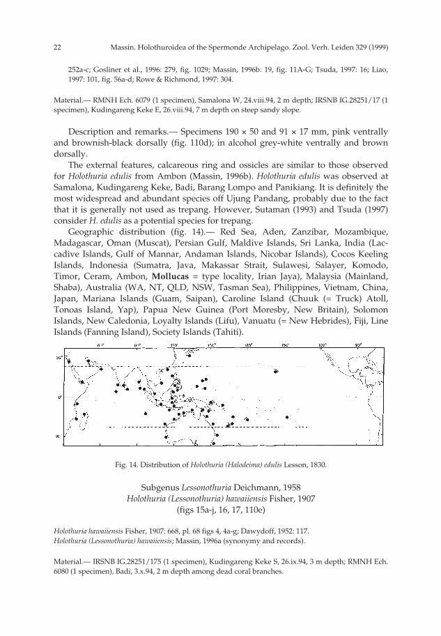
The external features, calcareous ring and ossicles are similar to those observedfor Holothuria edulis from Ambon (Massin, 1996b). Holothuria edulis was observed atSamalona, Kudingareng Keke, Badi, Barang Lompo and Panikiang. It is definitely themost widespread and abundant species off Ujung Pandang, probably due to the factthat it is generally not used as trepang. However, Sutaman (1993) and Tsuda (1997)consider H. edulis as a potential species for trepang.
Geographic distribution (fig. 14).— Red Sea, Aden, Zanzibar, Mozambique,Madagascar, Oman (Muscat), Persian Gulf, Maldive Islands, Sri Lanka, India (Lac-cadive Islands, Gulf of Mannar, Andaman Islands, Nicobar Islands), Cocos KeelingIslands, Indonesia (Sumatra, Java, Makassar Strait, Sulawesi, Salayer, Komodo,Timor, Ceram, Ambon, Mollucas = type locality, Irian Jaya), Malaysia (Mainland,Shaba), Australia (WA, NT, QLD, NSW, Tasman Sea), Philippines, Vietnam, China,Japan, Mariana Islands (Guam, Saipan), Caroline Island (Chuuk (= Truck) Atoll,Tonoas Island, Yap), Papua New Guinea (Port Moresby, New Britain), SolomonIslands, New Caledonia, Loyalty Islands (Lifu), Vanuatu (= New Hebrides), Fiji, LineIslands (Fanning Island), Society Islands (Tahiti).
Subgenus Lessonothuria Deichmann, 1958Holothuria (Lessonothuria) hawaiiensis Fisher, 1907
(figs 15a-j, 16, 17, 110e)
Holothuria hawaiiensis Fisher, 1907: 668, pl. 68 figs 4, 4a-g; Dawydoff, 1952: 117.Holothuria (Lessonothuria) hawaiiensis; Massin, 1996a (synonymy and records).
Material.— IRSNB IG.28251/175 (1 specimen), Kudingareng Keke S, 26.ix.94, 3 m depth; RMNH Ech.6080 (1 specimen), Badi, 3.x.94, 2 m depth among dead coral branches.
Fig. 14. Distribution of Holothuria (Halodeima) edulis Lesson, 1830.
pp 003-144 02-01-2007 15:33 Pagina 22

23Massin. Holothuroidea of the Spermonde Archipelago. Zool. Verh. Leiden 329 (1999)
Fig. 15. Holothuria (Lessonothuria) hawaiiensis Fisher, 1907. a: calcareous ring (IR: interradial piece; R:radial piece); b: stone canal and madreporic plate; c: body wall tables; d: body wall buttons; e: irregu-lar buttons from body wall; f: irregular curved rods from body wall; g: tube foot tables; h: perforatedplates from tube feet; j: tube foot rods.
aR
IR
100 µm
c-h
b
c
d
efg
h
j a, b
j
50 µm
2 mm
pp 003-144 02-01-2007 15:33 Pagina 23

Massin. Holothuroidea of the Spermonde Archipelago. Zool. Verh. Leiden 329 (1999)24
Description.— Specimens are 57 × 28 and 50 × 21 mm, greyish with tiny whitespots corresponding to heaps of ossicles and a patchwork of rusty-brown dots (fig.110e); tentacles pink; edge of the mouth red; in alcohol greyish with white dots. Bodycylindrical, tapering anteriorly and posteriorly. Mouth and anus terminal; tentacles30; tube feet only along the ambulacra. In each ambulacrum 4-6 rows of tube feet;papillae scattered on the whole bivium. Body soft, skin gelatinous, smooth and thin,damaged on preserved specimens.
Calcareous ring stout composed of radial pieces twice as high as interradial ones(fig. 15a). Anterior notch of the radial pieces very deep. One very long Polian vesicle(15-16 mm long) and one very short contorted stone canal ending in an ovoidmadreporic plate (fig. 15b). Specimen from Badi with a well developed gonad madeof a bundle of very long, undivided tubules. Cuvierian tubules numerous.
Ossicles of body wall tables and irregular buttons, also a few irregular curvedrods (fig. 15f). Table discs 70-80 µm in diameter with 4 central holes, a crown of 8large peripheral holes and a second crown of smaller holes, sometimes alternatingwith the large ones (fig. 15c); 4 pillars forming a spire united by one cross beam,exceptionally by 2, and ending in a dense crown of blunt spines (fig. 15c). Buttons 40-110 µm long, with 3-6 pairs of holes (fig. 15d), very often irregular (fig. 15e). Buttonsgathered in heaps appearing as tiny white dots on the skin. In the tube feet tables,perforated plates, and rods. Tables similar to those of body wall but smaller, 50-65µm across (fig. 15g); perforated plates 80-140 µm long, with 2 main central rows oflarge holes and small peripheral ones (fig. 15h); rods 195-270 µm long, perforated atthe center and the extremities (fig. 15j); end plate 200-220 µm across. In the tentaclesspiny rods, strait or curved, 110-460 µm long (fig. 16).
Geographic distribution (fig. 17).— Madagascar, Australia (GBR, Tasman Sea),Indonesia (Sulawesi), Vietnam (Bay of Nhatrang), Cook Islands, Hawaiian Islands =type locality, Easter Island.
Remarks.— The specimen from Sulawesi are very similar to those of Easter Island(Massin, 1996a) and lack the tables with 3 cross beams described by Fisher (1907). This
Fig. 16. Holothuria (Lessonothuria) hawaiiensis Fisher, 1907. Tentacle rods.
100 µm
pp 003-144 02-01-2007 15:33 Pagina 24

25Massin. Holothuroidea of the Spermonde Archipelago. Zool. Verh. Leiden 329 (1999)
difference can be ascribed to the relatively large size of the specimens. Data presentedby Fisher (1907), Cherbonnier (1988), Massin (1996a) and the present study suggestthat the number of cross beams diminishes with increasing body size (see table 2).
The species is new to the fauna of Indonesia.
Table 2. Variation in the number of cross beams of Holothuria hawaiiensis‘s tables.
Author Cherbonnier 88 Fisher 07 Present study Massin 96a
Body size 40 mm 45 mm 50-57 mm 55-120 mmnbr cross beams 3-5 3 1-2 1
Holothuria (Lessonothuria) pardalis Selenka, 1867(figs 18a-j, 19)
Holothuria pardalis Selenka, 1867: 336, pl.19 fig. 85; Dawydoff, 1952: 117; Endean, 1956: 132; Endean,1957: 254; Tortonese, 1977: 275; Tortonese, 1979: 316; James, 1983: 93.
Holothuria (Holothuria) pardalis; Panning, 1935d: 3, fig. 106a-x (synonymy and records before 1935).Holothuria (Lessonothuria) pardalis; A.M. Clark & Taylor, 1971: 91; Liao, 1975: 216, fig. 16 (1-2); Rowe &
Doty, 1977: 233, fig. 4e; Levin, 1979: 21; Sloan et al., 1979: 122; Liao, 1980: 115; Tortonese, 1980:109; Mary Bai, 1980: 14, textfig. 10C; Humphreys, 1981: 34; Price, 1982: 11; Rowe, 1983: 156;Mukhopadhyay & Samanta, 1983: 311; Reyes-Leonardo, 1984a: 148, pl. 4 fig 3a-f; Liao, 1984: 222;A.M. Clark, 1984: 99; Price & Reid, 1985: 4; Richard, 1985: 457; Reyes-Leonardo et al., 1985: 274;James, 1985 [1988]: 404; Marsh, 1986: 73; Cannon & Silver, 1986: 22, figs 3g, 6e; Rho & Shin, 1986:248, pl. 2 figs 1-12; George & George, 1987: 246; Maluf, 1988: 97; Mukhopadhyay, 1988: fig. 1a-c;Cherbonnier, 1988: 117, fig. 47A-O (synonymy and records before 1975); Chao & Chang, 1989:119, figs 20, 30G; James, 1989: 127; Levin & Dao Tan Ho, 1989: 55; Maluf, 1991: 359; Marsh, 1994a:11; Marsh, 1994b: 57; Rowe & Gates, 1995: 292; Liao & A.M. Clark, 1995: 438, fig. 255a-c; James,1995b: 191, fig. 3D-E; Tahera, 1996: 106; Massin, 1996b: 19, figs 12A-D, 13A-E; Liao, 1997: 105, fig.59a-c.
Material.— IRSNB IG.28251/156A (1 specimen), Barang Lompo W, 23.ix.94, 5 m depth under coralrubble.
Description.— Specimen 22 × 10 mm. Colour in alcohol beige with brown spots
Fig. 17. Distribution of Holothuria (Lessonothuria) hawaiiensis Fisher, 1907.
pp 003-144 02-01-2007 15:33 Pagina 25

Massin. Holothuroidea of the Spermonde Archipelago. Zool. Verh. Leiden 329 (1999)26
dorsally and white-beige ventrally. Body cylindrical, wider posteriorly than anterior-ly. Mouth and anus terminal. Tube feet more numerous ventrally than dorsally, scat-tered over the whole body surface without alignment.
Calcareous ring composed of very large radial pieces (fig. 18a) and narrow inter-radial pieces; anterior notch of the radial pieces V-shaped and deep. One very shortstone canal, ending in a madreporic plate slightly larger than the stone canal. OnePolian vesicle 9 mm long; tentacle ampullae 3-3.5 mm long. No Cuvierian tubules.
Ossicles of body wall tables and buttons. Table discs (fig. 18b) 25-40 µm in diame-ter with the edge of the discs strongly undulated.; discs perforated by 4 large centralholes; spire very flat ending in a crown of blunt spines (fig. 18b); ventral tables small-er and fewer than dorsal ones, sometimes reduced to the disc. In each preparationseveral larger tables 45-55 µm across (fig. 18c) with 7-9 peripheral holes. Buttons (fig.
Fig. 18. Holothuria (Lessonothuria) pardalis Selenka, 1867. a: calcareous ring (IR: interradial piece; R:radial piece); b: body wall tables; c: large tables from body wall; d: body wall buttons; e: tube foottables; f: irregular buttons from tube feet; g: large perforated plate from tube feet; h: curved rods fromtube feet; j: tentacle rods.
a
R IR
50 µmb-j
b
d
fe
h
g
j
a2 mm
c
pp 003-144 02-01-2007 15:33 Pagina 26

27Massin. Holothuroidea of the Spermonde Archipelago. Zool. Verh. Leiden 329 (1999)
18d) 25-55 µm long, nodulous, with 2-4 pairs of holes often reduced to one row ofholes. In the tube feet tables (fig. 18e) similar to those of the body wall, very irregularbuttons (fig. 18f), large perforated plates (fig. 18g) and curved rods (fig. 18h), 90-170µm long, perforated at the extremities; end plate of tube feet 210-270 µm across. In thetentacles small spiny rods, 60-115 µm long (fig. 18j), located only in the shaft.
Geographic distribution (fig. 19).— Red Sea, Somalia, Republic of Yemen (Socotra),Kenya (Mombassa), Zanzibar (Tumbatu), Mozambique, Seychelles (Malé, Aldabra),Madagascar, Mauritius, Pakistan, Maldive Islands, Chagos Archipelago (Diego Gar-cia), Sri Lanka, India (Gulf of Kutch, Bombay, Laccadive Islands, Gulf of Mannar,Andaman Islands, Nicobar Islands), Myanmar (Mergui Archipelago), Malaysia, CocosKeeling Islands, Indonesia (Sumatra, Java, Flores, Makassar Strait, Sulawesi, Salayer,Tukang Besi Islands, Rotti, Savu Island, Timor, Lucipara Islands, Ambon, Obi Islands,Halmaheira Islands, Celebes Sea), Philippines, Vietnam, Taiwan, China (Hong Kong,Xisha Islands), Japan, Korea, Mariana Islands (Guam), Papua New Guinea (Madang),Australia (WA, NT, Timor Sea, QLD, GBR), New Caledonia, Ellice Islands (Funafuti),Samoa (Navigator), Hawaiian Islands = type locality, Gulf of California, Mexico,islands off Panama, Columbia, Cocos Islands, Galapagos Islands.
Remarks.— The specimen is small but it already presents the colour and most ofthe ossicles of the adult. Tables are markedly small, 25-40 µm across. According to theliterature they vary in diameter from 37 to 140 µm (Fisher, 1907; Domantay, 1933;Panning, 1935c; Liao, 1975; Cherbonnier, 1988; Chao & Chang, 1989; James, 1995b;Massin, 1996b) and there is no significant correlation between table diameter andbody size. However, it must be noted that the present specimen is the smallest everrecorded. The rim of the disc is undulated and not really spinose as in adult speci-mens. Mitsubishi (1912) already noted “a disc with smooth crenate margin” in a smallspecimen 27 mm long. The fact that the ossicles become more spiny with advancingage is in accordance with many other observations on holothurians belonging to dif-ferent orders (Massin, 1994).
Subgenus Mertensiothuria Deichmann, 1958Holothuria (Mertensiothuria) leucospilota Brandt, 1835
(figs 20a-g, 21)
Holothuria leucospilota Brandt, 1835: 51; Endean, 1953: 57; Endean, 1956: 131; Endean, 1957: 253;
Fig. 19. Distribution of Holothuria (Lessonothuria) pardalis Selenka,1867.
pp 003-144 02-01-2007 15:33 Pagina 27

Massin. Holothuroidea of the Spermonde Archipelago. Zool. Verh. Leiden 329 (1999)28
Endean, 1961: 295; Caso, 1962: 317, pl. 6 figs 1-6, pl. 7 figs 1-9; Bonham & Held, 1963: 305, fig. 3B;Caso, 1965: 266, figs 12-13; Townsley & Townsley, 1972: 176; Tortonese, 1977: 275; Lawrence,1980: 222; Grosenbaugh, 1981: 51; James, 1983: 93; Massin & Doumen, 1986: 188; Zoutendijk, 1989:2; Ong Che, 1992: 440; Allen & Steene, 1994: 244; Colin & Arneson, 1995: 262, fig. 1235; Baine &Forbes, 1998: 4.
Holothuria (Holothuria) vagabunda; Panning, 1935b: 67, fig. 45a-u (synonymy and records before 1935);Dawydoff, 1952: 117.
Holothuria vagabunda; Serene, 1937: 26; Loi & Sach, 1963: 240, pl. 1 fig. D, pl. 4 fig. 5.Holothuria (Mertensiothuria) leucospilota; A.M. Clark & Taylor, 1971: 91; Liao, 1975: 215; Rowe & Doty,
1977: 233, figs 4f, 7g; Levin, 1979: 21; Sloan et al., 1979: 122; Tortonese, 1980: 108; Liao, 1980: 115;Mary Bai, 1980: 14, textfig. 10B; Tan Tiu, 1981: 77, pl. 19 figs 1-2, pl. 29 figs 1, 2d; Humphreys,1981: 34; Rowe, 1983: 156; Price, 1983: 91, fig. 49a-d; Mukhopadhyay & Samanta, 1983: 305, fig.7A-G; Liao, 1984: 222; Cherbonnier & Féral, 1984a: 682, fig. 11A-M (synonymy and records before1975); A.M. Clark, 1984: 99; Price & Reid, 1985: 4; James, 1985 [1988]: 404; Marsh, 1986: 73; Can-non & Silver, 1986: 23, figs 3h, 6g; Féral & Cherbonnier, 1986: 84; George & George, 1987: 246;Maluf, 1988: 96; Conand, 1989: 24; Chao & Chang, 1989: 119, figs 21, 30H; James, 1989: 126; Cham-bers, 1989: 89; Levin & Dao Tan Ho, 1989: 56; Kalashnikov: 1989: 64, fig. 3; Maluf, 1991: 359;Marsh et al., 1993: 64; Kerr, 1994: 168; Cherbonnier, 1988: 112, fig. 45A-P; Marsh, 1994a: 11; Marsh,1994b: 57; Rowe & Gates, 1995: 293; Liao & A.M. Clark, 1995: 442, fig 258a-c; James, 1995b: 190,fig. 3A-C, pl. 2A; Tahera, 1996: 106; Gosliner et al., 1996: 280, fig. 1034; Britayev & Zamishliak,1996: 177; Liao, 1997: 109, fig. 62a-c; Rowe & Richmonds, 1997: 304.
Material.— IRSNB IG.28251/29 (1 specimen) and RMNH Ech. 6058 (1 specimen), Panikiang, 30.viii.94,reef flat at low tide; IRSNB IG.28251/201A (1 specimen), Kudingareng Keke S, 28.ix.94, 1 m depth.
Description.— Specimens 100 × 25, 140 × 40 and 110 × 25 mm, completely black orblack dorsally and deep brown ventrally; in alcohol beige-brown, somewhat lighterventrally than dorsally. Body cylindrical, somewhat broader posteriorly than anteri-orly. Skin thin and soft. Mouth ventral, anus terminal; tentacles 20. Tube feet morenumerous ventrally than dorsally; densely crowded along the ambulacra ventrallyand scattered over the whole surface dorsally. On one specimen, an eulimid gastro-pod attached dorsally.
Calcareous ring stout, quadrangular radial pieces (fig. 20a) with a deep narrowanterior notch and with an undulating edge posteriorly; interradial pieces low with astrong anterior tooth (fig. 20a) somewhat swollen at the extremity. One Polian vesicle,short tentacle ampullae and one short contorted stone canal ending in an ovoidmadreporic plate (fig. 20b). Cuvierian tubules numerous, long, narrow.
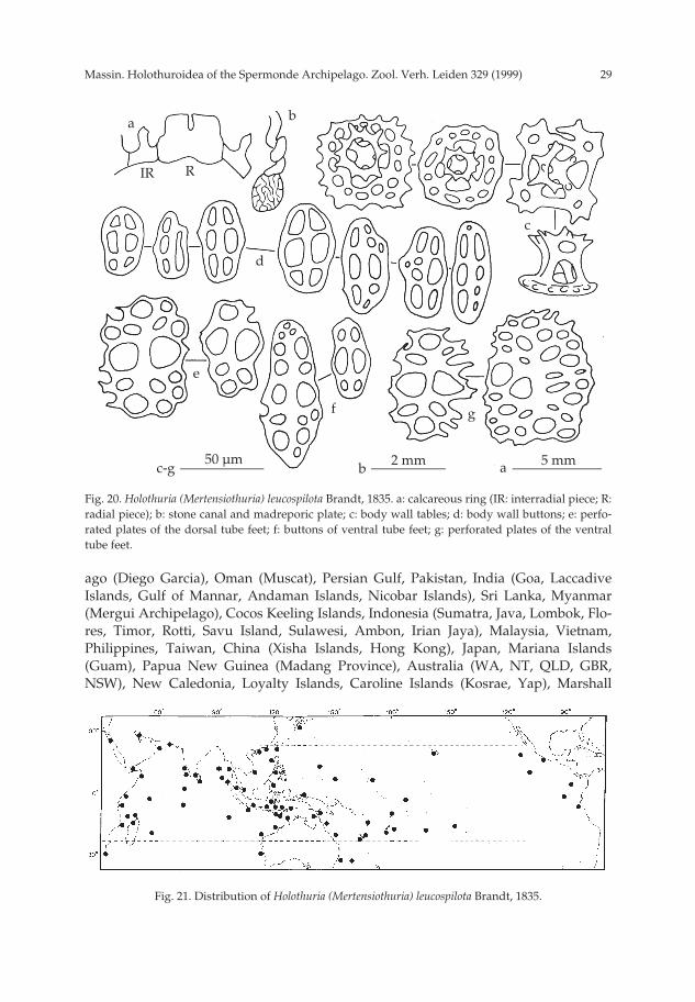
Ossicles of body wall tables and buttons. Tables (fig. 20c) numerous, disc diame-ters 40-75 µm; disc perforated by 4 large central holes and 4-15 small peripheral holes;rim of the disc spinose; 4 short pillars forming a spire united by one cross beam andending in a crown of sharp spines (fig. 20c), with a large central hole. Buttons few, 40-60 µm long with 3 pairs of holes (fig. 20d). Same ossicle types in dorsal and ventraltube feet. In dorsal tube feet small perforated plates, 65-75 µm long (fig. 20e) in addi-tion to tables and buttons, and an end plate 490-500 µm across. In the ventral tube feetsome buttons larger (fig. 20f), perforated plates larger than dorsally (fig. 20g) and endplate 580-600 µm across. No ossicles in the tentacles.
Geographic distribution (fig 21).— Red Sea, Aden, Somalia, Kenya, Zanzibar,Mozambique (Querimba Islands), Comore Islands, South Africa (Natal), Madagascar,Mauritius, Seychelles (Mahé, Amirantes, Aldabra), Maldive Islands, Chagos archipel-
pp 003-144 02-01-2007 15:33 Pagina 28
29Massin. Holothuroidea of the Spermonde Archipelago. Zool. Verh. Leiden 329 (1999)
ago (Diego Garcia), Oman (Muscat), Persian Gulf, Pakistan, India (Goa, LaccadiveIslands, Gulf of Mannar, Andaman Islands, Nicobar Islands), Sri Lanka, Myanmar(Mergui Archipelago), Cocos Keeling Islands, Indonesia (Sumatra, Java, Lombok, Flo-res, Timor, Rotti, Savu Island, Sulawesi, Ambon, Irian Jaya), Malaysia, Vietnam,Philippines, Taiwan, China (Xisha Islands, Hong Kong), Japan, Mariana Islands(Guam), Papua New Guinea (Madang Province), Australia (WA, NT, QLD, GBR,NSW), New Caledonia, Loyalty Islands, Caroline Islands (Kosrae, Yap), Marshall
Fig. 20. Holothuria (Mertensiothuria) leucospilota Brandt, 1835. a: calcareous ring (IR: interradial piece; R:radial piece); b: stone canal and madreporic plate; c: body wall tables; d: body wall buttons; e: perfo-rated plates of the dorsal tube feet; f: buttons of ventral tube feet; g: perforated plates of the ventraltube feet.
Fig. 21. Distribution of Holothuria (Mertensiothuria) leucospilota Brandt, 1835.
a
RIR
50 µmc-g
b
c
d
e
f g
2 mm 5 mmb a
pp 003-144 02-01-2007 15:33 Pagina 29

Massin. Holothuroidea of the Spermonde Archipelago. Zool. Verh. Leiden 329 (1999)30
Islands (Enewetok, Rongelap, Ualan = type locality), Vanuatu (= New Hebrides),Ellice Islands (Funafuti), Phoenix Islands (McKean Island), Cook Islands, Samoa,Tonga Islands, Fiji, Hawaiian Islands, Line Islands (Fanning Island), Cook Islands,Society Islands (Tahiti), Mexico (Revillagigedos, Zihuatanejo), Clipperton Islands,Cocos Islands, Galapagos Islands, Peru.
Remarks.— Holothuria leucospilota is a little variable species, its ossicles showingno noticiable variation in specimens from areas as remote as Madagascar (Cherbon-nier, 1988), the Galapagos Islands (Deichmann, 1958) or Hawaiian Islands (Théel,1886). The species, which is usually very common on reef flats, has been observedand collected only at two sites (Panikiang and Kudingareg Keke) off Ujung Pandang.It is typically found with several specimens being together hidden under a coral slabwith only the anterior part of the body protruding.
Subgenus Metriatyla Rowe, 1969Holothuria (Metriatyla) scabra Jaeger, 1833
(figs 22a-l, 23, 110f)
Holothuria scabra Jaeger, 1833: 23; Serene, 1937: 26; Dawydoff, 1952: 117; Endean, 1953: 57; Endean,1956: 132; Endean, 1957: 234; Loi & Sach, 1965: 241, pl. 2 figs A, D, pl. 4 fig. 6; Tortonese, 1979: 316;James, 1983: 93, pl. 1A; Brouns & Heijs, 1985: 175; Shelley, 1985: 298; Adams, 1992: 13.
Holothuria (Holothuria) scabra; Panning, 1935b: 80, fig. 66a-f (synonymy and records before 1935).Holothuria (Metriatyla) scabra; Cherbonnier, 1980: 647, fig. 16 A-L (synonymy and records before 1980);
Mary Bai, 1980: 15, textfig. 10G; Liao, 1980: 116; Tan Tiu, 1981: 83, pl. 25 figs 1-3; Humphreys,1981: 34; Price, 1982: 12; A.M. Clark, 1982: 489; Liao, 1984: 237; Reyes-Leonardo, 1984a: 149, pl. 6fig. 1a-g; A.M. Clark, 1984: 99; Reyes-Leonardo et al., 1985: 275; James, 1985 [1988]: 404; Cannon &Silver, 1986: 23, figs 4a, 6i; Féral & Cherbonnier, 1986: 86; Cherbonnier, 1988: 135, fig. 55A-O;Conand, 1989: 24, fig. 1; Zoutendijk, 1989: 3; Chambers, 1989: 89; Levin & Dao Tan Ho, 1989: 56;VandenSpiegel et al., 1992: 168; Marsh et al., 1993: 64; Kerr, 1994: 168, fig. 4c; Marsh, 1994a: 11;Holland, 1994: 2; James, 1994: 27; James & James, 1994: 17, fig. 11; Rowe & Gates, 1995: 294; Liao& A.M. Clark, 1995: 446, fig. 262a-c; James, 1995b: 196, fig.4B-C, pl. 2C ; Sant, 1995: 27; Massin,1996b: 25, figs 16A-F, 17A-D; Liao, 1997: 115, fig. 66a-c; Rowe & Richmond, 1997: 304.
Holothuria (Halodeima) scabra; Mortensen, 1934: 6.Sandfish (= Holothuria scabra); Anon., 1996: 13.
Material.— RMNH Ech. 6081 (1 specimen), Panikiang, 30.viii.94, reef flat at low tide; IRSNBIG.28251/36 (1 specimen), Panikiang, 30.viii.94, reef flat at low tide.
Description.— Specimens 120 × 27 and 175 × 40 mm, grey dorsally with transver-sal greenish bands, grey-white ventrally (fig. 110f). In alcohol white-grey ventrallyand grey-olive dorsally; ventral surface speckled with dark tiny dots correspondingto the tube feet; small specimen lighter coloured dorsally with dark, transverse bands.Body arched dorsally and more or less flat ventrally. Mouth ventral, surrounded by20 short tentacles; anus terminal. Tube feet and papillae densely crowded without
Fig. 22. Holothuria (Metriatyla) scabra Jaeger, 1833. a: calcareous ring (IR: interradial piece; R: radialpiece); b: tables from dorsal body wall; c: large table from from dorsal body wall; d: buttons from dor-sal body wall; e: large buttons from dorsal body wall; f: rods from dorsal body wall; g: table from ven-tral body wall; h: buttons from ventral body wall; j: tube foot buttons; k: perforated rods from tubefeet; l: tentacle rods.
pp 003-144 02-01-2007 15:33 Pagina 30

31Massin. Holothuroidea of the Spermonde Archipelago. Zool. Verh. Leiden 329 (1999)
a
RIR
100 µm
b-k
b
d
e
h
f
k
j
l
l
a5 mm
50 µm
c
g h
pp 003-144 02-01-2007 15:33 Pagina 31

Massin. Holothuroidea of the Spermonde Archipelago. Zool. Verh. Leiden 329 (1999)32
alignment ventrally and dorsally. One median groove ventrally; skin rough, 2-3 mmthick.
Calcareous ring composed of quadrangular, large radial pieces with a deep V-shaped anterior notch (fig. 22a); interradial pieces narrow with a sharp anterior tooth(fig. 22a); posterior edge of the calcareous ring undulating. One very long stone canal(12-15 % of body length) with a very characteristic madreporic plate. One Polian vesi-cle; tentacle ampullae as long as the stone canal. No Cuvierian tubules.
In the dorsal body wall tables, buttons and rods. Table discs 60-95 µm in diame-ter; discs with undulating or spiny edge, perforated by one large central hole andnumerous peripheral small holes (fig. 22b); in large tables central hole of the discreplaced by several small ones; 4 short pillars forming a spire united by one crossbeam and ending in a dense crown of spines. A few very large tables 120 µm across,perforated by numerous holes and with the pillars reduced to knobs (fig. 22c). But-tons nodulous, 40-55 µm long with 3 pairs of holes (fig. 22d); a few larger with 5-7pairs of holes (fig. 22e). Rods rare, 115-150 µm long, perforated or unperforated cen-trally and at the extremities (fig. 22f). Ventrally in the body wall tables (fig. 22g) andbuttons (fig. 22h) very similar to dorsal ones. Tables somewhat smaller and no verylarge tables without pillars. Buttons more numerous, more massive, 40-75 µm long;small much more numerous than larger ones. In the tube feet nodulous buttons 40-90µm long (fig. 22j), perforated rods 110-175 µm long (fig. 22k) and tables 50-100 µmacross (identical to those of the body wall). In the dorsal papillae a few rods andnumerous buttons similar to those of the tube feet; tables rare or absent. In the tenta-cles spiny rods, 80-440 µm long (fig. 22l).
Geographical distribution (fig. 23).— Red Sea, Somalia, Kenya, Zanzibar, Mozam-bique (Querimba Islands), South Africa (Natal), Madagascar, Seychelles (Mahé,Aldabra), Mauritius, Maldive Islands, Sri Lanka, India (Gulf of Kutch, LaccadiveIslands, Gulf of Mannar, Andaman Islands, Nicobar Islands), Myanmar (MerguiArchipelago), Cocos Keeling Islands, Indonesia (Sunda Strait, Java, Bali Sea, Sulawesi= type locality, Makassar Strait, Salayer, Sula Islands, Ceram, Rotti, Timor, Ambon),Philippines, Vietnam, Japan, China (Hong Kong), Australia (WA, NT, Timor Sea,QLD, GBR, NSW), Papua New Guinea (Port Moresby, Madang Province), SolomonIslands, Palau Islands, Caroline Islands (Kosrae), New Caledonia, Vanuatu (= NewHebrides), Fiji, Tonga, Cook Islands.
Remarks.— Holothuria scabra is a common species whose ossicles show little varia-
Fig. 23. Distribution of Holothuria (Metriatyla) scabra Jaeger, 1833.
pp 003-144 02-01-2007 15:33 Pagina 32

33Massin. Holothuroidea of the Spermonde Archipelago. Zool. Verh. Leiden 329 (1999)
tion throughout its distribution area (Cherbonnier, 1955a, 1988; VandenSpiegel et al.,1992; Massin, 1996b). The colour is highly variable as observed by Conand (1989) andVandenSpiegel et al. (1992). Conand (1989) proposed a new variety, H. scabra var. ver-sicolor based not only on colour pattern but also on ecological and physiological dif-ferences. However, she recognised that ossicles, calcareous ring and anatomy areinsufficiently different to describe a new species. It is the most prized species fortrepang and as a consequence has been overfished. Off Ujung Pandang it is now arare species.
Subgenus Microthele Brandt, 1835Holothuria (Microthele) nobilis (Selenka, 1867)
(figs 24a-e, 25a-k, 26a-d, 27, 110g, h)
Mülleria nobilis Selenka, 1867: 313, pl. 17 figs. 13-15.Holothuria nobilis; Serene, 1937: 26; Dawydoff, 1952: 117; Tortonese, 1977: 275; Tortonese, 1979: 316;
Grosenbaugh, 1981: 51; Zoutendijk, 1989: 2; Dalzeel, 1990: 12; Allen & Steene, 1994: 244; Mc Elroy,1990: 3; James & James, 1994: 15, fig. 9.
Microthele nobilis; Domantay, 1953: 122; Cherbonnier, 1967: 56; James, 1969: 61; A.M. Clark & Taylor,1971: 92; Anon., 1979: 6; James, 1983: 93; Brouns & Heijs, 1985: 175; Massin & Doumen, 1986: 188;Dalzeel et al., 1993: 27.
Holothuria (Microthele) nobilis; Panning, 1929 [1931]; 131, fig. 15a-s (synonymy and records before1929); Domantay, 1936: 398; Romimohtarto, 1975: 72; Levin, 1979: 21; Mary Bai, 1980: 15, textfig.10H; Cherbonnier, 1980: 626, fig. 5A-N (synonymy and records before 1975); Liao, 1980: 116; TanTiu, 1981: 84, pl. 27 figs 1-4; Humphreys, 1981: 12, 35; James, 1982b: 128; Price, 1982: 11; Rowe,1983: 157; Mukhopadhyay & Samanta, 1983: 311; Liao, 1984: 222; A.M. Clark, 1984: 99; Reyes-Leonardo, 1984a: 150, pl. 7 fig. 1a-g; Price & Reid, 1985: 4, fig. 2; Reyes-Leonardo et al., 1985: 275;Féral & Cherbonnier, 1986: 88, fig. 40L; Cannon & Silver, 1986: 24, figs 2i, 4b, 7a; Conand, 1986:28, figs 7, 19, 20, 44D, 46 (4); Marsh, 1986: 73; Cherbonnier, 1988: 142, fig. 58A-L (records); Jan-goux et al., 1989: 163; James, 1989: 127, fig. 26; Conand, 1989: 26, fig. 1; Chao & Chang, 1989: 117,figs 15, 30B, 33A; Chambers, 1989: 89; Levin & Dao Tan Ho, 1989: 56; Kalashnikov, 1989: 66;Marsh et al., 1993: 64; Kerr, 1994: 169; Holland, 1994: 2; Marsh, 1994a: 5, 11; Marsh, 1994b: 57;James, 1994: 27, pl. 1C; James & Manikfan, 1994: 102, pl. 1B; James, 1995b: 199, pl. 2D fig 4D ;Rowe & Gates, 1995: 295; Liao & A.M. Clark, 1995: 449, fig. 264a-c; Massin, 1996a: 151, figs 1A-F,2A-D, pl. 1B; Belhadjali, 1997: 3; Liao, 1997: 121, fig. 69a-c; Rowe & Richmond, 1997: 304.
Actinopyga nobilis; H.L. Clark, 1925: 106; Townsley & Townsley, 1972: 176.Argiodia maculata; Domantay, 1933: 55, pl. 1 fig. 1a-f.Holothuria (Microthele) fuscogilva Cherbonnier, 1980: 628, fig. 7A-L, pl. 1 fig. c; Conand, 1981: 523, figs
1c, c’; Cannon & Silver, 1986: 24; Féral & Cherbonnier, 1986: 88; Conand, 1989: 26, fig. 1; Liao &A.M. Clark, 1995: 448; Gosliner et al., 1996: 2789, fig. 1030; Liao, 1997: 119, fig. 68a-l.
Holothuria fuscogilva; Adams, 1992: 13; Allen & Steene, 1994: 244; Holland, 1994: 2; Sant, 1995: 27;Conand & Tuwo, 1996: 18.
White teatfish (= Holothuria nobilis); Anon., 1996: 13.
Material.— RMNH Ech. 6082 (1 specimen), Kudingareng Keke SE, 15.ix.94, 20 m depth; IRSNBIG.28251/185 (1 specimen), Kudingareng Keke S, 28.ix.94, 30 m depth; IRSNB IG.28251/211 (1 speci-men), Kapoposang, 30.ix.94, 37 m depth; specimen not measured and released after taking smallpieces of body wall, tube feet, papillae and tentacles; IRSNB IG.28251/255 (1 specimen), KudingarengKeke S, 5.x.94, 25 m depth.
Description.— Specimens 230 × 130, 190 × 55 and 475 × 85 mm, white-cream to
pp 003-144 02-01-2007 15:33 Pagina 33

Massin. Holothuroidea of the Spermonde Archipelago. Zool. Verh. Leiden 329 (1999)34
light brown dorsally. Dorsally each papilla black with at its base a black area goingfrom a small dot to a large spot (figs 110g, h). Several adjoining spots sometimesfused to form large black areas (figs 110g, h). In between the black areas body wallmottled with brown. Black surface very variable. Ventrally white-grey to light brownmottled with a few black spots; in alcohol same colour pattern but faded with lightbrown instead of black. Tube feet numerous, densely crowded over the whole ventralsurface, without alignment. Papillae over the whole dorsale surface, from a simplepoint to large conical warts; laterally, on each side, a row of 5-6 prominent papillae.Body cylindrical or ovoid, mouth ventral, anus terminal; tentacles 20 surrounded by acollar of papillae; anus with five prominent, yellow anal teeth (fig. 110h) each fol-lowed by 3-5 papillae; body wall thick (10-14 mm).
Calcareous ring composed of very large radial pieces having a well marked ante-
Fig. 24. Holothuria (Microthele) nobilis (Selenka, 1867). a: calcareous ring (IR: interradial piece; R: radialpiece); b: body wall tables; c: reduced tables from body wall; d: body wall ellipsoids; e: body wall but-tons.
a
RIR
50 µmb-e
b
d
c
e
a 5 mm
pp 003-144 02-01-2007 15:33 Pagina 34

35Massin. Holothuroidea of the Spermonde Archipelago. Zool. Verh. Leiden 329 (1999)
rior notch and two small blunt, posterior points (fig. 24a); interradial pieces narrowwith one large anterior tooth (fig. 24a). One Polian vesicle and one very short stonecanal (2-3 mm long) ending in a yellowish spherical madreporic plate. Tentacleampullae very long (15-25 % of body length). Cuvierian tubules absent or few andshort. Right respiratory tree very long, going up to the calcareous ring. Illustratedspecimen IRSNB IG.28251/185 (fig. 110g) with Cuvierian tubules, specimen RMNHEch. 6082 (fig. 110h) without.
Body wall ossicles tables, ellipsoids and buttons. Table discs 50-75 µm in diame-ter, edge smooth, undulating, sometimes with a few knobs; discs perforated by 4large central holes and 4-12 peripheral holes (fig. 24b); some specimens with reducedtables both in the body wall (fig. 24c) and in the tube feet and papillae; 4 pillars form-ing a spire united by one cross beam and ending in a dense crown of spines (fig. 24b);central hole of the crown often concealed by spines (fig. 24b); body wall tables similardorsally and ventrally. Ellipsoids 40-80 µm long, numerous, knobbed (fig. 24d); inone specimen ventral ellipsoids up to 100 µm long. Ventral ellipsoids not as complexas dorsal ones; in two specimens reduced to knobbed buttons with 3-5 pairs of holes(fig. 24e); the presence of ventral body wall buttons linked to the absence of Cuvieriantubules. In the dorsal papillae, tables, ellipsoids and perforated plates. Tables few,similar to those of the body wall, 60-90 µm across (fig. 25a), with 4-14 peripheralholes; ellipsoids 55-90 µm long, more irregular (fig. 25b) than in the body wall; perfo-rated plates 150-330 µm long (fig. 25c), derived from rods 85-125 µm long (fig. 25d).Tube feet with numerous ossicles , viz. tables, buttons and three kinds of perforatedplates; tables 55-65 µm across, similar to those of the body wall (figs 25e, 26a); buttons60-105 µm long, smooth or knobbed (fig. 26b); regular elongated, perforated plateslooking like large buttons, 100-200 µm long (fig. 25f); irregular narrow plates, 160-375µm long, derived from spiny rods (figs 25g-j); irregular large plates, 150-230 µm long(fig. 25k), close to the end plate; end plate 600-750 µm across, made of several plates.In the tentacles spiny rods, 40-410 µm long, straight or slightly curved (fig. 26c),sometimes with curved, forked or perforated extremities (fig. 26d).
Geographic distribution (fig. 27).— Red Sea, Somalia, Kenya, Zanzibar = typelocality, South Africa (Durban), Madagascar, Mauritius, Seychelles (Mahé, Aldabra),Maldive Islands (Malé), Chagos Archipelago (Diego Garcia), India (Laccadive Islands,Andaman Islands), Sri Lanka, Cocos Keeling Islands, Indonesia (Sumatra, Java,Sulawesi, Sumbawa, Timor, Flores, Rotti, Roma, Salayer, Tukang Besi Islands, Ceram,Banda Sea, Irian Jaya), Malaysia (Sabah), Philippines, Vietnam, China, Taiwan, Japan(RyuKyu Islands), Mariana Islands (Guam = Guahan), Caroline Islands (Yap), PapuaNew Guinea (Port Moresby, Madang Province), Australia (WA, Timor Sea, QLD,GBR, NSW, Tasman Sea), Solomon Islands, New Caledonia, Loyalty Islands (Lifu),Niue, Vanuatu (= New Hebrides), Fiji, Tonga Islands, Samoa, Ellice Islands (Tuvalu),Line Islands (Fanning Island), Cook Islands, Hawaiian Islands, Easter Island.
Remarks.— Holothuria (Microthele) nobilis is a highly variable species both asregards body colour and ossicles. Cherbonnier (1980) recognised a second species,H.(M.) fuscogilva, based on a mottled colour pattern, on the absence of Cuvieriantubules, on the presence of buttons in the ventral body wall and its supposed occur-rence in a different habitat. The four observed specimens from the Spermonde Archi-pelago present the same mottled colour pattern and only two of the specimens have
pp 003-144 02-01-2007 15:33 Pagina 35

Massin. Holothuroidea of the Spermonde Archipelago. Zool. Verh. Leiden 329 (1999)36
a
100 µm
a, b, e, j
b
c
d
e
f
g
h
k
j
50 µm
200 µm
c, d, f, h, k
g
k
pp 003-144 02-01-2007 15:33 Pagina 36

37Massin. Holothuroidea of the Spermonde Archipelago. Zool. Verh. Leiden 329 (1999)
ventral body wall buttons and no Cuvierian tubules. The two others have ventralbody wall ellipsoids and Cuvierian tubules, supposed to be typical of H. (M.) nobilis.Moreover, three of the studied specimens are from the same locality and from thesame type of habitat. The two specimens without Cuvierian tubules present variationin their ossicles, mainly in the size of the tables and the buttons. All these observa-tions reduce the differences between the two species to such an extend that I agreewith Rowe & Gates (1995), considering H. (M.) fuscogilva as a synonym of H. (M.)nobilis.
The mottled colour pattern, corresponding to what Cherbonnier (1980) calls H.(M.) fuscogilva, has previously been observed in the Red Sea (Panning 1941, fig. 7b),India (James & James 1994, fig. 9), Sulawesi (Panning, 1941: fig. 7c; present study),New Caledonia (Cherbonnier, 1980; Féral & Cherbonnier, 1986; Conand, 1989), Papua
Fig. 25. Holothuria (Microthele) nobilis (Selenka, 1867). a: tables from dorsal papillae; b: ellipsoids fromdorsal papillae; c: perforated plate from dorsal papillae; d: rods from dorsal papillae; e: tube foottables; f: large buttons from tube feet; g & j: tube foot rods: h: narrow plates from tube feet; k: largeplates from tube feet.
Fig. 26. Holothuria (Microthele) nobilis (Selenka, 1867). a: tube foot tables; b: tube foot buttons; c: tenta-cle rods; d: forked or perforated extremities from tentacle rods.
a
100 µmc, d
b
c
d
a, b 50 µm
pp 003-144 02-01-2007 15:33 Pagina 37
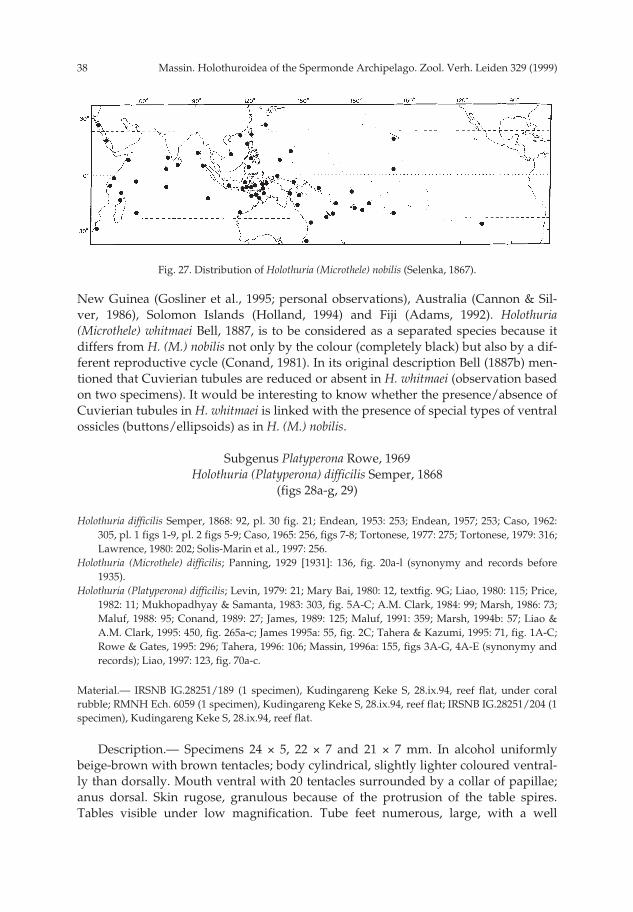
Massin. Holothuroidea of the Spermonde Archipelago. Zool. Verh. Leiden 329 (1999)38
New Guinea (Gosliner et al., 1995; personal observations), Australia (Cannon & Sil-ver, 1986), Solomon Islands (Holland, 1994) and Fiji (Adams, 1992). Holothuria(Microthele) whitmaei Bell, 1887, is to be considered as a separated species because itdiffers from H. (M.) nobilis not only by the colour (completely black) but also by a dif-ferent reproductive cycle (Conand, 1981). In its original description Bell (1887b) men-tioned that Cuvierian tubules are reduced or absent in H. whitmaei (observation basedon two specimens). It would be interesting to know whether the presence/absence ofCuvierian tubules in H. whitmaei is linked with the presence of special types of ventralossicles (buttons/ellipsoids) as in H. (M.) nobilis.
Subgenus Platyperona Rowe, 1969Holothuria (Platyperona) difficilis Semper, 1868
(figs 28a-g, 29)
Holothuria difficilis Semper, 1868: 92, pl. 30 fig. 21; Endean, 1953: 253; Endean, 1957; 253; Caso, 1962:305, pl. 1 figs 1-9, pl. 2 figs 5-9; Caso, 1965: 256, figs 7-8; Tortonese, 1977: 275; Tortonese, 1979: 316;Lawrence, 1980: 202; Solis-Marin et al., 1997: 256.
Holothuria (Microthele) difficilis; Panning, 1929 [1931]: 136, fig. 20a-l (synonymy and records before1935).
Holothuria (Platyperona) difficilis; Levin, 1979: 21; Mary Bai, 1980: 12, textfig. 9G; Liao, 1980: 115; Price,1982: 11; Mukhopadhyay & Samanta, 1983: 303, fig. 5A-C; A.M. Clark, 1984: 99; Marsh, 1986: 73;Maluf, 1988: 95; Conand, 1989: 27; James, 1989: 125; Maluf, 1991: 359; Marsh, 1994b: 57; Liao &A.M. Clark, 1995: 450, fig. 265a-c; James 1995a: 55, fig. 2C; Tahera & Kazumi, 1995: 71, fig. 1A-C;Rowe & Gates, 1995: 296; Tahera, 1996: 106; Massin, 1996a: 155, figs 3A-G, 4A-E (synonymy andrecords); Liao, 1997: 123, fig. 70a-c.
Material.— IRSNB IG.28251/189 (1 specimen), Kudingareng Keke S, 28.ix.94, reef flat, under coralrubble; RMNH Ech. 6059 (1 specimen), Kudingareng Keke S, 28.ix.94, reef flat; IRSNB IG.28251/204 (1specimen), Kudingareng Keke S, 28.ix.94, reef flat.
Description.— Specimens 24 × 5, 22 × 7 and 21 × 7 mm. In alcohol uniformlybeige-brown with brown tentacles; body cylindrical, slightly lighter coloured ventral-ly than dorsally. Mouth ventral with 20 tentacles surrounded by a collar of papillae;anus dorsal. Skin rugose, granulous because of the protrusion of the table spires.Tables visible under low magnification. Tube feet numerous, large, with a well
Fig. 27. Distribution of Holothuria (Microthele) nobilis (Selenka, 1867).
pp 003-144 02-01-2007 15:33 Pagina 38

39Massin. Holothuroidea of the Spermonde Archipelago. Zool. Verh. Leiden 329 (1999)
marked brown end plate; tube feet more densely crowded posteriorly than anteriorly,arranged in 2 rows along the ventral lateral ambulacra and 4 rows along the ventralcentral ambulacrum. Papillae few, scattered over the whole dorsal surface, withoutalignment; most of them arising from a conical wart.
Calcareous ring stout, radial pieces quadrangular with an anterior and posteriornotch (fig. 28a); interradial pieces with a well marked anterior tooth (fig. 28a). Tenta-cle ampullae short (1 mm long). One short, contorted stone canal ending in a coneshaped madreporic plate. One Polian vesicle 1/10 of body length. Cuvierian tubuleslarge and brown.
Ossicles of body wall tables and buttons. Table discs 70-80 µm in diameter; discrounded to quadrangular and perforated by 4 central holes, 8 large and 1-8 smallperipheral holes (fig. 28b); edge of the disc smooth; 4 pillars forming a spire united by1-2 cross beams and ending in a small crown of spines (fig. 28b). Buttons 105-145 µmlong with 2-5 small pairs of holes and a median line (fig. 28c); more numerous dorsal-ly than ventrally. In the tube feet tables similar to those of the body wall, large but-tons 145-155 µm long (fig. 28d) and perforated plates 125-150 µm long (fig. 28e); end
Fig. 28. Holothuria (Platyperona) difficilis Semper, 1868. a: calcareous ring (IR: interradial piece; R: radialpiece); b: body wall table; c: body wall buttons; d: large buttons from tube feet; e: perforated platesfrom tube feet; f & g: tentacle rods.
a
RIR
200 µmf
b
c
d
ed
f
g
100 µm
50 µm
2 mm
e
b-d, g
a
pp 003-144 02-01-2007 15:33 Pagina 39

Massin. Holothuroidea of the Spermonde Archipelago. Zool. Verh. Leiden 329 (1999)40
plate 350-410 µm across. In the tentacles a few tables similar to those of the body walland curved spiny rods, 40-620 µm long (figs 28f, g).
Geographic distribution (fig. 29).— Red Sea, Madagascar, Mauritius, Seychelles(Mahé, Aldabra), Pakistan, Maldive Islands, Sri Lanka, India (Laccadive Islands,Andaman Islands, Nicobar Islands), Indonesia (Java, Sulawesi, Timor, Banda Sea,Irian Jaya), Philippines, China, Taiwan, Japan, Australia (WA, NT, QLD, GBR, NSW,Norfolk Islands, Tasman Sea), Mariana Islands (Guam), Palau Islands, CarolineIslands (Kosrae), New Caledonia, Marshall Islands (Enewetok), Samoa (NavigatorIslands = type locality), Ellice Islands, Fiji (Rotuma), Hawaiian Islands, SocietyIslands (Tahiti), Easter Island, Clipperton Island, Gulf of California, Mexico (Revil-lagigedos, Zihuatanejo) , Cocos Islands, Galapagos Islands.
Remarks.— There is little variation in the form of ossicles throughout the geo-graphic range of Holothuria difficilis, from Madagascar (Cherbonnier, 1988) to EasterIsland (Massin, 1996a) through Taiwan (Chao & Chang, 1989) and Sulawesi (presentstudy).
Holothuria (Platyperona) excellens Ludwig, 1875(figs 30a-j, 31)
Mülleria excellens Ludwig, 1875: 98, pl. 7 fig. 32; Lampert, 1885: 97; Théel, 1886: 199; Ludwig, 1889-92:329;
Argiodia excellens; Pearson, 1914: 170.Holothuria (Microthele) excellens; Panning, 1929 [1931]: 132, fig. 16.Holothuria (Platyperona) excellens; Cherbonnier, 1988: 94, fig. 37A-N; Kerr et al., 1992: 209, figs 3g-h, 4a-
c, 5a-e, pl. 1e-f.
Material.— IRSNB IG.28251/242 (1 specimen), Badi, 3.x.94, 1 m depth.
Description.— Small specimen, 24 × 9 mm, completely black; in alcohol beige-white. Body flat ventrally, arched dorsally; mouth ventral surrounded by 18 tentacles;anus terminal surrounded by 5 large calcareous teeth. In each ambulacrum tube feetwide, short, arranged in 2 rows with a zig-zag pattern. Papillae few, small, scatteredon the whole bivium.
Radial and interradial pieces of the calcareous ring of nearly the same width, but
Fig. 29. Distribution of Holothuria (Platyperona) difficilis Semper, 1868.
pp 003-144 02-01-2007 15:33 Pagina 40

41Massin. Holothuroidea of the Spermonde Archipelago. Zool. Verh. Leiden 329 (1999)
the radials twice the hight of the interradials (fig. 30a); radial pieces squarish with alarge anterior notch; interradial pieces with anterior triangular tooth. One Polian vesi-cle; one short, contorted stone canal ending in a muriform madreporic plate (fig. 30b);tentacle ampullae short.
Ossicles of body wall tables and buttons. Table discs 50-80 µm in diameter andspire 25-30 µm high; disc perforated by one large central hole and 8-12 large peripher-al holes; in the largest tables 1-6 additionnal small holes; rim of the disc smooth or
Fig. 30. Holothuria (Platyperona) excellens Ludwig, 1875. a: calcareous ring (IR: interradial piece; R: radi-al piece); b: stone canal and madreporic plate; c: body wall tables; d: body wall buttons; e: tube foottables; f: tube foot buttons; g: perforated plates from tube feet; h & j: tentacle rods.
a
RIR
100 µm
c-e
b
c
d
g
e
hj
a, b
f-h
j
50 µm
2 mm
200 µm
f
pp 003-144 02-01-2007 15:33 Pagina 41

Massin. Holothuroidea of the Spermonde Archipelago. Zool. Verh. Leiden 329 (1999)42
slightly undulating; 4 pillars forming the spire with one cross beam, ending in adense crown of spines with a highly variable diameter (fig. 30c). Buttons 80-100 µmlong, with 3-5 pairs of holes; button surface with a few small knobbs and often a rowof knobbs along the edge (fig. 30d). In the tube feet tables 100-140 µm across (fig. 30e),a few buttons similar to those of the body wall (fig. 30f) and smooth perforated plates,110-200 µm long, with a median longitudinal line (fig. 30g); end plate 350-380 µmacross. In the tentacles curved spiny rods (figs 30h, j), 50-600 µm long.
Geographic distribution (fig. 31).— Madagascar, Indonesia (Sulawesi), MarianaIslands (Guam), Samoa = type locality.
Remarks.— I concur with Cherbonnier (1988) and Kerr et al. (1992) that Holothuriaexcellens and H. difficilis are closely related, though separate species. They differ main-ly in the form of the buttons which are knobbed in H. excellens and smooth in H. diffi-cilis. The distributional range of H. excellens is most probably much wider than shownin fig. 31 because many authors have considered H. excellens conspecific with H. diffi-cilis. The species is new to the fauna of Indonsia.
Subgenus Semperothuria Deichmann, 1958Holothuria (Semperothuria) flavomaculata Semper, 1868
(figs 32a-g, 33, 110j)
Holothuria flavomaculata Semper, 1868: 87, pl. 30 fig. 26; Grosenbaugh, 1981: 51.Holothuria (Holothuria) flavomaculata; Panning, 1935a: 42, fig. 35a-e (synonymy and records before
1935).Semperothuria flavomaculata; Deichmann, 1958: 303.Holothuria (Semperothuria) flavomaculata; Rowe, 1969: 135; Liao, 1980: 115; Price, 1982: 11; Liao, 1984:
230, fig. 11 (1-6); Price & Reid, 1985: 6; Féral & Cherbonnier, 1986: 90, fig. 40D; Maluf, 1988: 95;Cherbonnier, 1988: 67, fig. 26A-K (synonymy and records before 1984); Conand, 1989: 16, 27; Kerret al., 1992, 208, pl. 1 fig. 3e; Rowe & Gates, 1995: 298; Liao & A.M. Clark, 1995: 454, fig. 269a-f;Liao, 1997: 128, fig. 74a-f.
Material.— IRSNB IG.28251/251 (1 specimen), Badi, 3.x.94, 2 m depth.
Description.— Specimen 68 × 16 mm, brown-red with yellow-orange papillae andyellowish tentacles (fig. 110j); in alcohol brown-beige with white papillae and greyish
Fig. 31. Distribution of Holothuria (Platyperona) excellens Ludwig, 1875.
pp 003-144 02-01-2007 15:33 Pagina 42

43Massin. Holothuroidea of the Spermonde Archipelago. Zool. Verh. Leiden 329 (1999)
tentacles. Body club-shaped, wider posteriorly than anteriorly; mouth and anus ter-minal. Papillae scattered without alignment over the whole bivium. Tube feet alongthe ambulacra, more crowded posteriorly; in one row along the lateral ambulacra and2 rows along the central one. Tentacles 22. Five groups of 3 papillae around the anus.
Calcareous ring composed of radial pieces larger than the interradial ones (fig.32a); radial pieces with a long, narrow anterior tooth (fig. 32a); posterior edge of thecalcareous ring undulating. Several Polian vesicles, one of which being particularlylarge. Five stone canals at the right hand side of the dorsal mesentery. No Cuvieriantubules.
Ossicles of body wall tables and rods. Tables without disc, but spire 40-47 µm inheight, with the 4 pillars forming the spire united by one cross beam and ending in abundle of spines forming a Maltese cross when seen from above (fig. 32b). Rodsspiny, massive, 85-105 µm long (fig. 32c). In the tube feet tables (fig. 32e) similar tothose of the body wall; rods with perforated extremities, 160-200 µm long (fig. 32f)and perforated plates 130-210 µm long (fig. 32d); end plate 475-500 µm across. In thetentacles rods curved or straight, spinose, 95-355 µm long (fig. 32g).
Fig. 32. Holothuria (Semperothuria) flavomaculata Semper, 1868. a: calcareous ring (IR: interradial piece;R: radial piece); b: body wall tables; c: massive rods from body wall; d: perforated plated from tubefeet; e: tube foot tables; f: perforated rods from tube feet; g: tentacle rods.
a
RIR
100 µmd, f, g
bc
d
e
f
g
5 mm50 µm ab, c, e
pp 003-144 02-01-2007 15:33 Pagina 43

Massin. Holothuroidea of the Spermonde Archipelago. Zool. Verh. Leiden 329 (1999)44
Geographic distribution (fig. 33).— Red Sea, Madagascar, Sri Lanka, Indonesia(Sumatra, Java, Sulawesi), China (Xisha Islands), Philippines, Australia (AbrohlosIslands), Mariana Islands (Guam), Palau Islands, Caroline Islands (Yap), New Caledo-nia, Samoa = type locality, Society Islands (Tahiti), Clipperton Island.
Remarks.— The ossicles of the specimen from Sulawesi are similar to those ofspecimens from Samoa (Semper, 1868), Tahiti (Cherbonnier, 1955b), Madagascar(Cherbonnier, 1988) and China (Liao, 1984). Colour seems to be more variable, fromblue-black (Panning, 1935a; Cherbonnier, 1955b, 1988) to brown-red (present study)via brown-mauve (Féral & Cherbonnier, 1986). The number of tentacles also variesfrom 20 (Panning, 1935a) to 31 (Cherbonnier, 1955b). This is only the third record ofHolothuria flavomaculata for Indonesia.
Holothuria (Semperothuria) imitans Ludwig, 1875 (figs 34a-k, 35, 111a)
Holothuria imitans Ludwig, 1875: 109, fig. 41; Caso, 1962: 299; Maluf, 1988: 96; Solis-Marin et al., 1997:256.
Holothuria (Holothuria) imitans; Panning, 1935a: 39, fig. 33a-n (synonymy and records before 1935).Holothuria (Halodeima) imitans; Panning, 1928: 223, figs 19-31.Semperothuria imitans; Deichmann, 1958: 305, pl. 4 figs 8-12.Holothuria (Semperothuria) imitans; Rowe, 1969: 135; A.M. Clark & Rowe, 1971: 178, pl. 27 fig. 16; Price
& Reid, 1985: 6; James, 1995: 48.Holothuria pseudo-imitans Cherbonnier, 1951: 18, pl. 4 figs 1-15.Holothuria imitans var. polymorpha Caso, 1962: 310, pl. 4 figs 1-16, pl. 5 figs 1-17; Caso, 1965: 267, figs
14-15.
Material.— IRSNB IG.28251/237 (1 specimen), Badi, 3.x.94, 2 m depth in between branches of deadcorals.
Description.— Specimen 34 × 7 mm, uniformly chocolate brown, somewhatlighter ventrally than dorsally; extremity of dorsal papillae, ventral tube feet and ten-tacles bright yellow (fig. 111a); in alcohol light brown dorsally, beige-white ventrally;extremity of dorsal papillae, ventral tube feet and tentacles grey-white. Body cylindri-cal with mouth ventral and anus terminal. Tentacles 19, surrounded by a crown ofpapillae. Dorsal papillae few, scattered without alignment except 2 lateral rows. Tube
Fig. 33. Distribution of Holothuria (Semperothuria) flavomaculata Semper, 1868.
pp 003-144 02-01-2007 15:33 Pagina 44

45Massin. Holothuroidea of the Spermonde Archipelago. Zool. Verh. Leiden 329 (1999)
feet few, on one row along the lateral ambulacra and 2 rows along the central ambu-lacrum, more crowded near the anus.
Calcareous ring composed of radial pieces twice as large as the interradial ones(fig. 34a); posterior edge of the calcareous ring undulating; interradial pieces with asharp anterior tooth. Tentacle ampullae long (1/10 of body length). One very large (5mm long) Polian vesicle. A bundle of 7 short contorted stone canals ending each in aspherical madreporic plate. Very fine white Cuvierian tubules.
Ossicles of the body wall tables only, with spire 40-50 µm high, and reduced disc
Fig. 34. Holothuria (Semperothuria) imitans Ludwig, 1875. a: calcareous ring (IR: interradial piece; R:radial piece); b: tables from dorsal body wall; c: tables from ventral body wall; d: tables from dorsalpapillae; e: rods from dorsal papillae; f: extremity of a spiny rods from dorsal papillae; g: tube foottables; h: perforated tables from tube feet; j: tube foot rods; k: tentacle rods.
a
RIR
100 µme, k
bc
f
e
d
h
gj
k50 µm1 mm b-d, f-ja
pp 003-144 02-01-2007 15:33 Pagina 45
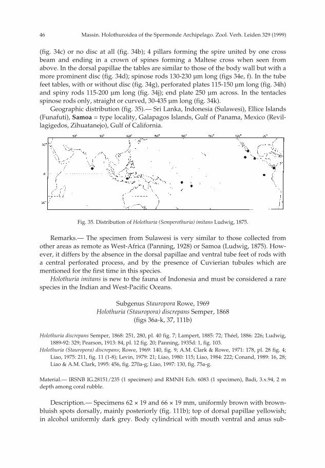
Massin. Holothuroidea of the Spermonde Archipelago. Zool. Verh. Leiden 329 (1999)46
(fig. 34c) or no disc at all (fig. 34b); 4 pillars forming the spire united by one crossbeam and ending in a crown of spines forming a Maltese cross when seen fromabove. In the dorsal papillae the tables are similar to those of the body wall but with amore prominent disc (fig. 34d); spinose rods 130-230 µm long (figs 34e, f). In the tubefeet tables, with or without disc (fig. 34g), perforated plates 115-150 µm long (fig. 34h)and spiny rods 115-200 µm long (fig. 34j); end plate 250 µm across. In the tentaclesspinose rods only, straight or curved, 30-435 µm long (fig. 34k).
Geographic distribution (fig. 35).— Sri Lanka, Indonesia (Sulawesi), Ellice Islands(Funafuti), Samoa = type locality, Galapagos Islands, Gulf of Panama, Mexico (Revil-lagigedos, Zihuatanejo), Gulf of California.
Remarks.— The specimen from Sulawesi is very similar to those collected fromother areas as remote as West-Africa (Panning, 1928) or Samoa (Ludwig, 1875). How-ever, it differs by the absence in the dorsal papillae and ventral tube feet of rods witha central perforated process, and by the presence of Cuvierian tubules which arementioned for the first time in this species.
Holothuria imitans is new to the fauna of Indonesia and must be considered a rarespecies in the Indian and West-Pacific Oceans.
Subgenus Stauropora Rowe, 1969Holothuria (Stauropora) discrepans Semper, 1868
(figs 36a-k, 37, 111b)
Holothuria discrepans Semper, 1868: 251, 280, pl. 40 fig. 7; Lampert, 1885: 72; Théel, 1886: 226; Ludwig,1889-92: 329; Pearson, 1913: 84, pl. 12 fig. 20; Panning, 1935d: 1, fig. 103.
Holothuria (Stauropora) discrepans; Rowe, 1969: 140, fig. 9; A.M. Clark & Rowe, 1971: 178, pl. 28 fig. 4;Liao, 1975: 211, fig. 11 (1-8); Levin, 1979: 21; Liao, 1980: 115; Liao, 1984: 222; Conand, 1989: 16, 28;Liao & A.M. Clark, 1995: 456, fig. 270a-g; Liao, 1997: 130, fig. 75a-g.
Material.— IRSNB IG.28151/235 (1 specimen) and RMNH Ech. 6083 (1 specimen), Badi, 3.x.94, 2 mdepth among coral rubble.
Description.— Specimens 62 × 19 and 66 × 19 mm, uniformly brown with brown-bluish spots dorsally, mainly posteriorly (fig. 111b); top of dorsal papillae yellowish;in alcohol uniformly dark grey. Body cylindrical with mouth ventral and anus sub-
Fig. 35. Distribution of Holothuria (Semperothuria) imitans Ludwig, 1875.
pp 003-144 02-01-2007 15:33 Pagina 46

47Massin. Holothuroidea of the Spermonde Archipelago. Zool. Verh. Leiden 329 (1999)
Fig. 36. Holothuria (Stauropora) discrepans Semper, 1868. a: calcareous ring (IR: interradial piece; R:radial piece); b: stone canal and madreporic plate; c: body wall tables; d: body wall buttons; e: tubefoot tables; f: tube foot buttons; g: perforated plates from tube feet; h & j: tentacle rods; k: heaps ofsmall spines on tentacle rods.
a
RIR
200 µm
j, k
b
c
e
d
f
g
k
hj
100 µm
50 µm
2 mma, b
c-g
h
pp 003-144 02-01-2007 15:33 Pagina 47

Massin. Holothuroidea of the Spermonde Archipelago. Zool. Verh. Leiden 329 (1999)48
dorsal; 24-26 yellowish tentacles surrounded by a crown of papillae. Papillae few,without alignment and scattered over the whole bivium. Tube feet more or lessaligned along the ambulacra and more numerous along the central ambulacrum thanalong the lateral ones. Skin soft, fragile, covered by a thick coating of mucus; underthis coating, numerous tables form a continuous layer of ossicles, visible under lowmagnification; this layer easily torn away from preserved specimens.
Calcareous ring stout; large radial pieces with a deep V-shaped notch anteriorly(fig. 36a). One long contorted stone canal (fig. 36b) ending in an ovoid madreporicplate. One huge Polian vesicle, 1/5 of body length; tentacle ampullae 1/12 of bodylength; very fine, white Cuvierian tubules, readily expelled when animal disturbed.
Ossicles of body wall tables and buttons. Tables very abundant, discs 40-45 µm indiameter; disc with a central cross shaped perforation and 4-8 peripheral holes with1-2 in each quadrant delineated by the cross hole; 4 pillars forming the spire ending ina crown of blunt spines (fig. 36c). Buttons 25-50 µm long with 2-4 pairs of holes (fig.36d), some irregular and knobbed (fig. 36d). In the tube feet tables similar to those ofthe body wall (fig. 36e), buttons 30-60 µm long (fig. 36f) and perforated plates (fig.36g) 80-180 µm long, especially numerous close to the end plate (300-320 µm across).In the tentacles rods (figs 36h, j) straight or curved, 70-720 µm long and covered byheaps of small spines (fig. 36k).
Geographic distribution (fig. 37).— Maldive Islands, Indonesia (Sulawesi), China,New Caledonia, Samoa = type locality.
Remarks.— Ossicles and calcareous ring of the Sulawesi specimens match those ofspecimens from the Maldive Islands (Pearson, 1913) and the Samoa (Semper, 1868). Thenumber of tentacles seems to be variable: 30 according to Semper (1868), 24-30 accord-ing to Liao (1975, 1997) and 24-26 according to the present study. This number does notseem to be linked to body size because the specimens observed by Semper (1868) and inthe present study are similar in length: 60-65 mm and 62-66 mm, respectively.
Holothuria discrepans is new to the fauna of Indonesia.
Holothuria (Stauropora) fuscocinerea Jaeger, 1833(figs 38a-n, 39, 111c)
Holothuria fuscocinerea Jaeger, 1833: 22; Panning, 1935d: 4, fig. 107a-n (records before 1935); Endean,1956: 131; Endean, 1957: 253; James, 1983: 92; Maluf, 1988: 95; Ong Che, 1992: 440; Salis-Marin et
Fig. 37. Distribution of Holothuria (Stauropora) discrepans Semper, 1868.
pp 003-144 02-01-2007 15:33 Pagina 48
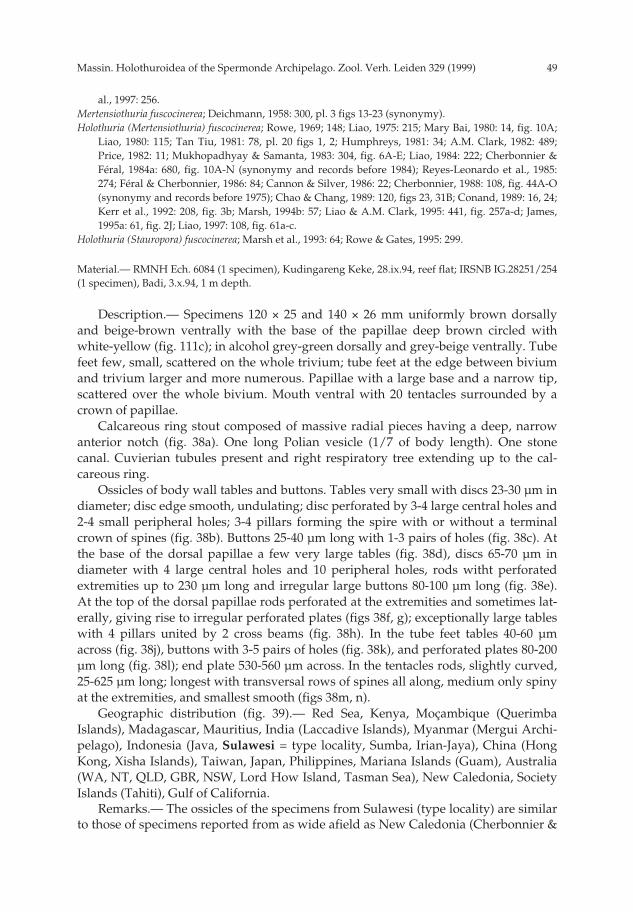
49Massin. Holothuroidea of the Spermonde Archipelago. Zool. Verh. Leiden 329 (1999)
al., 1997: 256.Mertensiothuria fuscocinerea; Deichmann, 1958: 300, pl. 3 figs 13-23 (synonymy).Holothuria (Mertensiothuria) fuscocinerea; Rowe, 1969; 148; Liao, 1975: 215; Mary Bai, 1980: 14, fig. 10A;
Liao, 1980: 115; Tan Tiu, 1981: 78, pl. 20 figs 1, 2; Humphreys, 1981: 34; A.M. Clark, 1982: 489;Price, 1982: 11; Mukhopadhyay & Samanta, 1983: 304, fig. 6A-E; Liao, 1984: 222; Cherbonnier &Féral, 1984a: 680, fig. 10A-N (synonymy and records before 1984); Reyes-Leonardo et al., 1985:274; Féral & Cherbonnier, 1986: 84; Cannon & Silver, 1986: 22; Cherbonnier, 1988: 108, fig. 44A-O(synonymy and records before 1975); Chao & Chang, 1989: 120, figs 23, 31B; Conand, 1989: 16, 24;Kerr et al., 1992: 208, fig. 3b; Marsh, 1994b: 57; Liao & A.M. Clark, 1995: 441, fig. 257a-d; James,1995a: 61, fig. 2J; Liao, 1997: 108, fig. 61a-c.
Holothuria (Stauropora) fuscocinerea; Marsh et al., 1993: 64; Rowe & Gates, 1995: 299.
Material.— RMNH Ech. 6084 (1 specimen), Kudingareng Keke, 28.ix.94, reef flat; IRSNB IG.28251/254(1 specimen), Badi, 3.x.94, 1 m depth.
Description.— Specimens 120 × 25 and 140 × 26 mm uniformly brown dorsallyand beige-brown ventrally with the base of the papillae deep brown circled withwhite-yellow (fig. 111c); in alcohol grey-green dorsally and grey-beige ventrally. Tubefeet few, small, scattered on the whole trivium; tube feet at the edge between biviumand trivium larger and more numerous. Papillae with a large base and a narrow tip,scattered over the whole bivium. Mouth ventral with 20 tentacles surrounded by acrown of papillae.
Calcareous ring stout composed of massive radial pieces having a deep, narrowanterior notch (fig. 38a). One long Polian vesicle (1/7 of body length). One stonecanal. Cuvierian tubules present and right respiratory tree extending up to the cal-careous ring.
Ossicles of body wall tables and buttons. Tables very small with discs 23-30 µm indiameter; disc edge smooth, undulating; disc perforated by 3-4 large central holes and2-4 small peripheral holes; 3-4 pillars forming the spire with or without a terminalcrown of spines (fig. 38b). Buttons 25-40 µm long with 1-3 pairs of holes (fig. 38c). Atthe base of the dorsal papillae a few very large tables (fig. 38d), discs 65-70 µm indiameter with 4 large central holes and 10 peripheral holes, rods witht perforatedextremities up to 230 µm long and irregular large buttons 80-100 µm long (fig. 38e).At the top of the dorsal papillae rods perforated at the extremities and sometimes lat-erally, giving rise to irregular perforated plates (figs 38f, g); exceptionally large tableswith 4 pillars united by 2 cross beams (fig. 38h). In the tube feet tables 40-60 µmacross (fig. 38j), buttons with 3-5 pairs of holes (fig. 38k), and perforated plates 80-200µm long (fig. 38l); end plate 530-560 µm across. In the tentacles rods, slightly curved,25-625 µm long; longest with transversal rows of spines all along, medium only spinyat the extremities, and smallest smooth (figs 38m, n).
Geographic distribution (fig. 39).— Red Sea, Kenya, Moçambique (QuerimbaIslands), Madagascar, Mauritius, India (Laccadive Islands), Myanmar (Mergui Archi-pelago), Indonesia (Java, Sulawesi = type locality, Sumba, Irian-Jaya), China (HongKong, Xisha Islands), Taiwan, Japan, Philippines, Mariana Islands (Guam), Australia(WA, NT, QLD, GBR, NSW, Lord How Island, Tasman Sea), New Caledonia, SocietyIslands (Tahiti), Gulf of California.
Remarks.— The ossicles of the specimens from Sulawesi (type locality) are similarto those of specimens reported from as wide afield as New Caledonia (Cherbonnier &
pp 003-144 02-01-2007 15:33 Pagina 49
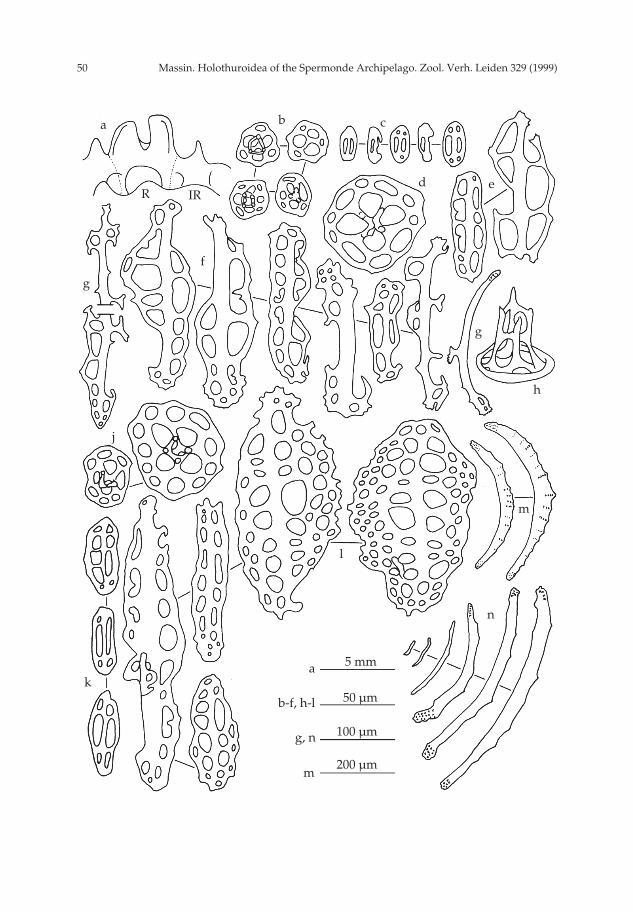
Massin. Holothuroidea of the Spermonde Archipelago. Zool. Verh. Leiden 329 (1999)50
a
R IR
200 µmm
b c
d e
g
h
f
g
j
l
m
n
k
100 µm
50 µm
5 mm
g, n
b-f, h-l
a
pp 003-144 02-01-2007 15:33 Pagina 50

51Massin. Holothuroidea of the Spermonde Archipelago. Zool. Verh. Leiden 329 (1999)
Féral, 1984a) and Taiwan (Chao & Chang, 1989). Specimens from Tahiti (Cherbonnier,1955b) and Madagascar (Cherbonnier, 1988) have tables with higher spire (up to 3-4cross beams). The colour of Holothuria fuscocinerea is highly variable as mentioned byFéral & Cherbonnier (1986).
Holothuria (Stauropora) olivacea Ludwig, 1888(figs 40a-h, 41)
Holothuria olivacea Ludwig, 1888: 811, pl. 30 figs 8-17.Holothuria (Holothuria) olivacea; Panning, 1935b: 76, fig. 58a-e (synonymy and records before 1935).Holothuria (Stauropora) olivacea; Rowe, 1969: 141; Liao, 1975: 212, fig. 12 (1-7); Liao, 1980, 115; Cherbon-
nier, 1988: 78, fig. 3A-P (synonymy and records before 1975); Rowe & Gates, 1995: 299; Liao &A.M. Clark, 1995: 457, fig. 271a-g; Liao, 1997: 132, fig. 76a-g.
Holothuria (Stauropora) fuscoolivacea; Price, 1982: 11; Cannon & Silver, 1986: 25.Holothuria fusco-olivacea; Endean, 1956: 137.
Material.— IRSNB IG.28251/239 (2 specimens) and RMNH Ech. 6060 (1 specimen), Badi, 3.x.94, 2 mdepth; IRSNB IG.28251/252 (1 specimen), Badi, 3.x.94, 2 m depth.
Description.— Specimens 95 × 21 , 86 × 17, 83 × 16 and 48 × 13 mm. Colour in alco-hol uniformly olive-gray; largest dorsal papillae emerging from a whitish wart; baseof the dorsal papillae circled with a brown line. Body cylindrical tapering slightlyanteriorly. Mouth ventral, anus dorsal surrounded by 5 groups of 3 long papillae; 19-20 yellowish tentacles surrounded by a collar of large papillae. Papillae numerous,small, scattered over the whole bivium. Tube feet large, numerous, in 6-7 rows with azig-zag pattern along each ambulacrum. Skin solid, 2 mm thick, rough to the touch.
Calcareous ring stout; large radial pieces with an anterior and posterior notch (fig.40a). Tentacle ampullae long: 1/14 of body length. One very long Polian vesicle, 1/4of body length. One contorted stone canal ending in a large ovoid madreporic plate(fig. 40b). Cuvierian tubules long and fine.
Fig. 38. Holothuria (Stauropora) fuscocinerea Jaeger, 1833. a: calcareous ring (IR: interradial piece; R:radial piece); b: body wall tables; c: body wall buttons; d: table from dorsal papillae (base); e: irregularlarge buttond from dorsal papillae (base); f & g: perforated rods from dorsal papillae (top); h: tablefrom dorsal papillae (top); j: tube foot tables; k: tube foot buttons: l: perforated plates from tube feet;m & n: tentacle rods.
Fig. 39. Distribution of Holothuria (Stauropora) fuscocinerea Jaeger, 1833.
pp 003-144 02-01-2007 15:33 Pagina 51
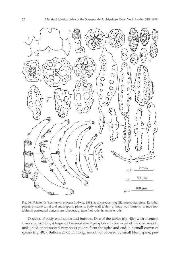
Massin. Holothuroidea of the Spermonde Archipelago. Zool. Verh. Leiden 329 (1999)52
Ossicles of body wall tables and buttons. Disc of the tables (fig. 40c) with a centralcross shaped hole, 4 large and several small peripheral holes; edge of the disc smoothundulated or spinose; 4 very short pillars form the spire and end in a small crown ofspines (fig. 40c). Buttons 25-55 µm long, smooth or covered by small blunt spine; per-
Fig. 40. Holothuria (Stauropora) olivacea Ludwig, 1888. a: calcareous ring (IR: interradial piece; R: radialpiece); b: stone canal and madreporic plate; c: body wall tables; d: body wall buttons; e: tube foottables; f: perforated plates from tube feet; g: tube foot rods; h: tentacle rods.
a
RIR
100 µmg, h
b c
d
e
f
gh
50 µm
2 mm
c-f
a, b
pp 003-144 02-01-2007 15:33 Pagina 52

53Massin. Holothuroidea of the Spermonde Archipelago. Zool. Verh. Leiden 329 (1999)
forations reduced or obliterated (fig. 40d). In the tube feet tables similar to those ofthe body wall but sometimes larger (fig. 40e), perforated plates 80-140 µm long withsmooth or spinose edge (fig. 40f), and rods 125-245 µm long, perforated centrally andat the extremities (fig. 40g); edge of the rods somewhat ragged; end plate 325-330 µmacross. In the tentacles curved rods, 35-500 µm long, covered with heaps of smallspines (fig. 40h).
Geographic distribution (fig. 41).— Red Sea, Madagascar, Indonesia (Sulawesi,Mollucas, Ambon = type locality, Savu Islands), China (Xisha Islands), Australia(WA, NT, QLD, GBR), Solomon Islands, Hawaiian Islands.
Remarks.— Ossicles of Holothuria olivacea show little variation throughout thegeographical range of the species (Ludwig, 1888; Liao, 1975; Cherbonnier, 1988). Onlythe edge of the perforated plates from the tube feet is more ragged in specimens fromMadagascar (Cherbonnier, 1988). Three of the specimens from Badi are relativelylarge: 83, 86 and 95 mm long. Only Liao (1997) mentioned larger specimens (100 mmlong) from China. The other known specimens are smaller: 58 mm long (Fisher, 1907;Tortonese, 1953), 70 mm long (Ludwig, 1888; Liao 1975); 80 mm long (Cherbonnier,1988).
Subgenus Theelothuria Deichmann, 1958Holothuria (Theelothuria) turriscelsa Cherbonnier, 1980
(figs 42a-l, 43)
Holothuria (? Theelothuria) turriscelsa Cherbonnier, 1980: 644, fig. 15A-L, pl. 1E.Holothuria (Theelothuria) turriscelsa; Féral & Cherbonnier, 1986: 92, fig. 40M; Kerr et al., 1992: 209, fig.
3f, pl. 1d.
Material.— IRSNB IG.28251/219 (1 specimen) and RMNH Ech. 6085 (1 specimen), Kapoposang,30.ix.94, 12 m depth, night dive; MNHNP slides of the holotype.
Description.— Specimens 165 × 32 and 200 × 40, grey-brown dorsally and beigeventrally; in alcohol grey-brown dorsally with beige papillae and beige ventrally.Papillae large, wart-like, scattered over the whole bivium. Tube feet numerous cover-ing ambulacral and interambulacral areas. Mouth ventral, surrounded by a collar ofpapillae; 18 tentacles are visible; anus terminal. Skin 6.5 mm thick.
Fig. 41. Distribution of Holothuria (Stauropora) olivacea Ludwig, 1888.
pp 003-144 02-01-2007 15:33 Pagina 53

Massin. Holothuroidea of the Spermonde Archipelago. Zool. Verh. Leiden 329 (1999)54
Fig. 42. Holothuria (Theelothuria) turriscelsa Cherbonnier, 1980. a: calcareous ring (IR: interradial piece;R: radial piece); b: body wall tables; c: buttons from dorsal body wall; d: buttons from ventral bodywall; e: table from dorsal papillae; f: buttons from dorsal papillae; g: rods from dorsal papillae; h: tubefoot tables; j: tube foot rods; k: perforated plates from tube feet; l: tentacle rods.
a
RIR
100 µmf, g, j-l
b
c
d
g
fe
h
k
j
l
50 µm
5 mm
b-e, h
a
pp 003-144 02-01-2007 15:33 Pagina 54

55Massin. Holothuroidea of the Spermonde Archipelago. Zool. Verh. Leiden 329 (1999)
Calcareous ring stout composed of massive radial pieces characterized by a deeprounded anterior notch and two large posterior points; interradial pieces with a smallanterior median tooth (fig. 42a). Three large (15-25 mm long) and several small (1-3mm long) Polian vesicles. The free extremity of the Polian vesicles is enlarged and fullof brown bodies; Cuvierian tubules numerous, thick, readily expelled.
In the body wall numerous tables and few buttons. Tables (fig. 42b) with discs 60-80 µm in diameter; disc perforated by 4 large central holes and numerous smallperipheral holes arranged in 1-2 concentric circles; rim of the disc spinose; spire short,made of 4 pillars united by 1-2 cross beams and ending in a small, very spiny crown.Buttons (figs 42c, d) very nodulous, often irregular, perforated by numerous holes;some buttons less nodulous ventrally than dorsally. In the dorsal papillae, few tables(fig. 42e), few buttons (fig. 42f) and numerous rods perforated laterally and/or at theextremities (fig. 42g). In the tube feet tables (fig. 42h) with central holes larger thanthe ones of the body wall tables, rods (fig. 42j), and perforated plates 130-290 µm long(fig. 42k). In the tentacles rods only (fig. 42l), 60-540 µm long; smallest smooth, medi-um spiny at the extremities and largest spiny on the whole length.
Geographic distribution (fig. 43).— Indonesia (Sulawesi), Mariana Islands(Guam), New Caledonia = type locality, Society Islands (Tahiti).
Remarks.— Minor variations occurs between the specimens from Sulawesi andthe holotype. The specimens from Sulawesi do not have bridges on the body wallbuttons, their rods of the dorsal papillae are less massive and their tentacle rods aresmaller than the ones of the holotype. The body wall with the wart-like papillae isvery characteristic and similar to specimens from New Caledonia (Cherbonnier, 1980;Cherbonnier & Féral, 1986), and Guam (Kerr et al., 1992). Holothuria turriscelsa is noc-turnally activ and has only been collected at night. It is a new species for the fauna ofIndonesia.
Subgenus Thymiosycia Pearson, 1914Holothuria (Thymiosycia) hilla Lesson, 1830
(figs 44, 111d)
Holothuria hilla Lesson, 1830: 226, pl. 78; Lawrence, 1980: 202; Grosenbaugh, 1981: 51; Kropp, 1982: 446;James, 1983: 93; Zoutendijk, 1989: 2; Colin & Arneson, 1995: 262, fig. 1234; Baine & Forbes, 1998: 4.
Holothuria (Holothuria) monacaria; Panning, 1935b: 69, fig. 47a-u (synonymy and records before 1935).
Fig. 43. Distribution of Holothuria (Theelothuria) turriscelsa Cherbonnier, 1980.
pp 003-144 02-01-2007 15:33 Pagina 55
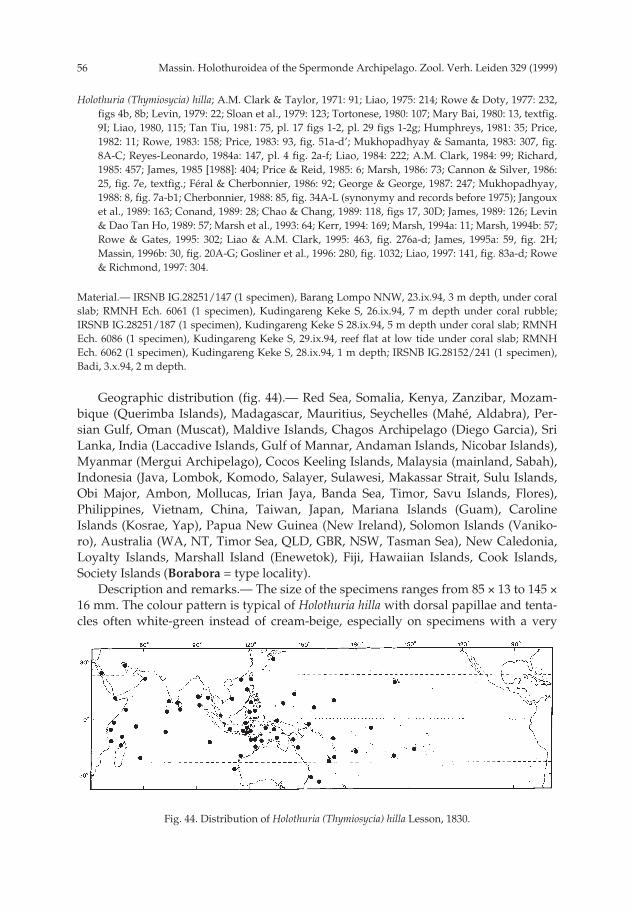
Massin. Holothuroidea of the Spermonde Archipelago. Zool. Verh. Leiden 329 (1999)56
Holothuria (Thymiosycia) hilla; A.M. Clark & Taylor, 1971: 91; Liao, 1975: 214; Rowe & Doty, 1977: 232,figs 4b, 8b; Levin, 1979: 22; Sloan et al., 1979: 123; Tortonese, 1980: 107; Mary Bai, 1980: 13, textfig.9I; Liao, 1980, 115; Tan Tiu, 1981: 75, pl. 17 figs 1-2, pl. 29 figs 1-2g; Humphreys, 1981: 35; Price,1982: 11; Rowe, 1983: 158; Price, 1983: 93, fig. 51a-d’; Mukhopadhyay & Samanta, 1983: 307, fig.8A-C; Reyes-Leonardo, 1984a: 147, pl. 4 fig. 2a-f; Liao, 1984: 222; A.M. Clark, 1984: 99; Richard,1985: 457; James, 1985 [1988]: 404; Price & Reid, 1985: 6; Marsh, 1986: 73; Cannon & Silver, 1986:25, fig. 7e, textfig.; Féral & Cherbonnier, 1986: 92; George & George, 1987: 247; Mukhopadhyay,1988: 8, fig. 7a-b1; Cherbonnier, 1988: 85, fig. 34A-L (synonymy and records before 1975); Jangouxet al., 1989: 163; Conand, 1989: 28; Chao & Chang, 1989: 118, figs 17, 30D; James, 1989: 126; Levin& Dao Tan Ho, 1989: 57; Marsh et al., 1993: 64; Kerr, 1994: 169; Marsh, 1994a: 11; Marsh, 1994b: 57;Rowe & Gates, 1995: 302; Liao & A.M. Clark, 1995: 463, fig. 276a-d; James, 1995a: 59, fig. 2H;Massin, 1996b: 30, fig. 20A-G; Gosliner et al., 1996: 280, fig. 1032; Liao, 1997: 141, fig. 83a-d; Rowe& Richmond, 1997: 304.
Material.— IRSNB IG.28251/147 (1 specimen), Barang Lompo NNW, 23.ix.94, 3 m depth, under coralslab; RMNH Ech. 6061 (1 specimen), Kudingareng Keke S, 26.ix.94, 7 m depth under coral rubble;IRSNB IG.28251/187 (1 specimen), Kudingareng Keke S 28.ix.94, 5 m depth under coral slab; RMNHEch. 6086 (1 specimen), Kudingareng Keke S, 29.ix.94, reef flat at low tide under coral slab; RMNHEch. 6062 (1 specimen), Kudingareng Keke S, 28.ix.94, 1 m depth; IRSNB IG.28152/241 (1 specimen),Badi, 3.x.94, 2 m depth.
Geographic distribution (fig. 44).— Red Sea, Somalia, Kenya, Zanzibar, Mozam-bique (Querimba Islands), Madagascar, Mauritius, Seychelles (Mahé, Aldabra), Per-sian Gulf, Oman (Muscat), Maldive Islands, Chagos Archipelago (Diego Garcia), SriLanka, India (Laccadive Islands, Gulf of Mannar, Andaman Islands, Nicobar Islands),Myanmar (Mergui Archipelago), Cocos Keeling Islands, Malaysia (mainland, Sabah),Indonesia (Java, Lombok, Komodo, Salayer, Sulawesi, Makassar Strait, Sulu Islands,Obi Major, Ambon, Mollucas, Irian Jaya, Banda Sea, Timor, Savu Islands, Flores),Philippines, Vietnam, China, Taiwan, Japan, Mariana Islands (Guam), CarolineIslands (Kosrae, Yap), Papua New Guinea (New Ireland), Solomon Islands (Vaniko-ro), Australia (WA, NT, Timor Sea, QLD, GBR, NSW, Tasman Sea), New Caledonia,Loyalty Islands, Marshall Island (Enewetok), Fiji, Hawaiian Islands, Cook Islands,Society Islands (Borabora = type locality).
Description and remarks.— The size of the specimens ranges from 85 × 13 to 145 ×16 mm. The colour pattern is typical of Holothuria hilla with dorsal papillae and tenta-cles often white-green instead of cream-beige, especially on specimens with a very
Fig. 44. Distribution of Holothuria (Thymiosycia) hilla Lesson, 1830.
pp 003-144 02-01-2007 15:33 Pagina 56
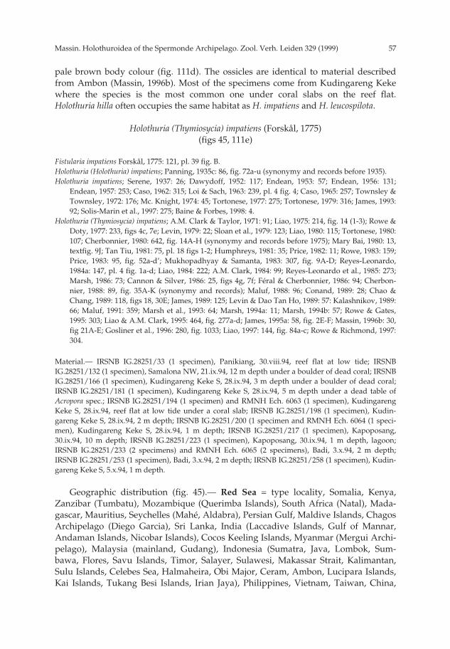
57Massin. Holothuroidea of the Spermonde Archipelago. Zool. Verh. Leiden 329 (1999)
pale brown body colour (fig. 111d). The ossicles are identical to material describedfrom Ambon (Massin, 1996b). Most of the specimens come from Kudingareng Kekewhere the species is the most common one under coral slabs on the reef flat.Holothuria hilla often occupies the same habitat as H. impatiens and H. leucospilota.
Holothuria (Thymiosycia) impatiens (Forskål, 1775)(figs 45, 111e)
Fistularia impatiens Forskål, 1775: 121, pl. 39 fig. B.Holothuria (Holothuria) impatiens; Panning, 1935c: 86, fig. 72a-u (synonymy and records before 1935).Holothuria impatiens; Serene, 1937: 26; Dawydoff, 1952: 117; Endean, 1953: 57; Endean, 1956: 131;
Endean, 1957: 253; Caso, 1962: 315; Loi & Sach, 1963: 239, pl. 4 fig. 4; Caso, 1965: 257; Townsley &Townsley, 1972: 176; Mc. Knight, 1974: 45; Tortonese, 1977: 275; Tortonese, 1979: 316; James, 1993:92; Solis-Marin et al., 1997: 275; Baine & Forbes, 1998: 4.
Holothuria (Thymiosycia) impatiens; A.M. Clark & Taylor, 1971: 91; Liao, 1975: 214, fig. 14 (1-3); Rowe &Doty, 1977: 233, figs 4c, 7e; Levin, 1979: 22; Sloan et al., 1979: 123; Liao, 1980: 115; Tortonese, 1980:107; Cherbonnier, 1980: 642, fig. 14A-H (synonymy and records before 1975); Mary Bai, 1980: 13,textfig. 9J; Tan Tiu, 1981: 75, pl. 18 figs 1-2; Humphreys, 1981: 35; Price, 1982: 11; Rowe, 1983: 159;Price, 1983: 95, fig. 52a-d’; Mukhopadhyay & Samanta, 1983: 307, fig. 9A-D; Reyes-Leonardo,1984a: 147, pl. 4 fig. 1a-d; Liao, 1984: 222; A.M. Clark, 1984: 99; Reyes-Leonardo et al., 1985: 273;Marsh, 1986: 73; Cannon & Silver, 1986: 25, figs 4g, 7f; Féral & Cherbonnier, 1986: 94; Cherbon-nier, 1988: 89, fig. 35A-K (synonymy and records); Maluf, 1988: 96; Conand, 1989: 28; Chao &Chang, 1989: 118, figs 18, 30E; James, 1989: 125; Levin & Dao Tan Ho, 1989: 57: Kalashnikov, 1989:66; Maluf, 1991: 359; Marsh et al., 1993: 64; Marsh, 1994a: 11; Marsh, 1994b: 57; Rowe & Gates,1995: 303; Liao & A.M. Clark, 1995: 464, fig. 277a-d; James, 1995a: 58, fig. 2E-F; Massin, 1996b: 30,fig 21A-E; Gosliner et al., 1996: 280, fig. 1033; Liao, 1997: 144, fig. 84a-c; Rowe & Richmond, 1997:304.
Material.— IRSNB IG.28251/33 (1 specimen), Panikiang, 30.viii.94, reef flat at low tide; IRSNBIG.28251/132 (1 specimen), Samalona NW, 21.ix.94, 12 m depth under a boulder of dead coral; IRSNBIG.28251/166 (1 specimen), Kudingareng Keke S, 28.ix.94, 3 m depth under a boulder of dead coral;IRSNB IG.28251/181 (1 specimen), Kudingareng Keke S, 28.ix.94, 5 m depth under a dead table ofAcropora spec.; IRSNB IG.28251/194 (1 specimen) and RMNH Ech. 6063 (1 specimen), KudingarengKeke S, 28.ix.94, reef flat at low tide under a coral slab; IRSNB IG.28251/198 (1 specimen), Kudin-gareng Keke S, 28.ix.94, 2 m depth; IRSNB IG.28251/200 (1 specimen and RMNH Ech. 6064 (1 speci-men), Kudingareng Keke S, 28.ix.94, 1 m depth; IRSNB IG.28251/217 (1 specimen), Kapoposang,30.ix.94, 10 m depth; IRSNB IG.28251/223 (1 specimen), Kapoposang, 30.ix.94, 1 m depth, lagoon;IRSNB IG.28251/233 (2 specimens) and RMNH Ech. 6065 (2 specimens), Badi, 3.x.94, 2 m depth;IRSNB IG.28251/253 (1 specimen), Badi, 3.x.94, 2 m depth; IRSNB IG.28251/258 (1 specimen), Kudin-gareng Keke S, 5.x.94, 1 m depth.
Geographic distribution (fig. 45).— Red Sea = type locality, Somalia, Kenya,Zanzibar (Tumbatu), Mozambique (Querimba Islands), South Africa (Natal), Mada-gascar, Mauritius, Seychelles (Mahé, Aldabra), Persian Gulf, Maldive Islands, ChagosArchipelago (Diego Garcia), Sri Lanka, India (Laccadive Islands, Gulf of Mannar,Andaman Islands, Nicobar Islands), Cocos Keeling Islands, Myanmar (Mergui Archi-pelago), Malaysia (mainland, Gudang), Indonesia (Sumatra, Java, Lombok, Sum-bawa, Flores, Savu Islands, Timor, Salayer, Sulawesi, Makassar Strait, Kalimantan,Sulu Islands, Celebes Sea, Halmaheira, Obi Major, Ceram, Ambon, Lucipara Islands,Kai Islands, Tukang Besi Islands, Irian Jaya), Philippines, Vietnam, Taiwan, China,
pp 003-144 02-01-2007 15:33 Pagina 57
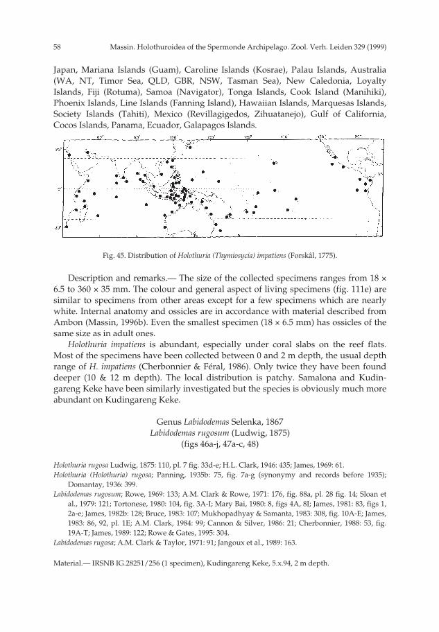
Massin. Holothuroidea of the Spermonde Archipelago. Zool. Verh. Leiden 329 (1999)58
Japan, Mariana Islands (Guam), Caroline Islands (Kosrae), Palau Islands, Australia(WA, NT, Timor Sea, QLD, GBR, NSW, Tasman Sea), New Caledonia, LoyaltyIslands, Fiji (Rotuma), Samoa (Navigator), Tonga Islands, Cook Island (Manihiki),Phoenix Islands, Line Islands (Fanning Island), Hawaiian Islands, Marquesas Islands,Society Islands (Tahiti), Mexico (Revillagigedos, Zihuatanejo), Gulf of California,Cocos Islands, Panama, Ecuador, Galapagos Islands.
Description and remarks.— The size of the collected specimens ranges from 18 ×6.5 to 360 × 35 mm. The colour and general aspect of living specimens (fig. 111e) aresimilar to specimens from other areas except for a few specimens which are nearlywhite. Internal anatomy and ossicles are in accordance with material described fromAmbon (Massin, 1996b). Even the smallest specimen (18 × 6.5 mm) has ossicles of thesame size as in adult ones.
Holothuria impatiens is abundant, especially under coral slabs on the reef flats.Most of the specimens have been collected between 0 and 2 m depth, the usual depthrange of H. impatiens (Cherbonnier & Féral, 1986). Only twice they have been founddeeper (10 & 12 m depth). The local distribution is patchy. Samalona and Kudin-gareng Keke have been similarly investigated but the species is obviously much moreabundant on Kudingareng Keke.
Genus Labidodemas Selenka, 1867Labidodemas rugosum (Ludwig, 1875)
(figs 46a-j, 47a-c, 48)
Holothuria rugosa Ludwig, 1875: 110, pl. 7 fig. 33d-e; H.L. Clark, 1946: 435; James, 1969: 61.Holothuria (Holothuria) rugosa; Panning, 1935b: 75, fig. 7a-g (synonymy and records before 1935);
Domantay, 1936: 399.Labidodemas rugosum; Rowe, 1969: 133; A.M. Clark & Rowe, 1971: 176, fig. 88a, pl. 28 fig. 14; Sloan et
al., 1979: 121; Tortonese, 1980: 104, fig. 3A-I; Mary Bai, 1980: 8, figs 4A, 8I; James, 1981: 83, figs 1,2a-e; James, 1982b: 128; Bruce, 1983: 107; Mukhopadhyay & Samanta, 1983: 308, fig. 10A-E; James,1983: 86, 92, pl. 1E; A.M. Clark, 1984: 99; Cannon & Silver, 1986: 21; Cherbonnier, 1988: 53, fig.19A-T; James, 1989: 122; Rowe & Gates, 1995: 304.
Labidodemas rugosa; A.M. Clark & Taylor, 1971: 91; Jangoux et al., 1989: 163.
Material.— IRSNB IG.28251/256 (1 specimen), Kudingareng Keke, 5.x.94, 2 m depth.
Fig. 45. Distribution of Holothuria (Thymiosycia) impatiens (Forskål, 1775).
pp 003-144 02-01-2007 15:33 Pagina 58

59Massin. Holothuroidea of the Spermonde Archipelago. Zool. Verh. Leiden 329 (1999)
Description.— Specimen 133 × 20 mm, white with yellow tube feet; in alcohol uni-formly white. Body cylindrical, tapering posteriorly with mouth and anus terminal;tentacles 20. Tube feet very long, narrow, mainly along the ambulacra, a few in theinterambulacra; latero-ventral ambulacra with 2 rows of tube feet, central ambu-lacrum with 4 rows of tube feet. Papillae large, less numerous than the tube feet, coni-cal, forming more or less 12 rows on the bivium; lateral rows particularly prominent.
Calcareous ring fine composed of massive radial pieces and ribbon-like interradi-
Fig. 46. Labidodemas rugosum (Ludwig, 1875). a: calcareous ring (IR: interradial piece; R: radial piece);b: stone canal and madreporic plate; c: tables from dorsal body wall; d: buttons from dorsal bodywall; e: table from ventral body wall; f: buttons from ventral body wall; g: tables from dorsal papillae;h: buttons from dorsal papillae; j: irregular buttons from dorsal papillae.
a
RIR
50 µmc-j
b
c
d
e
f
g
h j
h
5 mma, b
pp 003-144 02-01-2007 15:33 Pagina 59

Massin. Holothuroidea of the Spermonde Archipelago. Zool. Verh. Leiden 329 (1999)60
al pieces (fig. 46a). One Polian vesicle, and one stone canal going upward and endingin a quadrangular madreporic plate (fig. 46b).
In the body wall, dorsal papillae and tube feet tables and buttons. Tables of thebody wall with discs 80-95 µm in diameter; disc perforated by 4 large central holesand 10-12 peripheral holes (figs 46c, e); rim of the disc very spinose; height of thespire equal to disc diameter; 4 pillars, spiny near the top, united by 1-2 cross beamsand ending in a bundle of very strong spines; most of the spines bifurcated at theirextremities. Buttons smooth, regular, perforated by 2-6 pairs of holes, 45-75 µm longand more numerous dorsally than ventrally (fig. 46g). Tables of dorsal papillae (figs46d, f) smaller (35-60 µm long) than those of body wall, whereas buttons (fig. 46h) arelarger (55-95 µm long) and sometimes irregular (fig. 46j). Tables of tube feet 45-85 µmacross (fig. 47a), buttons 50-70 µm long (fig. 47b) and end plate 640-660 µm across;close to the end plate, rounded perforated plates (fig. 47c) derived from buttons.
Geographic distribution (fig. 48).— Somalia, Seychelles (Mahé, Aldabra), Mada-gascar, Maldive Islands, Chagos Archipelago (Diego Garcia), India (LaccadiveIslands, Andaman Islands), Indonesia (Sulawesi, Komodo, Lucipara Islands, IrianJaya), Philippines, Palau Islands (Palau = Pelew), Australia (Torres Strait), PapuaNew Guinea (New Britain), Fiji (Rotuma), Samoa = type locality.
Remarks.— The tables of the body wall are characteristic of Labidodemas rugosumand little variable throughout the distribution range. The buttons show more varia-tion. For example, they are more irregular in specimens from Madagascar (Cherbon-nier, 1988). The ribbon-like aspect of the calcareous ring is also variable. It is said tobe massive according to James (1981) and Cherbonnier (1988) and more ribbon-likeaccording to Ludwig (1875), Pearson (1913), Mary Bai (1980) and the present study.
Fig. 47 Labidodemas rugosum (Ludwig, 1875). a: tube foot tables; b: tube foot buttons; c: perforatedplates from tube feet.
a
50 µma-c
c
b
pp 003-144 02-01-2007 15:33 Pagina 60

61Massin. Holothuroidea of the Spermonde Archipelago. Zool. Verh. Leiden 329 (1999)
Labidodemas semperianum Selenka, 1867(figs 49a-c, 50, 111f)
Labidodemas semperianum Selenka, 1867: 309, pl. 17 figs 1-3; H.L. Clark, 1921: 172; Endean, 1956: 130;Endean, 1957: 253; Cherbonnier, 1970: 566, figs A-P (synonymy and records before 1970); A.M.Clark & Rowe, 1971: 176, pl. 18 fig. 12; Liao, 1975: 209, fig. 9 (1-7); Rowe & Doty, 1977: 223, 230,figs 3b, 5g; Tortonese, 1977: 275; Mary Bai, 1980: 8, fig. 8J; Liao, 1980: 115; Humphreys, 1981: 33;Price, 1982: 10; Rowe, 1983: 155; Price, 1983: 88, fig. 45a-d; Liao, 1984: 221: Cannon & Silver, 1986:21, figs 3c, 6a; George & George, 1987: 246; Conand, 1989: 16, 23; Marsh et al., 1993: 63; Kerr, 1994:170; Allen & Steene, 1994: 245; Marsh, 1994a: 10; Rowe & Gates, 1995: 305.
Material.— IRSNB IG.28251/197 (1 specimen), Kudingareng Keke, 28.ix.94, 1.5 m depth, under a coralslab; RMNH Ech. 6087 (1 specimen), Kapoposang, 30.ix.94, 5 m depth, under a coral slab.
Description.— Specimens 120 × 10 and 155 × 20 mm, white-pink (111f) with yel-
Fig. 48. Distribution of Labidodemas rugosum (Ludwig, 1875).
Fig. 49. Labidodemas semperianum Selanka, 1867. a: calcareous ring (IR: interradial piece; R: radialpiece); b: body wall tables; c: tube foot tables.
a
RIR
50 µmb, c
b
c
a 5 mm
pp 003-144 02-01-2007 15:33 Pagina 61

Massin. Holothuroidea of the Spermonde Archipelago. Zool. Verh. Leiden 329 (1999)62
low tube feet; largest specimen pink-brown around the mouth; in alcohol uniformlywhite. Body cylindrical with mouth and anus terminal; mouth surrounded by 20 ten-tacles; anus surrounded by 5 radial pairs of papillae. Few, long, slender, slightly coni-cal papillae scattered over the whole bivium, presenting a vague alignment in 3 dou-ble rows close to the mouth and anus. Tube feet very long, restricted to the ambu-lacra; in each ambulacrum 2 rows of tube feet with a zig-zag pattern.
Calcareous ring composed of massive radial pieces and ribbon-like interradialpieces (fig. 49a). One Polian vesicle and one stone canal going upwards and ending ina muriform madreporic plate. Digestive tract filled with very rough calcareous sand.
In the body wall and tube feet tables only. Tables of body wall with discs 65-80µm in diameter; disc perforated by 5-6 large central holes and 7-10 peripheral holes(fig. 49b); rim of disc smooth, undulating; spire low with 4 pillars united by one crossbeam and ending in a crown of huge spines, bifurcated at the apex; the crown ofspines larger than the disc. In the tube feet tables the same size as those of the bodywall and in addition smaller ones (fig. 49c), often reduced to the disc; reduced tableswith the rim of the disc spinose; end plate 370-400 µm across.
Geographic distribution (fig. 50).— Red Sea, Kenya, Persian Gulf, MaldiveIslands, India (Andaman Islands), Cocos Keeling Islands, Indonesia (Sulawesi, Timor,Sumba, Aroe, Irian Jaya), Malaysia (Sabah), Australia (WA, NT, QLD, NSW), Philip-pines, China, Mariana Islands (Guam), Caroline Islands (Kosrae), New Caledonia,Fiji, Hawaiian Islands = type locality, Society Islands (Tahiti).
Remarks.— The specimens from Sulawesi differ slightly from those from the Mar-shall Islands (Cherbonnier, 1970) with the edge of the disc of the large tables beingsmooth instead of spiny and by the absence of buttons in the dorsal body wall. Theaspect of the main tables is very characteristic and makes the identification of Labido-demas semperianum very easy.
Genus Pearsonothuria Levin, Kalinin & Stonik, 1984Pearsonothuria graeffei (Semper, 1868)
(figs 51, 111g, h)
Holothuria graeffei Semper, 1868: 78, pl. 30 fig. 9.Holothuria (Bohadschia) graeffei; Panning, 1929 [1931]: 124, fig. 6a-f (synonymy and records before 1929).Bohadschia graeffei; Levin, 1979: 19; Tortonese, 1979: 316; Tan Tiu, 1981: 68, pl. 9 figs 1-2; Price, 1982: 10;
Fig. 50. Distribution of Labidodemas semperianum Selanka, 1867.
pp 003-144 02-01-2007 15:33 Pagina 62

63Massin. Holothuroidea of the Spermonde Archipelago. Zool. Verh. Leiden 329 (1999)
Reyes-Leonardo, 1984a: 144, pl. 1 fig. 1a-h; Cherbonnier & Féral, 1984a: 669, fig. 6A-J (synonymyand records); Price & Reid, 1985: 3; Brouns & Heijs, 1985: 175; Reyes-Leonardo et al., 1985: 270;Marsh, 1986: 73; George & George, 1987: 246, pl. 11 fig. G; Jangoux et al., 1989: 163; Marsh et al.,1993: 63; Allen & Steene, 1994: 243; Marsh, 1994a: 10; Holand, 1994: 2; Sant, 1995: 27; Colin &Arneson, 1995: 238, 261, figs 1226-1227; Gosliner et al., 1996: 278, fig. 1025; Britayev & Zamisliak,1996: 177; Baine & Forbes, 1998: 4.
Bohadschia (Pearsonothuria) graeffei; Kalashnikov, 1989: 67, fig. 4.Pearsonothuria graeffei; Cherbonnier, 1988: 492, fig. 17A-F (records); Levin & Dao Tan Ho, 1989: 55;
Massin, 1996b: 33, fig. 22A-H (synonymy and records); Clouse, 1997: 191, fig. 5a-e; Liao, 1997:147, fig. 85a-f; Rowe & Richmond, 1997: 302.
Material.— One specimen observed at Samalona.
Geographic distribution (fig. 51).— Red Sea, Comores, Madagascar, MaldiveIslands, India (Laccadive Islands), Cocos Keeling Islands, Indonesia (Sumatra, Java,Sumba, Timor, Sulawesi, Halmaheira Sea, Ceram, Ambon, Banda Island, Kai Islands),Malaysia (Mainland, Sabah), Vietnam, China, Philippines, Taiwan, Mariana Islands(Guam), Caroline Islands (Kosrae), Papua New Guinea (Madang Province, PortMoresby), Solomon Islands, Australia (WA, Timor Sea, NT, QLD, GBR), New Caledo-nia, Fiji (Viti Island = type locality).
Description and remarks.— Pearsonothuria graeffei has a very characteristic colourpattern for adult (fig. 111g) and subadult specimens (fig. 111h) and can be identifiedin the field without hesitation. It is surprising that only a single specimen wasobserved since it is normally a common coral reef species between 2-10 m depth. Thisscarcity is difficult to relate to human fishing pressure because P. graeffei is normallynot used as trepang (Anon., 1979; Conand, 1986, 1989; Mc Elroy, 1990). In theSolomon Islands it is only fished for in recent times (Holland, 1994) but is considereda species of mediocre quality (Sant, 1995).
Family Stichopodidae Haeckel, 1886Genus Stichopus Brandt, 1835
Stichopus herrmanni Semper, 1868(fig. 52a-n)
Stichopus variegatus Herrmanni Semper, 1868: 73, pl. 17, pl. 30 fig. 2.
Fig. 51. Distribution map of Pearsonothuria graeffei (Semper, 1868).
pp 003-144 02-01-2007 15:33 Pagina 63
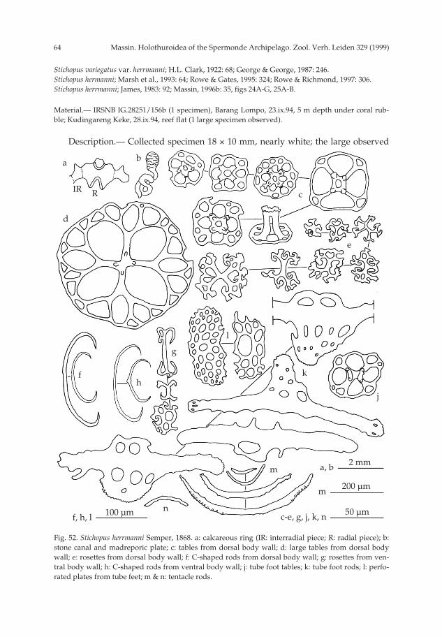
Massin. Holothuroidea of the Spermonde Archipelago. Zool. Verh. Leiden 329 (1999)64
Stichopus variegatus var. herrmanni; H.L. Clark, 1922: 68; George & George, 1987: 246.Stichopus hermanni; Marsh et al., 1993: 64; Rowe & Gates, 1995: 324; Rowe & Richmond, 1997: 306.Stichopus herrmanni; James, 1983: 92; Massin, 1996b: 35, figs 24A-G, 25A-B.
Material.— IRSNB IG.28251/156b (1 specimen), Barang Lompo, 23.ix.94, 5 m depth under coral rub-ble; Kudingareng Keke, 28.ix.94, reef flat (1 large specimen observed).
Description.— Collected specimen 18 × 10 mm, nearly white; the large observed
Fig. 52. Stichopus herrmanni Semper, 1868. a: calcareous ring (IR: interradial piece; R: radial piece); b:stone canal and madreporic plate; c: tables from dorsal body wall; d: large tables from dorsal bodywall; e: rosettes from dorsal body wall; f: C-shaped rods from dorsal body wall; g: rosettes from ven-tral body wall; h: C-shaped rods from ventral body wall; j: tube foot tables; k: tube foot rods; l: perfo-rated plates from tube feet; m & n: tentacle rods.
a
RIR
200 µmm
b
c
e
l
k
j
fh
n
m a, b
c-e, g, j, k, nf, h, l
2 mm
50 µm100 µm
d
g
pp 003-144 02-01-2007 15:33 Pagina 64

65Massin. Holothuroidea of the Spermonde Archipelago. Zool. Verh. Leiden 329 (1999)
specimen yellow-greenish with numerous brown spots dorsally; in alcohol uniformlywhite. Mouth ventral, anus terminal. Papillae scattered over the whole bivium with-out alignment. Ventrally 2-3 rows of tube feet along each ambulacrum.
Calcareous ring well developed; very large interradial pieces with 2 short posteri-or projections and narrow interradial pieces with one anterior tooth (fig. 52a). Onecontorted stone canal going upwards and ending in a rounded madreporic platelocated against the calcareous ring (fig. 52b). One Polian vesicle. Tentacle ampullaeshort (same length as the radial pieces). No gonads.
Ossicles of dorsal body wall include tables, rosettes and C-shaped rods. Tableswith discs 35-65 µm in diameter (fig. 52c); disc round or quadrangular with 4 largecentral holes and 4-11 small peripheral holes; 4 short pillars ending in a small crownof spines, sometimes forming a Maltese cross when see from above; a few large tableswith disc diameter up to 135 µm but with spire reduced to spine or knobs (fig. 52d).Rosettes abundant, 15-55 µm across (fig. 52e), varying in shape from a simple crosswith bifurcated extremities to much more complicated forms. C-shaped rods 95-180µm long, very numerous (fig. 52f). Ventrally in the body wall tables, rosettes and C-shaped rods; tables similar to the dorsal ones but more numerous; rosettes (fig. 52g)nearly of the same size as dorsally, 15-50 µm long, but more simple; C-shaped rods(fig. 52h) smaller (80-150 µm long) and less numerous than dorsally. In the tube feet afew tables (fig. 52j) similar to those of the body wall, rods with enlarged and perforat-ed central process (fig. 52k), 160-190 µm long and perforated plates 125-150 µm long(fig. 52l), located close to the end plate; end plate 250-290 µm across. In the tentaclescurved spiny rods 45-600 µm long (figs 52m, n).
Remarks.— Judging from its size, the reduced number of tube feet in each ambu-lacrum and the absence of gonads the specimen is a juvenile. This is further con-firmed by the 4 very large holes in the table discs, typical of juveniles of other Sticho-pus species (H.L. Clark, 1922; Cherbonnier, 1988). However, most of the ossicles of thespecimen have already reached the condition found in adult.
Stichopus herrmanni has a wide Indo-West Pacific distribution (Rowe & Gates,1995). However, its distribution map is problematical because in most of the literaturebefore 1995 it is cited as Stichopus variegatus Semper, 1868, a name which is no longervalid (Rowe & Gates, 1995). The specimens formerly gathered under S. variegatus arenow divided among two species: S. herrmanni and S. monotuberculatus (Quoy &Gaimard, 1833). Many of the references from the literature give only lists of holothuri-an names and it is not possible to ascertain the species we are dealing with.
Stichopus noctivagus Cherbonnier, 1980(figs 53a-m, 54, 112a)
Stichopus noctivagus Cherbonnier, 1980: 654, fig. 19A-P, pl. 1A; Féral & Cherbonnier, 1986: 96; Kerr etal., 1992: 213, pl. 1C; Allen & Steene, 1994: 245; Colin & Arneson, 1995: 262, fig. 1239; ? Gosliner etal., 1996: 281, fig. 1033; Erhardt & Baensch, 1998: 1094.
Material.— IRSNB IG.28251/108 (1 specimen), Kudingareng Keke NW, 15.ix.94, 12 m depth, underrubble of coral; MNHN Paris, slides of the holotype.
Description.— Specimen 80 × 21 mm, rust brown to beige with a patchwork of
pp 003-144 02-01-2007 15:33 Pagina 65

Massin. Holothuroidea of the Spermonde Archipelago. Zool. Verh. Leiden 329 (1999)66
Fig. 53. Stichopus noctivagus Cherbonnier, 1980. a: calcareous ring (IR: interradial piece; R: radial piece);b: tables from dorsal body wall; c: tables from ventral body wall; d: C- and S-shaped rods from dorsalbody wall; e: tables from dorsal papillae; f: C- and S-shaped rods from dorsal papillae; g: rods from dor-sal papillae; h: tube foot tables; j: perforated plate from tube feet; k: tube foot rods; l & m: tentacle rods.
a RIR
100 µm
a
b
d
ec
hg
f
j k
l
m
50 µm
5 mm
200 µm
b-f, h, m
g, j, k
l
pp 003-144 02-01-2007 15:33 Pagina 66

67Massin. Holothuroidea of the Spermonde Archipelago. Zool. Verh. Leiden 329 (1999)
darker spots (fig. 112a); base of the papillae dark brown, tip white; anus deep brown;in alcohol colour disappear completely. Body flat ventrally, arched dorsally; mouthventral, anus terminal. Dorsal papillae few, long, narrow at the tip, without align-ment except on the flanks. Ventral tube feet only along the ambulacra; lateral ambu-lacra with 2 rows of tube feet, central one with 3-4 rows. Tentacles 20.
Calcareous ring composed of massive radial pieces having 2 short posterior pro-jection and one anterior tooth split at the apex (fig. 53a); the interradial pieces twice aswide as high with a small central, blunt spine (fig. 53a). One Polian vesicle and onestone canal ending in a conical madreporic plate.
In the body wall tables, C- and S-shaped rods. Tables with discs 45-55 µm across;disc perforated by 4 large central holes and 0-12 peripheral holes; rim of the discsmooth and undulating (fig. 53b); ventrally maximally 4 peripheral holes; spire shortmade of 4 pillars ending in a crown of spines (fig. 53c). S- and C-shaped rods (fig.53d) irregular, rare dorsally, absent ventrally. In the dorsal papillae, tables, C- and S-shaped rods and rods with enlarged, perforated central process. Tables discs 60-80µm in diameter (fig. 53e) with the tip of the spire ending in one point giving the tablea tack-like appearance. S- and C-shaped rods (fig. 53f) very abundant and regular. Atthe tip of the dorsal papillae long curved rods, 170-350 µm long (fig. 53g) with a cen-tral, perforated process; also a few spiny perforated plates located at the extreme tipof the papillae. In the tube feet tables (fig. 53h), perforated plates (fig. 53j), rods withenlarged perforated central part (fig. 53k) and an end plate 430-450 µm across; no C-or S-shaped rods. In the tentacles spiny curved rods (figs 53l, m), 20-1000 µm long,some with lateral processes.
Geographic distribution (fig. 54).— Indonesia (Sulawesi), Philippines, MarianaIslands (Guam), Palau Islands, Papua New Guinea, New Caledonia = type locality,(?) Hawaiian Islands.
Remarks.— The ossicles of the specimen from Sulawesi are similar to those of theholotype. Specimens from Palau (Colin & Arneson, 1995), New Caledonia (Féral &Cherbonnier, 1986) and Sulawesi (present work) have the same colour pattern. Speci-mens from Guam depicted by Kerr et al. (1992) and Erhardt & Baensch (1998) aremore reddish, whereas the one pictured by Allen & Steene (1994) has the base of thepapillae blue and the back ground more brown. The specimen from Hawaiian Islands(Gosliner et al. 1996) is so different from the general aspect of the species that I doubtthe correctness of the identification.
Fig. 54. Distribution of Stichopus noctivagus Cherbonnier, 1980.
pp 003-144 02-01-2007 15:33 Pagina 67

Massin. Holothuroidea of the Spermonde Archipelago. Zool. Verh. Leiden 329 (1999)68
Stichopus quadrifasciatus spec. nov.(figs 55a-r, 56a-k, 112b, c)
Material.— IRSNB IG.28251/32 (paratype), Panikiang S, 30.viii.94, reef flat; IRSNB IG.28251/131(paratype), Samalona NW, 21.ix.-94, 12 m depth, under coral slabs; RMNH Ech. 6088 (paratype),Samalona NW, 21.ix.-94, 8 m depth, under coral slabs, a scale worm creeping on body wall, foureulimid gastropods attach to the mouth inside the tentacle crown and one 97 mm long Carapus spec.in the cloaca.; IRSNB IG.28251/193 (holotype), Kudingareng Keke S, 28.ix.94, reef flat.
Description.— Holotype 155 × 40 mm, paratypes 91 × 24, 125 × 40 and 45 × 23mm. Colour yellow-grey with 4 large transversal, black-grey bands (fig. 112b). Tip ofthe dorsal papillae brown to red (fig. 112c), dorsal papillae white; in alcohol com-pletely white. Dorsal papillae scattered, highly variable in size, without alignmentexcept along the zone between bivium and trivium. Body flattened with mouth ven-tral an anus terminal to subdorsal. Mouth with 19-20 tentacles surrounded by a collarof papillae united by a web; collar of the holotype 7 mm height; tentacles small, yel-lowish with large, flat extremities. Tube feet along the ambulacra; lateral ones with 3-4 rows of tube feet, central one with 5-6 rows; only a few tube feet in the interambu-lacra; juvenile (45 mm long) with only one row of tube feet in the lateral ambulacraand 2 rows in the central one; each row in a zig-zag pattern. Skin firm, granulous,rough to the touch.
Calcareous ring composed of large radial and narrow interradial pieces (fig. 55a).Dorsal radial piece with a deep posterior slit, V-shaped in the holotype (fig. 55b) butmore narrow in smaller specimens (fig. 55a); anteriorly, radial pieces with a centralnotch and two lateral teeth (fig. 55a). One Polian vesicle (10 % of body length) andone long, contorted stone canal, going upwards and ending in a large circularmadreporic plate, 2-3 mm across (fig. 55b), located close to the median dorsal interra-dial piece. Tentacle ampullae medium size (10-13 % of body length). Gonads in 2 bun-dles, one on each side of the dorsal mesentery; no gonads in the juvenile.
Body wall ossicles rosettes, tables and C-shaped rods; rosettes 20-35 µm long (figs27c, g, l) abundant to very abundant dorsally, very rare to absent ventrally. Tablediscs 35-50 µm in diameter; edge of the disc undulating, smooth or spinose; disc per-forated by 4 large central holes and 0-16 small peripheral ones; 4 pillars forming thespire united by one cross beam and ending in a crown of spine making a Maltesecross when seen from above (figs 55d, e); tables rare to abundant dorsally, always
Fig. 55. Stichopus quadrifasciatus spec. nov. a: calcareous ring (IR: interradial piece; R: radial piece)(paratype L=125 mm; IRSNB IG.28251/131); b: stone canal and madreporic plate (holotype; IRSNBIG.28251/193); c: rosettes from dorsal body wall (holotype; IRSNB IG.28251/193); d: tables from dor-sal body wall (holotype; IRSNB IG.28251/193); e: tables of ventral body wall (holotype; IRSNBIG.28251/193); f: tables from dorsal body wall (paratype L=45 mm; RMNH Ech. 6088); g: rosettesfrom ventral body wall (holotype; IRSNB IG.28251/193); h: C-shaped rods from dorsal body wall(holotype; IRSNB IG.28251/193); j: C-shaped rods from ventral body wall (holotype; IRSNBIG.28251/193); k: C-shaped rods from dorsal body wall (paratype L=45 mm; RMNH Ech. 6088); l:rosettes from dorsal body wall (L=45 mm; RMNH Ech. 6088); m: tables from ventral body wall(paratype L=45 mm; RMNH Ech. 6088); n: tube foot tables (holotype; IRSNB IG.28251/193); p: C-shaped rods from tube feet (paratype L=45 mm; RMNH Ech. 6088); q: tube foot rods (holotype;IRSNB IG.28251/193); r: perfotated plates from tube feet (holotype; IRSNB IG.28251/193).
pp 003-144 02-01-2007 15:33 Pagina 68

69Massin. Holothuroidea of the Spermonde Archipelago. Zool. Verh. Leiden 329 (1999)
aR IR
2 mma
bc
h
f
e
j gk
l
m
q
r
n
p 2 mm
100 µm50 µm
b
q, rc-p
d
pp 003-144 02-01-2007 15:33 Pagina 69

Massin. Holothuroidea of the Spermonde Archipelago. Zool. Verh. Leiden 329 (1999)70
abundant ventrally. Central holes of the disc much larger in the juvenile (fig. 55f) thanin the adults. C-shaped rods 35-50 µm long (figs 55h, j), rare dorsally and very rareventrally or enterely absent; juvenile with large C-shaped rods, 50-100 µm long (fig.55k) located dorsally. In the dorsal papillae tables, C- and S-shaped rods, rosettes,perforated plates and spiny rods. Tables similar to those of the body wall with the
a
200 µmj
Fig. 56. Stichopus quadrifasciatus nov. spec. (a-h: holotype; IRSNB IG.28251/193; j-k: paratype L=91mm; IRSNB IG.28251/32). a: tables from dorsal papillae; b: large table from dorsal papillae (holotype;IRSNB IG.28251/193); c: reduced table from dorsal papillae; d: C-shaped rods from dorsal papillae; e:S-shaped rods from dorsal papillae; f: rosettes from dorsal papillae; g: perforated plates from dorsalpapilllae; h: rods from dorsal papillae; j & k: tentacle rods.
b
c
g
f
d
h
k
j
100 µm50 µmh, ka-g
e
pp 003-144 02-01-2007 15:33 Pagina 70

71Massin. Holothuroidea of the Spermonde Archipelago. Zool. Verh. Leiden 329 (1999)
edge of the disc more spinose (fig. 56a); a few large tables, diameter 80-100 µm (fig.56b), with numerous peripheral holes; pillars and crown of spines similar to those ofthe small tables; some tables reduced to disc (fig. 56c); C-shaped rods 50-135 µm long(fig. 56d); S-shaped rods (fig. 56e) and rosettes (fig. 56f) rare; perforated plates withspiny edge (fig. 56g); spiny rods 150-280 µm long (fig. 56h), some with central perfo-rated process. In the tube feet tables, C-shaped rods, spiny rods and perforated plates.Tables numerous, discs 25-50 µm in diameter with or without pillars (fig. 55n); C-shaped rods very rare, 45 µm long (fig. 55p); spiny rods 240-360 µm long (fig. 55q)with very large central perforated process; perforated plates 155-240 µm long (fig.55r), located close to the end plate; end plate 750-900 µm across, composed of severalpieces. In the tentacles spiny rods, 100-1000 µm long, slightly curved (fig. 56j); a fewirregular with perforated or forked extremities (fig. 56k).
Etymology.— quadrifasciatus means circled with four bands refering to the verycharacteristic four black-grey transversal bands on the body wall.
Remarks.— Stichopus quadrifasciatus spec. nov. looks very much like Stichopus hor-rens Selenka, 1867. However, the absence of large tack-like tables in the dorsal papil-lae of the new species clearly separates both species. The ossicles and calcareous ringof S. quadrifasciatus are close to those of S. herrmanni, but S. quadrifasciatus does nothave large tables in the ventral tube feet and in the dorsal body wall as S. herrmanni.Moreover, S. quadrifasciatus has spiny perforated plates in the dorsal papillae, plateswhich have never been mentioned for S. herrmanni. The ossicles of Stichopus monotu-berculatus (Quoy & Gaimard, 1833) ( see Massin, 1996b) present also many similaritieswith those of S. quadrifasciatus. Both species are separated by their colour pattern, bytheir calcareous ring (no posterior projections on the radial pieces of S. monotubercual-tus; see Cherbonnier, 1952b; Massin, 1996b) and by the size of the large tables in thedorsal papillae (70 µm versus 100 µm across).
All the other Stichopus spp. of tropical Indo-Pacific waters are very different bytheir colour, the presence of tack-like tables, and the absence of rosettes, C-shapedrods or spiny perforated plates (see Lampert, 1885; Erwe, 1913; H.L. Clark, 1922, 1938;Cherbonnier, 1967, 1980; Liao, 1980).
Stichopus vastus Sluiter,1887(figs 57a-l, 58a-m, 59a-g, 60a-d, 61, 112d, e)
Stichopus vastus Sluiter, 1887: 198, pl.2 figs 46-48; Sluiter, 1895: 79; Rowe & Gates, 1995: 326.Stichopus spec.; Allen & Steene, 1994: 246; Colin & Arneson, 1995: 262, fig. 1242; Gosliner et al., 1996:
282, fig. 1039.Stichopus “variegatus”; Colin & Arneson, 1995: 262, fig. 1240.
Material.— RMNH Ech. 6089, (1 specimen), Panikiang S, 30.viii.94, reef flat; IRSNB IG.28251/54, (1specimen), Samalona W, 2.ix.94, 11 m depth.
Description.— Specimens 190 × 55 and 310 × 100 mm, yellow or green withnumerous brown lines dorsally (figs 112d, e) and brown with yellow-orange interam-bulacra ventrally; most of the brown lines transversal, discountinuous, encircling thebase of the large papillae; in alcohol nearly white, only a few faded brown lines stillvisible. Ventral sole flat with tube feet along the ambulacra; 4-5 rows of tube feet in
pp 003-144 02-01-2007 15:33 Pagina 71

Massin. Holothuroidea of the Spermonde Archipelago. Zool. Verh. Leiden 329 (1999)72
Fig. 57. Stichopus vastus Sluiter, 1887. a: calcareous ring (IR: interradial piece; R: radial piece) (L=190mm; RMNH Ech. 6089); b: stone canal and madreporic plate (L=190 mm; RMNH Ech. 6089); c: tablesfrom dorsal body wall (L=190 mm; RMNH Ech. 6089); d: tables from ventral body wall (L=190 mm;RMNH Ech. 6089); e: tables of dorsal body wall (L=310 mm; IRSNB IG.28251/54); f: rosettes from dor-sal body wall (L=190 mm; RMNH Ech. 6089); g: rosettes from ventral body wall (L=190 mm; RMNHEch. 6089); h: C-shaped rods from dorsal body wall (L=190 mm; RMNH Ech. 6089); j: C-shaped rodsfrom ventral body wall (L=190 mm; RMNH Ech. 6089; k: S-shaped rods from ventral body wall(L=310 mm; IRSNB IG.28251/54); l: perforated plates from dorsal body wall (L=310 mm; IRSNBIG.28251/54).
a
R
IR
50 µmc-l
b
h
c
e
f
l
g
kj
d
5 mm5 mm ba
pp 003-144 02-01-2007 15:33 Pagina 72
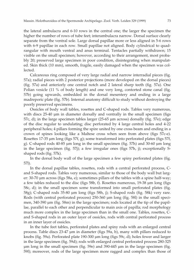
73Massin. Holothuroidea of the Spermonde Archipelago. Zool. Verh. Leiden 329 (1999)
the lateral ambulacra and 6-10 rows in the central one; the larger the specimen thehigher the number of rows of tube feet; interambulacra narrow. Dorsal surface clearlyseparate from the ventral sole. Large dorsal papillae more or less aligned in 5-6 rowswith 6-9 papillae in each row. Small papillae not aligned. Body cylindrical to quad-rangular with mouth ventral and anus terminal. Tentacles partially withdrawn; 15visible on the small specimen; however, according to their arrangement, most proba-bly 20; preserved large specimen in poor condition, disintegrating when manipulat-ed. Skin thick (10 mm), smooth, fragile, easily damaged when the specimen was col-lected.
Calcareous ring composed of very large radial and narrow interradial pieces (fig.57a); radial pieces with 2 posterior projections (more developed on the dorsal pieces)(fig. 57a) and anteriorly one central notch and 2 lateral sharp teeth (fig. 57a). OnePolian vesicle (11 % of body length) and one very long, contorted stone canal (fig.57b) going upwards, embedded in the dorsal mesentery and ending in a largemadreporic plate (fig. 57b). Internal anatomy difficult to study without destroying thepoorly preserved specimens.
Ossicles of body wall tables, rosettes and C-shaped rods. Tables very numerous,with discs 25-40 µm in diameter dorsally and ventrally in the small specimen (figs57c, d); in the large specimen tables larger (25-65 µm across) dorsally (fig. 57e); edgeof the disc regular to undulating; disc perforated by 4 large central holes and 4-22peripheral holes; 4 pillars forming the spire united by one cross beam and ending in acrown of spines looking like a Maltese cross when seen from above (figs 57c-e).Rosettes 17-35 µm long (figs 57f, g); some transformed into perforated plates (figs 57f,g). C-shaped rods 40-85 µm long in the small specimen (fig. 57h) and 30-60 µm longin the large specimen (fig. 57j); a few irregular ones (figs 57h, j); exceptionally S-shaped rods (fig. 57k).
In the dorsal body wall of the large specimen a few spiny perforated plates (fig.57l).
In the dorsal papillae tables, rosettes, rods with a central perforated process, C-and S-shaped rods. Tables very numerous, similar to those of the body wall but larg-er: 30-70 µm across (figs 58a, e); sometimes pillars of the tables with a spine half-way;a few tables reduced to the disc (figs 58b, f). Rosettes numerous, 19-38 µm long (figs58c, d); in the small specimen some transformed into small perforated plates (fig.58g); C-shaped rods 35-80 µm long (figs 58h, j); S-shaped rods (fig. 58k) very rare.Rods (with central perforated process) 250-360 µm long (fig. 58l) in the small speci-men, 340-390 µm (fig. 58m) in the large specimen; rods located at the tip of the papil-lae, parallel to each other and perpendicular to main axis of papilla; rod morphologymuch more complex in the large specimen than in the small one. Tables, rosettes, C-and S-shaped rods in an outer layer of ossicles, rods with central perforated processin an inner layer of ossicles.
In the tube feet tables, perforated plates and spiny rods with an enlarged centralprocess. Table discs 23-47 µm in diameter (figs 59a, b), many with pillars reduced toknobs (fig. 59a). Perforated plate 190-300 µm long (figs 59c, d); holes fewer and largerin the large specimen (fig. 59d); rods with enlarged central perforated process 280-325µm long in the small specimen (fig. 59e) and 390-445 µm in the large specimen (fig.59f); moreover, rods of the large specimen more ragged and complex than those of
pp 003-144 02-01-2007 15:33 Pagina 73

Massin. Holothuroidea of the Spermonde Archipelago. Zool. Verh. Leiden 329 (1999)74
a
100 µm
a-k
b c
h k
g
le
f
d
m
j50 µm
l, m
pp 003-144 02-01-2007 15:33 Pagina 74

75Massin. Holothuroidea of the Spermonde Archipelago. Zool. Verh. Leiden 329 (1999)
Fig. 58. Stichopus vastus Sluiter, 1887. a: tables from dorsal papillae (L=190 mm; RMNH Ech. 6089); b:reduced table from dorsal papillae; c: rosettes from dorsal papillae (L=390 mm; RMNH Ech. 6089); d:rosettes from dorsal papillae (L=310 mm; IRSNB IG.28251/54); e: tables from dorsal papillae (L=310mm; IRSNB IG.28251/54); f: reduced table from dorsal papillae (L=310 mm; IRSNB IG.28251/54); g:perforated plates from dorsal papillae (L=190 mm); h: C-shaped rods from dorsal papillae (L=190mm; RMNH Ech. 6089); j: C-shaped rods from dorsal papillae (L=310 mm; IRSNB IG.28251/54); k: S-shaped rod from dorsal papillae (L=190 mm; RMNH Ech. 6089); l: apical rods from dorsal papillae(L=190 mm; RMNH Ech. 6089); m: apical rods from dorsal papillae (L=310 mm; IRSNB IG.28251/54).
Fig. 59. Stichopus vastus Sluiter, 1887. a: tube foot tables (L=190 mm; RMNH Ech. 6089); b: tube foottables (L=310 mm; IRSNB IG.28251/54); c: perforated plates from tube feet (L=190 mm; RMNH Ech.6089); d: perforated plates from tube feet (L=310 mm; IRSNB IG.28251/54); e: tube foot rods (L=190mm; RMNH Ech. 6089); f: tube foot rods (L=310 mm; IRSNB IG.28251/54); g: tube foot rosettes (L=310mm; IRSNB IG.28251/54).
c
50 µma-d, g
e
a
b
d
f
g
100 µme, f
pp 003-144 02-01-2007 15:33 Pagina 75

Massin. Holothuroidea of the Spermonde Archipelago. Zool. Verh. Leiden 329 (1999)76
the small specimen; exceptionally rosettes (fig. 59g) and C-shaped rods in the tubefeet of the large specimen; end plates 330-380 µm across in the small specimen and650 µm in the large specimen.
In the tentacles curved spiny rods, 90-425 µm long in the small specimen (figs 60a,b) and 100-1000 µm in the large specimen (fig. 60c); extremities of the rods particular-ly spiny (fig. 60d).
Geographic distribution (fig. 61).— Indonesia (Java = type locality, Sulawesi),Papua New Guinea, Palau Islands, Micronesia, Australia (GBR, NE coast, QLD)
Remarks.— Stichopus vastus has a unique colour pattern which has already beenillustrated several times in field guides (Allen & Steene, 1994; Colin & Arneson, 1995;
Fig. 60. Stichopus vastus Sluiter, 1887. a & b: tentacle rods (L=190 mm; RMNH Ech. 6089); c & d: tenta-cle rods (L=310 mm; IRSNB IG.28251/54).
b
50 µmb
a
d
c
b
100 µm
200 µm
a, d
c
Fig. 61. Distribution of Stichopus vastus Sluiter, 1887.
pp 003-144 02-01-2007 15:33 Pagina 76

77Massin. Holothuroidea of the Spermonde Archipelago. Zool. Verh. Leiden 329 (1999)
Gosliner et al., 1996). It presents some variation in the background colour and thedensity of the brown lines. Moreover, the larger the specimen the larger some ossicletypes: tables of the dorsal body wall, rods of the tube feet, end plate of the tube feetand tentacle rods. By its ossicles and calcareous ring, S. vastus is close to S. herrmanni.
Genus Thelenota H.L. Clark, 1921Thelenota ananas (Jaeger, 1833)
(figs 62, 112f)
Trepang ananas Jaeger, 1833: 24, pl. 3 fig. 1.Thelenota ananas ; H.L. Clark, 1921: 184, pl. 18 fig. 2; Dawydoff, 1952: 117; Endean, 1957: 252; James,
1969: 60; Meyer-Rochow, 1977: 582; Levin, 1979: 22; Liao, 1980: 116; Tan Tiu, 1981: 64, pl. 6 figs 1-2; Grosenbaugh, 1981: 51;Humphreys, 1981: 36; Conand, 1981: 523, figs 1a-a’; Mukhopadhyay &Samanta, 1983: 312; Liao, 1984: 222; A.M. Clark, 1984: 99; Cherbonnier & Féral, 1984b: 829, fig.62A-S (synonymy and records before 1979); Brouns & Heijs, 1985: 175; Price & Reid, 1985: 6;Richard, 1985: 457; Reyes-Leonardo et al., 1985: 267, pl. 2 fig. 4a-c; Féral & Cherbonnier, 1986: 100;Cannon & Silver, 1986: 28, figs 2a, 7i; Marsh, 1986: 74; George & George, 1987: 246, pl. 12a;Conand, 1989: 31; Jangoux et al., 1989: 163; Chao & Chang, 1989: 116, figs 10, 29E; James, 1989:130, fig. 28; Chambers, 1989: 89; Zoutendijk, 1989: 2; Levin & Dao Tan Ho, 1989: 57; Kalashnikov,1989: 70, fig. 6; Marsh et al., 1993: 64; Dalzeel et al. 1993: 27; Kerr, 1994: 171; Allen & Steene, 1994:246; Marsh, 1994a: 11; Holland, 1994: 2; Fransen, 1994: 123; James, 1994: 28; James & James, 1994:14, fig. 8; James & Manikfan, 1994: 102, pl. 1A; Sant, 1995: 27; Colin & Arneson, 1995: 263, fig.1244; Rowe & Gates, 1995: 326; Liao & A.M. Clark, 1995: 469, fig. 282a-b; Gosliner et al., 1996: 282,fig. 1040; Conand & Tuwo, 1996: 18; Belhadjali, 1997: 3; Liao, 1997: 157, fig. 91a-b; Rowe & Rich-mond, 1997: 306; Baine & Forbes, 1998: 4.
Thelenota anax; Ballard, 1981: 18, fig. 4.Prickly redfish (= Thelenota ananas); Anon., 1996: 13
Material.— Samalona SE, 16.ix.94, a single specimen, about 50 cm long, observed at 20 m depth (fig.112f).
Geographic distribution (fig. 62).— Kenya, Madagascar, South Africa (Natal), Sey-chelles (Aldabra), Mauritius, Maldive Islands, India (Laccadive Islands), Cocos Keel-ing Islands, Indonesia (Sumatra, Java, Sumba, Sulawesi = type locality, BandaIslands, Irian Jaya), Malaysia (Tioman, Sabah), Australia (WA, NT, QLD, NSW),Philippines, Vietnam, Taiwan, China, Japan, Mariana Islands (Guam), Palau Islands,Caroline Islands (Kosrae, Yap), Papua New Guinea (Madang Province, New Ireland),
Fig. 62. Distribution of Thelenota ananas (Jaeger, 1833).
pp 003-144 02-01-2007 15:33 Pagina 77

Massin. Holothuroidea of the Spermonde Archipelago. Zool. Verh. Leiden 329 (1999)78
Solomon Islands, New Caledonia, Niue, Vanuatu (= New Hebrides), Fiji, Tonga,Ellice Islands (Tuvalu), Cook Islands, French Polynesia.
Remarks.— No specimen of this unmistakable and large species was collected inthe Spermonde Archipelago. Thelenota ananas is normally very abundant from thesurface to 30 m depth. The species, being highly prized as trepang, and overexploitedby local fishermen, is now rare in the Spermonde Archipelago.
Thelenota anax H.L. Clark, 1921(figs 63a-k, 64)
Thelenota anax Clark, 1921: 185, pl. 18 fig. 3; Liao, 1975: 205, fig. 6 (1-10); Lamberson, 1978: 115, figs 1-4;Levin, 1979: 22; Cherbonnier, 1979: 9, fig. 5A-N; Liao, 1980: 116; Liao, 1984: 222, 243; Brouns &Heijs, 1985: 175; Féral & Cherbonnier, 1986: 100, figs 39, 40O; Cannon & Silver, 1986: 28; Marsh,1986: 74; Thandar, 1987: 262; Cherbonnier, 1988: 156, fig. 64A-L (synonymy and records before1975); Conand, 1989: 31; Massin & Lane, 1991: 57; Lane, 1992: 176; Marsh et al., 1993: 64; Allen &Steene, 1994: 246; Holland, 1994: 2; James & Manikfan, 1994: 103; Sant, 1995: 27; Colin & Arneson,1995: 263, fig. 1245; Rowe & Gates, 1995: 327; Liao & A.M. Clark, 1995: 470, fig. 283a-d; Gosliner etal., 1996: 282, fig. 1041; Conand & Tuwo, 1996: 18; Liao, 1997: 158, fig. 92a-d; Rowe & Richmond,1997: 306.
Stichopus spec. George & George, 1987: 246, pl. 12b.Stichopus variegatus; A.M. Clark, 1984: 88, fig. 2.
Material.— IRSNB IG.28251/183 (1 specimen), Kudingareng Keke S, 28.ix.94, 6 m depth.
Description.— Specimen 206 × 50 mm, greyish-cream with numerous reddishspots and on each side a more or less continuous reddish line located in a groove; inalcohol uniformly grey-white. Body quadrangular, mouth ventral, anus subdorsal.Ventral sole covered by numerous, densely crowded tube feet. Border of ventral soleclearly marked by a row of large papillae, some of which fused; papillae scatteredover the whole dorsal surface. Skin very thick (15 mm).
Calcareous ring well developed but relatively small for such a large species; radialpieces large with 2 posterior points (fig. 63a), especially well marked on the dorsalones; interradial pieces half the width of the radial ones and with a short anteriortooth. One very short and contorted stone canal ending in a large, cylindricalmadreporic plate (fig. 63b). Three Polian vesicles; tentacle ampullae 20-25 mm long.
Ossicles of the body wall dichotomously branched rods (fig. 63c), pseudotables50-60 µm high (fig. 63d) and countless small rounded miliary granules 1.5-4 µmacross (fig. 63e). In the tube feet rods (fig. 63f) and dichotomously branched rods (fig.63g) smaller than in the body wall and some perforated plates (fig. 63h); end plate460-530 µm across. In the tentacles spiny perforated plates (fig. 63j) derived fromdichotomously branched rods (fig. 63k), 50-100 µm long in the tips of the tentaclesand up to 130 µm long in the shafts.
Geographic distribution (fig. 64).— Madagascar (Glorious Islands), Seychelles(Mahé), Maldive Islands, Malaysia (Shaba), Indonesia (Sulawesi), Australia (WA, NT,Torres Strait = type locality, QLD), China, Japan (Ryukyu Islands), Palau Islands,Mariana Islands (Guam), Marshall Islands (Enewetok), Papua New Guinea, SolomonIslands, New Caledonia, Fiji, Society Island (Moorea).
Remarks.— Thelenota anax has a characteristic, very consistent colour and body
pp 003-144 02-01-2007 15:33 Pagina 78

79Massin. Holothuroidea of the Spermonde Archipelago. Zool. Verh. Leiden 329 (1999)
form throughout its distribution range. Some characters of its internal anatomy andossicles show more variation. The number of Polian vesicles varies from 30 (Cherbon-nier, 1988) to 6 (Cherbonnier & Féral, 1984a) or even 3 (present study). Apparentlypseudo-tables are not always present; they have been mentioned by Yulin (1975),Lamberson (1978) and in the present study, but not by Cherbonnier (1979, 1988) andCherbonnier & Féral (1984b).
This is the first reliable record for Indonesia. Conand & Tuwo (1996) previouslyobserved the species on the market of Ujung Pandang (Sulawesi). However, most ofthe holothurians sold as trepang in the Sulawesi are fished in remote areas, oftenclose to or in Australian waters.
Fig. 63. Thelenota anax H.L. Clark, 1921. a: calcareous ring (IR: interradial piece; R: radial piece); b:stone canal and madreporic plate; c: branched rods from body wall; d: body wall pseudo-tables; e:miliary granules from body wall; f: tube foot rods; g: branched rods from tube feet; h: perforated platefrom tube feet; j: perforated plate from tentacles; k: branched rods from tentacles.
a
R
IR
50 µm
a
b
c
e
d
fg
kj
k
h
20 µm
5 mm
e
b-d, f-k2 mmb
pp 003-144 02-01-2007 15:33 Pagina 79

Massin. Holothuroidea of the Spermonde Archipelago. Zool. Verh. Leiden 329 (1999)80
Order Dendrochirotida Grube, 1840Family Cucumariidae Ludwig, 1894
Subfamily Colochirinae Panning, 1949Genus Colochirus Troschel, 1846
Colochirus robustus Oestergren, 1898(figs 65a-g, 66a-f, 67, 112g)
Colochirus robustus Oestergren, 1898b: 134; Ekman, 1918: 14, pl. 3 figs 8-9; Rowe & Gates, 1995: 273;Gosliner et al., 1996: 283, fig. 1043.
Pentacta robusta; H.L. Clark, 1946: 393; A.M. Clark & Rowe, 1971: 180; Panning, 1971: 39, fig. 4a-n;James, 1984: 112; Cannon & Silver, 1986: 30.
Colochirus luteus Sluiter, 1901: 100, pl. 2 fig. 6, pl. 7 fig. 2.Colochirus squamatus Sluiter, 1901: 101, pl. 8 fig. 3.Colochirus robustoides Ekman, 1918: 18, pl. 1 figs 5-6, pl. 3 figs 10-12.
Material.— IRSNB IG.28251/271 (10 specimens) and RMNH Ech. 6067 (11 specimens), BarangLompo, 24.xi.95, 20-25 m depth, on algae and hydroids (collected by B.W. Hoeksema).
Description.— Specimens varying in size from 22 × 7 to 37 × 12 mm, uniformlybright yellow (fig. 112g); in alcohol uniformly beige. Body wall rough to the touch,rigid, leathery, quadrangular in section. Ventral sole with tube feet only along theambulacra (fig. 65b); 2-4 rows of tube feet in the lateral ambulacra and 3-5 rows in thecentral one. Interambulacral areas narrow; contracted tube feet deep in a hollow, inextended state long and slender, ending in a large sucker. Dorsally 4 rows (2 lateraland 2 dorsal) of 5-18 prominent, rigid tapering papillae, 1-10 mm long (figs 65a, 113f);some of them in pairs, sometimes with a common base. Number of papillae increas-ing with increasing body length: 33 + 6.9 (n = 6) for 22-25 mm long specimens, 46 +5.3 (n = 7) for 28-32 mm long specimens and 55 + 7.5 (n = 8) for 33-37 mm long speci-mens. Mouth terminal closed by 5 triangular plates; anus dorsal surrounded by 5large papillae and lined by rounded plates.
Tentacles 10 with the 2 ventral ones smaller. Calcareous ring narrow, withoutposterior projections (fig. 65c); radial and interradial pieces of the same width; radialones with a large anterior tooth notched at the apex; posterior edge of the calcareousring strongly undulating. One short dorsal stone canal going upwards, ending in a
Fig. 64. Distribution of Thelenota anax H.L. Clark, 1921.
pp 003-144 02-01-2007 15:33 Pagina 80
81Massin. Holothuroidea of the Spermonde Archipelago. Zool. Verh. Leiden 329 (1999)
Fig. 65. Colochirus robustus Oestergren, 1898. a: general dorsal view; b: general ventral view; c: calcare-ous ring (IR: interradial piece; R: radial piece); d: incomplete sphere from body wall; e: knobbed per-forated plates from body wall; f: flat cups from dorsal body wall; g: flat cups from ventral body wall.
a
R IR
50 µm
a, b
b c
d
gd
e
f
5 mm
5 mm
c
d-g
e
pp 003-144 02-01-2007 15:33 Pagina 81

Massin. Holothuroidea of the Spermonde Archipelago. Zool. Verh. Leiden 329 (1999)82
narrow elongate madreporic plate; 2 ventral Polian vesicles: one large and one veryshort; retractor muscles of the pharynx attached at the anterior third of the body.Gonad well developed, forming a bundle of undivided tubules running parallel toeach other.
Body wall ossicles thick reticulated bodies, incomplete spheres and flat, knobbed,squarish cups. Thick reticulated bodies ovoid or rounded, present dorsally and ven-trally; ventrally up to 650 × 400 µm, dorsally up to 450 × 200 µm and 750 × 600 µm inthe dorsal papillae. Incomplete spheres derived from perforated plates, flat cups (fig.65d) or some kind of knobbed perforated plates (fig. 65e); incomplete spheres moreabundant ventrally than dorsally, rare in the dorsal papillae. Flat knobbed cups 50-100 µm across, more nodulous dorsally (fig. 65f) than ventrally (fig. 65g); numerousin the dorsal papillae.
Fig. 66. Colochirus robustus Oestergren, 1898. a: perforated rods from tube feet; b: flat cups from tubefeet; c: perforated rods from tentacles; d: small rods from tentacles; e: irregular rods from tentacles; f:perforated rod from tentacles.
a
100 µmc, f
b
fc
ed
d
50 µma, b, d, e
pp 003-144 02-01-2007 15:33 Pagina 82

83Massin. Holothuroidea of the Spermonde Archipelago. Zool. Verh. Leiden 329 (1999)
Tube feet with an end plate 300-350 µm across, perforated rods (fig. 66a) 100-200µm long, and irregular flat knobbed cups (65-90 µm) (fig. 66b). In the tentacles, perfo-rated rods (figs 66c, d) 45-400 µm long (smallest ones perforated only at the extremi-ties, fig. 66d), small irregular rods (fig. 66e) and exceptionally enlarged rods lookinglike perforated plates (fig. 66f).
Geographic distribution (fig. 67).— Korea Straits = type locality, Philippines,Palau (=Belau) Islands, Indonesia (Sulawesi, Tukang Besi, Flores, Timor), Australia(NW oceanic, WA up to Cape Jaubert).
Remarks.— Colochirus robustus Oestergren, 1898, C. quadrangularis Troschel, 1846and C. crassus Ekman, 1918 are closely related species which have been often con-fused. According to Rowe (personal communication) C. robustus can be distinguishedby its squarish, knobbed flat cups. The other two species have asymmetric flat cups,denticulated at one extremity, and with a smooth (C. quadrangularis) or slightlyknobbed (C. crassus) surface. Moreover, C. robustus has fenestrated spheres morerobust than in the two other species.
Colochirus robustus is obviously a variable species. Rowe & Gates (1995) considerthat Colochirus squamatus Sluiter, 1901, C. luteus Sluiter, 1901 and C. robustoidesEkman, 1918 are conspecific with it. Even so, C. robustus has a restricted geographicalrange (fig. 67).
Colochirus robustus has been listed once in a paper dealing with echinoderms fromIndian Seas (James, 1984). However, I have not succeeded tracing any primary sourcesupporting this record.
Genus Plesiocolochirus Cherbonnier, 1946Plesiocolochirus australis (Ludwig, 1875)
(figs 68a-k, 69a-j, 70, 112h)
Colochirus australis Ludwig, 1875: 88, pl. 6 fig. 15a-c; Bell, 1884a: 506; Bell, 1884b: 148; Lampert, 1885:123; Théel, 1886: 83, 122, pl. 6 fig. 6, pl.14 figs 5-6; Sluiter, 1887: 205, pl. 8 figs 20-22; Lampert,1889a: 304; Lampert, 1889b: 819; H.L. Clark, 1909: 561; Panning, 1949: 441, figs 39-40.
Pentacta australis; H.L. Clark, 1938: 445; H.L. Clark, 1946: 392; Endean, 1957: 251; Endean, 1965: 234;A.M. Clark & Rowe, 1971: 180; Cherbonnier & Féral, 1984b: 833, fig 20A-K; Féral & Cherbonnier,1986: 100, figs 39, 40Q; Cannon & Silver, 1986: 30; Jangoux et al., 1989: 163.
Plesiocolochirus australis; Rowe & Gates, 1995: 278 (synonymy).
Fig. 67. Distribution of Colochirus robustus Oestergren, 1898.
pp 003-144 02-01-2007 15:33 Pagina 83

Massin. Holothuroidea of the Spermonde Archipelago. Zool. Verh. Leiden 329 (1999)84
Pentacta minuta Ludwig, 1875: 89, pl. 6 fig. 16a-c.Colochirus doliolum; Erwe, 1913: 351, pl. 5 fig. 1; Ekman, 1918: 29, pl. 2 figs 2-3, pl. 3 figs 20-22, pl. 4 figs
23-24.
Material.— IRSNB IG.28251/141 (92 specimens) and RMNH Ech. 6068 (30 specimens), Samalona NW,21.ix.94, 5 m depth on a Clavularia spec. growing on a dead branch of Acropora spec. (collected byC.H.J.M. Fransen); IRSNB IG.28251/144B (9 specimens), Samalona NW, 21.ix.94, 5 m depth; IRSNB
Fig. 68. Plesiocolochirus australis (Ludwig, 1875). a: calcareous ring (IR: interradial piece; R: radialpiece); b: nodulous perforated plates from body wall; c: spinose baskets from body wall: d:spinose/nodulous baskets from body wall; e: nodulous baskets from body wall; f & g: perforatedplates from tube feet; h: rod like perforated plates from tube feet; j: spiny baskets from tube feet; k:nodulous baskets from tube feet.
a
RIR
1 mm
b-f, j, k
IRb
c
e
k
f
j
gh
f
100 µm50 µm
a
g, h
d
pp 003-144 02-01-2007 15:33 Pagina 84
85Massin. Holothuroidea of the Spermonde Archipelago. Zool. Verh. Leiden 329 (1999)
IG.28251/272 (8 specimens) and RMNH Ech. 6069 (5 specimens), Gusung, 31.v.94, 3 m depth amongHalimeda spec. (collected by B.W. Hoeksema).
Description.— Specimens ranging in size from 4 × 2.3 to 15 × 2.7 mm; widest spec-imen 3.7 mm across. Colour varying from dark brown to beige dorsally, lighter ven-trally; tentacle shafts speckled with brown, tentacle tips speckled with yellow (fig.112h); in alcohol light brown to beige dorsally, white ventrally, tentacles shafts speck-led with brown, tentacle tips white. Body quadrangular, narrower posteriorly thananteriorly. Mouth terminal, closed by 5 large triangular flaps; anus terminal, sur-rounded by 5 triangular teeth. Tentacles 10, the 2 ventral ones smaller. Tube feet onlyalong the ambulacra; in 2 rows, much more crowded ventrally than dorsally; the ven-tral rows shorter, starting behind the mouth at 1/5 of body length. Skin rigid.
Fig. 69. Plesiocolochirus australis (Ludwig, 1875). Ossicles from tentacles. a & b: rosettes; c & d: largeperforated rods; e: arrowhead perforated plates; f & g: X-shaped rods; h: small perforated plates; j:small perforated rods, some rosette-like.
e
100 µma, b, f-j
c
f
h
a
g
e d
j
b50 µm
c-e
pp 003-144 02-01-2007 15:33 Pagina 85

Massin. Holothuroidea of the Spermonde Archipelago. Zool. Verh. Leiden 329 (1999)86
Calcareous ring stout, without posterior projections but with an undulating poste-rior edge (fig. 68a); radial and interradial pieces of the same length with a strong ante-rior triangular tooth; radial pieces with a notch at the tip of the tooth (fig. 68a). Retrac-tor muscles of the pharynx attached at half-way the body length. One large Polianvesicle and one contorted stone canal going upwards up to the calcareous ring andending in an hemispherical mareporic plate.
Ossicles of body wall nodulous or spiny baskets, small nodulous perforatedplates and large oval or conical, multilayered, knobbed plates. Multilayered plates150-950 µm long, whatever body length. Small nodulous perforated plates 40-100 µmlong (fig. 68b); the larger the specimen, the more numerous these nodulous perforat-ed plates. Baskets highly variable, 30-55 µm long, spinose (fig. 68c), spinose/nodu-lous (fig. 68d) or nodulous (fig. 68e); central part with a cross only (fig. 68e) or with abar perpendicular to the cross (fig. 68d); the different kinds of baskets not linked tobody size. In the tube feet perforated plates, round or elongate (figs 68f, g), and some,nearly rod-like (fig. 68h), spiny (fig. 68j) or nodulous (fig. 68k) baskets; end plates 160-210 µm across. In the tentacles numerous rosettes 15-70 µm across (figs 69a, b), perfo-rated rods 85-315 µm long (figs 69c, d, j), arrowhead perforated plates, 180-280 µmlong (fig. 69e), X-shaped rods, 30-50 µm (figs 69f, g) and small perforated plates, 40-45µm long (fig. 69h).
Geographic distribution (fig. 70).— Indonesia (Java Sea, Sulawesi, Sumba), Aus-tralia (WA, NT, QLD, Bowen = type locality, NSW), New Caledonia.
Remarks.— The external aspect of all the specimens is similar. However, the bas-kets show considerable variation: they can be spinose with a bar perpendicular to thecentral X-shaped rods, or nodulous without this bar. These two types of basket corre-spond to the main difference previously used to separate Plesiocolochirus australis andP. minutus (Ludwig, 1875) (see Panning, 1949). Since these two types of basket arepresent in a single population (IRSNB IG.28251/141), I agree with Rowe & Gates(1995) that P. minutus is conspecific with P. australis.
Plesiocolochirus cf. australis (Ludwig, 1875)(Fig. 71a-n)
Material.— IRSNB IG.28251/68 (1 specimen), Kudingareng Keke SW, 5.ix.94, 12 m depth.
Fig. 70. Distribution of Plesiocolochirus australis (Ludwig, 1875).
pp 003-144 02-01-2007 15:33 Pagina 86

87Massin. Holothuroidea of the Spermonde Archipelago. Zool. Verh. Leiden 329 (1999)
Description.— Specimen 25 × 6 mm. Colour in alcohol brown dorsally and white-beige ventrally; base of the tentacles speckled with brown-black, tips with white.Body quadrangular with mouth and anus terminal. Five large triangular flaps aroundthe mouth; 5 small calcareous teeth around the anus. Tentacles 10, with the 2 ventralones smaller. Two rows of tube feet along each ambulacrum; tube feet more crowded
Fig. 71. Plesiocolochirus cf. australis (Ludwig, 1875). a: calcareous ring (IR: interradial piece; PV: Polianvesicle; R: radial piece; SC: stone canal); b: nodulous perforated plates from body wall; c: baskets fromdorsal body wall; d: baskets from ventral body wall; e: perforated rods from tube feet; f: nodulousperforated plate from tube feet; g: tube foot baskets; h & j: perforated rods from tentacles; k: X-shapedrods from tentacles; l: triangular perforated plates from tentacles; m: nodulous basket from tentacles;n: smooth basket from tentacles.
a RIR
100 µmb-d, f, g, j, k, m, n
IR
SC
PV
b
c
f
e
gd
l h
j k j n
m
2 mm
50 µm
a
e, h, l
pp 003-144 02-01-2007 15:33 Pagina 87

Massin. Holothuroidea of the Spermonde Archipelago. Zool. Verh. Leiden 329 (1999)88
ventrally than dorsally. Dorsally, 4-5 very small warts around each tube foot. Skinthick and rigid.
Calcareous ring without posterior projections (fig. 71a); radial and interradialpieces of the same length; radial ones with a notch anteriorly; posterior edge of thecalcareous ring undulating. One large conical Polian vesicle 2.5 mm long (fig. 71a);one contorted stone canal ending in a madreporic plate adjoining the calcareous ring(fig. 71a). Retractor muscles of the pharynx flat, thin, attached at the anterior third ofthe body length.
Ossicles of body wall large pine cone plates, nodulous perforated plates and bas-kets. Pine cone ossicles 200-800 µm long, multilayered; nodulous perforated plates 65-95 µm long (fig. 71b), more numerous ventrally than dorsally; baskets nodulous, 45-60µm long dorsally (fig. 71c) and 35-40 µm long ventrally (fig. 71d); most of them with abar perpendicular to the central X-shaped rod. In the tube feet perforated rods (fig.71e), nodulous perforated plates similar to those of the body wall (fig. 71f) but smaller(45-55 µm long) and baskets (fig. 71g); some baskets merging into nodulous plates upto 100 µm long (fig. 71g); end plate 200-220 µm across. In the tentacles perforated rods(figs 71h, j) 40-170 µm long, branched rods (fig. 71k), triangular perforated plates 45-210 µm long (fig. 71l) and a few nodulous (fig. 71m) or smooth (fig. 71n) baskets.
Remarks.— The specimen at hand looks very much like Plesiocolochirus australis.However, the tentacle ossicles are very different, mainly by the absence of rosettes.This difference can not be attributed to the size of the specimen (25 mm long versus 4-15 mm for the other P. australis observed in the present study) because Cherbonnier &Féral (1984b) working on specimens 18-30 mm long observed numerous rosettes inthe tentacles. Obviously, species of the genera Plesiocolochirus and Colochirus showlarge ossicle variations and therefore, I am reluctant to describe the single specimenas a new species. It appears closely related and comparable to P. australis. More mate-rial and especially large specimens are necessary to solve this problem.
Family Phyllophoridae Oestergren, 1907Subfamily Semperiellinae Heding & Panning, 1954
Genus Neothyonidium Deichmann, 1938Neothyonidium magnum (Ludwig, 1882)
(figs 72a-k, 73, 113a)
Thyodinium magnum Ludwig, 1882: 132; Lampert, 1885: 170; Théel, 1886: 146.Phyllophorus magnus; Ludwig, 1889-92: 347; Sluiter, 1901: 112, pl. 4 fig. 5, pl. 5 fig. 1; Domantay, 1933:
38, pl. 2 fig. 11a-g; Domantay, 1936: 399.Neothyodinium magnum Heding & Panning, 1954: 197, fig. 98; A.M. Clark & Rowe, 1971: 182; Cherbon-
nier, 1980: 656, fig. 20A-Q; Féral & Cherbonnier, 1986: 102, fig. 40T; George & George, 1987: 246;Rowe & Gates, 1995: 312 (synonymy); Colin & Arneson, 1995: 262, fig. 1247; Gosliner et al., 1996:283, fig. 1045.
Material.— RMNH Ech. 6090 (1 specimen), Barang Lompo, 23.ix.94, 3 m depth, in sand; IRSNBIG.28251/169 (1 specimen) Kudingareng Keke S, 26.ix.94, 14 m depth in coral rubble.
Description.— Only tentacle crown and introvert collected. Body deep in the sedi-ment and animals autoeviscerating readily when manipulated. Tentacles 20, from yel-
pp 003-144 02-01-2007 15:33 Pagina 88

89Massin. Holothuroidea of the Spermonde Archipelago. Zool. Verh. Leiden 329 (1999)
Fig. 72. Neothyonidium magnum (Ludwig, 1882). a: calcareous ring (IR: interradial piece; R: radialpiece); b: base of the calcareous ring (DT: digestive tract; MP: madreporic plate; PV: Polian vesicles;ST: stone canal); c: introvert tables; d: reduced table from introvert; e: perforated plates from tube feet;f: tube foot rosettes; g: tables from tentacle base; h: rosettes from tentacle base; j: rosettes from tentaclehalf length; k: rosettes from tentacle tips.
a R IR
1 cma, b
PV
DT
ST MP
PV
b
d
c
e
g h
j k
50 µmc-k
f
pp 003-144 02-01-2007 15:33 Pagina 89

Massin. Holothuroidea of the Spermonde Archipelago. Zool. Verh. Leiden 329 (1999)90
low to brown (fig. 113a), in two concentric circles of 10; outer circle with very largetentacles, inner with very small ones; shaft of large tentacles yellow with black annuliat the base. On the introvert tube feet densely crowded along the ambulacra.; due tothe state of contraction of the introvert tube feet arranged on 5-6 rows in each ambu-lacrum, except the first ones which are on two rows (fig. 113a).
Calcareous ring very long (76 and 79 mm); radial and interradial pieces of thesame length, non- fragmented (fig. 72a); radial pieces with deep anterior notch. FourPolian vesicles, 40-81 mm long, and one stone canal 15-16 mm long (fig. 72b).
In the introvert tables only, with discs 50-80 µm in diameter (fig. 72c); disc roundor ovate, smooth with 4 large central holes and 1-20 peripheral holes in 1-2 irregularcircles; 2 pillars forming a spire connected by 1-2 cross beams, each ending in a bun-dle of spines; pillars sometimes reduced to knobs (fig. 72d). Tube feet with elongatedperforated plates 85-165 µm long (fig. 72e) and rosettes 15-55 µm long (fig. 72f). At thebase of the tentacles tables similar to those of the introvert (fig. 72g) and rosettes (fig.72h); in the shaft half-way tentacle length rosettes only (fig. 72j); in the smallest shaftsvery small rosettes (fig. 72k); no ossicles at the tip of the tentacles.
Geographic distribution (fig. 73).— Indonesia (Sulawesi, Ambon = type locality),Malaysia (Sabah), Philippines, Australia (WA, QLD, GBR), Papua New Guinea, NewCaledonia.
Remarks.— Complete specimens of Neothyonidium magnum are difficult to collectbecause they live deeply buried in sand or coral rubble and because there is a strongconstriction at the base of the introvert, which easily detaches from the main bodywhen a collector tries to extract a specimen from the substratum (Sluiter, 1901; Hed-ing & Panning, 1954; Cherbonnier, 1980). However, even on the basis of incompletespecimens there is no doubt about their identification.
Genus Phyrella Heding & Panning, 1954Phyrella trapeza (H.L. Clark, 1932)
(figs 74a-e, 75, 113b)
Phyllophorus trapezus H.L. Clark, 1932: 224, figs 4-9; H.L. Clark, 1946: 409; Endean, 1956: 137, 139;Endean, 1957: 252.
Phyrella trapeza; Heding & Panning, 1954: 182, figs a-e; A.M. Clark & Rowe, 1971: 184, fig. 95k, pl. 30fig. 2; Rowe & Gates, 1995: 314.
Fig. 73. Distribution of Neothyonidium magnum (Ludwig, 1882).
pp 003-144 02-01-2007 15:33 Pagina 90

91Massin. Holothuroidea of the Spermonde Archipelago. Zool. Verh. Leiden 329 (1999)
Material.— IRSNB IG.28251/259 (1 specimen), Kudingareng Keke S, 5.x.94, reef flat at low tide.
Description.— Specimen 21 × 8 mm, uniformly chocolate brown with translucenttube feet, white at the tip (fig. 113b); in alcohol uniformly beige-white. Body cylindri-cal, curved, strongly tapering posteriorly. Tube feet densely crowded over the wholebody surface (fig. 113b).
Calcareous ring small, brittle, radial pieces with an anterior notch and two poste-rior processes composed of 5 elements (fig. 74a); interradial pieces with only a longanterior tooth (fig. 74a). One short Polian vesicle (1/20 of body length). Gonad welldeveloped, formes by a bundle of large undivided tubules.
Ossicles of body wall tables only (fig. 74b) with discs 70-95 µm in diameter, some-times irregular or reduced to the disc (fig. 74c); disc perforated by one central holeand 8 peripheral holes; some additional smaller holes also present; edge of the disc
Fig. 74. Phyrella trapeza (H.L. Clark, 1932). a: calcareous ring (IR: interradial piece; R: radial piece); b:body wall tables; c: reduced table from body wall; d: perforated plates from tube feet; e: end platesfrom tube feet.
a
R IR
50 µmb, c, e
R
d
e
c
b
100 µm1 mmda
pp 003-144 02-01-2007 15:33 Pagina 91

Massin. Holothuroidea of the Spermonde Archipelago. Zool. Verh. Leiden 329 (1999)92
undulated, smooth to spiny; 4 short pillars forming a spire ending in a crown ofspines. Tube feet with perforated plates 130-170 µm long (fig. 74d) and located closeto the end plate; end plate 200-270 µm across. Anterior tube feet with a very smallend plate, 80-125 µm across (fig. 74e).
Geographic distribution (fig. 75).— Australia (WA, NT, QLD (Low Isles = typelocality), GBR), Indonesia (Sulawesi)
Remarks.— The specimens from Sulawesi is small in comparison with the holo-type and material studied by Heding & Panning (1954): 21 mm versus 57 and 60 mm,respectively. However, its well developed gonad and the similarity of its ossicles withthose of the holotype indicate that we are dealing with an adult. Because of the smallsize of the specimen and of the tentacles being completely withdrawn, no preparationhas been made of the ossicles of the introvert.
Phyrella trapeza is new to the fauna of Indonesia.
Subfamily Thyoninae Panning, 1949Genus Hemithyone Pawson, 1967Hemithyone semperi (Bell, 1884)
(figs 76a-h, 77)
Cucumaria semperi Bell, 1884b: 147, pl. 9 fig. A; Bell, 1889: 7; Pearson, 1910a: 169; Pearson, 1910b: 185,fig. 18; Vaney, 1912: 291; Dawydoff, 1952: 118; Endean, 1957: 251.
Cucumaria Semperi; Lampert, 1885: 138.Hemithyone semperi; Pawson, 1967: 159, figs 1-10 (synonymy); James, 1969: 60; A.M. Clark & Rowe,
1971: 180, fig. 92J, J’, pl. 29, fig. 15; Rowe, 1983: 159; James, 1984: 115, fig. 2b-c (synonymy); James,1985 [1988]: 405; Cherbonnier, 1988: 208, fig. 90A-H (synonymy); Rowe & Gates, 1995: 311.
Material.— IRSNB IG.28251/114 (1 specimen), Kudingareng Keke NW, 16.ix.94, 12 m depth on theskeleton of Halomitra pileus.
Description.— Specimen 14 × 4.5 mm. Colour in alcohol uniformly white-beige.Body pentagonal in section, tapering posteriorly, curved upward at both ends. Mouthand anus terminal. Tube feet in a zig-zag pattern along each ambulacrum; a few tubefeet also in the interradii. Anus surrounded by 5 minute triangular teeth, each nearlycompletely concealed by 1-2 long tube feet.
Fig. 75. Distribution of Phyrella trapeza (H.L. Clark, 1932).
pp 003-144 02-01-2007 15:33 Pagina 92

93Massin. Holothuroidea of the Spermonde Archipelago. Zool. Verh. Leiden 329 (1999)
Calcareous ring tubular, composed of a mosaic of plates (fig. 76a); radial pieceswith 2 posterior projections composed of 5-6 pieces and with one anterior tooth; inter-radial pieces with an anterior tooth. One short Polian vesicle. Gonad well developed,formed of several long tubules.
Ossicles in the body wall ellipsoids only, 25-55 µm long, very often on one sidewith 2 bridges crossing at a right angle and on the other side a central bar with bifur-cated extremities (fig. 76b). In the tube feet rods (fig. 76c), 70-155 µm long, perforatedcentrally and at the extremities, and ellipsoids 20-45 µm long (fig. 76d), some beingirregular (fig. 76e); end plate 125-175 µm across (fig. 76f). In the tentacles small perfo-rated rods (fig. 76g) and rosettes, 20-90 µm long (fig. 76h).
Fig. 76. Hemithyone semperi (Bell, 1884). a: calcareous ring (IR: interradial piece; R: radial piece); b:body wall ellipsoids; c: tube foot rods; d: tube foot ellipsoids; e: irregular ellipsoids from tube feet; f:tube foot end plates; g: perforated rods from tentacles; h: tentacle rosettes.
aR IR
100 µmc
R
b
c
e
f
d
h
g
50 µm
2 mm
b, d-h
a
pp 003-144 02-01-2007 15:33 Pagina 93

Massin. Holothuroidea of the Spermonde Archipelago. Zool. Verh. Leiden 329 (1999)94
Geographic distribution (fig. 77).— Mozambique (Querimba Islands), Madagas-car, Pakistan (Karachi), Sri Lanka, India (Gulf of Mannar, Tuticorin), Myanmar (Mer-gui Archipelago), Indonesia (Java Sea, Sulawesi, Aru Islands), Vietnam, Australia(WA, NT, QLD, Torres Straits = type locality, GBR).
Remarks.— The specimen from Sulawesi is the smallest ever recorded. Previouslyrecorded specimens range from 20 to 51 mm in length: Bell, 1884b: 25-36 mm; Lam-pert, 1885: 36 mm; Koehler & Vaney, 1908: 27, 40 mm; Pearson, 1910a: 45 mm; Pear-son, 1910b: 20 mm; H.L. Clark, 1938: 25-30 mm; Pawson, 1967: 23-50 mm; James, 1984:51 mm; Cherbonnier, 1988: 24-40 mm. Nevertheless the specimen is an adult (gonadwell developed) with the ossicles typical of the species.
Subfamilly Phyllophorinae Oestergren, 1907Genus Lipotrapeza H.L.Clark, 1938
Lipotrapeza spec.(fig. 78a-e)
Material.— IRSNB IG.28251/269 (1 specimen), Barang Baku W, 14.v.97, 9 m depth (collected by B.W.Hoeksema).
Description.— Specimen 48 × 22 mm. Colour in alcohol uniformly brown. Bodyclub-shaped with the largest diameter located anteriorly. Tube feet large, numerous,densely crowded over the whole body surface. Specimen auto-eviscerated, lackingtentacles, introvert, calcareous ring, gonad and digestive tract.
No ossicles in the body wall except close to the anus: tables, rods and miliarygranules. Tables (fig. 78a) with very irregular disc, perforated by 4 large central holesand 4-20 peripheral holes; 2-4 pillars, each ending in a spine bent at an angle of 45°;some pillars united by a cross beam; rods (fig. 78b) 50-175 µm long, curved, simple,X- or Y-shaped with perforated extremities; some rods with perforations on the wholelength (fig. 78c); miliary granules (fig. 78d) numerous, 30-100 µm across. In the tubefeet perforated plates 170-200 µm long (fig. 78e), located close to the end plate; endplate 400-410 µm across.
Remarks.— The specimen is obviously a dendrochirote. However, in the absenceof tentacles, introvert and calcareous ring, it is very difficult to allocate the specimento a genus. The position of the tube feet, the absence of ossicles in the body wall
Fig. 77. Distribution of Hemithyone semperi (Bell, 1884).
pp 003-144 02-01-2007 15:33 Pagina 94

95Massin. Holothuroidea of the Spermonde Archipelago. Zool. Verh. Leiden 329 (1999)
(except some rosettes, rods and tables close to the anus) and the ossicles of the tubefeet being reduced to the end plate and a few perforated plates are congruent withsome species belonging to the genus Lipotrapeza (H.L. Clark, 1938; Heding & Panning,1954; Cherbonnier, 1988). It certainly shows affinities with Lipotrapeza ambigua Cher-bonnier, 1988, from Madagascar. However, it differs from that species in havingmuch larger ossicles (miliary granules 30-100 µm versus 25-40 for L. ambigua; tube feetend plate 400-410 µm across versus 200-220 µm for L. ambigua) and by the presence ofrods close to the anus. The specimen is also close to Lipotrapeza vestiens (Joshua, 1914).According to H.L. Clark (1938) only small specimens (about 50 mm long) shownumerous miliary granules, minute rods and a few tables around the anus. This fitsthe specimen from Sulawesi which is 48 mm long. However, unlike the Sulawesispecimen, L. vestiens does not have perforated plates in the tube feet (Joshua, 1914;H.L. Clark, 1938; Hickman, 1962). Moreover, that species is only known from Aus-tralian temperate waters: Victoria, south and western Australia and Tasmania (Rowe& Gates, 1995). In conclusion, it is impossible to positively identify the single incom-plete specimen to species level.
Fig. 78. Lipotrapeza spec. a: tables from anal region; b: rods from anal region; c: perforated rods fromanal region; d: miliary granules from anal region; e: perforated plates from tube feet.
a
50 µma-d
d b
c
e 100 µme
pp 003-144 02-01-2007 15:33 Pagina 95

Massin. Holothuroidea of the Spermonde Archipelago. Zool. Verh. Leiden 329 (1999)96
Family Sclerodactylidae Panning, 1949Subfamily Cladolabinae Heding & Panning, 1954
Genus Afrocucumis Deichmann, 1944Afrocucumis africana (Semper, 1868)
(figs 79, 113c)
Cucumaria africana Semper, 1868: 53, 270, pl. 15 fig. 16.Afrocucumis africana; James, 1969: 60; Lawrence, 1980: 222; Tortonese, 1980: 109; Humphreys, 1981: 36;
A.M. Clark, 1982: 489; Mukhopadhyay & Samanta, 1983: 312; James, 1983: 92; James, 1984: 118,fig. 1e-g; A.M. Clark, 1984: 99; Rho & Shin, 1986: 250, pl. 3 figs 1-13; James, 1989: 131; Levin & DaoTan Ho, 1989: 57; Marsh, 1994a: 11; Liao & A.M. Clark, 1995: 487, fig. 294a-b; Massin, 1996b: 39,fig. 27A-E (synonymy and records); Liao, 1997: 179, fig. 104a-b; Rowe & Richmond, 1997: 302.
Discucumaria africana; Endean, 1953: 56; Endean, 1956: 137; Endean, 1957: 252; Endean, 1961: 295.
Material.— IRSNB IG.28251/144a (1 specimen) and RMNH Ech. 6091 (1 specimen), Samalona NW,21.ix.94, 5 m depth at the foot of a Galaxea fascicularis.
Geographic distribution (fig. 79).— Somalia, Kenya, Zanzibar, Mozambique(Querimba Islands = type locality), South Africa (Natal), Madagascar, Seychelles(Mahé, Aldabra), Mauritius, India (Laccadive and Andaman Islands), Myanmar(Mergui Archipelago), Cocos Keeling Islands, Indonesia (Sumatra, Kalimantan,Makassar Strait, Celebes Sea, Sulawesi, Salayer, Komodo, Sumba, Flores, Rotti,Ambon, Banda Island, Aru Islands), Australia (WA, NT, QLD, GBR), Vietnam, Tai-wan, China, Korea, Japan, Mariana Islands (Guam), Caroline Islands (Kosrae), NewCaledonia, Marshall Islands (Enewetok), Ellice Islands (Funafuti), Fiji (Rotuma).
Description and remarks.— Specimens 20 × 4.5 and 20 × 5 mm (fig. 113c), uni-formly black. The external and internal anatomy, together with the ossicles are inaccordance with specimens from Ambon (Massin, 1996b).
Genus Cladolabes Brandt, 1835Cladolabes schmeltzii (Ludwig, 1875)
(figs 80a-e, 81, 113d)
Thyonidium Schmeltzii Ludwig, 1875: 94, fig. 20a, b; Lampert, 1885: 173.Thyonidium schmeltzii; Bell, 1884b: 150; Théel, 1886: 148; H.L. Clark, 1921: 168, 196.
Fig. 79. Distribution of Afrocucumis africana (Semper, 1868).
pp 003-144 02-01-2007 15:33 Pagina 96

97Massin. Holothuroidea of the Spermonde Archipelago. Zool. Verh. Leiden 329 (1999)
Phyllophorus schmeltzii; Ludwig, 1889-92: 347, pl. 3 fig. 8; Sluiter, 1901: 110.Urodemas schmeltzii; H.L. Clark, 1938: 501; H.L. Clark, 1946: 410; Endean, 1956: 130; Endean, 1957: 252.Cladolabes schmeltzii; Heding & Panning, 1954: 126, fig. 54; A.M. Clark & Rowe, 1971: 182, fig. 95d, pl.
30 fig. 7; Rowe & Gates, 1995: 322.Cladolabes schmeltzi; Reyes-Leonardo, 1984b: 8; Reyes-Leonardo et al., 1985: 266, pl. 1 fig. 2a-d; Cannon
& Silver, 1986: 37, fig. 10A; Liao & A.M. Clark, 1995: 490, fig. 296a-b; Liao, 1997: 183, fig. 107a-b.Phyllophorus hamatus; Ekman, 1918: 54, pl. 5 figs. 52-55 (not Phyllophorus hamatus Sluiter, 1914).
Material.— RMNH Ech. 6070 (1 specimen), Samalona NW, 21.ix.94, 8 m depth under a coral slab;IRSNB IG.28251/139 (1 specimen), Samalona NW, 21.ix.84, 8 m depth under a mushroom coral (Fun-gia scutaria); IRSNB IG.28251/162 (1 specimen), Kudingareng Keke S, 26.ix.94, 3 m depth.
Description.— Specimens 17 × 4, 12.5 × 3.5 and 77 × 7 mm. Largest specimen yel-low-orange (fig. 113d), the two others yellow-brown; tip of the tube feet reddish in allthe specimens; in alcohol body wall and tube feet of the largest specimen beige-brown; in the two others, body wall beige with white tube feet. Body pentagonal insection, tapering posteriorly. Small specimens with large tube feet on a double rowalong each ambulacrum, twice as numerous ventrally as dorsally; largest specimenwith tube feet over the whole body surface, more densely crowded along the ambu-lacra (fig. 113d). Mouth and anus terminal. Small specimens with 5 minute anal teeth.
Calcareous ring tubular, large (1/5 of body length) (fig. 80a); radial pieces twiceas wide as the interradial ones and with 2 narrow posterior processes (fig. 80a). Sevenlong Polian vesicles and numerous small ones; numerous very short stone canals.Retractor muscles of the pharynx attached at half –way the body length.
Fig. 80. Cladolabes schmeltzii (Ludwig, 1875). a: calcareous ring (IR: interradial piece; R: radial piece); b:club-shaped rods from body wall; c: perforated plates from tube feet; d: tentacle rosettes; e: tentaclerods.
a
RIR
100 µm
b, d, e
b c
d
e 50 µm
5 mma
c
pp 003-144 02-01-2007 15:33 Pagina 97

Massin. Holothuroidea of the Spermonde Archipelago. Zool. Verh. Leiden 329 (1999)98
In the body wall club-shaped rods (modified tables), perforated at one extremityand spiked at the other (fig. 80b); rods 80-100 µm long in the two small specimensand 110-150 µm long in the large one. Tube feet with perforated plates (fig. 80c); endplate 250-300 µm across in the two small specimens and 500-525 µm across in thelarge one. In the tentacles numerous, oval or rounded, rosettes (fig. 80d), 35-55 µmacross and sometimes small irregular rods (fig. 80e).
Geographic distribution (fig. 81).— Australia (QLD, GBR, Bowen = type locality),Philippines, Palau (= Pelew) Islands, Indonesia (North of Sumbawa, East of Flores,East Kalimantan, Sulawesi), China.
Remarks.— Cladolabes schmeltzii has very characteristic ossicles in the body wallthat can not be confused with any other species. Within its restricted geographicrange (fig. 81) it shows no variations of ossicles or calcareous ring. Théel (1886) notedthat a very small specimen (12 mm long) had the same ossicles as larger ones. Theossicles of the specimen from Sulawesi have also a constant form but increase in sizewith increasing body length.
Genus Ohshimella Heding & Panning, 1954Ohshimella ehrenbergii (Selenka, 1868)
(figs 82a-h, 83, 113e)
Urodemas Ehrenbergii Selenka, 1868: 114, figs 6-8.Phyllophorus ehrenbergii; Semper, 1868: 245; Cherbonnier, 1955a: 169, pl. 44 figs i-n (synonymy).Phyllophorus ehrenbergi; Ludwig, 1889-92: 347.Ohshimella ehrenbergii; Heding & Panning, 1954: 133, figs 57-59 (synonymy and records before, 1954).Ohshimella ehrenbergi; James, 1969: 60; A.M. Clark & Taylor, 1971: 91; Liao, 1975: 201, fig. 2 (1-4); Price,
1982: 12; Price, 1983: 97, fig. 54a-e; Cherbonnier, 1988: 216, fig. 94A-J (records before 1975); Liao &A.M. Clark, 1995: 492, fig. 298a-d; Liao, 1997: 186, fig. 90a-d.
Material.— IRSNB IG.28251/234 (1 specimen), badi, 3.x.94, 1 m depth.
Description.— Specimen 30 × 5 mm, reddish (fig. 113e) with white tube feetringed by grey-black bands; in alcohol beige with white tube feet. Body cylindrical,slightly tapering at both ends (fig. 113e); mouth and anus terminal; anus surroundedby 5 small anal teeth. Tube feet only along the ambulacra, in 2 rows dorsally and 3rows ventrally.
Fig. 81. Distribution of Cladolabes schmeltzii (Ludwig, 1875).
pp 003-144 02-01-2007 15:33 Pagina 98

99Massin. Holothuroidea of the Spermonde Archipelago. Zool. Verh. Leiden 329 (1999)
Calcareous ring stout, radial pieces with 2 posterior processes each composed of 4units and with a deep central anterior notch (fig. 82a); at the left side of the notch agroove for the insertion of the longitudinal muscle; interradial pieces more or less tri-angular, somewhat shorter than the radial pieces; retractor muscles of the pharynxsingle in their anterior half and double in their posterior half; attached about half-waythe body length. One Polian vesicle.
Ossicles of the body wall rods, often cruciform, (fig. 82b), 65-75 µm long, withwell-developed spines, usually around the middle and at each end of the rod. In thetube feet small perforated plates (fig. 82c), rods with perforated extremities (fig. 82d)and mulberry like rosettes (fig. 82e); end plate 275-300 µm across. In the tentaclesrods, 60-260 µm long, with perforated, spiny extremities (figs 82f, g) and elongatedrosettes (fig. 82h), 35-95 µm long, less granular than those of the tube feet.
Geographic distribution (fig. 83).— Red Sea = type locality, Somalia, South Africa
Fig. 82. Ohshimella ehrenbergii (Selenka, 1867). a: calcareous ring (IR: interradial piece; R: radial piece);b: body wall rods; c: perforated plates from tube feet; d: perforated rods from tube feet; e: tube footrosettes; f & g: tentacle rods; h: elongated rosettes from tentacles.
a
R IR
100 µmf
c
e
b
d
fg
h
b-e, g, h
a
50 µm
2 mm
R
pp 003-144 02-01-2007 15:33 Pagina 99

Massin. Holothuroidea of the Spermonde Archipelago. Zool. Verh. Leiden 329 (1999)100
(Natal), Madagascar, Seychelles (Mahé), Maldive Islands, Chagos Archipelago (DiegoGarcia), Persian Gulf, Sri Lanka, Indonesia (Sulawesi), China (Xisha Islands).
Remarks.— Ohshimella ehrenbergii has very characteristic body wall ossicles whoseform is similar throughout its distribution area. The only variation is the presence orabsence of the mulberry-like rosettes in the body wall, the tube feet and the tentacles.According to Cherbonnier (1955a, 1988) those are abundant in the body wall, whereasabsent according to Heding & Panning (1954) and the present study. In the tentaclesthey may also be present (Cherbonnier, 1955a; Heding & Panning, 1954; presentstudy) or absent (Cherbonnier, 1988). Furthemore the alignment of the tube feet alongthe ambulacra varies considerably from one specimen to another as previously notedby Selenka (1868). Presence or absence of the mulberry-like rosettes and the align-ment of the tube feet can not be related to body size nor to geographic origin.
The species is new to the fauna of Indonesia.
Order Apodida Brandt, 1835Family Synaptidae Burmeister, 1837
Subfamily Synaptinae Burmeister, 1837Genus Euapta Oestergren, 1898Euapta godeffroyi (Semper, 1868)
(figs 84a-k, 85, 113f)
Synapta Godeffroyi Semper, 1868; 231, pl. 39 fig. 13.Euapta godeffroyi; Endean, 1957: 254; James, 1969: 62; James, 1982a: 98, fig. 1i; Price, 1982: 12;
Mukhopadhyay & Samanta, 1983: 312; A.M. Clark, 1984: 99; Maluf, 1988: 103; James, 1989: 132;Levin & Dao Tan Ho, 1989: 59; Kalashnikov, 1989: 71; Marsh, 1994a: 11; Allen & Steene, 1994: 247;Colin & Arneson, 1995: 264, fig. 1252; Liao & A.M. Clark, 1995: 526, fig. 322a-f; Gosliner et al.,1996: 283, fig. 1046; Massin, 1996a: 164, fig. 11A-F (synonymy and records before 1994); Solis-Marin et al., 1997: 256; Liao, 1997: 255, fig. 151a-c.
Material.— IRSNB IG.28251/184 (1 specimen), Kudingareng Keke S, 28.ix.94, 5 m depth under a coralslab.
Description.— Specimen 400 × 22 mm. Body cylindrical, skin sticky to the touch.Colour white-grey with transversal black-brown stripes (fig. 113f); five brown radiallines on the whole body length, tentacles grey; in alcohol white-grey with transversal
Fig. 83. Distribution of Ohshimella ehrenbergii (Selenka, 1867).
pp 003-144 02-01-2007 15:33 Pagina 100

101Massin. Holothuroidea of the Spermonde Archipelago. Zool. Verh. Leiden 329 (1999)
stripes strongly faded. Tentacles 15, feather- like, each with 24-27 pairs of digits unit-ed by a web up to half-way of their length.
Calcareous ring greenish; two interradial pieces alternating with one radial piece(fig. 84a); radial pieces perforated for the nerve. No cartilaginous ring. NumerousPolian vesicles. One narrow contorted stone canal going upwards and ending in awhite hemispherical madreporic plate.
In the body wall anchors, anchor-plates and miliary granules. Anchors few, 270-280 × 170 µm (fig. 84b); arms smooth, vertex with 4-9 granules or blunt spines (fig.84c); stock branched and granulous (fig. 84d).
Anchor-plates few, 180-220 × 130-145 µm, oval with a narrow posterior part; 7large serrated holes, 2 smooth articular holes and 3-4 small posterior holes (fig. 84e);bridge well developed, smooth or with 1-2 knobs sometimes irregular (fig. 84f); mil-iary granules numerous, round or oval, 20-30 µm across (fig. 84g). In the tentaclesspiny rods with knobbed extremities, 110-230 µm long (figs 84h, j) and miliary gran-ules 20 µm across (fig. 84k), less numerous than in the body wall.
Geographic distribution (fig. 85).— Red Sea, Zanzibar, Seychelles (Aldabra),Madagascar, Mauritius, South Africa (Natal), Maldive Islands, India (LaccadiveIslands), Cocos Keeling Islands, Indonesia (Sulawesi, Celebes Sea), Philippines, Viet-nam, China, Mariana Islands (Guam), Palau Islands, Caroline Islands (Kosrae), Papua
Fig. 84. Euapta godeffroyi (Semper, 1868). a: calcareous ring (IR: interradial piece; R: radial piece); b:body wall anchor; c: anchor vertex; d: anchor stock; e & f: body wall anchor-plates; g: miliary granulesfrom body wall; h & j: tentacle rods; k: miliary granule from tentacles.
a RIR
100 µmb, e, f, h
b
c
d
g
k
jh
e
f
5 mm
20 µm
50 µm
a
g, k
c, d, j
IR
pp 003-144 02-01-2007 15:33 Pagina 101

Massin. Holothuroidea of the Spermonde Archipelago. Zool. Verh. Leiden 329 (1999)102
New Guinea (Madang Province), Australia (WA, NT, Timor Sea, QLD, GBR, NSW,Tasman Sea), New Caledonia, Fiji (Rotuma, Viti Island), Samoa (Navigator = typelocality), Hawaiian Islands, Society Island, Easter Island, Gulf of California.
Remarks.— The specimen from Sulawesi is similar to specimens from Madagas-car (Cherbonnier, 1988), New Caledonia (Cherbonnier & Féral, 1984b) and HawaiianIslands (Heding, 1928). Only a specimen collected at Easter Island presents some vari-ation (Massin, 1996a). Euapta godeffroyi is generally assumed to be common. However,the present record is only the second for Indonesia and obviously the species isuncommon off Ujung Pandang.
Genus Opheodesoma Fisher, 1907Opheodesoma grisea (Semper, 1868)
(figs 86a-j, 87)
Synapta grisea Semper, 1868: 11, pl. 4 figs 6-7; Ludwig, 1882: 128; Bell, 1884b: 146; Lampert, 1885: 219;Ludwig, 1889-92: 357; Sluiter, 1894: 105; Sluiter, 1895: 82.
Opheodesoma grisea; Endean, 1957: 254; Cherbonnier, 1955a: 171, pl. 49 figs k-t (records); Endean, 1957:254; Mary Bai, 1980: 4, 28, textfig. 13B; Tan Tiu, 1981: 62, pl. 4 figs 1-3, pl. 29 figs 1, 2a; Price, 1982:12, Reyes-Leonardo, 1984a: 153, pl.11 fig. 3a-b; Reyes-Leonardo et al., 1985: 277; Cherbonnier,1988: 244, fig. 109A-F (records); Chao & Chang, 1989: 120, figs 24, 31C; Marsh et al., 1993: 64; Kerr,1994: 171, fig. 4e-f; Marsh, 1994a: 11; Rowe & Gates, 1995: 334; Liao & A.M. Clark, 1995: 530, fig.325a-c; Massin, 1996b: 45, fig. 32; Liao, 1997: 261, fig. 155a-c.
Opheodesoma griseum; Tortonese, 1977: 275; Tortonese, 1979: 316.Ophiodesma grisea; James, 1969: 62; James, 1982a: 97, fig. 1h; James, 1982b: 5; James, 1983: 93; James,
1989: 132.
Material.— IRSNB IG.28251/28 (1 specimen), Garong Kong, 30.viii.94, muddy beach at low tide;RMNH Ech. 6092 (1 specimen), Panikiang, 30.viii.94, reef flat at low tide; IRSNB IG.28251/37 (1 speci-men), Panikiang, 30.viii.94, reef flat at low tide.
Description.— Specimen 370 × 14, 160 × 14 and 275 × 13 mm. Specimen RMNHEch. 6092 is narrower posteriorly (9 mm across). Colour of specimens from Panikiang(reef flat) dark grey-brown, lighter ventrally than dorsally); specimen from GaronKong (muddy beach of the main land) brown dorsally and orange ventrally; in alco-hol specimens from Panikiang uniformly grey-brown, specimen from Garong Kong
Fig. 85. Distribution of Euapta godeffroyi (Semper, 1868).
pp 003-144 02-01-2007 15:33 Pagina 102

103Massin. Holothuroidea of the Spermonde Archipelago. Zool. Verh. Leiden 329 (1999)
dark grey dorsally, light grey ventrally. Tentacles 15, feather-like, with 18-30 pairs ofdigits not united by a web.
Calcareous ring stout, white, with 2 interradial pieces alternating with one radialpiece; posterior margin undulating (fig. 86a); radial pieces with a very large quadrangu-lar hole for the nerve; anterior tooth of the interradial pieces very long. Cartilaginousring present, as high as the calcareous ring with opening in it below the calcareous ring(fig. 86a). Numerous Polian vesicles, very long (1/10 of body length); numerous veryshort (<1 mm) stone canals ending in a hemispherical madreporic plate (fig. 86b).
Fig. 86. Opheodesoma grisea (Semper, 1868). a: calcareous ring (Cal R: calcareous ring; Cart R: cartilagi-nous ring; IR: interradial piece; R: radial piece); b: madreporic plate; c: body wall anchor; d: anchorvertex; e: anchor stocks; f: body wall anchor-plates; g: miliary granules from body wall; h: miliarygranules from oral disc; j: oral disc rods.
a R IR
100 µm
d, e, j
Cal Rb
c
e
d
f
g
h
j
50 µm
5 mm
1 mm
20 µmc, f
a
b
g, h
Cart R
IR
pp 003-144 02-01-2007 15:33 Pagina 103

Massin. Holothuroidea of the Spermonde Archipelago. Zool. Verh. Leiden 329 (1999)104
Ossicles of the body wall anchors, anchor-plates and miliary granules. Anchors(fig. 86c) slightly larger anteriorly (310-320 × 180-200 µm) than posteriorly (255-285 ×160-175 µm); arms smooth, vertex with 4-12 small knobs (fig. 86d), stock branchedand granulous (fig. 86e). Anchor-plates (fig. 86f) quadrangular, 210-240 × 160-175 µm,of the same size anteriorly and posteriorly; 7 serrated holes with a double row ofteeth on one side of each hole; posterior part very narrow with 4-7 small smoothholes; bridge knobbed, centrally attached to a bar horizontally dividing the large pos-terior serrated hole. Miliary granules gathered in heaps, 17-22 µm across (fig. 86g);abundance varying a great deal from one specimen to the other. In the oral disc mil-iary granules (fig. 86h) similar to those of the body wall, and rods (fig. 86j) 60-80 µmlong with rounded or slightly spinose extremities. In the tentacles miliary granulesonly, 17-20 µm across, similar to those of the body wall.
Geographic distribution (fig. 87).— Red Sea, Zanzibar, Madagascar, India(Andaman & Nicobar Islands), Myanmar (Mergui Archipelago), Cocos KeelingIslands, Indonesia (Java, Sumbawa, Paternoster Islands, Sulawesi, Timor, Celebes Sea,Ceram, Ambon), Philippines (Bohol = type locality), China, Taiwan, Mariana Islands(Guam), Australia (WA, NT, Timor Sea, QLD, GBR), Caroline Islands (Kosrae),Samoa, Hawaiian Islands.
Remarks.— The very characteristic calcareous ring, the absence of a web linkingthe tentacle digits, and the presence of rods in the oral disc but not in the tentacles aretypical of Opheodesoma grisea. The difference in colour pattern between the specimensfrom Panikiang (grey-brown) and Garong Kong (brown-orange) could be explainedby the nature of the sediment type in which they live. In this content I refer to Cher-bonnier & Féral (1984b) who noted previously that the colour of Opheodesomaspectabilis Fisher, 1907, is variable and that the red-orange specimens were found in abay with sediment rich in laterite.
Genus Polyplektana H.L. Clark, 1907Polyplektana kallipeplos (Sluiter, 1887)
(figs 88a-h, 89, 113g)
Synapta kallipeplos Sluiter, 1887: 217, pl. 2 figs 41-43; Ludwig, 1889-92: 357; Domantay, 1933: 88.Chondrocloea kallipeplos; Oestergren, 1898a: 114.Polyplektana kallipeplos; Heding, 1928; 142.
Fig. 87. Distribution of Opheodesoma grisea (Semper, 1868).
pp 003-144 02-01-2007 15:33 Pagina 104
105Massin. Holothuroidea of the Spermonde Archipelago. Zool. Verh. Leiden 329 (1999)
? Polyplektana kefersteini; Chao & Chang, 1989: 121, figs 25, 31E, 35C (A).
Material.— IRSNB IG.28251/188 (1 specimen), Kudingareng Keke S, 28.ix.94, 4 m depth under a coralslab; ZMA H2498 (1 specimen), holotype, Jakarta Bay.
Description.— Specimen 128 × 10 mm, uniformly brown with numerous whitedots corresponding to heap of branched miliary granules (fig. 113g). Tentacles some-what darker than body wall with white dots corresponding to heaps of branched mil-iary granules on the inner side at the tip; in alcohol uniformly white-beige. Skin stickyto the touch. Tentacles 24, feather-like with 18-24 pairs of digits united by a web attheir base.
Calcareous ring white, posterior margin undulating (fig. 88a); two interradial
Fig. 88. Polyplektana kallipeplos (Sluiter, 1887). a: calcareous ring (Cal R: calcareous ring; Cart R: carti-laginous ring; IR: interradial piece; R: radial piece); b: stone canal and madreporic plate; c: body wallanchor; d: anchor vertex; e & f: body wall anchor-plates; g: branched ossicles from body wall; h:branched ossicles from tentacles.
a RIR
100 µmc, e, f
Cal R
Cart R
IR IR IR
b
c
d
e
f
g
h
50 µm
20 µm
2 mma, b
g, h
d
pp 003-144 02-01-2007 15:33 Pagina 105
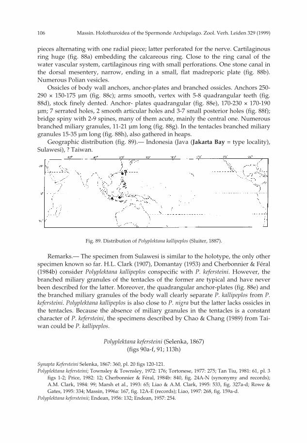
Massin. Holothuroidea of the Spermonde Archipelago. Zool. Verh. Leiden 329 (1999)106
pieces alternating with one radial piece; latter perforated for the nerve. Cartilaginousring huge (fig. 88a) embedding the calcareous ring. Close to the ring canal of thewater vascular system, cartilaginous ring with small perforations. One stone canal inthe dorsal mesentery, narrow, ending in a small, flat madreporic plate (fig. 88b).Numerous Polian vesicles.
Ossicles of body wall anchors, anchor-plates and branched ossicles. Anchors 250-290 × 150-175 µm (fig. 88c); arms smooth, vertex with 5-8 quadrangular teeth (fig.88d), stock finely dented. Anchor- plates quadrangular (fig. 88e), 170-230 × 170-190µm; 7 serrated holes, 2 smooth articular holes and 3-7 small posterior holes (fig. 88f);bridge spiny with 2-9 spines, many of them acute, mainly the central one. Numerousbranched miliary granules, 11-21 µm long (fig. 88g). In the tentacles branched miliarygranules 15-35 µm long (fig. 88h), also gathered in heaps.
Geographic distribution (fig. 89).— Indonesia (Java (Jakarta Bay = type locality),Sulawesi), ? Taiwan.
Remarks.— The specimen from Sulawesi is similar to the holotype, the only otherspecimen known so far. H.L. Clark (1907), Domantay (1953) and Cherbonnier & Féral(1984b) consider Polyplektana kallipeplos conspecific with P. kefersteini. However, thebranched miliary granules of the tentacles of the former are typical and have neverbeen described for the latter. Moreover, the quadrangular anchor-plates (fig. 88e) andthe branched miliary granules of the body wall clearly separate P. kallipeplos from P.kefersteini. Polyplektana kallipeplos is also close to P. nigra but the latter lacks ossicles inthe tentacles. Because the absence of miliary granules in the tentacles is a constantcharacter of P. kefersteini, the specimens described by Chao & Chang (1989) from Tai-wan could be P. kallipeplos.
Polyplektana kefersteini (Selenka, 1867)(figs 90a-f, 91; 113h)
Synapta Kefersteini Selenka, 1867: 360, pl. 20 figs 120-121.Polyplektana kefersteini; Townsley & Townsley, 1972: 176; Tortonese, 1977: 275; Tan Tiu, 1981: 61, pl. 3
figs 1-2; Price, 1982: 12; Cherbonnier & Féral, 1984b: 840, fig. 24A-N (synonymy and records);A.M. Clark, 1984: 99; Marsh et al., 1993: 65; Liao & A.M. Clark, 1995: 533, fig. 327a-d; Rowe &Gates, 1995: 334; Massin, 1996a: 167, fig. 12A-E (records); Liao, 1997: 268, fig. 159a-d.
Polyplektana kefersteinii; Endean, 1956: 132; Endean, 1957: 254.
Fig. 89. Distribution of Polyplektana kallipeplos (Sluiter, 1887).
pp 003-144 02-01-2007 15:33 Pagina 106
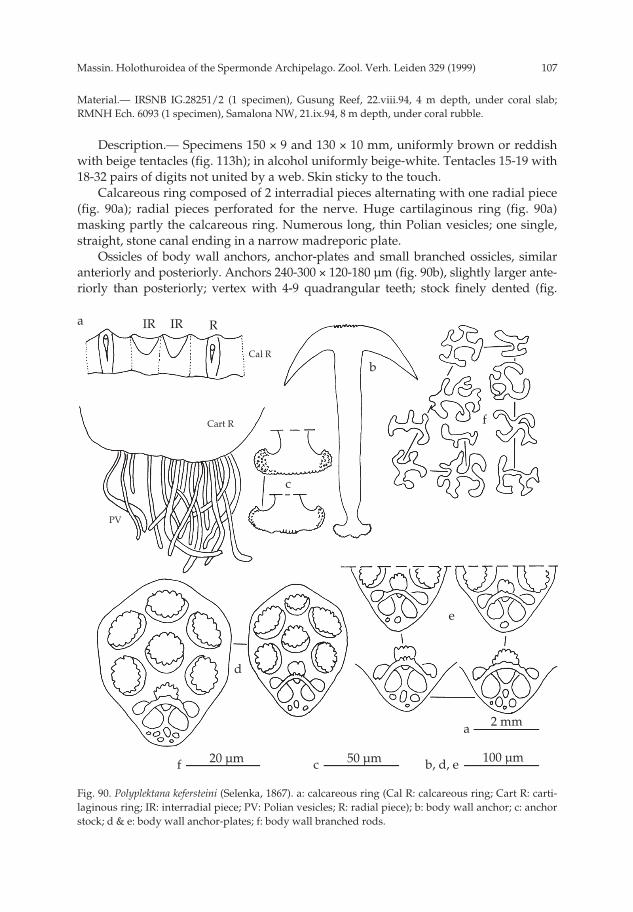
107Massin. Holothuroidea of the Spermonde Archipelago. Zool. Verh. Leiden 329 (1999)
Material.— IRSNB IG.28251/2 (1 specimen), Gusung Reef, 22.viii.94, 4 m depth, under coral slab;RMNH Ech. 6093 (1 specimen), Samalona NW, 21.ix.94, 8 m depth, under coral rubble.
Description.— Specimens 150 × 9 and 130 × 10 mm, uniformly brown or reddishwith beige tentacles (fig. 113h); in alcohol uniformly beige-white. Tentacles 15-19 with18-32 pairs of digits not united by a web. Skin sticky to the touch.
Calcareous ring composed of 2 interradial pieces alternating with one radial piece(fig. 90a); radial pieces perforated for the nerve. Huge cartilaginous ring (fig. 90a)masking partly the calcareous ring. Numerous long, thin Polian vesicles; one single,straight, stone canal ending in a narrow madreporic plate.
Ossicles of body wall anchors, anchor-plates and small branched ossicles, similaranteriorly and posteriorly. Anchors 240-300 × 120-180 µm (fig. 90b), slightly larger ante-riorly than posteriorly; vertex with 4-9 quadrangular teeth; stock finely dented (fig.
Fig. 90. Polyplektana kefersteini (Selenka, 1867). a: calcareous ring (Cal R: calcareous ring; Cart R: carti-laginous ring; IR: interradial piece; PV: Polian vesicles; R: radial piece); b: body wall anchor; c: anchorstock; d & e: body wall anchor-plates; f: body wall branched rods.
a RIR
100 µmb, d, e
Cal R
Cart R
b
c
f
d
e
IR
2 mm
50 µm20 µm
a
cf
PV
pp 003-144 02-01-2007 15:33 Pagina 107

Massin. Holothuroidea of the Spermonde Archipelago. Zool. Verh. Leiden 329 (1999)108
90c). Anchor-plates ovate, 170-225 × 135-185 µm (fig. 90d) with 7 serrated holes, twosmooth articular holes and 1-6 small posterior holes (fig. 90e); bridge with 1-5 small,blunt spines. Branched ossicles 9-17 µm long (fig. 90f). No ossicles in the tentacles.
Geographic distribution (fig. 91).— Red Sea, Seychelles (Mahé, Aldabra), Indone-sia (Java, Sulawesi, Ambon, Halmaheira), Philippines, Taiwan, China, MarianaIslands (Guam), Australia (WA, NT, Timor Sea, QLD, NSW, Tasman Sea), New Cale-donia, Fiji (Rotuma), Samoa (Navigator), Line Islands (Fanning Island), HawaiianIslands = type locality, Society Islands (Tahiti), Easter Island.
Remarks.— The number of tentacles and the number of digits per tentacle varygreatly in Polyplektana kefersteini (Heding, 1928; Cherbonnier & Féral, 1984b) even inspecimens from a single locality. However, calcareous ring, anchors, anchor-platesand branched ossicles are constant throughout the range of distribution. Only speci-mens from Easter Island lack branched miliary granules in the body wall (Massin,1996a). Poyplektana kefersteini is characterized by the absence of miliary granules in thetentacles (Selenka, 1867; H.L. Clark, 1907; Heding, 1928; Cherbonnier & Féral, 1984b;Féral & Cherbonnier, 1986; Massin, 1996a). Only H.L. Clark (1924) described minuteoblong granules (6-10 µm long) from the very tip of the tentacles of specimens fromHawaiian Islands. The specimens described by Chao & Chang (1989) from Taiwanhave small rods, up to 45 µm long, in the tentacles and quadrangular anchor-plateswith a spiny bridge. These characters are totally atypical for P. kefersteini and there-fore we are possibly dealing with P. kallipeplos.
Genus Synapta Eschscholtz, 1829Synapta maculata (Chamisso & Eysenhardt,1821)
(fig. 92)
Holothuria maculata Chamisso & Eysenhardt, 1821: 325, pl. 25.Synapta maculata; H.L. Clark, 1907: 78, pl. 1, pl. 4 figs 17-19, 26 (synonymy); Serene, 1937: 26; Dawyd-
off, 1952: 117; Endean, 1956: 132, 138; Endean, 1957: 255; Endean, 1961: 295; A.M. Clark &Spenser-Davies, 1966: 600; James, 1969: 62; Liao, 1975: 220, fig. 22 (1-3); Tortonese, 1977: 276;Sloan et al., 1979: 124; Tortonese, 1979: 316; Mary Bai, 1980: 28, textfig. 13C; Humphreys, 1981: 36;Tan Tiu, 1981: 60, pl. 2 figs 1-2, pl. 29 figs 1, 2b; Price, 1982: 12; James, 1982a: 99, fig. 1l-m; James,1982b: 5; Mukhopadhyay & Samanta, 1983: 310, textfig. 11A-B; Rowe, 1983: 161; James, 1983: 93;Reyes-Leonardo, 1984a; 153, pl. 11 fig. 1a-b; A.M. Clark, 1984: 99; Price & Reid, 1985: 7; Brouns &Heijs, 1985: 176; Reyes-Leonardo et al., 1985: 276; Marsh, 1986: 74; Massin & Doumen, 1986: 188;
Fig. 91. Distribution of Polyplektana kefersteini (Selenka, 1867).
pp 003-144 02-01-2007 15:33 Pagina 108

109Massin. Holothuroidea of the Spermonde Archipelago. Zool. Verh. Leiden 329 (1999)
Féral & Cherbonnier, 1986: 106, fig. 40X; Mukhopadhyay, 1988: 10, fig. 10a-b; Cherbonnier, 1988:251, fig. 112A-F (synonymy and records); Jangoux et al., 1989: 163; Chao & Chang, 1989: 120, figs26, 31D; James, 1989: 132; Zoutendijk, 1989: 2; Chambers, 1989: 89; Levin & Dao Tan Ho, 1989: 59;Marsh et al., 1993: 65; Kerr, 1994: 171; Marsh, 1994a: 11; Fiege et al., 1994: 86; Allen & Steene, 1994:247; Rowe & Gates, 1995: 336; Liao & A.M. Clark, 1995: 539, fig. 333a-c; Gosliner et al., 1996: 284,fig. 1047; Massin, 1996b: 46, fig. 33A-B; Liao, 1997: 278, fig. 166a-c; Rowe & Richmond, 1997: 306.
Material.— Panikiang, 30.viii.94, several specimens observed on reef flat; Kapoposang, 1.x.-94,numerous specimens observed in seagrass-bed.
Geographic distribution (fig. 92).— Red Sea, SE Arabia, Kenya, Zanzibar, Sey-chelles (Mahé, Aldabra), Madagascar, Mauritius, Maldive Islands, India (LaccadiveIslands, Andaman Islands, Nicobar Islands), Sri Lanka, Cocos Keeling Islands,Indonesia (Sumatra, Java, Lombok, Sumbawa, Sumba, Savu, Rotti, Timor, Salayer,Sulawesi, Makassar Strait, Celebes Sea, Halmaheira, Ceram, Ambon, LuciparaIslands, Irian Jaya), Philippines, Vietnam, Taiwan, China, Japan, Mariana Islands(Guam), Papua New Guinea (Port Moresby, Samarai, Madang Province), Australia(WA, NT, Timor Sea, GBR, QLD), New Caledonia, New Hebrides, Caroline Islands(Kosrae), Gilbert Islands, Marshall Islands = type locality, Vanuatu (= NewHebrides), Fiji, Samoa, Cook Island, Society Islands (Tahiti).
Description and remarks.— Synapta maculata was observed at Panikiang andKapoposang. At both sites the species is abundant in shallow seagrass-beds of thereef flat and lagoon. No specimens were collected because the species can be readilyidentified without doubt in the field. Synapta maculata has been reported from manylocalities in Indonesia (see Sluiter, 1901).
Genus Synaptula Oersted, 1849Synaptula cf denticulata Heding, 1928
(figs 93a-l, 94, 113j)
Synaptula denticulata Heding, 1928: 177, fig. 22 (1-4); A.M. Clark & Rowe, 1971: 186.
Material.— RMNH Ech. 6094 (1 specimen), Samalona NW, 2.ix.94, 10 m depth; IRSNB IG.28251/137(1 specimen), Samalona NW, 21.ix.94, 5 m depth in between living corals.
Fig. 92. Distribution of Synapta maculata (Chamisso & Eysenhardt, 1821).
pp 003-144 02-01-2007 15:33 Pagina 109

Massin. Holothuroidea of the Spermonde Archipelago. Zool. Verh. Leiden 329 (1999)110
Description.— Specimens 136 × 9 and 176 × 8 mm, light brown with white dotsand orange muscular bands (fig. 113j); in alcohol yellowish. Tentacles 13 with 18-30pairs of digits united by a web. Skin thin, translucent, very sticky to the touch.
Calcareous ring white, completely embedded in a huge cartilaginous ring; radialpieces perforated for the nerve and with scars at the insertion points of the longitudi-nal muscles (fig. 93a); laterally, 2 interradial pieces alternating with a radial one. Car-tilaginous ring with small perforations through the posterior margin (fig. 93a). Atleast 10 Polian vesicles 2.5 mm long; one stone canal, straight, going upwards and
Fig. 93. Synaptula cf denticulata Heding, 1928. a: calcareous ring (Cal R: calcareous ring; Cart R: carti-laginous ring; IR: interradial piece; MP: madreporic plate; PV: Polian vesicles; R: radial piece; SC:stone canal); b: anchor from anterior body wall; c: stock of anterior body wall anchor; d: anchor fromposterior body wall; e: stock of posterior body wall anchor; f & g: anchor-plates from anterior bodywall; h & j: anchor-plates from posterior body wall; k: miliary granules from body wall; l: miliarygranules from tentacles.
a RIR
100 µmb, d, f-j
Cal R
Cart R
c
b
f
d
g
h
k
j
e
l
50 µm 20 µm
5 mm
c, e k, l
a
MP
SC
PV
pp 003-144 02-01-2007 15:33 Pagina 110

111Massin. Holothuroidea of the Spermonde Archipelago. Zool. Verh. Leiden 329 (1999)
ending in a elongated madreporic plate (fig. 93a). Gonad well developed, formedfrom branched tubules.
Ossicles of body wall anchors, anchor-plates and miliary granules. Anchors withthe same size anteriorly (fig. 93b) and posteriorly (fig. 93d): 282.0+13.3 (n = 15) µmlong in the 136 mm long specimen, 299.3+20.4 (n = 20) µm long in the 176 mm longspecimen; arms smooth, a few knobs on the vertex, stock finely dented (figs 93e).Anchor-plates ovates (figs 93f, h) with 6 serrated holes, 3 articular holes [central onenearly always serrated (figs 93 f, j), lateral ones smooth or serrated (figs 93g, j)], and abridge with 3-4 blunt spines (figs 93f, h). Large specimen posteriorly with sharp teethon the bridge, central tooth being sometimes prominent (fig. 93j). The number of pos-terior small holes is 4.1+1.1 (n = 32) in anteriorly located anchor-plates and 3.8+1.0 (n= 36) in posteriorly located ones. Most of the miliary granules as dissociated grains;when intact, 13 µm across (fig. 93k). In the tentacles miliary granules only, 14-29 µmacross (fig. 93l).
Geographic distribution (fig. 94).— Indonesia (Sulawesi, Kai Islands = type locali-ty).
Remarks.— According to the key given by Heding (1928) the specimens fromSulawesi must be Synaptula denticulata. However, because of the colour of the bodywall (light brown with white dots instead of colourless mottled with green and white)and the calcareous ring colour (white instead of green), and added to this the averagenumber of posterior holes through the anchor-plates (4 instead of 7) doubt remains.The bulk of the type material of Heding (1928) consists of small specimens of 6-8 cmlong. This disparity in size with the present material could explain some differenceswith the type material.
Synaptula cf. lamperti Heding, 1928(figs 95a-h, 96, 114a)
Synaptula lamperti Heding, 1928: 193, fig. 25 (6-8); Cheng & Lewin, 1984: 95; Hammond & Wilkinson,1985: 3, fig. 1; Rowe & Gates, 1995: 337.
Material.— IRSNB IG.28251/125 (1 specimen), Samalona NW, 16.ix.94, 7 m depth.
Description.— Specimen 46 × 4.2 mm, white with 5 radial purple stripes over the
Fig. 94. Distribution of Synaptula denticulata Heding, 1928.
pp 003-144 02-01-2007 15:33 Pagina 111

Massin. Holothuroidea of the Spermonde Archipelago. Zool. Verh. Leiden 329 (1999)112
whole body length (fig. 114a); anterior quart of the stripes bifurcate; some very finelongitudinal purples lines are present in the interrambulacral areas; in alcohol purewhite. Tentacles 10, each with about 20 pairs of digits. Presence or absence of a webbetween the digits could not be verified due to the strong contraction of the tentacles.
Calcareous ring faint, white; radial pieces perforated for the nerve (fig. 95a). Carti-laginous ring well developed, translucent, with holes of various size close to the ringcanal of the water vascular system (fig. 95a). Polian vesicles 11 and one stone canalgoing upwards with a long cylindrical madreporic plate (fig. 95a). Prominent intesti-nal loop.
Ossicles of body wall anchors, ancho-plates and miliary granules. Anchors (fig.95b) 215-245 × 140-160 µm; arms smooth, vertex with 4-7 quadrangular teeth (fig.95c), stock finely dented (fig. 95d). Anchor-plates (fig. 95e) 175-190 × 145-160 µm,quadrangular with 7 serrated holes, 2 smooth or serrated articular holes and 5-8 smallposterior holes (fig. 95f); bridge spiny with central tooth sometimes longer than theothers. Miliary granules very abundant (fig. 95g), 11-14 µm across. In the tentaclesmiliary granules only, 9-14 µm across (fig. 95h).
Geographic distribution (fig. 96).— Indonesia (Sulawesi, Banda = type locality);Palau Islands; Australia (NE coast, N coast, QLD, NT).
Remarks.— By body size and colour, size and form of the ossicles (anchors,
Fig. 95. Synaptula cf. lamperti Heding, 1928. a: calcareous ring (Cal R: calcareous ring; Cart R: cartilagi-nous ring; IR: interradial piece; MP: madreporic plate; PV: Polian vesicles; R: radial piece); b: bodywall anchor; c: anchor vertex; d: anchor stock; e & f: body wall anchor-plates; g: miliary granules frombody wall; h: miliary granules from tentacles.
a R IR
100 µmb, e, f
Cal R
Cart R
b
c
d
ge
f
hc, d
g, h
a
50 µm
20 µm
5 mm
MP
PV
pp 003-144 02-01-2007 15:33 Pagina 112

113Massin. Holothuroidea of the Spermonde Archipelago. Zool. Verh. Leiden 329 (1999)
anchor-plates and miliary granules), number of tentacles and digits per tentacle, cal-careous and cartilaginous rings, number of Polian vesicles, the Sulawesi specimen isvery similar to Synaptula lamperti Heding, 1928. However, Heding (1928) mentionedthe absence of an intestinal loop (present in the Sulawesi specimen) and the constantpresence of 9 holes in the posterior end of the anchor-plates (versus 5-8 in the Sulawe-si specimen). Because of these differences I am not convinced that we are dealingwith S. lamperti and I prefer to refer to the specimen as Synaptula cf. lamperti.
Synaptula media Cherbonnier & Féral, 1984(figs 97a-f, 98)
Synaptula media Cherbonnier & Féral, 1984b: 843, fig. 25A-F, pl. 4A-B; Féral & Cherbonnier, 1986: 106,fig. 40Y; Colin & Arneson, 1995: 264, fig. 1237.
Material.— IRSNB IG.28251/215 (1 specimen), Kapoposang NE, 30.ix.94, 31 m depth, observed on 3different species of sponges.
Description.— Specimen 115 × 6 mm, grey-white with numerous longitudinal,purple lines; in alcohol completely white. Tentacles 10, each with 14-16 digits unitedby a web; skin thin, sticky to the touch.
Calcareous ring very fine, white; radial pieces perforated for the nerve; posterioredge of the calcareous ring undulating (fig. 97a). Cartilaginous ring moderatelydeveloped (fig. 97a), translucent; 10 Polian vesicles; one short stone canal; gonad com-posed of several large branched tubules. Longitudinal muscle bands narrow.
Ossicles of body wall anchors, anchor-plates and miliary granules. Anchors locat-ed posteriorly (fig. 97b) slightly smaller (216+5.5 × 140+6.8, n = 8) than those locatedanteriorly (fig. 97c) (230+10.3 × 150.5+6.0, n = 9); arms smooth, vertex with 3-6 bluntspines, stock finely dented. Size of anchor-plates similar anteriorly and posteriorly(fig. 97d) (176+3.0 × 140+4.0, n = 10); quadrangular with 6 serrated holes and onesmooth hole (fig. 97d); articular holes smooth, 3-7 posterior holes in the anterioranchor-plates and 3-4 in the posterior ones; bridge spinose. Countless small miliarygranules, 10-14 µm across (fig. 97e), most of them as dissociated grains. In the tenta-cles miliary granules only, few, 11-15 µm across (fig. 97f).
Geographic distribution (fig. 98).— Indonesia (Sulawesi), Micronesia, Papua New
Fig. 96. Distribution of Synaptula lamperti Heding, 1928.
pp 003-144 02-01-2007 15:33 Pagina 113

Massin. Holothuroidea of the Spermonde Archipelago. Zool. Verh. Leiden 329 (1999)114
Guinea, New Caledonia = type locality.Remarks.— The specimen from Sulawesi is much larger than those of the type
series (115 mm versus 10-40 mm). However, the ossicles and calcareous ring are iden-tical to the type series, both as regrads types and sizes (see Cherbonnier & Féral,1984b). The species seems always related to sponges, living between 8 and 31 mdepth. Synaptula media is new to the fauna of Indonesia.
Fig. 97. Synaptula media Cherbonnier & Féral, 1984. a: calcareous ring (Cal R: calcareous ring; Cart R:cartilaginous ring; IR: interradial piece; PV: Polian vesicles; R: radial piece); b: anchor from anteriorbody wall; c: anchor from posterior body wall; d: body wall anchor-plates; e: miliary granules frombody wall; f: miliary granules from tentacles.
Fig. 98. Distribution of Synaptula media Cherbonnier & Féral, 1984.
a RIR
100 µmb-d
Cal R
Cart R
b c
d
f
e
PV
20 µm 2 mme, f a
pp 003-144 02-01-2007 15:33 Pagina 114

115Massin. Holothuroidea of the Spermonde Archipelago. Zool. Verh. Leiden 329 (1999)
Synaptula psara (Sluiter, 1887)(figs 99a-f, 100)
Synapta psara Sluiter, 1887: 219, pl. 2 figs 36-39; Ludwig, 1889-92: 357; Sluiter, 1895: 82.Chondrocloea psara; Oestergren, 1898: 114: Sluiter, 1901: 126.Synaptula psara; H.L. Clark, 1907: 84; H.L. Clark, 1924: 475, pl. 3 figs 7-8, pl. 4 fig. 5; Heding, 1928: 176,
fig. 24 (4-11): A.M. Clark & Rowe, 1971: 186.
Material.— IRSNB IG.28251/13 (2 specimens), Samalona N, 24.viii.94, 9 m depth; IRSNBIG.28251/249 (1 specimen) and RMNH Ech. 6095 (1 specimen), Badi, 3.x.94, 2 m depth among coralrubble.
Description.— Length ±175 × 14 mm, 22 × 3 mm (anterior part only), >200 × 10 and>200 × 9 mm, estimated because specimens fallen apart into fragments; body cylindricaltapering posteriorly; diameter given corresponds to the anterior part at the level of thecalcareous ring. Colour varied from light to dark brown with white spots correspond-ing to heaps of miliary granules; in alcohol yellowish to pinkish with the white spotsstill visible. Tentacles 13, with 22-29 pairs of digits united by a web; the 2 ventral tenta-cles shorter, sometimes reduced to finger without digits; two specimens with 1-3 regen-erating tentacles. Skin sticky to the touch, especially thick (1-2 mm) anteriorly.
Fig. 99. Synaptula psara (Sluiter, 1887). a: calcareous ring (IR: interradial piece; R: radial piece); b: stonecanal and madreporic plate; c: body wall anchor; d & e: body wall anchor-plates. f: miliary granulesfrom tentacles.
a
R IR
100 µmc-e
b
c
d
e
f
d
f
a, b
20 µm
2 mm
R
pp 003-144 02-01-2007 15:33 Pagina 115

Massin. Holothuroidea of the Spermonde Archipelago. Zool. Verh. Leiden 329 (1999)116
Calcareous ring (fig. 99a) white, completely embedded in a large cartilaginousring. Radial pieces perforated for the nerve; laterally, 2 interradial pieces alternatingwith one radial piece (fig. 99a), whereas dorsally and ventrally one radial alternatingwith one interradial piece. Cartilaginous ring with small perforations through its pos-terior margin. At least 10 Polian vesicles, 4-8 mm long. One stone canal goingupwards ending in a long madreporic plate (fig. 99b). Gonad well developed, com-posed of branched tubules.
Ossicles of body wall anchors, anchor-plates and miliary granules. Statistically,anchors (fig. 99c) of the same size anteriorly (321.5+6.7 µm long) and posteriorly(326+21.4 µm long); arms smooth, vertex with 4-8 small blunt teeth, stock finely dent-ed (fig. 99c). Anchor-plates (fig. 99d) slightly larger posteriorly (241+7.7 µm long)than anteriorly (220+6.2 µm long); plates ovate (fig. 99d) with 6 anterior, serratedholes; articular holes smooth or with a few spines (fig. 99e); bridge smooth or with 1-4blunt spines; posterior holes more numerous (5.1+1.5; n = 22) on anterior anchor-plates than on posterior ones (4.3+1.1; n = 24). Miliary granules only as dissociatedgrains. In the tentacles miliary granules only, concentrated in heaps. Most of them asdissociated grains; the intact ones 17-20 µm across (fig. 99f).
Geographic distribution (fig. 100).— Indonesia (Java: Jakarta Bay = type locality),Sulawesi, Salayer, Kai Islands).
Remarks.— Synaptula psara can readily be identified by its relatively thick anteriorbody wall, by the heaps of miliary granules and by its ovate, elongated anchor-plates.According to H.L. Clark (1924), Heding (1928) and the present work, the size of theanchors and anchor-plates increases with increasing body length.
Synaptula recta (Semper, 1868)(figs 101a-k, 102, 114b, c)
Synapta recta Semper, 1868: 14, pl. 4 figs 2-3, pl. 5 fig. 18, pl.8 fig. 2; Lampert, 1885: 220; Bedford, 1899,142; Koehler, 1895b: 385.
Chondrochloea recta; Oestergren, 1898a: 114; Sluiter, 1901: 125; Erwe, 1913: 390, pl.8 fig. 26a-b (syn-onymy and records); Ekman, 1918: 63; Endean, 1953: 57; Endean, 1956: 132; Endean, 1957: 255;Endean, 1961: 295.
Synaptula recta; H.L. Clark, 1907: 84 (synonymy); H.L. Clark, 1924: 475, pl. 1 fig. 13, pl. 3 fig. 9, pl. 4 fig.3; Heding, 1928: 167, figs 16 (4-6), 17 (3-8); H.L. Clark 1932: 221; Tortonese, 1953: 46; A.M. Clark &
Fig. 100. Distribution of Synaptula psara (Sluiter, 1887).
pp 003-144 02-01-2007 15:33 Pagina 116
117Massin. Holothuroidea of the Spermonde Archipelago. Zool. Verh. Leiden 329 (1999)
Spenser-Davies, 1966: 600; Tortonese, 1977: 276; Tortonese, 1979: 316; James, 1982a: 101, fig. 1n(synonymy and records); James, 1982b: 5; Price, 1982: 12; James, 1984 [1988]: 405; Mukhopadhyay,1988: 15; Levin & Dao Tan Ho, 1989: 59; Marsh et al., 1993: 65; Marsh, 1994a: 11; Marsh, 1994b: 57;Rowe & Gates, 1995: 338; Tahera, 1996: 106; Tahera, 1997: 92; Baine & Forbes, 1998: 4.
Synapta striata Sluiter, 1887: 216, pl. 2 figs 39-40.
Material.— IRSNB IG28.251/136 (1 specimen), Samalona NW, 21.ix.94, 5 m depth, in between livingcorals; IRSNB IG.28252/138 (1 specimen), Samalona NW, 21.ix.94, 5 m depth; RMNH Ech. 6072 (1specimen), Badi, 3.x.94, 2 m depth.
Description.— Specimens 190 × 9, 84 × 16 and 194 × 10 mm, colour varied fromnearly black to brown, speckled with white spots corresponding to heaps of miliarygranules (figs 114b, c); in alcohol grey-beige speckled with white spots. Skin sticky tothe touch. Tentacles 13, with 19-20 pairs of digits not united by a web. Heaps of mil-iary granules sometimes forming white transversal bands in the tentacles.
Calcareous ring composed of radial and interradial pieces of the same size (fig.101a); radial pieces perforated for the nerve. Huge cartilaginous ring partly maskingthe calcareous ring (fig. 101a); posterior part of the calcareous ring straight or slightlyundulating. A few perforations at the base of the cartilaginous ring. Polian vesicles15-20, very variable in size. One stone canal going upwards and ending in amadreporic plate 1/3 the length of the stone canal.
Ossicles of body wall anchors, anchor-plates and miliary granules. Anchors of thetwo largest specimens (fig. 101b) of similar size anteriorly and posteriorly (330.5+13.2× 224.0+5.2 µm versus 326.5+18.3 × 210.5+15.5 µm; n = 10); a few tooth on the vertex,arms smooth and stock finely dented (fig. 101c). Anchors of the small specimen alsoof the same size anteriorly and posteriorly (274.5+10.7 × 172.5+8.3 µm and 273.0+15.7× 161.5+8.5 µm; n = 10); arms smooth, vertex with a few knobs, stock finely dented,occasionally divided into several lobes (fig. 101c). Anchor-plates of the two largespecimens (figs 101d, f) of the same size anteriorly and posteriorly (225.5+5.5 ×166.0+5.2 µm and 235.0+11.5 × 185.5+9.0 µm; n = 10), more ovoid anteriorly (fig.101d); 6 serrated holes; articular holes smooth or serrated; bridge spiny with spinesmore prominent on posteriorly located anchor-plates; sometimes a strong medianspine (figs 101e, g); 3-7 posterior holes with an average of 5.0+1.0 whatever the loca-tion of the anchor-plates (figs 101e, g). On one slide (IRSNB IG 28256/136) posteriorholes of posteriorly located anchor-plates less numerous (2-7 with an average of3.7+1.0). Anchor-plates of the small specimen of the same size anteriorly and posteri-orly (203.5+6.3 × 154.5+4.4 µm and 191.0+10.7 × 157.0+5.9 µm; n = 10), ovate to quad-rangular with 6 serrated holes; central articular hole serrated, lateral ones smooth;bridge knobbed to spiny, exceptionally with a strong median tooth; posterior holes 3-7 with an average of 5.7+1.1 (n = 15) in anteriorly located anchor-plates and 4.6+1.3 (n= 21) in posteriorly located ones. Miliary granules 12-16 µm across irrespective of theposition along the body wall (figs 101h, j). In the tentacles miliary granules only (fig.101k), somewhat larger (16-19 µm across) than in the body wall. In the small speci-men, most of them as dissociated grains.
Geographic distribution (fig. 102).— Red Sea, French Somalia, Pakistan, MaldiveIslands, Sri Lanka, India (Laccadive Islands, Gulf of Mannar, Madras, Andaman &Nicobar Islands), Myanmar (Mergui Archipelago), Cocos Keeling Islands, Malaysia,
pp 003-144 02-01-2007 15:33 Pagina 117

Massin. Holothuroidea of the Spermonde Archipelago. Zool. Verh. Leiden 329 (1999)118
Fig. 101. Synaptula recta (Semper, 1868). a: calcareous ring (Cal R: calcareous ring; Cart R: cartilaginousring; IR: interradial piece; R: radial piece); b: body wall anchor; c: anchor stock; d-g: body wall anchor-plates; h: miliary granules from anterior body wall; j: miliary granules from posterior body wall; k:miliary granules from tentacles.
a R IR
100 µmb, d-g
Cal R
Cart R
IR R
b
c
d
e
h
cf
g
j
k
50 µm
5 mm
20 µm
a
ch-k
pp 003-144 02-01-2007 15:33 Pagina 118

119Massin. Holothuroidea of the Spermonde Archipelago. Zool. Verh. Leiden 329 (1999)
Indonesia (Java, Salayer, Sulawesi, Timor, Kai Islands), Philippines (Bohol = typelocality, Cebu, Samar), Vietnam, Australia (WA, NT, Timor Sea, QLD), Papua NewGuinea (Samarai), Caroline Islands (Ponape).
Remarks.— The specimens from Sulawesi fit particularly well the descriptiongiven by Heding (1928), except for the colour. However, the colour of Synaptula rectais highly variable ranging from bright reddish to deep brownish-purple. According toPearson (1903) it can also be “striped lilac on a white ground” or according to Endean(1961) “olive”. H.L. Clark (1907, 1946) noted that the species is “extraordinarily vari-able in colour”. Notwithstanding these, the colour patterns described by Pearson(1903) and Endean (1961) are so different from the average found in S. recta that itthrows some doubt on the identification of these two authors. The small specimenwith a slightly undulating posterior margin of the calcareous ring and the very darkcolour (fig. 116c) is close to Synaptula striata (Sluiter, 1887). However, I agree with asH.L. Clark (1907) and Erwe (1913) that S. striata could be a synonym of S. rectabecause the main character to separate both species is based on the body wall colour(Heding, 1928) which, as said, is highly variable in S. recta.
Synaptula reticulata (Semper, 1868)(figs 103a-e, 104)
Synapta reticulata Semper, 1868: 13, pl.4 figs 4-5, pl. 5 figs 12, 23, pl. 6 fig.9; Lampert, 1885: 226; Théel,1886: 27; Sluiter, 1887: 214; Lampert, 1889b: 845; Koehler, 1895b: 385; Sluiter, 1895: 82.
Synapta reticulata var. nigro-punctata Bedford, 1899: 338.Chondrochloea reticulata ; Oestergren, 1898: 114; Sluiter, 1901: 127.Synaptula reticulata ; H.L. Clark, 1907: 86; H.L. Clark, 1924: 477; Heding, 1928: 191, fig 25 (1-4); A.M.
Clark & Rowe, 1971: 188; Rowe & Doty, 1995: 338; Liao & A.M. Clark, 1995: 541, fig. 334a-c; Liao,1997: 279, fig 167a-c.
Synaptula oestergreni Heding, 1928: 199, fig.27 (7-10).
Material.— IRSNB IG.28251/228 (1 specimen) and RMNH Ech. 6096 (1 specimen), Kapoposang,1.x.94, 20 m depth, on sponges and on sand around the sponges; ZMC: 3 paratypes (14.3 × 2.3, 11.2 ×1.8 and 8.8 × 1.9 mm) of Synaptula oestergreni Heding, 1928.
Description.— Specimens 11.8 × 1.8 and 13.0 × 3.0 mm, white because of the hugeamount of miliary granules in the body wall; in alcohol uniformly white. Ossicles
Fig. 102. Distribution of Synaptula recta (Semper, 1868).
pp 003-144 02-01-2007 15:33 Pagina 119

Massin. Holothuroidea of the Spermonde Archipelago. Zool. Verh. Leiden 329 (1999)120
give a shiny aspect to the specimens. Tentacles 10 with 6-7 pairs of digits. Because ofthe state of contraction of the tentacles, the presence or absence of a membrane inbetween the digits is difficult to ascertain. Tentacles of the 11.2 mm long paratype ofS. oestergreni with 6-8 pairs of digits.
Calcareous ring white, with radial and interradial pieces of the same size (fig.103a); radials perforated for the nerve; posterior margin of the calcareous ring undu-lating. Cartilaginous ring translucent, weakly developed. Polian vesicles 8 in 2 clus-ters of 4 (fig. 103a); one stone canal going upwards, ending in a narrow madreporicplate. No gonad.
Ossicles of body wall anchors, anchor-plates and miliary granules. Anchors few,158.8+11.8 × 100.0+0.0 µm (n = 4), arms smooth, a few knobs on the vertex and stockfinely dented (fig. 103b). Anchor-plates 127.7+10.1 × 109.1+8.6 µm (n = 11), nearlyrounded (fig. 103c), perforated by 6 serrated holes; lateral articular holes smooth, cen-tral one smooth or with 1-2 teeth (fig. 103c); bridge smooth or spiny; 1-5 posteriorholes (fig. 103c) with an average of 3.1+1.0 (n = 22). Miliary granules 10-17 µm across(fig. 103d), not dissociated. In the tentacles miliary granules (fig. 103e) similar to thoseof the body wall.
Geographic distribution (fig. 104).— China, Philippine (Bohol = type locality),Indonesia (Java, Sulawesi, Biaru Islands, Salayer, Sumbawa, Flores, Salawatti, BandaIslands, Kai Islands, Aru Islands), Australia (NE coast, NW coast, GBR, QLD, WA),New Caledonia.
Remarks.— The small size of the specimens and the absence of gonad indicatethat we are dealing with juveniles. As many characters (size of ossicles, number of
Fig. 103. Synaptula reticulata (Semper, 1868). a: calcareous ring (IR: interradial piece; MP: madreporicplate; PV: Polian vesicles; R: radial piece; SC: stone canal); b: body wall anchor; c: body wall anchor-plates; d: miliary granules from body wall; e: miliary granules from tentacles.
a R IR
100 µmb, c
SC
MP
PV
bc
d
e
ad, e 1 mm20 µm
R
pp 003-144 02-01-2007 15:33 Pagina 120

121Massin. Holothuroidea of the Spermonde Archipelago. Zool. Verh. Leiden 329 (1999)
tentacles, number of digits per tentacle, etc.) evolve with increasing body size, juve-niles are very difficult to identify. The two specimens from Kapoposang look verymuch like the paratypes of S. oestergreni. All agree in the number of tentacles, numberof Polian vesicles, size, general form and the number of posterior holes of the anchor-plates, and the size of the anchors. Heding (1928) mentioned 16-20 pairs of digits forspecimens 10-40 mm long. However, the 16-20 pairs of digits correspond only to thelargest specimen. Smaller ones have the same number of digits as the specimens fromSulawesi (6-8 versus 6-7). It may be emphasized once more that in Synaptidae, thenumber of digits increases with body length, and that it is important to compare spec-imens of the same size, when using this character to separate species.
The only difference with the type material of S. oestergreni is the amount of mil-iary granules which is much higher in the specimens from Indonesia than in thosefrom China. Heding (1928) recognised that the type material of S. oestergreni corre-sponds also to juveniles and he was not sure to be dealing with a new species. SinceS. oestergreni is only known from southern China and since Liao & A.M. Clark (1995)and Liao (1997) recognise only one Synaptula, S. reticulata, in southern China, I provi-sionally consider S. oestergreni as a juvenile form of Synaptula reticulata. A study ofgrowth series of S. reticulata is necessary to solve this problem
Synaptula cf. reticulata (Semper, 1868)(figs 105a-q, 106a-j, 114d-f)
Synaptula lamperti; Gosliner et al., 1996: 284, fig. 1049.
Material.— IRSNB IG 28251/5 (1 specimen), Samalona S, 24.viii.94, 22 m depth; IRSNB IG.28251/9 (31specimens) and RMNH Ech. 6073 (35 specimens), Samalona S, 24.viii.94, 7 m on sponges; IRSNBIG.28251/10 (2 specimens) and RMNH Ech. 6097 (1 specimen), Samalona S, 24.viii.94, 29 m depth;IRSNB IG.28251/48 (1 specimen) and RMNH Ech. 6098 (1 specimen), Samalona W, 1.ix.94, 8 m depth,on coral rubble; IRSNB IG.28251/55 (7 specimens), Samalona W, 2.ix.94, 12 m depth, on sponges;IRSNB IG.28251/57 (1 specimen), Samalona W, 2.ix.94, 12 m depth, on a sponge and on sand near thesponge; IRSNB IG.28251/165 (one specimen) and RMNH Ech. 6074 (1 specimen), Kudingareng KekeS, 26.ix.94, 15 m depth, on sponges, Millepora spec. and Culcita spec.
Description.— Eighty-two specimens from 17 × 2.3 to 95 × 5.5 mm. Colour highlyvariable, from nearly white with a few brown dots (fig. 114f) to white with rusty-red
Fig. 104. Distribution of Synaptula reticulata (Semper, 1868).
pp 003-144 02-01-2007 15:33 Pagina 121

Massin. Holothuroidea of the Spermonde Archipelago. Zool. Verh. Leiden 329 (1999)122
a RIR
100 µmb-e, h, k-m
Cal R
Cart R
b
d
f
g
c
e
hj
n
l nk
q
p
mj
f, g, n-q
a
5 0
2 0
2 mm
IR
PV
l
pp 003-144 02-01-2007 15:33 Pagina 122

123Massin. Holothuroidea of the Spermonde Archipelago. Zool. Verh. Leiden 329 (1999)
longitudinal lines and speckled with brown dots (fig. 114e). All these colour patternsobserved in a single large sample collected on a single sponge (fig. 114d). Generallyon sponges [mainly Xetospongia spp. (fig. 114d) and Amphimedon spp. (fig. 114e)] butalso on Millepora spec. (Hydrozoa), or Culcita spec. (Asteroidea) and on sand or coralrubble. Colour not linked to the colour of the sponge. In small specimens (17-40 mmlong) miliary granules very abundant forming broad longitudinal white bands; inalcohol uniformly light grey to white, longitudinal white bands sometimes still visi-ble. Tentacles 10 irrespective of body length; number of digits increasing with increas-ing body size: from 7-8 pairs of digits (17 mm long specimens) to 15-18 pairs (60-95mm long specimens); digits united by a web.
Calcareous ring white composed of radial and interradial pieces of the same size;radials perforated for the nerve (fig. 105a); posterior margin of the calcareous ringundulating. Cartilaginous ring translucent, with large posterior perforations (fig.105a). Seven-15 Polian vesicles and one stone canal going upwards, ending in anelongate madreporic plate.
Ossicles of body wall anchors, anchor-plates and miliary granules. Anchors withsmooth arms; vertex with a few knobs and stock finely dented (figs 105b, c, h, k). Pos-terior anchors nearly always somewhat larger than anterior ones; anchors 200-240 ×120-150 µm. No increase of anchor size with increasing body length. Anchor-plates150-180 × 125-150 µm, quadrangular with 6 serrated holes (figs 105e, d, l, m); centralarticular hole smooth or with 1-3 small spines; lateral articular holes smooth; posteri-or holes 1-7 (figs 105e, d, l, m), always more numerous on anterior anchor-plates thanon posterior ones. Average number of posterior holes in the anterior anchor-plates, 3-4 in small animals (17-30 µm long) and 4-5 in large ones (40-95 µm long). For the pos-terior anchor-plates average number of posterior holes 3-4 irrespective of bodylength. Bridge knobbed or spiny (figs 105e, d, l, m) sometimes with a large centralspine (fig. 106f). One specimen with some anomalous anchor-plates (figs 106c, g)among normal anchors (figs 106a, e) and anchor-plates (figs 106b, f). Miliary granules(figs 105f-g, n, p, 106d, h) very abundant in small specimens and with a patchy distri-bution. In large specimens not so abundant and more regularly distributed. Miliarygranules 8-18 µm across with size not related to body size. Most of the miliary gran-ules as dissociated grains.
In the tentacles, miliary granules only; same size as the ones in the body wall (figs105q, 106j) and absent in the largest specimens.
Remarks.— Synaptula cf. reticulata is very abundant in shallow-water (7-22 mdepth) off Ujung Pandang, but only at Samalona (80 of the 82 collected specimens).
This species has previously been depicted by Gosliner et al. (1996), as Synaptulalamperti Heding, 1928, a misidentification because of the presence of a large intestinal
Fig. 105. Synaptula cf. reticulata (Semper, 1868). (a-g: L=95 mm; IRSNB IG.28251/9; h-q: L=27 mm;IRSNB IG.28251/9). a: calcareous ring (Cal R: calcareous ring; Cart R: cartilaginous ring; IR: interradi-al piece; PV: Polian vesicles; R: radial piece); b: anchor from anterior body wall; c: anchor from poste-rior body wall; d: anchor-plates from anterior body wall; e: anchor-plates from posterior body wall; f:miliary granules from anterior body wall g: miliary granules from posterior body wall; h: anchor fromanterior body wall; j: anchor stock; k: anchor from posterior body wall; l: anchor-plates from anteriorbody wall; m: anchor-plates from posterior body wall; n: miliary granules from anterior body wall; p:miliary granules from posterior body wall; q: miliary granules from tentacles.
pp 003-144 02-01-2007 15:33 Pagina 123

Massin. Holothuroidea of the Spermonde Archipelago. Zool. Verh. Leiden 329 (1999)124
lope (absent in S. lamperti) and because of the number of posterior holes in theanchor-plates (9 in S. lamperti according to Heding (1928); 3-5 (average) in Synaptulacf. reticulata).
The single anomalous specimen has modified anchor-plates which are very simi-lar to those of Synaptula virgata (Sluiter, 1901) (see Heding 1928). However, S. virgatadiffers from this specimen mainly by the higher number of tentacles (13 versus 10), bythe cartilaginous ring reduced to tentacle canals and by the higher number of holes inthe posterior part of the anchor-plates.
According to the Heding’s key to the species of Synaptula (Heding 1928), the pre-sent species is close to the group formed by Synaptula maculata (Sluiter, 1888), Synap-tula reticulata (Semper, 1868) and Synaptula purpurea Heding, 1928. Synaptula cf. reticu-lata from Sulawesi presents a mixture of characters present in the three above men-tioned species. It agrees with S. reticulata in the number of pair of tentacle digits, withS. purpurea more or less in colour, and with S. maculata in the calcareous ring and ossi-cle size. It differs from S. purpurea by the number of posterior holes in the anchor-plates and by the presence of rosettes, and from S. maculata by the number of tentacle
Fig. 106. Synaptula cf. reticulata (Semper, 1868). (a-j: L=32 mm; IRSNB IG.28251/55). a: anchor fromanterior body wall; b: anchor-plates from anterior body wall; c: abnormal anchor-plate from anteriorbody wall; d: miliary granules from anterior body wall; e: anchor from posterior body wall; f: anchor-plates from posterior body wall; g: abnormal anchor-plate from posterior body wall; h: miliary gran-ules from posterior body wall; j: miliary granules from tentacles.
a
100 µma-c, e-g
b
c
d
g
h
e
j
f
d, h, j 20 µm
pp 003-144 02-01-2007 15:33 Pagina 124
125Massin. Holothuroidea of the Spermonde Archipelago. Zool. Verh. Leiden 329 (1999)
digits and by the number of holes in the posterior part of the anchor-plates. It showsthe highest affinity with Synaptula reticulata, but the colour of the Sulawesi specimensis so different from typical S. reticulata that I prefer to refer to it as Synaptula cf. reticu-lata.
Synaptula spec.(figs 107a-j, 114g)
Synaptula media; Gosliner et al., 1996: 284, fig 1048 (not Synaptula media Cherbonnier & Féral, 1984).
Material.— IRSNB IG.28251/130 (1 specimen), Samalona NW, 21.ix.94, 9 m depth on corals.
Description.— Specimen 220 × 6-8 mm, deep black to brown with large whitespots anteriorly (fig. 114g) and small white spots or longitudinal bands posteriorly(fig. 116f). Five ambulacra corresponding to 5 white bands; in alcohol uniformlywhite. Tentacles 13, each with 22-24 pairs of digits united by a web.
Calcareous ring white, composed of radials and interradials pieces of the samesize. Radial pieces perforated for the nerve; posterior edge of the calcareous ringundulating (fig. 107a). Huge cartilaginous ring masking partly the calcareous ring(fig. 107a); cartilaginous ring with posterior perforations. At least 13 Polian vesiclesand one stone canal going upwards and ending in a short madreporic plate fixed atthe level of the calcareous ring (fig. 107a).
Ossicles of body wall anchors, anchor-plates and miliary granules. Anchorsslightly longer posteriorly (325.5+8.0 × 200.5+9.8 µm; n = 10) (fig. 107c) than anterior-ly (301.1+14.3 × 210.0+17.3 µm; n = 5) (fig. 107b); arms smooth, a few knobs on vertexand stock finely dented (figs 107b, c). Anchor-plates slightly larger and more quad-rangular posteriorly (219.5+11.9 × 170.5+11.2 µm; n = 10) (fig. 107e) than anteriorly(208.3+5.0 × 153.3+3.5 µm; n = 10) (fig. 107d); articular holes smooth or serrated (figs107d-g); bridge spiny or knobbed (figs 107d-g); posterior holes 3-7, average of 4.8+1.6(n = 9), on the anterior anchor-plates (figs 107d, f) and 1-4, average of 2.8+0.6 (n = 24)on the posterior ones (figs 107e, g). Miliary granules (fig. 107h) 10-16 µm across, mostin the form of separated grains. In the tentacles miliary granules only, 17-25 µmacross (fig. 107j), mostly as dissociated grains.
Geographic distribution- Indonesia (Sulawesi), Philippines.Remarks.— If we look at the external anatomy (number of tentacles, number of
digits per tentacle, presence or absence of a web between the digits), internal anatomy(shape of calcareous and cartilaginous rings, number of Polian vesicles, shape of themadreporic plate) and ossicles (size and shape of the anchors, anchor-plates, miliarygranules), the present specimen is very close to Synaptula denticulata Heding, 1928,and Synaptula psara (Sluiter, 1887). Apart from the colour it differs from both speciesonly by the number of posterior holes in the posterior anchor-plates (2.8+0.6 in thepresent specimen versus 3.8+1.0 and 4.3+1.1 in S. denticulata and S. psara, respective-ly). This difference is possibly not significant because based on a single specimen.Only the colour is very characteristic and it exactly match a specimen from the Philip-pines, misidentified as Synaptula media by Gosliner et al (1996, fig. 1048). However,colour vanishes in preserved specimens and is not sufficient to recognize a new
pp 003-144 02-01-2007 15:33 Pagina 125
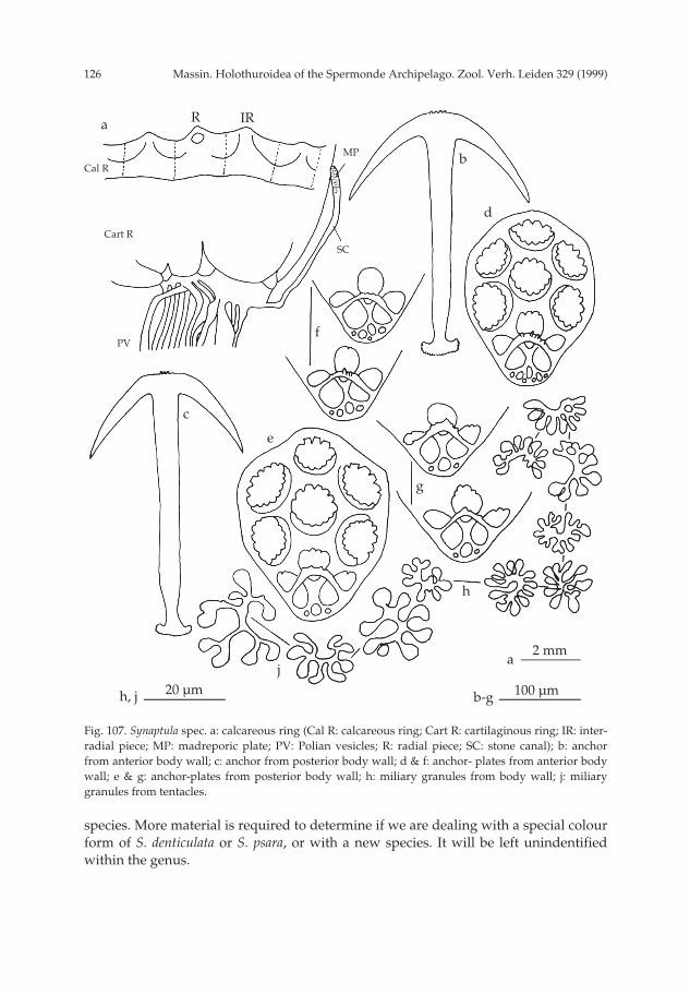
Massin. Holothuroidea of the Spermonde Archipelago. Zool. Verh. Leiden 329 (1999)126
species. More material is required to determine if we are dealing with a special colourform of S. denticulata or S. psara, or with a new species. It will be left unindentifiedwithin the genus.
Fig. 107. Synaptula spec. a: calcareous ring (Cal R: calcareous ring; Cart R: cartilaginous ring; IR: inter-radial piece; MP: madreporic plate; PV: Polian vesicles; R: radial piece; SC: stone canal); b: anchorfrom anterior body wall; c: anchor from posterior body wall; d & f: anchor- plates from anterior bodywall; e & g: anchor-plates from posterior body wall; h: miliary granules from body wall; j: miliarygranules from tentacles.
a R IR
100 µmb-g
Cal R
Cart R
SC
PV
MP b
d
f
c
g
h
j20 µm
2 mma
h, j
e
pp 003-144 02-01-2007 15:33 Pagina 126

127Massin. Holothuroidea of the Spermonde Archipelago. Zool. Verh. Leiden 329 (1999)
Family Chiridotidae Oestergren, 1898Genus Chiridota Eschscholtz, 1829Chiridota stuhlmanni Lampert, 1896
(figs 108a-g, 109)
Chiridota stuhlmanni Lampert, 1896: 67; Oestergren, 1898a: 118; Ludwig, 1899: 563; H.L. Clark, 1907:115; Heding, 1928: 302, fig. 64 (1-6); Heding, 1931: 676, fig. 12 (1-6); Dawydoff, 1952: 117; Cher-bonnier, 1967: 67, fig. 6l-p; A.M. Clark & Rowe, 1971: 188; Liao, 1975: 224, fig. 27 (1-8); Tortonese,1977: 276; Sloan et al., 1979: 125; Price, 1982: 12; A.M. Clark, 1984: 99; Price & Reid, 1985: 32; Cher-bonnier, 1988: 270, fig 122A-G; Marsh et al., 1993: 65; Rowe & Gates, 1995: 267; Liao & A.M. Clark,1995: 543, fig. 336a-e; Liao, 1997: 282, fig. 169a-h.
Material.— IRSNB IG.28251/244 (1 specimen), Badi, 3.x.94, 2 m depth.
Description.— Specimen 68 × 4 mm, pink when fully extended and brown-redwhen contracted; in alcohol uniformly beige-white. Body cylindrical with mouth andanus terminal. Tentacles 12, each with 8-9 pairs of digits. Wheel papillae scattered
Fig. 108. Chiridota stuhlmanni Lampert, 1896. a: calcareous ring (IR: interradial piece; R: radial piece);b: position of the wheel papillae along the body wall; c: body wall wheels; d: ambulacral C-shapedrods; e: inter-ambulacral C-shaped rods; f: rods from tentacle base; g: rods from tentacle digits.
a RIR
100 µmd, f
IR
d
b
c
e
f
g
5 mm
2 mm
50 µm
b
a
c, e, g
pp 003-144 02-01-2007 15:33 Pagina 127
Massin. Holothuroidea of the Spermonde Archipelago. Zool. Verh. Leiden 329 (1999)128
between the ambulacra over the whole length of the body (fig. 108b).Calcareous ring stout and thick (fig. 108a), with radial and interradial pieces of
the same width and both with an anterior tooth; radial pieces perforated for thenerve; posterior edge of the calcareous ring straight.
Ossicles of body wall wheels gathered in the wheel papillae; wheels 40-115 µmacross anteriorly and 55-125 µm posteriorly (fig. 108c). Along the ambulacra, stout C-shaped rods (fig. 108d) with rounded, spiny extremities (fig. 108d). In the interambu-lacral areas fine C-shaped rods (fig. 108e), rare anteriorly and numerous posteriorly.At the base of the tentacles stout C-shaped rods similar to those of the body wall (fig.108f); in the digits of the tentacles C-shaped rods with irregular spines at the extremi-ties (fig. 108g).
Geographic distribution (fig. 109).— Red Sea (Eilat), Zanzibar (Tumbatu = typelocality), Madagascar, Seychelles (Aldabra), Indonesia (Sumatra, Sulawesi), Australia(WA, Timor Sea, QLD, GBR), Vietnam (Gulf of Siam), China (Xisha Islands), Fiji.
Remarks.— Chiridota stuhlmanni shows no variation in its ossicles throughout itsrange of distribution (Heding, 1928; Cherbonnier, 1967, 1988; Liao, 1975). As previ-ously observed by H.L. Clark (1907) and Massin (1996a) for other Chiridota spp., thenumber of tentacle digits of C. stuhlmanni increases with increasing body length: ten-tacles of a 25 mm long specimen had 4 pairs of digits (Cherbonnier, 1967), of a 50 mmlong specimen 7 pairs (Heding, 1928) and of a 68 mm long specimen 8-9 pairs (pre-sent study). However, the number of tentacles (12) is constant irrespective of bodylength.
It is only the second record for Indonesia, the first one being from Sumatra (Price& Reid, 1985).
General discussion on distribution
Table 1 gives the list of the species for each locality sampled in the SpermondeArchipelago. According to this list some islands seem very specific regarding thecomposition of their holothurian fauna (qualitatively as well as quantitatively).Gusung, Barang Lompo and Bone Tambung have a very low diversity whereas Badiseems particularly rich. Samalona and Kudingareng Keke which have been sampledwith a comparable frequency (12 and 14 dives, respectively), yielded almost similar
Fig. 109. Distribution of Chiridota stuhlmanni Lampert, 1896.
pp 003-144 02-01-2007 15:33 Pagina 128

129Massin. Holothuroidea of the Spermonde Archipelago. Zool. Verh. Leiden 329 (1999)
numbers of species: 22 versus 26; and extrapolation would suggest these islands to beequally rich. However, the species composition found, with only six species in com-mon, differs strikingly between these two islands. Samalona is particularly rich inSynaptidae (8 species versus only 3 for Kudingareng Keke) but relatively poor inspecies of the genera Holothuria and Labidodemas (3 species versus 12 for KudingarengKeke). In comparison, the number of species of regular echinoids proved to be higheron Kudingareng Keke than on Samalona: 12 versus 8 species, respectively (de Beer,1990). Moreover, both islands show a high similarity in the regular echinoid fauna (7species in common).
No specimens were collected at Bone Tambung but remarkably, observation onthis reef seemed to indicate absence of aspidochirote holothurians. Only a few synap-tids were observed during the two dives at this locality.
Badi and Kudingareng Keke are equally rich in Holothuria spp. (10 species foreach island). However, only half of the species collected are common to both islands.These differences cannot be ascribed to the different zones (1 to 4) defined by Moll(1983), Hoeksema (1990) and de Beer (1990) working on scleractinian corals and seaurchins because both islands belong to the same zone (the third one); moreover, theyare similar in size and orientation with a similar reef flat (Moll, 1983: fig. 1B). Bothislands are also close to each other (18 km) and human fishery pressure must be thesame. With the data at hand it is not possible to ascribe the low similarity inholothurian fauna to known biotic and/or abiotic factors.
According to the number of dives performed around each island, Badi seem tohave the highest holothurian diversity. This could be linked to the coral species com-position which is more diverse on Badi (Moll, 1983). The general increase in speciesdiversity observed from zone one to three (Moll, 1983; Hoeksema, 1990; de Beer, 1990)fits for holothurians with islands as Gusung (zone one), Samalona (zone two) andBadi (zone three). However, this is not always true, and Gusung (zone one), BarangLompo (zone two) and Bone Tambung (zone three) seem to have a similar lowholothurian diversity.
The majority (70%) of the holothurian species described in the present paper (newand undetermined species excluded) have an Indo-Pacific (43%) or a more restrictedIndo-West Pacific (27%) distribution. Sixteen percent have an East Indian to WestPacific distribution and only 6% are restricted to the Pacific Ocean. The remaining 8%are “endemic species”, restricted to Indonesia and adjacent waters (northern Aus-tralia, southern Philippines and Malaysia). Among the alleged endemic species, mostare Synaptidae.
The present list of 56 species (table 1) is far from being complete because somebiotopes (mangrove areas, muddy bottoms) have not been investigated and only afew islands of the Spermonde Archipelago have been visited. Moreover, only onenight dive was carried out, although sampling in the dark usually yields species thatnever appear during day time. On the well investigated island of Samalona (12 dives)Patinapta ooplax (Marenzeller, 1881) has not been observed during the present survey,nor Protankyra pseudodigitata (Semper, 1868). Both species were recorded by Heding(1928) from Samalona, P. ooplax being common (82 specimens collected on its sandyshore).
Another factor to be taken into account in the study of holothurian diversity is
pp 003-144 02-01-2007 15:33 Pagina 129
Massin. Holothuroidea of the Spermonde Archipelago. Zool. Verh. Leiden 329 (1999)130
human pressure by trepang fishery. Through this activity some reefs off Ujung Pan-dang have nearly been stripped from the largest and commercially important speciessuch as Holothuria nobilis, H. scabra, Stichopus spp., Thelenota spp., and Bohadschia spp.
Acknowledgements
It is a pleasure to thank Drs F.W.E. Rowe, B.W. Hoeksema and J.C. den Hartog forcritically reading the manuscript and improving the English; Dr A. Smirnov for hisadvice on the taxonomy of several synaptids; sincere thanks to Dr B.W. Hoeksemaand his family for their hospitality and assistance in the field; Drs C.H.J.M. Fransenand B.W. Hoeksema for collecting specimens; Dr M. Borel Best gave advice on obtain-ing research visa. I am grateful to the Indonesian authorities in general and to Dr A.Noor (University Hasanuddin) in particular for support.
Thanks are also due to Dr C. Nielsen (Zoologisk Museum, Copenhagen) and Dr J.Bleeker (Zoölogisch Museum, Amsterdam) for the loan of material and to Dr N.Ameziane-Cominardi for welcoming me at the Laboratoire de Biologie desInvertébrés Marins et Malacologie (MNHN, Paris).
This work was financially supported by the King Léopold III Fund for NatureExploration and Conservation, the Fund for Scientific Research-Flanders (project G2908-90) and the Royal Belgian Institute of Natural Sciences. This work is part of theUNHAS Buginesia Programme.
References
Adams, T., 1992. Resource aspects of the Fiji beche-de-mer industry.— S.P.C. Beche-de-Mer Inf. Bull.4: 13-16.
Adjeroud, M., 1997. Long term changes of epibenthic macrofauna communities in a closed lagoon(Taioro Atoll, French Polynesia): 1972-1994.— Hydrobiologia 356: 11-19.
Allen, G.R. & R. Steene, 1994. Indo-Pacific Coral Reef Field Guide, i-vi + 1-378.— Singapore, TropicalReef Research.
Anon., 1979. La Beche-de-Mer dans le Pacifique tropical. Manuel à l’usage des pêcheurs. Manuel n°18:1-31.— Commission Pacifique Sud, Noumea.
Anon., 1996. Status and management of inshore fisheries in the Kingdom of Tonga: Beche-de-mer.—SPC Beche-de-mer Inf. Bull. 8: 12-19.
Baine, M. & B. Forbes, 1998. The taxonomy and exploitation of sea cucumbers in Malaysia.— S.P.C.Beche-de-mer Inf. Bull. 10: 2-7.
Bakus, G.J., 1973. The biology and ecology of tropical holothurians. In: O.A. Jones & R. Endean, eds,Biology and Geology of Coral Reefs: vol. 2 Biology 1 (10): 325-367.— Academic Press, N.Y. &London: i-xxi,1-480.
Ballard, J., 1991. The pearlfish.— Afri. Wildlife 45 (1): 16-19.Bedford, F.P., 1899. Holothurians.— A. Willey’s Zool. Results II: 141-150.Beer, de M., 1990. Distribution patterns of regular sea urchins (Echinodermata: Echinoidea) across the
Spermonde Shelf, SW Sualwesi, Indonesia. In: Ch. Deridder, Ph. Dubois, M.C. Lahaye & M. Jan-goux, eds, Echinoderm Research, Proc. 2nd European Conf. Echinoderms: 165-169.— Balkema,Rotterdam: i-xi, 1-343.
Belhadjali, K., 1997. Production de bêche-de-mer à Tuvalu.— C.P.S., Beche-de-mer Inform. Bull. 9: 2-3.Bell, F.J., 1884a. Notes on a collection of Echinodermata from Australia.— Proc. Linn. Soc. N.S. Wales
9: 496-507.Bell, F.J., 1884b. Echinodermata. In: Coppinger, R.W., ed., Report on the Zoological Collections made
pp 003-144 02-01-2007 15:33 Pagina 130
131Massin. Holothuroidea of the Spermonde Archipelago. Zool. Verh. Leiden 329 (1999)
in the Indo-Pacific Ocean during the voyage of H.M.S. “Alert”, 1881-1882: 117-171 + 10 pls.—London.
Bell, F.J., 1887a. Report on a Zoological collection made by the Officers of H.M.S. ‘Flying Fish’ atChristmas Island, Indian Ocean. VIII. Echinodermata.— Proc. Zool. Soc. London 1887: 523.
Bell, F.J., 1887b. Studies in the Holothuroidea. VI. Descriptions of New Species.— Proc. Zool. Soc.London 1887: 531-534 + 1 pl.
Bell, F.J., 1889. Additions to the echinoderm fauna of the Bay of Bengal.— Proc. Zool. Soc. London1889: 6-7.
Bonham, K. & E.E. Held, 1963. Ecological Observation on the Sea Cucumbers Holothuria atra and H.leucospilota at Rongelap Atoll, Marshall Islands.— Pacif. Sci. 17 (3): 305-314.
Brandt, J.F., 1835. Prodomus descriptionis animalium ab H. Mertensio in orbis terrarum circumnavi-gatione observatorum. 1: 1-75 + 1 pl.— Petropoli.
Britayev, A.T. & E.A. Zamishliak, 1996. Association of the commensal scale worm Gastrolepidia clavig-era (Polychaeta: Polynoidae) with holothurians near the coast of South Vietnam.— Ophelia 45 (3):175-190.
Brouns, J.J.W.M. & F.M.L. Heijs, 1985. Tropical seagrass ecosystems in Papua New Guinea. A generalaccount of the environment, marine flora and fauna.— Mar. Biol. 88 (2): 145-182.
Bruce, A.J., 1983. A further note on Pycnocaris chagoae Bruce (Decapoda, Gnathophyllidae).— Crus-taceana 45 (1): 107-108.
Cannon, L.R.G. & H. Silver, 1986. Sea Cucumbers of Northern Australia: i-viii, 1-60.— Brisbane,Queensland Museum.
Caso, M.E., 1962. Estudios sobre equinodermos de Mexico. Contribución al conocimiento de losequinodermos de las islas Revillagigedo.— An. Inst. Biol. (Univ. Méx.) 33 (1-2): 293-330.
Caso, M.E., 1965. Estudios sobre equinodermos de Mexico. Contribución al conocimiento de losholothuroideos de Zihuatanejo y de la isla de Ixtapa (primero parte).— An. Inst. Biol. (Univ.Méx.) 36: 253-291.
Chambers, M.R., 1989. Beche-de-mer. In: T. Done & K.F Navin, eds, The marine resources of Vanuatu:86-91.— Townsville, Australian Institute of Marine Science.
Chamisso, A. & C.C. Eysenhardt, 1821. De animalibus quibusquam e classe vermium Linneana, in cir-cumnavigatione terrae, auspicante comite N. Romanzoff, Duce Ottone de Kotzbue, 1815, 1818,peracta observatis.— II. Nova Acta Acad. Caesar. Leop. Carol. 10: 345-374 + 10 pls.
Chang, F.Y., 1934. Report on the holothurians collected from the coast of China.— Contr. Inst. Zool.nat. Acad. Peiping 2 (1): 1-46 + 52 + 3pls.
Chao, S.M. & K.H. Chang, 1989. The Shallow-water Holothurians (Echinodermata: Holothurioidea) ofSouthern Taiwan.— Bull. Inst. Zool., Acad. Sinica 28 (2): 107-137.
Chao, S.M. & K.H. Chang, 1990. First record of two holothurians (Echinodermata: Holothurioidea)from Taiwan.— Bull. Inst. Zool., Acad. Sinica 29 (1): 65-69.
Cheng, L. & R.A. Lewin, 1984. Prochloron on Synaptula.— Bull. Mar. Sci. 35 (1): 95-98.Cherbonnier, G. 1951., Les Holothuries de l’Institut Royal des Sciences Naturelles de Belgique.— Inst.
r. Sci. nat. Bel. Mém. 2ème sér. 41: 1-65 + 28 pls.Cherbonnier, G., 1952a. Contribution à la connaissance des Holothuries de l’Afrique du Sud.— Trans.
R. Soc. South Africa 33 (4): 469-509 + 16 pls.Cherbonnier, G., 1952b. Les holothuries de Quoy et Gaimard.— Mém. Inst. r. Sci. nat. Belg. 2ème sér.
44: 1-50 + 3 pls.Cherbonnier, G., 1955a. Les Holothuries de la mer Rouge. Résultats scientifiques des campagnes de la
Calypso.— Ann. Inst. Océanog. Monaco NS 30: 129-183 + 28 pls.Cherbonnier, G., 1955b. Holothuries récoltées en Océanie française par G. Ranson en 1952. 2ème
note.— Bull. Mus. natn. Hist. nat. (2) 27 (1): 77-82.Cherbonnier, G., 1955c. Holothuries récoltées en Océanie française par G. Ranson en 1952. 3ème
note.— Bull. Mus. natn. Hist. nat. (2) 27 (2): 135-141.Cherbonnier, G., 1967. Deuxième contribution à l’étude des Holothuries de la mer Rouge collectées
par des Israéliens.— Bull. Sea. Fish. Res. Sta. Haifa 43: 55-68.
pp 003-144 02-01-2007 15:33 Pagina 131

Massin. Holothuroidea of the Spermonde Archipelago. Zool. Verh. Leiden 329 (1999)132
Cherbonnier, G., 1970. Note sur l’holothurie Aspidochirote Labidodemas semperianum Selenka.— Bull.Mus. natn. Hist. nat. Paris 42 (3): 566-569.
Cherbonnier, G., 1979. Description d’Actinopyga flammea nov. sp., et données nouvelles sur deuxespèces connues d’Holothuries Aspidochirotes (Echinodermes).— Bull Mus. natn. Hist. nat. Paris4ème sér, 1, sect. A (1): 3-12.
Cherbonnier, G., 1980. Holothuries de Nouvelle-Calédonie.— Bull. Mus. natn. Hist. nat. Paris 4èmesér., 2, section A (3): 615-667.
Cherbonnier, G., 1988. Echinodermes: Holothurides.— Faune de Madagascar 70: 1-292.Cherbonnier, G. & J.-P. Féral, 1984a. Les Holothuries de Nouvelle-Calédonie. Deuxième contribution
(Première partie: Synallactidae et Holothuriidae).— Bull. Mus. natn. Hist. nat. Paris 4ème sér, 6,section A (3): 659- 700.
Cherbonnier, G. & J.-P. Féral, 1984b. Les Holothuries de Nouvelle-Calédonie. Deuxième contribution(Deuxième partie: Stichopodidae, Cucumariidae, Phyllophoridae et Synaptidae).— Bull. Mus.natn. Hist. nat. Paris 4ème sér., 6, section A (4): 827-851.
Clark, A.M., 1962. Starfishes and related echinoderms: 1-160.— Brit. Mus. (Nat. Hist.), London.Clark, A.M., 1982. Echinoderms of Hong Kong. In: B.S. Morton & C.K. Tseng, eds, The Marine Flora
and Fauna of Hong Kong and Southern China, Proc. 1st Int. Mar. Biol. Workshop vol. 1: 485-501.— Hong Kong University Press, Hong Kong.
Clark, A.M., 1984. Echinodermata of the Seychelles. In: D.R. Stoddart, ed., Biogeography and Ecologyof the Seychelles Islands, Monographiae biologicae 55: 83-102.— W. Junk, The Hague.
Clark, A.M. & F.W.E. Rowe, 1971. Monograph of shallow-water Indo-West Pacific echinoderms: i-vii,1- 238, pls 1-31.— London, Trust. Br. Mus. nat. Hist.
Clark, A.M. & P. Spencer-Davies, 1966. Echinoderms of the Maldive Islands.— Ann. Mag. nat. Hist.(13) 8: 597-612.
Clark, A.M. & J.B. Taylor, 1971. Echinoderms from Diego Garcia.— Atoll Res. Bull. 149: 89-92.Clark, H.L., 1907. The apodous holothurians. A monograph of the Synaptidae and Molpadiidae.
Smithson.— Contr. Knowl. 35: 1-231, pls 1-13.Clark, H.L., 1909. Echinodermata.— Austr. Mus. Sidney Mem. IV, 2 (11): 517-564. + 12 pls.Clark, H.L., 1921. The Echinoderm Fauna of the Torres Strait.— Pap. Dep. mar. Biol. Carnegie Instn
Wash. 10: i-viii, 1-223, pls 1-38.Clark, H.L., 1922. The holothurians of the genus Stichopus.— Bull. Mus. comp. Zool. Harvard 65 (3):
39-74.Clark, H.L., 1924. The holothurians of the Museum of Comparative Zoology. The Synaptinae.— Bull.
Mus. comp. Zool. 65 (13): 459-501, pls 1-12.Clark, H.L., 1932. Echinodermata (other than Asteroidea) of the Great Barrier Reel Expedition 1928-
29.— Scient. Rep. Gt Barrier Reef Exped. 4 (7): 197-239.Clark, H.L., 1938. Echinoderms from Australia.— Mem. Mus. comp. Zool. Harvard 55 (VIII): i-viii, 1-
596, pls 1-28.Clark, H.L., 1946. The Echinoderms Fauna of Australia.— Publs Carnegie Instn 566: i-iv, 1-567.Clastres A., A. Ahond, C. Poupat, P. Potier & A. Intes, 1978. Invertébrés marins du lagon de Nouvelle-
Calédonie. I. Etude structurale d’une nouvelle sapogénine extraite d’une holothurie: Bohadschiavitiensis Semper.— Experientia 34 (8): 973-974.
Clouse, R.M., 1997. End-Plate Spicules in Bohadschia marmorata (Jaeger) (Holothuroidea, Holothuri-idae).— Micronesica 30 (1): 187-192.
Colin, P.L. & Ch. Arneson, 1995. Tropical Pacific Invertebrates. A field Guide to the Marine Inverte-brates Occurring on Tropical Pacific Coral Reefs, Seagrass Beds and Mangroves: i-viii , 1- 296.—Beverly Hills, Coral Reef Press.
Conand, C., 1981. Sexual cycle of 3 commercially important holothurian species (Echinodermata)from the lagoon of New Caledonia.— Bull. Mar. Sci.31 (3): 523-543.
Conand, C., 1986. Les resources halieutiques des pays insulaires du Pacifique. Deuxième partie: Lesholothuries.— F.A.O., Doc. Tech. Pêches 272.2: 1-108.
Conand, C., 1989. Les holothuries aspidochirotes du lagon de Nouvelle-Calédonie. Biologie, écologieet exploitation: 1-393.— Thèse de Doctorat, Brest.
pp 003-144 02-01-2007 15:33 Pagina 132

133Massin. Holothuroidea of the Spermonde Archipelago. Zool. Verh. Leiden 329 (1999)
Conand, C. & A., Tuwo, 1996. Pêche et mariculture des holothuries d’importance commerciale dansles Célèbes du Sud (Indonésie).— S.P.C. Beche-de-mer Inf. Bull. 8: 17-21.
Dalzeel, P., 1990. A review of fisheries research activities in the Republic of Vanuatu.— Inshore Fish-eries Research Project. County Assignment Report, South Pacific Commission: 1-34. - Noumea,New Caledonia.
Dalzeel, P., S.R. Lindsay & H. Patiale, 1993. Fisheries resources survey of the Island of Niue.— InshoreFisheries Research Project. Technical Document n°3: i-xiv, 1-68.
Dawydoff, E., 1952. Contribution à l’étude des invertébrés de la faune marine benthique de l’Indo-chine.— Bull. Biol. France & Belgique. Suppl. 37: 1-158.
Deichmann, E., 1958. The Holothurioidea collected by the Velero III and during the years 1932 to1954, part II. Aspidochirota.— Allan Hancock Pacif. Exped. 11 (2): 239-349, pls 1-9.
Domantay, J.S., 1933. Littoral Holothurioidea of Port Galera Bay and adjacent waters.— Nat. Appl.Sci. Bull. Univ. Philipp. 3 (1): 41-101, pls 1-4.
Domantay, J.S., 1936. The ecological distribution of the echinoderm fauna of the Puerto Galera MarineBiological Station.— Nat. Appl. Sci. Bull. Univ. Philipp. 5: 385-403, pls 1-7.
Domantay, J.S., 1953. Littoral holothurians from Zamboanga and vicinity.— Philipp. J. Sci. 82 (2): 109-131.
Ebert, T.A., 1978. Growth and size of tropical sea cucumber Holothuria (Halodeima) atra Jaeger atEnewetok Atoll, Marshall Islands.— Pac. Sci. 32 (2): 183-191.
Ekman, S., 1918. Resuts of Dr. E. Mjöbergs Swedish Scientific Expedition to Australia 1910-1913. XIX.Holothurioidea.— K. swenska VetensAkad. handl. 58 (6): 1-70, pls 1-5.
Endean, R., 1956. Queensland Faunistic Records. Part IV. Further Records of Echinodermata (exclud-ing Crinoidea).— Pap. Dept. Zool. Univ. Queensland 1 (5): 121-140.
Endean, R., 1957. The biogeography of Queensland’s shallow-water Echinoderm fauna (excludingCrinoidea) with a rearrangement of the faunistic province of tropical Australia.— Aust. J. Mar.Freshw. Res. 8 (3): 233-273.
Endean, R., 1961. Queensland Faunistic Records. Part VII. Additional Records of Echinodermata(Excluding Crinoidea).— Pap. Dept. Zool. Univ. Queensland 1: 289-298.
Endean, R., 1965. Queensland Faunistic Records. Part VIII. Further Records of Echinodermata(Excluding Crinoidea) from Southern Queensland.— Pap. Dept. Zool. Univ. Queensland 2 (11):225-235.
Erwe, W., 1913. Holothurioidea. Die Fauna Südwest-Australiens.— Ergebnisse der Hamburger süd-west-australischen Forschungsreise 1905 4 (9): 349-402 , pls 5-8.
Féral, J.-P. & G. Cherbonnier, 1986. Les holothurides. In: A. Richard, P. Laboute & J.-L. Menou, eds,Guide des étoiles de mer, oursins et autres échinodermes du lagon de Nouvelle-Calédonie: 55-107.— ORSTOM, Paris: 1-238.
Fiege, D., V. Neuman & Jinhe Li, 1994. Observations on Coral Reefs of Hainan Island, South ChinaSea.— Mar. Poll. Bull. 29 (1-3): 84-89.
Fisher, W.K., 1907. The holothurians of the Hawaiian Islands.— Proc. U.S. natn. Mus. 32 (1555): 637-744, pls 64-82.
Forskål, P., 1775. Descriptiones animalium, avium, amphibiorum, piscium, insectorum, vermium: 1-164.— Havniae, Carsten Nieburh.
Fransen, C.H.J.M., 1984. Marine palaemonoid shrimps of the Netherlands Seychelles Expedition 1992-1993. In: J. Van der Land, ed., Results of the Oceanic Reefs Expedition to the Seychelles (1992-1993). Vol. 1.— Zool. Verhandl. 297: 85-152.
George, J.D. & J. George, 1987. The coral reefs of the Bodgaya Islands (Sabah: Malaysia) and PulauSipadan.— Malay. Nat. J. 40 (3-4): 225-260.
Gosliner, T.M., D.W. Behrens & G.C. Williams, 1996. Animal life from Africa to Hawai’i exclusive ofthe vertebrates. Coral Reef Animal of the Indo-Pacific: i-vi, 1-314.— Monterey, Sea Challengers.
Grosenbaugh, D.A., 1981. Qualitative assessment of the asteroids, echinoids and holothuroids in YapLagoon.— Atoll Res. Bull. 255: 49-54.
Hammond, L.S. & C.R. Wilkinson, 1985. Exploitation of sponge exudates by coral reef holothuroids.—J. exp. mar. Biol. Ecol. 94 (1): 1-9.
pp 003-144 02-01-2007 15:33 Pagina 133
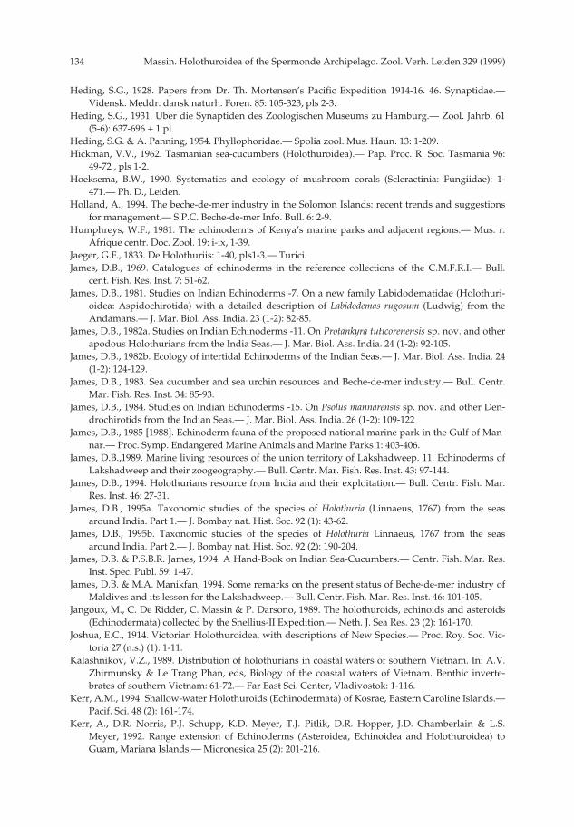
Massin. Holothuroidea of the Spermonde Archipelago. Zool. Verh. Leiden 329 (1999)134
Heding, S.G., 1928. Papers from Dr. Th. Mortensen’s Pacific Expedition 1914-16. 46. Synaptidae.—Vidensk. Meddr. dansk naturh. Foren. 85: 105-323, pls 2-3.
Heding, S.G., 1931. Uber die Synaptiden des Zoologischen Museums zu Hamburg.— Zool. Jahrb. 61(5-6): 637-696 + 1 pl.
Heding, S.G. & A. Panning, 1954. Phyllophoridae.— Spolia zool. Mus. Haun. 13: 1-209.Hickman, V.V., 1962. Tasmanian sea-cucumbers (Holothuroidea).— Pap. Proc. R. Soc. Tasmania 96:
49-72 , pls 1-2.Hoeksema, B.W., 1990. Systematics and ecology of mushroom corals (Scleractinia: Fungiidae): 1-
471.— Ph. D., Leiden.Holland, A., 1994. The beche-de-mer industry in the Solomon Islands: recent trends and suggestions
for management.— S.P.C. Beche-de-mer Info. Bull. 6: 2-9.Humphreys, W.F., 1981. The echinoderms of Kenya’s marine parks and adjacent regions.— Mus. r.
Afrique centr. Doc. Zool. 19: i-ix, 1-39.Jaeger, G.F., 1833. De Holothuriis: 1-40, pls1-3.— Turici.James, D.B., 1969. Catalogues of echinoderms in the reference collections of the C.M.F.R.I.— Bull.
cent. Fish. Res. Inst. 7: 51-62.James, D.B., 1981. Studies on Indian Echinoderms -7. On a new family Labidodematidae (Holothuri-
oidea: Aspidochirotida) with a detailed description of Labidodemas rugosum (Ludwig) from theAndamans.— J. Mar. Biol. Ass. India. 23 (1-2): 82-85.
James, D.B., 1982a. Studies on Indian Echinoderms -11. On Protankyra tuticorenensis sp. nov. and otherapodous Holothurians from the India Seas.— J. Mar. Biol. Ass. India. 24 (1-2): 92-105.
James, D.B., 1982b. Ecology of intertidal Echinoderms of the Indian Seas.— J. Mar. Biol. Ass. India. 24(1-2): 124-129.
James, D.B., 1983. Sea cucumber and sea urchin resources and Beche-de-mer industry.— Bull. Centr.Mar. Fish. Res. Inst. 34: 85-93.
James, D.B., 1984. Studies on Indian Echinoderms -15. On Psolus mannarensis sp. nov. and other Den-drochirotids from the Indian Seas.— J. Mar. Biol. Ass. India. 26 (1-2): 109-122
James, D.B., 1985 [1988]. Echinoderm fauna of the proposed national marine park in the Gulf of Man-nar.— Proc. Symp. Endangered Marine Animals and Marine Parks 1: 403-406.
James, D.B.,1989. Marine living resources of the union territory of Lakshadweep. 11. Echinoderms ofLakshadweep and their zoogeography.— Bull. Centr. Mar. Fish. Res. Inst. 43: 97-144.
James, D.B., 1994. Holothurians resource from India and their exploitation.— Bull. Centr. Fish. Mar.Res. Inst. 46: 27-31.
James, D.B., 1995a. Taxonomic studies of the species of Holothuria (Linnaeus, 1767) from the seasaround India. Part 1.— J. Bombay nat. Hist. Soc. 92 (1): 43-62.
James, D.B., 1995b. Taxonomic studies of the species of Holothuria Linnaeus, 1767 from the seasaround India. Part 2.— J. Bombay nat. Hist. Soc. 92 (2): 190-204.
James, D.B. & P.S.B.R. James, 1994. A Hand-Book on Indian Sea-Cucumbers.— Centr. Fish. Mar. Res.Inst. Spec. Publ. 59: 1-47.
James, D.B. & M.A. Manikfan, 1994. Some remarks on the present status of Beche-de-mer industry ofMaldives and its lesson for the Lakshadweep.— Bull. Centr. Fish. Mar. Res. Inst. 46: 101-105.
Jangoux, M., C. De Ridder, C. Massin & P. Darsono, 1989. The holothuroids, echinoids and asteroids(Echinodermata) collected by the Snellius-II Expedition.— Neth. J. Sea Res. 23 (2): 161-170.
Joshua, E.C., 1914. Victorian Holothuroidea, with descriptions of New Species.— Proc. Roy. Soc. Vic-toria 27 (n.s.) (1): 1-11.
Kalashnikov, V.Z., 1989. Distribution of holothurians in coastal waters of southern Vietnam. In: A.V.Zhirmunsky & Le Trang Phan, eds, Biology of the coastal waters of Vietnam. Benthic inverte-brates of southern Vietnam: 61-72.— Far East Sci. Center, Vladivostok: 1-116.
Kerr, A.M., 1994. Shallow-water Holothuroids (Echinodermata) of Kosrae, Eastern Caroline Islands.—Pacif. Sci. 48 (2): 161-174.
Kerr, A., D.R. Norris, P.J. Schupp, K.D. Meyer, T.J. Pitlik, D.R. Hopper, J.D. Chamberlain & L.S.Meyer, 1992. Range extension of Echinoderms (Asteroidea, Echinoidea and Holothuroidea) toGuam, Mariana Islands.— Micronesica 25 (2): 201-216.
pp 003-144 02-01-2007 15:33 Pagina 134

135Massin. Holothuroidea of the Spermonde Archipelago. Zool. Verh. Leiden 329 (1999)
Koehler, R., 1895a. Echinodermes de la baie d’Amboine.— Revue suisse Zool. 3 (2): 275-293.Koehler, R., 1895b. Catalogue raisonné des Echinodermes recueillis par Mr Korotnev aux îles de la
Sonde.— Mem. Soc. Zool. France 8: 374-423.Koehler, R. & C. Vaney, 1908. An account of the littoral Holothurioidea collected by the royal Indian
marine survey ship Investigator.— Trust. Indian Mus. Calcutta: 1-54, pls. 1-3.Kropp, R.K., 1982. Responses of Five Holothurian Species to Attacks by a Predatory Gastropod, Tona
perdrix.— Pac. Sci. 36 (4): 445-452.Lamberson, J.O., 1978. Notes on the Morphology, Ecology and Distribution of Thelenota anax H.L.
Clark (Holothuroidea: Stichopodidae).— Micronesica 14 (1): 115-122.Lampert, K., 1885. Die Seewalzen. Eine systematische Monographie. In: Semper, C., ed., Reisen im
Archipel der Philippinen, Teil 2, Wiss. Res. 4: 1-310, pl. 1.— Wiesbaden.Lampert, K., 1889a. Verzeichnis der wärhend des Reise S.M.S. “Gazelle” gesammelten Holothurien.
In: Th. Studer, ed., Die Forschungsreise S.M.S. “Gazelle” in dem Jahren 1875 bis 1876, 3 Theil,Zool. & Geol.: 301-309 .— E.S. Mittler & Sohn, Berlin: i-vi, 1-322, pls 1-33.
Lampert, K., 1889b. Die während der Expedition S.M.S. “Gazelle” 1874-1876 von Prof Th. Studergesammelten Holothurien.— Zool. Jahrb. 4: 806-858.
Lampert, K., 1896. Die von Dr. Stuhlmann in den Jahren 1888 und 1889 an der Ostküste Afrikasgesammelten Holothurien.— Mitt. Naturhist. Mus. Hamburg 13: 49-71.
Lane, D.J.W., 1992. Biogeographical note on the northward extension of the known latitudinal rangefor the tropical stichopodid sea-cucumber Thelenota anax H.L. Clark (Echinodermata:Holothuroidea).— Raffles Bull. Zool. 40 (2): 175-178.
Lawrence, J.M., 1980. Numbers and biomass of the common holothuroids on the windward reef flat atEnewetak atoll, Marshall Islands. In: M. Jangoux, ed., Echinoderms: Present and Past, Proc. Eur.Coll. Echinoderms, Brussels 3-8 September 1979: 202-204.— Balkema, Rotterdam: i-xviii, 1-428 .
Lesson, R.P., 1830. Centurie zoologique ou choix d’animaux rares, nouveaux ou imparfaitement con-nus: 1-244, pls 1-80.— Paris.
Levin, V.S., 1979. Aspidochirote holothurians of the upper sublittoral zone of Indo-West Pacific:species composition and distribution.— Biol. Morya 5: 17-23.
Levin, V.S. & Dao Tan Ho, 1989. Holothurians of the upper sublittoral zone of the coastal waters ofPhukhan Province (southern Vietnam). In: A.V. Zhirmunsky & Le Trang Phan, eds, Biology ofthe coastal waters of Vietnam. Benthic invertebrates of southern Vietnam: 54-60.— Far East Sci.Center, Vladivostok: 1-116.
Liao, Y., 1975. The echinoderms of Xisha Islands. I. Holothurioidea, Guandong Province, China.—Studia mar. Sinica 10: 199-230.
Liao, Y., 1980. The Aspidochirote holothurians of China with erection of a new genus. In: M. Jangoux,ed., Echinoderms: Present and Past, Proc. Europ. Coll. Echinoderms, Brussels 3-8 September 1979:115-120.— Balkema, Rotterdam: i-xviii, 1-428.
Liao, Y., 1984. The Aspidochirote Holothurians of China.— Studia Marina Sinica. 23: 221-247.Liao, Y., 1997. Fauna Sinica. Phylum Echinodermata: class Holothuroidea: i-ix, 1-334, pls 1-2.— Sci-
ence Press, Beijing.Liao, Y. & A.M. Clark, 1995. The echinoderms of southern China: i-iii, 1-1.614, pls 1-23.— Science
Press, Beijing, New York.Loi, T.N. & N.V. Sach, 1963. Les holothuries de la baie de Nhatrang (1ère note).— Ann. Fac. Sci.
Saigon 2: 237-248.Ludwig, H., 1875. Beiträge zur Kenntnis der Holothurien.— Arb. d. Zoolzootom Inst. Würzburg 2 (2):
77-118 , pls 6-7.Ludwig, H., 1882. List of the holothurians in the collection of the Leyden Museum.— Notes Leyden
Mus. IV (10): 127-137.Ludwig, H., 1883. Verzeichniss der Holothurien des Kieler Museums.— Ber. Oberhess. Ges. Nat. u.
Heilk. 22: 155-176.Ludwig, H., 1888. Die von Dr. J. Brock in Indischen Archipel gesammelten Holothurien.— Zool. Jb.
Abt. Syst. 3 (6): 805-820, pl. 30.Ludwig, H., 1889-92. Echinodermen: Die Zeewalzen. In: H.G. Bronn, ed., Broon’s Klassen und Ord-
pp 003-144 02-01-2007 15:33 Pagina 135
Massin. Holothuroidea of the Spermonde Archipelago. Zool. Verh. Leiden 329 (1999)136
nungen des Thier-Reichs, Bd 2, Abtheilung 3, Buch 1: i-vi, 1-460, pls 1-17.— C.F. Winter’sche,Leipzig.
Ludwig, H., 1899. Echinodermen des Sansibargebietes.— Abh. Senckerberg Naturfor. Gesel. 21 (1):537-563.
Mc.Elroy, S., 1990. Beche-de-mer species of commercial value- an update.— S.P.C., Beche-de-mer Info.Bull. 2: 2-7.
Mc. Knight, D., 1974. Echinoderms from Manihiki lagoon.— N.Z. Oceanog. Inst. Mem. n°31: 45-47.Machida, Y., 1989. New distribution Records of the Pearlfish, Carapus mourlani, with Notes on Its Mor-
phometry.— Jap. J. Ichthyol. 36 (3): 363-368.Maluf, L.Y., 1988. Composition and distribution of the central eastern Pacific echinoderms.— Nat.
Hist. Mus. Los Angeles County, Tech. Rep. 2: 1-242.Maluf, L.Y., 1991. Echinoderm Fauna of the Galápagos Islands. In: M.J. James, ed., Galápagos Marine
Invertebrates. Taxonomy, Biogeography, and Evolution in Darwin’s Islands: 245-267.— PlenumPress, NY & London.
Marsh, L.M., 1986. Echinoderms.— Rec. West. Aust. Mus. Suppl. 25: 63-74.Marsh, L.M., 1994a. Echinoderms of the Cocos (Keeling) Islands.— Atoll Res. Bull. 411: 1-12.Marsh, L.M., 1994b. Echinoderms of the Houtman Abrolhos Islands, Western Australia and their rela-
tionship to the Leeuwin Current. In: B. David, J.P. Féral & M. Roux, eds, Echinoderms throughTime, Proceedings of the 8th Int. Echinoderm Conf.: 55-61.— Balkema, Rotterdam: i-xxiii, 1-940.
Marsh, L.M., L.L. Vail, A.K. Hoggett & F.W.E. Rowe, 1993. Echinoderms of Ashmore Reef and CartierIsland. In: P.F. Berry, ed., Marine faunal surveys of Ashmore Reef and Cartier Island, North-west-ern Australia.— Rec. W. Aust. Mus. Suppl., 44: 53-65.
Mary Bai, M., 1980. Monograph on Holothuria (Metriatyla) scabra Jaeger.— Mem. zool. Survey India 16(2): 1-75, pl. 1.
Massin, C., 1994. Ossicle variation in Antarctic dendrochirote holothurians (Echinodermata).— Bull.Inst. r. Sci. nat. Belg. Biol. 64: 129-146.
Massin, C., 1996a. The holothurians of Easter Island.— Bull. Inst. r. Sci. nat. Belg. Biol. 66: 152-178.Massin, C., 1996b. Result of the Rumphius Biohistorical Expedition to Ambon (1990). Part. 4. The
Holothurioidea (Echinodermata) collected at Ambon during the Rumphius Biohistorical Expedi-tion.— Zool. Verhandl. 307: 1-53.
Massin, C. & Ch. Doumen, 1986. Distribution and feeding of epibenthic holothuroids on the reef flatof Laing Island (Papua New Guinea).— Mar. Ecol. Prog. Ser. 31: 185-195.
Massin, C. & D. Lane. 1991, Description of a New Species of Sea Cucumber (Stichopodidae,Holothuroidea, Echinodermata) from the Eastern Indo-Malayan Archipelago: Thelenota rubralin-eata n. sp.— Micronesica 24 (1): 57-64.
Meyer-Rochow, W.B., 1977. Comparison between 15 Carapus mourlani in a single holothurian and 19C. mourlani from starfish.— Copeia 3: 582-584.
Mitsukuri, K., 1896. A List of Holothurians known to occur in Japan.— Zool. Mag. 8: 405-413.Mitsukuri, K., 1912. Studies on Actinopodous Holothurioidea.— J. Coll. Sci. Imp. Univ. Tokyo 39 (2):
1-284, pls 1-8.Moll, H., 1983. Zonation and diversity of Scleractinia on reefs off S.W. Sulawesi, Indonesia: 1-170.—
Ph. D., University of Leiden, Leiden.Mortensen, Th., 1934. Echinoderms of Hong Kong.— The Hong Kong Nat. Suppl. 3: 3-14, pls 1-8.Mukhopadhyay, S.K., 1988. On some Holothurians from the Gulf of Mannar, India.— Rec. zool. Surv.
India. 85 (1): 1-17.Mukhopadhyay, S.K. & T.K., Samanta, 1983. On a collection of shallow-water Holothurians from the
Lakshadweep.— Rec. zool. Surv. India 81 (1-2): 299-314.Oestergren, H., 1898a. Das System der Synaptiden.— Öfvers. K. VetenskAkad. Förh. 55: 111-120.Oestergren, H., 1898b. Zur Anatomie der Dendrochiroten, nebst Beschreibungen neuer Arten.— Zool.
Anz. 21: 102-110 & 133-136.Ong Che, R., 1992. Niche partitioning by intertidal Hong Kong holothurians. In: B. Morton, ed., The
marine flora and fauna of Hong Kong and southern China. 3., vol. 1: introduction, taxonomy andecology: 439-445.— Hong Kong Univ. Press, Hong Kong.: i-xxx , 1-526.
pp 003-144 02-01-2007 15:33 Pagina 136

137Massin. Holothuroidea of the Spermonde Archipelago. Zool. Verh. Leiden 329 (1999)
Panning, A., 1928. Echinoderma III: Holothurioidea I (Fam. Holothuriidae). In: W. Michaelsen, ed.,Beiträge zur Kenntnis der Meeresfauna Westafrikas: 205-249.— L. Friederichsen & Co, Hamburg.
Panning, A., 1929 [1931]. Die Gattung Holothuria. (1. Teil).— Mitt. Zool. StaatInst. Zool. Mus. Ham-burg 44: 91-138.
Panning, A., 1935a. Die Gattung Holothuria. (2. Teil).— Mitt. Zool. Staatsinst. Zool. Mus. Hamburg 45:24-50.
Panning, A. 1935b. Die Gattung Holothuria. (3. Teil).— Mitt. Zool. Staatsinst. Zool. Mus. Hamburg 45:65-84.
Panning, A., 1935c. Die Gattung Holothuria. (4. Teil).— Mitt. Zool. Staatsinst. Zool. Mus. Hamburg 45:85-107.
Panning, A., 1935d. Die Gattung Holothuria. 5. Teil, Schluss.— Mitt. Zool. Staatsinst. Zool. Mus. Ham-burg 46: 1-18.
Panning, A., 1941. Über einige ostafrikanische Seewalzen und ihre Eignung zur Trepanggewin-nung.— Thalassia 4 (8): 1-18.
Panning, A., 1944. Die Trepangfischerei.— Mitt. hamb. Zool. Mus. Inst. 49: 2-76.Panning, A., 1949. Versuch einer Neuordnung der Familie Cucumariidae. (Holothurioidea, Den-
drochirota).— Zool. Jb. Abt. Syst. Okol. Geog. Tiere 78 (4): 404-470.Panning, A., 1971. Bemerkungen über die Holothurien-Familie Cucumariidae (Ordnung Dendrochi-
rota. 6. Teil (Schluss).— Mitt. hamb. Zool. Mus. Inst. 67: 29-51.Pawson, D., 1967. Redescription of Cucumaria semperi Bell, an Indo-West-Pacific holothurian echino-
derm.— Proc. biol. Soc. Wash. 80: 157-162.Pearson, J., 1903. Report on the Holothurioidea collected by Prof. Herdman, at Ceylon, in 1902.— Rep.
Ceylon Pearl Oyster Fish. Suppl. Rep. 5: 181-208, pls 1-3.Pearson, J., 1910a. On marine fauna from Querimba Archipelago. 2. Littoral Marine Fauna: Kerimba
Archipelago, Portuguese East Africa: Holothurioidea.— Proc. zool. Soc. London 1910: 167-182.Pearson, J., 1910b. On marine fauna from Mergui Archipelago. 3. Marine Fauna: Mergui Archipelago,
Lower Burma. Holothurioidea.— Proc. zool. Soc. London 1910: 183-194.Pearson, J., 1913 [1915]. Notes on the Holothurioidea of the Indian Ocean.— Spolia Zeylanica 9 (34):
49-101, pls 5-14.Pearson, J., 1913. Notes on the Holothurioidea of the Indian Ocean. II. The sub-genera Argiodia and
Actinopyga.— Spolia Zeylanica 9 (35): 173-190, pls 27-29.Price, A.R.G., 1982. Comparison between Echinoderm faunas of Arabian Gulf, SE Arabia, Red Sea and
Gulfs of Aqaba and Suez.— Fauna of Saudi Arabia 4: 3-21.Price, A.R.G., 1983. Echinoderms of Saudi Arabia. Echinoderms of the Arabian Gulf Coast of Saudi
Arabia.— Fauna of Saudi Arabia 5: 28-108.Price, A.R.G. & C.E. Reid, 1985. Indian Ocean echinoderms collected during the Sindbad Voyage
(1980-81): 1. Holothurioidea.— Bull. Br. Mus. nat. Hist. (Zool.) 48 (1): 1-9.Quoy, J.R.C. & J.P. Gaimard, 1833. Zoologie: Zoophytes. In: Voyage de la corvette de l’”Astrolabe”,
exécuté par ordre du roi pendant les années 1826-1829 sous le commandement de M. J. Dumontd’Urville: 1-390 , pls 1-26 .— Paris.
Reyes-Leonardo, L.D., 1984a. A taxonomic report of shallow-water Holothurians of Calatagan, Batan-gas.— Philipp J. Sci. 113 (3-4): 137-172.
Reyes-Leonardo, L.D., 1984b. Note on three species of dendrochirote holothurians including a newrecord.— Nat. Appl. Sci. Bull. (Quezon City) 36 (1): 7-12.
Reyes-Leonardo, L.D., R.B. Monzon & V.C. Navarro, 1985. A taxonomic account of shallow waterholothurians of Bolinao, Pangasinan.— Nat. Appl. Sci. Bull. (Quezon City) 37 (4): 261-284.
Rho, B.J. & S. Shin, 1986. A systematic study on the Holothuroidea in Cheju-do.— Korean J. Zool. 29(4): 245-256.
Richard, G., 1985. Faune et Flore. Premier abrégé des organismes marins de Polynésie française. In: B.Delasalle, R. Galzini & B. Salvat, eds, Les récifs coralliens de Polynésie française, 5ème CongrèsInt. Récifs Coralliens, vol. 1: 456-458.— Antenne Museum-EPHE, Moorea: 1-554.
Romimohtarto, K., 1975. Some problems in the development of the benthic resources of Pari Islands(Seribu Islands, Jakarta Bay).— 13th Pacif. Sci. Congress. Rec. Proc. 1: 72-73.
pp 003-144 02-01-2007 15:33 Pagina 137

Massin. Holothuroidea of the Spermonde Archipelago. Zool. Verh. Leiden 329 (1999)138
Rowe, F.W.E., 1969. A review of the family Holothuriidae (Holothurioidea: Aspidochirotida).— Bull.Br. Mus. nat. Hist. (Zool.) 18 (4): 119-170.
Rowe, F.W.E., 1983. A collection of holothurians in the Leiden Museum from the East Indies and NewGuinea, with the description of a new species of Protankyra (Holothurioidea: Synaptidae) fromJava.— Zool. Meded. Leiden 57 (17): 149-154.
Rowe, F.W.E. & J.E. Doty, 1977. The Shallow-Water Holothurians of Guam.— Micronesica 13 (2): 217-250.
Rowe, F.W.E. & J. Gates, 1995. Echinodermata. In: A. Wells, ed., Zoological Catalogue of Australiavol. 33: i-xiii, 1-510.— CSIRO Australia, Melbourne.
Rowe F.W.E. & M.D. Richmond, 1997. Echinodermata. In: M.D. Richmond, ed., A guide to theseashores of eastern Africa and the Western Indian Ocean islands: 290-321.— The SEA Trust,Zanzibar: 1-448.
Sant, G., 1995. Marine invertebrates of the South Pacific. An examination of the trade: 1-81.— TRAF-FIC International, Cambridge (U.K.).
Satyamarti, S.T., 1976. The Echinodermata in the collection of the Madras Museum.— Bull. MadrasGovt. Mus. (Nat. Hist.) 7 (3): 1-279.
Selenka, E., 1867. Beiträge zur Anatomie und Systematik der Holothurien.— Z. wiss. Zool. 17 (2): 291-374, pls 17-20.
Selenka, E., 1868. Nachtrage zu den Beiträgen zur Anatomie und Systematik der Holothurien.— Z.wiss. Zool. 18: 109-119, pl. 8.
Semper, C., 1868. Reisen im Archipel der Philippinen. Holothurien.2. Wissenschaftliche Resultate: i-x ,1-288 , pls 1-40.— Leipzig.
Serene, R., 1937. Inventaire des invertébrés marins de l’Indochine (1re liste).— Notes Inst. Océanog.Indochine 30: 1-83.
Shelley, C., 1985. Growth of Actinopyga echinites and Holothuria scabra (Holothuroidea: Echinodermata)and their fisheries potential (as bêche-de-mer) in Papua New Guinea. In: M. Harmelin-Vivien &B. Salvat, eds, Miscellaneous papers (A), , Proc. 5th. Int. Coral Reef Cong. Tahiti, vol 5: 297-302.—Antenne Muséum-EPHE, Moorea: 1-616.
Sloan N.A., A.M. Clark & J.D. Taylor, 1979. The echinoderms of Aldabra and their habitats.— Bull. Br.Mus. nat. Hist. (Zool.) 37 (2): 81-128.
Sluiter, C. Ph., 1887. Die Evertebraten aus der Sammlung des königlichen naturwissenschaftlichenVereins in Niederländisch Indien in Batavia.— Natuurk. Tijdschr. Ned.-Indië 47, 8ste ser. (8): 181-220, pls 1-2.
Sluiter, C. Ph., 1894. Holothurien.— Zool. Forsch. Aust. malay Archipel. 5 (1): 101-106.Sluiter, C. Ph., 1895. Die Holothuren Sammlung des Museums zu Amsterdam.— Bijdr. Dierk. Ams-
terdam 17: 75-82.Sluiter, C. Ph., 1901. Die Holothurien der Siboga Expedition.— Siboga Exped. 44: 1-142 , pls 1-10.Solis-Marin, F.A., H. Reyes-Bonilla, M.D. Herrero-Pérezrul, O. Arizpe-Covarrubias & A. Laguarda-
Figueras, 1997. Systematics and distribution of the echinoderms from Bahia de la Paz.— CienciasMarinas 23 (2): 249-263.
Sutaman, 1993. Petunjuk praktis budidaya Teripang: 1-68 .— Penerbit Kanisius, Yogyakarta.Tahera, Q., 1996. A checklist of echinoderm fauna of Pakistan.— Scient. Khyber 9 (2): 103-106.Tahera, Q., 1997. Note on Synaptula recta Semper, 1868 (Echinodermata, Holothuroidea, Synaptidae)
new to Pakistan waters.— Pakistan J. Zool. 29 (1): 92-94.Tahera, Q. & Q.B. Kazumi, 1995. First record of Holothuria (Platyperona) difficilis Semper, 1868 (Echino-
dermata: Holothuroidea) from the northern Arabian Sea (Pakistan).— Pakistan J. mar. Sci. 4 (1):71-73.
Tan Tiu, A.S., 1981. The Intertidal Holothurian Fauna (Echinodermata: Holothuroidea) of Mactan andthe Neighboring Islands, Central Philippines.— Philipp. Scient. 18: 45-119.
Thandar, A.S., 1987. The southern African stichopodid holothurians, with notes on the changes inspicule composition with age in the endemic Neostichopus grammatus (H.L. Clark).— S. Afr. J.Zool. 22 (4): 278-286.
Théel, Hj., 1886. Holothurioidea. Part 2.— Rep. scient. Results Voy. Challenger (Zool.) 39: 1-290, pls 1-16.
pp 003-144 02-01-2007 15:33 Pagina 138

139Massin. Holothuroidea of the Spermonde Archipelago. Zool. Verh. Leiden 329 (1999)
Thurston, E., 1890. Notes on the pearl and chank fisheries and marine fauna of the Gulf of Mannar.-Bull. Madras Govt. Mus. 3: 113-114.
Thurston, E. ,1894. Rameswaram Island and fauna of the Gulf of Mannar.— Madras Govt. Mus. Sci.ser 1: 78-138.
Tortonese, E., 1953. Spedizione subacquea italiana nel Mar Rosso. Richerche Zoologiche. II. Echino-derme.— Rivista Biol. Colon. 13: 25-48 + 1 pl.
Tortonese, E., 1977. Report on echinoderms from the Gulf of Aqaba (Red Sea).— Monit. Zool. Ital.N.S. Suppl. 9 (12): 273-290.
Tortonese, E., 1979. Echinoderms collected along the eastern shore of the Red Sea (Saudi Arabia).—Atti. Soc. ital. Sci. nat. Museo civ. Stor. nat. Milano 120 (3 & 4): 314-319.
Tortonese, E., 1980. Researches on the coast of Somalia. Littoral Echinodermata.— Monit. Zool. Ital.N.S. Suppl. 13 (5): 99-139.
Townsley, S.J. & M.P. Townsley, 1972. A preliminary Investigation of the Biology and Ecology of theHolothurians at Fanning Island. In: Fanning Island Expedition, July and August, 1972.— HawaiiInstitute of Geophysics Rept., HIG-73-13: 173-186.
Tsuda, R.T., 1997. Survey of commercially-valued sea-cucumbers in the Saipan Lagoon, CNMI.—S.P.C . Beche-de-mer Inf. Bull. 9: 15-17.
VandenSpiegel, D., A., Ovaere & C., Massin, 1992. On the association between the crab Hapalonotusreticulatus (Crustacea, Brachyura, Eumedonidae) and the sea cucumber Holoturia (Metriatyla)scabra (Echinodermata, Holothuridae).— Bull. Inst. r. Sci. nat. Belg. Biol. 62: 167-177.
Vaney, C., 1912. Holothurien der Aru-Insel, nach den Sammlungen von Dr. H. Merton.— Abhand.Senckenb. Naturf. Gesel. Frankfurt 34 (3): 189-191.
Zoutendijk, D.A., 1989. Bêche de Mer. Rori of the Cook Islands.— Ministery of Marines Resources,Rarotonga, Cook Islands, Resource Profil n°6: 1-17.
Received: 19.ii.1999Accepted: 28.vii.1999 (after revision)Edited: J.C. den Hartog
pp 003-144 02-01-2007 15:33 Pagina 139
Massin. Holothuroidea of the Spermonde Archipelago. Zool. Verh. Leiden 329 (1999)140
Fig. 110. a: Actinopyga lecanora (Jaeger, 1833) (IRSNB, IG 28252/126).b: Actinopyga miliaris (Quoy & Gaimard, 1833) (IRSNB, IG 28252/31).c: Bohadschia argus Jaeger, 1833.d: Holothuria (Halodeima) edulis Lesson, 1830.e: Holothuria (Lessonothuria) hawaiiensis Fisher, 1907 (RMNH Ech. Ech 6080).f: Holothuria (Metriatyla) scabra Jaeger, 1833. Photo B.W. Hoeksema.g: Holothuria (Microthele) nobilis (Selenka, 1867) (IRSNB, IG 28252/185).h: Holothuria (Microthele) nobilis (Selenka, 1867) (IRSNB, IG 28252/211).j: Holothuria (Semperothuria) flavomaculata Semper, 1868 (IRSNB, IG 28251/251).
3 cm
a
f
e g h j
d
b
c
3 cm
10 cm
5 cm 10 cm
1 cm 3 cm 10 cm 2 cm
pp 003-144 02-01-2007 15:33 Pagina 140

141Massin. Holothuroidea of the Spermonde Archipelago. Zool. Verh. Leiden 329 (1999)
Fig. 111. a: Holothuria (Semperothuria) imitans Ludwig, 1875 (IRSNB, IG 28251/237).b: Holothuria (Stauropora) discrepans Semper, 1868 (RMNH Ech. 6083).c: Holothuria (Stauropora) fuscocinerea Jaeger, 1833 (IRSNB, IG 28251/254).d: Holothuria (Thymiosycia) hilla Lesson, 1830 (RMNH Ech. 6061).e: Holothuria (Thymiosycia) impatiens (Forskål, 1775) (IRSNB, IG 28251/132.f: Labidodemas semperianum Selanka, 1867 (RMNH Ech. 6087).g: Pearsonothuria graeffei (Semper, 1868). Adult; photo B.W. Hoeksema.h: Pearsonothuria graeffei (Semper, 1868). Sub-adult; photo B.W. Hoeksema.
a b c d
e
g
f
h
1 cm 3 cm
2 cm
5 cm
2 cm
3 cm
6 cm 1 cm
pp 003-144 02-01-2007 15:33 Pagina 141

Massin. Holothuroidea of the Spermonde Archipelago. Zool. Verh. Leiden 329 (1999)142
Fig. 112. a: Stichopus noctivagus Cherbonnier, 1980 (IRSNB, IG 28251/108).b: Stichopus quadrifasciatus spec. nov. (RMNH Ech. 6088, paratype).c: Stichopus quadrifasciatus spec. nov. (IRSNB, IG 28251/131, paratype).d: Stichopus vastus Sluiter, 1887 (RMNH Ech. 6089).e: Stichopus vastus Sluiter, 1887. Photo B.W. Hoeksema.f: Thelenota ananas (Jaeger, 1833). g: Colochirus robustus Oestergren, 1898. Photo B.W. Hoeksema.h: Plesiocolochirus australis (Ludwig, 1875) (IRSNB, IG 28251/141).
a b c
d e
f g h
2 cm 1 cm 2 cm
5 cm 10 cm
10 cm 3 cm
1 cm
pp 003-144 02-01-2007 15:33 Pagina 142
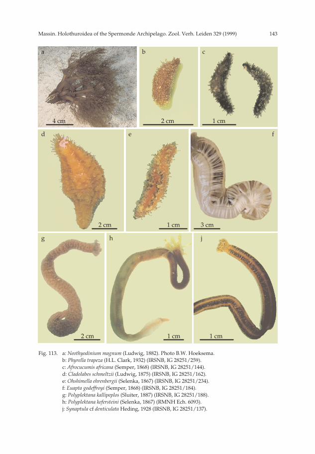
143Massin. Holothuroidea of the Spermonde Archipelago. Zool. Verh. Leiden 329 (1999)
Fig. 113. a: Neothyodinium magnum (Ludwig, 1882). Photo B.W. Hoeksema.b: Phyrella trapeza (H.L. Clark, 1932) (IRSNB, IG 28251/259).c: Afrocucumis africana (Semper, 1868) (IRSNB, IG 28251/144).d: Cladolabes schmeltzii (Ludwig, 1875) (IRSNB, IG 28251/162).e: Ohshimella ehrenbergii (Selenka, 1867) (IRSNB, IG 28251/234).f: Euapta godeffroyi (Semper, 1868) (IRSNB, IG 28251/184).g: Polyplektana kallipeplos (Sluiter, 1887) (IRSNB, IG 28251/188).h: Polyplektana kefersteini (Selenka, 1867) (RMNH Ech. 6093).j: Synaptula cf denticulata Heding, 1928 (IRSNB, IG 28251/137).
a b c
d e f
g h j
4 cm 2 cm 1 cm
2 cm 1 cm 3 cm
2 cm 1 cm 1 cm
pp 003-144 02-01-2007 15:33 Pagina 143

Massin. Holothuroidea of the Spermonde Archipelago. Zool. Verh. Leiden 329 (1999)144
Fig. 114. a: Synaptula cf. lamperti Heding, 1928 (IRSNB, IG 28251/125).b: Synaptula recta (Semper, 1868) (IRSNB, IG 28251/136).c: Synaptula recta (Semper, 1868) (IRSNB, IG 28251/138).d: Synaptula cf. reticulata (Semper, 1868) on Xetospongia spec.e: Synaptula cf. reticulata (Semper, 1868) on Amphimedon spec.f: Synaptula cf. reticulata (Semper, 1868) (IRSNB, IG 28251/48).g: Synaptula spec. (IRSNB, IG 28251/130).
a b c
d e
f g
2 cm 3 cm 2 cm
10 cm 10 cm
1 cm 2 cm
pp 003-144 02-01-2007 15:33 Pagina 144