
Recurrent swellings of the parotid gland · Gut, 1961, 2, 210 Recurrent swellings of the parotid...
8
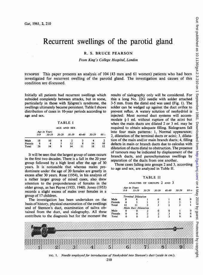
Gut, 1961, 2, 210 Recurrent swellings of the parotid gland R. S. BRUCE PEARSON From King's College Hospital, London SYNOPSIS This paper presents an analysis of 104 (43 men and 61 women) patients who had been investigated for recurrent swelling of the parotid gland. The investigation and causes of this condition are discussed. Initially all patients had recurrent swellings which subsided completely between attacks, but in some, particularly in those with Sjogren's syndrome, the swellings ultimately became persistent. Table I shows distribution of cases in 10-year periods according to age and sex. TABLE I AGE AND SEX Age in Years 0-9 10-19 20-29 30-39 40-49 50-59 60 + Male 18 10 Female 8 4 Total 26 14 4 5 9 2 12 14 3 8 11 4 14 18 It will be seen that the largest group of cases occurs in the first two decades. There is a fall in the 20 year group followed by a high level after the age of 30 years. It is noticeable that whereas males pre- dominate under the age of 20 females are greatly in excess after 30 years. Rose (1954), in his analysis of a rather larger group of mixed cases, also drew attention to the preponderance of females in the older group, as has Payne (1933, 1940). Jones (1953) records a slight excess of males over females in a group of 17 children. The investigation has been undertaken on the basis of history, physical examination of the swellings and of Stenson's duct, examination of saliva ob- tained from the duct, and sialography. All these contribute to the diagnosis but for the moment the results of sialography only will be considered. For this a long No. 22G needle with solder attached 3-5 mm. from the distal end was used (Fig. 1). The solder can be wedged up against the duct orifice to prevent reflux. A watery solution of neohydriol is injected: Most normal duct systems will accom- modate i-1 ml. without rupture of the acini but when the main ducts are dilated 2 or 3 ml. may be required to obtain adequate filling. Sialograms fall into four main patterns: 1, Normal appearance; 2, dilatation of the terminal ducts or acini; 3, dilata- tion of the main and/or main branch ducts; 4, filling defects in main or branch ducts due to calculus with dilatation of ducts distal to obstruction. The presence of tumours may be indicated by displacement of the branch ducts, and parenchymatous swellings by separation of the ducts from one another. Those cases falling into groups 2 and 3, according to age and sex, are analyzed in Table II. TABLE II ANALYSIS OF GROUPS 2 AND 3 Age in Years 0-9 10-19 20-29 30-39 Male Female Total Male Female Total 40-49 50-59 60 + Terminal Dilatation 8 8 1 0 1 0 4 3 1 3 1 2 12 11 2 3 2 2 Main Duct Dilatation 0 0 2 0 0 1 0 0 2 2 3 9 0 0 2 2 3 9 0 1 1 0 6 6 FIG. 1. Needle employed for introduction of Neohvdriol into Stenson's duct (scale in cm.). 210 W 1 :h S 1111 11 on 4 May 2019 by guest. Protected by copyright. http://gut.bmj.com/ Gut: first published as 10.1136/gut.2.3.210 on 1 September 1961. Downloaded from
-
Upload
vuongnguyet -
Category
Documents
-
view
221 -
download
0
Transcript of Recurrent swellings of the parotid gland · Gut, 1961, 2, 210 Recurrent swellings of the parotid...
Gut, 1961, 2, 210
Recurrent swellings of the parotid glandR. S. BRUCE PEARSON
From King's College Hospital, London
SYNOPSIS This paper presents an analysis of 104 (43 men and 61 women) patients who had beeninvestigated for recurrent swelling of the parotid gland. The investigation and causes of thiscondition are discussed.
Initially all patients had recurrent swellings whichsubsided completely between attacks, but in some,particularly in those with Sjogren's syndrome, theswellings ultimately became persistent. Table I showsdistribution of cases in 10-year periods according toage and sex.
TABLE IAGE AND SEX
Age in Years0-9 10-19 20-29 30-39 40-49 50-59 60+
Male 18 10Female 8 4Total 26 14
459
21214
38
11
41418
It will be seen that the largest group of cases occursin the first two decades. There is a fall in the 20 yeargroup followed by a high level after the age of 30years. It is noticeable that whereas males pre-dominate under the age of 20 females are greatly inexcess after 30 years. Rose (1954), in his analysis ofa rather larger group of mixed cases, also drewattention to the preponderance of females in theolder group, as has Payne (1933, 1940). Jones (1953)records a slight excess of males over females in agroup of 17 children.The investigation has been undertaken on the
basis of history, physical examination of the swellingsand of Stenson's duct, examination of saliva ob-tained from the duct, and sialography. All thesecontribute to the diagnosis but for the moment the
results of sialography only will be considered. Forthis a long No. 22G needle with solder attached3-5 mm. from the distal end was used (Fig. 1). Thesolder can be wedged up against the duct orifice toprevent reflux. A watery solution of neohydriol isinjected: Most normal duct systems will accom-modate i-1 ml. without rupture of the acini butwhen the main ducts are dilated 2 or 3 ml. may berequired to obtain adequate filling. Sialograms fallinto four main patterns: 1, Normal appearance;2, dilatation of the terminal ducts or acini; 3, dilata-tion of the main and/or main branch ducts; 4, fillingdefects in main or branch ducts due to calculus withdilatation of ducts distal to obstruction. The presenceof tumours may be indicated by displacement of thebranch ducts, and parenchymatous swellings byseparation of the ducts from one another.Those cases falling into groups 2 and 3, according
to age and sex, are analyzed in Table II.
TABLE IIANALYSIS OF GROUPS 2 AND 3
Age in Years0-9 10-19 20-29 30-39
MaleFemaleTotal
MaleFemaleTotal
40-49 50-59 60 +
Terminal Dilatation8 8 1 0 1 04 3 1 3 1 212 11 2 3 2 2Main Duct Dilatation0 0 2 0 0 10 0 2 2 3 90 0 2 2 3 9
011
066
FIG. 1. Needle employedfor introduction of Neohvdriol into Stenson's duct (scale in cm.).210
W
1
:h
S
1111
11
on 4 May 2019 by guest. P
rotected by copyright.http://gut.bm
j.com/
Gut: first published as 10.1136/gut.2.3.210 on 1 S
eptember 1961. D
ownloaded from

Recurrent swellings of the parotid gland
Terminal duct dilatation therefore occurs mostfrequently in the first two decades, with males pre-dominating over females in the proportion of two toone. Main duct dilatation occurs very much morecommonly in females and is rare under 40 years ofage. Those with normal sialograms on the wholereflect the findings in the group with terminal ductdilatation, occurring most commonly before the ageof 20 with a 4:1 excess of males over females. Thosein whom sialography was not carried out clearlyinclude cases from both the youthful group and theolder female cases (Table III).
TABLE IIISIALOGRAM FINDINGS RELATED TO AGE AND SEX
Age in Years0-9 10-19 20-29 30-39
MaleFemaleTotal
MaleFemaleTotal
Normal Sialograms6 2 11 1 37 3 4
Sialograms Not Done4 0 03 0 07 0 0
45
0
40-49 50-59 60-69
2
033
0
022
000
011
There remain 14 patients with filling defects andlocalized distensions; these were all over 20, and in-cluded sevenmalesand seven females. All had calculusformation and will be more fully discussed later.For the purpose of this paper cases have been
classified on clinical grounds under five mainheadings: 1, Infective; 2, Sjogren's syndrome;3, calculus formation; 4, allergic; and 5, doubtfulorigin. It should be pointed out that infection of theduct system is not limited to group 1. Many cases ofparotid swelling in Sjogren's syndrome are infected,and infection, possibly secondary, is present in manycases of calculus obstruction and may also occur inthose with allergy.
INFECTIVE GROUP
These patients have periodic attacks of swelling ofthe parotid gland, usually for several days or weeksat a time with intervals of freedom. The gland isoften tender, the duct orifice reddened, and purulentdebris can sometimes be expressed from it. Organ-isms, including Strep. viridans, pneumococcus, Staph.aureus, haemolytic streptococci, and Staph. albus,have all been cultivated from the parotid secretions,but it is difficult to avoid contamination with mouthorganisms, and cultures from saliva which do notcontain pus should be accepted with caution.Patients frequently complain of an unpleasant tastein the mouth. In cases of doubt resolution after treat-ment with tetracycline or penicillin may provideconfirmatory evidence of infection.
The group contains 39 patients: 21 were under theage of 20 and of these 15 were males. All thoseinvestigated by sialography had terminal duct dilata-tion (15) or normal ducts (4). Two also had irregulardilatation of one main duct. There were 16 cases over30: 15 were females and one male. Ten had dilatedmain ducts, three terminal dilatation, and one anormal sialogram. In two cases sialograms werenot obtained. Of those with dilatations of theterminal ducts, 12 had bilateral swellings, seven uni-lateral. Two of the latter showed terminal dilatationon the unaffected side as well as on the affected.
CASE 1 This boy, aged 10 years, is the only diabetic inthe series. He has had recurrent swellings of the parotidglands for six years. The left side has been repeatedlyaffected but the right side on one occasion only. Pressureover the distended glands on the left side caused exuda-tion of pus from Stenson's duct. Strep. pneumoniae wasobtained on culture. Sialography showed that the terminalducts were dilated on both sides (Fig. 2). A biopsyobtained from the left side (Fig. 3) shows dilated aciniwith fine interstitial fibrosis and patchy infiltration bychronic inflammatory cells. Polymorphs are present insome of the smaller ducts. The appearance is typical of arecurrent parotitis in an active phase.
The patients with main duct obstruction can bedivided into those in whom obstruction was at theorifice of the duct (seven cases) and those in whomthe obstruction occurred at some point in the courseof the main duct distal from the orifice (six cases).Four in each group had bilateral swellings. Twopatients with unilateral swellings showed obstructionat some distance from the duct orifice with anappearance very suggestive of calculus formation(filling defects in duct) but no confirmatory evidencewas obtained. Terminal duct dilatation was present intwo cases with dilatation of the main duct. In two
FIG. 2. Case 1 showing bilateral terminal dilatation ofducts. Recurrent infection.
211
on 4 May 2019 by guest. P
rotected by copyright.http://gut.bm
j.com/
Gut: first published as 10.1136/gut.2.3.210 on 1 S
eptember 1961. D
ownloaded from
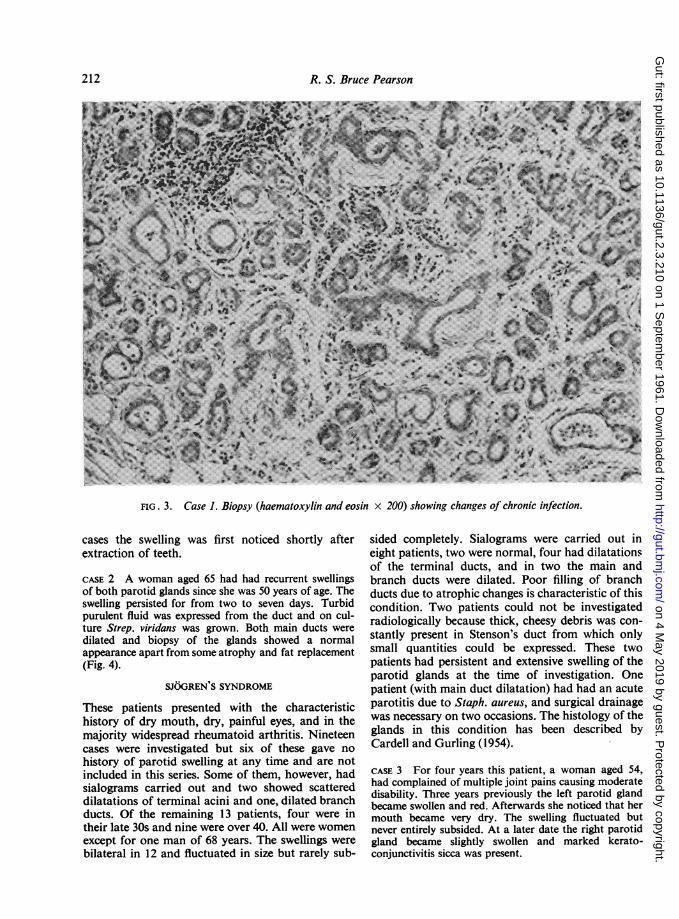
R. S. Bruce Pearson
FIG. 3. Case 1. Biopsy (haematoxylin and eosin x 200) showing changes of chronic infection.
cases the swelling was first noticed shortly afterextraction of teeth.
CASE 2 A woman aged 65 had had recurrent swellingsof both parotid glands since she was 50 years of age. Theswelling persisted for from two to seven days. Turbidpurulent fluid was expressed from the duct and on cul-ture Strep. viridans was grown. Both main ducts weredilated and biopsy of the glands showed a normalappearance apart from some atrophy and fat replacement(Fig. 4).
SJOGREN'S SYNDROME
These patients presented with the characteristichistory of dry mouth, dry, painful eyes, and in themajority widespread rheumatoid arthritis. Nineteencases were investigated but six of these gave nohistory of parotid swelling at any time and are notincluded in this series. Some of them, however, hadsialograms carried out and two showed scattereddilatations of terminal acini and one, dilated branchducts. Of the remaining 13 patients, four were intheir late 30s and nine were over 40. All were womenexcept for one man of 68 years. The swellings werebilateral in 12 and fluctuated in size but rarely sub-
sided completely. Sialograms were carried out ineight patients, two were normal, four had dilatationsof the terminal ducts, and in two the main andbranch ducts were dilated. Poor filling of branchducts due to atrophic changes is characteristic of thiscondition. Two patients could not be investigatedradiologically because thick, cheesy debris was con-stantly present in Stenson's duct from which onlysmall quantities could be expressed. These twopatients had persistent and extensive swelling of theparotid glands at the time of investigation. Onepatient (with main duct dilatation) had had an acuteparotitis due to Staph. aureus, and surgical drainagewas necessary on two occasions. The histology of theglands in this condition has been described byCardell and Gurling (1954).
CASE 3 For four years this patient, a woman aged 54,had complained of multiple joint pains causing moderatedisability. Three years previously the left parotid glandbecame swollen and red. Afterwards she noticed that hermouth became very dry. The swelling fluctuated butnever entirely subsided. At a later date the right parotidgland became slightly swollen and marked kerato-conjunctivitis sicca was present.
212
on 4 May 2019 by guest. P
rotected by copyright.http://gut.bm
j.com/
Gut: first published as 10.1136/gut.2.3.210 on 1 S
eptember 1961. D
ownloaded from

Recurrent swellings of the parotid gland
FIG. 4. Case 2. Biopsy (haematoxylin and eosin x 200) showing minimal glandular atrophy andfat replacement.
FIG. 5. A sialogram of Case 3 showing terminal dilatation.3
A sialogram showed extensive dilatation of the terminalducts and some irregular dilatation of the main duct(Fig. 5). Section on the left side confirmed the dilatationwith evidence of chronic inflammation and residualglandular tissue undergoing hyalinization (Fig. 6).
CASE 4 A woman aged 36 first had symptoms ofrheumatoid arthritis at the age of 23. These cleared upbut returned after pregnancy at 29 and her condition hassteadily deteriorated since then. At 28 she noticed swellingof the parotid glands on eating (for the past two years theswelling has been persistent). Treatment with cortico-trophin caused the swelling to subside temporarily but noincrease in secretion. For two years her mouth has beenpersistently dry and she has had a pricking sensation inthe eyes. A sialogram showed dilatation of the terminalducts. The branches of Stenson's duct were character-istically scanty due to atrophy of glandular tissue (Fig. 7).Section showed parenchymal atrophy and replacementby lymphoid tissue, typical of the changes described byCardell and Gurling.
CALCULUS FORMATION
These cases presented with a history of pain andswelling on the side of the calculus formation. There
213
on 4 May 2019 by guest. P
rotected by copyright.http://gut.bm
j.com/
Gut: first published as 10.1136/gut.2.3.210 on 1 S
eptember 1961. D
ownloaded from

R. S. Bruce Pearson
FIG. 6. Case 3. Biopsy ofparotid gland (haematoxylin and eosin x 200) showing glandular atrophy with evidence ofchronic inflammation.
FIG. 7. Sialogram of Case 4, showing scanty branch ductswith terminal dilatation.
was evidence of infection in seven out of 14 casesbut it was certainly not invariably present. Forexample, a man aged 43 noticed an acute, painfulswelling of the left parotid gland while eating on twooccasions, and on the second of these he pressedfirmly over the swelling, which caused a sharp paindeep in the gland, followed by expulsion of a smallphosphate calculus the size of a grape pip and a gushof pent-up secretion. Since this patient was seen, twoother patients have been able to express stones bypressure in this way, one after nine years and theother after four years. Stones or grit were passedspontaneously in four other cases and in another acalculus was removed from the main duct at opera-tion. Swellings were unilateral in all cases except two.
Sialography was carried out in 13 cases. In onlytwo was a stone observed in a straight radiograph,but filling defects were present in the main duct infive and in branch ducts in two other patients. Intwo the main duct on the affected side was com-pletely blocked. Dilatation of the main duct distalto the obstruction was demonstrated in eight cases,and localized dilatations of branch ducts in six (in
214
on 4 May 2019 by guest. P
rotected by copyright.http://gut.bm
j.com/
Gut: first published as 10.1136/gut.2.3.210 on 1 S
eptember 1961. D
ownloaded from

Recurrent swellings of the parotid gland
these both main duct and localized dilatations werepresent). Twelve patients were 37 years of age ormore and the sex distribution was equal.
CASE 5 A man aged 37 complained of swelling of the leftside of the face intermittently for one year. It was oftenmade worse by eating and might persist for two weeks ata time. Pressure over the gland caused turbid fluid toexude from Stenson's duct. A calculus, approximately1 cm. in length, could be felt at the edge of the masseter.A sialogram showed a convex filling defect in the mainduct from which a radiotranslucent stone was subsequentlyremoved (Fig. 8).
CASE 6 A man aged 59 had had recurrent swelling of theright parotid gland at irregular intervals during six orseven years. Small pieces of grit have been expelled fromStenson's duct from time to time. A sialogram showed afilling defect at the point of subdivision of the main ductwith localized swelling of the branch duct (Fig. 9).
CASES WITH EVIDENCE OF ALLERGIC AETIOLOGY
These include 16 cases, 11 females and five males.Six patients, all women between the ages of 37 and 70with bilateral swellings, had dilated main ducts, inone case limited to one side. The swellings appearedvery frequently, sometimes during every meal. Thesecases have been described in detail elsewhere(Pearson, 1951). All gave a history of expellingmucus plugs followed by clear saliva from the ductorifices by pressure over the dilated gland, andmicroscopical examination showed that the plugscontained numerous eosinophils and in one poly-morphs also. Four had asthma and two rhinorrhoea.All except one had a high eosinophil count in the
blood. In some cases the eating of specific foods wasconsidered by the patient to cause swelling, but thiscould not always be confirmed by observation. Thereis no reasonable doubt that eosinophilic mucus plugswhich blocked the orifices of Stenson's ductsaccounted for the swellings in these cases.There were 10 other patients in whom allergy was
thought to play some part in the aetiology. Three ofthese had terminal duct dilatations and five normalsialograms, including one of a patient with a historyof many years' recurrent swellings. The ages of thisgroup varied from 4 to 61 but only three were over20. In none of these patients was purulent materialever seen emerging from Stenson's duct. The dura-tion of episodes of swelling was brief in most cases.Four patients had asthma, two had had migraine atthe same time as swellings were present, and onepatient developed parotid swellings as part of a goldsensitivity reaction.
CASE 7 A girl aged 17 suffered from periodic swelling ofboth parotid glands for one day at a time. These swellingsoften followed the eating of egg in cake or pudding. Eggshave always caused vomiting, and eating a boiled eggwas followed the next day by migraine, vomiting, andbilateral parotid swellings. A sialogram (Fig. 10) showedscattered dilatation of terminal ducts. She had no moreswellings after the age of 19. The patient continues toavoid eggs.
SWELLINGS OF DOUBTFUL ORIGIN
This group includes 22 cases (11 males and 11females). Twelve had bilateral swellings. Ten wereunder the age of 10, and six were over 30 years of
FIG. 8. Case 5. Sialogram showing radiotranslucent stone FIG. 9. Case 6. Sialogram with filling defect due toin Stenson's duct. calculus.
215
on 4 May 2019 by guest. P
rotected by copyright.http://gut.bm
j.com/
Gut: first published as 10.1136/gut.2.3.210 on 1 S
eptember 1961. D
ownloaded from

R. S. Bruce Pearson
FIG. 10. Case 7 showing terminal duct dilatation inrecurrent swelling of allergic origin.
age. One woman of 66 had a dilated main duct, fivehad terminal dilatations, and eight had normal sialo-grams. Three were asthmatic. Two were siblings, andone other, a woman of 26, was the sister of a boy inthe infected group. In some cases the swelling wasalmost certainly confined to the glandular or inter-stitial tissues and unassociated with duct obstruction.
TRAUMA TO DUCT ORIFICE
One patient, a woman of 58, was observed to have alesion of the duct papilla on one side, probablycaused by trauma from ill-fitting dentures.
DISCUSSION
Recurrent swellings of the parotid glands are notuncommon and present a difficult and complicatedproblem in pathogenesis. The tendency to develop bi-lateral swellings, often simultaneously, the changingsex ratio with increasing age accompanied by changein the sialographic appearances from dilated terminalducts or acini in youth to dilatation of the mainducts in older people, all offer a tempting bait forspeculation. There has been no lack of suggestedaetiological factors, including infection (Payne, Rose,Jones), allergy (Pearson, 1935, 1936; Serafini, 1951;Waldbott and Shea, 1947; Findeisen, 1956; Riley,1956), dental trauma (Rose, 1954), dehydration,diabetes (Lyon, 1943), improper mastication (Payne,
1940), heredity (Smith, 1953), auto-immune disease(Mosbech, Kristensen, 1960), and sialorrhoea(Reimann and Lindquist, 1952).
Parotid calculi are recognized but generally con-sidered rare. In the present series of cases parotidcalculi were identified in 14 patients. This was theonly group of middle-aged or elderly patients inwhom males were as numerous as females, and theonly group in which unilateral swellings were moreoften seen than bilateral. The diagnosis is not alwayseasy because the calculus may be small or radio-translucent. Sialography may reveal a filling defectin the main or branch duct, with dilatation beyondit, and is an essential feature of the investigation ofsuspected cases. Infection may be present, but isprobably secondary to obstruction in most cases.
In Sjogren's syndrome the dry mouth and poorsalivary flow may be considered to account at leastpartly for the recurrent swellings found in this con-dition by predisposing to ascending infection. Thereduction in size of the glands after treatment withcorticotrophin suggests that there is also inflamma-tory swelling of the interstitial tissues (Gurling,Pearson, and Pond, 1954). The patients are almostexclusively female, and in the present series this wasthe only group of elderly women in whom dilatationof the terminal ducts was common. Main ductdilatation may also occur. The most characteristicfeature of the sialogram is the small number ofbranch ducts. This is accounted for by atrophy ofthe gland and its replacement with fibro-adiposetissue (Cardell and Gurling, 1954).
Allergy can also reasonably be accepted as of someimportance as an aetiological factor. The olderpatients are exclusively female and the swellings,which are chiefly of the main and branch ducts, arecaused by eosinophilic plugs obstructing the orificesof Stenson's ducts from which they can readily beejected by pressure. In these cases dilatation of theterminal ducts has not been seen. In the youngerpatients, however, males predominate and the sialo-graphic appearances are either normal or withterminal duct dilatation. No patient under 30 wasfound to have a dilated main duct.A similar relationship between age and sex and in
the sialographic pattern is seen in the infected cases,and expression of mucous plugs containing poly-niorphs is sometimes encountered in the middle-agedwomen who mainly form this group. The organismsisolated from the purulent secretions in thesepatients are most commonly Strep. viridans and lessfrequently pneumococci and Staph. aureus (Payne,1933, 1900). These findings have been confirmed inthe present series.
It is difficult to understand why the pattern of ductobstruction should differ in patients of different ages,
216
on 4 May 2019 by guest. P
rotected by copyright.http://gut.bm
j.com/
Gut: first published as 10.1136/gut.2.3.210 on 1 S
eptember 1961. D
ownloaded from

Recurrent swellings of the parotid gland 217
even when the aetiological factors are not identical,e.g., infection, allergy, and cases without obviousaetiology. There is general agreement that duct in-fection is almost invariably an ascending one. Itseems that in young patients the infection mayascend rapidly into the branch ducts, where swellingof the mucosal lining of the intercalary ducts or ofthe interstitial tissue itself obstructs the lumen.Biopsy specimens from such cases confirm this view,revealing inflammatory change in the interstitialtissues with purulent material in small ducts. Inthese cases there is a definite tendency for improve-ment during adolescence when recurrent swellingsdisappear. Spontaneous remissions also take placein young patients with an allergic basis and thosewithout obvious aetiological factors. In adults themain reaction to both ascending infection andallergic processes appears to be in the main duct,which responds by secretion of mucus and sheddingof the lining cells of the duct to form masses whichobstruct the main duct orifice causing it and itsmain branches to dilate. In other cases constrictionsseen in the course of the main duct may indicatefibrous stricture. Biopsy of the gland in these casesshows little change in the acini and interstitial tissues.The tendency to form tenacious mucus is of coursemore apparent in the bronchial tract in olderpatients, especially those with bronchitis. It seemspossible that the mucus-secreting glands which arefound in Stenson's duct are also more active in thisage group and pour out their secretions as a resultof infective, allergic, or other stimuli.
Hereditary or congenital abnormalities of theducts have been suggested as a possible basis forrecurrent swellings. In the present series of casesthere are two pairs of siblings, the first' a boy aged 12with infective sialodochitis and terminal duct dilata-tion with early involvement of the main ducts, andhis sister aged 26 years. She had had recurrentswellings as a child but at the time of investigationhad been free of symptoms for some years but sialo-graphy still revealed bilateral terminal dilatation ofthe ducts.' The second pair were brothers aged 4jand 1 year 9 months. Both were asthmatics but theaetiology of the swellings was considered uncertainand sialograms were not carried out. Two otherpatients who gave a history of unilateral swellingassociated with infection had terminal duct dilata-tions on both sides. Rose quotes 10 cases in childrenbetween the age of 3 and 15 years who had advancedchanges in the duct system in spite of a short andcomparatively mild history, which he regards asevidence of pre-existing acinar dilatations. Smith(1953) records recurrent parotid swellings with radio-
logical evidence of duct dilatation in three genera-tions of a family.
Recurrent attacks of sialorrhoea may account forsome of the transient bilateral swellings withoutevidence of infection or allergy (Reimann andLindquist, 1952). Improper mastication with a con-sequently inadequate salivary flow (Payne, 1940)may well predispose to ascending infection, but thisis difficult to determine. It may help to explain theonset of infection after dental extractions. Stenosisof the duct or duct papilla due to trauma from afaulty bite has been regarded as a relatively commoncause of parotid swelling (Rose). Although the ductorifices are sometimes small and a cannula may bedifficult to pass, especially when the orifice is on amobile papilla, we have only encountered one casewhere there was evidence of trauma from a newlyacquired set of dentures. The swelling was unilateral.We have found no evidence that diabetes mellitus
is a predisposing factor of importance. In spite of acareful search in the Diabetic Department at King'sCollege Hospital, only one case of pneumococcalsialodochitis, in a diabetic boy aged 11, has beenreferred.
It is clear that there is a group of patients inwhom the aetiological factor is obscure, and evenwhen infection is present the underlying predispos-ing abnormality often remains unrecognized.I should like to express my thanks to Dr. B. S. Cardellfor help and advices.
REFERENCESCardell, B. S., and Gurling, K. J. (1954). Observations on the pathology
of Sjogren's syndrome. J. Path. Bact., 68, 137-146.Findeisen, D. G. R. (1956). Allergy to dentures: A report of two cases
presenting asthma and parotitis. Int. Arch. Allergy, 9, 281-284.Gurling, K. J., Pearson, R. S. B., and Pond, M. H. (1954). Sjogren's
syndrome treated with A.C.T.H. Brit. J. Opthal., 38, 619-652.Jones, H. Everley. (1953). Recurrent parotitis in children. Arch. Dis.
Childh., 28, 182-186.Lyon, E. (1943). Swelling of the parotid gland and diabetes mellitus.
Int. J. Gastroent., 68, 139-147.Mosbech, S., and Kristensen, H. P. 0. (1960). Chronic Sialoadenitis
caused by autoimmunisation. Acta med. scand., 168, 147-150.Payne, R. T. (1933). Recurrent pyogenic parotitis. Lancet, 1, 348-353.- (1940). Pneumococcal parotitis. Brit. med. J., 1, 287-292.Pearson, R. S. B. (1935). Recurrent swelling of the parotid glands.
Arch. Dis. Childh., 10, 363-376.(1936). Two cases of recurrent swelling of the parotids. Guy'sHosp. Rep., 86, 333-342.
- (1951). Parotid swelling due to allergic obstruction of the mainduct. Proceedings of First International Congress for Allergy,Basel, pp. 868-873.
Riley, H. D. (1956). Recurrent parotid swelling. Sth. med. J. (Bgham,Ala.), 49, 523-528.
Rose, S. S. (1954). A clinical and radiological survey of 192 cases ofrecurrent swellings of the salivary glands. Ann. roy. Coll. Surg.,15, 374-401.
Reimann, H. A., and Lindquist, J. N. (1952). Periodic sialorrhea.J. Amer. med. Ass., 149, 1465-1467.
Serafini, U. (1951). Prime osservazioni su alcuni casi di tumefazioneacuta ricorrente della parotide di probabile origine allergica.Minerva med. (Torino), 17, 271-275.
Smith, M. (1953). Familial incidence of sialectatis. Brit. med. J., 2,1359.
Waldbott, G. L., and Shea, J. J. (1947). Allergic parotitis. J. Allergy,18, 51-54.'Previously reported (Pearson, 1935).
on 4 May 2019 by guest. P
rotected by copyright.http://gut.bm
j.com/
Gut: first published as 10.1136/gut.2.3.210 on 1 S
eptember 1961. D
ownloaded from


![Parotid Lesions in Children Undergoing Parotidectomy. The … · 2018. 8. 8. · of salivary gland masses occur within the parotid gland [1-4]. Parotid gland lesions are infrequent](https://static.fdocuments.us/doc/165x107/60d3cf2c7c14947d7f31fea4/parotid-lesions-in-children-undergoing-parotidectomy-the-2018-8-8-of-salivary.jpg)


