Reconstructive plastic surgery in head and neck oncology S ... · The tumor was negative for...
37
Reconstructive plastic surgery in head and neck oncology S. Tuinder Plastic Surgery unit MUMC+ Maastricht Maastricht, 21-5-15
Transcript of Reconstructive plastic surgery in head and neck oncology S ... · The tumor was negative for...

Reconstructive plastic surgery in head and neck oncology
S. Tuinder
Plastic Surgery unit MUMC+ Maastricht
Maastricht, 21-5-15

s.tuinder
History
from: J. M. Bourgery u. Claude Bernard „Traité complet de l`anatomie
de l`homme“, Paris 1866

s.tuinder
History
from: Gaspare Tagliacozzi „De curtorum chirurgia per insitionem“,
Venedig 1597

s.tuinder
History

s.tuinder
Principles of plastic surgery
• Team
• Skin lines
• Aesthetic units
• Reconstructive ladder
• Contour and function

s.tuinder

s.tuinder
Tension area
to be radical
RT
Aesthetic
function

s.tuinder
Aesthetic units

s.tuinder
Skin lines

s.tuinder
Reconstructive ladder

s.tuinder
Primary intention

s.tuinder
Skin graft

s.tuinder
Tissue expansion

s.tuinder
Local tissue transfer
s.tuinder
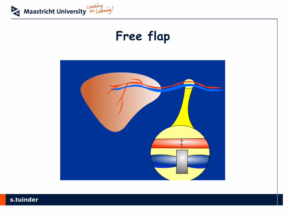
Free flap

s.tuinder
Free flap

s.tuinder
Contour and function
• Example: face and facial palsy surgery….
s.tuinder
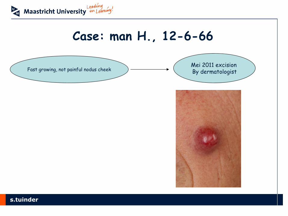
Case: man H., 12-6-66
Fast growing, not painful nodus cheek
Mei 2011 excision By dermatologist

s.tuinder
PA: tumor localized in the diep dermis en subcutis. Irregular nests and fascicles were formed, which consisted of a mix of epithelioid and spindled cells. Between the tumor cells multiple delicately branching capillaries are present. The tumor cells had abundant eosinophilic cytoplasm, containing enlarged polymorphic and vesicular nuclei with prominent nucleoli. Mitoses were seen at a rate of 5 mitoses per 10 high power fields. There was no necrosis. Immunohistochemically, the tumor cells expressed several melanocytic markers, with exception of S100. There was a strong immunoreactivity for CD68, CD10 and vimentin and weak, but focal expression of alpha smooth muscle actin. Other muscle markers were negative. The tumor was negative for epithelial markers and neuro-endocrine markers. Based on this histological and immunohistochemical profile, a perivascular epithelioid cell tumor (PEComa) was diagnosed. Because of the mitotic rate of 5 mitosis per high power field and the
nuclear atypia, this tumor was considered a malignant PEComa.
PEComa: Malignant perivascular epithelioid cell tumor

s.tuinder
Head-neck team decision: excision with 2 cm marge followed by RT
Ultrasound neck,PET-CT en CT thorax (neg.)

s.tuinder
Head-neck team decision: excision with 2 cm marge followed by RT
• What does it mean?
• Defect through and trough of the cheek ,6 by 5 cm, with sacrifice of the buccal en marginali branch of the facial nerve, zigomatici muscles, part of the levator alequae nasi en labii superior, part of the orbicularis oris, masseter, parotis gland, mucosa.

s.tuinder
Reconstruction: 2 weeks after
• Functional gracilis flap, een split skin graft for the mucosa. Mustarde’ flap for the skin defect.
• Re-or because of problems with the SSG because of saliva lekkage.

s.tuinder
Radiotherapy
• He received a total dosis of 51 Gy in 30 fractions in the reconstructed area and 60 Gy in 30 fractions around.

s.tuinder
Facial palsy

s.tuinder
CAUSES
Intracranial region: trauma, infection, congenital problems (moebius syndrome), tumors...
Temporal bone: infections, trauma, tumors....
Parotid region: tumor, trauma....
Bells palsy (25:100,000 p.a.• Spontaneous onset• 85% good resolution
within 2 months• Aetiology viral vs inflammatory• Steroids +/- NSAIDs• DIAGNOSIS OF EXCLUSION)

s.tuinder
TIMING AND MODALITY

s.tuinder
Possibilities…
new Old (more
than 2 years)
Congenital (complete or
partial)

s.tuinder
2 problems
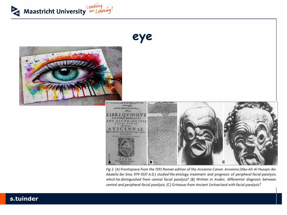
eye
mouth
s.tuinder
eye

s.tuinder
eye new (1 to 2 years)
GRAFT (n. suralis, great auricular nerve, nervus safenus)

s.tuinder
eye new (1 to 2 years)
BABYSITTER PROCEDURE, jump anastomosis: hypoglossus f-facials anastomosis and masseteric-facialis anastomosis

s.tuinder
eye new (1 to 2 years)
BABYSITTER PROCEDURE, jump anastomosis: hypoglossus f-facials anastomosis and masseteric-facialis anastomosis

s.tuinder
mouth

s.tuinder
Mouth: new
• Graft facial-facial
• Graft facial-masseter
• Graft facial-hypoglossus
• Combinations

s.tuinder
Mouth: old en new Labbè procedure

s.tuinder
Free muscle transfer (old or congenital)

s.tuinder
Thank you!