Recommended Proton Therapy...
25
1 April 2015 Recommended Proton Therapy Indications National Association of Proton Therapy (NAPT) member medical directors under the leadership of William Hartsell, MD, Robert Foote, MD, and Nancy Mendenhall, MD developed this document summarizing recommended proton beam therapy indications by tumor site. NAPT is a non-profit organization promoting education and public awareness of the clinical benefits of proton beam radiation therapy for cancer treatment. More information on NAPT is available on its website (www.proton-therapy.org). 1. Breast Cancer 2. Esophagus Cancer 3. Gastrointestinal (GI) Cancer 4. Hematologic Cancer 5. Prostate Cancer 6. Thoracic Cancer
Transcript of Recommended Proton Therapy...

1
April 2015
Recommended Proton Therapy Indications
National Association of Proton Therapy (NAPT) member medical directors under the leadership of William Hartsell, MD, Robert Foote, MD, and Nancy Mendenhall, MD developed this
document summarizing recommended proton beam therapy indications by tumor site. NAPT is a non-profit organization promoting education and public awareness of the clinical benefits of proton beam radiation therapy for cancer treatment. More information on NAPT is available on
its website (www.proton-therapy.org).
1. Breast Cancer
2. Esophagus Cancer
3. Gastrointestinal (GI) Cancer
4. Hematologic Cancer
5. Prostate Cancer
6. Thoracic Cancer

2
Breast Cancer
Recommended Proton Therapy Indications
Indications:
Left or Right-sided early or locoregionally advanced breast cancer requiring
breast or chest wall plus regional nodal irradiation (i.e. lymph node positive disease,
advanced T stage and/or medial tumor location)
Adjuvant radiotherapy improves survival in breast cancer patients, suggesting
that persistence of locoregional tumor is associated with an increased risk of
developing metastases and death.1,2. Results of modern randomized controlled
clinical trials highlight the importance of regional nodal irradiation in reducing
distant events in this population.3-7 Targeting of the regional lymphatics results in
lung and heart doses associated with increased major cardiac events, cardiac
deaths, lung cancer, and lung cancer deaths in a patient population where
advances in systemic therapy and other multidisciplinary care has resulted in
decreasing breast cancer specific mortality.8-10 Proton radiotherapy improves
coverage of the regional lymphatics while substantially reducing mean lung and
mean heart doses to levels significantly correlated with reduced cardiac events,
lung cancer, and symptomatic pneumonitis.11-15
Inclusion criteria:
o Age ≥ 18 years
o Histologic confirmation of breast cancer
o Lumpectomy or mastectomy with or without immediate reconstruction
o The axilla must be staged by sentinel node biopsy alone, sentinel node biopsy followed by axillary node dissection, or axillary lymph node dissection alone
o Whole breast/chest wall and regional nodal irradiation indicated (lymph node positive disease, T3-T4, medial tumor location) .
o pStage T1-T4N0-N3M0 or ypStage T0-4N0-N3M0
o Breast implants and expanders allowed
o Improved target coverage for regional nodes or absolute difference in mean heart dose between proton and photon plans (with photon cardiac sparing technique such as breath hold, IMRT, or prone positioning) >1.35 Gy or absolute difference in ipsilateral lung volume receiving 20 Gy between proton and photon plans >10%
Exclusion criteria:
o Medical contraindication to receipt of radiotherapy.
o Severe active co-morbid systemic illnesses or other severe concurrent disease which, in the judgment of the physician, would contraindicate any radiation therapy.
o Active systemic lupus or scleroderma.

3
o Pregnancy or women of childbearing potential who are sexually active and not willing/able to use medically acceptable forms of contraception
Early stage Breast cancer with indications for whole breast radiotherapy
Darby et al. have established that rates of coronary events increase linearly with
mean dose to the heart by 7.4% per gray with no apparent threshold.6 The
increase begins within a few years after exposure, and continues for at least 20
years. Proton whole breast radiotherapy achieves mean heart doses below
<0.5Gy. Best evidence suggests proton radiotherapy is associated with a
clinically significant >10% reduction in the rate of major coronary events in
patients determined to have a >1.35Gy improvement in mean heart dose with
proton, compared with photon planning.8-10
Inclusion criteria:
o Female
o Age ≥ 18 years.
o Histological confirmation of breast cancer
o Lumpectomy
o For invasive breast cancer the axilla must be staged by sentinel node biopsy alone, sentinel node biopsy followed by axillary node dissection, or axillary lymph node dissection alone
o pStage T0-T3N0-N1M0 or ypStage T0-T3N0-N1micM0
o Whole breast irradiation with or without a lumpectomy cavity boost indicated
o Absolute difference in mean heart dose between proton and photon plans (with photon cardiac sparing technique such as breath hold, IMRT, or prone positioning) >1.35 Gy or absolute difference in ipsilateral lung volume receiving 20 Gy between proton and photon plans >10%
Exclusion criteria:
o Medical contraindication to receipt of radiotherapy.
o Severe active co-morbid systemic illnesses or other severe concurrent disease which, in the judgment of the physician, would contraindicate any radiation therapy.
o Active systemic lupus or scleroderma.
o Pregnancy or women of childbearing potential who are sexually active and not willing/able to use medically acceptable forms of contraception
Left and Right-sided early stage (invasive and non-invasive) Breast cancer with
clinical indications for partial breast irradiation as described below
The safety and efficacy of proton partial breast irradiation is established for early
stage breast cancer.13 Proton PBI provides a more homogeneous dose
distribution and reduction in exposure to the normal breast, heart, and lung
4
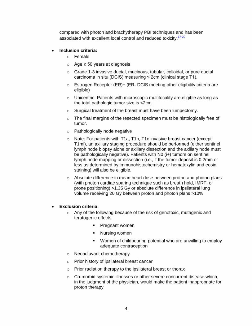
compared with photon and brachytherapy PBI techniques and has been
associated with excellent local control and reduced toxicity.17-20
Inclusion criteria:
o Female
o Age ≥ 50 years at diagnosis
o Grade 1-3 invasive ductal, mucinous, tubular, colloidal, or pure ductal carcinoma in situ (DCIS) measuring ≤ 2cm (clinical stage T1).
o Estrogen Receptor (ER)+ (ER- DCIS meeting other eligibility criteria are eligible)
o Unicentric: Patients with microscopic multifocality are eligible as long as the total pathologic tumor size is <2cm.
o Surgical treatment of the breast must have been lumpectomy.
o The final margins of the resected specimen must be histologically free of tumor.
o Pathologically node negative
o Note: For patients with T1a, T1b, T1c invasive breast cancer (except T1mi), an axillary staging procedure should be performed (either sentinel lymph node biopsy alone or axillary dissection and the axillary node must be pathologically negative). Patients with N0 (i+) tumors on sentinel lymph node mapping or dissection (i.e., if the tumor deposit is 0.2mm or less as determined by immunohistochemistry or hematoxylin and eosin staining) will also be eligible.
o Absolute difference in mean heart dose between proton and photon plans (with photon cardiac sparing technique such as breath hold, IMRT, or prone positioning) >1.35 Gy or absolute difference in ipsilateral lung volume receiving 20 Gy between proton and photon plans >10%
Exclusion criteria:
o Any of the following because of the risk of genotoxic, mutagenic and teratogenic effects:
Pregnant women
Nursing women
Women of childbearing potential who are unwilling to employ adequate contraception
o Neoadjuvant chemotherapy
o Prior history of ipsilateral breast cancer
o Prior radiation therapy to the ipsilateral breast or thorax
o Co-morbid systemic illnesses or other severe concurrent disease which, in the judgment of the physician, would make the patient inappropriate for proton therapy

5
o Active collagen-vascular disease that, in the opinion of the treating physician, would make proton therapy hazardous for the patient
o Paget’s disease of the breast
o Proven multicentric carcinoma (DCIS or invasive) in more than one quadrant or separated by 4 or more centimeters or diffuse (>1 quadrant) suspicious calcifications
o Histologic evidence of angiolympatic invasion (ALI). Note: Cases termed focally suspicious for ALI but where no definitive ALI is found are eligible.
o Surgical margins that cannot be microscopically assessed or that are positive
o Pathologic tumor >2cm in size
o Metastatic disease
o Invasive lobular carcinoma or lobular carcinoma in situ
o BRCA1 or BRCA2 mutation
o Breast implants (patients who have had implants removed are eligible).
o Extensive intraductal component
o Active connective tissue disease
o Reduction mammoplasty if 3DCRT or proton APBI are planned
Scientific Evidence:
1. Ebctcg Early Breast Cancer Trialists' Collaborative Group. Effect of radiotherapy after mastectomy and axillary surgery on 10-year recurrence and 20-year breast cancer mortality: meta-analysis of individual patient data for 8135 women in 22 randomised trials. Lancet. Mar 19 2014.
2. Darby S, McGale P, Correa C, et al. Effect of radiotherapy after breast-conserving surgery on 10-year recurrence and 15-year breast cancer death: meta-analysis of individual patient data for 10,801 women in 17 randomised trials. Lancet. Nov 12 2011;378(9804):1707-1716.
3. Ragaz J, Jackson SM, Le N, et al. Adjuvant radiotherapy and chemotherapy in node-positive premenopausal women with breast cancer. N Engl J Med. Oct 2 1997;337(14):956-962.
4. Overgaard M, Jensen MB, Overgaard J, et al. Postoperative radiotherapy in high-risk postmenopausal breast-cancer patients given adjuvant tamoxifen: Danish Breast Cancer Cooperative Group DBCG 82c randomised trial. Lancet. May 15 1999;353(9165):1641-1648.
5. Overgaard M, Hansen PS, Overgaard J, Rose C, Andersson M, Bach F, Kjaer M, Gadeberg CC, Mouridsen HT, Jensen MB, Zedeler K. Postoperative radiotherapy in high-ris premenopausal women with breast cancer who receive adjuvant chemotherapy. Danish Breast Cancer Cooperative Group 82b Trial. N Engl J Med 1997; 237 (14): 949-55.
6. Whelan TJ, Olivotto I, Ackerman I, Chapman JW, Chua B, Nabid A, Vallis KA, White JR, Rousseau P, Fortin A, Pierce LJ, Manchul L, Craighead P, Nolan MC, Bowen J, McCready DR, Pritchard KI, Levine MN, Parulekar W. NCIC-CTG MA.20: An

6
intergroup trial of regional nodal irradiation in early breast cancer [abstract]. J Clin Oncol. 2011;29(18 suppl):LBA1003.
7. Poortmans P, Struikmans H, Collette S, Kirkove C, Budach V, Maingon P, Valli MC, Fourquet A, den Bogaert WV, Bartelink H. Lymph node RT improves survival in breast cancer: 10 years results of the EORTC, ROG, AND BCG phase III trial 22922/10925. Presented at ESTRO 33. April 4-8, 2014. Vienna, Austria.
8. Darby SC, McGale P, Taylor CW, Peto R. Long-term mortality from heart disease and lung cancer after radiotherapy for early breast cancer: prospective cohort study of about 300,000 women in US SEER cancer registries. Lancet Oncol. Aug 2005;6(8):557-565.
9. Darby SC, Ewertz M, McGale P, et al. Risk of ischemic heart disease in women after radiotherapy for breast cancer. N Engl J Med. Mar 14 2013;368(11):987-998.
10. Clarke M, Collins R, Darby S, et al. Effects of radiotherapy and of differences in the extent of surgery for early breast cancer on local recurrence and 15-year survival: an overview of the randomised trials. Lancet. Dec 17 2005;366(9503):2087-2106.
11. Ares C, Khan S, Macartain AM, Heuerger J, Goitein G, Gruber G, Lutters G, Hug EB, Bodis S, Lomas AJ. Postoperative proton radiotherapy for localized and locoregional breast cancer: potential for clinically relelvant improvements? Int J Radiat Oncol Biol Phys. 2010. 76(3): 685-97.
12. Macdonald SM, Patel SA, Hickey S, et al. Proton therapy for breast cancer after mastectomy: early outcomes of a prospective clinical trial. Int J Radiat Oncol Biol Phys. Jul 1 2013;86(3):484-490.
13. MacDonald SM, Jimenez R, Paetzold P, et al. Proton radiotherapy for chest wall and regional lymphatic radiation; dose comparisons and treatment delivery. Radiat Oncol. 2013;8:71.
14. Xu N, Ho MW, Li Z, Morris CG, Mendenhall NP. Can proton therapy improve the therapeutic ratio in breast cancer patients at risk for nodal disease? Am J Clin Oncol 2014. 37(6): 568-74.
15. Depauw N, Batin E, Daartz J, et al. A novel approach to postmastectomy radiation therapy using scanned proton beams. Int J Radiat Oncol Biol Phys. Feb 1 2015;91(2):427-434.
16. Johansson J, Isacsson U, Lindman H, Montelius A, Glimelius B. Node-positive left-sided breast cancer patients after breast-conserving surgery: potential outcomes of radiotherapy modalities and techniques. Radiother Oncol. Nov 2002;65(2):89-98.
17. Bush DA, Do S, Lum S, et al. Partial breast radiation therapy with proton beam: 5-year results with cosmetic outcomes. Int J Radiat Oncol Biol Phys. Nov 1 2014;90(3):501-505.
18. Haviland JS, Owen JR, Dewar JA, et al. The UK Standardisation of Breast Radiotherapy (START) trials of radiotherapy hypofractionation for treatment of early breast cancer: 10-year follow-up results of two randomised controlled trials. Lancet Oncol. Oct 2013;14(11):1086-1094
19. Presley CJ, Soulos PR, Herrin J, et al. Patterns of use and short-term complications of breast brachytherapy in the national medicare population from 2008-2009. J Clin Oncol. Dec 10 2012;30(35):4302-4307.

7
20. Olivotto IA, Whelan TJ, Parpia S, et al. Interim cosmetic and toxicity results from RAPID: a randomized trial of accelerated partial breast irradiation using three-dimensional conformal external beam radiation therapy. J Clin Oncol. Nov 10 2013;31(32):4038-4045.

8
Esophagus Cancer Recommended Proton Therapy Indications
Indications: 1) Adenocarcinoma or Squamous Cell Carcinoma of proximal, middle, or lower
esophagus or gastroesophageal junction
Inclusion criteria:
Clinical stage T1-4 N0-3 M0
Treatment intent is curative
ECOG performance status 0-2
Receiving high dose (≥ 40 Gy) definitive or neoadjuvant radiotherapy
Receiving concurrent chemotherapy
Dose-volume parameters for photon RT predict for ≥ 20% absolute risk of cardiac or pulmonary toxicity when the following cannot be met.
Lung mean dose ≤ 7 Gy or V5 ≤ 60%
V5 <1500 CC (Volume of total lung exposed to less than 5 Gy)
Heart mean dose ≤ 26 Gy, V30 < 46% (with no surgery)
V50<20% (Shirai, 2011)
The threshold parameters for considering proton therapy based on mean heart dose should be reduced if trimodality (including surgery) is used, although precise parameters are not clear at this point.
Exclusion criteria:
Clinical stage Tx, Nx, M1
Treatment intent is palliative
Total radiotherapy dose is < 40 Gy
Scientific Evidence:
1. Ling TC, Slater JM, Nookala P, Mifflin R, Grove R, Ly AM, Patyal B, Slater JD, Yang GY.
Analysis of Intensity-Modulated Radiation Therapy (IMRT), Proton and 3D Conformal
Radiotherapy (3D-CRT) for Reducing Perioperative Cardiopulmonary Complications in
Esophageal Cancer Patients. Cancers (Basel). 2014 Dec 5;6(4):2356-68. doi:
10.3390/cancers6042356. PMID: 25489937
2. Wang J, Wei C, Tucker SL, Myles B, Palmer M, Hofstetter WL, Swisher SG, Ajani JA,
Cox JD, Komaki R, Liao Z, Lin SH. Predictors of postoperative complications after
trimodality therapy for esophageal cancer. Int J Radiat Oncol Biol Phys. 2013 Aug
1;86(5):885-91. doi: 10.1016/j.ijrobp.2013.04.006. PMID: 23845841

9
3. Lin SH, Komaki R, Liao Z, Wei C, Myles B, Guo X, Palmer M, Mohan R, Swisher SG,
Hofstetter WL, Ajani JA, Cox JD. Proton beam therapy and concurrent chemotherapy for
esophageal cancer. Int J Radiat Oncol Biol Phys. 2012 Jul 1;83(3):e345-51. doi:
10.1016/j.ijrobp.2012.01.003. Epub 2012 Mar 13. PMID: 22417808
4. Mizumoto M, Sugahara S, Okumura T, Hashimoto T, Oshiro Y, Fukumitsu N, Nakahara
A, Terashima H, Tsuboi K, Sakurai H. Hyperfractionated concomitant boost proton beam
therapy for esophageal carcinoma. Int J Radiat Oncol Biol Phys. 2011 Nov
15;81(4):e601-6. doi: 10.1016/j.ijrobp.2011.02.041. Epub 2011 Apr 19. PMID: 21511402
5. Welsh J, Gomez D, Palmer MB, Riley BA, Mayankkumar AV, Komaki R, Dong L, Zhu
XR, Likhacheva A, Liao Z, Hofstetter WL, Ajani JA, Cox JD. Intensity-modulated proton
therapy further reduces normal tissue exposure during definitive therapy for locally
advanced distal esophageal tumors: a dosimetric study. Int J Radiat Oncol Biol Phys.
2011 Dec 1;81(5):1336-42. doi: 10.1016/j.ijrobp.2010.07.2001. Epub 2011 Apr 4. PMID:
21470796
6. Mizumoto M, Sugahara S, Nakayama H, Hashii H, Nakahara A, Terashima H, Okumura
T, Tsuboi K, Tokuuye K, Sakurai H. Clinical results of proton-beam therapy for
locoregionally advanced esophageal cancer. Strahlenther Onkol. 2010 Sep;186(9):482-
8. doi: 10.1007/s00066-010-2079-4. Epub 2010 Aug 30. PMID:20803187
7. Pan X, Zhang X, Li Y, Mohan R, Liao Z. Impact of using different four-dimensional
computed tomography data sets to design proton treatment plans for distal esophageal
cancer. Int J Radiat Oncol Biol Phys. 2009 Feb 1;73(2):601-9. doi:
10.1016/j.ijrobp.2008.09.042. PMID: 19147024
8. Zhang X, Zhao KL, Guerrero TM, McGuire SE, Yaremko B, Komaki R, Cox JD, Hui Z, Li
Y, Newhauser WD, Mohan R, Liao Z. Four-dimensional computed tomography-based
treatment planning for intensity-modulated radiation therapy and proton therapy for distal
esophageal cancer. Int J Radiat Oncol Biol Phys. 2008 Sep 1;72(1):278-87. doi:
10.1016/j.ijrobp.2008.05.014. PMID: 18722278
9. Wei X, Liu HH, Tucker SL, Wang S, Mohan R, Cox JD, Komaki R, Liao Z. Risk factors
for pericardial effusion in inoperable esophageal cancer patients treated with definitive
chemoradiation therapy. Int J Radiat Oncol Biol Phys. 2008 Mar 1;70(3):707-14. doi:
10.1016/j.ijrobp.2007.10.056. Epub 2008 Jan 11. PMID:18191334
10. Wang SL, Liao Z, Vaporciyan AA, Tucker SL, Liu H, Wei X, Swisher S, Ajani JA, Cox JD, Komaki R. Investigation of clinical and dosimetric factors associated with postoperative pulmonary complications in esophageal cancer patients treated with concurrent chemoradiotherapy followed by surgery. Int J Radiat Oncol Biol Phys. 2006 Mar 1;64(3):692-9. Epub 2005 Oct 19. PMID: 16242257
11. Shirai K, Tamaki Y, Kitamoto Y, Murata K, Satoh Y, Higuchi K, Nonaka T, Ishikawa H, Katoh H, Takahashi T, Nakano T. Dose-volume histogram parameters and clinical factors associated with pleural effusion after chemoradiotherapy in esophageal cancer patients. Int J Radiat Oncol Biol Phys. 2011 Jul 15;80(4):1002-7.

10
Gastrointestinal (GI) Cancer
Recommended Proton Therapy Indications Indications:
1) Hepatocellular Carcinoma
2) Intrahepatic Cholangiocarcinoma
Inclusion criteria:
Clinical stage T1-4 N0-1 M0
Curative treatment intent
Definitive radiotherapy receiving doses ≥ 40 Gy
ECOG performance status 0-2
Child-Pugh score A or B
Dose-volume parameters for photon RT are unable to meet mean liver (defined as liver – gross tumor volume) dose constraints specified on RTOG 1112 (Hepatocellular Carcinoma) and NRG GI001 (Cholangiocarcinoma)
o 5 fractions: mean liver dose ≤ 13 Gy; V10<70%;
o 15 fractions: mean liver dose ≤ 22 Gy
Exclusion criteria:
Clinical stage Tx, Nx, M1
Treatment intent is palliative
Total radiotherapy dose is < 40 Gy
Scientific evidence:
1. Bush DA, Hillebrand DJ, Slater JM, Slater JD. High-dose proton beam radiotherapy of hepatocellular carcinoma: preliminary results of a phase II trial. Gastroenterology. 2004 Nov;127(5 Suppl 1):S189-93. PMID: 15508084
2. Bush DA, Kayali Z, Grove R, Slater JD. The safety and efficacy of high-dose proton beam radiotherapy for hepatocellular carcinoma: a phase 2 prospective trial. Cancer. 2011 Jul 1;117(13):3053-9. doi: 10.1002/cncr.25809. Epub 2011 Jan 24. PMID: 21264826
3. Chiba T, Tokuuye K, Matsuzaki Y, Sugahara S, Chuganji Y, Kagei K, Shoda J, Hata M, Abei M, Igaki H, Tanaka N, Akine Y. Proton beam therapy for hepatocellular carcinoma: a retrospective review of 162 patients. Clin Cancer Res. 2005 May 15;11(10):3799-805.PMID: 15897579
4. Dionisi F, Ben-Josef E. The use of proton therapy in the treatment of gastrointestinal cancers: liver. Cancer J. 2014 Nov-Dec;20(6):371-7. doi: 10.1097/PPO.0000000000000082. PMID: 25415681[PubMed - in process]
5. Fukumitsu N, Sugahara S, Nakayama H, Fukuda K, Mizumoto M, Abei M, Shoda J, Thono E, Tsuboi K, Tokuuye K. A prospective study of hypofractionated proton beam therapy for patients with hepatocellular carcinoma. Int J Radiat Oncol Biol Phys. 2009

11
Jul 1;74(3):831-6. doi: 10.1016/j.ijrobp.2008.10.073. Epub 2009 Mar 21. PMID: 19304408
6. Hashimoto T, Tokuuye K, Fukumitsu N, Igaki H, Hata M, Kagei K, Sugahara S, Ohara K, Matsuzaki Y, Akine Y. Repeated proton beam therapy for hepatocellular carcinoma. Int J Radiat Oncol Biol Phys. 2006 May 1;65(1):196-202. Epub 2006 Mar 24. PMID: 16563656
7. Hata M, Tokuuye K, Sugahara S, Tohno E, Nakayama H, Fukumitsu N, Mizumoto M, Abei M, Shoda J, Minami M, Akine Y. Proton beam therapy for aged patients with hepatocellular carcinoma. Int J Radiat Oncol Biol Phys. 2007 Nov 1;69(3):805-12. Epub 2007 May 24. PMID: 17524568
8. Hata M, Tokuuye K, Sugahara S, Fukumitsu N, Hashimoto T, Ohnishi K, Nemoto K, Ohara K, Matsuzaki Y, Akine Y. Proton beam therapy for hepatocellular carcinoma patients with severe cirrhosis. Strahlenther Onkol. 2006 Dec;182(12):713-20. PMID: 17149578
9. Hata M, Tokuuye K, Sugahara S, Fukumitsu N, Hashimoto T, Ohnishi K, Nemoto K, Ohara K, Matsuzaki Y, Akine Y. Proton beam therapy for hepatocellular carcinoma with limited treatment options. Cancer. 2006 Aug 1;107(3):591-8. PMID: 16804931
10. Hata M, Tokuuye K, Sugahara S, Kagei K, Igaki H, Hashimoto T, Ohara K, Matsuzaki Y, Tanaka N, Akine Y. Proton beam therapy for hepatocellular carcinoma with portal vein tumor thrombus. Cancer. 2005 Aug 15;104(4):794-801. PMID: 15981284
11. Hong TS, DeLaney TF, Mamon HJ, Willett CG, Yeap BY, Niemierko A, Wolfgang JA, Lu HM, Adams J, Weyman EA, Arellano RS, Blaszkowsky LS, Allen JN, Tanabe KK, Ryan DP, Zhu AX. A prospective feasibility study of respiratory-gated proton beam therapy for liver tumors. Pract Radiat Oncol. 2014 Sep-Oct;4(5):316-22. doi: 10.1016/j.prro.2013.10.002. Epub 2013 Nov 22. PMID: 25194100
12. Kim TH, Park JW, Kim YJ, Kim BH, Woo SM, Moon SH, Kim SS, Koh YH, Lee WJ, Park SJ, Kim JY, Kim DY, Kim CM. Phase I dose-escalation study of proton beam therapy for inoperable hepatocellular carcinoma. Cancer Res Treat. 2015 Jan;47(1):34-45. doi: 10.4143/crt.2013.218. Epub 2014 Sep 11. PMID: 25381830 [PubMed]
13. Komatsu S, Fukumoto T, Demizu Y, Miyawaki D, Terashima K, Sasaki R, Hori Y, Hishikawa Y, Ku Y, Murakami M. Clinical results and risk factors of proton and carbon ion therapy for hepatocellular carcinoma. Cancer. 2011 Nov 1;117(21):4890-904. doi: 10.1002/cncr.26134. Epub 2011 Apr 14. PMID: 21495022
14. Lee SU, Park JW, Kim TH, Kim YJ, Woo SM, Koh YH, Lee WJ, Park SJ, Kim DY, Kim CM. Effectiveness and safety of proton beam therapy for advanced hepatocellular carcinoma with portal vein tumor thrombosis. Strahlenther Onkol. 2014 Sep;190(9):806-14. doi: 10.1007/s00066-014-0604-6. Epub 2014 Mar 4. PMID: 24589917
15. Ling TC, Kang JI, Bush DA, Slater JD, Yang GY. Proton therapy for hepatocellular carcinoma. Chin J Cancer Res. 2012 Dec;24(4):361-7. doi: 10.3978/j.issn.1000-9604.2012.10.09. PMID: 23359779 [PubMed]
16. Mizumoto M, Okumura T, Hashimoto T, Fukuda K, Oshiro Y, Fukumitsu N, Abei M, Kawaguchi A, Hayashi Y, Ookawa A, Hashii H, Kanemoto A, Moritake T, Tohno E, Tsuboi K, Sakae T, Sakurai H. Proton beam therapy for hepatocellular carcinoma: a comparison of three treatment protocols. Int J Radiat Oncol Biol Phys. 2011 Nov 15;81(4):1039-45. doi: 10.1016/j.ijrobp.2010.07.015. Epub 2010 Oct 1. PMID: 20888707

12
17. Mizumoto M, Tokuuye K, Sugahara S, Nakayama H, Fukumitsu N, Ohara K, Abei M, Shoda J, Tohno E, Minami M. Proton beam therapy for hepatocellular carcinoma adjacent to the porta hepatis. Int J Radiat Oncol Biol Phys. 2008 Jun 1;71(2):462-7. doi: 10.1016/j.ijrobp.2007.09.056. Epub 2008 Feb 19. PMID: 18243571
18. Nakayama H, Sugahara S, Tokita M, Fukuda K, Mizumoto M, Abei M, Shoda J, Sakurai H, Tsuboi K, Tokuuye K. Proton beam therapy for hepatocellular carcinoma: the University of Tsukuba experience. Cancer. 2009 Dec 1;115(23):5499-506. doi: 10.1002/cncr.24619. PMID: 19645024
19. Petersen JB, Lassen Y, Hansen AT, Muren LP, Grau C, Høyer M. Normal liver tissue sparing by intensity-modulated proton stereotactic body radiotherapy for solitary liver tumours. Acta Oncol. 2011 Aug;50(6):823-8. doi: 10.3109/0284186X.2011.590526. PMID: 21767180
20. Qi WX, Fu S, Zhang Q, Guo XM. Charged particle therapy versus photon therapy for patients with hepatocellular carcinoma: A systematic review and meta-analysis. Radiother Oncol. 2014 Dec 9. pii: S0167-8140(14)00538-6. doi: 10.1016/j.radonc.2014.11.033. [Epub ahead of print] PMID: 25497556
21. Skinner HD, Hong TS, Krishnan S. Charged-particle therapy for hepatocellular carcinoma. Semin Radiat Oncol. 2011 Oct;21(4):278-86. doi: 10.1016/j.semradonc.2011.05.007. Review. PMID: 21939857 [PubMed - indexed for MEDLINE]
22. Sugahara S, Nakayama H, Fukuda K, Mizumoto M, Tokita M, Abei M, Shoda J, Matsuzaki Y, Thono E, Tsuboi K, Tokuuye K. Proton-beam therapy for hepatocellular carcinoma associated with portal vein tumor thrombosis. Strahlenther Onkol. 2009 Dec;185(12):782-8. doi: 10.1007/s00066-009-2020-x. Epub. PMID: 20013087
23. Sugahara S, Oshiro Y, Nakayama H, Fukuda K, Mizumoto M, Abei M, Shoda J, Matsuzaki Y, Thono E, Tokita M, Tsuboi K, Tokuuye K. Proton beam therapy for large hepatocellular carcinoma. Int J Radiat Oncol Biol Phys. 2010 Feb 1;76(2):460-6. doi: 10.1016/j.ijrobp.2009.02.030. Epub 2009 May 7. PMID: 19427743
24. Taddei PJ, Howell RM, Krishnan S, Scarboro SB, Mirkovic D, Newhauser WD. Risk of second malignant neoplasm following proton versus intensity-modulated photon radiotherapies for hepatocellular carcinoma. Phys Med Biol. 2010 Dec 7;55(23):7055-65. doi: 10.1088/0031-9155/55/23/S07. Epub 2010 Nov 12. PMID: 21076199
25. Toramatsu C, Katoh N, Shimizu S, Nihongi H, Matsuura T, Takao S, Miyamoto N, Suzuki R, Sutherland K, Kinoshita R, Onimaru R, Ishikawa M, Umegaki K, Shirato H. What is the appropriate size criterion for proton radiotherapy for hepatocellular carcinoma? A dosimetric comparison of spot-scanning proton therapy versus intensity-modulated radiation therapy. Radiat Oncol. 2013 Mar 5;8:48. doi: 10.1186/1748-717X-8-48. PMID: 23497543
26. Rusthoven KE, Kavanagh BD, Cardenes H, Stieber VW, Burri SH, Feigenberg SJ, Chidel MA, Pugh TJ, Franklin W, Kane M, Gaspar LE, Schefter TE Multi-institutional phase I/II trial of stereotactic body radiation therapy for liver metastases. . J Clin Oncol 2009 Apr 1;27(10):1572-8.
27. Hong, MGH, ongoing prospective trial.
13
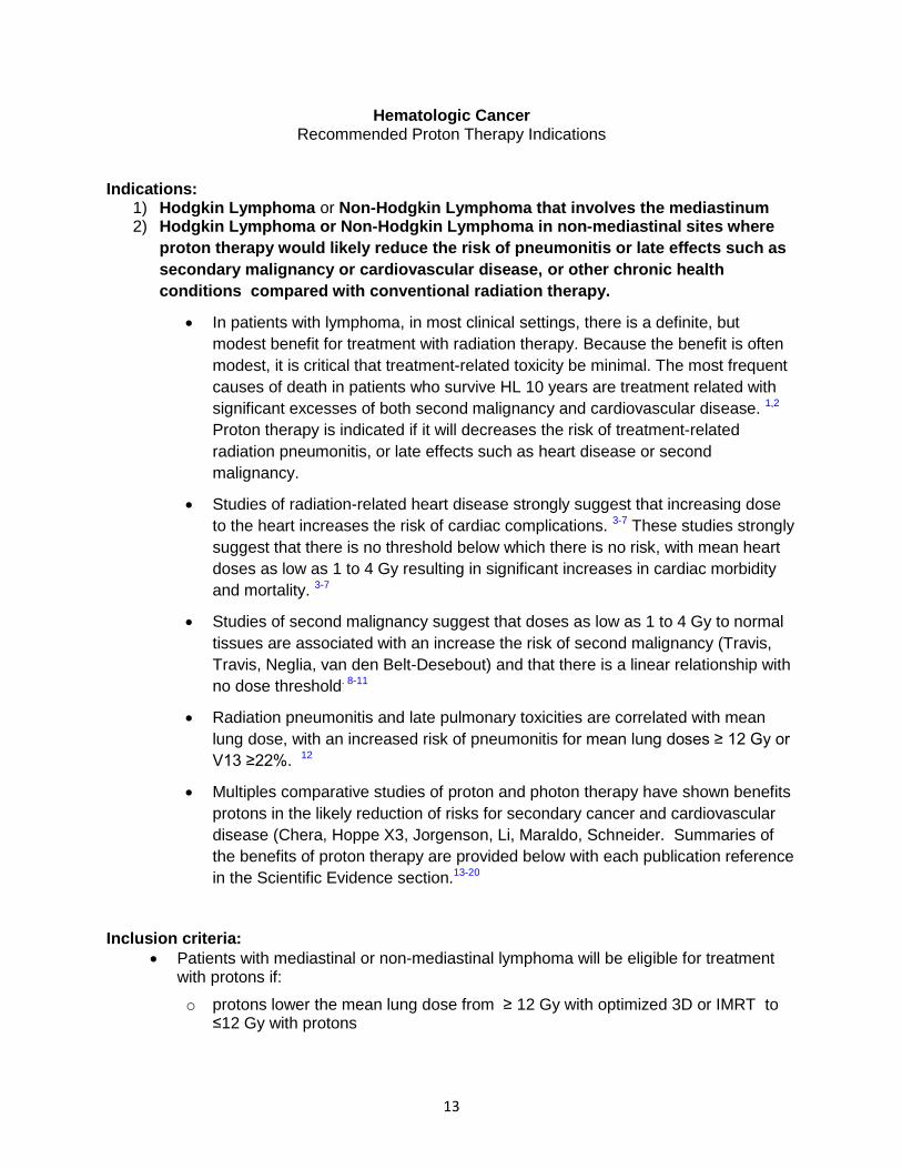
Hematologic Cancer Recommended Proton Therapy Indications
Indications: 1) Hodgkin Lymphoma or Non-Hodgkin Lymphoma that involves the mediastinum 2) Hodgkin Lymphoma or Non-Hodgkin Lymphoma in non-mediastinal sites where
proton therapy would likely reduce the risk of pneumonitis or late effects such as
secondary malignancy or cardiovascular disease, or other chronic health
conditions compared with conventional radiation therapy.
In patients with lymphoma, in most clinical settings, there is a definite, but
modest benefit for treatment with radiation therapy. Because the benefit is often
modest, it is critical that treatment-related toxicity be minimal. The most frequent
causes of death in patients who survive HL 10 years are treatment related with
significant excesses of both second malignancy and cardiovascular disease. 1,2
Proton therapy is indicated if it will decreases the risk of treatment-related
radiation pneumonitis, or late effects such as heart disease or second
malignancy.
Studies of radiation-related heart disease strongly suggest that increasing dose
to the heart increases the risk of cardiac complications. 3-7 These studies strongly
suggest that there is no threshold below which there is no risk, with mean heart
doses as low as 1 to 4 Gy resulting in significant increases in cardiac morbidity
and mortality. 3-7
Studies of second malignancy suggest that doses as low as 1 to 4 Gy to normal
tissues are associated with an increase the risk of second malignancy (Travis,
Travis, Neglia, van den Belt-Desebout) and that there is a linear relationship with
no dose threshold. 8-11
Radiation pneumonitis and late pulmonary toxicities are correlated with mean
lung dose, with an increased risk of pneumonitis for mean lung doses ≥ 12 Gy or
V13 ≥22%. 12
Multiples comparative studies of proton and photon therapy have shown benefits
protons in the likely reduction of risks for secondary cancer and cardiovascular
disease (Chera, Hoppe X3, Jorgenson, Li, Maraldo, Schneider. Summaries of
the benefits of proton therapy are provided below with each publication reference
in the Scientific Evidence section.13-20
Inclusion criteria:
Patients with mediastinal or non-mediastinal lymphoma will be eligible for treatment with protons if:
o protons lower the mean lung dose from ≥ 12 Gy with optimized 3D or IMRT to ≤12 Gy with protons

14
o protons lower the volume of lung receiving 13 Gy from ≥ 22% with optimized 3D or IMRT to ≤22% with protons
o protons result in a ≥ 1 Gy reduction in the average dose to the heart
o protons lower the mean dose to breast, stomach, brain, or other organ by ≥ 1 Gy
.
Exclusion criteria:
Lymphoma that is beyond any reasonable hope of cure unless :
o protons lower the mean lung dose from ≥ 12 Gy with optimized 3D or IMRT to ≤12 Gy with protons
o protons lower the volume of lung receiving 13 Gy from ≥ 22% with optimized 3D or IMRT to ≤22% with protons
Scientific Evidence:
1. Oeffinger KC, Mertens AC, Sklar CA, Kawashima T, Hudson MM, Meadows AT,
Friedman DL, Marina N, Hobbie W, Kadan-Lottick NS, Schwartz CL, Leisenring W,
Robison LL. Chronic health conditions in adult survivors of childhood cancer. N Engl
J Med 2006. 355(15): 1572-82.
Among survivors, the cumulative incidence of a chronic health condition reached
73.4% 30 years after the cancer diagnosis.
2. Castellino SM, Geiger AM, Mertens AC, Leisenring WM, Tooze JA, Goodman P,
Stovall M, Robison LL, Hudson MM. Morbidity and mortality in long-term survivors of
Hodgkin lymphoma: a report from the Childhood Cancer Survivor Study. Blood
2011. 117: (6): 1806-16.
There is an excess of overall mortality, death from second malignant neoplasms
and cardiovascular disease in survivors of Hodgkin lymlphoma with persists >20
years after treatment.
3. Char ZA, et al. Coronary heart disease after radiotherapy for peptic ulcer disease. Int
J R Oncol Biol Phys 61:842-850, 2005
A statistically significant relationship was observed between coronary heart
disease average dose to the heart in the 0-7.6 Gy range. The study is important
in that it shows that even very low doses (2 Gy or more) may be associated with
increased risk of coronary artery heart disease.
4. Darby, SC, et al. Risk of ischemic heart disease in women after radiotherapy for
breast cancer. New Eng J Med 11:987-998, 2013
Cardiac risk strongly related to cardiac dose with no obvious threshold
5. Hancock, et al. Factors affecting later mortality from heart disease after treatment of
Hodgkin’s Disease. JAMA. 270:1949-1955, 1993:.
A cardiac dose of more than 30 Gy was associated with a three-fold higher risk of
death from cardiac disease.

15
6. Mulrooney DA, Yeazel MW, Kawashima T, Mertens AC, Mitby P, Stovall M,
Donaldson SS, Green DM, Sklar CA, Robison LL, Leisenring WM. Cardiac
outcomes in a cohort of adult survivors of childhood and adolescent cancer:
retrospective analysis of the Childhood Cancer Survivor Study cohort. BMJ 2009.
339.
Cardiac radiation exposure of 15 Gy or more increased the relative hazard of
congestive heart failure, myocardial infarction, pericardial disease, and valvular
abnormalities by twofold to sixfold compared to non-irradiatied survivors.
7. Tukenova M, Guibout C, Oberlin O, Doyoon F, Mousannif A, Haddy N, Guierin S,
Pacquement H, Aouba A, Hawkins M, Winter D, Bourhis J, Lefkopoulos D, Diallo I,
De Vathaire F. Role of cancer treatment in long-term overall and cardiovascular
mortality after childhood cancer. J Clin Oncol. 2010.. 28(8): 1308-15.
A cardiac dose of more than 5 Gy was associated an increase relative risk of
cardiac mortality of 12.5 and of 25 for >15 Gy to the heart.
8. Travis LB, Hill DA, Dores GM, Gospodarowicz M, van Leeuwen FE, Holowaty E,
Glimelius B, Andersson M, Wiklund T, Lynch CF, Van’t Veer MB, Glimelius I, Storm
H, Pukkala E, Stovall M, Curtis R, Boice JD, Jr, Gilber E. Breast cancer following
radiotherapy and chemotherapy among young women with Hodgkin disease. JAMA
2003. 290(4): 465-475.
A radiation dose of 4 Gy or more to the breast was associated with a 3.2 fold
increased risk of breast cancer in HD survivors. The dose response was linear.
9. Travis LB, Gospodarowicz M, Curtis RE. et al. Lung cancer following chemotherapy and radiotherapy for Hodgkin's disease. J Natl Cancer Inst.2002;94:182-192.
A radiation dose of 5 Gy or more was associated with a 5.9 fold increased relative risk of lung cancer in Hodgkin’s disease survivors; the dose response was linear.
10. Neglia JP, Robison LL, Stovall M, Liu Y, Packer RJ, Hammond S, Yasui Y, Kasper
CE, Mertens AC, Donaldson SS, Meadows AT, Inskip PD. New primary neoplasms
of the central nervous system in survivors of childhood cancer: a report from the
Chidhod Cancer Survivor Study. J natl Cancer Inst 2006 98(21): 1528-37.
Radiation exposure was associated with increased rsk of subsequent glioma (OR
= 6.78) and meningioma (OR = 9.94) with a linear relative risk dose response.
11. Van den Belt-Desebout, Aleman BM, Besseling G, de Bruin ML, Haupfmann M, van’t
Veer MB, de Wit R, Ribot JG, Noordijk EM, Kerst JM, Gietema JA, van Leeuwen FE.
Roles of radiation dose and chemotherapy in the etiology of stomach cancer as a
second malignancy. Int J Radiat Oncol Biol Phys 2009. 75(5): 1420-9.
The risk of stomach cancer after treatment for Hodgkin’s lymphoma or testicular
cancer increased with increasing mean stomach dose at an estimated relative
risk of 0,84 per Gy.
16
12. Sepenwoolde Y, et al. Comparing different NTCP models that predict the incidence
of radiation pneumonitis. Int J Rad Oncol Biol Phys 55:724-735, 2003.
The risk of radiation pneumonitis in 382 patients with breast cancer, lymphoma
and lung cancer was assessed in relation to a variety of measures of radiation
dose to the lungs. The risk of pneumonitis was estimated to be more than 5% if
the mean lung dose was greater than approximately 12 Gy or if the volume of
lung receiving more than 13 Gy (V13) was more than 23% (see Figure 2).
13. Chera, et al. Dosmetric comparison of three different involved nodal irradiation
techniques for stage II Hodgkin’s lymphoma patients: Conventional radiotherapy,
intensity-modulated radiotherapy and three-dimensional proton radiotherapy. Int. J.
Radiation Oncology Biol. Phys., Vol. 75, No. 4, pp. 1173–1180, 2009
Mean breast dose lower; 1.94 Gy for CRT, 3.74 Gy for IMRT, 1.59 Gy for
3DPRT.
Mean lung dose: 4.83, 5.38 and 30.4
In general, the advantage for protons is seen in volume receiving relatively low
dose (<15 Gy, see figures 3, 4 and 5).
14. Hoppe, Bradford et al, Involved-Node Proton Therapy in Combined Modality Therapy
for Hodgkin Lymphoma: Results of a Phase 2 Study. Int J Radiation Oncol Biol Phys,
Vol. 89, No. 5, pp. 1053e1059, 2014
Progressively lower average integral dose, and average dose to heart, lungs,
breast, thyroid and esophagus when 3D, IMRT and proton plans were compared
in 15 patients treated with INPT after chemotherapy. Three year EFS 93%.
15. Hoppe, Bradford, et al. Effective dose reduction to cardiac structures using protons
compared with 3DCRT and IMRT in mediastinal Hodgkin Lymphoma. Int J Radiation
Oncol Biol Phys, Vol. 84, No. 2, pp. 449e455, 2012
Highly significant decrease in dose with comparison of PT vs 3D or IMRT to
multiple critical organs with proton therapy, including heart, L ventricle, R
ventricle, L atrium, mitral valve, tricuspid valve, aortic valve (significant only for
3D vs PT), LAD, L circumflex, R circumflex (significant only for 3D vs PT),
pulmonary artery (significant only for 3D vs PT), and ascending aorta (significant
only for IMRT vs PT).
16. Hoppe, Bradford, et al. Consolidative Involved-Node Proton Therapy for Stage IA-IIIB
Mediastinal Hodgkin Lymphoma Preliminary Dosimetric Outcomes From a Phase II
Study. Int J Radiation Oncol Biol Phys, Vol. 83, No. 1, pp. 260e267, 2012
“PT provided the lowest mean dose to the heart, lungs, and breasts for all 10
patients compared with either 3D-CRT or IMRT.”
17. Jorgensen, et al. The effect of esophagus after different radiotherapy techniques for
early stage Hodgkin’s lymphoma. Acta Oncologica, 52: 1559–1565, 2013

17
“Mean dose to the esophagus was 16.4, 16.4, 14.7 and 34.2 Gy (p 0.001) with
3DCRT, VMAT, PT and MF treatment, respectively. No differences were seen in
the estimated risk of developing esophagitis, stricture or cancer with 3DCRT
compared to VMAT (p =1.000, p =1.000, p = 0.356). PT performed significantly
better with the lowest risk estimates on all parameters compared to the photon
treatments, except compared to 3DCRT for stricture (p = 0.066).”
18. Li, et al. Rationale for and preliminary results of proton beam therapy for mediastinal
lymphoma. International Journal of Radiation Oncology, Biology, Physics. 81(1):167-
74, 2011
10 patients, “PBT delivered lower mean doses to the lung (6.2 vs. 9.5 Gy),
esophagus (9.5 vs. 22.3 Gy), and heart (8.8 vs. 17.7 Gy) but not the breasts (5.9
vs. 6.1 Gy) than did conventional RT.”
19. Maraldo, et al. Estimated risk of cardiovascular disease and secondary cancers with
modern highly conformal radiotherapy for earl-stage mediastinal Hodgkin lymphoma.
Annals of Oncology 24: 2113–2118, 2013
Compared to arc IMRT (VMAT) or 3D conventional therapy, highly significant
estimated benefit for protons as measured by cardiac mortality, cardiac
morbidity, MI, valvular disease (only VMAT vs PT significant), lung ca, breast ca
and life years lost.
20. Scheneider, et al. Comparative risk assessment of secondary cancer incidence after
treatment of Hodgkin’s disease with photon and proton radiation. Radiation Research
154, 382--388 (2000)
This is basically a case report in which the risk of second cancer is calculated for
several different kinds of plans (2 field photons, IMRT, and two different proton
plans). “Irradiation with protons using the spot scanning technique decreases the
avoidable cancer incidence compared to photon treatment by a factor of about
two.”

18
Prostate Cancer Recommended Proton Therapy Indications
Indications:
1) Delivering a high radiation dose to the primary tumor in the prostate and/or seminal vesicles has become an important aspect of the optimal management of clinically localized prostate carcinoma in the radiotherapy setting. This is the result of multiple phase III studies demonstrating that a higher radiation dose reduces the risk of prostate cancer recurrence1-6. However, a higher radiation dose is inherently associated with an increased risk of radiation-related gastrointestinal and genitourinary toxicity1-6. Unlike conventional X-rays, protons have a physical property to deposit most of their energy only when they reach their target. This allows protons to deliver a radiation dose to the target more preferentially, while minimizing a dose to the nearby normal organs. Thus, a delicate balance of delivering a high radiation dose to eradicate prostate cancer while largely sparing the nearby normal organs (such as rectum and bladder) can be achieved much better with proton therapy than with conventional x-rays.
2) Proton therapy can be at least as efficacious as conventional external beam radiotherapy in treating clinically localized (encompassing low-risk, intermediate-risk, and high-risk) prostate cancer, while reducing the risk of acute and late side effects of radiotherapy6-8, 14. At present, a phase III study is in progress to compare proton therapy with conventional external beam radiotherapy using intensity modulated radiotherapy (IMRT). Table 1 depicts the comparison between conventional external beam radiotherapy and proton therapy with respect to therapeutic efficacy in terms of biochemical relapse-free rate, based on some of published manuscripts and abstracts. Biochemical relapse [also called ‘PSA (prostate specific antigen) relapse’] is a widely accepted surrogate representing prostate cancer recurrence. Table 2 compares the incidences of acute and late radiation toxicity between conventional external beam radiotherapy and proton therapy. Additionally, the comparison of patient-reported quality of life between two prospectively collected databases suggests approximately 50% reduction in problems with significant bowel urgency and frequency in patients treated with proton therapy compared with IMRT13.
3) Clinical and laboratory studies have suggested that prostate cancer has a relatively slow rate of proliferation, characterized by a low α/β value (1.5 to 3 Gy) in a linear quadratic model for cell survival after irradiation. This implies that a larger radiation dose per fraction (i.e. hypofractionation) is more effective in cell killing for prostate cancer than simply adding more fractions. Another major advantage of hypofractionation is its convenience and cost effectiveness, as it allows a shorter treatment duration with a reduced number of radiation fractions. Table 3 shows phase III studies comparing a conventional dose-fractionation regimen with a moderate hypofractionation regiment in a conventional external beam setting. It has been shown that a moderate hypofractionation regimen can be efficacious as a conventional dose-fractionation schedule without an increase in the risk of radiation morbidity. Thus, in a proton therapy setting, the incorporation of a moderate hypofractionation regimen is appropriate for properly selected clinical situations. The University of Florida recently submitted an abstract to the annual meeting of 2015 ASTRO that describes the outcome of a prospective trial of hypofractionated proton therapy in 228 men with low- or intermediate-risk prostate cancer who had 5 years of follow-up. This study reports that a hypofractionated proton therapy (28-29 fractions over 5 ½ weeks) can yield outcomes

19
similar to those achieved with a standard fractionation proton therapy (39-42 fractions over 8 weeks) in selected patients 15. Patient included in the trial were men with prostates < 60 cc in volume, IPSS (International Prostate Sympton Score) <15, and no previous required treatment with either alpha reductase inhibitors (Flomax, Hytrin, etc) or anticoagulation (Plavix, Coumadin, etc). This hypofractionation strategy would be similar in cost to standard fractionation IMRT, but more convenient for patients and potentially yielding better disease control and quality of life.
Inclusion criteria:
Age ≥ 18 years
Histologic confirmation of prostate cancer
Low, intermediate, or high risk prostate cancer without evidence of distant metastases
When regional pelvic node irradiation is required in patients with high risk disease, proton therapy can be used for both the pelvic nodes and the prostate, or as a prostate boost following conventional external beam radiotherapy to the pelvic nodes.
Whenever clinically appropriate, a moderate hypofractionation regimen (26 to 28 fractions) can be utilized to maximize cost-effectiveness of proton therapy.
Clinically palpable or radiographically evident local recurrence of prostate cancer following radical prostatectomy for which definitive salvage radiotherapy is indicated
Special circumstances in which definitive radiation is required, and the effort to minimize a radiation dose to normal organs is critically important due to other co-morbid medical issues: patients with inflammatory bowel disease, prior pelvic irradiation for non-prostate cancer, or hip prosthesis.
Exclusion criteria:
Presence of distant metastasis
Medical contraindication to receipt of radiotherapy.
Severe active co-morbid systemic illnesses or other severe concurrent disease which, in the judgment of the clinician, would make the patient inappropriate for radiotherapy.
Very low risk prostate cancer (CST1C and PSA <10 and PSA index <0.15 and <3 cores involved and <50% maximum core involvement and Gleason ≤6) and life expectancy of < 10 years.
Scientific Evidence:
1. Al-Mamgani A, van Putten WL, Heemsbergen WD, van Leenders GJ, Slot A, Dielwart
MF, et al. Update of Dutch multicenter dose-escalation trial of radiotherapy for localized
prostate cancer. Int J Radiat Oncol Biol Phys. 2008;72:980-8.
2. Beckendorf V, Guerif S, Le Prise E, Cosset JM, Bougnoux A, Chauvet B, et al. 70 Gy
versus 80 Gy in localized prostate cancer: 5-year results of GETUG 06 randomized trial.
Int J Radiat Oncol Biol Phys. 2011;80:1056-63.

20
3. Dearnaley DP, Sydes MR, Graham JD, Aird EG, Bottomley D, Cowan RA, et al.
Escalated-dose versus standard-dose conformal radiotherapy in prostate cancer: first
results from the MRC RT01 randomised controlled trial. Lancet Oncol. 2007;8:475-87.
4. Kuban DA, Tucker SL, Dong L, Starkschall G, Huang EH, Cheung MR, et al. Long-term
results of the M. D. Anderson randomized dose-escalation trial for prostate cancer. Int J
Radiat Oncol Biol Phys. 2008;70:67-74.
5. Michalski J, Moughan J, Purdy J, Bosch W, Bahary J-P, Lau HY, et al. A randomized
trial of 79.2Gy versus 70.2Gy radiation therapy (RT) for localized prostate cancer
[abstract]. J Clin Oncol 33, 2015 (suppl 7, abstr 4).
6. Zietman AL, Bae K, Slater JD, Shipley WU, Efstathiou JA, Coen JJ, et al. Randomized
trial comparing conventional-dose with high-dose conformal radiation therapy in early-
stage adenocarcinoma of the prostate: long-term results from proton radiation oncology
group/american college of radiology 95-09. J Clin Oncol. 2010;28:1106-11.
7. Mendenhall NP, Hoppe BS, Nichols RC, Mendenhall WM, Morris CG, Li Z, et al. Five-
year outcomes from 3 prospective trials of image-guided proton therapy for prostate
cancer. Int J Radiat Oncol Biol Phys. 2014;88:596-602.
8. Slater JD, Rossi CJ, Jr., Yonemoto LT, Bush DA, Jabola BR, Levy RP, et al. Proton
therapy for prostate cancer: the initial Loma Linda University experience. Int J Radiat
Oncol Biol Phys. 2004;59:348-52.
9. Pollack A, Walker G, Horwitz EM, Price R, Feigenberg S, Konski AA, et al. Randomized
trial of hypofractionated external-beam radiotherapy for prostate cancer. J Clin Oncol.
2013;31:3860-8.
10. Arcangeli S, Strigari L, Gomellini S, Saracino B, Petrongari MG, Pinnaro P, et al.
Updated results and patterns of failure in a randomized hypofractionation trial for high-
risk prostate cancer. Int J Radiat Oncol Biol Phys. 2012;84:1172-8.
11. Michalski JM, Yan Y, Watkins-Bruner D, Bosch WR, Winter K, Galvin JM, et al.
Preliminary toxicity analysis of 3-dimensional conformal radiation therapy versus
intensity modulated radiation therapy on the high-dose arm of the Radiation Therapy
Oncology Group 0126 prostate cancer trial. Int J Radiat Oncol Biol Phys. 2013;87:932-8.
12. Arcangeli G, Fowler J, Gomellini S, Arcangeli S, Saracino B, Petrongari MG, et al. Acute
and Late Toxicity in a Randomized Trial of Conventional Versus Hypofractionated Three-
Dimensional Conformal Radiotherapy for Prostate Cancer. Int J Radiat Oncol.
2011;79:1013-21.
13. Hoppe BS, Michalski JM, Mendenhall NP, Morris CG, Henderson RH, Nichols RC, et al.
Comparative effectiveness study of patient-reported outcomes after proton therapy or
intensity-modulated radiotherapy for prostate cancer. Cancer. 2014;120:1076-82.
14. Nihei K, Ogino T, Onozawa M, Murayama S, Fuji H, Murakami M, et al. Multi-institutional
phase II study of proton beam therapy for organ-confined prostate cancer focusing on
the incidence of late rectal toxicities. Int J Radiat Oncol Biol Phys. 2011;81:390-6

21
15. Henderson RH, personal communication from abstract submitted to ASTRO for 2015 fall
meeting.
Appendix:
Table 1: Comparison between photon vs. proton for biochemical relapse-free rate, based on
some of the prospective studies
Study F/U
(years)
N Treatment Biochemical relapse-free rate
Photon
RTOG0126 (2015)5 7 748 79.2 Gy in 44 fractions
(intermediate risk)
84% at 5 years
Fox Chase Cancer
Center (2013)9
5.7 153 76 Gy in 38 fractions
(mainly, intermediate- and high-risk)
78.6% at 5 years
Italy (2012)10
5.8 85 80 Gy in 40 fractions
(intermediate- and high-risk)
79% at 5 years
Proton
Univ. Florida
(2014)7
5.2 211 78 GyE (Gy): low risk
78-82 GyE (Gy): intermediate risk
78 GyE (Gy) + weekly docetaxel + 6-month
ADT: high risk
99% at 5 yrs for low risk;
99% at 5 yrs for intermediate risk;
76% at 5 yrs for high risk
Japan (2011)14
3.6 151 74GyE in 37 fractions
(low- and intermediate-risk)
94% at 3 years
Proton Radiation
Oncology Group
(2010)6
8.9 197 28.8 GyE in 16 fractions (proton) +
50.4 Gy in 28 fractions (photon)
( low- and intermediate-risk)
82.6% at 10 years
ADT: Androgen deprivation therapy

22
Table 2: Comparison between photon vs. proton for radiation toxicity
Study Tool for toxicity
assessment
RT
dose
F/U
(yrs)
N Acute toxicity Late toxicity
GI GU GI GU
G2 ≥ G3 G2 ≥ G3 G2 ≥ G3 G2 ≥ G3
Photon
RTOG0126
(IMRT) (2013)11
CTCAE v2.0
and
RTOG/EORTC
79.2
Gy
in 44 f
3.5 257 ≥ G2 GI or GU: 9.7% ≥ G2:
15.1%
at 3 yrs
≥ G3:
2.6%
at 3 yrs
RTOG0126
(IMRT/3D-CRT)
(2015)5
7 748 ≥ G2 GI:
2.4%
≥ G2 GU:
11.1%
≥ G2:
21%
at 5 yrs
≥ G3:
5%
at 5 yrs
≥ G2:
12%
at 5 yrs
≥ G3:
3%
at 5 yrs
Fox Chase
Cancer Center
(IMRT) (2013)9
LENT/RTOG
(similar to
CTCAE v4.0)
76 Gy
in 38 f
5.7 153 20.5%
at 5 yrs
2%
at 5 yrs
≥ G2: 37.9%
at 5 yrs
(or 13.4% with
modified criteria)
70.2
Gy
in 26 f
154 16.1%
at 5 yrs
2%
at 5 yrs
≥ G2: 39.1%
at 5 yrs
(or 21.5% with
modified criteria)
Italy (3D-CRT)
(2011)12
RTOG/EORTC
for acute
toxicity;
LENT-SOMA
for late toxicity
80 Gy
in 40 f
2.9 85 ≥ G2 GI:
21%
≥ G2 GU:
40%
≥ G2 rectal: 12% ≥ G2 GU: 6%
62 Gy
in 20 f
2.7 83 ≥ G2 GI:
35%
≥ G2 GU:
47%
≥ G2 rectal: 14% ≥ G2 GU: 8%
Proton
Univ. Florida
(2014)7
CTCAE v3.0 78-82
GyE
5.2 211 0.5% 0.5
%
1% at 5
yrs
(0.5%, if
CTCAE
v4.0)
5.4%
at 5 yrs
(0.9%, if
CTCAE
v4.0)
Japan (2011)14
CTCAE v2.0 74
GyE
3.6 151 0.7
%
0% 12% 0% 2%
at 2 yrs
0%
at 2 yrs
4.1%
at 2 yrs
0%
at 2 yrs
Table 3: Phase III studies evaluating hypofractionation regimens: photons
Study F/U
(years)
Patients N Treatment Biochemical
relapse-free rate
Fox Chase Cancer
Center (2013)9
5.7 Low- to High-risk;
(mainly, intermediate-
and high-risk); (ADT
for intermediate- and
high-risk)
153 76 Gy in 38 fractions
(2 Gy/f)
78.6% at 5 years
P =
0.745 154 70.2 Gy in 26 fractions
(2.7 Gy/f)
76.7% at 5 years
Italy (2012)10
5.8
Intermediate- and High-
risk; (all had 9-month
ADT)
85
80 Gy in 40 fractions
(2 Gy/f)
79% at 5 years
P =
0.065
83
62 Gy in 20 fractions over
5 weeks (4 fractions/week)
(3.1 Gy/f)
85% at 5 years
ADT: Androgen deprivation therapy

23
Thoracic Cancer Recommended Proton Therapy Indications
Indications:
1) Stage III Lung cancer, which is pathologically confirmed.
Inclusion criteria:
Specific dosimetric conditions must be present for a patient to be qualified to receive PBT rather than Conventional radiotherapy (C-RT (either Intensity modulated RT or 3D-RT)). This will require generating comparative plans demonstrating any one of the following:
o PBT keeps the mean lung dose < 20 Gy when conventional radiotherapy cannot. (C-RT=3D-RT or IMRT)
This will keep the risk of grade >3 radiation pneumonitis to <20% instead of >30%; Wang et al.1)
o PBT keeps the V20 (Volume of lung receiving >20 Gy) less than 30% when C-RT cannot.
This will keep pneumonitis fatalities to <1% instead of >2.9% (Palma et al.2)
o PBT is indicated to spare cardiac toxicity/fatalities by reducing V5 (volume of heart receiving >5Gy) &/or V30
RTOG 0617 found these were associated with survival3. However, the exact cut-offs have not been firmly established. This will likely be defined in the next 2 years.
o PBT is indicated to spare cardiac toxicity by reducing the mean cardiac dose to <13.5 Gy when C-RT cannot.
This will keep long term cardiac event to less than 2x the normal risk (ref. Darby4)
o PBT can keep the V60 for the esophagus to <17% when C-RT cannot.
This will keep the incidence of grade > 3 radiation esophagitis to <10% instead of 22% (Palma et al.5).
Exclusion criteria:
Patients treated for palliation alone (generally this is with doses less than 60 Gy and
often without chemotherapy)

24
Stage I lung cancer
Indications:
o Stage I (T2N0M0) lung cancer, when treated in a hypofractionated
fashion
o Stage I (T1N0M0) lung cancer, treated in a hypofractionated fashion, if
organ-at-risk constraints cannot be met with photon SBRT
Inclusion criteria: Protons are appropriate for patients with T2N0 non-small cell lung cancer, when given in a hypofractionated course. Protons given in SBRT/SABR or hypofractionated course may be appropriate for T1N0, if the following parameters cannot be met with IMRT/ 3-D conformal techniques:
Lung V5 – 40%
Lung V20 – 20%
Heart maximum dose 40 Gy
Spinal cord maximum dose 20 Gy
Bronchial tree maximum dose 40 Gy
Scientific Evidence: In an analysis of compiled data from a national database, patients with T2N0 non-small cell lung cancer treated with higher effective doses of SBRT (stereotactic body radiation therapy) – significantly better overall survival at 2 and 4 years.6 Survivals at 2 years were approximately 57% for higher biologically equivalent doses vs 40% for lower doses, and at 4 years the differences persisted (approximately 35% vs 20%). In a review from multiple series of patients who have received high dose (high BED) protons as stereotactic ablative treatment (SABR) or hypofractionated treatment (typically 10 or fewer treatments), the two year overall survivals are 95-98%, 3 year survivals are 60-90%, with 4 years survival of 51%.7 In addition, planning studies show that many of the patients with centrally located T1 or T2 lesions are not candidates for SBRT/SABR with x-rays, because of the inability to meet dose constraints for heart, esophagus, lungs, brachial plexus or bronchial tree.8
1. Wang S, Liao Z, Wei X, et al. Analysis of clinical and dosimetric factors associated with
treatment-related pneumonitis (TRP) in patients with non-small-cell lung cancer
(NSCLC) treated with concurrent chemotherapy and three-dimensional conformal
radiotherapy (3D-CRT). Int J Radiat Oncol Biol Phys. 2006;66: 1399-1407.
2. Palma DA, Senan S, Tsujino K, et al. Predicting radiation pneumonitis after
chemoradiation therapy for lung cancer: an international individual patient data meta-
analysis. Int J Radiat Oncol Biol Phys. 2013;85: 444-450.
3. Bradley JD, Paulus R, Komaki R, et al. Standard-dose versus high-dose conformal
radiotherapy with concurrent and consolidation carboplatin plus paclitaxel with or without
cetuximab for patients with stage IIIA or IIIB non-small-cell lung cancer (RTOG 0617): a
randomised, two-by-two factorial phase 3 study. Lancet Oncol. 2015;16: 187-199.a

25
4. Darby SC, Ewertz M, McGale P, et al. Risk of ischemic heart disease in women after
radiotherapy for breast cancer. N Engl J Med. 2013;368: 987-998.
5. Palma DA, Senan S, Oberije C, et al. Predicting esophagitis after chemoradiation
therapy for non-small cell lung cancer: an individual patient data meta-analysis. Int J
Radiat Oncol Biol Phys. 2013;87: 690-696.
6. Koshy et al, Increasing radiation therapy dose is associated with improved survival in
patients undergoing stereotactic body radiation therapy for stage I non-small-cell lung
cancer. Int J Radiat Oncol Biol Phys 2015; 91:344-50.
7. Grant JD, Chang JY. Proton-based stereotactic ablative radiotherapy in early-stage
non-small-cell lung cancer. Biomed Res Int, 2014; 2014:389048,
doi:10.1155/2014/389048, Epub 2014 Jul 17.
8. Register, et al, Proton stereotactic body radiation therapy for clinically challenging cases
of centrally and superiorly located stage I non-small-cell lung cancer. Int J Radiat Oncol
Biol Phys 2010; 80:1015-22.