RECOMMENDATIONS IN FORENSIC recomend ANTHROPOLOGY · Association of Forensic Anthropology and...
145
RECOMMENDATIONS IN FORENSIC ANTHROPOLOGY SPANISH ASSOCIATION OF FORENSIC ANTHROPOLOGY AND ODONTOLOGY June 2013 Coordinator: F Serrulla
Transcript of RECOMMENDATIONS IN FORENSIC recomend ANTHROPOLOGY · Association of Forensic Anthropology and...

RECOMMENDATIONSIN FORENSICANTHROPOLOGY
SPANISH ASSOCIATION OF FORENSICANTHROPOLOGY AND ODONTOLOGY
June 2013 Coordinator: F Serrullareco
men
d


It´s a publication ofAssociation of Forensic Anthropology and Odontology
www.aeaof.com
DEPOSITO LEGAL; OU 117-2013
PRINTED IN: Valpapeis S.L.


Coordinator of the documentDr. Fernando Serrulla Rech
Translation Spanish into EnglishHande Arli
Pinar Çelebioglu Cera SanzPedro Garamendi
(EU Twinning Project TR 08 IB JH 01 Improvingthe skills of Forensic Experts in Turkey)

Introduction
Since the eighties, in which Forensic Anthropology take its first steps in Spain as a discipline embedded in Forensic Science, a number of concerns were outlined among the professionals dedicated to it that are tackled initially in an individual way and later collectively. This makes us consider, on the one hand, the need to organize ourselves and have a series of activities and tools that allow us to get to know each other and the works and activities we develop in our respective centers, on the other hand, the desire to overcome this first stage through agreements that will enable all of us to have and to use if necessary, some protocols that might be used as basis for the reports carried out in each laboratory.
The first of these concerns, the need to organize ourselves, is reflected after a number of attempts and the start up of the creation of the Spanish Association of Forensic Anthropology and Odontology (AEAOF) in 2006. The second one, to have some available protocols, begins to be developed with the proposal of creating some working groups among the AEAOF's members who will be in charge of drawing what at first was proposed as "AEAOF's protocols". Subsequently and following analysis and discussion of the proposals it was concluded most appropriate to establish a number of recommendations to assist and inspire the elaboration of anthropological-forensic reports. Finally, at the meeting held in May 2012, at the National Institute of Toxicology and Forensic Science in Madrid (IV Scientific Meeting), great unanimity was achieved on the final text that under the name "Recomendations in Forensic Anthropology" we offer to all AEAOF's members and also to those professionals who might be interested in this discipline.
I would only like to add that I hope and we all hope that this protocols will be found useful for everyone.
Jose Antonio Sánchez Sánchez.President of the AEAOF.
RECOMENDATIONS IN FORENSIC ANTHROPOLOGY
Spanish Association of Forensic Anthropology and Odontology. June 2013
5


Recommendations
This text has been drawn up in the 2nd Scientific Meeting of the Spanish Association of Forensic Anthropology and Odontology (AEAOF) held in Donostia-San Sebastián in 2010. It's the result of the need to standardize procedures as a step towards the accreditation of professionals and laboratories in Spain. This need is recognized by the majority of professionals of Forensic Anthropology in Spain.
The work was divided among several partners who showed their interest in the different topics raised and after the elaboration of a basic text for debate, it was forwarded for consideration by all the members of the association, by e-mail and during the 3rd and 4th Scientific Meetings of the AEAOF. Efforts have been made to ensure that the authors highlighted in every topic the internationally accepted methods. At the 3rd Scientific Meeting held in Madrid, the different topics provided were intensively discussed, and some specific issues were decided by the majority (age-range, terminology of the age,…). With many of the modifications proposed it was agreed to differentiate on the one hand what we called 'Diagnostic Criteria for Identification' (sex, age, size and ancestral origin estimation and identification criteria) and the rest of recommendations.
With all of these we get to the 4th Scientific Meeting held at the National Institute for Toxicology and Forensic Science Headquarters in Madrid, during which little proposals were made for changing the text of Diagnostic Criteria for Identification. Aditionally, during this meeting it was also decided a change of the term “protocols” as it was originally written in the document by the term “recommendations”.
During 2013 the rest of the documents have been subject to discussion ending up with a few little modifications.
We thank to the team of the Twinning Project in Turkey (Pedro Garamendi, Hande Arli and Pinar Çelebioglu) for his kindness, dedication and time in the English translation of the “Recomendations in Forensic Anthropology”.
RECOMENDATIONS IN FORENSIC ANTHROPOLOGY
Spanish Association of Forensic Anthropology and Odontology. June 2013
7

It is also necessary to thank all our authors for their efforts, not only for drafting the texts but for accepting the modifications and critics done during these years too. Therefore it is already a document that belongs to everyone and is for everyone.
It is as well, an open document, that is to say, a not definitive document, a document that will have to be improved over the years to come with the contributions of those willing to make them.
Fernando Serrulla RechCoordinator of the document
RECOMENDATIONS IN FORENSIC ANTHROPOLOGY
Spanish Association of Forensic Anthropology and Odontology. June 2013
8

Authors
Inmaculada Alemán
Miguel Botella
Manuel Francisco Carrillo
Santiago Crespo
Enrique Dorado
Francisco Javier Fernández
Concha Magaña
Manuel Polo
José Luis Prieto
Mª Mar Robledo
José Antonio Sánchez
Fernando Serrulla
Victor Verano
Joan Viciano
RECOMENDATIONS IN FORENSIC ANTHROPOLOGY
Spanish Association of Forensic Anthropology and Odontology. June 2013
9


Table of Contents
1. Removal and Exhumation of Skeletal Remains. Crespo S, Polo M . . . . 13
2. Laboratory Guidelines. Alemán I, Botella MC . . . . . . . . . . . . . . . . . . . . 23 3. Anthropometric Data. Carrillo MF, Dorado E . . . . . . . . . . . . . . . . . . . . 33
4. Integrated Forensic Anthropology Report. Serrulla F, Verano V. . . . . . . 47
5. Diagnostic Criteria for Identification . . . . . . . . . . . . . . . . . . . . . . . . . . 53
Sex Estimation. Alemán I; Botella MC . . . . . . . . . . . . . . . . . . . . . . . 55
Age Estimation. Robledo MM, Sanchez JA . . . . . . . . . . . . . . . . . . . 71
Height Estimation. Dorado E, Carrillo MF . . . . . . . . . . . . . . . . . . . . 83
Ancestry Estimation. Prieto JL, Magaña C . . . . . . . . . . . . . . . . . . . 111
Identification Criteria. Serrulla F, Prieto JL, Verano V . . . . . . . . . . . 127
RECOMENDATIONS IN FORENSIC ANTHROPOLOGY
Spanish Association of Forensic Anthropology and Odontology. June 2013
11


REMOVAL AND EXHUMATIONOF SKELETAL REMAINS
13


The scope of Forensic Anthropology has grown over time, due to the growing problem of corpse identification. A clear example is the so-called events with multiple fatalities from natural disasters, collective accidents or terrorist attacks.
However, most elements under study in an anthropological-forensic laboratory belong to one or a few corpses which can be found partially or totally skeletonized, in bad conservation status or in advanced process of putrefaction.
These remains can be found above ground or buried. From the forensic point of view, most cases belong to the first group, while those in the second one are often found as a result of building works or as a result of a confession or testimony obtained during a police investigation.
On the other hand we need to take into consideration, and above all, depending on the work organization from the different Institutes of Legal Medicine, that not all medical-forensic experts have in-depth anthropology knowledge.
Due to all the above is necessary to set out the guidelines when finding a corpse to identify, that allow to collect as much information as possible from the corpses, the environment, the surrounding circumstances and its subsequent transfer to the anthropological and forensic laboratory.
Needless to say, the importance of this information for a judicial investigation.
May serve as an example that the study of the area of the discovery and the way the remains were found may lead to the origin of death. Moreover, data on death may be better reached if we study the scene's elements and if we relate those with the corpse.
1. GUIDELINES
At the news of the appearance of remains to identify, the first thing to do be done is to contact the anthropological-forensic Unit. The unit will consider, in the light of the preliminary information provided, the need to have a Forensic
PROTOCOL
AUTHORS
CONTACT
DATE
REMOVAL - EXHUMATION OF SKELETAL REMAIN
CRESPO, S. POLO, M.
[email protected] • [email protected]
February 2, 2013
RECOMENDATIONS IN FORENSIC ANTHROPOLOGY
Spanish Association of Forensic Anthropology and Odontology. June 2013
15

Anthropologist on the scene, and if he goes, he will act as adviser to the medical-forensic expert and will provide guidance on the different stages of the removal.
One point that has to be mentioned, it will be an action that will require time, therefore there is no rush. It can even be postponed if environmental or personal security circumstances are adverse.
Our actions will depend on whether the remains are above the surface or buried.
1.1. Above ground corpse.
We will establish two phases; a non-invasive one and an invasive one, depending on whether we enter or not the area surrounding the remains.
The area will be cordoned off.
In the non-invasive phase, we will be on the periphery of the remains and we will make an orderly and meticulous observation of the scene.
We will set the scene through sketches, photography or video. When using sketches, we will always point out the geographic north and measurements shall be made taking fixed not removable elements as reference.
The observation has to include the zone where the elements to study are (apartment, empty field, outdoors, stream, etc), the circumstances of the finding (first report, previous contamination), the actual environmental conditions (temperature, season), the annual environmental conditions (rainy, sunny, wet, dry climate), the circumstances previous to the finding (floods, works on public or private spaces), characteristics of the remains (corpse in deep decomposing, in conservation status or skeletonized), when finding bone remains will be relevant any abnormal distribution, concentration or position.
In the invasive phase, we will get into the perimeter where the remains to study are. Without touching, we make a first assessment of the origin (whether are or not bones or remains) and specie (human or animal). We are looking for elements that provide data about the identity. Thus, we pay special attention to the clothing, of which we shall describe type, fabric, size, brand, drawings, particular details (zippers, pockets), identification labels (particularly in persons who are institutionalized). Personal items (glasses, jewelry), personal documents (identity, social security number, credit cards). Surveys for biological signs like dry spills (blood, sperm, etc).
The next step would be to focus on the body or remains and conduct a preliminary study, on the basis of which some answers may be advanced that,
RECOMENDATIONS IN FORENSIC ANTHROPOLOGY
Spanish Association of Forensic Anthropology and Odontology. June 2013
16

although provisional, can shed light on the possible identification. Thus, for example, we could point out the sex and the approximate age interval. In the same manner, with minimal handling we may see if it is its original place or if there is the possibility of a previous transfer. We will study the loss of skeletal remains valuing a vital or postmortal origin in that loss.
If we notice any injury we will make the same assessment about whether or not its origin is vital. As an example, sometimes the position of a fractured limb offers guidance on this subject.
When this preliminary observations have been made, the collection of every fragment or remain will take place. This collection shall be carried out in an orderly manner, so that the bones and feet will be placed separately and indicating the side they belong to. Special protection shall be taken with ribs and head, given its fragility.
Pack must be made of a dry material such as paper bags. In the case of using plastic, we need to make sure there are small holes to allow ventilation. Concerning the teeth, we must always consider that they are easily detachable due to attachment loss, and we need to make sure that they are not to be lost.
All packaged bones will be put in a box designed to transfer remains or, failing this, in a shock-absorbing material box. Together with the remains shall be sent a judge's order to do an anthropological study and all kind of questions that may be raised. Likewise, the documentation consisting of the removal report or the collecting of remains report, that shall contain all the information about the activities taken on the place of the finding, will be sent. Obviously, this information is crucial to the forensic analysis.
1.2. Buried corpse.
In the case of buried remains, it will be necessary the involvement of an archaeologist or of qualified persons or with sufficient archeological knowledge, since the exhumation shall be done following the methods and techniques of this discipline.
First an appropriate planning is to be achieved, preventing in advance any contingency from the meteorological ones to the necessary materials and utensils.
All possible ante-mortem information need to be gathered, like identifying characteristics of the buried person (age, gender), clothing, associated pathology, dental work, particular features, complimentary clinic tests (image mainly), type of violence suffered, disappearance time, etc.
If the grave has to be located, careful attention would need to be paid to the
RECOMENDATIONS IN FORENSIC ANTHROPOLOGY
Spanish Association of Forensic Anthropology and Odontology. June 2013
17

characteristic of the terrain, irregularities, vegetation, etc.
It needs to be established whether the burial is individual or collective, isolated or adjacent to other places of burial or graves, primary or secondary (depending on whether or not there has been a previous transfer), if the burial is intact or has been altered (due to human actions, animals or other events). In the latter case we must point out that many forensic cases are altered exhumations due to the existence of previous construction work.
2. STEPS:
The area shall be clearly delimited using stakes and ropes, fixating the area, with photographs, mapping of the area through global positioning system.
Elimination of waste products and vegetation. Special care of the biological remains in the surface such as hair and clothes must be taken.
Uniform and horizontal elimination of the soil's upper layer. The outlines of the grave will be delimited. Any color change at the zone will be examined.
Elimination of excess soil. As the first remains appear, they will be determined by description, photography and cartography. Excavation taking special care not to damage skeletal remains. Avoid tool marks. Sift the soil for evidences (projectiles, ammunition shells, cigarettes, paper tissues or handkerchiefs, wrappers, cigarette butts, etc).
Ensure every finding. Confine the corps digging by its sides up to 50cm deep.
Display everything without altering the evidences. Special care must be taken when collecting the soil around the skull for any hair to submit it later to the laboratory.
Collect related remains and evidences. An inventory has to be conducted. Hands and feet kept separately, indicating laterality. Special care should be taken with the teeth since they may easily be lost. Record every evidence found, both those that may identify both the victim and those that may identify the aggressor.
Excavation must be continued up to the intact soil layer.
Package with caution. Use paper bags and cardboard boxes. Be cautious when using plastic bags since encourage the appearance of moisture and thus of microorganisms. Every evidence must be numbered.
Final photograph.
RECOMENDATIONS IN FORENSIC ANTHROPOLOGY
Spanish Association of Forensic Anthropology and Odontology. June 2013
18

Keep the chain of custody at all times.
Annex
FIELD NOTEBOOK / REMOVAL REPORT / ABOVE THE SURFACE REMAINS
Court of originProsecutionMunicipality of originMedical-forensic expert in charge (name and e-mail)Date and time of discovery or exhumationDate of despatch
Types of remains (skeletal remains, putrefaction, conservation)Description of the circumstances and the way they were found (who found them, background of the place, preliminary proceedings, manipulations)
Geographical characteristics of the area (rural, urban, others)Climatic characteristics (general, previous days, the day of the discovery)Location of the remains Above the surface: covered (material), uncovered, spread range, position. Buried: burial type and description grave, characteristics), position of the remains, previous manipulations, technique of extraction.Fauna of the area (scavengers) and observed at the sceneSupplementary remarksSketch of the area with coordinates.
Data for guidance on how to identify the remains (suspicion of identity, previous information, documents found, personal belongings, clothing)Circumstances of the deathData of the death (cadaveric fauna). In case of identity suspicion, last time it was seen alive.
Samples collected on the area. Remains above the surface: material covering the corpse, soil from under the corpse, others. Buried: samples of the soil covering the remains, of the lower area, of inside its cavities (thoracic and abdominal), others. Remains collected from the water: temperature of the water, vegetation, sample of the water, others.
Inventory of the remains (diagram and description)Inventory of the bone pieces (diagram)
Chain of custody Responsible for the collection: ( identification, day/time) Responsible for the packaging: (identification, date/time, packaging materials) Responsible for the transport: (identification, date/time)
RECOMENDATIONS IN FORENSIC ANTHROPOLOGY
Spanish Association of Forensic Anthropology and Odontology. June 2013
19
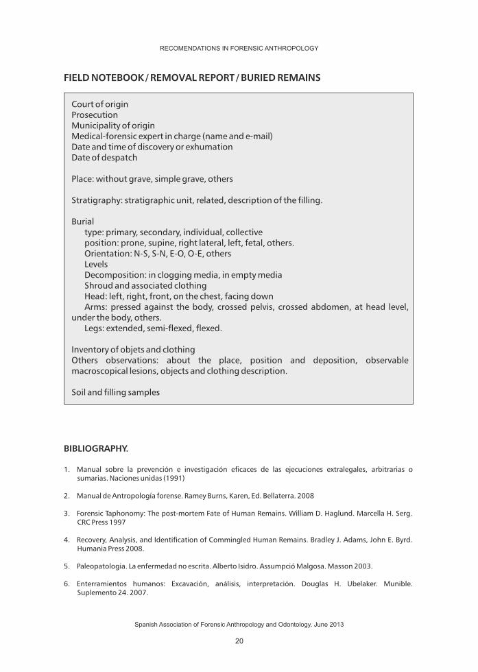
FIELD NOTEBOOK / REMOVAL REPORT / BURIED REMAINS
BIBLIOGRAPHY.
1. Manual sobre la prevención e investigación eficaces de las ejecuciones extralegales, arbitrarias o sumarias. Naciones unidas (1991)
2. Manual de Antropología forense. Ramey Burns, Karen, Ed. Bellaterra. 2008
3. Forensic Taphonomy: The post-mortem Fate of Human Remains. William D. Haglund. Marcella H. Serg. CRC Press 1997
4. Recovery, Analysis, and Identification of Commingled Human Remains. Bradley J. Adams, John E. Byrd. Humania Press 2008.
5. Paleopatologia. La enfermedad no escrita. Alberto Isidro. Assumpció Malgosa. Masson 2003.
6. Enterramientos humanos: Excavación, análisis, interpretación. Douglas H. Ubelaker. Munible. Suplemento 24. 2007.
Court of originProsecutionMunicipality of originMedical-forensic expert in charge (name and e-mail)Date and time of discovery or exhumationDate of despatch
Place: without grave, simple grave, others
Stratigraphy: stratigraphic unit, related, description of the filling.
Burial type: primary, secondary, individual, collective position: prone, supine, right lateral, left, fetal, others. Orientation: N-S, S-N, E-O, O-E, others Levels Decomposition: in clogging media, in empty media Shroud and associated clothing Head: left, right, front, on the chest, facing down Arms: pressed against the body, crossed pelvis, crossed abdomen, at head level, under the body, others. Legs: extended, semi-flexed, flexed.
Inventory of objets and clothingOthers observations: about the place, position and deposition, observable macroscopical lesions, objects and clothing description.
Soil and filling samples
RECOMENDATIONS IN FORENSIC ANTHROPOLOGY
Spanish Association of Forensic Anthropology and Odontology. June 2013
20

7. Resolució IRP/4072/2010, de 15 de desembre, per la qual s'aproven els protocols d'aplicació a les actuacions previstes en la Llei 10/2009, de 30 de juny, sobre la localització i la identiicació de les persones desaparegudes durant la Guerra Civil i la dictadura franquista, i la digniicació de les fosses comunes, i en el Decret 111/2010, de 31 d'agost, pel qual es desenvolupa reglamentàriament la Llei 10/2009, de 30 de juny. Diari Oficial de la Generalitat de Catalunya. 5784. 28/12/2010.
8. Fco-José Puchalt Fortea. Hallazgos y excavaciones. En Identificación antropológica policial y forense. JD Villalain Blanco. FJ Puchalt Fortea. Tirant lo blanch. Valencia. 2000: 31-39.
9. Orden PRE/2568/2011, de 26 de septiembre, por la que se publica el Acuerdo del Consejo de Ministros de 23 de septiembre de 2011, por el que se ordena la publicación en el Boletín Oficial del Estado del Protocolo de actuación en exhumaciones de víctimas de la guerra civil y la dictadura. BOE Nº 232 del 27 de septiembre de 2011.
10. Orden de 7 de septiembre de 2009, por la que se aprueba el Protocolo Andaluz de actuación en exhumaciones de víctimas de la Guerra Civil y la Posguerra. BOJA nº 190 de 28 de septiembre de 2009.
RECOMENDATIONS IN FORENSIC ANTHROPOLOGY
Spanish Association of Forensic Anthropology and Odontology. June 2013
21


LABORATORYGUIDELINES
23


This section includes information about the routine laboratory work before the anthropological analysis take place. Aforementioned laboratory work comprises of the traditionally applied techniques including the bibliographic search, the recommendations of anatomists who have sound professional background and craftsman coming from diverse places of the world such as Granada, Mexico and Brazil. In addition, the section sheds light on the mistakes that have been committed in this field and the lessons learned from those malpractices.
It is important to apply practical techniques that shall give rise to more efficiency at the workplace while taking into account that there are other existing methods/techniques that are in use and provide equally credible results.
1. ADMISSION OF THE MATERIALS TO THE LABORATORY:
All the materials to be admitted by the laboratory should be accompanied by affidavits both signed by the deliverer and recipent and these documents should include the number of the containers, the content of each chest, the origin, chronology and the date of the delivery.
In the archaeological cases, it is essential to record the excavated findings and/or discoveries. Ideally, the graves that are under the investigation should be marked level by level in line with the chronological phase accompanied by photographs and if it is necessary , stratigraphic profile is recommended. If it is not possible, archeological sites are identified with numbers to indicate the histo-chronological period that the findings belong. It is the only way to conduct a research that provide reliable paleodemographic results. Findings without any references should not be accepted.
If the received material is a forensic case, the measures concerning chain of custody should be taken. It is essential to comply with the Act JUS/1291/2010 BOE 122 dated 19th of May. The below mentioned points should be addressed in the documents;
PROTOCOL
AUTHOR
CONTACT
DATE
LABORATORY GUIDELINES
ALEMÁN I, BOTELLA MC
[email protected] • [email protected]
May 6,2011
RECOMENDATIONS IN FORENSIC ANTHROPOLOGY
Spanish Association of Forensic Anthropology and Odontology. June 2013
25

- The date and the hour of the admission - The judicial authority that sends the materials - The person and/or the company that delivers the material - The type of packaging and seal and the method of conservation - The person that receives the exhibits, deals with the opening procedures
and allocating identification numbers. - The place where the materials are kept until the opening process. - The description of the package, the type of the containers, possible
damages or anomalies detected , the attached documents etc. - The description of the label, whether it is complete or incomplete,
readable or not, without label etc. - Type of manipulation - The place where the exhibits are kept until the analysis start
It is recommended to unpack the materials (exhibits) and lay them on the metal trays covered with blotting paper in order to compare the nature of the exhibits received.
Most probably the bones are received in plastic and airtight bags and prior to the packaging, they might have lost their humidity. If they are left as mentioned above, the bones will become more fragile and brittle or it will result in proliferation of fungi, which results in destruction of the exhibits. In other words, effective measures should be taken to avoid information loss stemming from deterioration of the materials. Each tray should be labeled by the laboratory so that the materials under investigation can be traced in all process. The materials shall be traced with the numbers displayed on the labels in cleaning , analysis and research phases of the entire process. This issue requires constant emphasis and constitutes the most fundamental part.
The exhibits should be kept in an environment where the temperatures are controlled, it is recommended to place the exhibits in the shade until they get completely dried.
Although the very first step of the process is to dry the bones completely, the marks of humidity can go unnoticed. In our laboratory, we have a drying closet that provides improved ventilation and heating with the capacity of 12 trays functioning simultaneously and result in complete dryness in few hours.
Once the bones reach complete dryness, they shall be kept in a stable environment where their conservation is assured until the laboratory analysis starts.
RECOMENDATIONS IN FORENSIC ANTHROPOLOGY
Spanish Association of Forensic Anthropology and Odontology. June 2013
26

2. CLEANING OF SKELETAL REMAINS:
The bones received are usually fragile and sometimes brittle. It is important to know that preliminary drying should take place in the shade or in the closet, which will increase the consistency in terms of humidity and therefore, the bones will be more durable against manipulation. After the drying process in the shade or the closet , if it is necessary, the bones can be moisturized again. The bones will be less likely to get detoriated in this way.
If the bones are archeological remains, the important point is to get rid of the sediments on the bones. In order to wipe off the sediments, little brushes wih soft bristles can be used. The most appropriate way to wipe off the sediments is to sop the brush in weak stream and to clean the surface of the bones. Please take into account that the humidity can easily cause layers of mud. It is better not to immerse the bones into water. In order to remove the soil from the interior part of cranium or from the small cavities (orbits, nasal opening, medullary canal). It is essential to moisten the soil slowly to soften and remove the soil particles using odontologic tools. The use of spray gun is recommended.
Skeletonized remains are generally brought with soft tissues and putrilageous tissues.If these remains go through anthropological investigations, it is convenient to disinfect the remains before manipulating the bones however there should not be any loss of forensic information.
In order to preserve them well, the remains are immersed in soapy water and then cleaned with the help of a brush in the running water. Later on, place them in %30 diluted sodium hypochlorite during 15 minutes and rinse the remains well until the residues of the solution completely vanish. The reason is that the solution residues may destroy the bones when they crystallize in time. The better solution to this problem is to sink the remains into a container filled with water for a few hours and to keep on changing the water constantly. After taking them out, ph of the water used should be checked.
3.CADAVER REMAINS.
The samples should be softened in order to remove the soft tissues. In the past, the anatomists would scrape the flesh from the bones and they would leave them in the water which would be refreshed every 3 or 4 days until get to reduce body any decomposed remains soft, and then the anatomists were able to remove the soft tissues along with the ligaments. Nowadays, the process of scraping the flesh from the bones is shorter as chemical products are used as accelerants. Previously the same process used to take almost 3 months and it
RECOMENDATIONS IN FORENSIC ANTHROPOLOGY
Spanish Association of Forensic Anthropology and Odontology. June 2013
27
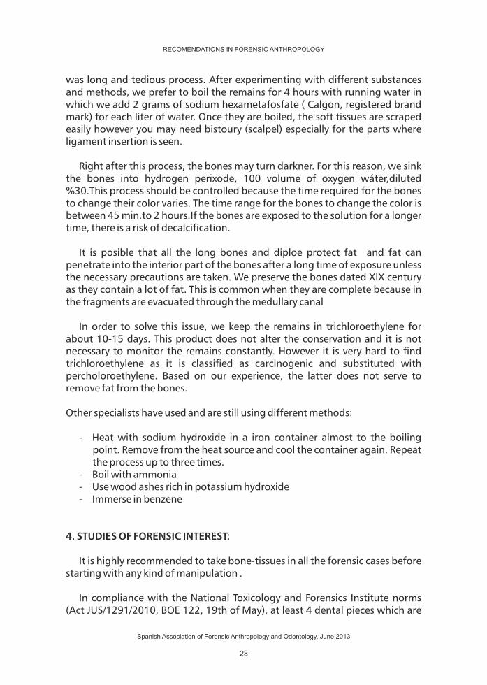
was long and tedious process. After experimenting with different substances and methods, we prefer to boil the remains for 4 hours with running water in which we add 2 grams of sodium hexametafosfate ( Calgon, registered brand mark) for each liter of water. Once they are boiled, the soft tissues are scraped easily however you may need bistoury (scalpel) especially for the parts where ligament insertion is seen.
Right after this process, the bones may turn darkner. For this reason, we sink the bones into hydrogen perixode, 100 volume of oxygen wáter,diluted %30.This process should be controlled because the time required for the bones to change their color varies. The time range for the bones to change the color is between 45 min.to 2 hours.If the bones are exposed to the solution for a longer time, there is a risk of decalcification.
It is posible that all the long bones and diploe protect fat and fat can penetrate into the interior part of the bones after a long time of exposure unless the necessary precautions are taken. We preserve the bones dated XIX century as they contain a lot of fat. This is common when they are complete because in the fragments are evacuated through the medullary canal
In order to solve this issue, we keep the remains in trichloroethylene for about 10-15 days. This product does not alter the conservation and it is not necessary to monitor the remains constantly. However it is very hard to find trichloroethylene as it is classified as carcinogenic and substituted with percholoroethylene. Based on our experience, the latter does not serve to remove fat from the bones.
Other specialists have used and are still using different methods:
- Heat with sodium hydroxide in a iron container almost to the boiling point. Remove from the heat source and cool the container again. Repeat the process up to three times.
- Boil with ammonia - Use wood ashes rich in potassium hydroxide - Immerse in benzene
4. STUDIES OF FORENSIC INTEREST:
It is highly recommended to take bone-tissues in all the forensic cases before starting with any kind of manipulation .
In compliance with the National Toxicology and Forensics Institute norms (Act JUS/1291/2010, BOE 122, 19th of May), at least 4 dental pieces which are
RECOMENDATIONS IN FORENSIC ANTHROPOLOGY
Spanish Association of Forensic Anthropology and Odontology. June 2013
28

unharmed should be kept concerning the simple admission and preparation. When possible, molars and a long bone,preferably femur, are recommended to be kept for the genetic identification studies.
According to the protocols set by the United States in 1991, it is recommended to have all the skeleton X-rayed before the cleaning process kicks off in order to handle the investigation and prevention of illegal executions and referees and summaries.
Keeping the dental radiographies of the bitemarks, periapicals as well as panoramics and of all the bones of the skeleton is vital. Attention should be attaced to the documentation of the fractures and anomalies of growth and surgical interventions. In addition, the radiography of front sinus can help the identification phase.
It is important to keep some of the bones in their original state as stated in the protocol. In line with the protocol, two lumbar vertebras shall be sufficient. Then it is only possible to wash the rest of the bones but without being rinsed nor scrubbed.
In these cases, we had better not wash the bones, we can only get rid of the dirt on the surface of the bones using brushes. We should not use water or any other product. While manipulating the bones in all the stages we use nitril gloves.
If the bones are just received prior to the identification , it is important to preserve the followng samples for post analysis
- Transverse cut in the middle of each femur , 2 cm height or more de 2 cm. - Transverse cut in the middle of each fibula, 2 cm height or more. - A 4cm cut on the extremity of sternum and one of the ribs (the sixth one,if
it is possible). - A tooth, preferably a mandibular incisor. - Molars for a possible subsequent identification in genetic analysis. - Plaster cast of the cranium in case of a facial reconstruction.
In our laboratory, we have substituted the last phase of forming a cast for facial reconstruction with an IT supported system of three dimensional modelling of a cranium with the help of 3D scanner (vivid 910 Konica-Minolta)
Make sure that all the exhibits are given an identification number, the date and the name of the person dealing with them
RECOMENDATIONS IN FORENSIC ANTHROPOLOGY
Spanish Association of Forensic Anthropology and Odontology. June 2013
29

Once all the precautions are taken for the samples to remain unaltered, the cleaning method of cadaver remains shall continue.
Before we start to work on the remains, it is advisable to create an inventory by marking the remains with an undeletable marker indicating the origin and when possible, the bones of the different individuals should be separated. To exemplify, attached please see the inventory that we use for sub adults.
BIBLIOGRAPHY:
1. Guirao Gea, M. (1953): Techical Anatomy Manual. Guide for organic macrotechnics. Scientific and medical Editorial-. Barcelona.
2. United Nations (1991): Manual on the Effective Prevention and Investigation of Illegal Summaries and Referees United Nations.
3. http://www.boe.es/boe/dias/2010/05/19/pdfs/BOE-A-2010-8030.pdf
4. Brothwell, D.R. (1987): Unearthing Bones Excavation, treatment and study of human skeletal remains.. Fondo de Cultura Económica. México.
5. Bass, W. (1987): Human osteology: A laboratory and field manual. Missoury Archaeological Society.
RECOMENDATIONS IN FORENSIC ANTHROPOLOGY
Spanish Association of Forensic Anthropology and Odontology. June 2013
30

F I C H A I N V E N T A R I OINDIVIDUOS SUBADULTOS
RECOMENDATIONS IN FORENSIC ANTHROPOLOGY
Spanish Association of Forensic Anthropology and Odontology. June 2013
31


ANTHROPOMETRICDATA
33


ANTHROPOMETRIC DATA.
Measuring the bones has historically been the main study method in Physical Anthropology. Used as method for documenting the findings, facilitates the description of the individual as well as comparing the groups. This will allow us, in the practice of Forensic Anthropology, the estimation of various features, especially the size, sex and race.of the bones has historically been the main study method in physical anthropology. Used as method for documenting findings facilitates the description of the individual as well as comparison of groups. This will allow us, in the practice of Forensic Anthropology, the estimation of various features, especially the size, sex and ancestry.
Until the 60s comparisons between groups, and therefore their assignments, were made based on; individual or so-called indexes measurements, which combine two individual measurement, allowing an approximate estimation of the form. Meanwhile, the development of statistical methods and information technology currently has carried out the calculation of parameters using the multivariate analysis, as reflected, for example, in the FORDISC (Jantz and Ousley, 2005). Today, the systematic use of that software, is very recommended, provided that we apply with due caution when accepting its inferences, which sometimes have severe limitations, mainly due to the reference populations used (Elliott and Collard, 2005 Ramsthaler et al., 2007). At the moment, the development and the application of complex mathematical methods, such as Fourier analysis, which aims to reduce the form to a mathematical curve, goes even further and allows to start from the 90s a boom in morphometric studies (Claude , 2008) in Physical Anthropology, whose future applications already starting to be reflected in forensic anthropology work (Gonzalez et al., 2009, Wilson et al., 2011).
Historically, the utmost importance has also been given the to cranial measurements and measurement proposals were multiplied to infinity. Today, however a consensus has been reached in terms of the importance of postcranial measures and the necessity of reducing the number of mandatory measures to those which have proven to be truly useful in practice, leaving as optional those that are only of interest in research or in exceptional circumstances.
In this chapter, after an introduction to the general techniques of measurement and instrumentation, we will describe the measurements considered standard in -Forensic Anthropology from those listed in Standards for
PROTOCOL
AUTHORS
CONTACT
DATE
ANTHROPOMETRIC DATA
MANUEL F. CARRILLO, ENRIQUE DORADO
[email protected] • [email protected]
October 4, 2011
RECOMENDATIONS IN FORENSIC ANTHROPOLOGY
Spanish Association of Forensic Anthropology and Odontology. June 2013
35

Data Collection from Human Skeletal Remains (Buikstra and Ubleaker, 1994) -, by proposing 24 cranial, 10 mandibular and 44 postcranial, as well as corresponding reference points. However, it should be pointed out, that these set of measurements were designed for the comprehensive data collection in Physical Anthropology in previous studies to the return of remains to indigenous communities in North America. Therefore, while maintaining its reference value and completeness, rarely it will be required as a whole in a Forensic Anthropology case.
For specific aspects of data collection, such as identification of single bones, laterality, etc., we refer to the consultation of the typical manuals of general osteology whose international references are those of William Bass (2005) and Tim D. White (1999 and 2005).
1. GENERAL CONDITIONS OF MEASUREMENT.
A good measurement in order to be useful, must provide homogeneous and repeatable results, by trying to limit the most, the error in both intra -and also- interobserver. For this you need to start from reference points whose determination leave the least space possible for ambiguity. For the same reason there is also a tendency to use maximum measurements, which is easier to obtain than minimum measurements and if necessary, can be obtained by trial and error techniques with the usual instruments, and are more reproducible than arbitrary measures.
The measurements shall always be taken in millimeters not fractional rounding up or down to the most immediate.
In general, for measurements in bilateral structures, we will take the ones on the left and only if that take is impossible, will carry out the measurements on the right side, always outlining this circumstances-with an "R" next to the measurement if collected a data sheet. Fragmented or deformated bones must not be measured in dimension corresponding to the measurement. However, we do need to estimate the measurements for remains slightly eroded or rebuilt, outlining this situation with an "*" - star-in the data sheet.
2. MEASUREMENT INSTRUMENTS.
Multiple measuring instruments are suggested which are useful without doubt in most specialized research. However, for our purposes, and for the collection of the proposed measures, the list above is sufficient:
- Tape measure: an ordinary metallic or non-distensible material is sufficient. It is
RECOMENDATIONS IN FORENSIC ANTHROPOLOGY
Spanish Association of Forensic Anthropology and Odontology. June 2013
36

used to measure circumferences and it is not recommended to use it for other measurements.
- Calibre or vernier caliper: Formed by a ruler which has a fixed branch at one end and another that, when sliding on the ruler allows to read on itself, on a dial or a digital display, the distance between the branches. Many models have different branch points to measure both external diameters of an object placed between the ends and internal diameters ends of a hole wherein the points are introduced.
- Calliper compasses : derived from pelvimeters used by obstetricians, comprise of two question mark shaped branches connected by the edge of the straight side. Connected to one of the branches, it is a ruler marked with a scale and with a sliding support for the other branch, so that it allows to read in the opening between the extremes of the branches.
- Osteometric table: This is millimetric horizontal surface with a longitudinal ruler and, a perpendicular fixed stop at one end and another slide at the other end, which by moving on the millimetric surface, the ruler allows to measure the opening between the stops.
- Jaw meter: is, in fact, a modified osteometric table, in which the fixed stop is not, nevertheless perpendicular to the horizontal surface, but rather tilting on it, and with a sliding stop at the opposite side of the joint. Thus we can put in the jaw on the horizontal surface and, by adjusting the tilt stop, we can read the scale on height of the branch, as well as the angle it forms with the horizontal by an incorporated goniometer.
3. MEASUREMENTS OF THE SKULL.
3.1. The Frankfurt plane.
It is the standart atomic orientation and measurement of the skull, established by Congress of Anthropology in Frankfurt in 1877. It is defined by a horizontal plane passing through the two porion and the left orbital. All positions and measures in the skull will refer to this plane.
3.2. Odd or mid craniometric points:
1. Bregma (b): T Center formed by the intersection of the coronal suture and sagittal.
2. Vértex: Highest point of the skull, located it in the Frankfurt plane, and thus,
RECOMENDATIONS IN FORENSIC ANTHROPOLOGY
Spanish Association of Forensic Anthropology and Odontology. June 2013
37

the farthest from the basion.3. Glabela (g): Most prominent point in the interciliary area.4. Nasion (n): T Center formed by the intersection of frontal-nasal suture with
the suture of the nose bones.5. Nasoespinal (ns): The point where a line drawn tangent to the most inferior
points of the pyriform aperture cuts the midsagittal plane.6. Prostion (pr): Lowest point on the midline of the maxillary alveolar process
between the two upper middle incisors.7. Alveolon (alv): Point of the hard palate located at the intersection of the
midline with the line tangent to the most posterior edges of the maxillary alveolar process.
8. Infradental (id): Upper most point on the midline of the septum between the media mandibular incisors. Opposed to prostion.
9. Gnation (gn): Lowest point of the mandible in the midline.10. Opistocranion (op): Most posterior point of the skull in the occipital region,
not in the external occipital protuberance and instrumentally determined as the one corresponds to the maximum distance from the glabella.
11. Lambda (l): Point of intersection of the sagittal suture and the lambdoid suture.
12. Basion (ba): Point of the anterior edge of foramen magnum located in the midline, thus it is the most anterior of the foramen magnum.
13. Opistion (o): Point of rear edge of the foramen magnum located in the midline, thus opposed to the basion.
3.3. Pairs or laterale craniometric points:
1. Porion (po): Is the uppermost point of the margin of the ear hole. 2. Asterion: Is the point of intersection of the occipital, parietal and temporal
sutures.3. Dacrion (d): Point of binding of the frontal, maxillary and tear in the mid rim
orbital4. Orbital (or): The lowest point of the orbital rim.5. Ectoconquion (ec): Most anterior point of the lateral rim of the orbit.6. Zigion (zy): Most lateral point of the zygomatic arch, determined
instrumentally by measuring the width-maximum-bizygomatic.7. Eurion (eu): Most lateral point of the skull. Determined instrumentally by
measuring the maximum width of skull. Its location may vary, between the parietal and temporal.
8. Lateral Condilon (CDL) Most lateral point on the mandibular condyles.9. Ectomolar (ecm) most lateral point on the outer surface of the alveolar
process of the maxilla, usually at the level of the second molar.10. Gonion (go): Lowest, most posterior and most lateral point of the mandibular
angle, at the junction of the ramus and mandibular body.11. Alar (al): It is determined instrumentally such as the most lateral points of the
RECOMENDATIONS IN FORENSIC ANTHROPOLOGY
Spanish Association of Forensic Anthropology and Odontology. June 2013
38

nasal opening, thus corresponding to the maximum width of the hole piriformis.
12. Front temporal (ft): Most anterior-medial point of the timeline, in the zygomatic process of the frontal.
13. Front malar-temporal (fmt): Most lateral point of the front-malar suture.14. Atrial (au): Point located above the ear hole in the root of the zygomatic arch
at its deepest incuvation area.15. Mastoid (ms): Lowest point of the mastoid process.
3.4. Measurements of the skull.
1. Maximum skull length (g-op) GOL: Measured with callipers between glabella and the farthest point in the midsagittal plane of the skull (opistocráneo).
2. Maximum skull width (eu-eu) XCB: Maximum width measured with callipers in the horizontal plane, excluding the lower temporal lines.
3. Bizygomatic width (average facial width) (zy-zy) ZYB: Distance measured with the callipers or calibrate between the two lateral points of the zygomatic arches.
4. Maximum skull height (ba-b) BBH: Distance measured with callipers between bregma and basion.
5. Length of the cranial base) (ba-n) BNL: Measured with the calibrate or callipers between nasion.
6. Length of basion - prosthion (b-pr) BPL: Measured with callipers or calibrate between the basion and prosthion.
7. Maxiloalveolar Width (ecm-ecm) MAB: Measured with the the caliber between both ectomolares.
8. Length of maxilla - alveolar (pr-alv) MAL: Length between the prosthion and alveolon. Measured with the caliber only if there is loss of incisors and, otherwise, with the callipers .
9. Biatrial width (base of the skull) (au-au) AUB: Measured by the calliper or calibrate between both auricular points.
10. Height of upper face (n-pr) UFH: Measured by the caliber between the nasion and prostion.
11. Minimum front width (ft-ft) WFB: Minimum horizontal measurement between the two frontal temporary lines using the caliber.
12. Upper facial width (fmt-fmt) UFB: Measured by caliber between the two most lateral point of the suture frontomalar.
13. Nasal height (n-ns) NLH: Measured by the caliber between nasion and nasospinal.
14. Nasal width (al-al) NLB: Maximum distance measured by the caliber between the lateral edges of the pyriform aperture.
15. Orbital width (d-ec) OBB: Distance measured with the caliber between dacrion and ectoconquion drawing a line that divides into equal parts the orbit.
RECOMENDATIONS IN FORENSIC ANTHROPOLOGY
Spanish Association of Forensic Anthropology and Odontology. June 2013
39

16. Orbital height OBH: Distance measured caliber between the upper and lower edge scanning perpendicular to the Orbital OBB width of.
17. Biorbital Width (ec-ec) EKB: Distance measured with the caliber between both ectoconquion.
18. Interorbital width (d-d) DKB: Distance measured with the caliber between both dacrion.
19. Front Cord (n-b) FRC: Distance measured with the caliber between nasion and bregma in midsagittal plane.
20. Parietal Cord (b-l) PAC: Distance measured with the caliber between bregma and lambda in midsagittal plane.
21. Occipital Cord (l-o) OCC: Distance measured with the caliber between lambda and opisthion in midsagittal plane.
22. Foramen magnum length (ba-o) FOL: Distance measured with the caliber between basion and opisthion.
23. Foramen magnum width FOB: Maximum width with the caliber between the lateral edges.
24. Mastoid length MDH: Vertical projection of the mastoid process under and perpendicular to the plane of Frankfurt.
3.5.- Mandibular Measurements.
25. Length of the chin (id-gn) GNI: Distance measured with the caliber between gnation and infradental.
26. Height of the body HMF: Distance measured with the caliber between the edge of the alveolar process and the lower edge of the jaw taken perpendicular to the base level of the mental foramen.
27. Thickness of the mandibular body TMF: Maximum thickness measured with the caliber of the the mandibular body at the level of mental foramen.
28. Bigoniáca Width (go-go) GOG: Distance measured with the caliber between both gonion.33
29. Bicondilar width (CDL-CDL) CBD: Maximum distance measured with the caliber between the outer edges of the condyles.
30. Minimum Width of the ramus WRB: Minimum distance measured with the caliber between the anterior and posterior edges of the mandibular ramus perpendicular to the maximum height XRH.
31. Maximum width of the ramus XRB: Distance measured with the caliber between the most anterior points of the ramus and the tangent that connects the most posterior point of the condyle with the angle of the mandibular.
32. Maximum height of the ramus XRH: Distance from the uppermost point of the condyle to the gonion, measured by the caliber or jaw meter.
33. Mandibular length MLN: Distance measured by the jaw meter between the pogonio and the midpoint of the line connecting both gonion.
34. Mandibular angle MAN: Angle formed by the lower edge of the body and the posterior edge of the ramus, measured with the jaw meter.
RECOMENDATIONS IN FORENSIC ANTHROPOLOGY
Spanish Association of Forensic Anthropology and Odontology. June 2013
40

4. POSTCRANIAL MEASUREMENTS.
4.1.Clavicle.
35. Maximum length: Maximum distance between the outermost points with the osteometric table or caliber.
36. Medium sagittal diameter (anterior-posterior): Distance measured with the caliber between the anterior and posterior surfaces at the level of the midpoint diaphyseal specified in the osteometric table.
37. Medium vertical diameter (upper-lower): Direct distance between the upper and lower surfaces of the collarbone at the level of midpoint diaphyseal.
4.2. Scapula.
38. Scapular height (anatomical width): Distance measured with the caliber between the uppermost point of the cranial angle and the lowermost point of the caudal angle.
39. Scapular width (anatomical length): Distance measured with calliper compasses between the midpoint of the dorsal edge of the glenoid cavity to the point between the lips of the scapular spine at its medial border.
4.3. Humerus.
40. Maximum length: Distance between the most proximal -superior- point of the head and the most distal-inferior-point of the trochlea in the osteometric table.
41. Epicondylar width: Distance measured with the caliber or osteomteric table between the most lateral points of the epicondyles.
42. Vertical diameter of the head: Measure with the caliber between the most upper and lower points of the edge of the articular surface.
43. Maximum diameter of the diaphysis: Measured with the caliber at the midpoint level of the diaphysis, specified in osteometric table.
44. Minimum diameter of the diaphysis: Measured, like the previous one, with the caliber at the midpoint level the of the diaphysis, specified in osteometric table.
4.4.Radius.
45. Maximum length of the radius: Distance measured in the osteomteric table between the most proximal point of the head and the most distal point of the styloid processes.
RECOMENDATIONS IN FORENSIC ANTHROPOLOGY
Spanish Association of Forensic Anthropology and Odontology. June 2013
41
46. Sagittal diameter (anterior-posterior) of the diaphysis: After the determination of the midpoint of the diaphysis in the osteometric table, the distance at this level between the anterior and posterior surfaces is measured by the caliber.
47. Transverse diameter (medial-lateral) of the diaphysis: Maximum diameter between the medial and lateral surface of the diaphysis at the midpoint level the of the diaphysis, as in the previous case.
4.5. Ulna.
48. Maximum length: Maximum distance between the uppermost point of the olecranon and the lowest point of the styloid process in the osteometric table.
49. Dorsum-fly diameter (anterior-posterior): Maximum diameter with the caliber of the diaphysis at the point of greater development of the ridge.
50. Transverse diameter (medial-lateral): Diameter at the point of greater development of the ridge perpendicular to the previous measure.
51. Physiological length: Measured with the calliper compasses between the most distal-lower point, which is, the deepest point of the concave-on the coronoid process surface and the most distal point of the lower surface of the distal epiphysis.
52. Minimum circumference: Measured with tape measure proximate to the distal end.
4.6. Sacrum.
53. Anterior Length: Distance measured with the caliber between the point of the promontory located in the midsagittal plane and the point of the same plane at the top of the sacrum.
54. Anterosuperior Width: Maximum transversal width measured with the caliber of the sacrum at the level of previous projections of the auricular surfaces.
55. Maximum width of the base: Distance measured with the caliber between the most lateral superior articular surface - sacral base, measured perpendicular to the midsagittal plane.
4.7. Pelvis.
56. Height: Distance measured with calliper compass between the uppermost point of the iliac crest and the lowest of the ischial tuberosity.
57. Iliac Width: Distance measured with calliper compasses between the anterior-superior iliac spine and the posterior superior iliac spine.
58. Length of Pubic: Distance measured with the caliber between the point of the
RECOMENDATIONS IN FORENSIC ANTHROPOLOGY
Spanish Association of Forensic Anthropology and Odontology. June 2013
42

acetabulum where the three parts of the hip bone and the upper edge of syphilis join.
59. Ischial Length: Distance measured with the caliber between the point of the acetabulum where the three parts of the hip bone and lowest point of the tuberosity of the isquiátca join, approximately perpendicular to the length of the pubic.
4.8. Femur.
60. Maximum length (anatomical): Maximum distance in osteometric table between uppermost point of the head and the lowest point of the distal condyles.
61. Bicondylar length (physiological oblique): Distance in osteometric table between the uppermost point of the head and a plane tangential to the lower surfaces of both distal condyles.
62. Epicondylar width: Distance in the osteometric table between the most lateral points of the epicondyles.
63. Maximum diameter of the femoral head measured with the caliber.64. Subtrochanteric sagittal: Diameter (anterior-posterior) distance measured
with the caliber between anterior and posterior surfaces at the proximal end of the diaphysis, measured perpendicularly to the medial-lateral diameter.
65. Subtrochanteric transverse diameter (medial-lateral): Distance measured with the caliber between lateral and medial surfaces on the proximal end of the diaphysis, perpendicular to the previous measurement of the sagittal diameter at the highest level of lateral expansion under the lesser trochanter.
66. Diaphyseal sagittal diameter (anterior-posterior): Distance measured with the caliber between the anterior and posterior surfaces at approximately the midpoint diaphyseal level, located at the maximum point of development of the linea aspera.
67. Diaphysial transversal diameter (medial-lateral): Distance with the caliber between the medial and lateral surfaces, measured perpendicular to the previous measurement.
68. Circumference of the diaphysis: Measured with a tape measure at the level of midpoint of the diaphysis as the two previous measurements.
4.9. Tibia.
69. Maximum length: Distance in osteometric table between the superior articular surface of the lateral condyle and the tip of the medial malleolus.
70. Maximum width in the proximal epiphysis: Maximum distance in osteometric table between the most lateral and medial point of the condyles of the proximal epiphysis.
71. Maximum width of the distal epiphysis: Maximum distance in osteometric
RECOMENDATIONS IN FORENSIC ANTHROPOLOGY
Spanish Association of Forensic Anthropology and Odontology. June 2013
43

table between the most lateral point of the medial malleolus and the lateral surface of the distal epiphysis.
72. Maximum diameter at the level of nutrient foramen: Maximum distance with the caliber between the crest anterior and the posterior surface at the level of the nutrient foramen.
73. Transversal diameter (medial-lateral) to the nutrient foramen: Direct distance measured with the caliber between the medial edge and interosseous crest at the level of nutrient foramen, perpendicular to the previous measurement.
74. Circumference at the height of nutritional hole: Measured with tape measure at the level of the nutrient foramen.
4.10.Fibula.
75. Maximum length: Maximum distance in osteometric table between the proximal end of the head and the distal end of the lateral malleolus.
76. Maximum diameter of the diaphysis: Maximum distance measured with caliber at a semicircular mid-diaphysis level, located by osteometric table by determining the maximum length.
4.11. Calcaneus.
77. Maximum length: Distance measured with the caliber between the posterior most prominent point of the tuberosity and most anterior point of the upper edge of the facet joint with the cuboid. Measured on the midsagittal plane and projected onto the underlying surface.
78. The medium width: Distance measuered with the caliber between laterally most prominent point of the dorsal facet joint and the most medial point of the sustentaculum tali. Since these two points do not have the same height nor in the same plane perpendicular to the sagittal, the measurement must necessarily be projected in both dimensions.
5. RECOMMENDATIONS.
Only standard measures collected here will be taken, which are basically the accepted ones for data collection in Physical Anthropology by Buikstra and Ubleaker (1994). If it is necessary to supplement them, for example due to lack of materials or its fragmentation, in the case of the postcranial skeleton, the alternative measures proposed by the aforementioned authors (Table 1) can be applied.
Generally in forensic cases, only postcranial skeleton measurements which lead us to calculate the size or estimate the gender will be of interest. In the case
RECOMENDATIONS IN FORENSIC ANTHROPOLOGY
Spanish Association of Forensic Anthropology and Odontology. June 2013
44

of the size thus, we focus on the maximum lengths of the humerus, radius, ulna, femur -physiological length -, tibia and fibula, the latter (tibia and femur) being the most reliable.
For the estimation of gender, we are interested especially in maximum diameters of the head of the humerus and femur.
Minimum diameters are generally not very useful in Forensic Anthropology and also are very susceptible to error in its determination therefore they should be avoided.
All possible standard measures of the cranial segment should be taken, the more mesurement the more reliable FORDISC inferences, particularly useful in the estimation of race (Jantz and Ousley, 2005).
Table1 (Buikstra and Ubleaker, 1994)
RECOMENDATIONS IN FORENSIC ANTHROPOLOGY
Spanish Association of Forensic Anthropology and Odontology. June 2013
45
BIBLIOGRAPHY.
1. Bass, William M. Human Osteology: A Laboratory and Field Manual. 5th ed. Missouri Archaeological Society, 2005.
2. Buikstra, Jane E. Standards for Data Collection from Human Skeletal Remains: Proceedings of a Seminar at the Field Museum of Natural History. Arkansas Archeological Survey, 1994.
3. Claude, Julien. Morphometrics with R. New York, NY: Springer New York, 2008. Cox, Margaret, y Simon Mays. Human Osteology: In Archaeology and Forensic Science. Greenwich
Medical Media, 2000.
4. Elliott, Marina, y Mark Collard. "FORDISC and the determination of ancestry from cranial measurements". Biology Letters 5, nº. 6 (Diciembre 23, 2009): 849-852.
5. Gonzalez, Paula N., Valeria Bernal, y S. Ivan Perez. "Geometric morphometric approach to sex estimation of human pelvis". Forensic Science International 189, nº. 1-3 (Agosto 10, 2009): 68-74.
6. Jantz R. L., Ousley S. D. Fordisc, version 3.0. Knoxville, TN: University of Tennessee, 2005.
7. Kranioti, Elena F., Markus Bastir, Andrea Sánchez-Meseguer, y Antonio Rosas. "A geometric-morphometric study of the cretan humerus for sex identification". Forensic Science International 189, nº. 1-3 (Agosto 10, 2009): 111.e1-111.e8.
8. Ramsthaler, F, K Kreutz, y M A Verhoff. "Accuracy of metric sex analysis of skeletal remains using Fordisc based on a recent skull collection". International Journal of Legal Medicine 121, nº. 6 (Noviembre 2007): 477-482.
9. Reichs, Kathleen J., y William M. Bass. Forensic Osteology: Advances in the Identification of Human, Second Edition. 2nd ed. Charles C. Thomas Publisher, 1998.
10. White, Tim D., y Pieter A. Folkens. Human Osteology, Second Edition. 2nd ed. Academic Press, 1999.
11. White TD, Folkens PA The Human Bone Manual. 1st ed. Academic Press, 2005.
12. Wilson, Laura A.B., Hugo F.V. Cardoso, y Louise T. Humphrey. "On the reliability of a geometric morphometric approach to sex determination: A blind test of six criteria of the juvenile ilium". Forensic Science International 206, nº. 1-3 (Marzo 20, 2011): 35-42.
RECOMENDATIONS IN FORENSIC ANTHROPOLOGY
Spanish Association of Forensic Anthropology and Odontology. June 2013
46

INTEGRATED FORENSICANTHROPOLOGY REPORT
47


1. DEFINITION AND GENERAL CONCEPTS:
The Integrated Forensic Anthropological Report (IAFI) is the document that gathers together all the scientific and technical actions of the professionals involved in the researches within the competence of Forensic Anthropologist and which may include-if the report already has not -a final inclusive document by way of conclusion, of all the results obtained.
It is both an expert report and a document that allows to have a global vision, composed of all the research conducted. The Spanish Association of Forensic Anthropology and Odontology (AEAOF) recommend using this type of report in cases where possible.
It would therefore be right to issue a joint report or to issue separate interim reports but with a common synthesis report which answers for scientific consensus to at least five basic issues of all Forensic Pathology expert report: 1) Identification, 2 ) Origin of death: Natural or violent (Accidental, Suicide or Homicide) 3) Time of death 4) Causes of death and 5) Circumstances of death.
It is recommended that the integration of all the information is coordinated by the forensic anthropologist who shall be obliged to:
1) Incorporate to the IAFI all interim reports of the different professionals involved.
2) To highlight the agreements and disagreements between them. 3) To prepare a synthesis document of scientific consensus among all the
professionals involved.
In addition to the study of remains, Police Report or certain data of legal medical interest of the same, Historical Research, Reports, protocols or Antemortem files, Archaeological Report, Report of lifting of corpse or bones remains, Technical Reports of Ground Penetrating Radar or other close remote sensing systems, conclusive samples sampling report with its corresponding sheets of informed consent, report of chain of custody for each of the samples submitted, dental-forensic report, reports from results of the analyses performed as the radiological, chemical, toxicological, entomological, histopathological, biological, genetic, experimental, criminalistic, environmental, etc...are considered eligible to join the IAFI.
PROTOCOL
AUTHORS
CONTACT
DATE
INTEGRATED FORENSIC ANTHROPOLOGY REPORT
SERRULLA F, VERANO V
[email protected] • [email protected]
February 28, 2013
RECOMENDATIONS IN FORENSIC ANTHROPOLOGY
Spanish Association of Forensic Anthropology and Odontology. June 2013
49

2. GENERAL OUTLINE OF THE IAFI:
The reports will tend to be drafted in a way that it is possible that other experts with the results obtained can reach conclusions. It is recommended that the reports that are responsible of Forensic Anthropologists are structured with the following outline:
2.1. Identification of the expert and the institution to which he belongs:
This section must include the name or identification code of the expert, his basic data of professional accreditation and the institution to which he belongs with his postal address and contact telephone number.
2.2. Equipment and Methods:
It is recommended to include in this section, at least, the relationship of the material received with explicit reference to its abbreviations, and the data referring to the chain of custody and all the methodological aspects that are considered to be of interest. It is recommended to use a neutral descriptive language without any evaluative reference.
2.3.Results:
In this section all positive and negative findings of interest will be described without employing any evaluative language. It will not be possible to ignore any result that has been used in support of the conclusions.
2.4. Discussion of the results or Forensic Anthropological analysis:
Evaluative section of the report that integrates all the information obtained from the case by analyzing all the results obtained so that it is possible to hold scientific or reasonable conclusions.
2.5.Conclusions:
Final section of synthesis of all the information obtained that must respond at least to the following questions. 1) Medical legal origin of death; 2) Time of death; 3) Causes and/or mechanism of death; 4) Circumstances of death and 5) identification.
RECOMENDATIONS IN FORENSIC ANTHROPOLOGY
Spanish Association of Forensic Anthropology and Odontology. June 2013
50

2.6. References:
It is recommended to use a final section with used or recommended bibliographical references.
BIBLIOGRAPHY.
1. White, Tim D., y Pieter A. Folkens. Human Osteology, Second Edition. 2nd ed. Academic Press, 1999.
2. White TD, Folkens PA The Human Bone Manual. 1st ed. Academic Press, 2005.
3. Ramey Buns K, Forensic Anthropology Training Manua. Second edition. Pearson Prentice Hall 2007.
4. Buikstra, Jane E. Standards for Data Collection from Human Skeletal Remains: Proceedings of a Seminar at the Field Museum of Natural History. Arkansas Archeological Survey, 1994.
RECOMENDATIONS IN FORENSIC ANTHROPOLOGY
Spanish Association of Forensic Anthropology and Odontology. June 2013
51


DIAGNOSTIC CRITERIAFOR IDENTIFICATION
53


SEX ESTIMATION CRITERIA
RECOMENDATIONS IN FORENSIC ANTHROPOLOGY
Spanish Association of Forensic Anthropology and Odontology. June 2013
55


RECOMENDATIONS FOR SEX ESTIMATION.
Sex determination is one of the basic tasks when studying skeletal remains, of both ancient collections for the possibility of establishing the demographic conditions of past societies and personal identification in forensic cases.
To estimate sex differences, it is always recommended to analyze the skeleton as a whole; however, this is not generally possible, since in many cases there is only a part of it or isolated and fragmented bones are encountered.
Overall, it could be applied as a rule that the more remains there are, the easier the sex determination will be. In practice there are key elements in the analysis that allow to estimate sex with great precision.
In general, male bones are bigger and more robust, with more pronounced muscular insertions and reliefs than the female.
If a detailed inspection of the bones is made, the sex can be established with close to 100% reliability, in case of conserving the whole skeleton; the results are less satisfactory when working with isolated remains, even though it has been estimated that the sex can be properly identified in 98% of the cases when conserving only the pelvis and in 92% if considering only the characteristics of the skull.
Although it is recommended to evaluate all the bones as a whole, the pelvis is the anatomic region in which sex differences are best reflected. If observed in anatomical connection, in other words, both innominate bones articulated by the sacrum, the female pelvis is wider and the lateral projection of the iliac bones is more pronounced than in males; the sagittal and transverse dimensions of the inner pelvis are bigger in women than in men, and it could be said that in the former ones, there is a horizontal predominance, while in men, it is the vertical one.
In order to clearly expose the criteria that would allow the establishment of sexual differentiation based on the pelvis, a table containing the specific characteristics of each gender has been developed.
PROTOCOL
AUTHORS
CONTACT
DATE
SEX ESTIMATION
ALEMAN I, BOTELLA MC, VICIANO J
[email protected] • [email protected] • [email protected]
April 30, 2012
RECOMENDATIONS IN FORENSIC ANTHROPOLOGY
Spanish Association of Forensic Anthropology and Odontology. June 2013
57

Table 1: Features of sexual differentiation in the pelvis.
Ischiatica major incisure.
Sulcus preauricularis.
Acetabulum.
Foramen obturatum.
Os pubis.
Arcus subpubianum.
Ramus isquiopubianum.
Articulatio sacrum-iliac.
Os iliacum.
MALE
Narrow, tending to adopt a V shape. Usually its angle doesn't exceed 30º.
Generally absent, if it appears, it is very slightly marked.
Large and deep, with tendency to be directed laterally.
Big and ovoid.
Narrower and higher.
Forms a steeper angle, V-shaped.
Wide and flat area.
Big.
Tall and with a tendency to verticality. The iliac crest is sinuous and adopts a pronounced S-shaped.
FEMALE
Wide and open, U-shaped. Describes an angle of about 60º.
Always present and well defined.
Smaller and located anterolaterally.
Smaller and triangular.
Lower and wider.
Forms a more open angle.
Narrow with a medial crest.
Small and oblique.
Lower and more diverging laterally. The iliac crest is less sinuous.
RECOMENDATIONS IN FORENSIC ANTHROPOLOGY
Spanish Association of Forensic Anthropology and Odontology. June 2013
58

These criteria would define a hypermasculine or hyperfemenine pelvis; nevertheless, another possibility is that not all the features are present or that they are not equally emphasized.
Phenice (1969) established the gender in a multigrupal sequence, paying attention only to the following structures: ventral arc, subpubic concavity and
Figure 1: Morphological characteristics of sexual differentiation in the pelvis.
MALE FEMALE
RECOMENDATIONS IN FORENSIC ANTHROPOLOGY
Spanish Association of Forensic Anthropology and Odontology. June 2013
59

medial aspect of the ischiopubic ramus.
The ventral arc is a bone roughly located on the ventral part of the pubis and it descends through the subpubic ramus, it is without a doubt the best gender indicator, since it is always present in women and is absent in men. With this method, he correctly classified 96% of the individuals. He indicated that the medial aspect of the ischiopubic ramus is the most variant element and therefore, it is the least efficient for the determination.
As we have verified, the percentage of success of untrained persons who were only taught about this feature is 92%, while those who trained and worked with bones -once they received the explanation- reached the 96%.
Figure 2: Gender variations in the pubis: A. Female ventral arc; B. Male light parallel ridge; C. Female subpubic concavity; D. Male pubic dorsal aspect; E. Female crest and narrowing of the ischiopubic ramus; F. Male wide ischiopubic ramus. (Modified from Phenice, 1969. Figure 1).
RECOMENDATIONS IN FORENSIC ANTHROPOLOGY
Spanish Association of Forensic Anthropology and Odontology. June 2013
60

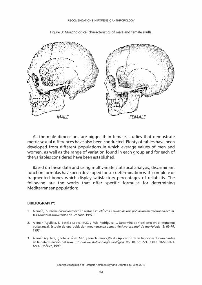
Skull morphology also varies in either sex. Just as in the rest of the skeleton, the female skulls are more graceful and rounded than the male, in which muscular insertions are more pronounced. These differences also affect the size and it is considered that cranial capacity is of 150 to 200 cc. higher in men than women, and it oscillates widely due to the variability and sexual dimorphism in human groups.
The male skull in sagittal view show a front profile that is usually slightly oblique, with a glabella and supraorbital arcs more pronounced, so are the temporal muscle and supramastoid crest. The mastoids are medium to large sized and generally rough, rounded and protruding. The occipital region is often rough and it shows a prominent inion; all this because the neck muscles are inserted in this zone, which tend to be more developed in men. In the facial region orbits have rounded edges and are relatively thick; the nasal opening is bigger, higher and the massive malar has a rough lower edge. The palate is generally wider, longer and deeper, with an U shape.
In women the frontal is more vertical and it usually shows rounded frontal protuberances more pronounced than in men; so it is with the parietal protuberances. The zygomatic arches are smaller and weaker, so are the mastoid process, smoother, smaller and more pointed; the orbits are generally more oval, with sharp edges. The palate is relatively shorter and narrower, with a tendency to adopt a parabolic shape.
The foramen magnum is generally bigger in men than in women, and so are the occipital condyles and the glenoid cavity.
Regarding the jaw, the male is more massive than female, with a higher body, wider ascending ramus and longer condyles. In men, the angle formed by the arm and the body is more closed than in women. The male chin tends to be square and thick, while in women is sharper and thinner.
RECOMENDATIONS IN FORENSIC ANTHROPOLOGY
Spanish Association of Forensic Anthropology and Odontology. June 2013
61

These features are reliable for the sex determination of the people aged between 20 to 55 years or so. For the infants, the traits are not yet completely developed and some of them may be affected by the changes due to advanced age.
Cranial capacityOccipital.
Frontal.
Superciliary arches.
Orbits.
Zygomatic arches.
Mastoid apophysis.
Jaw.
Teeth.
Men
Well marked muscle attachments.
More inclined.
Strong.
Rather low; quadrangular.
Strong.
Well developed.Massive, high and robust.
More bulkier.
Women
150 to 200 cc. Less.
R o u n d e d , w i t h o u t roughness.
Convex and high.
Minimal or absent.
Higher and more rounded, with the upper sharp edgeLittle robust.
Less developed and pointed y puntiagudas.
Less robust, graceful appearance, more obtuse mandibular angle and reduced condyles.
Less bulky.
SOME SEX DIFFERENCES IN THE SKULL
Table 2: Morphological characteristics more common in male and female skulls.
RECOMENDATIONS IN FORENSIC ANTHROPOLOGY
Spanish Association of Forensic Anthropology and Odontology. June 2013
62
As the male dimensions are bigger than female, studies that demostrate metric sexual differences have also been conducted. Plenty of tables have been developed from different populations in which average values of men and women, as well as the range of variation found in each group and for each of the variables considered have been established.
Based on these data and using multivariate statistical analysis, discriminant function formulas have been developed for sex determination with complete or fragmented bones which display satisfactory percentages of reliability. The following are the works that offer specific formulas for determining Mediterranean population:
BIBLIOGRAPHY:
1. Aleman ,�I.:�Determinacion del sexo en restos esquelet icos. Estudio de una poblacion mediterran ea actual.
Tesis�doctoral.�Universidad�de�Granada.�1997.
2. Aleman �Aguilera,� I.;� Botella� Lop ez,�M.C.� y� Ruiz� Rodrig uez,� L.�Determinacion �del� sexo� en� el� esqueleto�postcraneal.�Estudio�de�una�poblacion �mediterran ea�actual.�Archivo espano l de morfologia , 2:�69-79,�1997.
3. Aleman �Aguilera,�I.;�Botella�Lop ez,�M.C.�y�Souich�Henrici,�Ph.�du.�Aplicacion �de�las�funciones�discriminantes�en� la�determinacion �del�sexo.�Estudios de Antropologia Biolog ica. Vol.� IX.�pp:�221-�230.�UNAM-INAH-AMAB.�Mexico,�1999.
Figure 3: Morphological characteristics of male and female skulls.
MALE FEMALE
RECOMENDATIONS IN FORENSIC ANTHROPOLOGY
Spanish Association of Forensic Anthropology and Odontology. June 2013
63

4. Aleman ,�I.;�Botella,�M.C.;�Souich,�Ph.du�y�Yoldi,�A.�Estudio�de�poblaciones�prehistor icas�mediante�aplicacion �de�anal isis�discriminante.�Aspectos�metodolog icos.�Antropologia y Biodiversidad. Vol.�1;�pp:�25-�32.�Aluja,�MP.;�Malgosa,�A.;�Nogues ,�RM.�(eds.).�Bellaterra.�Barcelona.�2003.
5. Aleman ,�I.;�Yoldi,�A.;�Botella,�MC.;�Mastrangelo,�P.�Funciones�isquiopub icas�discriminantes�del�sexo�en�una�poblacion �mediterran ea�de�sexo�y�edad�conocidos.�Diversidad humana y antropologia aplicada. pp:�613-627;�2010
6. Amores,�A.�Determinacion del sexo a partir de la base del cran eo, atlas, axis, c7 y t12. Trabajo�de�investigacion �tutelada.�Master�de�Antropologia �Fisica�y�Forense.�Universidad�de�Granada.�2009.
7. Barrio,�P.;�Trancho,�G.;�San chez,�JA.:�Metacarpal�sexual�determination�in�a�Spanish�population.�Journal of
Forensic Science, 51 (5), 990�–�995,�2006.
8. Mastrangelo,�P.:�Diagnos tico del sexo a traves de huesos del carpo en una poblacion de sexo y edad
conocidos. Trabajo�de� investigacion �tutelada.�Master�de�Antropologia �Fis ica�y�Forense.�Universidad�de�Granada.�2008.
9. Mastrangelo,� P.;� Aleman,� I.;� De� Luca,� S.;� Botella,� MC.� Determinacion � del� sexo� mediante� los� huesos�proximales�del�carpo.�Diversidad humana y antropologia aplicada. pp:�689-696;�2010
10. Mastrangelo,�P.;�De�Luca,�S.;�Aleman �I.;�Botella�MC.:�Sex�assessment�from�the�carpals�bones:�Discriminant�function� anal isis� in� a� 20th� century� Spanish� simple.� Forensic Science Internacional. 2011.� DOI:�10.1016/j.forsciint.2011.01.007
11. Trancho,� G.J.,� Robledo,� B.� ,� Lop ez-Bueis,� I.,� San chez,� J.A.:� Sexual� determination� of� the� femur� using�discriminant� functions.�Analysis�of� a� Spanish�population�of� known� sex� and� age. Journal of Forensic
Sciences, Volume�42,�Issue�2,�March�1997,�Pages�181-185
12. Viciano,�J.:�La odontometria como tec nica de determinacion del sexo en individuos subadultos. Estudio de
la poblacion de Herculano (s.I d.C., Nap oles, Italia). Trabajo� de� investigacion � tutelada.� Master� de�Antropologia �Fis ica�y�Forense.�Universidad�de�Granada.�2008.
13. Viciano� J.;�Aleman� I.;�D'Anastasio�R.;�Capasso� L.;�Botella�MC.�Odontometric�Sex�Discrimination� in� the�Herculaneum�Sample�(79�AD,�Naples, Italy),�with�Application�to�Juveniles.�American Journal of Physical
Anthropology. AJPA-2010-0008�2011.�DOI:�10.1002/ajpa.21471
14. Yoldi,�A.;�Aleman ,� I.;�Botella,�MC.�Funciones�discriminantes�del�sexo�a�partir�del� ilion�en�una�poblacion �mediterranea�de�sexo�conocido.�Revista Espano la de Antropologia Biolog ica. Vol.�22.�pp:�23-38.�2001.
15. Yoldi,�A.;�Aleman,�I.�y�Botella,�M.C.�Funciones�discriminantes�del�sexo�en�el�extremo�esternal�de�la�cuarta�costilla�en�una�poblacion �mediterran ea�de�sexo�y�edad�conocidos.�Antropologia y Biodiversidad. Vol.�1;�pp:�594-602.�Aluja,�MP.;�Malgosa,�A.;�Nogues ,�RM.�(eds.).�Bellaterra.�Barcelona.�2003.�
RECOMENDATIONS IN FORENSIC ANTHROPOLOGY
Spanish Association of Forensic Anthropology and Odontology. June 2013
64

SEX ESTIMATION IN SUBADULTS.
Sex determination in subadults is one the biggest challenges that Physical Anthropology currently faces and involves a significant hurdles in forensic expertise because it is necessary to have very accurate results. Children are not sexually mature, which results in the absence of secondary sexual characteristics -that are not discernible before puberty- hinders greatly the discrimination between boys and girls.
Several morphological and metric methods have been proposed for determining the sex of skeletal and rental remains of subadults. The metric ones have been used a lot and for a long time due to their objectivity, but they have a limited use, while methods based on skeletal morphology have shown great advantages. This may be due to the difficulty in locating anatomical reference points and reproducing the measurements, but also because morphological methods take into consideration the whole, it is possible to estimate the relationships between segments and offer a more complete view. It is true that it can be more subjective and that it requires training and more experience, but they are superior because they integrate a greater amount of associated data.
The best available methods based on metric characteristics refer to dental size (Ditch and Rose, 1972; Black, 1978; De Vito and Saunders, 1990; Cardoso, 2008), because teeth crowns are developed early in life (the first permanent molar crown its development at age 3). The size of the crown may be determined by direct measurement or by using radiographs.
Numerous papers have documented this difference in size, and usually emphasize that the tooth showing a greater sexual dimorphism is the permanent canine whose crown is completely developed at the age 7. For the children under the age of 3, primary dentition must be used. However, sexual dimorphism in the primary dentition is minor compared to the permanent dentition, and therefore it is preferable, whenever possible, to use permanent dentition.
Apart from teeth, there are several morphological characteristics of the skull and pelvis that can and should be evaluated.
Several skeletal nonmetric traits proposed by Schutkowski (1993) and Weaver (1980), subsequently reassessed by Loth and Hennenberg (2001) and Sutter (2003) show promising results in sex determination in subadults , and it is observed that these characteristics are more precise for assigning sex in boys than in girls. This may be due to the expression of skeletal characteristics associated with sex, especially in the pelvis, related to differential growth in women. Furthermore, sexual expression of the various features is related to
RECOMENDATIONS IN FORENSIC ANTHROPOLOGY
Spanish Association of Forensic Anthropology and Odontology. June 2013
65

age, so at certain age groups, some characteristics are more convenient to use than the others.
For children between the ages of 2 to 5, the depth and angle of the greater sciatic notch and the curvature of the iliac crest show acceptable accuracy for forensic applications. For children aged between 5 to 15 , the depth and angle of the greater sciatic notch, the criterion of the compounded arc and the shape of the anterior mandible arc can be considered.
Special precautions must be taken with individuals younger than 2 years old, since it does not offer any acceptable security for its application in forensic cases except the atrial surface elevation. However, there are several papers that show that some morphological characteristics of the pelvis and the skull present sexual dimorphism.
The morphological analysis of the humeral distal epiphysis has shown an acceptable sexual dimorphism for its use within the forensic context. It was proposed by Rogers (1999) and reassessed by Rogers (2009) and Falys et al. And it is applicable to adolescents between the ages of 11 and 15. The method is based on the fact that the observed sexual dimorphism would reflect the carrying capacity of the elbow. Given that women usually have narrower shoulders and wider hips than men, the angle that the elbow describes is greater in women than in men.
The following tables gather the characteristics that must be taken into consideration when determining the sex of children, according to the estimated age group.
RECOMENDATIONS IN FORENSIC ANTHROPOLOGY
Spanish Association of Forensic Anthropology and Odontology. June 2013
66

Table�3:�Morphological�and�metric�sex�differences�in�subadults.
Feature.
Pelvis.
Depth of the greater sciatic notch.
Angle of the greater sciatic notch.
Compound arch.
Iliac crest curvature.
Auricular surface elevation.
Jaw
Prominence of the chin.
Anterior dental arch form.
Diversion of the region goníaca.
Dentition.
Dental size.
Humerus.
Constriction of the trochlea.
Symmetry of the trochlea.
Shape olecranon fossa.
Angle of medial epicondyle.
Girls.
Shallow.
Greater angle of 90º.
The line crosses the auricular surface.
The crest forms an attenuated S.
High.
Smooth and flat surface.
Arch rounded, parabolic.
Angle not diverted, flat.
Smaller.
Notably constrained, loop shaped.
Symmetric.
Oval, deep.
Clearly high.
Boys.
Deep.
Narrower angle, approaches 90°.
The solid line along the lateral edge of the auricular.
The curvature is more pronounced, and draws a sharp S.
No high.
Prominent, quadrangular, wider.
Wider arch, U-shaped.
Angle diverted.
Larger.
Slightly constricted.
Asymmetric.
Triangular, shallow, not very deep.
Flat or slightly elevated.
RECOMENDATIONS IN FORENSIC ANTHROPOLOGY
Spanish Association of Forensic Anthropology and Odontology. June 2013
67

RECOMMENDATIONS.
1. If the conservation of the material allows, it is better to use skull and pelvis morphological characteristics rather than the metric data.
2. In adults, the pubic ventral arc and the subpubic concavity are the elements that offer greater reliability; differences of its morphology depend only on the sex and don't show variations related to population origin of individuals.
3. An overall evaluation of all morphological features must be made. It is true that some of them are easier to observe than the others, but not all are equally pronounced in every person. We must not forget that variability is an inherent characteristics of the population.
Tabla�4:�Morphological�and�metric�characteristics�most
�important�to�evaluate�distributed�by�age�groups.
Fetuses - 2 years oldAuricular surface elevationSize of dental deciduous dentition
2 - 5 years oldDepth of the greater sciatic notchAngle of the greater sciatic notchIliac crest curvatureSize of dental temporal and permanent dentition
5 - 11 years oldDepth of the greater sciatic notchAngle of the greater sciatic notchCompound ArchShape of the anterior mandible dental archSize of temporal and permanent dentition
11 - 15 years old Constriction of the trochleaSymmetry of the trochleaShape of olecranon fossaAngle of medial epicondyleDental size, permanent dentition
RECOMENDATIONS IN FORENSIC ANTHROPOLOGY
Spanish Association of Forensic Anthropology and Odontology. June 2013
68

4. Methods for estimating sex in subadults are less accurate and its reliability depends on the state of development of the skeleton to identify.
BIBLIOGRAPHY:
1. Black TK.. Sexual dimorphism in the tooth-crown diameters of the deciduous teeth. American Journal of Physical Anthropology, 48: 77-82. 1978.
2. De Vito C, Saunders SR. A discriminant function analysis of deciduous teeth to determine sex. Journal of Forensic Science, 35: 845-858. 1990.
3. Ditch LE, Rose JC. A multivariate dental sexing technique. American Journal of Physical Anthropology, 37: 61-64. 1972.
4. Falys CG, Schutkowski H, Weston DA. The distal humerus - A blind test of Rogers' sexing technique using a documented skeletal collection. Journal of Forensic Science, 50: 1289-1293. 2005.
5. Loth SR, Henneberg M. Sexually dimorphic mandibular morphology in the first few years of life. American Journal of Physical Anthropology, 115: 179-186. 2001.
6. Phenice TW. A newly developed visual method of sexing the os pubis. American Journal of Physical Anthropology, 30: 297- 302. 1969.
7. Rogers TL. A visual method of determining the sex of skeletal remains using the distal humerus. Journal of Forensic Science, 44: 55-59. 1999.
8. Rogers TL. Sex determination of adolescent skeletons using the distal humerus. American Journal of Physical Anthropology, 140: 143-148. 2009.
9. Schutkowski H. Sex determination of infant and juvenile skeletons: I. Morphognostic features. American Journal of Physical Anthropology, 90: 199-205. 1993.
10. Sutter RC. Nonmetric subadult skeletal remains traits. I. A blind test of the accuracy of eight previously proposed methods using prehistoric known-sex mummies from Northern Chile. Journal of Forensic Science, 48: 927-935. 2003.
11. Weaver D. Sex differences in the ilia of a known sex and age sample of fetal and infant skeletons. American Journal of Physical Anthropology, 52: 191-195. 1980.
RECOMENDATIONS IN FORENSIC ANTHROPOLOGY
Spanish Association of Forensic Anthropology and Odontology. June 2013
69


CRITERIA OF AGEESTIMATION
RECOMENDATIONS IN FORENSIC ANTHROPOLOGY
Spanish Association of Forensic Anthropology and Odontology. June 2013
71


PROTOCOL
AUTHORS
CONTACT
DATE
AGE ESTIMATION
ROBLEDO ACINAS MM, SANCHEZ SANCHEZ JA,
FERNANDEZ CAMACHO FJ
[email protected] • [email protected] ,
May 31, 2012
1. INTRODUCTION.
According�to�the�dictionary�of�the�Royal�Academy�of�the�Spanish�Language�(RAE),�the�concept�of�age�has�several�meanings,�and�in�reference�to�the�topic�at�hand,�the�RAE�conceptualized�a�person's�age�as�“ time�that�a�person,�certain�animals�or�vegetables�have�lived” or�“each�of�the�periods�in�which�human�life�is�considered�divided”.�From�the�point�of�view�of�Forensic�Anthropology,�we�have�to�differentiate�chronological�age,�which� is�referred�by�the�above�definition,�from�the�biological�and�in�particular�from�the�bone�age.�Indeed�when�we�face�a�case�in�which�a�person�must�be�identified�from�the�skeletal�remains�found,�what�we�have�to�do�is�to�infer�which�may�be�the�age�of�person�at�the�time�of�death�from� the� characteristics� shown�by� the�bones,� in�order� to�do� so,� �what�we�consider� is� the�bone� age� that�we� transfer� into� the� chronological� age.� This�transfer�is�inevitably�approximate,�since�the�variability�of�bone�characteristics�in�terms�of�aging�make� it� impossible� to�establish�beyond�any�doubt� the�exact�chronological�age,�so�what�we�do�is�an�estimation�of�that�chronological�age.�The�better�or�worse,�approximation�depends�on�a� large�number�of�variables�such� as:�personal� characteristics,�nutrition,�diseases,� ancestral�origin�of� the�subject,�etc.�
We�must�consider�these�conditions�when�we�are�about�to�carry�out�the�study�of�a�case�and�we�have�to�be�prudent�enough�not�to�venture�to�estimate�beyond�what�can�be,�that�is�we�will�always�have�to�give�an�age�period�and�not�an�exact�age.�This�age�period�will�depend�on�the�method�that�we�can�use,�its�reliability�and�of�course�the�age�period�in�which�the�subject�is�in�addition�to�the�conditions�that�we�have�previously�indicated.�With�this�protocol�or�guidelines�established�by�AEAOF,�we�are�trying to�offer�the�most�general�and�easiest�to�apply�methods�for� the� researcher� to�make� the� age� estimation�with� the� strongest�possible�guarantees.
As�also�the�dictionary�of�the�RAE�points�out� in�another�of� its�definitions:�“each�of�the�periods�in�which�human�life�is�considered�divided”,�in�our�case�the�
RECOMENDATIONS IN FORENSIC ANTHROPOLOGY
Spanish Association of Forensic Anthropology and Odontology. June 2013
73

methods�to�apply�for�age�estimation�are�divided�into�age�groups�and�because�it�is�not�the�same�the�bone�of�a�fetus�than�one�of�a�child�in�formation�and�growing�or�one�of�a�young�adult�in�which�the�bone�even�though�is�fully�formed,�it�has�not�yet� begun� to� be� altered,� a� situation� that� occurs� in� a� subject� of� middle� or�advanced�age.
2. AGE RANGES OR GROUPS.
The�age�ranges�proposed�are:
With�this�age�groups�we�try�to� limit,�to�the�extent�possible,� �the�different�periods�that�occur�during�the�human�bone�development�but�by�definition,�any�division�of�a�continuum�is�arbitrary.�Therefore�we�must�take�into�consideration�that�age�estimation�methods� implemented�by�various�authors,�may�overlap�among�different�age�ranges.
METHODS OF AGE ESTIMATION.
3.1- Subadults.
3.1.1 Fetuses.
Although�for�the�age�calculation�in�fetuses�the�onset�of�ossification�cores�is�mentioned,� this�method� is�not�useful� for�studying� the�skeletized� fetus�since�those�cores�have�disappeared.�The�metric�of�the�bone�remains�that�we�find�is�what�will�give�us�an�approximate�age.� �In�this�regard�the�method�followed�by
Fazekas�and�Kosa�(1978),�based�on�the�study�of�138�human�fetuses�(17�males�
SUBADULTS: - Fetal <0 - Children: 0-14 years - Juvenile: 15-18 years ADULTS: - Young: 18-35 years - Medium age: 36-60 years - Advanced age: > 60 years
RECOMENDATIONS IN FORENSIC ANTHROPOLOGY
Spanish Association of Forensic Anthropology and Odontology. June 2013
74

and� 67� females),�aged� from� the� third� to� the� tenth� lunar�month� is� the�most�complete� available� publication.� This� work� has� been� developed� using� 67�measures�on�37�bones�and�although�there�are�other�papers� (Balthazard�and�Dervieux�1921,�Olivier�and�Pineau�1958),�these�are�partial�unlike�Fazekas�and�Kosa�which�is�systematic.�
3.1.2 Children.
a)�Dental�eruption.�Probably�dental�eruption,�by�visual�inspection,�has�been�the�most�widely�used�and�although�there�is�a�variability�mediated�by�genetic�and�environmental�factors,�continues�to�be�an�useful�and�fast�method.
b)�Dental�mineralization.�The�mineralization�of�the�crows�in�the�deciduous�teeth�starts�at�3,�4�months�in�utero�and�continues�after�birth�during�the�neonatal�period� (Burdi� in�Moyers,� 1992).� For� its�part� the�development�of�permanent�dentition�mineralization�takes�approximately�9�years,�beginning�with�the�first�permanent�moral� (Evans�and�Knight,� 1981).� In�our�geographical� setting� the�dental�eruption�of�both�dentitions� is�published�by�Barberia� (1995).�We�must�consider�in�this�paper�that�the�data�from�the�eruption�were�obtained�in�the�clinic,�while� in� the�case�of�skeleton� remains� the�valuation�of� the�eruption� is�made�according�to�the�bone.
Another�of�the�most�used�methods� is�Demirjian's�et�al� (1973),�based�on�a�sample�of�French-Canadian�children.�Subsequently�this�same�author�developed�updates� of� the� original� method� (Demirjian,� 1976).� In� any� case,� since�maturational� development� is� different� in� both� � sexes,� the� sex� must� be�determined� previously.� Subsequent� to� this� paper,� many� others� have� been�published�in�which�the�accuracy�of�it�is�put�to�test,�showing�a�slight�delay�in�the�maturation�of�the�French-Canadian�population,�leading�to�an�overestimation
when� applying� this�method� to� other� populations.� In� 2007,�Cameriere� et� al�compared�Cameriere�methods� (Cameriere� et� al,� 2006),�Demirjian� y�Willems�(Willems� et� al,� 2001)� studying� radiographs�of� Spanish,� Italian� and�Croatian�children�between�5�and�15�years�old�and�they�indicating�that�the�best�method�is�the�one�advocated�by�Cameriere.
3.1.3 Children�and�youth.
When�dentition� is�not�available�for�age�estimation�the� length�of�the� long�bones�and�the�age�can�be�related.�In�this�regard�there�are�several�papers�such�as�those�by�Mares�(1943,�1955,�1970,�1972).�This�method�can�be�applied�between�2�
RECOMENDATIONS IN FORENSIC ANTHROPOLOGY
Spanish Association of Forensic Anthropology and Odontology. June 2013
75

months�and�18�years�old,�therefore�covers�the�child�and�the�juvenile�period.�It�would�be�a�method�to�apply�in�cases�where�the�study�of�the�dentition�was�not�possible.�Besides�Maresh,�there�are�other�authors�like�Giunhart�(1973),�in�radios�and�tibia,�Anderson�et�al�(1964)�in�femur�and�tibia,�Hoffman�(1979)�who�studies�bone� growth� in� relation� to� age� between� 2� months� and� 12� years.� Maresh,�Guinhart�and�Anderson�papers�can�be�found�in�Scheuer�and�Black�(2000).
3.2. Adults.
3.2.1 Synostosis�of�the�cranial�sutures.
The�closure�of�the�cranial�sutures�and�its�relation�to�the�individual's�age�has�been�studied�since�Todd�and�Lyon�in�1924�and�1925�published�the�first�studies�in�this� direction,� subsequently� there� are� a� number� of� authors� who� try� to�implement�and�modify� this�method� to�achieve�greater�accuracy,�Meind�and�Lovejoy� (1985)� are� who� applied� the� method� of� study� that� provides� most�reliability.� This� study�was� conducted� on� 236� skulls� from� the�Hamann-Todd�collection,�proposing�a�method�based�on�the�study�of�10�points�located�in�the�cranial�vault,�establishing�four�closing�stages.
3.2.2 Morphological�changes�of�the�fourth�rib.
� �Iscan�et�al�(1984)�studied�230�right�ribs�extracted�from�white�men�who�have�been�autopsied�whose�age�and�sex�are�known.�It�is�a�method�that�has�proven�to�be�reliable�and�relatively�easy�to�use.�This�method�has�been�also�seen�that� is�dependent�population.
3.2.3. Changes�in�the�public�symphysis.
� The�changes�in�the�pubis�symphysis�were�studied�first�by�Todd�(1920)�who�studied�a�collection�of�skeletons�of�known�age�white�males�and�proposed�10�stadiums�for�study�in�relation�to�age.
Mackern�and�Stewart�(1957)�presented�a�method�based�on�the�study�of�the�pubic�symphysis�divided�in�three�fields�of�study�(dorsal�facet,�ventral�wall�and�symphyseal� ring)� for�males.�Acsadi�and�Nemeskeri� (1970)� studied� the�pubic�symphysis� of� 61� men� and� 44� women,� that� later� extended� with� skeletons�extracted�from�cemeteries�in�Hungary.
RECOMENDATIONS IN FORENSIC ANTHROPOLOGY
Spanish Association of Forensic Anthropology and Odontology. June 2013
76

Gilbert�and�Mckern�(1973),�set�the�standards�for�women,�based�on�the�same�components� that� had� been� previously� established� for�men� by�Mckern� and�Stewart.�Suchey�(1979),�points�out�that�applying�the�test�of�Gilbert�and�Mckern�only�51%�of�the�cases�fall�withing�the�expected�age.
Suchey�et�al�(1986),�indicate�that�the�symphyseal�surface�of�men�is�not�a�good�indicator�for�ages�over�40�years.�Meindl�et�al�(1985),�compare�the�methods�of�Todd� (1920),�Mackern�and�Stewart� (1978),�Gilbert�and�Mackern� (1973)�and�Hanihara�and�Suzuki�(1978),�in�the�Hamman-Todd�collection�and�conclude�that�Todd�method�is�the�best�one.
Brooks�and�Suchey� (1990),�using�a�sample�of�739�males�and�273�females,�whose�symphysis�were�obtained�of�a�modern�and�well�documented�sample�of�autopsied�individuals�in�Los�Angeles�County,�and�they�establish�6�phases�with�its�features,�both�for�male�and�female,�with�the�standard�mean�and�deviation�and�range�of�age,�and�they�compare�their�results�with�those�obtained�by�Acsadi�and�Nemeskeri,�which�until�then�was�the�method�recommended�by�the�Workshop�of� European� Anthropologist,� concluding� that� the� technique� of� Acsadi� and�Nemeskeri�does�not�adapt�to�the�data�of�the�modern�sample�they�study.
3.2.4 Changes�in�the�auricular�surface�of�the�ilium.
� (Lovejoy�et�al,�1985),�they�develop�a�method�based�on�the�changes� in�the�auricular�surface�of�the� ilium,�and�as�previously�studied�by� Iscan�and�Derrick�(1984)�and�St�Hoyme�(1984),�who�conducted�a�study�on�growth�of�the�pelvis.�Lovejoy�et�al�(1985)�study�an�extensive�sample�of�250�skeletons�from�the�Libben�collection� (Ohio,� Indian� population),� and� 500� from� the� Hamman-Todd�collection.�Subsequently�they�carry�out�a�test�with�216�ilium.�According�to�the�authors�there�are�no�differences�in�the�evolution�of�the�features�of�this�surface�between�men�and�women.�After� this,�other�authors� such�as�Buckberry�and�Chamberlain� (2002),�Osborne�et�al� (2004),�Mulhern�and� Jones� (2004),�make�revisions�on�the�method�ob�Lovejoy�et�al�(1985),�or�apply�it�to�other�populations�as�Hens�and�Belcastro�(2012)�do�to�Italian�population.�
3.2.5. Dental�study.
Periodontosis�and�root�transparency�(Lamendin�et�al,�1992).�These�authors�conduct� their�paper�on� 306� single-rooted� teeth,� from� 208� individuals� aged�between�22�and�90�years�and�value�the�periodontosis�and�the�root�transparency.�This�study�shows�no�significant�differences�in�gender,�the�minimum�error�occurs�
RECOMENDATIONS IN FORENSIC ANTHROPOLOGY
Spanish Association of Forensic Anthropology and Odontology. June 2013
77

in�the�group�of�50-59�years�and��the�maximum�in�13,1�years�in�young�groups�(30-39�years),�so� its�use� in�young� individuals� is�not�advised.�Baccino�et�al� (1999),�comparing�in�French�population�various�methods�of�age�estimation,�observe�a�better�performance�of�the�method�of�Lamendin�when�facing�skeletal�changes�(pubis,�rib,�histology).
Other�authors� that�study� the� teeth� in� relation� to�age�are�Solheim� (1993),�López�Nicolas�et�al�(1996),�Kaal�and�Solheim�(1994),�Martínez�de�Mandojana�(1999).�There�are�also��papers�based�on�biochemical�methods,�there�are�a�large�number� of� publications�Heman� and� Bada� (1975),�Ohtani� (1992),�Mornstad�(1994),�based�on�the�study�of�the�D�and�L�forms�of�amino�acids�and�Martin�de�las�Heras�(1999)�based�on�the�study�of�deoxypyridinoline.�These�methods�show�a�good�correlation�with�age�and�we�can�consider�them�as�an�alternative�to�the�usual�methods.
RECOMENDATIONS.
1. Always consider that what we are doing is an estimation and therefore put the range of age given as the most likely.
2. Methods proposed:
2.1. SUBADULTS
2.1.1 Fetuses. In the fetal remains the method of choice for age estimation is the one proposed by Fazekas and Kosa, in their book Forensic Fetal Osteology.
2.1.2. Infants. - Method Demirjian et al (1973, 1976). - Method Maresh (1943, 1955, 1970, 1972), and collected in its entirety in
Sheuer and Black (2000).
2.1.3. Juveniles. - Method Demirjian et al (1973, 1976). - Method Maresh (1943, 1955, 1970, 1972), and collected in its entirety in
Scheuer and Black (2000).
RECOMENDATIONS IN FORENSIC ANTHROPOLOGY
Spanish Association of Forensic Anthropology and Odontology. June 2013
78
2.2. ADULTS.
- Synostosis of cranial sutures: Method Meinld y Lovejoy (1985). - Changes in the cost-sternal union of the fourth rib (Iscan y cols, 1984,
1985). - Changes in the auricular surface (Lovejoy and cols, 1985). - Dental study (Lamendin et al, 1992).
3. Summary and considerations.
As noted in the introduction, when facing an anthropologic-forensic case, in which we try to estimate the age, we must never forget the conditioning factors that the proposed methods have, regarding the source population that has given rise to this method and the particular constraints of each case. As to the fetuses, the method of Fazekas and Kosa, is the only full one existing in the biography for this age group, and therefore its application is mandatory. At older ages, ie from birth, the methods that have been implemented are more numerous and we must choose the one that offers greater reliability, and in these cases is proposed the use of Demirjian method for children and young ages based on the mineralization of teeth and more widespread among researchers and the one of Maresh, in the case that we do not have teeth. In adult subjects, that we have divided into young 18-35 years, medium age 36-60 and advanced age > 60 years, it has to be noted that actually with the existing methods we can approximate the age at the first two groups, while for the third there are no reliable methods. We also have to take into consideration that the estimation of age only by cranial sutures should not be done, this method will be used always as a complementary to another, and in terms of the cost-sternum union of the fourth rib and the study of the pubic symphysis, if it is possible to conduct the study of the fourth rib this would be the method of choice. The study of the atrial surface has the advantage of being independent of sex, so if sex is unknown this method would be the one to apply.
BIBLIOGRAPHY:
1. Acsad i,�G�y�Nemeskeri,�J.�(1970).�History�of�Human�Lifespan�and�Mortality,�Akademiai�Kiado,�Budapest.
2. Balthazard,� V.�Dervieux� (1921).� Etudes� anthropologiques� sur� le� foetus� humain.�Ann.�Med.� Leg.� 1:37�Barberia �Leache�E.�Odontopediatria.�Barcelona:�Masson;�1995.
3. Berkovitz,�B.K.B.,�Holland,�G.�R.,�Moxham,�B.J.(1995).�Atlas�en�color�y�texto�de�Anatomia �Oral,�Histologia �y�Embriologia �(2a�ed.).�Mosby/Doyma�libros,�S.A.�Madrid.
4. Brooks,�S�y�Suchey,�J.�(1990).�Skeletal�age�determination�base�on�the�Os�Pubis:�A�Comparison�of�the�Acsad i-Nemeskeri�and�Suchey-Brooks�Methods.�”Human Evolution”, 5:227-227-238.
5. Brotwell,�D.�R.�(1981).�Desenterrrando�huesos.�La�excavacion ,�tratamiento�y�estudio�de�restos�del�esqueleto�humano.�Fondo�de�Cultura�Econom ica.�S.A.�Madrid.
RECOMENDATIONS IN FORENSIC ANTHROPOLOGY
Spanish Association of Forensic Anthropology and Odontology. June 2013
79

6. Buckberry,�J.L.,�Chamberlain,�A.T.�(2002).�Age�Estimation�From�Auricular�Surface�of�the�Ilium:�A�revised�method.�Am�J.�of�Pys�Anthropol,�119:231-239.
7. Burdi,� A.R.� (1992).� Desarrollo� de� la� denticion� y� la� oclusion .� Moyers� R.E.� Manual� de� Ortodoncia.� Ed.�Panamericana,�Buenos�Aires.
8. Cameriere,�R.,�Ferrante,L.,Cingolani.,�M.�(2006).�Age�estimation�in�children�by�measurement�of�open�apices�in�teeth,�Int.�J.�Leg.�Med.�120:�49–53.
9. Demirjian,�A,�Goldstein,�H.,�Tanner,�J.M.�(1973).�A�new�system�of�dental�age�assement,.�Human�Biology,�45(2):�211-227.
10. Demirjian,�A.�(1976).�New�system�for�dental�maturity�based�on�seven�and�four�teeth.�Annals�for�Human�Biology,�3(5):�411-421.
11. Evans,�K.T.,�Knight,B.�(1981).�Radiology�in�Forensic�Medicine.�Oxford�Blackwell�Scientific�Publications.
12. Fazekas,�I.�Gy.,�Kos a,�F.�(1978).�Forensic�Fetal�Osteology.�Akademiai�Kiado, �Budapest.
13. Gilbert,�M.M.,�Mckern,�T.W.�(1973).�A�method�of�ageing�the�female�os�pubis.�Am.�J.�of�Pys.�Anthropol,�38:31-38.
14. Hens,�M.S.,�Belcastro,�M.G.(2012).�Auricular�surface�aging:�a�blind�test�of�the�revised�method�on�historic�Italians�from�Sardinia.�Forensic�Sciences�International,�214(1-3):1-5.
15. Hoffman,�J.M.�(1979).�Age�estimation�from�Diaphiseal�lengths:�Two�Months�to�Twelve�Years.�J.�For�Sci,�24:2.�
16. Iscan,�M.�Y.,�Loth,�S.R.,�Wright,�R.K.�(1984).�Age�estimation�from�the�rib�by�Phase�Analysis:�White�Males.�Jounal�of�Forensic�Sciences�29:1094-1104.
17. Iscan,�M.�Y.,�Loth,�S.R.,�Wright,�R.K.�(1985).�Age�estimation�from�the�rib�by�Phase�Analysis:�Females.�Jounal�of�Forensic�Sciences�30:853-863.
18. Lamedin�H,�Baccino�E�Humbert�J.�C�Tavernier�R.�M�Nossintchouk�Zerilli�A.�1992.�A�simple�technique�for�age�estimation�in�adult�corpses:�the�two�criteria�dental�method.�J Forensic Sci 37:1373-9.
19. Lovejoy,�C.O.,�Meindl,�R.S.,�Pryzbeck,�T.R.,�Mensforth,�R.�P.�(1985).�Chronological�Metamorphosis�of�the�Auricular�Surface�of�the�Ilium:�A�new�method�for�the�Determination�of�Adult�Skeletal�Age�at�Death.�Am.�J.�of�Phys.�Anthropol,�68:15-28.
20. Meindl,�R.S.�y�Lovejoy,�C.O.�(1985).�Ectocranial�Suture�Closure:�A�Revised�Method�for�the�Determination�of�Skeletal�Age�at�Death�Based�on�the�Lateral-Anterior�Sutures.�American�Journal�of�Physical�Anthropology�68:57-66.
21. Meindl,�R.S.�Lovejoy,�C.O.,�Mensford,�R.�P.,�Walker,�R.A.�(1985).�A�revised�method�of�age�determination�using�the�os�pubis,�with�a�review�and�test�of�accuracy�of�other�current�methods�of�pubis�symphyseal�ageing.�Am.�J.�Pys�Anthropol,�68:29-45.
22. Maresh,�M.M.� (1943).�Growth�of�major� long�bones� in�healthy�children.�Americam� Journal�of�Physical�Anthropology,�66:�227-257.
23. Maresh,�M.M.� (1955).�Linear�Growth�of� long�Bones�of�Extremities� from� Infancy�Through�Adolescence.�Continuing�Studies.�AMA�Am�J.�Dis�Child,�89(6):�725-742.
24. Maresh,�M.M.� (1970).�Measurements� from� roentgenograms.�Human�Growth� and�Development,�R.W.�McCammon,�Springfield.
25. Maresh,�M.M.�(1972).�A�forty-five�year�investigation�for�secular�changes�in�physical�maturation.�Americam�Journal�of�Physical�Anthropology,�36-103-109.
RECOMENDATIONS IN FORENSIC ANTHROPOLOGY
Spanish Association of Forensic Anthropology and Odontology. June 2013
80

26. Mulhern,�D.�M.,�Jones,�E.B.�(2004).�Test�of�revised�method�of�age�estimation�from�the�auricular�surface�of�the�ilium.�Am.�J.�of�Phys�Anthropol,�126(1):�61-65.
27. Olivier,�G.�Pineau,�H.�(1958).�Determination�de�l�a ge�du�foetus�et�de�l�e mbryon.�Arch.�D�A nat.�6:21.
28. Osborne,�D.L.,�Simmons,�T.L.,�Nawrocki,�S.�P.�(2004).�Reconsidering�the�Auricular�Surface�as�an�Indicator�of�Age�at�Death.�Journal�of�Forensic�Sciences,�49:5.
29. Scheuer,�L.�y�Black,�S.�(2000).�Developmental�Juvenile�Osteology.�Elsevier�Academic�Press, USA.
30. Suchey,�J.M.�(1979).�Problems�in�the�aging�of�females�using�the�Os�pubis.�Am.�J.�of�Phys�Anthropol,�51:�467-470.
31. Todd,�T.W.�(1920).�Age�changes�in�the�pubic�bone:�I.�The�male�White�pubis.�Am.�J.�of�Phys.�Anthropol.�3:285-334.
32. Todd,�T.W.,�Lyon�D.W.�(1924).�Endocranial�suture�closure,�its�progress�and�age�relationship.�Part I.�Adult�males�o�White�Stock.�Am.�J.�of�Phys.�Anthropol,�7:�325-384.
33. Todd,�T.W.,�Lyon�D.W.�(1925a).�Cranial�suture�closure,�its�progress�and�age�relationship.�Part�II.�Ectocranial�suture�in�adult�males�o�White�Stock.�Am.�J.�of�Phys.�Anthropol,�8:23-45.
34. Todd,� T.W.,� Lyon� D.W.� (1925b).� Cranial� suture� closure,� its� progress� and� age� relationship.� Part� III.�Endocranial�suture�in�adult�males�of�Negro�Stock.�Am.�J.�of�Phys.�Anthropol,�8:47-71.
35. Todd,�T.W.,�Lyon�D.W.�(1925c).�Cranial�suture�closure,�its�progress�and�age�relationship.�Part�IV.Ectocranial�closure�in�adult�males�o�Negro�Sock�Stock.�Am.�J.�of�Phys.�Anthropol,�8:149-168.�
36. Ubelaker,�D.H.�2007�(3a�Ed.).�Human�Skeletal�Remains.�Washington.�
37. Willems,�G.,�Van�Olmen,�A.,�Spiessens,B.,�Carels,C.� (2001).�Dental�age� estimation� in�Belgian� children:�Demirjian's�technique�revisited,�J.�Forensic�Sci.�46:�893–895.
RECOMENDATIONS IN FORENSIC ANTHROPOLOGY
Spanish Association of Forensic Anthropology and Odontology. June 2013
81

CRITERIA FOR HEIGHTESTIMATION
RECOMENDATIONS IN FORENSIC ANTHROPOLOGY
Spanish Association of Forensic Anthropology and Odontology. June 2013
83


PROTOCOL
AUTHORS
CONTACT
DATE
HEIGHT ESTIMATION
DORADO E., CARRILLO MF
[email protected] • [email protected]
March 31, 2013
CRITERIA FOR HEIGHT ESTIMATION IN FORENSIC ANTHROPOLOGY
1. INTRODUCTION.
It� is�meant�by�height�the�distance�between�the�highest�point�of�the�head�(vertex)�and�the�ground�while�the�individual�is�standing,�with�the�head�oriented�in�the�Frankfurt�plane.
Sex,� along�with� age� and� race� create� the� basic� identifying� parameters� in�forensic�anthropology.
Early� studies�about�height�determination�based�on� skeletal� remains�date�back�to�Sue� in�the�eighteenth�century�and�to�Orfila.� In�1888�Rollet�correlates�height� with� long� bones� in� 50� French� corpses,� sample� that� subsequently�Manouvrier�modifies�eliminating�those�older�than�60�years,�and�based�on�this�one�Pearson�creates�in�1899�the�first�regression�formula.�Countless�studies�on�different�populations,�different�bones�and�new�methods�of�measurement�have�happened�since�then,�including�the�studies�of Genoves�in�1967�on�Amerindian�population,�of�Stevenson�in�1929�on�the�Mongoloid�race�or�of�Yung-Hao�in�1970�on�Chinese�population.�Stand�out�for�its�greater�interest�in�the�field�of�Forensic
Anthropology�the�publication�of�the�regression�formulas�by�Trotter�and�Gleser�in�1952�and�1958,�the�ones�of Mendonça MC�in�1998�on�Iberian�population,�and�the�creation�of�the�Fordisc�computer�system�by�Ousley�and�Jantz.
The� height� is� the� result� of� the� interaction� of� multiple� exogenous� and�endogenous�factors,�there�are�variations� in�bone�proportions�between�races,�sexes� and� populations,� which� forces� us� to� be� cautious� with� the� resulting�estimates.�Authors�like�Steyn�and�Smith,�advice�to�simply�speak�of�tall,�medium�or� low� height� depending� on� the� percentile� on� which� is� situated� within� a�population.
The� formulas�available� for�height�determination�are� the� result�of� studies�carried�out�on�specific�samples,�which� implies,�when�discovering�some�bone�remains,�searching�for�the�formula�based�on�a�population�as�similar�as�possible.
RECOMENDATIONS IN FORENSIC ANTHROPOLOGY
Spanish Association of Forensic Anthropology and Odontology. June 2013
85

2. ISSUES ON TABLES AND FORMULAE CONSTRUCTION.
Estimates�of� the�height�will�be�based�on� the�application�of� formulas�and�tables.�We�will�discuss�some�issues�related�to�its�development�and�the�difficulties�involved,�as�it�provides�an�overview�of�the�current�status�of�this�issue�in�the�field�of�Forensic�Anthropology.
Influence� of� secular� changes:� it� has� been� noted� the� influence� of� secular�changes�in�populations,�which�in�the�last�century�has�resulted�to�an�increase�in�the�average�height�along�with�a�earlier�termination�of�growth�(Klepinger�2001).�In� this� respect,� the�growth�of� the�upper�extremities�has�been�categorized�as�isometric� and� of� the� lower� ones� as� allometric,� larger� in� tibia� than� in� fibula�(Meadows�and�Janz�1995).�Which�implies�the�potential�for�error�when�using�for�current�population�the�formulas�developed�based�on�older�populations.
Asymmetry�among�members:�although�the�asymmetry�between�the�bones�of�both�extremities,�right�and�left,�is�generally�irrelevant,�it�has�been�revealed�that�when� is� pronounced,� the� use� of� one� formula� for� both� sides� may� lead� to�erroneous�estimates�(Krishan�et�al�2010).
Small� sheight� of� the� samples:� typically� the� height� of� the� sample� used� in�studies� for�human�height�determination� is�small.�Manouvrier� for�his� famous�table�was�based�on�a�total�of�24�men�and�25�women,�while�Telkka�did�ti�on�114�men�and�39�women,�Genovés�on�22�men�and�15�women�and�Fully�in�a�total�of�102.�The�sample�of�Trotter�and�Gleser�is�an�exception,�reaching�over�7000�bodies.�All�of�which�have�raised�doubts�about� its statistical�validity.�Samples�are�also�limited�by�the�age�of�the�deceased,�since�the�bodies�of�persons�who�have�not�finished�growing�and�elderly�adults�are�excluded,�as�they�may�cause�distortions,�unless�of�course�they�are�the�specific�object�of�study.�
In�this�regard�it�should�be�noted�too�the�difficulty�involved�in�having�large�samples�which�height�is�well�documented.�In�an�exhumation�can�be�easy�to�have�data�on�sex�and�age�but�not�on�the�height.
Bones�analyzed:�the�most�important�works�have�targeted�the�calculation�of�the�height�based�on�the�long�bones�of�the�extremities,�with�better�correlation�with�the�lower�ones.�In�relation�to�this,�it�would�be�preferable�the�femur�for�white�race�and�the�tibia�for�black�race.
However,�other�bones�have�also�been�studied,�usually�without�sufficiently�satisfactory� results,�such�as� the�metatarsals� (Cordeiro� 2009,�Byers� 1989),� the�metacarpals� (Musgrave� and� Hameja� 1978,� Meadows� and� Janz� 1992),� the�calcaneus�and�talus�(Holland�1995),�the�spine�(Janson�and�Taylor�1995,�Terezawa
RECOMENDATIONS IN FORENSIC ANTHROPOLOGY
Spanish Association of Forensic Anthropology and Odontology. June 2013
86

1990,�Tibbetts�1981),�as�well�as�the�clavicle,�the�scapula,�the�hips,�the�sacrum�or�the�height�of�the�femoral�head�(Jit�I.�And�Singh�S.�1956,�Shulin�P.�and�Fangwu�Z.�1983).�
Neither�the�different�craneofacial�measures�(Krishan�2008,�Pelin�2010,�Ryan�and�Bidmos�2006)�or�the�dental�ones� (Ferreira�1993),�have�achieved�valuable�results�to�calculate�the�height.
Measurement�methods:�the�bone�measures�in�which�the�studies�are�based,�have�been�carried�out�in�different�ways.�Usually�by�direct�measurements�of�the�bone�with�the�osteometric�table�or�caliber.�Some�authors�have�taken�them�on�specific�anatomical�points�that�they�release�by�the�dissection�of�the�body�(Celbis,�2006)�or�on�percutaneously�accessible�reliefs�in�living�people,�generally�limited�to�tibia�and�ulna�(Agniotri�2009,�Allbrok�1961).
Measures� have� also� been� conducted� on� radiographic� images,� with� the�disadvantage�of�determining�accurately�the�coefficients�of�expansion.�It�has�also�been�suggested�(Petrovecki�2007,�Lowet�and�Van�Der�Perre�1996)�the�taken�of�ultrasound�measurements.
On�other�occasions�anthropometric�measurements�of�specific�body�parts�in�live�persons,�such�as�hands�or�feet,�of�forensic�utility�in�circumstances�such�as�major�catastrophes�or�dismemberments,�have�been�chosen.� �Even�pointing�its�application�to�the�traces�left�on�a�surface�(Habib�2010,�Bradley�2009).
Knowledge�of� the�height� in� life:�an� important�problem�when� selecting�a�sample�of�skeletons�or�bodies�on�which�formulas�calculating�height�are�going�to�be�carried�out,�is�to�know�which�one�was�this�in�life,�this�data�can�be�obtained�from�many�different�sources.�Thus,�by�directly�measuring� the�corpse,�case� in�which� should� be� considered� as� potential� sources� of� error� the� rigor� mortis�(apparently�minor,�but�often�it�is�chosen�to�take�the�measurements�once�that�is�resolved)�or�the� loss�of�muscle�tone�and� lack�of�weight�over�the�spine� in�the�decubitus,�which�motivate�that�the�lying�corpse�has�a�larger�height,�estimated�by�some�authors�in�2.5cms.
Several�studies�have�been�carried�out�on�collections�of�skeletons�that�have�anthropometric� data� of� the� deceased,� as� the� Raimond� A� Dart� Collection�(University�of�Witwatersrad, Johannesburg),�the�Hamann-Todd�(Natural�History�Museum�of�Cleveland),�the�W.�M.�Bass�Donated�Skeletal�Collection�(University�of�Tennessee, Knoxville)�and�the�Ferry�Collection�(Smithsonian�Institute).
Sometimes,�in�the�absence�of�previous�anthropometric�data,�Frank�method�is�used�to�estimate�the�theoretical�height�in�life,�considering�it�very�good.
RECOMENDATIONS IN FORENSIC ANTHROPOLOGY
Spanish Association of Forensic Anthropology and Odontology. June 2013
87

Official�records�or�manifestations�of�relatives�and�acquaintances�are�other�possible�sources�to�ascertain�what�was�the�height�in�life,�obviously�with�a�large�margin�of� error�because� the�height� shown� in� the�documents� is�usually�not�meticulously� taken� and� statements� suffer� from� subjectivity.� And� moreover,�height �is�not�a�constant�value�throughout�the�day,�but�decreases�along�with�loss�of� muscle� tone.� Furthermore,� several� studies� have� evidenced� that� height�declared�by�persons�themselves�in�life�tends�to�be�bigger�than�the�real�one�(Janz�2008,�Willey�1991).�Ousley�refers�to�these�heights�as�“ forensic�height”,�more�precise�to�him,�since�in�the�process�of�identification�we�make�the�comparison�with�such�documents�or�statements.
3. METHODS�FOR�HEIGHT�DETERMINATION.
They�are�based�on�measuring�one�or�more�bones�(mathematical�methods)�or�the�skeleton�(anatomical�methods)�
3.1. Mathematical.
The�most�interesting�ones�in�the�field�of�Forensic�Anthropology�are�discussed�below.
Manouvrier�tables� (FIG.�1�y�2):�are�based�on�French�population�of�the� late�nineteenth� century.� They� are� included� because� even� today� they� are� used,�although�on� current�population,� it�may�give�distorted� results,� calculated�by�some�up�to�10�cms.�The�height�obtained�is�the�one�corresponding�to�the�corpse,�so�that�2�cm�has�to�be�subtracted�from�the�result�to�obtain�the�height�in�life.
Telkka�regression�formulas�and�tables�(FIG.�3,�4�y�5):�conducted�in�1950�on�Finnish�population,�so�they�are�of�little�use�on�Mediterranean�population.�The�physiological�length�is�measured�on�the�ulna�and�the�radio,�while�in�the�femur�is�the�maximum�length.
Trotter-Gleser�regression�formulas�and�tables�(FIG.�6�to�10):�both�researchers�published� their� results� in� 1952,� based� on� the� study� of� bodies� of� American�soldiers�killed� in�World�War� II�and�on� skeletons�of� the�Terry�collection,� 2055�bodies� in� total.� In� 1958� they� expanded� the� samples� with� 5517� bodies� of�American�soldiers�killed�in�Korea,�bringing�a�total�of�7572�bodies�analyzed.�They�take�into�consideration�the�difference�of�2,35�between�the�height�of�a�live�person�and�the�corpse�and�from�the�age�30�applied�to�the� �height�reduction�of� � �0,06�x�(age-30).�In�1977�they�published�the�corrected�from�a�transcription�error�in�the�measurements�of�the�radius�of�black�women�ones.�
RECOMENDATIONS IN FORENSIC ANTHROPOLOGY
Spanish Association of Forensic Anthropology and Odontology. June 2013
88

In� 1995� Jantz�et�al.�expose�errors� in� the�measurement�of� the� tibia,�where�probably� would� not� have� been� included� the� malleolus,� leading� to� an�underestimation�of�the�height.
Mendonça MC�regression�formulas�and�tables�(FIG.�11�to�13):�this�Portuguese�anthropologist�makes�known�her�regressive�formulas�for�size�calculation�in�1998,�based�on�the�study�of�200�autopsied�in�the National Institute of Legal Medicien
(Oporto) �bodies.�She�measures�the�maximum�length�of�the�humerus�and�the�maximum�and�physiological�length�of�the�femur,�always�on�its�right�side.�They�are�all�contemporary�individuals,�Mediterraneans�Caucasians,�originating�in�the�Iberian�Peninsula,�what�makes�clear�her�interest�in�the�field�of�Spanish�Forensic�Anthropology.
� Belmonte MT regression formulas and tables (Figure 14). In recent publication (Belmonte, 2012), calculate height from maximun lenght of tibia, shich is measured in the cadaver through incisions in anatomical points. Usefull in our geographical area cause being based on contemporary spanish population.
If measurements are taken in dry bone, we need add 2mm. to the length obtainded. In any case, we must add 2cm. to extrapolate height in the living subject.
FORDISC:� software� developed� by� Stepphen� Ousley� and� Richard� Jantz,�currently�at�version 3.1.
The�formulas�to�calculate�the�height�are�elaborated�considering�the�height�in�life�as�the�one�shown�by�various�official�records�(such�as�driver's�licenses�or�police�records). Depending�on�which�osseous�measures�have�been�introduced,�the�program�will�allow�the�choice�between�different�equations.� It�also�gives�the�option�to�choose�the�“group” (race�and�sex),�as�well�as�permissible�margin�of�error. In� case� of� black� or�white� race�males,� the� program� allows� to� choose� the�calculation�using�Trotter�and�Gleser�formulas,�through�the�same�procedure.�Due�to�the�characteristics�of�the�sources�used�the�result�of�the�forensic�height�is�going�to�be�higher�than�the�one�offered�by�Trotter�and�Gleser�formulas.
RECOMENDATIONS IN FORENSIC ANTHROPOLOGY
Spanish Association of Forensic Anthropology and Odontology. June 2013
89

B.2. Anatomical
Based�on�the�skeleton�height,�they�have�their�preceding� in�Dwigh�method,�that�“articulated” in�a�specific�way�in�a�metric�table,�proposed�its�height,�adding�to�the�result�a�correcting�factor�of�32�mm�for�lost�of�soft�tissues.
Although�several�methods�have�been�proposed�in�this�regard,�like�Niscanen�and� Junno�or� variations� to�method�of� Fully�by� Fornicola�or�Raxter,� the�most�commonly�applied�is�still�the�one�proposed�by�Fully.�His�method�allows�a�more�accurate� determination� of� the� height,� presenting� a� clear� advantage� over�mathematical�method�in�the�case�of�individuals�with�atypical�body�proportions�or�with�very�long�or�very�short�height,�while�obviating�the�need�to�know�the�sex�or�race.� However� it� requires� having� a� serie� of� bones� in� good� condition,� which�together�with�its�greater�complexity�of�realization�has�limited�its�use.
More�specifically,�the�method�establishes�the�sum�of: - Basion-bregma�cranial�height. - Maximum�height�of�the�vertebral�bodies�L5�C2,�measured�separately. - Height�previous�to�the�first�sacral�segment. - Physiological�length�of�the�femur. - Maximum�length�of�the�tibia�(boneless�intercondylar�including�malleolus). - Articulated�calcaneus�and�talus�height,�from�the�uppermost�point�of�the�
talus�to�the�lower�one�of�the�calcaneus. - The�previous�three�measures�are�taken�on�both�sides�and�the�average�value�
is�used. - To�the�result�we�add�a�correction�factor�whose�value�is: - Height�equal�or�greater�than�153.5�cm:�10�cm. - Heights�between�153.6�and�165.4�cm:�10.5�cm. - Height�equal�to�or�greater�than�165.5�cm:�11.5�cms.
Subsequently�Fully�and�Pineau�(1960)�have�been�proposed�as�a�better�fit�of�the�correction�factor:
Height�=�Height�of�skeleton�+�10.8�+�/�-�2.015
The�same�authors�developed�a�more�simplified�method,�and�therefore�more�inaccurate,�from�only�measurements�of�lumbar�spine�and�femur�or�tibia:
Height�=�2.09�(L1-L5�Femur�+)�+�42.67�+�/�-�2.35
Height�=�2.32�(L1-L5�Tibia�+)�+�48.63�+�/�-�2.54
Fully�method�has�been�criticized,�often�noting�that�it�would�underestimate�the�actual�height.�Modifications�have�been�proposed,�such�as�measuring�the�height�
RECOMENDATIONS IN FORENSIC ANTHROPOLOGY
Spanish Association of Forensic Anthropology and Odontology. June 2013
90

of�the�vertebral�bodies�or�of�the�calcaneus/talus�set,�as�well�as�new�values�of�the�correction�factor�(Rexter�2006,�Byers�2001,�Maijanen�2009).
3.3 Determination of height in fetal period and subadults.
The main objective to determine fetal height is find out gestational age, which constitutes the obvious interest in forensic investigations.
While different formulas relating fetal bone measures and height exist, such as Oliver and Pineau, Scheuer and Black or Balthazard and Dervieux, those provided by Fazekas and Kósa (Fazekas and Kósa 1978) are still commonly used. We annexed as example on the long bones (Fig. 15).
Determining the height of subadult skeletal remains is as important as determining the age, albeit in certain cases (such as major disasters) the height can be an important identification data itself. Nevertheless, the available studies in this area are scarce as growth and bone development has always been discussed by a great variety of people and little has been done in the field. Until the age of 6 years old,the growth is fast, the bone development gets slower and uniform until the age of 10 years old. Again the development gets faster from 10 to 15 for men and 16 for women. There are different formules that are recognized for each group. Another drawback lies in anthropological studies on the frequent lack of epiphyses (lost or deteriorated), which makes it difficult to know the total length of the bone.
Some of the studies carried out in this field, such as Maresh and Smith, are displayed (Smith 2007): The latter is about North American population and follows the consultation of the formulas set forth by Telkkä (Telkkä 1962). The latter, made from Finnish subadult radiographs, distinguishable in terms of age and sex, and includes the margins of statistical confidence. They are included in the annex (FIG.16).
3.4. Determination�of�the�height�based�on�osseous�fragments.
When� finding� incomplete�bones,�an�estimation�of� the�height�based�on� its�fragments�may�be�performed,�but�always�evaluating�the�results�cautiously.
The� first� studies� on� the� subject� are�Muller's,� in� 1935,�who� calculated� the�percentage�that�particular�fragments,�delimited�between�a�certain�number�of�points�established�by�the�author,�represent�in�relation�to�the�total�length�of�the�bone.�It�was�developed�on�femur,�humerus�and�radius.�And�after�obtaining�the�length�of�the�bone,�he�deduced�the�height�using�Manouvrier�tables.
Subsequently�MacKern�and�Steel,�in�1969,�developed�a�parallel�method,�in�
RECOMENDATIONS IN FORENSIC ANTHROPOLOGY
Spanish Association of Forensic Anthropology and Odontology. June 2013
91

which�the�bones�studied�correspond�to�the�humerus,�tibia�and�femur.
They�define�5�points�in�humerus�and�femur,�and�6�in�tibia.�Some�fragments�are�delimited�among�these�points.�Using�suitable�formulas,�based�on�the�length�of� �these�fragments�the�total�length�of�the�bone�is�obtained.�
Following� is� the� description� of� each� segment� for� each� bone� and� its�corresponding�formula:
HUMERUS:
- H1:�between�the�most�proximal�point�of�the�humeral�head�and�the�most�distal�of�the�circumference�of�the�head.�
- H2:�between�the�most�distal�point�of�the�circumference�of�the�head�and�the�most�proximal�margin�of�the�olecranon�fossa.�
- H3:�between� the�most�proximal�margin�of� the�olecranon� fossa�and� the�most�distal�margin�of�the�olecranon�fossa�
- H4:�among�the�most�distal�margin�of�the�olecranon�fossa�and�the�most�distal�point�of�the�trochlea.
FEMUR:
- F1: between the most proximal point of the head and the center of the lesser trochanter.
- F2: between the center of the lesser trochanter and the maximum proximal extension of the popliteal surface at the place where the medial and lateral supracondylar lines get separated in parallel below the rough line.
- F3: between the most proximal extention of the popliteal surface at the place where the medial and lateral supracondylar lines get separated in parallel below the rough line and the most proximal point of the intercondylar fossa.
- F4: Among the most proximal intecondylar fossa and the most distal point of the medial condyle.
MALES FEMALES
RECOMENDATIONS IN FORENSIC ANTHROPOLOGY
Spanish Association of Forensic Anthropology and Odontology. June 2013
92

TIBIA:
- T1: between the most prominent point on the lateral side of the lateral condyle and the most proximal point of the tibial tuberosity.
- T2: between the most proximal point of the tibial tuberosity and the place of confluence of the lines extending from the lower edge of the tuberosity.
- T3: between the place of confluence of the lines extending from the lower edge of the tuberosity and where the anterior crest of the tibia intersects above the medial border of the axle above the medial malleolus (the measurement is taken on the point where the crest intersects the middle section of the axle).
- T4: between the spot where the anterior crest of the tibia intersects above the medial border of the axle above the medial malleolus (the measurement is taken on the point where the crest intersects with the middle section of the axle) and the proximal edge of the inferior articular facet (measured from the point opposite to the medial malleolus).
- T5: between the proximal edge of the inferior articular facet (measured from the point opposite to the medial malleolus) and the most distal point on the medial malleolus.
MALES FEMALES
MALES FEMALES
RECOMENDATIONS IN FORENSIC ANTHROPOLOGY
Spanish Association of Forensic Anthropology and Odontology. June 2013
93

Once obtained the length of the bone, height can be deduced. However, the authors also developed formulas to obtain directly the height for Negroids and Amerindian caucasoids (based on several fragments of a bone)
Subsequently, several studies have insisted on this area, such as those from Holland, Simmons, Prasad, Mendonça MC or Bismos, generally with uncertain or poor results.
RECOMMENDATIONS
1. The first step to estimate height we must estimate sex, necesary date in most formulas.
2. When enough material of the skeleton is available, height must be determined by the anatomical method proposed by Fully in atipical cases of skeletal proportions, high height and cases with osteopathology too.
3. In contemporary skeletal remains of Mediterranean origin, the equations proposed by Mendonça MC will be preferentially used, or Belmonte MT.
4. For central or northern European populations may be of interest to exhibit the result according to the formulas developed by Telkka.
5. For American population, Trotter and Gleser formulas are useful as well as FORDISC computer program.
6. The formula used with the population on which is based, as well as the margin of error ,if available, will be indicated on the reports.
7. It would be of great interest to run a more coordinated approach of new studies in order to have specific formulas for the current Spanish population.
RECOMENDATIONS IN FORENSIC ANTHROPOLOGY
Spanish Association of Forensic Anthropology and Odontology. June 2013
94

FIG.1. Manouvrier tables for men. Long bones length and height in milimeters.
RECOMENDATIONS IN FORENSIC ANTHROPOLOGY
Spanish Association of Forensic Anthropology and Odontology. June 2013
95

MALES
FEMALES
FIG. 2. Manouvrier tables for women. Long bones length and height in milimeters.
FIG. 3. Telkka regression formulas. T: Height; R: Radius; C: ulna; F: femur; T: Tibia; P: Fibula
RECOMENDATIONS IN FORENSIC ANTHROPOLOGY
Spanish Association of Forensic Anthropology and Odontology. June 2013
96

MALES
FIG. 4. Telkka table for men.
FEMALES
RECOMENDATIONS IN FORENSIC ANTHROPOLOGY
Spanish Association of Forensic Anthropology and Odontology. June 2013
97

FIG. 5. Telkka table for Women.
FIG. 6. Trotter y Gleser´s Regressive formulas.
FEMALES
RECOMENDATIONS IN FORENSIC ANTHROPOLOGY
Spanish Association of Forensic Anthropology and Odontology. June 2013
98
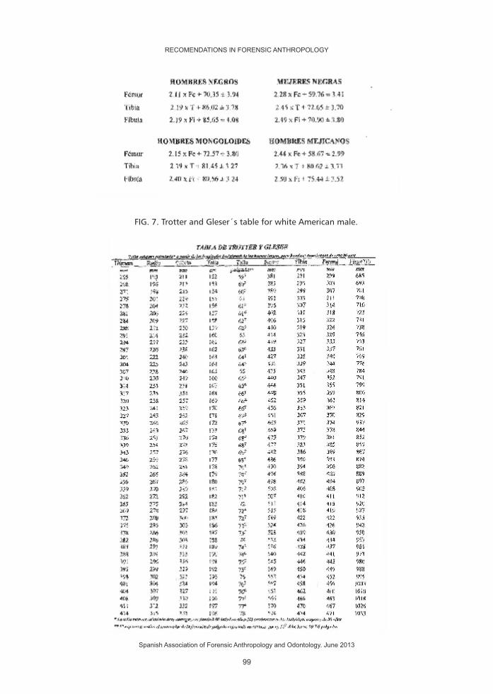
FIG. 7. Trotter and Gleser´s table for white American male.
RECOMENDATIONS IN FORENSIC ANTHROPOLOGY
Spanish Association of Forensic Anthropology and Odontology. June 2013
99

FIG. 8. Trotter and Gleser´s table for white American female.
RECOMENDATIONS IN FORENSIC ANTHROPOLOGY
Spanish Association of Forensic Anthropology and Odontology. June 2013
100

FIG. 9. Trotter and Gleser´s table for African American male.
RECOMENDATIONS IN FORENSIC ANTHROPOLOGY
Spanish Association of Forensic Anthropology and Odontology. June 2013
101

FIG. 10. Trotter and Gleser table for African American female.
RECOMENDATIONS IN FORENSIC ANTHROPOLOGY
Spanish Association of Forensic Anthropology and Odontology. June 2013
102

FIG. 11. Regressive formulas of Mendonça MC.
RECOMENDATIONS IN FORENSIC ANTHROPOLOGY
Spanish Association of Forensic Anthropology and Odontology. June 2013
FEMALES
MALES
103

FIG. 12. Table of Mendonça MC for male.
MALES
RECOMENDATIONS IN FORENSIC ANTHROPOLOGY
Spanish Association of Forensic Anthropology and Odontology. June 2013
104

FIG. 13. Table of Mendonça MC for female.
FEMALES
RECOMENDATIONS IN FORENSIC ANTHROPOLOGY
Spanish Association of Forensic Anthropology and Odontology. June 2013
105

FIG. 14. Regressive formulas of Belmonte MT (2012).
FIG. 15. Fazekas and Kosa regression formulas.
FIG. 16. Telkkä et al. regression formulas.
FÓRMULA GENERAL E= 69,74 + 2,693.T ± 8 cm
FÓRMULA PARA HOMBRES E= 85,807 + 2,279.T ± 6,9 cm
FÓRMULA PARA MUJERES E= 78,812 + 2,339.T ± 7,5 cm
E estatura en cms T longitud máxima de la tibia en cm
RECOMENDATIONS IN FORENSIC ANTHROPOLOGY
Spanish Association of Forensic Anthropology and Odontology. June 2013
FEMALESMALES
UNDER 1 YEAR
S.D. S.D.
106

BIBLIOGRAPHY:
1. Agnihotri AK, Kachhwaha S, Jowaheer V, Sing AP. Estimating stature from percutaneus length of tibia and ulna in Indo-Mauritian population. Forensic Sci Int 2009;187:109e1-109e3.
2. Allbrook D. The estimation of stature in British and East African males based on tibial and ulnar bone lengths. J Forensic Sci 1961;8(1):15-28.
3. Belmonte MªT. Determinación de la estatura a través de la tibia en población española contemporánea. Tesis doctoral. Universidad de Granada, 2012.
4. Bidmos MA. Fragmentary femora: evaluation of the accuracy of the direct and indirect methods in stature reconstruction. Forensic Sci Int 2009;192:131e1-131e5.
5. Bradley JA, Herrmann NP. Estimation of living stature from selected anthropometric (soft tissue) measurements: applications for forensic anthropology. J Forensic Sci 2009;54(4):753-760.
6. Byers SN. Introduction to forensic anthropology. Referido en Krenzer U: Compendio de métodos antropológico forenses. Referred to Krenzer U: Compendium of forensic anthropological methods
7. Byers S, Akoshima K, Curran B. Determination of adult stature from metatarsal length American Journal of Fisical Anthorpology 1989;79:275-279.
8. Celbis O, Agritmis H. Estimation of stature and determination of sex from radial and lunar bone lenghts in a Turkish corpse simple. Forensic Sci Int 2006;158:135-139.
9. Cordeiro C, Muñoz-Barús JI, Wasterlain S, Cunha E, Vieira DN. Predicting adult stature from metatarsal lenght in a Portuguese population. Forensic Sci Int 2009;193:131e1-131e4.
10. Fazekas IG, Kósa F. Forensic Fetal Osteology. Akadémiai Kiadó. Budapest, 1978.
11. Ferreira FJN, Pinto Da Costa J, Maia J, Pereira O. Estudo da relaçao entre a largura dos dentes e a altura dos individuos. em edade adulta. Recogido en Nunes de Mendonça MC: Contribución para la identificación humana a partir del estudio de las estructuras óseas.
FEMALESMALES
1 - 9 YEARS
S.D. S.D.
FEMALESMALES
10 - 15 YEARS
S.D. S.D.
RECOMENDATIONS IN FORENSIC ANTHROPOLOGY
Spanish Association of Forensic Anthropology and Odontology. June 2013
107

12. Fully G. Une nouvelle méthode de determination de la taille. Referido en Krogman WM, Iscan MY: The human skeleton in forensic medicine.
13. Habib SR, Kamal NN. Stature estimation from hand and phalanges lengths of Egyptians. J Forensic and Legal Medicine 2010;17:156-160.
14. Holland TD. Brief communication: estimation of adult stature from the calcaneus an talus. American Journal of Physícal Anthropology 1995;96:315-320.
15. Holland TD. Estimation of adult stature from fragmentary tibias. J Forensic Sci 1992;37(5):1223-1229.
16. Jantz RL, Hunt DR, Meadows L. The measure and mismeasure of the tibia: implications for stature estimation. J Forensic Sci 1995;40(5):758-761.
17. Jantz RL, Kimmerle EH, Baraybar JP. Sexing and statures estimation criteria for Balkan populations. J Forensic Sci 2008;55(3):601-605.
18. Jason DR, Taylor K. Estimation of stature from the length of the cervical thoracic and lumbar segments of the spine in american whites and blacks. J Forensic Sci 1995;40(1):59-62.
19. Jit I, Sing S. Estimation of stature from de clavicles. Recogido en Krogman WM, Iscan MY: The human skeleton in forensic medicine.
20. Klepinger LL. Stature, maturation variation and secular trends in forensic anthropology. J Forensic Sci 2001;46(4):788-790.
21. Krenzer U. Compendio de métodos antropológico forenses. CAFCA. Serie de antropología forense. Compendium of forensic anthropological methods. CAFCA. Series of forensic anthropology Guatemala 2006.
22. Krishan K, Kanchan T, DiMaggio JA. A study of limb asymmetry and its effect on estimation stature un forensic case work. Forensic Sci Int 2001;200:181e1-181e5.
23. Krishan K. Estimation of stature from cephalo-facial anthropometry in north Indian population. Forensic Sci Int 2008;181:52e1-52e6.
24. Krogman WM, Iscan MY. The human skeleton in forensic medicine. Charles C Thomas Publisher. Springfield, Illinois 1986.
25. Lowet G, Van Der Perre G. Ultrasound velocity measurement in long bones: measurement method and simulation of ultrasound wave propagation. Journal of Biomechanics 1996;29(10):1255-1262.55-62.
26. Maijanen H. Testing anatomical methods for stature estimation on individuals from the W. M. Bass Donated Skeletal Collection. J Forensic Sci 2009;54(4):746-752.
27. Manouvrier L. Le détermination de la taille d'aprés les grand os des embres: Mém de la Soc d'Anthropol de Paris. 4 (Ile sér): 347-402, 1893.Recogido en Reverte JM: Antropología forense.
28. Meadows L, Jantz RL. Estimation of stature from metacarpal lengths. J Forensic Sci 1992;37(1):147-154.
29. Meadows L, Jantz RL. Allometric secular change in the long bones from the 1800s to present. J Forensic Sci 1995;40(5):762-767.
30. Musgrave JH, Harneja NK. The estimation of adult bone stature from metacarpal bone length. Referido en Krogman WM, Iscan MY: The human skeleton in forensic medicine.
31. Mendonça MC. Contribución para la identificación humana a partir del estudio de las estructuras óseas. Determinación de la talla a través de la longitud de los huesos largos. Tesis Doctoral Universidad Complutense de Madrid. 1998.
32. Ousley S. Should we estimate biological or forensic stature?. J Forensic Sci 1995;40(5):768-773.
RECOMENDATIONS IN FORENSIC ANTHROPOLOGY
Spanish Association of Forensic Anthropology and Odontology. June 2013
108

33. Pelin C, Zagyapan R, Yazici C, Kürkcüoglu A. Body height estimation from head and face dimensions: a different method. J Forensic Sci 2010;55(5):1326-1330.
34. Petrovecki V, Mayer D, Slaus M, Strinovic D, Sk avic J. Prediction of stature based on radiographic measurements of cadaver long bones: a study of the Croatian population. J Forensic Sci 2007;52(3):547-552.
35. Prasad R, Vettivel S, Jeyaseelan L, IsaacB, Chandi G. Reconstruction of femur length from markers of its proximal end. Referido en Nunes de Mendonça MC: Contribución para la identificación humana a partir del estudio de las estructuras óseas. Referred to Nunes de Mendonça MC: Contribution for the human identification based on the study of bone structures.
36. Raxter MH, Auerbach BM, Fuff ChB. Revision of the Fully technique for estimating statures. American Journal of physical anthropology 2006;130:374-384.
37. Reverte JM. Antropología forense. Ministerio de Justicia. Madrid 1991. Forensic anthropology. Ministry of Justice.
38. Rodríguez, JV. Introducción a la Antropología forense. Universidad Nacional de Colombia. Colombia 2004. Introduction to Forensic Anthropology. National University of Colombia
39. Ryan I, Bidmos MA. Skeletal height reconstruction from measurements of the skull in indigenous South Africans. Forensic Sci Int 2007;167:16-21.
40. Shulin P, Fangwu Z. Estimation of stature from skull, clavicle, scapula and os coxa of male adult of Southern China. Recogido en Krogman WM, Iscan MY: The human skeleton in forensic medicine.
41. Simmons T, Jantz RL, Bas WM. Stature estimation from fragmentary femora: a revision of the Steele method. J Forensic Sci 1990;35(3):628-636.
42. Smith ShL. Stature estimation of 3-10 year old children from long bone lengths. J Forensic Sci 2007;52(3):538-546.
43. Steele G, Mckern TW. A method for assessment of maximum bone lenght and living stature from fragmentary long bones. Americam Journal of Physical Anthropology 1969;31:215-218.
44. Steyn M, Smith JR. Interpretation of ante-mortem stature estimates in South Africans. Forensic Sci Int 2007;171:97-102.
45. Telka A. On the prediction of human stature from the long bones. Recogido en Nunes de Mendonça MC: Contribución para la identificación humana a partir del estudio de las estructuras óseas.
46. Telkkä A, Palkama A, Virtama P. Prediction of stature from radiographs of long bones in children. J Forensic Sci, 7:474-479, 1962.
47. Terezawa K, Akabane H, Gotouda H, Mizukami K, Nagao M, Takatori T. Estimating stature from the length of the lumbar part of the spine in japanese. Medical Science and Law 1990;30(4):354-357.
48. Tibbets GL. Estimation of stature from the vertebral column in american blacks. J Forensic Sci 1981;26(4):715-723.
49. Trotter M, Glesser OC. A reevaluation of estimation of stature based on measurements of stature taken during life and of long bones after death. Referido en Nunes de Mendonça MC: Contribución para la identificación humana a partir del estudio de las estructuras óseas.
50. Willey P, Falsetti T. Inaccuracy of height information on driver´s licences. J Forensic Sci 1991;36(3):813-819.
RECOMENDATIONS IN FORENSIC ANTHROPOLOGY
Spanish Association of Forensic Anthropology and Odontology. June 2013
109


ANCESTRY ESTIMATIONCRITERIA
RECOMENDATIONS IN FORENSIC ANTHROPOLOGY
Spanish Association of Forensic Anthropology and Odontology. June 2013
111


PROTOCOL
AUTHORS
CONTACT
DATE
ANCESTRY ESTIMATION CRITERIA
PRIETO JL, MAGAÑA C
October 2, 2012
1. INTRODUCTION.�CONCEPT�OF�RACE��VS.�ANCESTRY.
The�reconstruction�of�the�biological�profile�constitutes�the�first�phase�of�the�process�of� identification�of�human� remains.�Within� this�process,�one�of� the�elements�to�evaluate� is�the�race�or� 'descent(ancestry)� ',�according�to� its�most�recent�and�frequent�denomination�in�the�forensic�field.
Unlike�other�diagnostic�elements�of�the�biological�profile,�such�as�sex,�age�or�height,� this� is�much�more� complicated�and� inaccurate.� The�ambiguity� in� its�definition�is�reflected�in�the�use�of�a�variety�of�denominations�(and�concepts)�such�as� race,�ancestry�origin,�etnic�origin�or�ancestry�and,�consequently,� the�translation�of�this�diagnosis�that�we�perform�when�it�comes�to�reconstruct�the�biological�profile�of�a�body�(skin�color,�geographical�origin,�socio-cultural�aspect,�physical�appearance,�...?).�Some�definitions�influence�this�idea.�Since�the�original�terminology�is�mainly�in�English-speaking�literature,�definitions�are�included�in�their�original�language.
1 Race.-� DRAE :� Each� one� of� the� groups� that� are� subdivided� into� some�biological�species�and�whose�differentiating�characters�are�are�perpetuated�by�inheritance.
Race.-�MERRIAM-WEBSTER:�a:�a�family,�tribe,�people,�or�nation�belonging�to�the�same�stock�b�:�a�class�or�kind�of�people�unified�by�shared�interests,�habits,�or�characteristics.
Ancestry.-� DRAE:� 1.� m.� ancestor;� 2.� m.� inheritance� (_� trait� or� traits� that�continue�warning�on�descendants).
Ancestry.-� MERRIAM-WEBSTER:� line� of� descent:� lineage;� DIE-NET:� 2:�inherited�properties�shared�with�others�of�your�bloodline.
1 DRAE: Dictionary of Royal Spanish Academy.
RECOMENDATIONS IN FORENSIC ANTHROPOLOGY
Spanish Association of Forensic Anthropology and Odontology. June 2013
113

Ancestor.-�MERRIAM-WEBSTER:�a�:�one�from�whom�a�person�is�descended�and�who� is�usually�more�remote� in�the� line�of�descent�than�a�grandparent�a�person's�ancestors�:�the�people�who�were�in�your�family�in�past�times.
Ancestor.-�DRAE:�1.�f.�Series�of�ancestors�or�predecessors�of�someone�2.�f.�Source,�origin�of�something.
However,�the�diagnosis�of�ancient�origin�is�a�major�factor�in�the�estimation�of�other� elements� of� reconstructive� profile� (age,� height),� whose� methods�application�need�to�know�in�advance�the�ancestral�origin�or�population.
Numerous� studies� have� demonstrated� widely� that� different� population�groups,� subject� to� different� ethnic� influences,� geographic� and� /� or� socio-environmental,�among�others,�show�differences�in�the�development�of�skeletal�and�dental�characters,�which�would�affect�the�mentioned�diagnostic�elements.However,�the�very�concept�of�population�origin,�race,�ethnic�origin�or�ancestry,�presents� serious� conflicts� when� our� intention� is� precisely� to� evaluate� this�population�origin.
Human�classification� in�biological�races,�serves�to�the�sharing�of�heritable�traits�that�make�them�similar�to�each�other�and�different�from�other�biological�races.
Since�the�division�of�the�human�beings�into�three�races:�white,�negroid�and�mongoloide,�as�primary� races�which�at� the�same� time�could�be�divided� into�secondary� races,� the� meaning� of� the� term� 'race'� has� not� been� accepted�universally,�and�its�use�in�Forensic�Anthropology�has�been�criticized�due�to�the�recent�emphasis�of�the�Biological�Anthropology�in�contradicting�the�biological�concept�of�race�minted�by�the�classical�physical�anthropology,�when�the�human�variation� is� discussed.� Nevertheless,� although� there� has� been� a� change� in�terminology�in�the�field�of�Forensic�Anthropology,�with�a�much�wider�use�of�the�term�'�ancestry�',�many�contemporary�texts�still�structure�the�human�variation�in�the�three�major�classical�ancestral�groups:�Mongoloid,�Negroid�and�Caucasoid,�under�the�concept�of�the�division�of�a�specie�based�on�the�frequency�with�which�certain�hereditary�traits�appear�among�its�members.�(Brues'77).
On� the�other�hand,� the� races� are� also� socially�defined� categories,�whose�members�overlap�by�social�factors�(Edgar�'�09).�Human�Biology�results�from�the�interaction�of�genetic�processes,�of�development�and�culturels,�the�'Biohistory',�which�reflects�the�broad�sequence�of�events�in�the�history�of�the�biosphere�and�the�Civilization,�from�the�beginning�of�life�until�our�days.
Serve�as�an�example,�the�classification�in�5�races�and�2�ethnicities�that�the�U.S.�
RECOMENDATIONS IN FORENSIC ANTHROPOLOGY
Spanish Association of Forensic Anthropology and Odontology. June 2013
114

Census�Bureau�makes�to�its�citizens:
� 1.-�American�Indians�or�Natives�of�Alaska.
� 2.-�Asian.
� 3.-�Blacks�or�Africans.
� 4.-�Native�Hawaiian�and�other�islands.
� 5.-�Whites.
1.-�Hispanic�or�Latin.
2.-�Not�Hispanic�or�Latin.
The�biological�and�social�definitions�of�'race'�does�not�mean�the�same�and�both� concepts� must� be� separated.� In� the� sense� 'American',� the� term� 'race'�corresponds�to�its�social�definition.�Sauer�('92)�in�an�excellent�article�entitled�"If�races�do�not� exist,�why� forensic� anthropologists� are� so�good� at� identifying�them?"�Concludes�that� forensic�anthropologists�are�good�at�recognizing�the�race�in�USA�because�there�is�good�correlation�between�the�American�social�races�and� the� skeleton� biology,� especially� in� the� skull� between� white� and� black�Americans.
When�these�circumstances�are�not�fulfilled,�the�diagnosis�of�race�is�ineffective.�In�fact,�Goodman�('97)�states�that�"in�the�best�case,�the�identification�of�race�is�depressingly�inaccurate,�and�at�worst�completely�capricious.".�In�the�same�sense,�authors�such�as�Armelagos�and�Goodman�('98)�or�Williams�and�colleagues�('05)�are�postulated.�Usually,�biological�phenotypes�are�associated�with�concepts�and��cultural� 'labels'� of� race,� something� changing� over� time.� Also� phenotypical�characteristics� are� altered� by� the� migration� and� the� mixture.� Such� is� the�uncertainty�of�this�variable�as� in� investigation�of�victims�of�genocide,�whose�pursuit� requires� to� identify� the� group� (national,� ethnic,� racial,� or� religious)�belonging�to�the�victims;�proposes�the�use�of�cultural�elements�as�a�reflection�of�ethnic�identity.
For�the�purposes�of�this�document,�only�the�term�'�ancestry'�will�be�used,�this�is�the�most�widely�used�in�current�forensic�anthropological�literature.
2. METHODS TO ESTIMATE ANCESTRY.
The�first�attempt�to�collect�a�comprehensive�reference�on�the�estimation�of�the� ancestry� for� forensic� anthropologists� is� the� text� of�Gill� and� Rhine� 1990�'skeletal�attribution�of�race:�methods�for�forensic�anthropology�'(Gill�and�Rhine�'90).�The�estimation�of� the�ascendancy� is�usually�carried�out� from� the�visual�observation�of�morphological�elements�(especially�the�skull�and�jaw)�or�through�
RECOMENDATIONS IN FORENSIC ANTHROPOLOGY
Spanish Association of Forensic Anthropology and Odontology. June 2013
115

the�metric�analysis�of�the�cranial�skeleton�and�postcranium.
The�non�metric�or�morphological�methods�are�based�on�the�assessment�of�the� presence� /� absence,� form� or� degree� of� development� of� a� certain�characteristic.�The�most�useful�characteristics�for�morphological�analysis,�are�those�whose�differences�between�the�various�study�groups�are�higher.�Given�that�with�only�one�characteristic�it�is�not�possible�to�determine�the�belonging�to�a�certain�group,�commonly�combinations�of�characteristics�are�used.
3. NON-METRIC METHODS
3.1. Cranials.
Hooton�('26)�in�a�study�carried�out�with�students,�obtained�a�very�low�level�of�matches�when�they�assessed�independently,�the�different�characteristics�of�the�ancestry� attribution,� which� led� him� to� conclude� that� it� was� necessary� to�standardize�the�observations�between�different�observers.�With�the�intention�of�reducing�the�mentioned�subjectivity�inherent�in�the�process�of�observation�of�the�morphological�characters,�developed�the�so-called�Harvard�Catalog�which�consisted�of�a�series�of�cranial�nonmetric�characteristics,�the�majority�of�which�are�still�used�by�forensic�anthropologists.
In� an� attempt� to� reduce� the� subjectivity�of� the� estimations,�he�designed�illustrative� schemes� for�each�of� the� characteristics,�although� the� latter�were�never�published.�Over�the�years�he�has�been�establishing�a�relationship�of�non-metric�characteristics,�the�ones,�considered�most�useful,�which�are�summarized�in�most�of�the�texts.�Below�you�can�find�in�detail,�the�ones�gathered�from�the�text�of�Ubelaker�('07).
The� author,� overcoming� the� difficulties� mentioned� above� regarding� the�diagnosis�of�ancestry,� refers� to� the� concentration�of�extreme�expressions�of�some�skeletal�features�could�suggest�affiliation�with�one�of�the�following�major�groups:
Asians�and�American�Indians.�The�skulls�present�a�very�projected�forward�malar�bones�and�comparatively�more�flattened�faces.�The�nasal�opening�is�of�moderate�width�and�it�has�a�lightly�developed�nasal�spine.The�orbits�tend�to�be�more�circular�than�those�of�other�groups�and�the�palate�is�moderately�broad.�At�least�among�American�Indians,�the�suture�between�the�maxilla�and�the�malar�tends�to�be�straight.
RECOMENDATIONS IN FORENSIC ANTHROPOLOGY
Spanish Association of Forensic Anthropology and Odontology. June 2013
116
Blacks:�The�skulls� tend� to�show�a� relatively�small�projection�of� the�malar,�more�rectangular�orbits�and�wider� �interorbital�distances.The�nasal�opening�is�very�wide�and�lacks�nasal�spine.�The�palate�tends�to�be�very�wide�and�somewhat�rectangular.�The�maxillary�anterior�alveoli�and� jaw� tend� to�be�very�projected�forward� (prognathism).� Many� skulls� show� a� slight� coronal� depression,� just�behind�the�coronal�suture.
White.�The�skulls�are�typically�characterized�by�a�very�flattened�malar�bones,�giving�the�face�a�sharp�appearance.The�nasal�opening�is�very�narrow�and�has�a�prominent�nasal�spine.�The�palate�is�relatively�narrow�and�triangular.�The�suture�between�the�maxilla�and�the�malar�tends�to�be�curved.�Metopism�frequency�(suture�from�nasion�to�bregma)�is�higher�than�among�other�groups.
Walker�('08),�as�already�done�by�Hooton�said�the�utility�of�using�comparative�illustrations� to� reduce� inter-observer�error,�and� that� the� results�obtained�are�integrated�and�interpreted�in�a�statistical�framework.
JT�Hefner�('09)�conducted�one�of�the�most�complete�works�with�11�features,�taken�from�747�individuals:
- Nasal�opening�width - Lower�nasal�opening - Interorbital�width - External�contour�of�the�nasal�surface - Postbregmatic�depression - Prominence�of�the�anterior�nasal�spine - Malar�tubercle - Nasal�bone�structure - Zygomatic�tubercle - Supranasal�suture - Shape�of�the�transverse�palatine�suture - Shaped�of�zigomaticamaxilar�suture
Each�feature�was�defined�and�drawn,�establishing�a�range�of�variability.�The�frequency�of�distribution,�the�correlation�between�different�features�is�studied,�and�the� inter-and� intraobserver�error� is�determined,�noting�that�cranial�traits�historically�used�in�the�diagnosis�of�ascendancy�(1-5�from�the�list)�show�a�good�correlation�with�the�three�major�ancestral�groups�(Mongoloid,�Caucasoid�and�Negroid)�especially�when�used�in�a�combined�way.
The�conclusions�of�the�study�indicate�that�the�prediction�of�ascendancy�based�on�the�valuation�of�morfoscópicos�traits�is�an�art,�not�a�scientific�process,�since�it�is�not�repeatable,�is�unreliable�and�not�valid.
RECOMENDATIONS IN FORENSIC ANTHROPOLOGY
Spanish Association of Forensic Anthropology and Odontology. June 2013
117

Ten�of�the�11�traits�had�distribution�frequencies�with�significant�differences,�but�the�range�of�variation�of�characteristics�far�exceeds�earlier�expectations.
Thus� it� is�demonstrated� that� the�expression�of� the�characteristics�are�not�reliable�for�estimating�ancestors�and�that�these�features�must�also�be�analyzed�within�a�statistical�framework.
3.2. Dental traits.
Although�there�are�significant�differences�at�the�population�level,�they�do�not�have�single�diagnostic�character.
With�regard�to�morphological�variations�of�the�crown,�the�most�celebrated�feature� is�called�tubercle�of�Carabelli,�small�additional�cusp�on�the�surface�of�mesio-lingual� of� upper� molars.� In� incisors,� and� sometimes� in� canines,� the�marginal�flange�can�be�especially�prominent�causing�a�deep�lingual�fossa.�These�teeths�are�called�"shovel-shaped�teeth".
Classical�texts�gather�the�observation�of�a�high�prevalence�of�teeth�"in�Pala"�in�Mongoloid�populations�and�low�prevalence�respect�to�the�presence�of�tubercle�of�Carabelli,�which�would�be�more�frequent�in�Caucasians�(Hanihara�'67).�In�a�study�in�which�data�sex,�age�and�dental�size�of�two�populations�of�Mongoloid�origin�were�recorded,�Hsu�and�colleagues�('97)�show�that�the�presence�of�teeth�on�blade�significantly� increases� the� likelihood�of� the�presence�of� tubercle�of�Carabelli,�particularly�in�male�subjects.
In� a� recent� work,� Edgar� ('09)� analyze� the� frequency� of� presentation� of�morphological� features� indicated� in� the� literature� (upper� incisors,tubercle�of�Carabelli,�mesial�crest�of�the�canine�and�seventh�peak,�comparing�samples�of�African�American,�Euro-Americans,�Hispanic�Americans�and�Native�Americans.�The� results� reveal� that� these�dental� traits�commonly� recognized�as� the�most�indicative�of�ancestry,�have�no�real�diagnostic�value.�From�the�data�evaluated,�only� the� canine� mesial� ridge� was� markedly� different� in� African-American�population,�but�it�showed�no�differences�between�the�other�three�groups.�On�the�other�hand,�due�to�its�low�frequency�of�presentation�in�all�groups�(2-21%)�the�absence�of�this�feature�would�not�be�indicative�of�ascendancy.
The�mandibular�molars�sometimes�present�a�cusp�or�tubercle�accessory� in�mesiobuccal�surface,�called� 'protoestiloide'.�The� frequency�of�protoestiloides�seems�to�be�higher�among�Asians�including�American�Indians�and�Eskimos.
Radiologically�the�finding�of�radicular�enamel�pearls,�the�absence�of�lower�
RECOMENDATIONS IN FORENSIC ANTHROPOLOGY
Spanish Association of Forensic Anthropology and Odontology. June 2013
118

incisors,�and�extremely�large�pulp�cavities�are�traits�that�have�been�associated�with�Mongoloids�groups.�(Evans 81).
However,� the�degree�of�probability�of� finding�one�or� another� trait� in� an�individual�depends�on�the�statistical�frequency�of�this�character,�judging�from�the�investigations�carried�out�for�the�population�concerned.�In�this�sense,�the�lack�of�research�evaluating�these�frequencies�in�subjects�of�different�ancestry,�and� the� poor� results� of� the� few� published� studies,�make� the� use� of� dental�features,�restricted�to�purely�presumptive�diagnosis�of�ancestry,�and�the�poor�results�of�the�few�published�studies,�make�the�use�of�dental�features,�restricted�to�purely�presumptive�diagnosis.
3.3. Postcranial.
Biological�differences�are�expressed�on�the�curvature�of�the�long�bones.�In�particular,�Blacks�tend�to�have�relatively�straight�femurs,�with�very�little�torsion�or�rotation�between�the�neck�and�head.� In�contrast,�Asian�femurs�tend�to�be�fairly� curved,�with�a� considerable�degree�of� torsion�of� the�neck.�Whites�are�situated�in�an�intermediate�position�respect�to�the�curvature�and�torsion.�The�American� Indian�femurs�often�show�a�marked�flattening�or�platimeria� in�the�anterior�zone�of�the��upper�end�diaphyseal�[References].
4. METRIC�METHODS.�
Metric�methods�are�based�on�differences�in�size,�both�in�cranial�characters�and�postcranial,�existing�between�different�human�groups.
4.1. Cranial.
The�skull�is�considered�to�be�the�most�useful�part�of�the�structure�in�order�to�use�in�the�assessment�of�ancestry�(Howells 73).
The� measures� used� for� diagnostic� purposes� of� ancestry,� were� originally�established� in� the� Agreement� of� Frankfurt� in 1882.� In� this� meeting� it� was�intended� standardize� the� points� of� reference� of� the� skull� to� be� used� for�anthropometric�data.
The� indices�obtained�by� the� combinations�of� these�measures� allowed� to�classify�the�skulls,�considering�the�predominant�morphology�resulting�from�the�measures� of� the� cranial� vault� (dolichocephalous,� mesocephalics,�
RECOMENDATIONS IN FORENSIC ANTHROPOLOGY
Spanish Association of Forensic Anthropology and Odontology. June 2013
119

brachycephalics)of� the� face� (prognathous,� mesognato,� retrognato),� of� the�orbital� morphology� (cameconcos,� mesoconcos,� hipsiconcos),� of� the� palate�(leptoestafilino,� mesoestafilino,� braquiestafilino)� or� of� the� nasal� passages�(leptorhine,�mesorhine,�platyrhine,�hyperplatyrhine),�among�others.
In�1962�Giles�and�Elliot�('62)�established�a�scoring�method�to�identify�racial�affinity.�This�work�was�conducted�with�1,022�skulls,�(187�whites,�221�blacks,�and�617� Indians)� where� they� determined� sex� and� race� with� eight� skull�measurements:
� 1.�Glabella-occipital�length
� 2.�Cranial�width
� 3.�Basion-bregma
� 4.�Basion-nasion
� 5.�Basion-prosthion
� 6.�Bizygomatica�width
� 7.�Prosthion-nasion
� 8.�Nasal�width
Sex�determination�was�correct�in�82.9%�of�cases,�and�the�determination�of�race�was�correct�in�85.1%�of�cases.�Starting�from�this�work�multiple�studies�have�conducted�comparing�the�different�groups�that�exist.
Snow�and�colleagues� ('79)�verified�the�discriminant�functions�of�Giles�and�Elliot�in�a�sample�of�52�skulls�of�known�sex�and�race.�Sex�assignment�was�correct�in�88%�of�cases�and�the�race�in�71%.�Most�errors�were�committed�in�the�group�corresponding�to�American�Indians.
MY�Iscan�and�Steyn�M�('99)�analyzed�a�sample�comprised�of�53�white�and�45�black�men�and�53�white�and�45�black�women� in�South�Africa.�They�used�13�cranial�measurements�and�4�mandibular.
Cranial�measurements: Cranial�longitude Cranial�width Maximum�frontal�width Minimum�frontal�width Bizigomática�width Basion�-�nasion
Basion�-�bregma
Basion�-�prosthion
Nasion�-�prosthion
Mastoids�height
RECOMENDATIONS IN FORENSIC ANTHROPOLOGY
Spanish Association of Forensic Anthropology and Odontology. June 2013
120

Biasterionica�width Nasal�height Nasal�width
Mandibular�measures: Bicondylar�longitude Bicondylar�width Bigonial�width Minimum�width�of�branch.�
The�results�were�correct�in�98%�of�cases�with�cranial�measurements�and�with�the�mandibular�in�74%�of�men�and�87%�of�women.
The�metric�method�most�used�today�is�the�result�of�the�computer�application�FORDISC.�This�application�was�created�by�Richard�Jantz�and�Steve�Ousley�and�uses�discriminant�function�analysis�derived�from�a�base�of�anthropometric�data�(Jantz�and�Moore� Jansen� '88)�containing�a�number�of�measures� taken� from�positively� identified�bodies,�from�forensic�cases.�The�program�was�created� in�1993� and� revised� in� 1996� and� 2005.� The� database� incorporates� 21� cranial�measurements,� and� the� application� calculates� the�probability� that� the� skull�belongs�to�a�certain�populational�group.�The�groups�represented�are:�African�Americans,�Indian�Americans,�whites�Americans�and�Japanese�of�both�sexes�as�well� as� male� Chinese,� Vietnamese� and� Hispanics,� most� of� the� latter� being�Mexican�Americans).
Although�the�method�is�objective,�there�are�still�many�mistakes,�mainly�inter�and�intra�observer�originating�from�taking�measurements.�The�results�of�recent�studies� (Elliot� and� Conrad� '09)� suggest� little� usefulness� of� the� FORDISC� in�diagnosing�descent,�which�generally�does�not�pass�40%�of�accuracy,�especially�in�cases�which�are�not�represented�in�the�database.
4.2. Postcranial.
The� postcranial� skeleton� is� the� result� of� gene-environment� interaction�occurred�during�the�processes�of�intravariación.�It�is�expected�that�the�form�and�size�of�the�various�elements�that�compose�it�is�reflected�in�differential�form�both�the�genetic� information,�and� the� influence�of�various�environmental� factors�(climate,�nutritional,�functional,�etc.)
Trudrell�('98).�Performing�a�work�to�discriminate�between�blacks�and�whites,�with� samples�of�different� collections� studying� the� anterior� curvature�of� the�femur.�The�measures�used�for�the�study�are�as�follows.
RECOMENDATIONS IN FORENSIC ANTHROPOLOGY
Spanish Association of Forensic Anthropology and Odontology. June 2013
121

Maximum�Length Oblique�Length Bicondylar�width Anteroposterior�diameter�mediodiafisario Transverse�diameter�mediodiafisario Subtrochanteric�transverse�diameter Distance�to�the�R1�table�(proximal�part�of�diaphysis) Distance�to�the�R2�table�(half�diaphysis) Distance�to�the�R3�table�(distal�part�of�diaphysis) Distance�to�the�proximal�table Distance�from�the�proximal�block�(blocks�where�you�place�the�femur) Distance�from�distal�block
These�measures�were�taken�in�both�right�and�left�femur�with�the�following�results:�88.1%�correct�with�right�femur�and�86%�with�left�femur.
Marino�('97)�performs�a�work�to�discriminate�whites�of�blacks�with�samples�from� the�collections�of�Terry�and�Todd� in� first�cervical�vertebra,� taking�eight�measurements�of�200�vertebrae,�the�result�is�correct�in�75%�of�men�and�in�the�76%�of�women.
Holliday�and�colleagues�('99)�working�with�body�proportions�with�a�sample�of�26�males�and�28�females�black�Americans�and�28�males�and�28�females�white�Americans,�all�from�the�Terrys'�Collection�of�the�Smithsonian�Institution.�On�the�other�hand�they�studied�thirteen�other�skeletons�which�are�not�belong�to�the�populations�mentioned�before,� 5�women�and� 7�men� from� the�Antropology�Museum�of�New�Mexico�and� 2�women�and� 4�men� from� the�Unı versity�of�Florida.
Working�with�the�following�measures:
� � 1.�Anteroposterior�diameter�of�the�femoral�head
� � 2.�Trunk�height
� � 3.�Bi-iliac�width
� � 4.�Bicondylar�femoral�longitude
� � 5.�Maximum�length�of�humerus
� � 6.�maximum�longtitud�of�the�tibia
� � 7.�maximum�longtitud�of�the�radius
They�obtain�that�of�the�samples�from�the�Terrys'�Collection,�87%�of�men�are�classified�correctly�as�well�as�100%�of�women,�but�of�the�other�thirteen�skeletons,�81.8%�of�men�are�classified�correctly�and�only�57.1�%�in�women.
RECOMENDATIONS IN FORENSIC ANTHROPOLOGY
Spanish Association of Forensic Anthropology and Odontology. June 2013
122

4.3. Dental.
Size.� In� general,� the� teeth� of� Australian� aborigines,� Melanesians,� Native�Americans,� and� Eskimos� are� among� the� largest.� The� teeth� of� Lapones� y�Bosquimanos�are�usually�among��the�smallest,�while�other�populations�presents�intermediate�values�(Lasker�and�Lee�'57).
DNA.�-�Although�it�is�possible�to�estimate�the�most�probable�geographical�origin�of�the�variation�of�a�particular�polymorphism�(polymorphism�variant)�or�a�profile� (sequence�profile),� it�must�be�emphasized� that� this�probability� is�not�synonymous�of�the�most�likely�population�of�origin�of�a�particular�person.
It�is�necessary�to�be�particularly�cautious�when�using�skin�pigmentation�as�a�factor�that�may�infer�ancestry.
It�is�possible�to�estimate�the�ancestry�of�a�person�based�on�DNA�analysis,�with�a�reasonable�number�of�genetic�markers.�But�there�is�the�question�of�how�many�SNPs� are� needed� to� reliably� infer� the� ancestry.� One� should� perform� global�populational�screening�to�assess�the�potential�of�specific�SNPs�for�estimating�ancestry,�not�necessarily�related�to�physical�characteristics�related�to�a�particular�population�(Salas�and�colleagues�'06).
RECOMMENDATIONS.
1.� Unlike�other�diagnostic�elements�of�the�biological�profile,�such�as�sex,�age�or�height,� the� diagnosis� of� ancestry� is� much� more� inaccurate� because� of� the�ambiguity�in�its�definition�and�the�absence�of�'pure'�populations.
2. �Only�in�those�cases�where�there�is�a�good�correlation�between�social�races�and�skeletal�biology�(in�case�of�African�Americans,�white�Americans�and�Native�Americans�groups�in�the�U.S.),�can�be�useful�as�integral�element�of�process� �of�identification�reconstructive�of�the�biological�profile.
3. When�these�circumstances�do�not�concur,�the�diagnosis�of�race�is�ineffective�and�should�be�limited�to�the�assessment�of�previous�hypotheses�(confirmation�/�exclusion�of�candidates),�yet�with�a�relative�reliability.
4. In�such�cases,�the�combined�use�of�all�those�methods�possible�to�apply,� is�recommended,�especially�the�cranial�features�used�historically�in�the�diagnosis�of�ancestry.
RECOMENDATIONS IN FORENSIC ANTHROPOLOGY
Spanish Association of Forensic Anthropology and Odontology. June 2013
123

5. Due�to�the�unreliability�of�the�results�derived�from�the�application�of�FORDISC�in�the�diagnosis�of�ascendancy,�in�cases�of�cadavers�of�unknown�identity,�its�use�in�this�process�is�not�recommended.
BIBLIOGRAPHY:
1.� Armelagos�G,�Goodman�AH.� 1998.�Race� racism,�and�anthropology.� In:�Goodman�AF,� Leatherman�TL,�editors.�Building�a�new�biocultural�synthesis:�political-economic�perspectives�on�human�biology.�Ann�Arbor�University�of�Michigan�Press�359-378.�Citado�en:�Ousley�S,�Jantz�R,�Freid�D.�2009.�Understanding�race�and�human�variation:�why�forensic�anthropologists�are�good�at�identifying�race.�Am�J�Phys�Anthrop�139:�68-76.
2.� Brues�AM.�1977.�People�and�races.�New�York:�Collier�Macmillan.�Citado�en:�Edgar�HJH.�2009.�Biohistorical�approaches�to�“Race”� in�the�United�States:�Biological�distances�among�African�Americans,�European�Americans,�and�their�ancestors.�Am�J�Phys�Anthrop�139:�58-67.
3.� Edgar�HJH.�2009.�Biohistorical�approaches�to�“Race”�in�the�United�States:�Biological�distances�among�African�Americans,�European�Americans,�and�their�ancestors.�Am�J�Phys�Anthrop�139:�58-67.
4.� Elliot�M,�Collard�M.�2009.�Fordisc�and�the�determination�of�ancestry�from�cranial�measurements.�Biol.�Lett.�5:849-852.
5.� Evans�K.T,�Knight�B.�1981.�Radiology�in�forensic�medicine.��Oxford,�Blackwell�Scientific�Publications.
6.� Gill�GW,�Rhine�S.�1990.�Skeletal�attribution�of�race:�methods�for�forensic�anthropology.�Alburquerque�NM:�Maxwell�Museum�of�Anthropology.�Citado�en:�Hefner�JT.�2009.�Cranial�nonmetric�variation�and�estimating�ancestry.�J�Forensic�Sci�54(5):�985-995.
7.� �Giles�E,Elliot�O.�1962.�Race�identification�from�craneal�measurements.�J�Forensic�Sci�7:147-57.
8.� Goodman�AH.�1997.�Bred�in�the�bone?�Sciences�37:�20-25.
9.� Hefner�JT.�2009.�Cranial�nonmetric�variation�and�estimating�ancestry.�J�Forensic�Sci�54�(5):�985-995.
10.�Hanihara�K.�1967.�Racial�characteristics�in�the�dentition.�J.�Dent.�Res.�46:923-8.
11.�Hsu�J.W,�Peiling�T�Kehmin�L�Ferguson�D.�1997.�Logistic�analysis�of�shovel�and�Carabelli's�tooth�traits�in�a�Caucasod�population.�Forensic�Sci.�Int.�89,�65-74.
12.�Holliday�TW.�Falsetti�AB.�1999.�A�new�method� for�discriminating�African-American� from�European�–�American�skeletons�using�postcranial�osteometrics�reflective�of�body�shape.�J�Forensic�Sci�44:�926-930.
13.�Hooton�EA.�1926.�Methods�of�racial�analysis.�Science�63:�75-81.�Citado�en:�Ousley�S,�Jantz�R,�Freid�D.�2009.�Understanding�race�and�human�variation:�Why�forensic�anthropologists�are�good�at�identifying�race.�Am�J�Phys�Anthrop�139:�68-76.
14.�Howells�WW.�1973.�Cranial�variation� in�man:�a�study�by�multivariate�analysis�of�patterns�of�difference�among� recent� human� populations.� Papers� of� the� Peabody Museum� of�Archaeology� and� Ethnology.�Harvard University� 67.�Cambridge,�MA:� The�museum.�Citado� en:�Hefner� JT.� 2009.�Cranial�nonmetric�variation�and�estimating�ancestry.�J�Forensic�Sci�54�(5):�985-995.
15.� Iscan�MY,�Steyn�M.�1999.�Craniometric�determination�of�population�affinity�in�South�Africans.�Int�J�Legal�Med�112:�91-7.
16.� Jantz,�R.�L.�and�Moore-Jansen,�P.�H.�A�database�for�forensic�anthropology:�Structure,�content�and�analysis.�In:�Report�of� Investigations�No.� 47,�Department�of�Anthropology,�University�of� Tennessee,�Knoxville,�
RECOMENDATIONS IN FORENSIC ANTHROPOLOGY
Spanish Association of Forensic Anthropology and Odontology. June 2013
124

Tennessee,�1988.
17.� Komar�DA,� Lathrop�S.� 2008.�The�use�of�material� culture� to�establish� the�ethnic� identity�of� victims� in�genocide�investigations:�a�validation�study�from�the�American�Southwest.�J�Forensic�Sci�53(5):�1035-1039.
18.� Lasker�GW,�Lee�MMC.�1957.�Racial�traits�in�the�human�teeth.�J�Forensic�Sci�2:�401-19.
19.�Marino�EA.� 1997.�A�pilot� study�using� the� first�cervical�vertebra�as�an� indicator�of� race.� J�Forensic�Sci�42(6):1114-1118.
20.� Salas�A,�Phillips�C,�Carracedo�A.� 2006.�Ancestry�vs�physical� traits:� the� search� for�ancestry� informative�markers�(AIMs).�Int�Legal�Med�120:�188-189.
21.� Sauer�NJ.� 1992.�Forensic�anthropology�and� the�concept�of� race:� if� races�don't�exist,�why�are� forensic�anthropologists�so�good�at�identifying�them?�Soc�Sci�Med�34:�107-111.
22.� Snow�CC,�Hartman�E,�Giles�E,�Young�FA.� 1979.�Sex�and� race�determination�of�crania�by�callipers�and�computer:�a�test�of�the�Giles�and�Elliot�discriminant�functions�in�52�forensic�science�cases.�J�Forensic�Sci�24:448-459.
23.� Trudell�MB.�1999.�Anterior�femoral�curvature�revisited:�Race�assessment�from�the�femur.�J�Forensic�Sci�44�(4):�700-707.
24.�Ubelaker�DH.�2007.�Enterramientos�humanos:�excavación,�análisis,�interpretación.�Munibe.�Suplemento�24:�139-140.
25.�Walker�PL.�2008.�Sexing�skulls�using�discriminant�function�analysis�of�visually�assessed�traits.�Am�J�Phys�Anthrop�136:�39-50.
26.�Williams� FL,� Belcher� RL,� Armelagos� GJ.� 2005.� Forensic� misclassification� of� ancient� Nubian� crania:�implications�for�assumptions�about�human�variation.�Curr�Anthropol�46:340-34.
RECOMENDATIONS IN FORENSIC ANTHROPOLOGY
Spanish Association of Forensic Anthropology and Odontology. June 2013
125


IDENTIFICATIONCRITERIA
RECOMENDATIONS IN FORENSIC ANTHROPOLOGY
Spanish Association of Forensic Anthropology and Odontology. June 2013
127

128
PROTOCOL
AUTHORS
CONTACT
DATE
IDENTIFICATION CRITERIA
SERRULLA F, PRIETO JL, VERANO V
[email protected] • [email protected]
February 10, 2012
PREVIOUS CONSIDERATIONS:
In the drafting of this work it has been taken into consideration that the criteria presented here be useful mainly for Laboratory of Forensic Anthropology and Odontology of the Administration of Justice in Spain. Therefore, expert working standards adapted to the Spanish civil and criminal proceedings were raised. However we consider that the criteria established given their general nature can be useful also for application in other areas.
1.THE CONCEPT OF INDIVIDUAL HUMAN IDENTIFICATION:
Identity is the character set that individualize a person, distinguishing it from the others.
In forensic practice, the identification of human remains is a scientific and technical process by which it is possible to attribute them a certain identity.
This concept allows us to make the following considerations:
1) Scientific-Technical Procedure: Identification methods should be scientifically sound, reliable, applicable under field conditions, able to be implemented in a reasonable period of time (INTERPOL DVI GUIDE) adapted to each context and applied by skilled and experienced professionals (according to criteria of the document The Missing
2ICRC) .
The investigation process of the individual human identification is governed by scientific principles and sometimes also technical. Therefore all methodological aspects of the process should be recorded and made in a way that they can be reproduced by other experts. Attended to the peculiarities of Forensic Anthropology, it should be considered that the identification
2 ICRC: International Committee of Red Cross.
RECOMENDATIONS IN FORENSIC ANTHROPOLOGY
Spanish Association of Forensic Anthropology and Odontology. June 2013
129

procedure begins in the investigation phase, it continues in Archaeology (individualization of the remains) and often ends with the introduction of genetic profile obtained in the police database on identifiers obtained from the DNA. In the whole procedure the registration and maintenance the so called 'chain of custody' of the samples has special significance.
2) Attribution of identity: The process of identification of human remains is multidisciplinary and includes the collection, integration and comparing all of the information available in each case, classified in antemortem and postmortem records.
Forensic Anthropology and Odontology constitute a part of this process, which should include all disciplines involved and requires comprehensive assessment of all information collected by them.
Whatever the technique used, it must be based on scientific criteria. It is recommended to take into account these effects called Daubert decision
[1]criteria used in the legal field in the U.S. to evaluate the expert evidence:
1) Whether the theory or technique used by the expert can be or has been tested.
2) Whether the theory or technique has been subjected to peer review and publication.
3) The known or potential margin of error of the method used. 4) The degree of acceptance of the method or conclusions between the
relevant scientific community.
For comparative purposes we call Doubted sample any of the samples obtained from human remains that we aim to identify and we call undoubted sample to all the samples that we assign to a known identity, generally with high certainty.
Special interest has the data collection procedure or antemortem registers. The expert must always ensure that the identity belongs to the antemortem record that will be compared in the case.
The antemortem data collection be made by multidisciplinary teams that will include among others professionals from Legal Medicine and as support staff it is considered necessary to obtain the maximum number of antemortem data.
RECOMENDATIONS IN FORENSIC ANTHROPOLOGY
Spanish Association of Forensic Anthropology and Odontology. June 2013
130

2.IMPORTANCE OF THE IDENTIFICATION.
The process of identification of human remains is one of the most important responsibilities of forensic anthropologist and should be prioritized in all cases, both in legal and judicial significance for the purposes of ongoing investigation, as a humanitarian response to the needs of the families of the missing persons.
3.THE IDENTIFICATION TECHNIQUES:
Identification techniques are considered to be all those technical or factual or circumstantial elements that contribute or could contribute to identifying human remains either because allow us to attribute a certain identity or because they allow us exclude it.
We believe that most of the scientific community share the idea that the various techniques currently used in human identification have different scientific value for identification purposes, whether or not published on its reliability. Some of the techniques shown in Table 1 have contrasted scientific value. Others however do not have scientific studies to support them but they can add value to the identification taken together.
TABLE 1 : Classification techniques applicable in human identification.
CLASS TECHNICAL
PRIMARY Fingerprint
Odontological
DNA polymorphisms
SECONDARY Visual Identification
Radiographic comparison
Documents and personal objects
Facial approximation
Anthropological Profile: Det Sex
Anthropological Profile: Det Height
Anthropological Profile: Det Age
Anthropological Profile: Det Ancestry
Pathobiography
Date of death evaluation
Photographic superimposition
Circumstances of the death
RECOMENDATIONS IN FORENSIC ANTHROPOLOGY
Spanish Association of Forensic Anthropology and Odontology. June 2013
131

3.1. Primary:
Primary techniques are considered to be as follows:
1) FINGERPRINTS:
This is a technique of high value identifier but generally they are no generally applicable in Forensic Anthropology. In some special cases it is possible to obtain fingerprints by mummified tissue regeneration or dried or by obtaining latent prints on some items that have been cut off from sources of deterioration of fingerprints. Expert intervention in Forensic Anthropology should allow to collect these prints in ideal conditions to provide the expert in Lofoscopia obtaining fingerprints in conditions of maximum quantity and quality. In any case the task of obtaining and regeneration of fingerprints must be carried out by experts in Lofoscopia. In general terms individual identification by fingerprints will be accepted if
3there matching at least 12 characteristic points .
2) ODONTOLOGICAL:
Dental techniques are generally recognized as high value identifying technics both because of its individualizing potential and for its exclusion capacity.
That is why in any case of identification of human remains, from the beginning, the dental information of people reported missing should be required. The need to have complete and original antemortem dental records, should be emphasized, all properly labeled with the patient's name, advising dentists to submit all the available information on a particular patient (odontographs, radiographic and photographic records, study models) and perform duplicate for their own archives.
The nomenclature recommended for working context of the AEAOF is the International Dental Federation (FDI) or digit two.
3 The requirement that there be on prints or footprints same twelve points derives from the tradition of Galton, Remus, Balthazard and others. More recent authors as Steinwender and Cooke, are inclined to the opinion that eight to twelve points of comparison may be sufficient for identification, depending on the nature of the points and drawing in general. However it must be noted that in Spain, the jurisprudence considers that eight or ten characteristic points are sufficient to establish the identity between two fingerprints or impressions, provided they are identical in topographic parameters (located in the same zona), morphological (likewise) and mathematical (same number of ridges between them), and showing no natural dissimilarity (S. de 15-06-88 Ar.5024, S. de 4-7-88 Ar.6477, S. de 25-11-89 Ar.9314, S. de 4-7-90 Ar.6220, S. de 15-3-91 Ar 2156). If several fingerprints are obtained, they can serve as a complement for more solid basis for the questioned authorship of the fact ( S T.S de 25-11-99, Ar.9314). On the other side, Spanish identification services, argue that it is possible to establish the identity with a smaller number of characteristic points depending on their qualitative value.
RECOMENDATIONS IN FORENSIC ANTHROPOLOGY
Spanish Association of Forensic Anthropology and Odontology. June 2013
132

For techniques and procedures recommended by some professional and s c i e n t i f i c b o d i e s c o n s u l t I N T E R P O L N e w D V I G u i d e : http://www.interpol.int/Public/DisasterVictim/guide/guide.pdf; International Organization of Forensic Odonto-Stomatology (IOFOS) Quality A s s u r a n c e i n F o r e n s i c O d o n t o - S t o m a t o l o g y : http://amalgam.uio.no/foreninger/iofos/quality/quality_assuarance.htm;American Board of Forensic Odontology (ABFO) Reference Manual. Body I d e n t i f i c a t i o n I n f o r m a t i o n ( p á g 1 1 8 - 1 7 0 ) : http://www.abfo.org/pdfs/ABFO_Reference_Manual_for_11-2010.PDF
There is no international consensus regarding the number of mathes of characteristic points. While in this North America it is evaluated individually, depending on the case, in other countries like South Africa a minimum of 12
[1]characteristic points of match is required .
In cases where it is necessary to manage a high volume of información, the use of specific computer programs is recommended. In this regard, the program WinID is recommended to use, it can be obtainable free at: www.winid.com
3) DNA polymorphisms:
The object of this document is not to describe the genetic techniques of identification. It is well-known by all that genetic techniques have high power of identification with a minimum overall margin of error.
The most extended current criteria in use of DNA as a procedure for identification is limited to cases where other methods are not appropriate (ICRC-INTERPOL).
Some aspects of interest for the anthropologist and forensic odontologist in relation with genetic identification techniques should be emphasized in this document.
In all cases of studies of human remains,bone and / or dental samples are collected for its eventual processing and analysis.
In cases of bodies in advanced putrefaction or skeletonized is it is advisable, as most ideal, the sampling of dense cortical bone of long bones of lower limbs (sections in 'window' of 4-6cm without separation of the diafisis)avoid to include areas that could compromise taking measures, articular margins or fractures, or 10gr of the cortical of other bones available and / or healthy teeth (preferably molars). In case of carbonized bodies, in addition to the above,
RECOMENDATIONS IN FORENSIC ANTHROPOLOGY
Spanish Association of Forensic Anthropology and Odontology. June 2013
133

impacted teeth or dental roots.
- It is recommended that all Anthropologists and Forensic Odontologists dedicated to studies of human identification and many professionals dedicated to obtain DNA samples have their genetic profile (autosomal STR markers, Identifiler).
- For all types of sampling as well as regarding the chain of custody requirements to be observed in this regard is proposed from the International Society for Forensic Genetic (ISFG) referenced below.
- In cases of disasters, multiple victims incidents or armed conflicts shall take into consideration the specific recommendations made on the matter
[16] [17] [4]both the ISFG as Interpol or the International Committee of the Red Cross .
- In the case studies of unidentified human remains, forensic anthropology laboratories in Spain request that the profile gene obtained is inserted in the DNA database.
3.2. Secondary:
Secondary techniques are considered among others, the followings:
1. VISUAL IDENTIFICACION:
Although this technique is often not possible to use in Forensic Anthropology, we include it because sometimes it can provide elements of assessment in the identification. A tattoo, a body or facial peculiarity can contribute to facilitate the identification. We have not found specific work to confirm the low reliability of this technique, although many professionals
[1],[4]confirm that this technique is capable of causing many errors .
2. RADIOGRAPHIC COMPARISON:
Depending on the specific case study, in general the application of radiological and imaging techniques(Conventional Radiology, TAC or RNM) of human identification has long shown to be a valuable technique.
a) Frontal sinuses: There is a belief that are unique for each individual including univitelline twins. They start their development from 6 months to 2 years of age. They are radiographically recognizable starting from 5 years and
RECOMENDATIONS IN FORENSIC ANTHROPOLOGY
Spanish Association of Forensic Anthropology and Odontology. June 2013
134

continue to develop through puberty. Although they are anatomically stable, once developed, they may suffer pathological and physiological modifications. Usually they present such as bilateral cavities although they can be unilateral, being totally absent in 5% of the population. The
[1]projection PA of skull is recommended for its identifying evaluation . For comparison of frontal sinuses it is recommended the digitization of radiographic images and the use of a graphic design computer program which allows to evaluate contour, general morphology, area, volume or symmetry in the case of having a recourse to TAC).
b) Radiological characteristics: In the use of radiographic plates for human identification the following must be taken into account:
- Confirm the identity of the person to whom belongs to plate/ s considered as undoubted sample.
- Consider the date of issue of the plate. - Consider the position of the subject as well as the projection and the
radiological technique used to fit them in the doubted sample as possible.
- Considering the presence of taphonomic phenomena on the doubted sample.
- Considering the presence of the sample taphonomic phenomena dubitada.
- Always consider the possible use of all the useful characteristics to the case (turkish sella, anatomical variations, osteopatological congenital
[7]alterations, hereditary or acquired, etc, ...) . - Some authors consider necessary to obtain eight matches to establish a
[1]positive identification .
3. DOCUMENTS AND PERSONAL ITEMS:
These objects are frequently used in the Judicial field to establish the preliminary identity and they generally constitute a source of essencial information throughout the investigation. However one should always keep in mind in forensic anthropology that documents and personal belongings are not elements of the cadaver but associated with it and therefore can be an important source of error.
4. FACIAL APPROXIMATION:
All those techniques that allow to obtain a good face starting from a skull or from the testimony of a witness are included under this name. Also the artistic
RECOMENDATIONS IN FORENSIC ANTHROPOLOGY
Spanish Association of Forensic Anthropology and Odontology. June 2013
135

techniques of aging faces are included here. Both sculptural techniques, such as drawings or computer-aided reconstructions are not acceptable as valid techniques for individual identification purposes for the high margin of error
[5],[6]shown in published studies . However these techniques can contribute to the identification process and be helpful deinvestigación purposes.
5. OBTAINING THE ANTHROPOLOGICAL PROFILE:
The margin of error for obtaining any of the features of anthropological profile vary depending on the quantity and quality of the skeletal material recovered and existence of standard populations,
- As for sex determination. In adults with a complete skeleton the accuracy is 100%, studying only the pelvis the accuracy lowers to 95%, studying only the skull to 90%, with skull and pelvis we got 98%. Studying the sex only from long bones the accuracy ranges between 80-90%. Using long bones and skull we got to 90-95% and studying long bones and pelvis the accuracy exceeds 95%. In subadults the accuracy is around 50% without pelvic remains. Counting with the pelvis accuracy may reach to
[8]75-80% . These degrees of accuracy should be taken into account when performing an identification or exclusion based on sex.
- As regards the determination of the height we have not found general studies that allow us to know the reliability of the techniques. If we have the complete skeleton it is recommended to use the method called Anatomical Method or Fully Method [9] when necessary using the height data for identification purposes. If it is not possible to apply the anatomical method a mathematical method should be used if possible whose data have been obtained from a population as similar as to the case of the study. There seems to be general recognition for the Trotter and Glesser Tables also for those of Manouvrier. In general many authors recognize that in the estimation of the height starting from the length of the long bones reduces the error the more long bones we employ. In our environment, it is recommended to use the Tables de Mendonça MC
[10](1998) whose accuracy in adults reaches 95% . It must also be taken especially into consideration that antemortem data of height frequently have low reliability either by the method used in measuring or by the time elapsed since the antemortem record. Therefore extreme caution it is recommended when obtaining height data for identification purposes.
- As for the age determination it is necessary to remember that the accuracy of the different methods is different and that in the case of study it can be further modified the conditions based on that the
RECOMENDATIONS IN FORENSIC ANTHROPOLOGY
Spanish Association of Forensic Anthropology and Odontology. June 2013
136

individual is an adult or subadult, in that we count on all the bones or not and also the state of conservation of the bones. Overall, the error decrease from older individuals to younger ages. In general, over 60-65 years the margin of error can be 20 years, in middle age (20-60 years)
[11]between 5 and 10 years and in children ages 1 to 2 years .
- In connection with the determination of ancestry origin there appears to be no reliable method from the anthropological point of view. Some population differences are apparent in the skeleton, but the variation within same group and the overlap between groups reduces the
[11].reliability of identification based on one skeleton The application of genetic techniques may help to locate the case of study in one of five populational groups (Europoides, Asian, African, Native American and
[12], [13]Australioids) .
6. PATHOBIOGRAPHIC EVALUATION:
Given the high and varied number of possible cases is not possible to talk about reliability of this technique. Sometimes the comparison is simply morphological, in other cases it can be more complex. There seems to be general agreement that isabout a technique that can have a high identification value. Antemortem pathobiographic data collection must be extremely careful. The expert must confirm the identity of antemortem registration in all cases. The comparative technique employed should be reproducible. The best source of pathobiographic data collection is medical history. Oral sources of pathobiographic information should always be contrasted. In recent years in our country the majority of osteosynthesis material that is implanted includes a registration number that the expert must verify.
7. EVALUATION OF TIME OF DEATH:
Sometimes estimating the time of death can help identify whether done by anthropological methods, biological or purely circumstantial. In general the contribution to the identification takes place because it allows excluding individuals if the error margin of the technique is safe enough.
8. CRANEOPHOTOGRAFIC SUPERIMPOSITION.
Although some authors have claimed the utility of identification of these [14]
techniques and even they have situate the reliability of the craneofotografic [15]
superposition in 91% , the most widely used approach is not to admit them as
RECOMENDATIONS IN FORENSIC ANTHROPOLOGY
Spanish Association of Forensic Anthropology and Odontology. June 2013
137

reliable methods of human identification, considering them useful only as excluding procedures.
9. CIRCUMSTANCES OF DEATH:
The assessment of the circumstances of death can contribute a very important way to the complex process of individual identification that sometimes appears before the Forensic Anthropology or expert responsible of the case. For this we believe that several conditions should meet:
- The process of investigation of the circumstances of death must be prior to the study of human remains or performed by persons or entities other than who do the forensic anthropological study of the case. The purpose of this condition is to reasonably exclude any methodological contamination in responsible for conducting the identification.
- The forensic expert, who is in charge of the case of identification must take into account the reliability of information sources.
- There should be as detailed as possible registration of the sources of information that support the circumstantial facts that will be subject to assessment for identification purposes. Overall anonymous sources of information are not admissible.
- The research process must gather sufficient conditions so that others as Forensic Expert can come to similar conclusions.
4. FORENSIC IDENTIFICATION PROCEDURE:
4.1. Preliminary aspects:
The anthropologist and forensic odontologist shall always consider all the circumstances of the death that may condition the identification process.
4.2. Sampling and chain of custody:
The rules regarding the chain of custody must be applied to all samples, but you should be especially careful with research samples for DNA polymorphisms.
RECOMENDATIONS IN FORENSIC ANTHROPOLOGY
Spanish Association of Forensic Anthropology and Odontology. June 2013
138
For samples to be sent to the National Institute of Toxicology and Forensic Sciences, with regard to shipping and chain of custody you should consider the Act of JUS/1291/2010 of 13 May with which the rules for the preparation and submission of samples analyzed by the National Institute of Toxicology and Forensic Sciences is approved (BOE 122 of May 19, 2010).
Download from:http://www.boe.es/boe/dias/2010/05/19/pdfs/BOE-A-2010-8030.pdf
With error correction from:http://www.boe.es/boe/dias/2010/06/16/pdfs/BOE-A-2010-9507.pdf
In rest of the cases it is proposed to follow:
1) The recommendations made by the Group of Spanish and Portuguese speakers of the International Society for Forensic Genetic whose recommendations can be downloaded from the links below:http://www.gep-isfg.org/documentos/Recogida%20de%20evidencias.pdfhttp://www.gep-isfg.org/documentos/Documento%20catastrofes.pdf
2) The recommendations proposed by the International Committee of the Red Cross in cases of armed conflict and other situations of armed violence for DNA samples [4] downloadable at:http://www.icrc.org/WEB/SPA/sitespa0.nsf/htmlall/p4010/$File/ICRC_003_4010.PDF
3) The recommendations of INTERPOL in cases of disasters downloadable at:http://www.interpol.int/Public/DisasterVictim/guide/guideES.pdf
4.3 Antemortem data collection:
Antemortem data collection should be done in a systematic way and if possible with references on the subject already exist in antemortem police records of missing persons or antemortem INTERPOL protocol. In cases of application of National Forensic Medicine and Scientific Police in multiple
[18]victims events Action Protocol , will be addressed as specified therein.
The AEAOF recommends the development of anthropological datas of antemortem chips in coordination with the datas that can be introduced into the data base unified for all the Police force in Spain of all missing persons and human remains (PDIRH).
RECOMENDATIONS IN FORENSIC ANTHROPOLOGY
Spanish Association of Forensic Anthropology and Odontology. June 2013
139

Forensic anthropologist will confirm the identity of all the elements that can be compared in the identification process and considered as undoubted samples (x-rays, undoubted antemortem DNA, etc).
5. IDENTIFICATION CATEGORIES:
As a resume to the criterias previously exposed, the identification of human remains is result of the comparison of the available antemortem and postmortem data, categorized according to their potential identification in primary and secondary. The coincidence of a primary element of identification must never exclude the verification of the coincidence of secondary elements such as sex, age and other personal characteristics.
It is commonly found in expert reports as well as standard designations in some texts or verbal predicates to refer to the level of certainty with which we
[1]can express opinions regarding the identification of a case . Often used as follows:
Tentative identification.
Indicates that the identification is suspected because it is based on circumstances or personal items (identity card, wallet, ...). It is frequently used in the field of police investigations, judicial and forensic new cadavers.
Presumptive identification.
Assumes a greater degree of certainty than the tentative, but less than the positive. For example, you can assign a presumptive identification when we found the body of a person in the interior of a house ('his house'), anthropological data coincide and no grounds for exclusion.
Positive identification.
Assumes that for the expert there is enough probative evidence to affirm the identity.
RECOMENDATIONS IN FORENSIC ANTHROPOLOGY
Spanish Association of Forensic Anthropology and Odontology. June 2013
140

Individualization.
The term itself makes clear that the identity is confirmed also excluding other individuals. The term is usually applied when genetic tests, the Forensic Odontology, the pathobiography or fingerprints confirmed it.
RECOMMENDATIONS:
We propose (at least for the final diagnosis, once integrated all the data from different procedures) to reduce the judgment on the identification is of these three categories:
a) POSITIVE IDENTIFICATION b) INCONCLUSIVE IDENTIFICATION c) EXCLUSIVE IDENTIFICATION
BIBLIOGRAPHY:
1. Komar DA, Buikstra JE. Forensic Anthropology. Contemporary Theory and Practice. Oxford University Press. 2008. New York-Oxford.
2. Christensen AM, 2005. Testing the reliabililty of frontal sinuses in positive identification. Journal of Forensic Sciences 49(1):5-10.
3. Koot MG, Sauer NJ, Fenton TW. 2005. Radiographic human identification using bones of the hand: a validation study. Journal of Forensic Sciences 50(2):263-268.
4. Personas desaparecidas, análisis forense de AND e identificación humana. Guía sobre prácticas idóneas en caso de conflicto armado y de otras situaciones de violencia armada. Segunda Edicion 2009. CICR.
5. Stephan CN, 2003. Anthropological facial reconstruction-recognizing fallacies, "unembracing" the errors and realizing methods limits. Science and Justice 43(4):193-200.
6. Stephan CN, and Henneberg M, 2006. Recognition of forensic facial approximation:case-specifics examples and empirical test. Forensic Science International 156(2-3):182-191.
7. Villalain Blanco JD, Puchalt Fortea FJ (Directores). Identificación Antropológica Policial y Forense. Ed Tiranch lo Blanch . Ciencia Policial. Valencia 2000.
8. Krogman WM, Iscan MY. The human skeleton in forensic medicine. Charles C Thomas Publisher. Springfield, Illinois 1986.
9. Fully G. Une nouvelle méthode de determination de la taille. Annales de Médecine Légale e Criminologie 1956; 36(5): 266-273.
10. Nunes de Mendonça MC. Contribución para la identificación humana a partir del estudio de las estructuras óseas. Determinación de la talla a través de la longitud de los huesos largos. Tesis Doctoral Universidad Complutense de Madrid. 1998.
11. Ubelaker DH. Enterramientos Humanos, excavación, análisis, interpretación. Suplemento 24 Munibe. Sociedad de Ciencias Aranzadi. Donostia-San Sebastián 2007.
RECOMENDATIONS IN FORENSIC ANTHROPOLOGY
Spanish Association of Forensic Anthropology and Odontology. June 2013
141

12. Phillips C et al. Inferring ancestral origin using a single multiplex assay of ancestry-informative marker SNPs. Forensic Science International Genetics (2007) 273-280.
13. Bamshad MJ et al. Human population genetic structure and inference of group memebership. Am. J Hum Genet. 72:578-589, 2003.
14. Angyal M, Dérczy K. Personal identification on the basis of antemortem and postmortem radiographs. J Forensic Sci. 1998 Sep;43(5):1089-93.
15. Jayaprakash PT, Srinivasan GJ, Amravaneswaran MG. Cranio-facial morphanalysis: a new method for enhancing reliability while identifying skulls by photo superimposition. Forensic Sci Int. 2001 Mar 1;117(1-2):121-43.
16. Prinz M, Carracedo A, Mayr WR, Morling N, Parsons TJ, Sajantila A, Sc heithauer R, Schmitter H, Schneider PM; International Society for Forensic Genetics. DNA Commission of the International Society for Forensic Genetics (ISFG): recommendations regarding the role of forensic genetics for disaster victim identification (DVI). Forensic Sci Int Genet. 2007 Mar;1(1):3-12. Epub 2006 Nov 28.
17. Organizacion internacional de Policía Criminal (INTERPOL), Guía de Identificación de víctimas de catástrofes, 1997. Descargable en www.interpol.int .
18. Real Decreto 32/2009, de 16 de enero, por el que se aprueba el Protocolo Nacional de actuación Médico-forense y de Policía Científica en sucesos con victimas multiples. BOE nº 32 de 6 de febrero de 2009.
RECOMENDATIONS IN FORENSIC ANTHROPOLOGY
Spanish Association of Forensic Anthropology and Odontology. June 2013
142