Recombineering with Red /ET Modification of the Bacterial Chromosome
6
Recombineering with Red ® /ET ® Modification of the Bacterial Chromosome Tim Zeppenfeld and Harald Kranz, Gene Bridges GmbH, Heidelberg Metabolic engineering to design and construct microorganisms suitable for the production of industrial products like ethanol or aromatic amino acids requires the disruption of specific genes on the bacterial chromosome. Regulatory circuits, the uptake of carbon and amino acids, the glycolytic and pentose phosphate pathway, as well as the common aromatic amino acid pathway have to be manipulated. The complexity of the necessary modifications requires a tool allowing the precise knock- out or alteration of multiple genes without leaving antibiotic selection markers. Red/ET Recombination (1) also known as recombineering is an easy to use modification system for prokaryotic functional genomics. There is proven evidence that Red/ET works not only in E.coli, but also in Salmonella, Shigella, Yersinia, Serratia and Citrobacter. Here we demonstrate the easy and precise knock-out of the major mannose transporter (manXYZ) of E. coli strain DH10B using a FRT flanked kanamycin-resistance cassette. The selection marker was subsequently removed by a FLP-recombinase step. References: (1) Zhang Y. et al 1998: “A new logic for DNA engineering using recombination in Escherichia coli“ Nature Genetics 20, 123-138.
Transcript of Recombineering with Red /ET Modification of the Bacterial Chromosome

Recombineering with Red®/ET®
Modification of the Bacterial Chromosome
Tim Zeppenfeld and Harald Kranz, Gene Bridges GmbH, Heidelberg
Metabolic engineering to design and construct microorganisms suitable for the production of industrial products like ethanol or aromatic amino acids requires the disruption of specific genes on the bacterial chromosome.
Regulatory circuits, the uptake of carbon and amino acids, the glycolytic and pentose phosphate pathway, as well as the common aromatic amino acid pathway have to be manipulated.
The complexity of the necessary modifications requires a tool allowing the precise knock-out or alteration of multiple genes without leaving antibiotic selection markers. Red/ET Recombination (1) also known as recombineering is an easy to use modification system for prokaryotic functional genomics.
There is proven evidence that Red/ET works not only in E.coli, but also in Salmonella, Shigella, Yersinia, Serratia and Citrobacter.
Here we demonstrate the easy and precise knock-out of the major mannose transporter (manXYZ) of E. coli strain DH10B using a FRT flanked kanamycin-resistance cassette. The selection marker was subsequently removed by a FLP-recombinase step.
References: (1) Zhang Y. et al 1998: “A new logic for DNA engineering using recombination in Escherichia coli“ Nature Genetics 20, 123-138.

manX manY manZ
manY manZ
Red/ET Recombination
Flp recombination
Inactivation of manX by insertion of a FRT-neo-cassette.
Replacement of the selection marker by subsequent FLP-recombinase step, manXYZ maintains iactivated.
Strategy
manY manZ
FRT-flanked cassette
+ FRT-neo-FRT cassette
+ 705-FLP plasmid
E.coli chromosome
40.9‘ 41.0‘
FRT

manY manZ
1 2 3 4 65
1 2 3 4 5 1 2 3 4 5
After Red/ET Recombination
After FLP recombination
Verification of the correct insertion site by PCR
PCR primer combinations 4/5 (lane 1), 4/6 (lane 2), 2/3 (lane 3) and 1/3 (lane 4) to confirm the correct insertion of the cassette by Red/ET recombination. After FLP recombination primer combination 2/5 (lane 5) amplifies the DNA fragment without the cassette.
FRT-neo-FRT

Verification of the expected phenotype by physiological tests
DH10B
DH10B manX::FRTneo DH10B manX::FRT
McConkey(Mannose)
LB Kan
McConkey(Glucose)
Only cells from strain DH10B show the red phenotype on McConkey (Mannose) plates indicating mannose metabolism (left plate). The red phenotype of all three strains on the McConkey (Glucose) plates indicates a specific knock-out of the mannose transport system (right plate). While the intermediate strain (DH10B manX::FRTneo) grows on LB Kan plates, the final strain does not grow on kanamycin indicating a complete removal of the resistance marker (middle plate).
0
0.5
1.0
1.5
2.0
2.5
0 60 120 180 260 305 365 430 490 530
FRT MM-GlcFRT MM-ManDH10B MM-GlcDH10B MM-ManFRTneo MM-Man
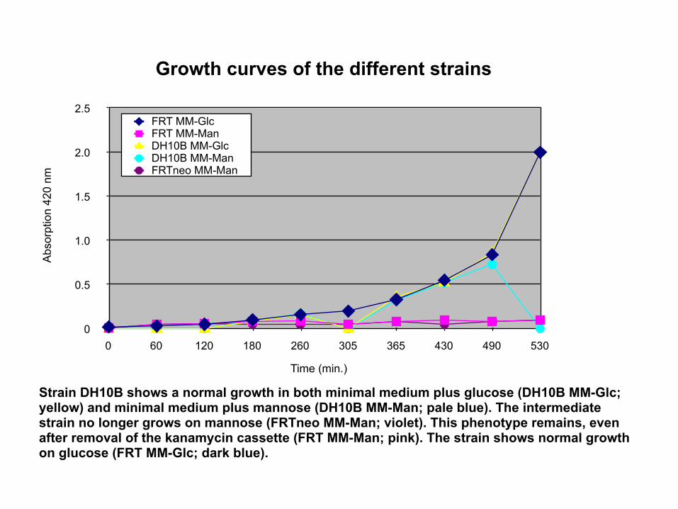
Strain DH10B shows a normal growth in both minimal medium plus glucose (DH10B MM-Glc; yellow) and minimal medium plus mannose (DH10B MM-Man; pale blue). The intermediate strain no longer grows on mannose (FRTneo MM-Man; violet). This phenotype remains, even after removal of the kanamycin cassette (FRT MM-Man; pink). The strain shows normal growth on glucose (FRT MM-Glc; dark blue).
Growth curves of the different strains
Time (min.)
Abs
orpt
ion
420
nm

Verification of specificity
1 2 3 4 5 6 M
291243
194
145
97
45
23
10
[kb]
Lanes 1 + 4: DH10B Lanes 2 + 5: DH10B manX::FRTneo Lane 3 + 6: DH10B manX::FRT
Due to an additional NotI site which flanks the FRT cassette, the 120kb NotI fragment on which the mannose transporter is located (see Heath et al. 1992) disappears after insertion of the FRT-flanked neomycin cassette while an additional 90kb fragment appears instead in lanes 5 and 6.
Beside this predicted difference in the NotI pattern, no other changes are visible, neither in the ApaI nor in the NotI digest indicating that no unintended rearrangements took place.
E.coli cells from all three strains were embedded in agarose and digested with ApaI and NotI. DNA fragments were separated by Pulsed Field Gel Electrophoresis.
ApaI NotI