Recognition of Objects and Their Component Parts ...mwo/Papers/Wachsmuth_etal...Recognition of...
14
Recognition of Objects and Their Component Parts: Responses of Single Units in the Temporal Cortex of the Macaque E. Wachsmuth, M. W. Oram, and D. I. Perrett School of Psychology, St Andrews University, Scotland, KY16 9JU, United Kingdom We investigated the role that different component parts play in the neural encoding of the visual appearance of one complex object in the temporal cortex. Cells responsive to the sight of the entire human body (but no to control stimuli) were tested with two subregions (head alone with the body occluded from sight and the body alone with the head occluded). Forty-two percent (22 of 53) of cells responded to the whole body and to one of the two body regions tested separately: 72% (17 of 22) responding to the head and 28% (5 of 22) to the rest of the body. Forty-two percent (22 of 53) of cells responded independently to both regions of the body when tested in isolation. The remaining cells (17%, 9 of 53) were selective for the entire body and unrespon- sive to component parts. The majority of cells tested (90%, 35 of 39) were selective for perspective view (e.g., some cells respond optimally to the side view of the body, others to die back view). Comparable levels of view sensitivity were found for responses to the whole body and its parts. Results indicate (1) separate neuronal analysis of body parts and (2) extensive in- tegration of information from different parts. Contrary to influential models of object recognition (Marr and Ni- shihara, 1978; Biederman, 1987), the results indicate view-specific processing both for the appearance of separate object components and for integration of in- formation across components. Visual object recognition is a fundamental part of our everyday activity. The brain is able to compare sensory information with internal representations of objects apparently independent of the object's orientation, distance, part occlusions, and lighting conditions. This raises the question of how the brain matches the in- finite number of different retinal images of one object to the representation (or representations) of the same object stored in memory. Two important issues have been raised in the con- text of object recognition. These concern (1) the role of the object's components and (2) the frame of ref- erence used for specifying the spatial relationship be- tween components of that object. Two types of frames of reference have been considered: object-centered descriptions relate an object's component parts to a framework based on the object itself, whereas viewer- centered descriptions relate an object's component parts to a framework based on the observer. This ar- ticle presents a physiological study of the role of the component parts and perspective view in the coding of one type of complex object, the body. The Role of Component Parts in Object Recognition Several theories of object recognition have suggested that the processing of an object's parts plays an im- portant role in the initial stages of recognition (Marr and Nishihara, 1978; Marr, 1982; Biederman, 1987). Re- cently, however, Baker Cave and Kosslyn (1993) have suggested that even though coding of parts is impor- tant for recognition, parts are processed only after the processing of the configuration of the whole object. To isolate the component parts, the image of the object may be segmented at regions of sharp concav- ity of the external boundary (Marr and Nishihara, 1978; Hoffman and Richards, 1984; Biederman, 1987). Each of the resulting image regions can then be treat- ed as if it corresponded to a volumetric primitive (i.e., a 3D component). Theories differ as to the type of volumetric primitives thought to be used in object recognition. Marr suggested that objects could be built up from a set of generalized cones (Binford, 1971; Marr and Nishihara, 1978). By contrast, Biederman pos- tulates a more extensive set of 36 types of cone com- ponents called geons (Biederman, 1987). Marr has suggested that the position, 3D orienta- tion, and size of each cone component of an object CcrcbraJ Cortex Scp/Oct 1994;5:5O9-522; 1047-3211/94/S4 00
Transcript of Recognition of Objects and Their Component Parts ...mwo/Papers/Wachsmuth_etal...Recognition of...

Recognition of Objects and TheirComponent Parts: Responses ofSingle Units in the TemporalCortex of the Macaque
E. Wachsmuth, M. W. Oram, and D. I. Perrett
School of Psychology, St Andrews University,Scotland, KY16 9JU, United Kingdom
We investigated the role that different component partsplay in the neural encoding of the visual appearanceof one complex object in the temporal cortex. Cellsresponsive to the sight of the entire human body (butno to control stimuli) were tested with two subregions(head alone with the body occluded from sight and thebody alone with the head occluded). Forty-two percent(22 of 53) of cells responded to the whole body and toone of the two body regions tested separately: 72% (17of 22) responding to the head and 28% (5 of 22) to therest of the body. Forty-two percent (22 of 53) of cellsresponded independently to both regions of the bodywhen tested in isolation. The remaining cells (17%, 9of 53) were selective for the entire body and unrespon-sive to component parts. The majority of cells tested(90%, 35 of 39) were selective for perspective view(e.g., some cells respond optimally to the side view ofthe body, others to die back view). Comparable levelsof view sensitivity were found for responses to thewhole body and its parts. Results indicate (1) separateneuronal analysis of body parts and (2) extensive in-tegration of information from different parts. Contrary toinfluential models of object recognition (Marr and Ni-shihara, 1978; Biederman, 1987), the results indicateview-specific processing both for the appearance ofseparate object components and for integration of in-formation across components.
Visual object recognition is a fundamental part of oureveryday activity. The brain is able to compare sensoryinformation with internal representations of objectsapparently independent of the object's orientation,distance, part occlusions, and lighting conditions. Thisraises the question of how the brain matches the in-finite number of different retinal images of one objectto the representation (or representations) of the sameobject stored in memory.
Two important issues have been raised in the con-text of object recognition. These concern (1) the roleof the object's components and (2) the frame of ref-erence used for specifying the spatial relationship be-tween components of that object. Two types of framesof reference have been considered: object-centereddescriptions relate an object's component parts to aframework based on the object itself, whereas viewer-centered descriptions relate an object's componentparts to a framework based on the observer. This ar-ticle presents a physiological study of the role of thecomponent parts and perspective view in the codingof one type of complex object, the body.
The Role of Component Parts in ObjectRecognitionSeveral theories of object recognition have suggestedthat the processing of an object's parts plays an im-portant role in the initial stages of recognition (Marrand Nishihara, 1978; Marr, 1982; Biederman, 1987). Re-cently, however, Baker Cave and Kosslyn (1993) havesuggested that even though coding of parts is impor-tant for recognition, parts are processed only after theprocessing of the configuration of the whole object.
To isolate the component parts, the image of theobject may be segmented at regions of sharp concav-ity of the external boundary (Marr and Nishihara,1978; Hoffman and Richards, 1984; Biederman, 1987).Each of the resulting image regions can then be treat-ed as if it corresponded to a volumetric primitive (i.e.,a 3D component). Theories differ as to the type ofvolumetric primitives thought to be used in objectrecognition. Marr suggested that objects could be builtup from a set of generalized cones (Binford, 1971;Marr and Nishihara, 1978). By contrast, Biederman pos-tulates a more extensive set of 36 types of cone com-ponents called geons (Biederman, 1987).
Marr has suggested that the position, 3D orienta-tion, and size of each cone component of an object
CcrcbraJ Cortex Scp/Oct 1994;5:5O9-522; 1047-3211/94/S4 00

are described in relation to the object's principal axis(the longest axis of the object; Marr and Nishihara,1978). Biederman (1987) relates geon components toother geons rather than to the object's principle axis.In Biederman's model the position and orientation ofeach geon are specified relative to other geons in qual-itative terms (e.g., geon 1 to the side of geon 2 andjoined at the expanded end). Both models suggest thatthe entire 3D object description can be accessed fromthe sight of key component parts. Thus, even whenparts of an object are occluded from sight, recognitioncan occur on the basis of the remaining visible geonsor cone components. From both Marr's and Bieder-man's theories, one might expect to find cellular unitslate in the visual pathway that respond selectively toone type of object and are activated by the sight ofany major part of that object (a major part of an objectcan include several geons).
The processes of accessing the entire 3D descrip-tion from the sight of one 3D part are similar to the"completion" property of parallel distributed process-ing (PDP) networks. Networks trained on a 2D imageand tested with any large part of the trained imagewill settle into a pattern of activity within the net-work, which is equivalent to activity pattern gener-ated by the complete training image.
View Specificity
Object-centered RepresentationBoth Marr's and Biederman's accounts of object rec-ognition can be considered object centered (Marr andNishihara, 1978; Biederman, 1987). In these modelsonly one descriptive representation of the object isstored in long-term memory (see also Lowe, 1987; Por-rill et al., 1988). This description should be accessiblefrom all viewpoints provided that the principle axis isfully visible (Marr and Nishihara, 1978)
As mentioned above, under Biederman's accountrecognition can be based on a very small number ofgeons. Thus, as long as a sufficient number of geonsare visible, recognition should not be affected by view.Under both object-centered schemes, perspectiveview should not affect the cellular mechanisms in-volved in the higher stages of object processing (be-yond the limitations noted). One would expect, fromboth Marr's and Biederman's theories, to find cellularunits late in the visual pathway that code an object ina way that is accessible from all views (i.e., cells thatrespond to all views of an object).
Recent psychological studies show that for brain-damaged and normal subjects, ease of recognition isinfluenced by the visibility of an object's salient fea-tures and cues to the 3D orientation of the object(Warrington and Taylor, 1973; Humphreys, 1984; War-rington and James, 1986; Quinlan, 1988; Humphrey,1989).
Viewer-centered RepresentationRepresentation of an object in a viewer-centered man-ner relies on descriptions of the object relative to dieviewer. Such description includes a collection of the
2D visual characteristics of the object that are visiblefrom a specific viewpoint. The number of character-istics present in any one viewer-centered descriptionis therefore smaller than that of an object-centereddescription. Two main types of viewer-centered rep-resentation have been considered, in which (1) onlyone view of the object is stored or (2) multiple viewsare stored in long-term memory (Palmer et al., 1981;Jolicoeur, 1985; Tarr and Pinker, 1989; UUman, 1989;Edelman and Biilthoff, 1990; McMullen and Farah,1991; Cutzu and Edelman, 1992; Verfaillie, 1992). Notethat under scheme 1, where a single view is stored,the orientation of the object's components would bespecified with respect to the viewer. Hence, this singledescription is not object centered.
Palmer et al. (1981) have defined the "canonicalviews" as the single view that reveals the maximal in-formation about an object's salient features. For mostobjects (8 of 12 objects of those studied by Palmer etal., 1981), the canonical view lies between the "front"and "side" views, with the object's principal axis beingoriented 45° to the observer's line of sight. In addition,for heads, the 45° (or %) view is the most readily rec-ognized in naming and matching tasks (Bruce et al.,1987). Models envisaging storage of a single viewer-centered description (such as the canonical view) relyon a transformation of the incoming image to matchthe stored description. The transformation may in-volve processes akin to mental rotation (Shepard andCooper, 1982). If an object is represented by a singlecanonical viewer-centered description, then onemight expect to find greater numbers of cells selec-tively tuned to this view of an object than to otherviews of the same object.
Most viewer-centered models of object recognitionsuggest that several views of an object are represent-ed in memory (Koenderink and van Doom, 1979;Tarrand Pinker, 1989; Ullman, 1989; Edelman and Bulthoff,1990;Poggio and Edelman, 1990;Seibert and Waxman,1991). The theories, however, differ as to the numberand nature of the views stored. In these models eitherthe incoming image is transformed to match to thenearest stored view, or alternatively the image is iden-tified by interpolation from a minimum of three sur-rounding stored views. Multirepresentational viewer-centered models suggest the presence of cells codingdifferent specific views of the same object.
Cellular Sensitivity to Objects and Tbeir PartsIt has been proposed that the inferotemporal cortex(IT) plays a central role in visual pattern and objectrecognition (Gross, 1973, Ungerleider and Mishkin,1982). Most physiological studies of this area havebeen concerned with the coding of geometrical fea-tures that may occur in several objects (such as bars,circular areas, Fourier descriptors; Gross et al., 1972;Schwartz et al., 1983; Desimone et al., 1984; Tanakaand Fujita, 1991; Fujita et al., 1992; Komatsu and Ideu-ra, 1993).
Studies in IT and neighboring cortex widiin thesuperior temporal sulcus (STS) have also revealedpopulations of cells with greater visual selectivity that
510 Ncuronal Responses to Object Components • Wachsmuth ct al.

respond preferentially to particular complex biologi-cally important stimuli such as hands and faces (Grosset al., 1969, 1972; Perrett et al., 1982, 1989; Kendrickand Baldwin, 1987;Desimone et al., 1990). These cellsoffer an opportunity to determine the importance ofan object's component parts in the processing of theentire form of the object. Hence, these cells can beused to investigate the neurobiological validity of psy-chological and computational models of object rec-ognition.
Early studies focused on the importance of facialcomponents for cell responses to the whole face. Dif-ferent cells were found to be selective for differentregions of the face (some tuned to the eyes, others tothe mouth; Perrett et al., 1982). Though not system-atical!)' studied, it was also noted that most cells re-sponded to several regions tested in isolation (Perrettet al., 1982; Desimone et al., 1984). Facial characteris-tics, particularly when seen from the front, are dennedby differences in pigmentation and surface structure.The parts cannot be segmented from points of maxi-mum concavity in the external contour (silhouette).Hence, models such as those of Marr and Biedermanthat embody segmentation at concave points in theexternal boundary may be less applicable to the pro-cessing of the internal structure of the face.
In the present study we extended the investigationof the role of components in the processing of wholeobjects by comparing the response of cells to the en-tire body and to two major parts: the head with thebody occluded from sight and the body with the headoccluded from sight.
Celhdar Sensitivity to Object ViewPhysiological studies have investigated the view sen-sitivity of cells responsive to faces in the temporalcortex (Desimone et al., 1984; Perrett et al., 1984,1985, 1989, 1991, 1992; Kendrick and Baldwin, 1987;Hasselmo et al., 1989a,b). All studies reveal that themajority of cells are sensitive to change in perspectiveview. That is, most cells code in a viewer-centeredfashion. Different cells are tuned to different optimalviews (some to the face, some to the back of the head,etc.). It is interesting to note that in humans evokedpotential studies also indicate viewer-centered pro-cessing of the face (Botzel and Griisser, 1989; Jeffreysand Turkmachi, 1992).
A small population of cells in the macaque STS hasbeen found to respond equally to all views of the head(Perrett et al., 1984, 1985, 1989, 1991, 1993; Hasselmoet al., 1989b). The insensitivity of these cells tochanges in view is in accordance with the definitionof object-centered coding. Object-centered cells couldarise from a combination of the outputs of differentviewer-centered cells (Perrett and Oram, 1993). Thissuggestion is supported by the finding that latenciesof object-centered cells are longer than those of view-er-centered cells (Perrett et al., 1992).
An additional aim of the present study was to com-pare view tuning for the whole body with view tuningfor component body parts. This aspect of the studycould provide potential insight in the role of view-
selective processes in the integration of informationabout separate object parts.
Preliminary results of have been reported previ-ously (Perrett et al., 1993; Wachsmuth et al., 1993).
Materials and MethodsRecordings of responses of single cells are from fivemacaque monkeys (Macaca mulatto; two females, 4-8 kg; three males, 5-8 kg). The techniques appliedhave been previously described (Perrett et al., 1991).
Training and Fixation TaskPresurgical training involved the following. Situated ina primate chair, the monkeys were trained to fixateon an LED light presented onto a white wall at eyelevel at a distance of 4 m. The monkey's task was todiscriminate the color of the I.KD that followed a shortsignal tone to obtain the monkey's attention. Lickingresulted in fruit juice reward for the green I.KD Themonkey was to withhold licking in order to avoid de-livery of a weak saline solution to the red LED. TheLED stimuli were presented in a pseudorandom orderunder computer control.
SurgeryUnder full sterile conditions two stainless steel rings(16 mm diameter, 10 mm deep) were surgically im-planted bilaterally under sodium pentothal (Sagital)anesthesia with their centers at predetermined stereo-taxic coordinates 12-14 mm midline and anterior tothe interaural plane. Two plastic tubes (5 mm diame-ter) were attached with dental acrylic horizontally infront and behind the wells. By passing a metal barthrough each tube, the monkey's head could be re-strained during recording sessions. The monkey wasallowed to recover from surgery and retrained untilpresurgical performance in the color discriminationtask was reached.
Recording TechniquesBefore each recording session a topical anesthetic (xy-locaine, 40 mg/ml) was applied to the dura. A DavidKopf micropositioner was fixed to the recording well,allowing a tungsten in glass microelectrode to be in-serted through a transdural guide tube into the tem-poral cortex, aiming for the anterior part of the STS(areas TPO, PGa, and TAa of Seltzer and Pandya, 1978).
The LED light and the visual stimuli were present-ed from behind a shutter wiui rise time of less than15 msec. A large-aperture (6.5 cm diameter) electro-mechanical shutter (Compur) or an alternative (20 cmsquare) liquid crystal shutter (Screen Print Technolo-gy Ltd.) was used. The shutter opened for 1.0 sec aftera 0.5 sec warning tone that allowed the monkey toprepare fixation of the LED position before the shut-ter opened. This enabled the monkey to lick severaltimes for multiple juice rewards during the trial peri-od. Visual stimuli (2D) were projected onto the whitewall in front of the monkey on which the LED lightwas positioned, and 3D stimuli were presented infront or to either side of the LED. The test stimuli wereinterspersed with control stimuli and a no-stimulus
Cerebral Cortex Sep/Oct 1994, V 4 N 5 511

Figure 1. Examples of stimuli used for testing: whole-body, head-only, andbody-only stimuli in different views.
condition (the I.KD alone, to measure spontaneous ac-tivity).
The neuronal activity recorded was amplified, fil-tered, and displayed on an oscilloscope using standardtechniques and equipment. The spike activity was alsoconverted to digital signals and stored on a PC-com-patible computer using CED1401 (Cambridge Elec-tronic Design) in 5 msec time bins. Eye movementswere recorded with each trial using an infrared reflec-tion system (ACS, modified to allow recording of bothhorizontal and vertical signals from one eye), andstored with 8-bit accuracy.
Visual StimuliNeuronal responses were recorded for both real 3Dobjects and 2D objects (video disk images and slides).The visual stimuli tested were the (1) whole humanbody (head and body), (2) head alone, and (3) bodyalone (defined here as torso, arms and legs; see Fig. 1).The posture in all cases was bipedal. The humanbody stimuli were photographed onto a 200 ASA ec-tochrome slide film, with the person standing againsta light gray background. Slides with occluded bodyparts were produced by first taking the whole-bodyslide and then occluding the unwanted parts withblack tape.
Alternatively the stimuli were filmed with a videocamera (JVC BY-110E), recorded on % inch U-Maticvideotape, edited on a JVC editing suite (control unit
RM-88U), and transferred on a laser video disc (RLVMk II, Optical Disc Corp.). The video stimuli werethen replayed with a video disk player (Philips VP4O6LaserVision Disc Drive) and projected onto the displayscreen (using a Sony color video projector VPH-1041QM).
Where 2D stimuli did not activate cells, 3D stimuliwere used. 3D head-only and body-only stimuli werecreated by occluding the unwanted parts with a largesheet of black cardboard or curtain material.
For most cells multiple views of the stimuli weretested: front view (0°), left profile (90°), back of head(180°), and right profile (270°). In addition to these,four intermediate views (45°, 135°, 225°, and 315°)were tested for some cells. For each cell, a number ofdifferent control stimuli were tested (2D and 3D).These included complex 3D objects of different sizes,shapes, and textures (lab coats, chairs, etc.), simple 2Dgeometrical shapes (bars, spots, and gratings) and sim-ple 3D forms (cylinders, balls, boxes, etc.).
Testing MethodsEvery cell from which neuronal activity was recordedwas first tested in an exploratory way by presentinga series of static and moving 3D objects (includingbodies), and tactile and auditory stimuli. Cells foundto be responsive to static views of the whole bodywere then further investigated for sensitivity to bodyparts. After identifying the optimal stimulus with a 3Dwhole body, further testing was carried out with 2Dstimuli. Stimulus order was presented in a computer-controlled pseudorandom order. The cell was thentested with at least five trials of each of the wholebody, its two parts (head only and body only), anddifferent control objects. Testing was performed,where possible, with stimuli of the (cell's) preferredview, the opposite view (180° rotation was usually theleast effective view), control stimuli, and no stimulus.The views used for testing components were the sameas used for testing the whole body.
Data AnalysisSince most cells in the STS respond with a latency of100 msec (±30 msec; Oram and Perrett, 1992), themagnitude of cell activity on individual trials was as-sessed over the 0.25 sec time period occurring 100-350 msec after stimulus onset. For some cells [withlate response onset (>200 msec) or inhibitory re-sponses) a 0.5 sec time period (100-600 msec post-stimulus) was used to assess cell activity.
Cell responses to the whole body and two com-ponents (head only, body only), controls, and sponta-neous activity were compared on line by using one-way ANOVA and post hoc tests [protected leastsignificant difference (PLSD); Snedecor and Cochran,1980]. The influence of view on cell responses to thewhole and component parts was analyzed off linewith two-way ANOVA with view and body parts testedas main factors.
Recording SitesAfter each recording session, frontal and lateral x-rayphotographs were taken to localize the electrode. Mi-
512 Ncuronal Responses to Object Components" Wachsmuth et al.

crolesions (10 JJLA DC for 30 sec) made at the end ofsome electrode tracks, subsequently identified usingstandard histological techniques, allowed reconstruc-tion of the electrode position within the brain. In ad-dition, reference markers were made by injection ofHRP and fluorescent dyes true blue and diamidino yel-low.
Once the last recording session had been complet-ed, the monkey was given a sedating dose of ketaminefollowed by a lethal dose of barbiturate anesthetic. Af-ter transcarinal perfusion with phosphate-buffered sa-line and 4% gluteraldehyde/paraformaldehyde fixative,the brain was removed and put into a series of sucrosesolutions with increasing concentration (10%, 20%,and 30%) or alternative!)' 2% dimethyl sulfoxide and20% glycerol (Rosene et al., 1986; see Harries and Per-rett, 1991, for detail).
ResultsFor five subjects, a total of 7287 cells were screenedfor neuronal responses. Of these, 23% (1691 of 7287)were found to be visually responsive, including cellsresponsive to moving or static visual stimuli (see, e.g.,Bruce et al., 1981;Oram et al., 1993) and visual generalcells (where no apparent specific visual stimulusdrives the cell). Of the visually responsive cells in thecortex of the STS, a total of 64 cells that were foundto be responsive to the whole body [i.e., have signif-icantly greater response to the whole human bodythan to control objects and spontaneous activity (S/A)] were tested for selectivity to component parts ofthe body. All the cells included in this study did notselectively respond to a wide variety of different con-trol objects tested. For 53 of these cells responseswere measured for two body parts [head only; bod}'only (torso, arms, and legs)] and the whole body (headand body). The remaining 11 cells were tested for thewhole body and only one part, and therefore are notincluded in the analysis of coding component parts,but are included in the view discrimination study.Cells were categorized on the basis of their responseto the two parts: they could either respond to none,one, or two pans.
View sensitivity was investigated for a total of 39(of 64) cells. For 28 of these cells, view sensitivity wastested for the whole and both parts, whereas for 11cells view sensitivity was measured for the wholebody and only one part.
The optimal stimuli for a given cell was defined asthe stimuli causing maximal change in the firing raterelative to baseline activity (S/A). For the majority (60of 64~) of cells, optimal stimuli produced excitatoryresponses. Four cells gave inhibitory responses to op-timal stimuli. For clarity of explanation, "greater" re-sponse is defined as greatest change from S/A (wheth-er excitatory or inhibitory).
Coding of PartsTwenty-two cells responded to only one of the twocomponent body parts when tested in isolation.
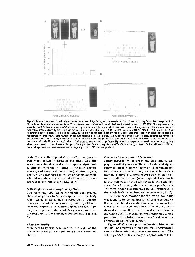
Cells Only Responsive to the HeadFor 17 cells (32% of the 53 cells tested) the responseto the sight of the head alone (with the rest of thebod)' occluded from sight) was significantly greaterthan that to controls and to S/A. Additionally, for thesecells the body alone (with the head occluded fromsight) did not produce a response that was signifi-cantly different from controls or S/A. This pattern ofresponse is shown in Figure 2/4. For this cell the pres-ence of the head was both necessary and sufficient toaccount for the response to the whole body.
Figure 2B-D also displays responses of the samecell recorded on the five individual trials with thewhole-body, head-alone, and body-alone stimuli. Re-sponses to the whole bod)' (Fig- 2fl) occurred at ap-proximately 120 msec after stimulus onset, with aninitial transient burst (lasting ~35O msec) following aresponse decline, though remaining substantiallygreater than the prestimulus activity typical for thecells found in STS (Oram and Perrett, 1992). Figure 2Cshows responses of similar latency and time course ofactivity during trials when the head alone was pre-sented. When the body was presented in isolation (Fig.ID), however, there was no change in cell activity incomparison to the prestimulus period. Thus, the bodypresented without the head was an ineffective stim-ulus for this particular cell.
In addition, Figure 2B-D indicated that for all stim-ulus types the position of the eyes was held constant(within ±5°) during the time of neuronal responseanalysis (100-350 msec). Thus, differences in cell re-sponsiveness for the different types of visual stimuli(effective or ineffective) were not due to different pat-terns of fixation.
Cells Only Responsive to the Body (Torso and Limbs)Nine percent (5 of 53) of the cells showed a responseto the body tested in isolation, which was significantlydifferent from the response to control objects and S/A (e.g., Fig. 3). For these cells the responses to headalone did not differ from S/A or the response to con-trol objects.
The 22 cells described in this section were respon-sive to one of the two body parts tested in isolation.This classification could include cells that show hid-den sensitivity to multiple body parts. Thus, eventhough a cell responds only to one component whentested in isolation, the other component may influ-ence the response to the whole object. For 12 of thesecells, there was no significant difference between theresponse to the effective component part and re-sponse to the whole body. For these cells the responseto one part was necessary and sufficient to accountfor the response to the whole body. The remaining 10cells showed a significant difference between the re-sponses to the effective component-part and thewhole-body stimuli (see, e.g., Fig. 3).
Coding the Entire Body
Cells Only Responsive to Whole BodySeventeen percent (9 of 53) of the cells showed aresponse depending on the visuality of the whole
Cerebral Concx Scp/Oct 1994, V 4 N 5 513
B
II I I I II I I
I I I I Il i i ; I i l l i n Pi n i l i n
i i M i l l I I I III I I I I I I I
"OST-STIMULUS TIME (ms) POST-STIMULUS TIME (ms)
Fignre 2. Neuronal responses of a cell only responsive to the head. kTop, Photographic representation of stimuli used for testng. Bottom, Mean responses ( ± 1SE) to the whole body, its components (view 0°), spontaneous activity [S/A], and control stimuli are illustrated for one cell (E29-33.56). The responses to thewhole-body and the head-only stimuli were not significantly different (p > 0.05), whereas both these stimuli produced a significantly higher neuronal responsethan activity rates produced by the body-alone stimulus, S/A, or control objects [p < 0.005 for each comparison; ANOVA F(7,39) = 26.1, p < 0.0005). B-D,Rastergram displays of responses of one cell (EZL33.56) on five trials for each of the stimulus conditions. Each trial (originally in pseudorandom order) isrepresented by a single row of ticks marks; each tick mark indicates one action potential. Prestimulus time is given at the figure base. Horizontal eye movementsare shown for each trial in the upper sections. The responses to the whole body (A far left column) and the head tested in isolation [second column from left)were not significantly different (p > 0.05), whereas both these stimuli produced a significantly higher neuronal response than activity rates produced by bodyalone (center column] or control objects [far right column) (p < 0.005 for each comparison) ANOVA: f(7,39) = 26.1, p < 0.0005. Vertical calibration: ± 100° forhorizontal (eye movements were recorded over a range of positions ±20° from straight ahead).
body. These cells responded to neither componentpart when tested in isolation. For these cells thewhole-body stimulus produced a response significant-ly different from that to either of the body compo-nents (head alone and body alone), control objects,and S/A. The responses to the components individu-ally did not show any statistical difference from re-sponses to controls or S/A (e.g., Fig. 4).
Cells Responsive to Multiple Body PartsThe remaining 42% (22 of 53) of the cells studiedshowed responses to both components of the bodywhen tested in isolation. The responses to compo-nents and the whole body were significantly differentfrom the responses to control objects and S/A Typi-cally the response to the whole body was greater thanthe response to the individual components (e.g., Fig.5).
View SensitivityView sensitivity was examined for the sight of thewhole body for 39 cells (of the 53 cells describedabove).
Cells with Viewer-centered PropertiesNinety percent (35 of 39) of the cells studied dis-played sensitivity to view. These cells showed signifi-cantly different responses between (a minimum of)two views of the whole body. As should be evidentfrom the Figures 2-5, different cells were found to betuned to different views (some responded maximallyto the front view of the body, others to the back, oth-ers to the left profile, others to the right profile, etc.).The view preference exhibited by cell responses tothe whole body generalized to the parts (e.g., Fig. 6).
The view discrimination for the whole and panswas found to be compatible for all cells (see below).If a cell exhibited view discrimination between twoviews of an isolated body part, then the cell alsoshowed the same direction of view discrimination forthe whole body. Two cells, however, responded to onepart tested in isolation but only displayed view dis-crimination for the whole body.
Figure 6B-D shows poststimulus time histograms(PSTHs) for a viewer-centered cell that discriminatedview for the whole body and its component parts. Thecell responded with a latency of approximately 100-
514 Neuronal Responses to Object Components • Wachsmuth et al

S/A
Figure 3. Neuronal responses of a cell only responsive to the body. Top,Photographic representation of stimuli used for testing. Bottom, Mean re-sponses ( ± 1 SE) to the whole body, its components (view 0°), spontaneousactivity {S/A), and control stimuli are illustrated for one cell (B77_21.54). Thewhole-body and the body-alone stimuli produced no significant difference inthe cell's activity rate (p > 0.051, whereas both these stimuli produced sig-nificantly higher neuronal activity than activity rates produced by head-alonestimulus, S/A, or control object stimuli (p < 0.005 for each comparison!.AN0VA: f(4,39) = 11.4, p < 0.0005.
Rgora 4. Neuronal responses of a cell only responsive to the whole body.Top, Photographic representation of stimuli used for testing. Bottom, Meanresponses ( ± 1 SE) to the whole-body, head-alone, and body-alone stimuli(view 180°) and spontaneous activity {S/A) are illustrated for one cell(D107J5.41). The whole-body stimuli gave a significantly higher responsethan the other stimuli tested (p < 0.0005 for each comparison). The headtested in isolation and the body tested in isolation did not show any signifi-cant difference from the S/A (p > 0.05 each comparison). AN0VA: 5(3,23) =35.3, p < 0.0005.
120 msec. After an initial transient burst Casting ~3OOmsec) the response declined, though remained greaterthan the prestimulus activity. This pattern of cellularactivity can be seen most easily for the whole-bodystimuli (C), but is also present to a lesser extent forthe head-alone (B) and body-alone (£>) stimuli. Note,that the activity profile for each of these stimuli pre-sented in the preferred view is enhanced, comparedto the activity profile for the nonpreferred view.
Cells with Object-centered PropertiesTen percent (4 of 39) of the cells showed no preferredview for either the whole body or its components(Fig. 7).
View Discrimination Indices
View Discrimination: Whole Body versus Head AloneThe responses of 18 viewer-centered cells were usedin a population analysis to compare the efficiency ofview discrimination for the whole body and the headpresented alone (Fig. 8/1). Cells were only included inthis analysis if they responded to the whole body andto the head when tested in isolation.
A view discrimination index was computed foreach cell from the view producing the greatest changein activity from S/A (best angle) and smallest changein activity (worst angle) to the whole-body stimulus.The cell's activity was then measured to the compo-nent stimulus (head alone) in the same two views.
The formula used to calculate these indices wasthe same as used previously (Oram and Perrett, 1992):/„ = [(response to best angle - S/A) — (response toworst angle - S/A)]/(response to best angle - S/A).
The distribution of /„ values is shown in Figure 8Afor the whole body and for the head alone. For anindex value of 1 0 response to the worst view was thesame as S/A. Index values >1.0 arise when the cellresponse to the best view was greater than S/A, andthe response to the worst view was less than S/A. lr
can have a negative value if a neuronal response is
S/A
aCO
I -•CO
IIICO 20-
oaCOCOUJ
Figure 5. Neuronal responses of a cell responsive to multiple parts. Top,Photographic representation of stimuli used for testing. Bottom, Mean re-sponses ( ± 1 SE) to the whole body, its components (view 270°), controlstimuli, and spontaneous activity {S/A) are illustrated for one cell (D30J7.72)The cell responded more to the whole-body stimuli (p < 0.05 each compar-ison) than to any other stimuli The cell also responded significantly more toeither of the body regions tested in isolation (head-alone, body-alone stimuli)than to S/A or control objects (p < 0.005). AN0VA: f(4,18) = 14.1, p < 0.0005.
Cerebral Cortex Scp/Oct 19SM, V 4 N 5 515

« . 30
(0UJOC 10
2in
S/A
-hrh r*i
POST-STIMULUS TIME (ms)
a »V)
D• J I l lJL J I.L
POST-STIMULUS TIME (ms)
POST-STIMULUS TIME (ms)
Figure 6. Neuronal responses of a cell with viewer-centered properties to the body and its components. A Histogram of response (spikes/sec) to different stimuli.Top, Photographic representation of stimuli used for testing. Bottom, Mean responses ( ± 1 SE) of one cell (J12S.33.66) tested to view 45° and view 225° of thewhole body and its components. Two-way ANOVA revealed a significant main effect of view [ f ( U 4 | = 23.28, p < 0.0005L body part tested (f(2^4) = 334, p <0.05), and interaction between these factors [F{2JU) = 2.49, p < 0.5]. Protected least significant difference tests (PLSD), post hoc tests, indicated significantresponse discrimination between the different views of the entire body (p < 0.0005) and of the body parts tested in isolation: head only (p < 0.05) and body only(p < 0.05). 8-O, Poststimulus time histograms (PSTHs) show averaged responses from five trials (bin width, 20 msec). The cell responded strongly to the sightof the whole body and the component parts presented in view 45° {white bars), but failed to respond to the same stimuli in an opposite view (black bars, view225°; stimulus onset at time 0). The ordinate of the PSTHs denotes the cell responstvrty for 100 spikes/sec.
numerically greater for the "worst" view than the"best" view. This can only occur when the index iscomputed for the component body parts, since thebest and worst views were defined on the basis ofresponses to the whole body.
For cells responsive to the head, the distribution of/,, for the entire body was not significantly differentfrom the distribution of /,, when the head was testedalone (matched pairs t = 0.20, df = 17,/? = 0.85).
View Discrimination: Whole Body versus Body AloneThe efficiency of view discrimination for the wholebody and the bod)' alone was computed in a similarway for 10 cells (Tig. 80). These cells were responsiveto both the whole body and to the body without thehead visible. The distribution of Ir values obtainedwith the whole body visible was not significantly dif-ferent from that obtained from the bod)' alone (t =-0.76, df = 9,p = 0.47).
This analysis was repeated for seven cells respon-sive to multiple parts and again it was shown that thedistribution of /,. for the entire body did not signifi-cantly differ from the distribution of Ir for head alone
(matched pairs t = -0.74, df = 6, p = 0.49) or bodyalone (matched pairs t = -1.5, df = 6,p = 0.19).
Thus, as a population of cells there was no signifi-cant difference of quality for view discrimination be-tween viewing the entire body or its isolated parts.
Histological LocalizationReconstruction of cell position in four monkeys re-vealed that cells responsive to the head and bodywere located in the upper bank (and to a certain ex-tent in the lower bank) of the anterior STS. Figure 9illustrates the position of the different cell types re-corded in one monkey. The different cell types wereintermixed within the cortical areas sampled. For onemonkey histology was unavailable but x-ray analysisrevealed the recording site to be in the same brainregion.
Discussion
Cell Sensitivity to tbe BodyPrevious studies of form processing of biological stim-uli in the temporal cortex have focused on cell re-
516 Neuronal Responses to Ob|ect Components • Wachsmuth et aJ

S/A
to
UJ* is-
111CO 10
HIE
Figure 7. Neuronal responses of a cell with object-centered properties tothe body and its components. Top, Photographic representation of stimuliused for testing. Bottom, Mean responses ( ± 1 SE) of one cell (J22.27.78) tofront (view 0°) and back (view 180°) views of the whole body and its com-ponents are shown. Two-way ANOVA showed a significant main effect of thebody part tested [F(2#) = 103, p < 0.005] but no effect of view [F(U4) =0.9, p > 02\ and no interaction between view and part tested0.2, p > 0.5].
sponses to the sight of the face and other views ofthe head (Perrett et al., 1982, 1984, 1985, 1989, 1991,1992; Kendrick and Baldwin, 1987; Hasselmo et al.,1989b). In the present study it was not surprising tofind cells responding selectively to the sight of thehead alone, since the face carries much social infor-mation.
The surprising result of the present study was theextent to which information arising from parts of theentire body, other than the head, influence cell re-sponses. The responses of the majority of cells (83%,44 of 53) carried information about regions of thebody other than the head. There were three types ofcell for which body information was found to be im-portant. For the five cells responsive to the bodyalone, information about the head in isolation was in-sufficient to drive responses. For 22 cells independentresponses could be measured both to the head and tothe rest of the body. Finally, for nine cells, informationfrom the head and the body was critical before anyresponse could be stimulated.
The high proportion of cells found in the presentstudy sensitive to body information suggests that oth-er studies overlooked the importance of the body. Itis unlikely that we have uncovered four new classesof cell; rather, we may have revealed additional infor-mation processing abilities of cells that have alreadybeen described. In previous studies cells have beendescribed as "face responsive" or even dubbed "facecells." The present analysis suggests such labeling maybe inappropriate, as it can underestimate or bias in-terpretation of cell selectivity and functions. The func-tion of cells may require an integration of informationfrom multiple body parts including the eyes, face,hands, body posture, and so on (Perrett et al., 1992).
UJ
o
UJ-1 0 1 2 >
VIEW DISCRIMINATION INDEX
B7
BJ .
S •O 4
£n 3
Z 2
1
( I . I IL
••
fl< -2 -1 0 1 2 >
VIEW DISCRIMINATION INDEX
Hgve 8. View discrimination indices. A, Whole body versus head alone. Anindex of view discrimination was computed (see text) for 18 viewer-centeredcells responsive to the whole body and the head alone. The black barsdisplay the ability of the cell population to discriminate between views of thewhole-body stimuli. The gray bars display the ability of the same cells todiscriminate between views of the head alone. The distributions of indexvalues were not significantly different (t = 0.20, df = 17, p = 0.85). fl, Wholebody versus body alone. A similar comparison was made for 10 viewer-cen-tered cells responding to the whole body and the body alone. The black barsdisplay the population's ability to discriminate between views of the whole-body stimuli. The gray bars display the population's ability to discriminatebetween views of the body-only stimuli. The distributions did not significantlydiffer (f = -0.75, df = 9, p = 0.47).
Implications for Models
Cells Selective for One Body PartForty-two percent (22 of 53) of the cells studied re-sponded to only one of the two body parts tested.Hence, the component parts of an object appear tobe coded separately within higher visual associationcortex. One might suppose that such findings fit mod-els suggesting an initial encoding of objects in termsof their component 3D volumetric parts (Marr andNishihara, 1978; Biederman, 1987). Detailed consider-ation (below) indicates that the present findings donot fit these models.
The recordings indicate response sensitivity tobody components that are more complex than simple3D volumetric shapes. First, the cells are unresponsiveto a wide variety of simple and complex control stim-uli. If a cell was selective for a particular geon (e.g., acylinder shape), then one would expect responsesboth to the shape of a human body that would includeseveral cylindrical components and to a great variety
Cerebral Cortex Scp/Oct 1994, V 4 N 5 517

O Head Alone• Body Alone
Whole OnlyMulti-Parts
Figure 9. 7bp left. Schematic drawing of the nght hemisphere of the macaque brain. Hatched region indicates the location area STPa. Top right Frontal sectionof the brain of one monkey (J) 9.5 mm antenor to the interaural plane The STS of the left and nght hemispheres is indicated by the hatched areas. Bottom,Series of frontal sections every 1 mm (from 15.5 to 6.5 mm anterior to the interaural plane) showing the location of cells responsive to the head alone [opencircle^, body alone (solid circle^, whole body only (open triangles], and responsive to all parts (solid Wangles). The thick lines indicate the brain surface; thethin lines show the boundary between white and gray matter. Cells were located in both the upper bank and fundus of the STS.
of control objects (e.g., a mug, a broom, etc.) that alsohave cylindrical components. Second, most cellsshowed sensitivity to body view. Indeed, some cellsdiscriminate between mirror-symmetrical views (e.g.,left profile but not right profile) that contain identicalgeometric shapes. If a cell response to one body partis to be explained by the presence of a particular 3Dvolumetric shape (geon or cylinder), then the cellshould be equally responsive to all views where thisshape remains visible. Though object components areencoded independently during visual processing, thecomplexity of the component parts receiving separateanalyses within the STPa cortex is greater than sug-gested by the models of Marr and Biederman.
The results are also incompatible with viewer-cen-
tered models of recognition suggesting that only theglobal shape of particular object view is represented(without independent part representation; Koender-ink and van Doom, 1979; Edelman and Biilthoff, 1990;Seibert and Waxman, 1991).
Cells Selective for Multiple Body PartsOne population of cells (42%, 22 of 53) studied wasresponsive to the whole body and to multiple partsof the body when tested in isolation. This populationof cells is in accordance with the models of Bieder-man (1987) and Marr and Nishihara (1978). Thesemodels predict one global description of an objectstored in memory that is independently accessiblefrom the sight of any major object component. How-
518 Ncuronal Responses to Ob(ect Components • Wachsmuth et al.

ever, the view tuning of these cells suggests that eventhese cells do not support these models (see below).
We note that categorization used may have under-estimated multicomponent coding. Sensitivity to infor-mation from both the head and the bod}' alone mayhave been present even for some of the cells catego-rized as responsive to only one part. First, for some ofthese cells (10 of 22) responses to the entire bodywere different from the response to the most effectivepart tested in isolation (e.g., Fig. 3), indicating that the"noneffective" part could influence response whenviewed together with the effective part. Second, forfour cells view tuning was present for the entire bodybut was not evident for the effective part tested inisolation.
Cells Selective for the Whole BodyThe models of Marr and Nishihara (1978), Biederman(1987), and Lowe (1987) do not predict the popula-tion of cells (17%, 9 of 53) that were only responsiveto the entire body. For these cells there was no activityprovoked by isolated parts. Thus, the cell activity pro-vides a description of the appearance of the entirebody but this description is not accessible from theisolated parts. The cell responses provide informationabout the overall appearance of the object but theydo not provide information about independent partsof the object.
The responses of such cells may fit suggestions ofBaker Cave and Kosslyn (1993). These authors pro-pose a model of visual processing in which the overallappearance of the entire object is processed beforethe parts. If the cells selective for the entire body rep-resent a description of the overall configuration thatis processed first, then one would not expect them tobe activated by independent parts. The overall config-uration of an object could perhaps be revealed froman analysis of coarse image attributes (e.g., low spatialfrequencies). This course of analysis could be inde-pendent of the detailed form of individual objectparts. It is noted, however, that the majority of cellsselective for the whole body (as all other cell classes)showed viewer<entered properties in coding. Viewdiscrimination would not be predicted from a verycoarse analysis of the body form.
With larger numbers of cells defined in each pop-ulation, an analysis of the latency of the different celltypes would be possible and may clarify the order ofprocessing. Such analysis could reveal whether partsof an object are processed prior to the configurationof the whole object that would be consistent with"bottom-up" processing models. Alternatively latencyanalysis could reveal the opposite order of processingas suggested by some models employing "top-down"processing (e.g., Baker Cave and Kosslyn, 1993).
Sensitivity to View
Object-centered CodingThe analysis of view sensitivity revealed that represen-tation of the body and its parts within area STPa oc-curs mostly in a viewer-centered fashion; 90% of cells
studied were selective for view. This result is inmarked contrast to models that employ object-cen-tered representations since these representationsshould be accessible from all views (e.g., Marr andNishihara, 1978; Biederman, 1987; Lowe, 1987; Porrillet al., 1988; Hasselmo et al., 1989b).
The remaining 10% of cells studied were found torespond to all views of the body that were tested andcan therefore be considered as coding in an object-centered manner. Multiple inputs from cells withviewer-centered properties may account for the re-sponses of object-centered cells. Latency estimates forcells sensitive to the head are consistent with thisscheme (Perrett et al., 1992). It is possible that object-centered coding will be more frequently encounteredin areas subsequent to the STPa.
Viewer-centered CodingIt emerged that different cells were selective for dif-ferent views of the whole body. This parallels the ob-servation that different cells in the STPa are tuned toa range of head views (Desimone et al., 1984; Perrettet al., 1985, 1991; Hasselmo et al., 1989b). Thus, nosingle canonical view of the body accounts for theentire range of view tuning (Palmer et al., 1981). Theresults are therefore consistent with models suggest-ing multiple viewer-centered representations stored inmemory (Koenderink and van Doom, 1979; Tarr andPinker, 1989; Ullman, 1989; Edelman and Bulthoff,1990; Poggio and Edelman, 1990; Seibert and Waxman,1991; Cutzu and Edelman, 1992). As noted above, theresults are consistent with a view-sensitive represen-tation of parts, rather than a view-sensitive represen-tation of the global form that is common to mostviewer-centered models.
The present study did not set out to define thedistribution of view tuning among cells responsive tothe body. Nevertheless, view tuning for the wholebody appears to have similar properties to view tun-ing for the component parts. For a minority of cells(4 of 22 cells) view tuning was apparent for the wholebody but was not present for component parts thatprovoked responses when tested in isolation. As apopulation of cells, analysis showed view discrimina-tion (as measured by our index) exhibited to thewhole body that was equivalent to the view discrim-ination manifest to the head alone or to the bodyalone. There was no improvement in quality of viewdiscrimination for the entire body, compared to a sit-uation in which only one body part was visible.
Coding of Isolated Object PartsOne question arising from the present study is whyparts of the body are coded independently of eachother. Under natural viewing conditions individualparts of an body, or indeed any object, are often notfully visible. Such situations arise when a body isviewed from behind an intervening object or whenone part of the body occludes the sight of anotherpart. It would be impossible to recognize objects insuch circumstances if the cortex only contained cellsselective for the intact or entire object (such as that
Cerebral Cortex Scp/Oct 1994, V 4 N 5 519

depicted in Fig. 4). The separate coding of object partsallows recognition under conditions of partial objectocclusion.
If each of the major (articulating) parts of an objectis coded separately from a small number of views,then recognition of the object is possible despitechanges in the configuration of these parts. An alter-native processing scheme could rely on cells selectivefor the entire object's configuration, with differentcells selective for different configurations. While thisscheme is possible, it is also inefficient and would re-quire large numbers of "templates" or cells, each se-lective for one of the huge range of possible config-urations. Thus, an advantage of separate coding fordistinct body parts is that such coding is compact andaccommodates the great variety in configurations ofthose body parts. We note that single-cell coding forspecific configurations may exist for some of the moremeaningful body postures (e.g., Perrett et al., 1984).
Learning the Association between PartsOne issue that is raised by the present study concernsthe mechanism by which the nervous system inte-grates information about different parts of the sameobject. Several unsupervised learning mechanisms(rules) have been proposed whereby output units inartificial or real neural networks "learn" the pairing ofindependent input patterns when these inputs are as-sociated over time (e.g., Rumelhart and Zipser, 1985;Foldiak, 1990,1991). After learning the association, theoutput units of the network are able to respond toany of the input paired patterns.
Studies of Miyashita and colleagues (Miyashita,1988, 1990; Miyashita and Chang, 1988) have demon-strated that single cells in the anterior and ventral tem-poral cortex do register the temporal association ofabstract patterns that are presented sequentially withan interval of 15 sec. A recent report by Sobotka andRingo (1993), however, failed to find evidence that as-sociation between complex arbitrary patterns (pre-sented simultaneously in pairs) is in cells of the infe-rior temporal (IT) cortex. After an extensivebehavioral training period, some IT cells exhibited se-lective responses for a particular pattern but thesecells were not more likely to be additionally selectivefor the paired pattern (Sobotka and Ringo, 1993). Theresults of the above studies suggest that associationsare learned when the stimuli are presented sequen-tially (Miyashita, 1988, 1990; Miyashita and Chang,1988) but not when presented simultaneously (Sobot-ka and Ringo, 1993).
Twenty-two of the cells described here were selec-tively responsive to either of the two components ofthe body tested in isolation. The body parts for whichthe cells' responses were conjointly sensitive are vi-sually distinct, but nonetheless these two parts arephysically related in the entire object. This physical(spatial) association means that the head and the body(in the same view) are frequently encountered togeth-er. We speculate, therefore, that the sensitivity of cellsresponsive to multiple parts is established through a
learned association of simultaneously presented in-puts from cells selective to only one body part.
The scheme described above implicitly assumesthat cells responsive to individual body parts shouldbe activated by the visual input before cells respon-sive to multiple parts. Further study of the latenciesof the different cell types could define the order ofprocessing and be used to determine the validity ofthe proposed processing scheme.
It was apparent in the present study that cellstuned for multiple parts exhibited compatible viewtuning for these parts. If such a cell responded moreto the front than the back view of the head, then thecell was also more responsive to the front than theback view of the rest of the body. This compatibleview tuning supports the speculation that sensitivityto multiple body parts arises through experience ofthe association between the parts. For example, theface is seen in association with the front view of thebody. If cells learn the association of parts, then it fol-lows that the cells will tend to be selective for thesame view of these parts (assuming that the parts arenormally coaligned).
NotesThis research was funded by project grants from the UnitedKingdom MRC, SERC, and the United States ONR. E.W wasawarded an SERC studentship. We acknowledge the contri-bution of L. K. Harrison, J. K. Hietanen, M. H. Harries, and P. J.Benson, who participated in some of the experiments. Weare grateful for the support given by University technical andphotographic staff.
Correspondence should be addressed to E. Wachsmuth,School of Psychology, St Andrews University, Scotland, KY169JU UK
ReferencesBaker Cave C, Kosslyn M (1993) The role of parts and spatial
relations in object identification. Perception 22:229-248.Biederman I (1987) Recognition by components: a theory
of human image understanding. Psychol Rev 94:115-145.Binford TO (1971) Visual perception by computer Paper
presented at the IEEE Conference on Systems and Con-trol, Miami, FL, December.
Botzel K, Grusser O-J (1989) Electric brain potentialsevoked by pictures of faces and non-faces: a search for"face specific" EEG potentials. Exp Brain Res 77:349-360.
Bruce CJ, Desimone R, Gross CG (1981) Visual propertiesof neurons in a polysensory area in superior temporalsulcus of the macaque. J Neurophysiol 46:369-384.
Bruce V, Valentine T, Baddcly A (1987) The basis of % viewadvantage in face recognition. Appl Cognit Psychol 1:109-120.
Cutzu F, Edelman S (1992) Viewpoint-dependence of re-sponse time in object recognition. Rehovot: WelzmannInstitute of Science.
Desimone R, Albright TD, Gross CG, Bruce C (1984) Stimu-lus-selective properties of inferior temporal neurons inthe macaque.J Neurosd 4:2051-2062.
Desimone R, Wessinger M, Thomas L, Schneider W (1990)Attentional control of visual perception: cortical and sub-cortical mechanisms. Cold Spring Harbor Symp QuantBiol 55:963-971.
Edelman S, Bulthoff H (1990) Viewpoint-specific represen-tations in three-dimensional object recognition. Cam-bridge, MA: MIT Press.
Foldiak P (1990) Forming sparse representation by localanti-Hebbian learning. Biol Cybern 64:165-170.
520 NeuronaJ Responses to Ob(cct Components • Wachsmuth el aJ.

Foldiak P (1991) Learning invariance from transformationsequences. Neural Comput 3:194-200.
Fujita 1, Tanaka K, Ito M, Cheng K (1992) Columns for visualfeatures of objects in monkey inferotemporal cortex. Na-ture 36:343-346.
Gross CG (1973) Visual functions of inferotemporal cortex.In: Handbook of sensor)' physiology, VII/3 (Jung Y ed).Berlin: Springer.
Gross CG, Bender DB, Rocha-Miranda CE (1969) Visual re-ceptive fields of neurons in inferotemporal cortex of themonkey. Science 166:1303-1306.
Gross CG, Rocha-Miranda CE, Bender DB (1972) Visual prop-erties of neurons in inferotemporal cortex of the ma-caque. J Neurophysiol 35:96-111.
Harries MH, Perrett DI (1991) Visual processing of faces inthe temporal cortex: physiological evidence for a modularorganization and possible anatomical correlates. J CognitNeurosci 3:9-24
Hasselmo ME, Rolls ET, Baylis GC (1989a) The role of ex-pression and identity in the face-selective responses ofneurons in the temporal visual cortex of the monkey Be-hav Brain Res 32:203-218.
Hasselmo ME, Rolls ET, Baylis GC, Nalwa V (1989b) Objectcentred encoding by face-selective neurons in the cortexof the superior temporal sulcus of the monkey. Exp BrainRes 75:417-429.
Hoffman DD, Richards WA (1984) Parts of recognition. Cog-nition 18:65-96.
Humphrey GK (1989) Visual object identification, some ef-fects of image foreshortening and monocular depth cues.In: Computational processes in human vision a interdis-ciplinary perspective (Pylyshyn Z, ed), pp 429-442.
Humphreys GW (1984) Shape constancy: the effects ofchanging shape orientation and the effects of changingfocal features. Percept Psychophys 36:50-64.
Jeffreys DA, Turkmachi ESA (1992) The vertex-positive scalppotential evoked by faces and objects. Exp Brain Res 91:340-350.
Jollcoeur P (1985) The time to name disorientated naturalobjects. Mem Cognit 13:289-303.
Kendrick KM, Baldwin BA (1987) Cells in temporal cortexof conscious sheep can respond preferentially to thesight of faces. Science 236:448-450.
Koenderink JJ, van Doom AJ (1979) The internal represen-tation of solid shape with respect to vision. Biol Cybern32:211-216.
Komatsu H, Idcura Y (1993) Relationships between color,shape, and pattern selectivities of neurons in the inferiortemporal cortex of the monkey. J Neurophysiol 70:677-694.
Lowe DG (1987) Three-dimensional object recognitionform single two-dimensional images. Artlf Intell 31 355-395.
Marr D (1982) Vision. San Francisco: Freeman.Marr D, Nishihara HK (1978) Representation and recogni-
tion of the spatial organization of three dimensionalshapes. Proc R Soc Lond [Biol] 200:269-294.
McMullen PA, Farah MJ (1991) Viewer-centred and object-centred representations in the recognition of naturalisticline drawings. Am Psychol Soc 2:275-277.
Miyashita Y (1988) Neuronal correlate of visual associativelong-term memory in the primate temporal cortex. Na-ture 335:817-820.
Miyashita Y (1990) Associative representation of visual longterm memory in the neurons of the primate temporalcortex. In: Vision, memory and the temporal lobe (Iwai E,Mishkin M, eds), pp 75-87. New York: FJsevier.
Miyashita Y, Chang HS (1988) Neuronal correlate of pictorialshort-term memory in the primate temporal cortex. Na-ture 331:68-70.
Oram MW, Perrett DI (1992) Time course of neural re-sponses discriminating different views of the face andhead.J Neurophysiol 68:70-84.
Oram MW, Perrett DI.HietanenJK (1993) Directional tuningof motion-selective cells in the anterior superior temporal
polysensory area of the macaque. Exp Brain Res 97:274-294.
Palmer SE, Rosch E, Chase P (1981) Canonical perspectiveand the perception of objects. In: Attention and perfor-mance IX (Long J, Baddely AD, eds). Hillsdale, NJ: Eribaum.
Perrett Dl.Oram MW (1993) Neurophysiology of shape pro-cessing. Image Vision Comput 11:317-333.
Perrett DI, Rolls ET, Caan W (1982) Visual neurons respon-sive to faces in the monkey temporal cortex. Exp BrainRes 47:329-342.
Perrett DI, Smith PAJ, Potter DD, Mistlin AJ, Head AS, MilnerAD, Jeeves MA (1984) Neurons responsive to faces inthe temporal cortex: studies of functional organization,sensitivity to identity and relation to perception. HumNeurobiol 3:197-208.
Perrett DI, Smith PAJ, Potter DD, Mistlin AJ, Head AS, MilnerAD, Jeeves MA (1985) Visual cells in the temporal cortexsensitive to face view and gaze direction. Proc R Soc Lond[Biol] 223:293-317
Perrett DI, Harries MH, Bevan R, Thomas S, Benson PJ, MistlinAJ, Chitty AJ, Hietanen J, Ortega JE (1989) Frameworksof analysis for the neural representation of animate ob-jects and actions. J Exp Biol 146:87-114.
Perrett DI, Oram MW, Harries MH, Bevan R, Hietanen JK, Ben-son PJ, Thomas S (1991) Viewer-centred and object-cen-tred coding of heads in the macaque temporal cortexExp Brain Res 86:159-173
Perrett DI, Hietanen JK, Oram MW, Benson PJ (1992) Orga-nization and functions of cells responsive to faces in thetemporal cortex. Pmlos Trans R Soc Lond [Bioll 335:23—30
Perrett DI, Oram MW, Wachsmuth E (1993) Understandingminds and expression from facial signals: studies at thebrain cell level. IEEE Conf Proc [Suppl] 22:138-139.
Poggio T, Edelman S (1990) A network that learns to recog-nise three-dimensional objects. Nature 343:263-266.
Porrill J, Pollard SB, Pridmore TR Bowen JB, Mayhew JEW,Frisby JP (1988) TINA: a 3D vision system for pick andplace. Image Vision Comput 6:91-99.
Quinlan P (1988) Evidence for the use of perceptual refer-ence frames in two-dimensional shape recognition. PhDthesis, University of London.
Rosenc DL, Roy NJ, Davis BJ (1986) A cryoprotection meth-od that facilitates cutting frozen sections of whole mon-key brains for histological and histochemical processingwithout freezing artifact. J Histochem Cytochem 34:1301-1315.
Rumelhart DE, Zipser D (1985) Feature discovery by com-petitive learning. Cognit Sci 9:75-112.
Schwartz EL, Desimone R, Albright TD, Gross CG (1983)Shape recognition and inferior temporal neurons. ProcNatl Acad Sci USA 80:5776-5778.
Seibert M, Waxman AM (1991) Learning aspect graph rep-resentations from view sequences. In: Advances in neuralnetwork information processing systems (Touretzky DS,ed), pp 258-265. San Mateo, CA: Kaufman.
Seltzer B, Pandya DN (1978) Afferent cortical connectionsand architectonics of the superior temporal sulcus andsurrounding cortex in the rhesus monkey. Brain Res 149.1-24.
Shepard RN, Cooper LA (1982) Mental images and theirtransformations. Cambridge, MA: MIT Press.
Snedecor GW, Cochran WG (1980) Statistical methods, 7thed. Ames: Iowa State University.
Sobotka S, Ringo JL (1993) Investigation of long term rec-ognition and association memory in unit responses frominferotemporal cortex. Exp Brain Res 96:28-38.
Tanaka K, Fujita M (1991) Coding visual images of objectsin the inferotemporal cortex of the macaque monkey. JNeurophysiol 66:170-189.
Tarr MJ, Pinker S (1989) Mental rotation and orientation de-pendence in shape recognition. Cognit Psychol 5:233-282.
Ullman S (1989) Aligning pictorial descriptions: an ap-proach to object recognition. Cognition 32:193-254.
Cerebral Cortex Scp/Oct 1994, V 4 N 5 521

Ungerleider LG, Mishkln M (1982) Two cortical visual sys-tems. In- Analysis of visual behaviour (Ingle DJ, GoodaleMA, Mansfield iyw, eds), pp 549-585, Cambridge, MA:MIT Press
Verfaillie K (1992) Variant points of view on viewpoint in-variance. Can J Psychol 46:215-236.
Wachsmuth E, Oram MW, Perren DI (1993) Response of sin-gle units in the anterior superior temporal polysensoryarea (STPa) of the macaque to objects and their compo-nents. Perception [Suppl] 22:138-139.
wanington EKJames M (1986) Visual object recognition inpatients with right hemisphere lesions: axes or features?Perception 15:355-366.
Warrington EK, Taylor AM (1973) The contribution of theright parietal lobe to object recognition. Cortex 9:152-164.
522 Neuronal Responses to Object Components • Wachsmuth et al.