Recinos revision Supporting Information - PNAS · Supporting Information Text S1. Materials and...
15
Supporting Information Text S1. Materials and methods Construction of mutants We generated unmarked deletions of the phenazine modifying genes phzH, phzM, phzS, the two redundant phenazine biosynthetic operons phzA1-G1 (phz1) and phzA2-G2 (phz2), and the quinolone biosynthetic genes pqsABC, pqsH and pqsL in various combinations in a panel of P. aeruginosa PA14 strain backgrounds (supporting table 1). The deletion plasmids for phzA1-G1 and phzA2-G2 have been described previously (1). All other deletion plasmids were generated using the yeast gap repair method (2). Here we describe the protocol for generating the unmarked deletion of phzA2-G2. All other deletion strains were generated according to a similar protocol. ~1 kb of the sequences flanking the gene(s) to be deleted were amplified using the primer pairs US-1 and US-2 (for the 5’ flanking region) and DS-1 and DS-2 (3′ flanking region) (supporting table 2). These flanking DNA fragments were joined and integrated into the linearized plasmid pMQ30 by gap repair cloning using the yeast strain InvSc1 (2). The resulting deletion plasmid was transformed into E. coli BW29427 and mobilized into PA14 using biparental conjugation. PA14 single recombinants (merodiploid containing the intact allele(s) and the null/deleted allele(s)) were selected on LB agar containing 100 µg/ml gentamicin. Potential deletion mutants were generated by selecting for loss of the plasmid (formation of the double recombinant) by identifying strains that grew in the presence of 10% sucrose (these strains lost the sacB- containing plasmid because sacB is toxic in the presence of sucrose). Strains with properties of a double recombination were verified by PCR.
Transcript of Recinos revision Supporting Information - PNAS · Supporting Information Text S1. Materials and...

Supporting Information
Text S1. Materials and methods
Construction of mutants
We generated unmarked deletions of the phenazine modifying genes phzH, phzM, phzS, the two
redundant phenazine biosynthetic operons phzA1-G1 (phz1) and phzA2-G2 (phz2), and the
quinolone biosynthetic genes pqsABC, pqsH and pqsL in various combinations in a panel of P.
aeruginosa PA14 strain backgrounds (supporting table 1). The deletion plasmids for phzA1-G1
and phzA2-G2 have been described previously (1). All other deletion plasmids were generated
using the yeast gap repair method (2). Here we describe the protocol for generating the unmarked
deletion of phzA2-G2. All other deletion strains were generated according to a similar protocol.
~1 kb of the sequences flanking the gene(s) to be deleted were amplified using the primer pairs
US-1 and US-2 (for the 5’ flanking region) and DS-1 and DS-2 (3′ flanking region) (supporting
table 2). These flanking DNA fragments were joined and integrated into the linearized plasmid
pMQ30 by gap repair cloning using the yeast strain InvSc1 (2). The resulting deletion plasmid
was transformed into E. coli BW29427 and mobilized into PA14 using biparental conjugation.
PA14 single recombinants (merodiploid containing the intact allele(s) and the null/deleted
allele(s)) were selected on LB agar containing 100 µg/ml gentamicin. Potential deletion mutants
were generated by selecting for loss of the plasmid (formation of the double recombinant) by
identifying strains that grew in the presence of 10% sucrose (these strains lost the sacB-
containing plasmid because sacB is toxic in the presence of sucrose). Strains with properties of a
double recombination were verified by PCR.
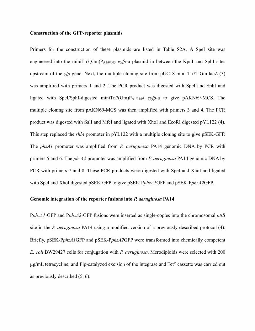
Construction of the GFP-reporter plasmids
Primers for the construction of these plasmids are listed in Table S2A. A SpeI site was
engineered into the miniTn7(Gm)PA1/04/03 eyfp-a plasmid in between the KpnI and SphI sites
upstream of the yfp gene. Next, the multiple cloning site from pUC18-mini Tn7T-Gm-lacZ (3)
was amplified with primers 1 and 2. The PCR product was digested with SpeI and SphI and
ligated with SpeI/SphI-digested miniTn7(Gm)PA1/04/03 eyfp-a to give pAKN69-MCS. The
multiple cloning site from pAKN69-MCS was then amplified with primers 3 and 4. The PCR
product was digested with SalI and MfeI and ligated with XhoI and EcoRI digested pYL122 (4).
This step replaced the rhlA promoter in pYL122 with a multiple cloning site to give pSEK-GFP.
The phzA1 promoter was amplified from P. aeruginosa PA14 genomic DNA by PCR with
primers 5 and 6. The phzA2 promoter was amplified from P. aeruginosa PA14 genomic DNA by
PCR with primers 7 and 8. These PCR products were digested with SpeI and XhoI and ligated
with SpeI and XhoI digested pSEK-GFP to give pSEK-PphzA1GFP and pSEK-PphzA2GFP.
Genomic integration of the reporter fusions into P. aeruginosa PA14
PphzA1-GFP and PphzA2-GFP fusions were inserted as single-copies into the chromosomal attB
site in the P. aeruginosa PA14 using a modified version of a previously described protocol (4).
Briefly, pSEK-PphzA1GFP and pSEK-PphzA2GFP were transformed into chemically competent
E. coli BW29427 cells for conjugation with P. aeruginosa. Merodiploids were selected with 200
µg/mL tetracycline, and Flp-catalyzed excision of the integrase and TetR cassette was carried out
as previously described (5, 6).

Supporting Table S1.
Strains and plasmids used in this work
Strain Comments/Genotype Source or Reference
Pseudomonas aeruginosaPseudomonas aeruginosaPseudomonas aeruginosaPA14 Clinical Isolate, UCBPP-14 (7)
Δphz PA14 with deletions in the phzA1-G1 and the phzA2-G2 operons (1)
Δphz1 PA14 with deletion of the phzA1-G1 operon this study
Δphz2 PA14 with deletions of the phzA2-G2 operon this study
ΔphzHMS PA14 with deletions of the phzM, phzH and phzS genes this study
ΔphzHMSΔphz1 PA14 with deletions of phzM, phzH ,phzS genes and phzA1-G1 operon this study
ΔphzHMSΔphz2 PA14 with deletions of phzM, phzH ,phzS genes and phzA2-G2 operon this study
ΔpqsAC PA14 with deletions of the pqsA-C genes this study
ΔpqsR PA14 with deletion of the pqsR gene Deborah Hogan, Hanover, NH
ΔpqsH PA14 with deletion of the pqsH gene Deborah Hogan Hanover, NH
ΔpqsE PA14 with deletion of the pqsE gene this studyΔpqsL PA14 with deletion of the pqsL gene this studyΔpqsHL PA14 with deletions of the pqsH and pqsL genes this study
ΔpqsACΔphzHMS∆phz1 PA14 with deletions of the pqsA-C genes in the ΔHMS1 background this study
ΔpqsACΔphzHMS∆phz2 PA14 with deletions of the pqsA-C genes in the ΔHMS2 background this study
ΔpqsHΔphzHMS∆phz1 PA14 with deletions of the pqsH gene in the ΔHMS1 background
this study
ΔpqsLΔphzHMS∆phz1 PA14 with deletions of the pqsL gene in the ΔHMS1 background
this study
ΔpqsHLΔphzHMS∆phz1 PA14 with deletions of the pqsH and pqsL genes in the ΔHMS1 background
this study
WT PmcsGFP PA14 with GFP insert with no promoter in the multiple cloning site
this study
WT PphzA1GFP PA14 with PphzA1GFP insert this study
WT PphzA2GFP PA14 with PphzA2GFP insert this study
ΔpqsAC PphzA2GFP ΔpqsAC with PphzA2GFP insert this study
ΔpqsR PphzA2GFP ΔpqsR with PphzA2GFP insert this study
ΔpqsHL PphzA2GFP ΔpqsHL with PphzA2GFP insert this study

Δphz2 pUCP18 Δphz2 with pUCP18 plasmid inserted this study
Δphz2 pUCP18-phz2 Δphz2 with pUCP18 plasmid with phz2 insert this study
Escherichia coliEscherichia coliEscherichia coli
UQ950E. coli DH5α λ(pir) host for cloning; F-Δ(argF-lac)169 Φ80 dlacZ58(ΔM15) glnV44(AS) rfbD1 gyrA96(NalR) recA1 endA1 spoT1 thi-1 hsdR17 deoR λpir+
D. Lies, Caltech
BW29427Donor strain for conjugation: thrB1004 pro thi rpsL hsdS lacZ ΔM15RP4–1360 Δ(araBAD)567 ΔdapA1341::[erm pir(wt)]
W. Metcalf, University of
IllinoisSaccharomyces cerevisiaeSaccharomyces cerevisiaeSaccharomyces cerevisiaeInvSc1 Used for yeast gap repair cloning (2)
Plasmids Description SourcepUCP18 Multi-copy plasmid with ColEI ORI; AmpR; lacZ α gene (8)pUCP18-phz2 pUCP18 plasmid with phz2 operon inserted this studypUC18-mini Tn7T-Gm-lacZ Broad host plasmid with mini Tn7T-GmR-lacZ (3)
miniTn7(Gm)PA1/04/03 eyfp-a Integration vector containing eyfp, GmR (9)
pAKN69-MCS Integration vector containing multiple cloning site (MCS) this studypYL122 rhlAB::gfp transcriptional reporter on mini-CTX, TetR (4)pSEK-GFP GFP reporter plasmid containing MCS this studypSEK-PphzA1GFP GFP reporter plasmid with phzA1 inserted this studypSEK-PphzA2GFP GFP reporter plasmid with phzA2 inserted this study
pMQ30 Yeast-based allelic exchange vector, sacB,a CEN/ARSH, URA3+, GentR (2)
pLD338 phzM deletion fragments cloned into pMQ30 this studypLD294 phzS deletion fragments cloned into pMQ30 this studypLD741 phzH deletion fragments cloned into pMQ30 this studypLD11 phz1 deletion fragments cloned into pSMV10 (1)pLD18 phz2 deletion fragments cloned into pSMV10 (1)pLD710 pqsABC deletion fragments cloned into pMQ30 this studypLD1140 pqsH deletion fragments cloned into pMQ30 this studypLD706 pqsL deletion fragments cloned into pMQ30 this study

Supporting Table S2.
A. Primers for fluorescent reporter constructs
Construction of pAKN69-MCSConstruction of pAKN69-MCS1. speI-MCS-FOR TGCCCGAGGCATAGACTGTA2. sphI-MCS-REV ggatggcatgcCTGTTTCCTGTGTGATAAAGAAAGConstruction of pSEK-GFPConstruction of pSEK-GFP3. salI-MCS-FOR tgaggtcgacTACCGCCACCTAACAATTCG4. mfeI-MCS-REV tcgacaattgTACCGGGCCCAAGCTTCTConstruction of pSEK-PphzA1GFPConstruction of pSEK-PphzA1GFP5. speI-PphzA1-FOR cgccactagtTTCCTGCGTACCGAAAGAAT6. xhoI-PphzA1-REV cgagctcgagCGAGAGGGCTCTCCAGGTATConstruction of pSEK-PphzA2GFPConstruction of pSEK-PphzA2GFP7. speI-PphzA2-FOR cgccactagtGCCTGCTCAACTGAATCGAC8. xhoI-PphzA2-REV cgagctcgagAGTTCGAATCGACTGGCATC
B. Primers for deletion strains
pqsABC deletion strainspqsABC deletion strainspqsABC-US-1 ggaattgtgagcggataacaatttcacacaggaaacagctAGAGGCTCCGATCACCCTATpqsABC-US-2 ctcagcacaccagcacctcGTCTGGCCCCGATAGTGATApqsABC-DS-3 tatcactatcggggccagacGAGGTGCTGGTGTGCTGAGpqsABC-DS-4 ccaggcaaattctgttttatcagaccgcttctgcgttCTGAACCGTAGGTCAGGACCAGpqsL deletion strainspqsL deletion strainspqsL-US-1 ggaattgtgagcggataacaatttcacacaggaaacagctCGCCTGTTCCTCAAGTACGpqsL-US-2 gctgataggaacgctcgcCCTGCTCCACTACCACCACpqsL-DS-3 gtggtggtagtggagcaggGCGAGCGTTCCTATCAGCpqsL-DS-4 ccaggcaaattctgttttatcagaccgcttctgcgttCTCGAACAGGTGTTCCTCAATCpqsH deletion strainspqsH deletion strainspqsH-US-1 ggaattgtgagcggataacaatttcacacaggaaacagctGATATCCACATCCACGGTGTCpqsH-US-2 tattcctcagccagacgctcGATGCCTGCCTTGGTGAATpqsH-DS-3 attcaccaaggcaggcatcCTGAGGAATACCCTCGTTCGpqsH-DS-4 ccaggcaaattctgttttatcagaccgcttctgcgttctgatGGAGATGCTCTGCACCTTGTpqsE deletion strainspqsE deletion strainspqsE-US-1 ggaattgtgagcggataacaatttcacacaggaaacagctGCAATCATGACCTGGTAGGG
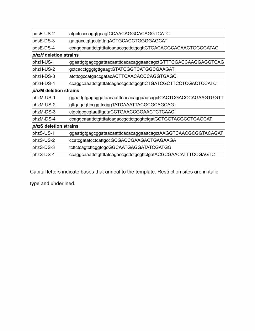
pqsE-US-2 atgctccccaggtgcagtCCAACAGGCACAGGTCATCpqsE-DS-3 gatgacctgtgcctgttggACTGCACCTGGGGAGCATpqsE-DS-4 ccaggcaaattctgttttatcagaccgcttctgcgttCTGACAGGCACAACTGGCGATAGphzH deletion strainsphzH deletion strainsphzH-US-1 ggaattgtgagcggataacaatttcacacaggaaacagctGTTTCGACCAAGGAGGTCAGphzH-US-2 gctcacctgggtgttgaagtGTATCGGTCATGGCGAAGATphzH-DS-3 atcttcgccatgaccgatacACTTCAACACCCAGGTGAGCphzH-DS-4 ccaggcaaattctgttttatcagaccgcttctgcgttCTGATCGCTTCCTCGACTCCATCphzM deletion strainsphzM deletion strainsphzM-US-1 ggaattgtgagcggataacaatttcacacaggaaacagctCACTCGACCCAGAAGTGGTTphzM-US-2 gttgagagttccggttcaggTATCAAATTACGCGCAGCAGphzM-DS-3 ctgctgcgcgtaatttgataCCTGAACCGGAACTCTCAACphzM-DS-4 ccaggcaaattctgttttatcagaccgcttctgcgttctgatGCTGGTACGCCTGAGCATphzS deletion strainsphzS deletion strainsphzS-US-1 ggaattgtgagcggataacaatttcacacaggaaacagctAAGGTCAACGCGGTACAGATphzS-US-2 ccatcgatatcctcattgccGCGACCGAAGACTGAGAAGAphzS-DS-3 tcttctcagtcttcggtcgcGGCAATGAGGATATCGATGGphzS-DS-4 ccaggcaaattctgttttatcagaccgcttctgcgttctgatACGCGAACATTTCCGAGTC
Capital letters indicate bases that anneal to the template. Restriction sites are in italic
type and underlined.

Supporting Figure S1
Supporting Figure 1 (Fig. S1). (A) Phenazine biosynthetic pathway. PCN, phenazine-1-carboxamide; 5-MCA, 5-methyl PCA; 1-OH-PHZ, 1-hydroxyphenazine.(B) Genomic arrangement of the phzA1-G1 (phz1) and phzA2-G2 (phz2) operons and their surrounding regions. phz1 is flanked by phzM and phzS, which encode the enzymes that convert PCA to pyocyanin (PYO). The arrow indicates the location of the las box upstream of phz1. phz2 is flanked by qscR, which encodes a transcription factor that senses acyl-homoserine lactones, and PA14_39870, which encodes a protein of unknown function that contains a HIT domain, characteristic of a superfamily of hydrolases and transferases.

Supporting Figure S2
Supporting Figure 2 (Fig. S2). HPLC quantification of PYO and PCA from colonies grown on 1% tryptone/1% agar plates. Quantification of phenazines extracted from the agar on which biofilms were grown for three or six days. The PYO+PCA (combined concentration of PCA and PYO) from wild type are similar to that of the Δphz1 strain, indicating that the phz2 operon is sufficient for production of wild-type levels of these phenazines. Error bars indicate standard deviation of three independent experiments.

Supporting Figure S3
Supporting Figure 3 (Fig. S3). HPLC traces of phenazines extracted from colony biofilms after six days. Phenazines were extracted from agar and submitted to HPLC analysis for separation and quantification at a wavelength of 366 nm. Wild type produced the phenazines PYO, PCN and PCA. The Δphz and Δphz2 strains did not produce detectable levels of any phenazines (arrow indicates where PCA peak would be expected in the Δphz2 strain). This suggests that phz2 is necessary for phenazine production in biofilms. HPLC conditions and protocol were adapted from (1). Observed retention times for PYO and PCA agree with their results (~10 min and ~20 min respectively).

Supporting Figure S4
Supporting Figure 4 (Fig. S4). Complementation with phz2 restores PCA production and restores wild type colony morphology. The phz2 complementation strain was made by inserting a multi-copy plasmid containing the entire phz2 operon into the Δphz2 mutant. (A) Colony morphology assay for wild type, Δphz, control strain containing empty vector (Δphz2-pUCP18), and complemented ∆phz2. Colonies were grown for three days. Scale bar is 1 cm. (B) Quantification of PCA production from deletion and complemented strains shows that complementation with phz2 restores PCA production. Error bars indicate standard deviation of three independent experiments.

Supporting Figure S5
Supporting Figure 5 (Fig. S5). Effect of temperature on phenazine production. WT, ∆phz, ∆phz1, ∆phz2 colonies were grown for 3 days on 1% tryptone, 1% agar at 25˚C and 37˚C. Phenazines were extracted from the agar into water and analyzed by HPLC. Phenazines could not be detected (n.d.) for ∆phz and ∆phz2 colonies. Error bars indicate standard deviation of biological triplicates.

Supporting Figure S6
Supporting Figure 6 (Fig. S6). HHQ positively regulates the expression of phz2 in planktonic cultures. We assayed for expression of the phz2 operon using a GFP-reporter fusion containing the 500 bp region upstream of phz2. We integrated this reporter into the wild type, ΔpqsAC (no quinolones) and ΔpqsHL (HHQ only) strains and monitored growth and GFP expression in planktonic cultures for 20 hours. (A) Quinolone signaling is necessary for wild-type expression of phz2 as ΔpqsAC-PphzA2GFP and ΔpqsR-PphzA2GFP exhibited a severe reduction in phz2 expression. (B) Quinolone-dependent expression of phz2 is achieved specifically through HHQ. The ΔpqsHL-PphzA2GFP strain produces HHQ (but no PQS or HQNO) and is able to induce expression of phz2 although not to wild type levels. Error bars represent the standard deviation of one experiment performed in biological triplicates. Experiment was repeated three additional times with similar results.

Supporting Figure S7
Supporting Figure 7 (Fig. S7). phz2 is expressed under anaerobic conditions. WT, ∆phz, ∆phz1, ∆phz2 colonies were grown on 1% tryptone, 1% agar supplemented with 40 mM potassium nitrate in an anaerobic glove box filled with 80% N2, 15% CO2 and 5% H2 (Coy) for five days. Phenazines were extracted from the agar into water and analyzed by HPLC. Error bars indicate standard deviation of biological triplicates.
References for Supporting Information
1. Dietrich LE, Price-Whelan A, Petersen A, Whiteley M, & Newman DK (2006) The
phenazine pyocyanin is a terminal signalling factor in the quorum sensing
network of Pseudomonas aeruginosa. Mol Microbiol 61(5):1308-1321.
2. Shanks RM, Caiazza NC, Hinsa SM, Toutain CM, & O'Toole GA (2006)
Saccharomyces cerevisiae-based molecular tool kit for manipulation of genes
from gram-negative bacteria. Appl Environ Microbiol 72(7):5027-5036.
3. Choi KH & Schweizer HP (2006) mini-Tn7 insertion in bacteria with single attTn7
sites: example Pseudomonas aeruginosa. Nat Protoc 1(1):153-161.
4. Lequette Y & Greenberg EP (2005) Timing and localization of rhamnolipid
synthesis gene expression in Pseudomonas aeruginosa biofilms. J Bacteriol
187(1):37-44.
5. Handfield M, et al. (1998) ASD-GFP vectors for in vivo expression technology in
Pseudomonas aeruginosa and other gram-negative bacteria. Biotechniques
24(2):261-264.
6. Hoang TT, Kutchma AJ, Becher A, & Schweizer HP (2000) Integration-proficient
plasmids for Pseudomonas aeruginosa: site-specific integration and use for
engineering of reporter and expression strains. Plasmid 43(1):59-72.
7. Rahme LG, et al. (1995) Common virulence factors for bacterial pathogenicity in
plants and animals. Science 268(5219):1899-1902.
8. Schweizer HP (1991) Escherichia-Pseudomonas shuttle vectors derived from
pUC18/19. Gene 97(1):109-121.

9. Lambertsen L, Sternberg C, & Molin S (2004) Mini-Tn7 transposons for site-
specific tagging of bacteria with fluorescent proteins. Environ Microbiol 6(7):
726-732.


![CHAPTER 9 CHEMISTRY Doctoral Thesescrl.du.ac.in/Doc.Bib/2014/Chemistry.pdf · for the synthesis of novel benzo[a]phenazine annulated heterocycles and their photophysical studies.](https://static.fdocuments.us/doc/165x107/605e5323a79a245d50771859/chapter-9-chemistry-doctoral-for-the-synthesis-of-novel-benzoaphenazine-annulated.jpg)