Receptor-based discovery of a novel AR · PDF filescreening methods have become quite reliable...
21
Receptor-based discovery of a novel AR antagonist 1 Structure-Based Virtual Screening and Identification of a Novel AR Antagonist* Chin-Hee Song 1,# , Su Hui Yang 2,# , Eunsook Park 1 , Suk Hee Cho 2 , Eun-Yeung Gong 1 , Daulat Bikram Khadka 2 , Won-Jea Cho 2,* and Keesook Lee 1,* 1 Hormone Research Center, School of Biological Sciences and Technology, 2 College of Pharmacy and Research Institute of Drug Development, Chonnam National University, Gwangju 500-757, Republic of Korea *Running title: Receptor-based discovery of a novel AR antagonist To whom correspondence should be addressed: Keesook Lee, Ph.D., Hormone Research Center, Chonnam National University, Gwangju, 500-757, Republic of Korea. Phone: +82-62-530-0509; Fax: +82-62-530-0500; E mail: [email protected]. Won-Jea Cho, Ph.D., College of Pharmacy, Chonnam National University, Gwangju 500-757, Republic of Korea. Phone: +82-62-530-2933; Fax: +82-62-530-2911; E mail: [email protected]. # These authors contributed equally to this work. Key words: androgen receptor, prostate cancer, anti-androgen, nicotinamide derivative Background: The androgen receptor (AR) is the primary drug target for prostate cancer treatment. Results: We have identified a novel AR antagonist, the compound 6-(3,4-dihydro-1H- isoquinolin-2-yl)-N-(6-methylpyridin-2- yl)nicotinamide (DIMN) that inhibits the growth of AR-positive prostate cancer cells. Conclusion: DIMN has been identified as a new lead structure targeting the AR. Significance: This novel AR antagonist could be a useful therapeutic agent for prostate cancer treatment. SUMMARY Hormonal therapies, mainly combinations of anti-androgens and androgen deprivation, have been the mainstay treatment for advanced prostate cancer because the androgen-androgen receptor (AR) system plays a pivotal role in the development and progression of prostate cancers. However, the emergence of androgen resistance, largely due to inefficient anti-hormone action, limits the therapeutic usefulness of these therapies. Here, we report that 6-(3,4-dihydro-1H-isoquinolin-2-yl)-N- (6-methylpyridin-2-yl)nicotinamide (DIMN) acts as a novel anti-androgenic compound that may be effective in the treatment of both androgen-dependent and androgen- independent prostate cancers. Through AR structure-based virtual screening using the FlexX docking model, fifty-four compounds were selected and further screened for AR antagonism via cell-based tests. One compound, DIMN, showed an antagonistic effect specific to AR with comparable potency to that of the classical AR antagonists, hydroxyflutamide and bicalutamide. Consistent with their anti- androgenic activity, DIMN inhibited the growth of androgen-dependent LNCaP prostate cancer cells. Interestingly, the compound also suppressed the growth of androgen-independent C4-2 and CWR22rv prostate cancer cells, which express a functional AR, but did not suppress the growth of the AR-negative prostate cancer cells PPC-1, DU145, and R3327-AT3.1. Taken together, the results suggest that the synthetic compound DIMN is a novel anti- androgen and strong candidate for useful therapeutic agent against early stage to advanced prostate cancer. Prostate cancer is the most commonly diagnosed malignancy and the second leading cause of cancer deaths in men in the United States (1,2). It is well established that androgens, such as testosterone and dihydrotestosterone http://www.jbc.org/cgi/doi/10.1074/jbc.M112.379107 The latest version is at JBC Papers in Press. Published on July 13, 2012 as Manuscript M112.379107 Copyright 2012 by The American Society for Biochemistry and Molecular Biology, Inc. by guest on May 23, 2018 http://www.jbc.org/ Downloaded from
-
Upload
truongkhue -
Category
Documents
-
view
216 -
download
2
Transcript of Receptor-based discovery of a novel AR · PDF filescreening methods have become quite reliable...

Receptor-based discovery of a novel AR antagonist
1
Structure-Based Virtual Screening and Identification of a Novel AR Antagonist*
Chin-Hee Song1,#
, Su Hui Yang2,#
, Eunsook Park1, Suk Hee Cho
2, Eun-Yeung Gong
1, Daulat
Bikram Khadka2, Won-Jea Cho
2,* and Keesook Lee
1,*
1 Hormone Research Center, School of Biological Sciences and Technology,
2 College of Pharmacy
and Research Institute of Drug Development, Chonnam National University, Gwangju 500-757,
Republic of Korea
*Running title: Receptor-based discovery of a novel AR antagonist
To whom correspondence should be addressed: Keesook Lee, Ph.D., Hormone Research Center,
Chonnam National University, Gwangju, 500-757, Republic of Korea. Phone: +82-62-530-0509; Fax:
+82-62-530-0500; E mail: [email protected]. Won-Jea Cho, Ph.D., College of Pharmacy,
Chonnam National University, Gwangju 500-757, Republic of Korea. Phone: +82-62-530-2933; Fax:
+82-62-530-2911; E mail: [email protected].
# These authors contributed equally to this work.
Key words: androgen receptor, prostate cancer, anti-androgen, nicotinamide derivative
Background: The androgen receptor (AR) is
the primary drug target for prostate cancer
treatment.
Results: We have identified a novel AR
antagonist, the compound 6-(3,4-dihydro-1H-
isoquinolin-2-yl)-N-(6-methylpyridin-2-
yl)nicotinamide (DIMN) that inhibits the growth
of AR-positive prostate cancer cells.
Conclusion: DIMN has been identified as a new
lead structure targeting the AR.
Significance: This novel AR antagonist could
be a useful therapeutic agent for prostate cancer
treatment.
SUMMARY
Hormonal therapies, mainly
combinations of anti-androgens
and
androgen deprivation, have been the
mainstay treatment for advanced
prostate
cancer because the androgen-androgen
receptor (AR) system plays a pivotal role in
the development and progression of prostate
cancers. However, the emergence of
androgen resistance, largely due to inefficient
anti-hormone action, limits the therapeutic
usefulness of these therapies. Here, we report
that 6-(3,4-dihydro-1H-isoquinolin-2-yl)-N-
(6-methylpyridin-2-yl)nicotinamide (DIMN)
acts as a novel anti-androgenic compound
that may be effective in the treatment of both
androgen-dependent and androgen-
independent prostate cancers. Through AR
structure-based virtual screening using the
FlexX docking model, fifty-four compounds
were selected and further screened for AR
antagonism via cell-based tests. One
compound, DIMN, showed an antagonistic
effect specific to AR with comparable
potency to that of the classical AR
antagonists, hydroxyflutamide and
bicalutamide. Consistent with their anti-
androgenic activity, DIMN inhibited the
growth of androgen-dependent LNCaP
prostate cancer cells. Interestingly, the
compound also suppressed the growth of
androgen-independent C4-2 and CWR22rv
prostate cancer cells, which express a
functional AR, but did not suppress the
growth of the AR-negative prostate cancer
cells PPC-1, DU145, and R3327-AT3.1.
Taken together, the results suggest that the
synthetic compound DIMN is a novel anti-
androgen and strong candidate for useful
therapeutic agent against early stage to
advanced prostate cancer.
Prostate cancer is the most commonly
diagnosed malignancy and the second leading
cause of cancer deaths in men in the United
States (1,2). It is well established that androgens,
such as testosterone and dihydrotestosterone
http://www.jbc.org/cgi/doi/10.1074/jbc.M112.379107The latest version is at JBC Papers in Press. Published on July 13, 2012 as Manuscript M112.379107
Copyright 2012 by The American Society for Biochemistry and Molecular Biology, Inc.
by guest on May 23, 2018
http://ww
w.jbc.org/
Dow
nloaded from
Receptor-based discovery of a novel AR antagonist
2
(DHT), play an essential role in the
tumorigenesis and progression of androgen-
dependent early stage prostate cancer (3).
Testosterone, predominantly produced by
Leydig cells in the testes, is converted to a more
active form, DHT, by the enzyme 5α-reductase
in the prostate. Early onset prostate cancer is
androgen-dependent; therefore, androgen-
ablation therapies that decrease the levels of
circulating androgens through chemical or
surgical castration have been the mainstay of
treatment for androgen-dependent prostate
cancer (ADPC). Unfortunately, androgen-
ablation therapy is only palliative. After 2-3
years of treatment, the cancer cells progress to a
more aggressive form, androgen-independent
prostate cancer (AIPC), or to a hormone
refractory state known as castration-resistant
prostate cancer (CRPC) (4,5).
The androgen receptor (AR), the mediator
of androgen action, is a member of the steroid
hormone receptor superfamily and contains a
DNA-binding domain, and a hormone-binding
domain. This receptor is activated by binding
with androgens in the cytoplasm and is then
translocated into the nucleus, where it regulates
the expression of target genes such as prostate-
specific antigen (PSA) and NK3 transcription
factor locus 1 (NKX3.1) in the human prostate.
AR signaling is known to regulate the
development and progression of normal, benign,
and malignant prostate cells (6-8). The AR is
expressed in the vast majority of both ADPC
and AIPC, and decreasing levels of AR protein
expression reduce both ADPC and AIPC growth,
suggesting a critical role of AR signaling in both
types of prostate cancers (9-11). Castration
resistance is attributed to the high expression of
the AR and AR-regulated genes, indicating that
AR transcriptional activity is reactivated. AR
reactivation in CRPC can be explained by AR
gene amplification (12,13), AR gene mutation
(14-17), activation by alternative androgens
(18,19), or ligand-independent AR activation
through other factors, including the increased
expression of transcriptional co-activators or the
activation of kinases and signal transduction
pathways that modulate AR function (7,20-23).
Due to the critical role of the AR in
prostate cancer, the AR has been the primary
target for the treatment of this disease, and AR
antagonists have been used for the treatment of
prostate cancer and prostatic hyperplasia. There
are two types of AR antagonists, structure-based
steroidal and non-steroidal. Cyproterone acetate,
one of the steroidal AR antagonists, inhibits
androgen action, but it also has weak
progestational and glucocorticoid activities
(24,25). Non-steroidal AR antagonists, such as
flutamide (hydroxyflutamide) and bicalutamide,
have been considered to be less problematic due
to their selective blockade of androgen action
and fewer side effects (26-28). Bicalutamide is
the most widely used of these compounds in the
treatment of prostate cancer because it is
believed to overcome some problems caused by
other anti-androgens (29). However, because the
classical AR antagonists are not effective for the
treatment of advanced prostate cancers, many
efforts have been undertaken to develop newer
and better AR antagonists that work effectively
on either early stage androgen-dependent or
later stage androgen-independent prostate
cancer cells.
Searching for new lead scaffolds that
induce equal or better biological responses than
the current drugs through the same receptor is a
challenging goal in drug design. Because new
therapeutic targets and their 3D structures have
been identified at a dramatic rate, computational
screening methods have become quite reliable
as a source of chemical starting points in the
drug design/discovery process. Compared to the
conventional high-throughput screening (HTS)
method, virtual screening (VS) extends the
screening possibilities to molecules that do not
exist physically in the collection but can be
purchased. In addition, out of the large number
of chemicals screened in silico, only a small
subset of chemicals is tested to quantify
biological activity based on the computational
prediction results. These substantial advantages
have made the VS approach increasingly
valuable for the identification of novel lead
scaffolds that bind to ligand-dependent receptors
(30,31).
Many trials have been conducted to
identify new leads for the development of better
AR antagonists that could provide new
treatments for prostate cancers. Most of the AR
antagonists that have been discovered thus far
have been developed through ligand-based drug
design, which relies on the pharmacophores of
known drugs. Because of the characteristics of
by guest on May 23, 2018
http://ww
w.jbc.org/
Dow
nloaded from

Receptor-based discovery of a novel AR antagonist
3
ligand-based design, most AR antagonists seem
to contain the same basic scaffolds as the known
drugs, such as bicalutamide and flutamide or
other non-steroidal AR agonists (32,33). As an
example, MDV3100, now in phase III clinical
trials, was developed from the non-steroidal AR
agonist RU59063 by modifying the chemical
structures systemically while maintaining the
key chemical scaffold (34).
In this study, keeping in mind the possible
switch from AR antagonism to agonism induced
by similar scaffolds to those of known ligands,
we carried out AR structure-based virtual
screening to discover a novel chemical scaffold
for AR antagonists. We have successfully
identified a new lead structure targeting the AR,
and verified the biological effects of the
compound as AR antagonist that works on early
stage prostate cancer cells as well as on late
stage cells.
EXPERIMENTAL PROCEDURES
Chemistry-Chemicals were purchased
from Aldrich Chemical Co. or Tokyo Chemical
Industry Co. Melting points were determined by
the capillary method on Electrothermal IA9200
digital melting point apparatus. 1H NMR data
were collected on a Varian 300 FT spectrometer
and were calibrated with tetramethylsilane. The
NMR data are displayed as follows: chemical
shifts (δ) are recorded in ppm, coupling
constants (J) in hertz (Hz), integrity in the
number of protons, and multiplicity in s (singlet),
d (doublet), t (triplet), and m (multiplet). Mass
spectra were obtained on a Shimadzu LCMS-
2010EV utilizing the electron-spray ionization
(ESI) method and on a JEOL JNS-DX 303 using
the electron-impact (EI) method. IR spectra
were recorded on a JASCO-FT IR spectrometer
using CHCl3 or KBr pellets. Thin-layer
chromatography (TLC) was carried out using
plates coated with silica gel 60 F254 purchased
from Merck. Column chromatography was
performed with Merck silica gel 60 (70-230
mesh).
Chemical synthesis of DIMN-DIMN was
prepared in three steps starting from 6-chloro-
nicotinic acid (1) (Fig. 1B). Nicotinic acid
chloride, formed by refluxing (1) with thionyl
chloride, was treated with pyridylamine (2) to
obtain the intermediate (3) with 83% yield.
Finally, an SNAr reaction with 1,2,3,4-
tetrahydroisoquinoline (4) in 2-propanol under
reflux conditions provided the desired DIMN
(5) with 72% yield.
Reagents-Cyproterone acetate (CPA),
bicalutamide (BIC), and 2-hydroxyflutamide
(OHF) were purchased from Sigma Chemical
Co., Sequoia Research Products Ltd., and LKT
Laboratories, Inc., respectively. Radiolabeled
dihydrotestosterone ([3H]-DHT) ([1,2,4,5,6,7-
3H(N)]-dihydrotestosterone (5α-androstan-17β-
ol-3-one)) and thymidine ([methyl-3H]-
thymidine, specific activity: 70-90 Ci (2.59-
3.33TBq/mmol) were obtained from Perkin
Elmer Life Science. Antibodies were purchased
from Santa Cruz Biotechnology, Inc. (AR (sc-
815), PSA (sc-7638) and α-tubulin (sc-5286))
and Epitomics, Inc. (GAPDH (cat.#2251-1)).
Plasmids-The mammalian expression
plasmids of the mouse AR (pcDNA3.AR),
mouse GR (pcDNA3.GR), pARE2-TATA-Luc,
PSA-Luc, MMTV-Luc, GFP-AR, pCR3.1-
SRC1, and pSG5-HA-GRIP-1 (SRC-2) have
been previously described (35-40). The
pcDNA3.ERα (human ERα expression plasmid)
and ERE-Luc reporter constructs were kindly
provided by Dr. J.W. Lee (Baylor College of
Medicine) (41). The mammalian expression
plasmids VP-AR1-660, GAL-AR624-919, and
5XGAL4-Luc3 (originally from Dr. Donald
McDonnell) were kindly provided as gifts by Dr.
Elizabeth M. Wilson (University of North
Carolina) (42).
Cell culture-COS-7, 293T, PPC-1, DU145,
HeLa, and MEF (mouse embryonic fibroblast)
cells were maintained in Dulbecco’s minimum
essential medium (Hyclone) supplemented with
10% fetal bovine serum (FBS). LNCaP cells
were purchased from the American Type
Culture Collection (ATCC CRL-1740). C4-2
and CWR22rv cells were kindly provided by Dr.
C. Jung (Chonnam National University Medical
School, Republic of Korea). LNCaP, C4-2, and
CWR22rv cells were maintained in RPMI 1640
(Hyclone) medium supplemented with 5% FBS.
R3327-AT3.1 cells were kindly provided by
Mazence, Inc., (Suwon, Republic of Korea) and
were maintained in RPMI 1640 (Hyclone)
medium supplemented with 10% FBS. All cells
were cultured at 37°C in a 95% humidified
atmosphere containing 5% CO2.
by guest on May 23, 2018
http://ww
w.jbc.org/
Dow
nloaded from
Receptor-based discovery of a novel AR antagonist
4
Transient transfection assay-
Transfections were carried out using the
SuperFect (QIAGEN) transfection reagent for
COS-7 and 293T cells and the Lipo2000
transfection reagent for PPC-1 cells, according
to the instructions of the manufacturer. Cells
plated in 24-well plates were transfected with
the indicated expression plasmids and a reporter
plasmid, along with the β-gal expression
plasmid pCMV-β (CLONTECH). Cells kept in
5% charcoal-stripped FBS (CSS) were treated
with chemicals in the presence or absence of the
ligand for 24 h and processed as described
previously (43). The levels of luciferase activity
were normalized to β-gal expression.
Competitive steroid binding assay-The
whole-cell binding assay was performed as
described previously (44). Briefly, COS-7 cells
were transiently transfected with pcDNA3.AR.
Twenty-four hours prior to the binding reaction,
the cells were placed in phenol red-free DMEM
supplemented with 5% CSS and incubated for 2
h at 37C with 5 nM [3H]-5α-DHT in the
presence and absence of increasing
concentrations of unlabeled chemicals.
Nonspecific binding of [3H]-5α-DHT was
assessed by adding a 100-fold molar excess of
unlabeled 5α-DHT. Dose-response data were
analyzed using the sigmoidal dose-response
function of Prism (GraphPad, San Diego, CA).
Fluorescent microscopy-HeLa cells plated
onto 0.1% gelatin-coated coverslips were
transfected with the GFP-AR expression vector.
After 16 h, transfected cells were fed with fresh
DMEM containing 5% CSS and treated for 1 h
with chemicals. Cells were processed for
fluorescent microscopy using an Olympus 1x70
fluorescent microscope (Tokyo, Japan) as
described previously (44).
Northern blot analysis-Northern blot
analysis was conducted as described previously
(45). Random-primed -32
P-labeled PSA,
NKX3.1, and GAPDH (glyceraldehyde-3-
phosphate dehydrogenase) cDNA probes were
used for hybridization. GAPDH expression was
used as an internal control.
Western blot analysis-Western blot
analysis was conducted as described previously
(46). In brief, the LNCaP and C4-2 cells were
incubated in RPMI supplemented with 5% CSS
for 2 days, and then treated with AR antagonists
in the presence of 10nM or 1nM DHT for 2 days,
respectively (47). The whole cell lysates were
separated by SDS-PAGE, transferred to
nitrocellulose and subjected to Western blot
analysis with anti-AR, anti-PSA, anti-α-tubulin
and anti-GAPDH antibodies. Signals were
detected using an ECL kit (Amersham
Pharmacia).
Thymidine incorporation assay-The
thymidine incorporation assay was conducted as
described previously (37). LNCaP cells were
seeded into 96-well plates at a density of 2x10³
cells per well. The cells were treated with
chemicals in the presence of 1 nM DHT for 72
hours and then treated with 10 µCi/ml of [3H]-
thymidine for another 4 h. Cells were harvested
onto a glass microfiber filter (Whatman, Inc.,
Florham Park, NJ) and processed for the
measurement of incorporated amount of
thymidine into DNA. All values represent the
mean ± SEM of at least three independent
experiments.
Cell viability-The cell growth and
cytotoxicity assays were conducted using the
CellTiter 96® aqueous non-radioactive cell
proliferation assay kit (Promega). Cells were
seeded into 96-well plates at a density of 2x10³
cells per well (LNCaP, C4-2, and MEF) or
5x10² cells per well (PPC-1, DU145, and
R3327-AT3.1). Cells cultured in media
supplemented with 5% CSS (LNCaP), 5% FBS
(C4-2 and MEF) or 10% FBS (PPC-1, DU145,
and R3327-AT3.1) were treated with indicated
chemicals for 4 and 6 days. Combined
MTS/PMS (ratio 20:1 by volume, 20 μl/well)
solution was added to cells in freshly prepared
media. After 2 h, the absorbance at 490 nm was
recorded using an ELISA plate reader. Cell
viability was also assessed by trypan blue dye
exclusion by counting cell numbers as described
previously (48). CWR22rv cells (4x10⁴ cells per
well) were incubated with chemicals for 5 days.
All values represent the mean ± SEM of at least
three independent experiments.
Statistical analysis-To identify significant
differences, the data were analyzed using
GraphPad Prism (GraphPad Software, Inc.).
Single comparisons between 2 experimental
by guest on May 23, 2018
http://ww
w.jbc.org/
Dow
nloaded from

Receptor-based discovery of a novel AR antagonist
5
groups were performed using an unpaired
Student's t-test. Data are shown as the means ±
standard error of the mean (SEM). For all
statistical analyses, P < 0.05 was used as the
criterion to determine statistical significance.
RESULTS
AR structure-based virtual screening for
AR antagonists-A key feature of ligand-
dependent receptors for use in rational drug
design is the ligand-binding domain (LBD),
which we have used in AR structure-based drug
discovery. Crystal structures of the AR LBD
bound to ligands have been determined, but no
structural information about the nature of the
antagonist-induced conformational change
exists because of the lack of defined
crystallization of the wild-type AR-antagonist
complex to date. We therefore selected the
structure of the AR-metribolone (R1881)
complex for screening (code from protein data
bank: 1E3G). Since R1881 is one of the
compounds known to bind most tightly to the
AR, the bound LBD structure would be
expected to offer some information on the native
AR in its strongest binding state.
A chemical library containing over
200,000 drug-like molecules extracted from
commercial and in-house databases was docked
into the AR LBD using the docking algorithm
FlexX. The binding affinity between the
chemicals and the AR LBD was predicted by
five different scoring functions and a consensus
score. To verify the prediction confidence of our
docking system, a root-mean-square deviation
(RMSD) calculation was performed by taking
into account the binding coordinate of R1881 in
the AR LBD. The use of FlexX resulted in the
RMSD value of 0.721, which is almost identical
to the RMSD value of the native AR-R1881
structure, indicating that our docking program is
highly confident. Following the screening, 54
compounds were acquired and numbered in
order of their consensus scores (data not shown).
To test whether the selected 54
compounds exhibit agonistic/antagonistic
activity toward the AR, we performed transient
transfection assays using a reporter system for
the AR, pARE2-TATA-Luc, which contains two
AREs of the androgen target gene C3. The
results revealed that several compounds
significantly inhibited the DHT-induced
transcriptional activation of AR at a
concentration of 1 µM in the presence of 0.3 nM
DHT (Supplementary Fig. 1, upper panel).
Among them, compound #1 (6-(3,4-dihydro-
1H-isoquinolin-2-yl)-N-(6-methylpyridin-2-
yl)nicotinamide), designated as DIMN, showed
the smallest agonistic effect (Supplementary Fig.
1, bottom panel) with a significant antagonistic
effect. Moreover, this compound survived
elimination on the basis of Lipinski’s rule, the
novelty of chemical structure, and other
parameters affecting the successful outcome of
lead optimization (Supplementary File 1).
Therefore, we selected DIMN for further study,
which has a completely novel scaffold
compared to the previously known agonists and
antagonists (Fig. 1A) of the AR, and prepared
the compound in three steps starting from 6-
chloro-nicotinic acid (1) (Fig. 1B). The
chemical 6-chloro-nicotinic acid (1) and 1,2,3,4-
tetrahydroisoquinoline (4) showed no agonistic
or antagonistic effect on AR transactivation
(data not shown).
Identification of DIMN as a new AR
antagonist-We first investigated whether the
inhibitory effect of DIMN was AR-specific by
testing its effect on the transactivation of the
steroid receptor GR and ER. COS-7 cells were
co-transfected with plasmids expressing the GR
and ER along with MMTV-Luc and ERE-Luc,
respectively. DIMN specifically inhibited the
transactivation of the AR, but not of the GR and
ER (Fig. 1C). DIMN was further analyzed for
AR antagonistic activity by transient
transfection assays using several reporter
systems for the AR. The PSA-Luc and MMTV-
Luc plasmids contain the prostate specific
antigen (PSA) and mouse mammary tumor virus
(MMTV) long terminal repeat promoters,
respectively, which are natural AR target
promoters (35,49). DIMN inhibited androgen-
induced AR transactivation at 1 M
concentration in all of the tested reporter
systems (Fig. 1D); this inhibitory potency was
similar to that of hydroxyflutamide (OHF). In
addition, DIMN inhibited AR transactivation in
a dose-dependent manner with the IC50 value at
3 μM (Fig. 1E), which is comparable to the IC50
value of BIC (1.6 μM). However, even at 10 μM
concentration, DIMN showed little agonistic
effect in contrast to BIC, which showed some
agonistic effect (Fig. 1F) as previously reported
by guest on May 23, 2018
http://ww
w.jbc.org/
Dow
nloaded from

Receptor-based discovery of a novel AR antagonist
6
(50). These results suggest that DIMN has a
strong AR-specific antagonistic effect with little
agonistic effect.
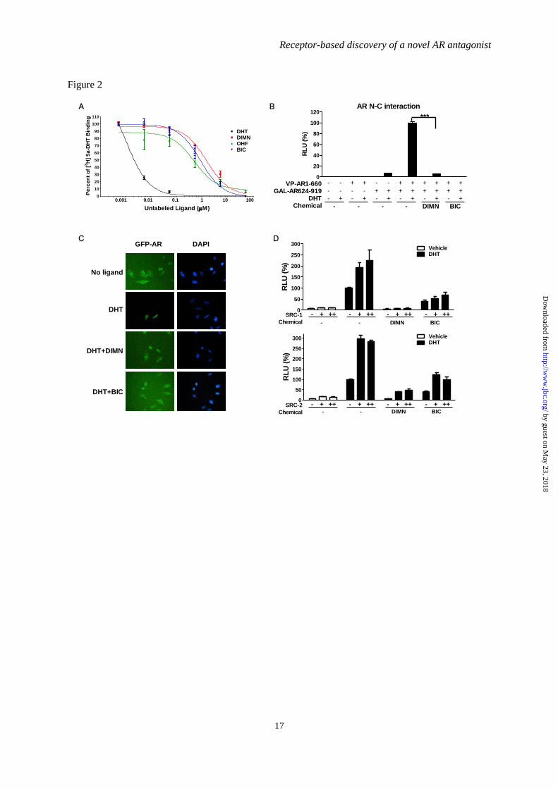
Binding of DIMN to AR- To characterize
the binding of the compound DIMN to the AR,
we performed competitive androgen binding
assay using [3H]-5α-DHT (a labeled androgen)
and AR expressed in COS-7 cells. We used
several known competitors (unlabeled DHT,
BIC, and OHF) of DHT as positive controls
(38,51). The IC50 (the concentration of ligand
that were able to inhibit AR-DHT binding by
50%) of DIMN was 1-2 µM, while the IC50s of
unlabeled DHT, OHF, and BIC were 1-2 nM,
0.4-0.5 µM, and 0.9 µM, respectively (Fig. 2A).
DIMN showed 2- to 4-fold lower AR binding
activity than OHF and 1- to 2-fold lower
binding activity than BIC. The compound
DIMN could not bind to the AR more tightly
than the conventional AR antagonists BIC and
OHF, but could bind nearly as well as those
antagonists, suggesting that it competes with
androgen for AR binding in a similar fashion.
Molecular basis for the anti-androgenic
effect of DIMN-Upon ligand binding, AR
dissociates from heat shock proteins and
translocates into the nucleus, binding to its
target gene promoters as a homodimer formed
by the intermolecular N/C interaction of two AR
molecules. To explore the anti-androgenic
effects of DIMN induced through mechanisms
other than the inhibition of androgen binding to
the AR, we investigated the ability of DIMN to
inhibit any of the AR activation steps, such as
the N/C interaction, nuclear translocation, and
co-activator recruitment.
The effect of DIMN on the AR N/C
interaction was tested using a mammalian two-
hybrid system. PPC-1 cells were transfected
with plasmids encoding the VP-AR1-660
(containing AR residues 1–660) and GAL-
AR624-919 (containing AR residues 624–919)
fusion proteins in conjunction with a luciferase
reporter gene regulated by tandem Gal4-
responsive elements (5XGAL4-Luc3) (42). The
DHT-induced N/C interaction was inhibited
strongly by DIMN. However, its inhibitory
effect on the N/C interaction was weaker than
that of BIC, which almost completely abolished
this interaction (Fig. 2B). The compound
induced no N/C interaction of AR in the absence
of DHT (Fig. 2B) as OHF and BIC (52,53).
These results suggest that DIMN inhibits the
dimerization of the AR.
The effect of DIMN on the dynamics of
the subcellular distribution of the AR was tested
using a GFP-AR fusion protein. When GFP-AR
was overexpressed in HeLa cells in the absence
of androgen, the fusion protein was mostly
distributed in the cytoplasmic compartment, but,
in the presence of 10 nM DHT, GFP-AR was
predominantly localized in the nucleus, as
previously described for the native AR (54) (Fig.
2C). The inhibitory effect of DIMN on the
nuclear import of the AR was assessed by
adding 10 µM DIMN in addition to 10 nM DHT.
The distribution of GFP-AR protein in cells
treated with both DHT and DIMN was dispersed
between the nuclear and the cytoplasmic
compartments, similar to cells challenged with
10 µM BIC (55,56). These results suggest that
DIMN interferes with the nuclear translocation
of the AR.
The elevated expression of SRC-1 and
SRC-2 has been reported to enhance AR activity
in the development of more aggressive prostate
cancers (reviewed in (57)). Therefore, we tested
the effects of DIMN on the action of the AR co-
activators SRC-1 and SRC-2 via transient
transfection assays using a reporter system with
pARE2-TATA-Luc. As shown in Fig. 2D,
overexpression of SRC-1 and SRC-2 enhanced
the transcriptional activity of the AR induced by
10 nM DHT. A 10 M dose of DIMN could
inhibit the SRC-1- and SRC-2-mediated
enhancement of AR transactivation. BIC
exhibited a similar effect on AR transcriptional
activity enhanced by the co-activators SRC-1
and SRC-2, consistent with previous reports
(53).
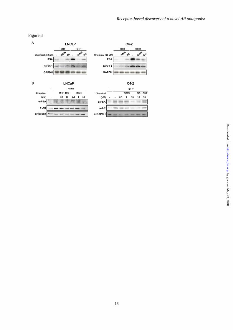
Suppression of androgen-induced AR
target gene expression by DIMN in prostate
cancer cells-Because DIMN has anti-androgenic
activity, we assessed its effect on the expression
of the AR target genes PSA and NKX3.1
(58,59) in androgen-dependent LNCaP and
androgen-independent C4-2 prostate cancer
cells (Fig. 3), both of which express functional
endogenous AR (60). The mRNA levels of PSA,
which is a prostate-specific tumor marker (61),
were reduced by treatment with DIMN in the
absence or presence of DHT in both LNCaP and
by guest on May 23, 2018
http://ww
w.jbc.org/
Dow
nloaded from

Receptor-based discovery of a novel AR antagonist
7
C4-2 cells, and this reduction of PSA mRNA
levels by DIMN was comparable to or greater
than that induced by BIC (Fig. 3A).
Interestingly, BIC activated the expression of
PSA in the absence of DHT in C4-2 cells, as
previously reported in LNCaP (62). The mRNA
levels of NKX3.1 showed similar patterns to
those of PSA upon DIMN treatment in both
LNCaP and C4-2 cells (Fig. 3A).
The change in PSA protein levels was
also assessed by Western blot analysis. The
expression levels of endogenous PSA induced in
the presence of DHT were reduced by treatment
with DIMN in a dose-dependent manner in both
LNCaP and C4-2 cells (Fig. 3B). DIMN reduced
PSA protein levels more effectively than the AR
antagonists BIC and OHF at the same
concentration (10 µM). Interestingly, DIMN
also reduced the protein level of the AR, as
previously reported with BIC and OHF
(53,63,64). Taken together, these results suggest
that the compound DIMN inhibits AR function
in prostate cancer cells and inhibit the
expression of endogenous AR target genes in a
similar fashion to the conventional AR
antagonists, BIC and OHF.
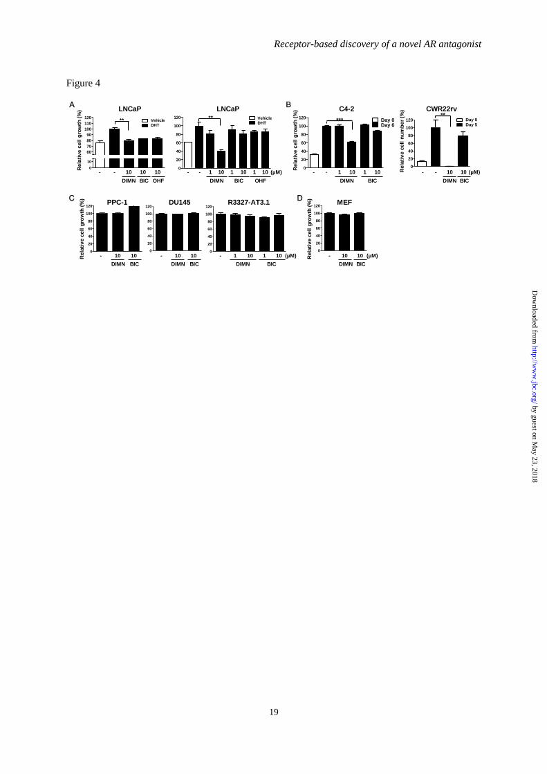
Inhibition of the growth of prostate
cancer cells by DIMN-To assess the effect of
DIMN on the androgen-induced proliferation of
prostate cancer cells, we measured the
proliferation rate of LNCaP cells by the MTS
assay. The growth of LNCaP cells induced by 1
nM DHT was highly inhibited by treatment with
10 µM DIMN as well as with BIC and OHF
treatment (Fig. 4A, left panel). To confirm this
effect of DIMN on the proliferation rate of
LNCaP cells, we also performed thymidine
incorporation assay (Fig. 4A, right panel). DNA
synthesis, which increased in the presence of
DHT, was inhibited approximately 20% and
60% by 1 µM and 10 µM DIMN, respectively.
However, DNA synthesis was inhibited only
10% and 20% by 1 µM and 10 µM BIC,
respectively, and up to 15% by OHF. These
results indicated that DIMN effectively inhibit
the proliferation of LNCaP cells, which
represent the early stage androgen-dependent
state, and that it is much more potent than BIC
or OHF.
To investigate the utility of DIMN as a
new generation of AR antagonists for the
treatment of CRPC, the more aggressive form of
prostate cancer, we next determined the
inhibitory effect of DIMN on the proliferation of
later stage androgen-independent C4-2 and
CWR22rv cells, which grow independently of
androgens while expressing AR protein (47,65).
The effect of DIMN on the proliferation rate of
C4-2 cells was measured by the MTS assay. The
growth of C4-2 cells was effectively inhibited to
approximately 40% by DIMN, which is more
potent than the inhibition by BIC at the same
dose, 10 µM (Fig. 4B). We also analyzed cell
viability by trypan blue staining in CWR22rv
cells, because MTS-based assay resulted in an
underestimation of the anti-proliferative effect
of DIMN in CWR22rv cells due to the
limitation of the method as previously described
(66). The viability of CWR22rv cells was
completely inhibited by DIMN, whereas there
was no significant inhibitory effect on cell
viability induced by BIC (Fig. 4B). However,
the DIMN showed no inhibitory effect on the
cell growth of the AR-negative and androgen-
independent prostate cancer cell lines PPC-1,
DU145, and R3327-AT3.1 (Fig. 4C). Because
DIMN also had an inhibitory effect on the
growth of androgen-independent prostate cancer
cells that express the AR, we investigated the
cytotoxicity of DIMN using mouse embryonic
fibroblast (MEF) cells as normal cells. The
result showed that DIMN exhibited no cytotoxic
effect, similar to BIC (Fig. 4D).
Taken together, these results suggest that
the synthetic compound DIMN has an effective
inhibitory effect on the growth of AR-positive
prostate cancer cells, both androgen-dependent
(LNCaP) and androgen-independent (C4-2 and
CWR22rv), unlike the conventional AR
antagonists BIC and OHF.
Modeling basis for the pure antagonistic
character of DIMN in WT and mutant ARs-To
investigate the basis for the finding that DIMN
acts as a pure AR antagonist in the wild type
(WT) and mutated ARs, we predicted the
binding mode of DIMN through modeling. A
series of AR mutations, including T877A and
W741C, has been identified from tissue
specimens of CRPC patients. In particular, the
T877A mutation has been found in patients who
were treated with flutamide and eventually
became refractory to the treatment (67). The
functional significance of the W741C mutation
was demonstrated by the bicalutamide-
by guest on May 23, 2018
http://ww
w.jbc.org/
Dow
nloaded from

Receptor-based discovery of a novel AR antagonist
8
stimulated tumor growth of a prostate xenograft
model derived from bicalutamide-treated
patients (68).
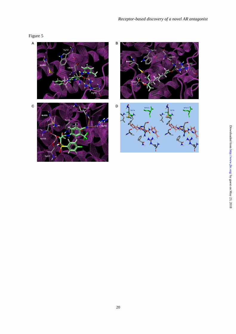
The modeling suggested that DIMN fit
well into the narrow cavity and formed four
possible hydrogen bonds with the backbones of
Gln711 and Arg752 (Fig. 5A, Supplementary
Fig. 2). These interactions are identical to
agonist R1881 with high AR binding affinity
(Fig. 5B), although R1881 forms another
hydrogen bond with Thr877 (69). Despite of the
obvious resemblance of DIMN with agonist, the
activity exhibited is solely antagonism which is
in turn the ability of a compound to displace
H12 from AR LBD. BIC in antagonistic mode,
as predicted from in silico simulations and
docking model (70,71), attains an extended
conformation with the sulfonyl-linked phenyl
ring orienting away from the indole ring of
Trp741 and facing H12. At this structural
orientation, the sulfonyl group of BIC overlaps
with Met895 of H12 and possibly displaces the
helix due to a steric clash (72,73). The steric
overlap of other antagonists such as OHF with
Thr877 is believed to cause the conformational
change for their antagonism in WT AR (74,75).
However, unexpectedly, BIC has a
completely folded conformation with two
phenyl rings stacked in the same site that
agonist R1881 binds in a docking model (Fig.
5C), which is consistent with the recently
illustrated conformation (70). As suggested in
the lowest energy state, BIC may act as an
agonist even in WT AR, to some extent. The
two distinct BIC-AR complexes of either the
former extended conformation or the latter
folded conformation seem to be accessible due
to their comparable binding energy in WT AR
(70), presenting a preference of a designed
antagonist to have a unfolded scaffold for its full
antagonism. Moreover, when W741L/W741C
mutation occurs, BIC acts as an agonist since
the phenyl moiety of BIC shifts up to occupy the
cavity created by the absence of the Trp indole
and thus allows H12 to fold, and this fashion is
similar to the occurrence of point mutation
T877A with the switch to agonist from the
antagonists (70,76,77).
On the other hand, importantly, DIMN
has conserved ligand-receptor hydrogen bonds
for high AR affinity and an extended linear
structure to extrude H12 owing to a steric
conflict with the bulky isoquinoline ring. It also
locates at a distance unaffected by the change in
the size of the active site due to point mutation
Trp741 and Thr877 (Fig. 5D), supporting the
assumption that DIMN works as a pure
antagonist regardless of WT or mutated AR.
DISCUSSION
AR antagonists have proven to be useful
targets for chemotherapeutic agents in the
treatment of prostate cancer. Non-steroidal AR
antagonists, such as bicalutamide, have been
widely used because of their selective blockade
of androgen action and fewer side effects.
However, these antagonists cause some side
effects due to an increased serum testosterone
level by interrupting the negative feedback
regulation in the brain (78) and hormone
resistance in advanced prostate tumors (67).
Therefore, novel potent AR antagonists with
fewer negative effects and working on both
hormone-dependent and -independent tumors
are highly desirable. In an effort to search for
such compounds, we performed AR structure-
based virtual screening as a tool to discover a
new compound, DIMN. DIMN showed strong
AR antagonistic effect and little AR agonistic
effect. Considering that the agonistic properties
of BIC are thought to cause hormone resistance
in advanced prostate tumors (79), our data
showing that DIMN has little agonistic effect
may suggest that this compound could be
developed into more effective AR antagonists
for the treatment of advanced prostate cancer.
The AR signaling pathway is essential for
the growth and progression of both androgen-
dependent and androgen-independent prostate
cancers. Because of this, AR-mediated signaling
and gene expression have been key targets of
advanced prostate cancer therapy through the
utilization of anti-androgens that prevent AR
activation and/or the disruption of endogenous
androgen production (80,81). However, in most
cases, these therapies ultimately fail as a result
of AR reactivation by various factors, including
non-physiological ligands, AR mutants, certain
growth factors, and signaling pathways such as
PI3K/PTEN/AKT and MAPK (82-89). For
example, the PI3K/AKT signaling pathway is
by guest on May 23, 2018
http://ww
w.jbc.org/
Dow
nloaded from
Receptor-based discovery of a novel AR antagonist
9
well-known to regulate the AR with respect to
both expression (90) and transactivation (91-93)
and to mediate the proliferation of both
androgen-dependent LNCaP and androgen-
independent C4-2 and CWR22rv cells (94).
CRPCs exhibit a high level of activation of
PI3K/AKT signaling, resulting in increased
proliferation (95-97). Interestingly, DIMN was
able to inhibit the growth of androgen-
independent prostate cancer cells as well as
androgen-dependent cells (Fig. 4), but only
those that expressed the AR. One explanation
for this AR-dependent inhibition could be a
DIMN-induced decrease in the levels of AR
protein (Fig. 3B), although this decrease did not
seem to fully explain the strong inhibition of
cell growth.
Recent studies have shown that
resveratrol, EPI-001, RD162, and MDV3100
also have the ability to inhibit both androgen-
independent and androgen-dependent
proliferation in prostate cancer cells (87,98,99).
Resveratrol and EPI-001 have been shown to
inhibit the growth of androgen-independent
prostate cancer cells by negatively regulating
PI3K/AKT pathway-activated AR activity.
Similarly, it will be worthwhile to investigate
whether and how DIMN disrupts AR signaling,
which is known to be activated by various
factors in CRPCs, to access the actual pathway
of AR inhibition. Further studies are indeed
required to characterize the mechanisms of AR
antagonist action of DIMN, and such a
characterization will help to develop DIMN as
favorable treatments for AR-related diseases,
including prostate cancer.
DIMN is a potent anti-androgen that
inhibits the proliferation of AR-positive human
prostate cancer cells, both androgen-dependent
and androgen-independent. Furthermore, DIMN
is better than BIC at inhibiting the SRC-1- and
SRC-2-mediated enhancement of AR
transactivation, although it has a little lower AR
binding activity and weaker inhibitory effect on
the N/C interaction (Fig. 2). Such AR
antagonistic activity of DIMN identifies this
class of compounds as potential replacements
for the current therapeutic prostate cancer drug,
BIC. However, further in vivo study is necessary
to confirm that sufficient levels of the
compound DIMN is attained in target tissues for
a sufficient time to alter AR-regulated processes,
which is critical in the final assessment of
chemicals as therapeutic drugs. In addition,
DIMN also would be expected to increase serum
testosterone level due to the loss of negative
feedback regulation at the hypothalamus and
pituitary in common with conventional AR
antagonists (78). Therefore, the in vivo study is
also necessary to evaluate whether DIMN acts
as selective androgen receptor modulators
(SARMs), a class of AR antagonists with
peripheral tissue selectivity (100). We are
currently conducting in vivo experiments.
In summary, we successfully identified a
novel chemical entity, the nicotinamide
compound DIMN, as a non-steroidal AR
antagonist through receptor-based virtual
screening. The potent AR antagonism of this
compound has been confirmed by AR-ligand
binding competition, the blocking of AR
activation steps, the reduced expression of AR
target genes, and the considerably inhibited
proliferation of AR-expressing prostate cancer
cells, either androgen-dependent or androgen-
independent. The remarkable potency of the
action of the compound DIMN on both prostate
cancer cell types as well as their strong anti-
androgenic activity suggests that this compound
could be potent drug candidate for the treatment
of early stage to advanced prostate cancers,
potentially replacing currently established
anticancer medicines such as BIC.
Acknowledgements-We thank Dr. J. W. Lee for the pcDNA3.ERα and ERE-Luc plasmids, Dr. E. M.
Wilson for the VP-AR1-660, GAL-AR624-919, and 5XGAL4-Luc3 plasmids, Dr. C. Jung for the C4-
2 and CWR22rv cells, and Mazence, Inc., for the R3327-AT3.1 cells.
REFERENCES
1. Parkin, D. M., Bray, F., Ferlay, J., and Pisani, P. (2005) CA Cancer J Clin 55, 74-108
by guest on May 23, 2018
http://ww
w.jbc.org/
Dow
nloaded from

Receptor-based discovery of a novel AR antagonist
10
2. Jemal, A., Siegel, R., Ward, E., Hao, Y., Xu, J., Murray, T., and Thun, M. J. (2008) CA
Cancer J Clin 58, 71-96
3. Kokontis, J. M., and Liao, S. (1999) Vitam Horm 55, 219-307
4. Scher, H. I., Steineck, G., and Kelly, W. K. (1995) Urology 46, 142-148
5. Eder, I. E., Haag, P., Bartsch, G., and Klocker, H. (2005) Future Oncol 1, 93-101
6. Heinlein, C. A., and Chang, C. (2004) Endocr Rev 25, 276-308
7. Rahman, M., Miyamoto, H., and Chang, C. (2004) Clin Cancer Res 10, 2208-2219
8. Setlur, S. R., and Rubin, M. A. (2005) Adv Anat Pathol 12, 265-270
9. Rowland, J. G., Robson, J. L., Simon, W. J., Leung, H. Y., and Slabas, A. R. (2007)
Proteomics 7, 47-63
10. Wang, Q., Li, W., Zhang, Y., Yuan, X., Xu, K., Yu, J., Chen, Z., Beroukhim, R., Wang, H.,
Lupien, M., Wu, T., Regan, M. M., Meyer, C. A., Carroll, J. S., Manrai, A. K., Janne, O. A.,
Balk, S. P., Mehra, R., Han, B., Chinnaiyan, A. M., Rubin, M. A., True, L., Fiorentino, M.,
Fiore, C., Loda, M., Kantoff, P. W., Liu, X. S., and Brown, M. (2009) Cell 138, 245-256
11. Chen, C. D., Welsbie, D. S., Tran, C., Baek, S. H., Chen, R., Vessella, R., Rosenfeld, M. G.,
and Sawyers, C. L. (2004) Nat Med 10, 33-39
12. Visakorpi, T., Hyytinen, E., Koivisto, P., Tanner, M., Keinanen, R., Palmberg, C., Palotie, A.,
Tammela, T., Isola, J., and Kallioniemi, O. P. (1995) Nat Genet 9, 401-406
13. Edwards, J., Krishna, N. S., Grigor, K. M., and Bartlett, J. M. (2003) Br J Cancer 89, 552-556
14. Taplin, M. E., Bubley, G. J., Ko, Y. J., Small, E. J., Upton, M., Rajeshkumar, B., and Balk, S.
P. (1999) Cancer Res 59, 2511-2515
15. Veldscholte, J., Ris-Stalpers, C., Kuiper, G. G., Jenster, G., Berrevoets, C., Claassen, E., van
Rooij, H. C., Trapman, J., Brinkmann, A. O., and Mulder, E. (1990) Biochem Biophys Res
Commun 173, 534-540
16. Wilding, G., Chen, M., and Gelmann, E. P. (1989) Prostate 14, 103-115
17. Hara, T., Miyazaki, J., Araki, H., Yamaoka, M., Kanzaki, N., Kusaka, M., and Miyamoto, M.
(2003) Cancer Res 63, 149-153
18. Tan, J., Sharief, Y., Hamil, K. G., Gregory, C. W., Zang, D. Y., Sar, M., Gumerlock, P. H.,
deVere White, R. W., Pretlow, T. G., Harris, S. E., Wilson, E. M., Mohler, J. L., and French,
F. S. (1997) Mol Endocrinol 11, 450-459
19. Culig, Z., Hobisch, A., Cronauer, M. V., Cato, A. C., Hittmair, A., Radmayr, C., Eberle, J.,
Bartsch, G., and Klocker, H. (1993) Mol Endocrinol 7, 1541-1550
20. Weber, M. J., and Gioeli, D. (2004) J Cell Biochem 91, 13-25
21. Gregory, C. W., He, B., Johnson, R. T., Ford, O. H., Mohler, J. L., French, F. S., and Wilson,
E. M. (2001) Cancer Res 61, 4315-4319
22. Culig, Z., Hobisch, A., Cronauer, M. V., Radmayr, C., Trapman, J., Hittmair, A., Bartsch, G.,
and Klocker, H. (1994) Cancer Res 54, 5474-5478
23. Whang, Y. E., Wu, X., Suzuki, H., Reiter, R. E., Tran, C., Vessella, R. L., Said, J. W., Isaacs,
W. B., and Sawyers, C. L. (1998) Proc Natl Acad Sci U S A 95, 5246-5250
24. Poyet, P., and Labrie, F. (1985) Mol Cell Endocrinol 42, 283-288
25. Hamann, L. G., Farmer, L. J., Johnson, M. G., Goldman, M. E., Mais, D. E., Davtian, A.,
Bender, S. L., and Jones, T. K. (1995) Ann N Y Acad Sci 761, 383-387
26. Reid, P., Kantoff, P., and Oh, W. (1999) Invest New Drugs 17, 271-284
27. Anderson, J. (2003) BJU Int 91, 455-461
28. Wirth, M. P., Hakenberg, O. W., and Froehner, M. (2007) Eur Urol 51, 306-313; discussion
314
29. Lefort, M., Diaz Curiel, M., Carrascal, M. T., Mendez-Davila, C., and de la Piedra, C. (2005)
Urol Int 74, 301-307
30. Soderholm, A. A., Viiliainen, J., Lehtovuori, P. T., Eskelinen, H., Roell, D., Baniahmad, A.,
and Nyronen, T. H. (2008) Journal of chemical information and modeling 48, 1882-1890
31. Axerio-Cilies, P., Lack, N. A., Nayana, M. R., Chan, K. H., Yeung, A., Leblanc, E., Guns, E.
S., Rennie, P. S., and Cherkasov, A. (2011) J Med Chem 54, 6197-6205
32. Kinoyama, I., Taniguchi, N., Kawaminami, E., Nozawa, E., Koutoku, H., Furutani, T., Kudoh,
M., and Okada, M. (2005) Chemical & pharmaceutical bulletin 53, 402-409
by guest on May 23, 2018
http://ww
w.jbc.org/
Dow
nloaded from
Receptor-based discovery of a novel AR antagonist
11
33. Kinoyama, I., Taniguchi, N., Toyoshima, A., Nozawa, E., Kamikubo, T., Imamura, M.,
Matsuhisa, A., Samizu, K., Kawanimani, E., Niimi, T., Hamada, N., Koutoku, H., Furutani, T.,
Kudoh, M., Okada, M., Ohta, M., and Tsukamoto, S. (2006) J Med Chem 49, 716-726
34. Jung, M. E., Ouk, S., Yoo, D., Sawyers, C. L., Chen, C., Tran, C., and Wongvipat, J. (2010) J
Med Chem 53, 2779-2796
35. Lee, Y. S., Kim, H. J., Lee, H. J., Lee, J. W., Chun, S. Y., Ko, S. K., and Lee, K. (2002) Biol
Reprod 67, 1580-1587
36. Chattopadhyay, S., Gong, E. Y., Hwang, M., Park, E., Lee, H. J., Hong, C. Y., Choi, H. S.,
Cheong, J. H., Kwon, H. B., and Lee, K. (2006) Mol Endocrinol 20, 984-995
37. Suh, J. H., Shong, M., Choi, H. S., and Lee, K. (2008) Mol Endocrinol 22, 33-46
38. Lee, H. J., Chattopadhyay, S., Gong, E. Y., Ahn, R. S., and Lee, K. (2003) Toxicol Sci 75, 40-
46
39. Onate, S. A., Tsai, S. Y., Tsai, M. J., and O'Malley, B. W. (1995) Science 270, 1354-1357
40. Hong, H., Kohli, K., Garabedian, M. J., and Stallcup, M. R. (1997) Mol Cell Biol 17, 2735-
2744
41. Lee, S. K., Anzick, S. L., Choi, J. E., Bubendorf, L., Guan, X. Y., Jung, Y. K., Kallioniemi, O.
P., Kononen, J., Trent, J. M., Azorsa, D., Jhun, B. H., Cheong, J. H., Lee, Y. C., Meltzer, P. S.,
and Lee, J. W. (1999) J Biol Chem 274, 34283-34293
42. Langley, E., Kemppainen, J. A., and Wilson, E. M. (1998) J Biol Chem 273, 92-101
43. Hong, C. Y., Park, J. H., Seo, K. H., Kim, J. M., Im, S. Y., Lee, J. W., Choi, H. S., and Lee, K.
(2003) Mol Cell Biol 23, 6000-6012
44. Yang, J., Bohl, C. E., Nair, V. A., Mustafa, S. M., Hong, S. S., Miller, D. D., and Dalton, J. T.
(2006) J Pharmacol Exp Ther 317, 402-408
45. Suh, J. H., Gong, E. Y., Hong, C. Y., Park, E., Ahn, R. S., Park, K. S., and Lee, K. (2008) J
Steroid Biochem Mol Biol 112, 117-121
46. Hong, C. Y., Gong, E. Y., Kim, K., Suh, J. H., Ko, H. M., Lee, H. J., Choi, H. S., and Lee, K.
(2005) Mol Endocrinol 19, 2245-2257
47. Ai, J., Wang, Y., Dar, J. A., Liu, J., Liu, L., Nelson, J. B., and Wang, Z. (2009) Mol
Endocrinol 23, 1963-1972
48. Butler, L. M., Agus, D. B., Scher, H. I., Higgins, B., Rose, A., Cordon-Cardo, C., Thaler, H.
T., Rifkind, R. A., Marks, P. A., and Richon, V. M. (2000) Cancer Res 60, 5165-5170
49. Lee, H. J., Hwang, M., Chattopadhyay, S., Choi, H. S., and Lee, K. (2008) Biochem Biophys
Res Commun 367, 481-486
50. Kawata, H., Arai, S., Nakagawa, T., Ishikura, N., Nishimoto, A., Yoshino, H., Shiraishi, T.,
Tachibana, K., Nakamura, R., and Sato, H. (2011) Prostate 71, 1344-1356
51. Gao, W., Kim, J., and Dalton, J. T. (2006) Pharm Res 23, 1641-1658
52. Kemppainen, J. A., Langley, E., Wong, C. I., Bobseine, K., Kelce, W. R., and Wilson, E. M.
(1999) Mol Endocrinol 13, 440-454
53. Masiello, D., Cheng, S., Bubley, G. J., Lu, M. L., and Balk, S. P. (2002) J Biol Chem 277,
26321-26326
54. Jenster, G., van der Korput, H. A., van Vroonhoven, C., van der Kwast, T. H., Trapman, J.,
and Brinkmann, A. O. (1991) Mol Endocrinol 5, 1396-1404
55. Georget, V., Terouanne, B., Nicolas, J. C., and Sultan, C. (2002) Biochemistry 41, 11824-
11831
56. Terouanne, B., Paris, F., Servant, N., Georget, V., and Sultan, C. (2002) Mol Cell Endocrinol
198, 143-147
57. Heinlein, C. A., and Chang, C. (2002) Endocr Rev 23, 175-200
58. Yoon, H. G., and Wong, J. (2006) Mol Endocrinol 20, 1048-1060
59. Nelson, P. S., Clegg, N., Arnold, H., Ferguson, C., Bonham, M., White, J., Hood, L., and Lin,
B. (2002) Proc Natl Acad Sci U S A 99, 11890-11895
60. Thalmann, G. N., Sikes, R. A., Wu, T. T., Degeorges, A., Chang, S. M., Ozen, M., Pathak, S.,
and Chung, L. W. (2000) Prostate 44, 91-103 Jul 101;144(102)
61. Kim, J., and Coetzee, G. A. (2004) J Cell Biochem 93, 233-241
62. Lu, S., Wang, A., and Dong, Z. (2007) Molecular cancer therapeutics 6, 2057-2064
by guest on May 23, 2018
http://ww
w.jbc.org/
Dow
nloaded from

Receptor-based discovery of a novel AR antagonist
12
63. Veldscholte, J., Berrevoets, C. A., Brinkmann, A. O., Grootegoed, J. A., and Mulder, E.
(1992) Biochemistry 31, 2393-2399
64. Furutani, T., Watanabe, T., Tanimoto, K., Hashimoto, T., Koutoku, H., Kudoh, M., Shimizu,
Y., Kato, S., and Shikama, H. (2002) Biochem Biophys Res Commun 294, 779-784
65. Lee, S. J., Zhang, Y., Lee, S. D., Jung, C., Li, X., Kim, H. S., Bae, K. H., Jeng, M. H., Kao, C.,
and Gardner, T. (2004) Mol Ther 10, 1051-1058
66. Wang, P., Henning, S. M., and Heber, D. (2010) PloS one 5, e10202
67. Taplin, M. E., Rajeshkumar, B., Halabi, S., Werner, C. P., Woda, B. A., Picus, J., Stadler, W.,
Hayes, D. F., Kantoff, P. W., Vogelzang, N. J., and Small, E. J. (2003) J Clin Oncol 21, 2673-
2678
68. Yoshida, T., Kinoshita, H., Segawa, T., Nakamura, E., Inoue, T., Shimizu, Y., Kamoto, T.,
and Ogawa, O. (2005) Cancer Res 65, 9611-9616
69. Matias, P. M., Donner, P., Coelho, R., Thomaz, M., Peixoto, C., Macedo, S., Otto, N.,
Joschko, S., Scholz, P., Wegg, A., Basler, S., Schafer, M., Egner, U., and Carrondo, M. A.
(2000) J Biol Chem 275, 26164-26171
70. Osguthorpe, D. J., and Hagler, A. T. (2011) Biochemistry 50, 4105-4113
71. Soderholm, A. A., Viiliainen, J., Lehtovuori, P. T., Eskelinen, H., Roell, D., Baniahmad, A.,
and Nyronen, T. H. (2008) J Chem Inf Model 48, 1882-1890
72. Bohl, C. E., Gao, W. Q., Miller, D. D., Bell, C. E., and Dalton, J. T. (2005) P Natl Acad Sci
USA 102, 6201-6206
73. Bohl, C. E., Miller, D. D., Chen, J. Y., Bell, C. E., and Dalton, J. T. (2005) Journal of
Biological Chemistry 280, 37747-37754
74. Bohl, C. E., Wu, Z. R., Miller, D. D., Bell, C. E., and Dalton, J. T. (2007) Journal of
Biological Chemistry 282, 13648-13655
75. Salvati, M. E., Balog, A., Shan, W., Wei, D. D., Pickering, D., Attar, R. M., Geng, J., Rizzo,
C. A., Gottardis, M. M., Weinmann, R., Krystek, S. R., Sack, J., An, Y., and Kish, K. (2005)
Bioorganic & Medicinal Chemistry Letters 15, 271-276
76. Taplin, M. E., Bubley, G. J., Ko, Y. J., Small, E. J., Upton, M., Rajeshkumar, B., and Balk, S.
P. (1999) Cancer Research 59, 2511-2515
77. Yoshida, T., Kinoshita, H., Segawa, T., Nakamura, E., Inoue, T., Shimizu, Y., Kamoto, T.,
and Ogawa, O. (2005) Cancer Research 65, 9611-9616
78. Miyamoto, H., Messing, E. M., and Chang, C. (2004) Prostate 61, 332-353
79. Culig, Z., Hoffmann, J., Erdel, M., Eder, I. E., Hobisch, A., Hittmair, A., Bartsch, G.,
Utermann, G., Schneider, M. R., Parczyk, K., and Klocker, H. (1999) Br J Cancer 81, 242-
251
80. Gregory, C. W., Hamil, K. G., Kim, D., Hall, S. H., Pretlow, T. G., Mohler, J. L., and French,
F. S. (1998) Cancer Res 58, 5718-5724
81. Craft, N., and Sawyers, C. L. (1998) Cancer Metastasis Rev 17, 421-427
82. Grigoryev, D. N., Long, B. J., Njar, V. C., and Brodie, A. H. (2000) J Steroid Biochem Mol
Biol 75, 1-10
83. Yeh, S., Lin, H. K., Kang, H. Y., Thin, T. H., Lin, M. F., and Chang, C. (1999) Proc Natl
Acad Sci U S A 96, 5458-5463
84. Kang, H. Y., Lin, H. K., Hu, Y. C., Yeh, S., Huang, K. E., and Chang, C. (2001) Proc Natl
Acad Sci U S A 98, 3018-3023
85. Miyamoto, H., Yeh, S., Wilding, G., and Chang, C. (1998) Proc Natl Acad Sci U S A 95,
7379-7384
86. Yeh, S., Miyamoto, H., Shima, H., and Chang, C. (1998) Proc Natl Acad Sci U S A 95, 5527-
5532
87. Wang, Y., Romigh, T., He, X., Orloff, M. S., Silverman, R. H., Heston, W. D., and Eng, C.
(2010) Human molecular genetics 19, 4319-4329
88. Feldman, B. J., and Feldman, D. (2001) Nature reviews. Cancer 1, 34-45
89. Rodriguez-Berriguete, G., Fraile, B., Martinez-Onsurbe, P., Olmedilla, G., Paniagua, R., and
Royuela, M. (2012) Journal of signal transduction 2012, 169170
by guest on May 23, 2018
http://ww
w.jbc.org/
Dow
nloaded from

Receptor-based discovery of a novel AR antagonist
13
90. Manin, M., Baron, S., Goossens, K., Beaudoin, C., Jean, C., Veyssiere, G., Verhoeven, G.,
and Morel, L. (2002) The Biochemical journal 366, 729-736
91. Wen, Y., Hu, M. C., Makino, K., Spohn, B., Bartholomeusz, G., Yan, D. H., and Hung, M. C.
(2000) Cancer Res 60, 6841-6845
92. Lin, H. K., Yeh, S., Kang, H. Y., and Chang, C. (2001) Proc Natl Acad Sci U S A 98, 7200-
7205
93. Sharma, M., Chuang, W. W., and Sun, Z. (2002) J Biol Chem 277, 30935-30941
94. Eng, C. (2003) Human mutation 22, 183-198
95. Mikhailova, M., Wang, Y., Bedolla, R., Lu, X. H., Kreisberg, J. I., and Ghosh, P. M. (2008)
Advances in experimental medicine and biology 617, 397-405
96. Wang, Y., Mikhailova, M., Bose, S., Pan, C. X., deVere White, R. W., and Ghosh, P. M.
(2008) Oncogene 27, 7106-7117
97. Ghosh, P. M., Malik, S. N., Bedolla, R. G., Wang, Y., Mikhailova, M., Prihoda, T. J., Troyer,
D. A., and Kreisberg, J. I. (2005) Endocr Relat Cancer 12, 119-134
98. Andersen, R. J., Mawji, N. R., Wang, J., Wang, G., Haile, S., Myung, J. K., Watt, K., Tam, T.,
Yang, Y. C., Banuelos, C. A., Williams, D. E., McEwan, I. J., Wang, Y., and Sadar, M. D.
(2010) Cancer cell 17, 535-546
99. Tran, C., Ouk, S., Clegg, N. J., Chen, Y., Watson, P. A., Arora, V., Wongvipat, J., Smith-
Jones, P. M., Yoo, D., Kwon, A., Wasielewska, T., Welsbie, D., Chen, C. D., Higano, C. S.,
Beer, T. M., Hung, D. T., Scher, H. I., Jung, M. E., and Sawyers, C. L. (2009) Science 324,
787-790
100. Mohler, M. L., Bohl, C. E., Jones, A., Coss, C. C., Narayanan, R., He, Y., Hwang, D. J.,
Dalton, J. T., and Miller, D. D. (2009) J Med Chem 52, 3597-3617
FOOTNOTES
*This research was supported by Basic Science Research Program through the National Research
Foundation of Korea (NRF) funded by the Ministry of Education, Science and Technology (NRF-
2012R1A2A2A01008388, NRF-2011-0015551). The funders had no role in study design, data
collection and analysis, decision to publish, or preparation of the manuscript.
The abbreviations used are: AR, androgen receptor; DHT, dihydrotestosterone; OHF,
hydroxyflutamide; BIC, bicalutamide; DIMN, 6-(3,4-dihydro-1H-isoquinolin-2-yl)-N-(6-
methylpyridin-2-yl)nicotinamide; ADPC, androgen-dependent prostate cancer; AIPC, androgen-
independent prostate cancer; CRPC, castration-resistant prostate cancer; HTS, high-throughput
screening; VS, virtual screening; RMSD, root-mean-square deviation; LBD, ligand-binding domain;
GFP, green-fluorescent protein
FIGURE LEGENDS
FIGURE 1. Identification of DIMN as a new AR antagonist. (A) The chemical structures of DIMN
and the classical AR antagonists hydroxyflutamide and bicalutamide. (B) The chemical synthesis of
DIMN. (C) The selective AR antagonist activity of DIMN. COS-7 cells were co-transfected with, AR/
pARE2-TATA-Luc, GR/MMTV-Luc or ER/ERE-Luc. The cells were treated with 1 µM DIMN in the
presence of 0.3 nM DHT (dihydrotestosterone), 100 nM DXM (dexamethasone) and 10 nM E2
(estradiol), respectively. (D) The repression of AR transactivation by DIMN. The transcriptional
activity was determined in COS-7 cells transiently co-transfected with pcDNA3.AR and an androgen-
responsive luciferase reporter (pARE2-TATA-Luc, PSA-Luc, or MMTV-Luc). After a 24-h
by guest on May 23, 2018
http://ww
w.jbc.org/
Dow
nloaded from

Receptor-based discovery of a novel AR antagonist
14
transfection, cells were treated with 1 µM OHF, CPA, or DIMN in the presence of 0.3 nM DHT for
an additional 24 h. OHF and CPA were used as positive controls for AR antagonism. The luciferase
data were normalized for β-galactosidase activity and expressed as a percentage of AR activity in the
presence of 0.3 nM DHT only. (E) Dose-dependent AR antagonistic activities of DIMN. After a 24-h
transfection with pcDNA3.AR and pARE2-TATA-Luc, COS-7 cells were treated with various
concentrations of BIC or DIMN in the presence of 10 nM DHT and incubated for 24 h. The IC50
values represent the concentrations of compounds that inhibited 50% of the response induced by 10
nM DHT. (F) The agonistic/antagonistic effect of DIMN on AR transactivation. Cells transfected as
in (E) were treated with 10 M of DIMN in the absence (white bar) or presence (black bar) of 10 nM
DHT. Each value represents the mean ± SEM of at least three independent experiments. RLU, relative
light units.
FIGURE 2. Effect of DIMN on androgen binding to AR and on androgen-induced AR activation
steps. (A) The effect of DIMN on [3H]-5α-DHT binding to the AR. The binding inhibition was
determined in COS-7 cells transiently transfected with pcDNA3.AR. The results are presented as
percent binding relative to [3H]-5α-DHT alone and are shown for unlabeled DHT, BIC, OHF, and
DIMN. (B) The inhibitory effects of DIMN on the AR N/C interaction. A mammalian two-hybrid
assay was performed in PPC-1 cells transfected with 5XGAL4-Luc3, VP-AR1-660, and GAL-
AR624-919. The interaction between the AR N- and C-termini was assessed after the addition of 10
M of the indicated compound in the presence of 10 nM DHT. Error bars indicate the standard
deviation. ***, P < 0.001. (C) The inhibitory effect of DIMN on the nuclear translocation of GFP-AR.
HeLa cells transfected with the GFP-AR expression plasmid were grown on gelatin-coated coverslips.
The subcellular localization of the GFP-AR in living cells was observed and recorded by fluorescence
microscopy after a 1-h treatment with 10 M BIC or DIMN in the presence of 10 nM DHT. (D) The
inhibition of SRC-1- and SRC-2-mediated enhancement of AR transactivation by DIMN. 293T cells
were co-transfected with pcDNA3.AR, pARE2-TATA-Luc, and increasing amounts of pCR3.1 SRC-1
or pSG5-HA-SRC-2 (+, 300 ng; ++, 600 ng). After a 24-h transfection, cells were treated with 10 M
BIC or DIMN in the presence of 10 nM DHT. Each value represents the mean ± SEM of at least three
independent experiments. BIC and OHF were used as positive controls.
FIGURE 3. Effects of DIMN on AR and AR target gene expression in prostate cancer cells. (A) The
suppression of AR target gene expression by DIMN in LNCaP and C4-2 cells. Human prostate cancer
LNCaP and C4-2 cells maintained in media containing 5% CSS were treated with 10 M DIMN or
BIC in the absence or presence of 10 nM DHT for 48 h prior to harvest. Total RNA was analyzed by
Northern blot analysis using cDNA probes for androgen receptor target genes (PSA and NKX3.1).
GAPDH expression was used as an internal control. (B) The suppression of AR and PSA protein
expression by DIMN in LNCaP and C4-2 cells. The protein expression levels of AR and PSA were
determined by Western blot analysis. The cells maintained as in (A) were treated with the indicated
concentrations of OHF, BIC, or DIMN in the presence of DHT (LNCaP, 10nM; C4-2, 1nM) for 48 h
prior to harvest. Specific antibodies against the AR and PSA were used for Western blot analysis.
Tubulin or GAPDH expression was used as an internal control.
FIGURE 4. Inhibition of the proliferation of prostate cancer cells by DIMN. (A) The inhibition of
androgen-dependent LNCaP cell growth by DIMN. The inhibitory effect on DHT-induced cell
proliferation was evaluated by the MTS colorimetric assay. LNCaP cells seeded into 96-well plates
were incubated with 1 nM DHT and 10 M of the indicated compound for 5 days. The values were
compared with those from vehicle-treated cells (left panel). The inhibition of DNA synthesis by
DIMN was measured by a [3H]-thymidine incorporation assay (right panel). (B) The inhibition of
androgen-independent, but AR-positive, C4-2 and CWR22rv cell growth by DIMN. C4-2 cells were
incubated with 1 M or 10 M DIMN or BIC for 6 days, and the inhibitory effects on cell
proliferation were evaluated by the MTS colorimetric assay (Day 6). CWR22rv cells were incubated
with 10 M DIMN or BIC for 5 days, and cell viability was assessed by trypan blue dye exclusion
(Day 5). White bar represents the starting cell number before chemical treatment (Day 0). Error bars
by guest on May 23, 2018
http://ww
w.jbc.org/
Dow
nloaded from

Receptor-based discovery of a novel AR antagonist
15
indicate the standard deviation. **, P < 0.01; ***, P < 0.001. (C) No inhibitory effect of DIMN was
observed on androgen-independent and AR-negative prostate cancer cell growth. For 4 days, PPC-1
and DU145 cells were incubated with 10 M chemicals, and R3327-AT3.1 cells were incubated with
1 M or 10 M chemicals. The inhibitory effects on cell proliferation were evaluated by the MTS
colorimetric assay. (D) The cytotoxic effect of DIMN on MEF cell proliferation. MEF cells were
incubated with 10 M DIMN or BIC in complete medium for 4 days, and the negative effect of
DIMN on cell proliferation was evaluated by the MTS colorimetric assay.
FIGURE 5. Computational analysis of the binding mode of DIMN into the AR. Low-energy binding
conformations of DIMN (A), R1881 (B), BIC (C) bound to WT AR, and DIMN (D) bound to point
mutated AR by virtual ligand docking. Hydrogen bonds are depicted as dotted lines. The green
colored amino acid residues represent W741C and T877A mutation.
by guest on May 23, 2018
http://ww
w.jbc.org/
Dow
nloaded from

Receptor-based discovery of a novel AR antagonist
16
Figure 1
DIM
NOHF
CPA
DIM
NOHF
CPA
DIM
NOHF
CPA
0
20
40
60
80
100
120 VehicleDHT
pARE2-TATA-Luc PSA-Luc MMTV-Luc
RL
U (
%)
A
B
DC
0
20
40
60
80
100
120
DHTDXM
E2DIMN
VehicleLigand
AR GR ER
- - + +
- + - + - + - + - + - +
- - + +- - + +
RL
U (
%)
0
50
100DIMN
BIC
0.01 0.1 1 10 100
Compound concentration (μ M)
RL
U (
% o
f co
ntr
ol)
Agonist assay
0
20
40
60
80
100
DIMN BIC
RL
U (
%)
Antagonist assay
0
20
40
60
80
100
DIMN BIC
VehicleDHT
E F
Compound#1 (DIMN) Hydroxyflutamide Bicalutamide
by guest on May 23, 2018
http://ww
w.jbc.org/
Dow
nloaded from
Receptor-based discovery of a novel AR antagonist
17
Figure 2
- - DIMN BIC
0
50
100
150
200
250
300VehicleDHT
SRC-1
Chemical
- + ++ - + ++ - + ++- + ++
RL
U (
%)
AR N-C interaction
0
20
40
60
80
100
120
VP-AR1-660GAL-AR624-919
DHTChemical
- - + + - - + + + + + +
- - - - + + + + + + + +
- + - + - + - + - + - +
DIMN BIC- - - -
***
RL
U (
%)
A B
C D
0
10
20
30
40
50
60
70
80
90
100
110
DHTDIMNOHFBIC
0.001 0.01 0.1 1 10 100
Unlabeled Ligand (M)
Pe
rce
nt
of
[3H
] 5
a-D
HT
Bin
din
g
- - DIMN BIC
0
50
100
150
200
250
300 VehicleDHT
- + ++ - + ++ - + ++- + ++SRC-2
Chemical
RL
U (
%)
GFP-AR DAPI
No ligand
DHT
DHT+DIMN
DHT+BIC
by guest on May 23, 2018
http://ww
w.jbc.org/
Dow
nloaded from
Receptor-based discovery of a novel AR antagonist
18
Figure 3
B C4-2
α-AR
α-GAPDH
α-PSA
- - 0.1 1 10 10 10
DIMN BIC OHF
+DHT
Chemical
(µM)
-
LNCaP
α-AR
α-tubulin
α-PSA
- - 0.1 1 10
DIMN
10
BIC
10
OHF
+DHT
Chemical
(µM)
-
A LNCaP
- -
+DHT-DHT
PSA
NKX3.1
GAPDH
Chemical (10 µM)
C4-2
- -
+DHT-DHT
PSA
NKX3.1
GAPDH
Chemical (10 µM)
by guest on May 23, 2018
http://ww
w.jbc.org/
Dow
nloaded from
Receptor-based discovery of a novel AR antagonist
19
Figure 4
D
0
20
40
60
80
100
120
C
0
20
40
60
80
100
120
0
20
40
60
80
100
120
0
20
40
60
80
100
120
Rela
tiv
e c
ell g
row
th (
%)
Re
lati
ve c
ell g
row
th (
%)
PPC-1 DU145 R3327-AT3.1 MEF
- 10 10 - 10 10 - 1 10 1 10 - 10 10
DIMN BIC DIMN BIC DIMN BIC DIMN BIC
(µM)(µM)
A
0
20
40
60
80
100
120 ** VehicleDHT
0
10
60
70
80
90
100
110
120VehicleDHT
**
LNCaP LNCaP
Re
lati
ve c
ell g
row
th (
%)
- 10 10
DIMN BIC
- 10 - 1 10 1 10 1 10- (µM)
OHF DIMN BIC OHF
B
0
20
40
60
80
100
120*** Day 0
Day 6
0
20
40
60
80
100
120**
Day 0Day 5
C4-2 CWR22rv
Re
lati
ve c
ell g
row
th (
%)
Re
lati
ve c
ell n
um
ber
(%)
- 10 10- 1 10 1 10 (µM)
DIMN BIC DIMN BIC
- -
by guest on May 23, 2018
http://ww
w.jbc.org/
Dow
nloaded from
Receptor-based discovery of a novel AR antagonist
20
Figure 5
A
C D
B
by guest on May 23, 2018
http://ww
w.jbc.org/
Dow
nloaded from
Bikram Khadka, Won-Jea Cho and Keesook LeeChin-Hee Song, Su Hui Yang, Eunsook Park, Suk Hee Cho, Eun-Yeung Gong, DaulatStructure-Based Virtual Screening and Identification of a Novel AR Antagonist
published online July 13, 2012J. Biol. Chem.
10.1074/jbc.M112.379107Access the most updated version of this article at doi:
Alerts:
When a correction for this article is posted•
When this article is cited•
to choose from all of JBC's e-mail alertsClick here
Supplemental material:
http://www.jbc.org/content/suppl/2012/07/13/M112.379107.DC1
by guest on May 23, 2018
http://ww
w.jbc.org/
Dow
nloaded from


![FOSTER, MICHAEL D., M.S. Computational Study of RTI ......adrenergic receptor ( β2-AR) [6-8], β1-adrenergic receptor ( β1-AR) [9], adenosine A2A receptor [10] and most recently](https://static.fdocuments.us/doc/165x107/607b302caf43ed024c5d3e7b/foster-michael-d-ms-computational-study-of-rti-adrenergic-receptor.jpg)