Real-time measurement of internal stress of dental tissue using holography
8
Real-time measurement of internal stress of dental tissue using holography Dejan Panteli 1 , Larisa Blaži 2 , Svetlana Savi -Ševi 1 , Branka Muri 1 , Darko Vasiljevi 1, 3 , Bratimir Pani 1 , Ilija Beli 1 1 Institute of Physics, Pregrevica 118, 11080 Zemun, Belgrade, Serbia [email protected] 2 University of Novi Sad, Faculty of Medicine, Dental Clinic, Novi Sad, Serbia 3 Faculty of Mechanical Engineering, Kraljice Marije 16, 11120 Belgrade 35, Serbia Abstract: We describe a real-time holographic technique used to observe dental contraction due to photo-polymerization of dental filling during LED lamp illumination. An off-axis setup was used, with wet in-situ processing of the holographic plate, and consequent recording of interference fringes using CCD camera. Finite elements method was used to calculate internal stress of dental tissue, corresponding to experimentally measured deformation. A technique enables selection of preferred illumination method with reduced polymerization contraction. As a consequence, durability of dental filling might be significantly improved. ©2007 Optical Society of America OCIS codes: (090.2880) Holographic interferometry; (170.1850) Dentistry; (120.2650) Fringe analysis; (160.5470) Polymers. ___________________________________________________________________________ References and links 1. K. F. Leinfelder, “Composite resin systems for posterior restorations,” Pract. Periodontics. Aesthet. Dent. Suppl. 1, 23-27 (1993). 2. C. J. Whitters, J. M. Girkin, and J. J. Carey, “Curing of dental composites by use of InGaN light-emitting diodes,” Opt. Lett. 24, 67-68 (1999). 3. R. W. Mills, A. Uhl, G. B. Blackwell and K. D. Jandt, “High power light emitting diode (LED) arrays versus halogen light polymerization of oral biomaterials: Barcol hardness, compressive strength and radiometric properties,” Biomaterials 23, 2955–2963 (2002). 4. G. A. Laughlin, J. L. Williams and J. D. Eick, “The Influence of System Compliance and Sample Geometry on Composite Polymerization Shrinkage Stress,” J. Biomed. Mater. Res. (Appl. Biomater.) 63, 671–678 (2002). 5. A. Versluis, D. Tantbirojn, M. R. Pintado, R. DeLong and W. H. Douglas, “Residual shrinkage stress distributions in molars after composite restoration,” Dent. Mater. 6, 554-564 (2004). 6. J. D. Eick and F. H. Welch, “Polymerization shrinkage of posterior composite resins and its possible influence on postoperative sensitivity,” Quintessence Int. 17, 103-111 (1986). 7. C. L. Davidson, A. J. de Gee and A. J. Feilzer, “The competition between the composite-dentin bond strength and the polymerization contraction stress,” J. Dent. Res. 12, 1396-1399 (1984). 8. E. A. Fogleman, M. T. Kelly and W. T. Grubbs, “Laser interferometric method for measuring linear polymerization shrinkage in light cured dental restoratives,” Dent. Mater. 18, 324 – 330 (2002). 9. H. Lang, R. Rampado, R. Müllejans and W. H. M. Raab, “Determination of the dynamics of restored teeth by 3D electronic speckle pattern interferometry,” Lasers in Surg. Med. 34, 300-309 (2004). 10. T. G. Oberholzer, S. Grobler, C. H. Pameijer and R. J. Rossouw, “A modified dilatometer for determining volumetric polymerization shrinkage of dental materials,” Meas. Sci. Technol. 13, 78–83 (2002). 11. P. Ausiello, A. Apicella, C. L. Davidson and S. Rengo, “3D-finite element analyses of cusp movements in a human upper premolar, restored with adhesive resin-based composites,” J. Biomech. 34, 1269 – 1277 (2001). 12. H. Ensaff, D.M. O’Doherty and P.H. Jacobsen, “The influence of the restoration-tooth interface in light cured composite restorations: a finite element analysis,” Biomaterials 22, 3097-3103 (2001). #80362 - $15.00 USD Received 22 Feb 2007; revised 24 Apr 2007; accepted 25 Apr 2007; published 18 May 2007 (C) 2007 OSA 28 May 2007 / Vol. 15, No. 11 / OPTICS EXPRESS 6823
Transcript of Real-time measurement of internal stress of dental tissue using holography
Real-time measurement of internal stress of dental tissue using holography
Dejan Pantelić1, Larisa Blažić2, Svetlana Savić-Šević1, Branka Murić1, Darko Vasiljević1, 3, Bratimir Panić1, Ilija Belić1
1Institute of Physics, Pregrevica 118, 11080 Zemun, Belgrade, Serbia
2University of Novi Sad, Faculty of Medicine, Dental Clinic, Novi Sad, Serbia
3Faculty of Mechanical Engineering, Kraljice Marije 16, 11120 Belgrade 35, Serbia
Abstract: We describe a real-time holographic technique used to observe dental contraction due to photo-polymerization of dental filling during LED lamp illumination. An off-axis setup was used, with wet in-situ processing of the holographic plate, and consequent recording of interference fringes using CCD camera. Finite elements method was used to calculate internal stress of dental tissue, corresponding to experimentally measured deformation. A technique enables selection of preferred illumination method with reduced polymerization contraction. As a consequence, durability of dental filling might be significantly improved.
©2007 Optical Society of America
OCIS codes: (090.2880) Holographic interferometry; (170.1850) Dentistry; (120.2650) Fringe analysis; (160.5470) Polymers.
___________________________________________________________________________
References and links 1. K. F. Leinfelder, “Composite resin systems for posterior restorations,” Pract. Periodontics. Aesthet. Dent.
Suppl. 1, 23-27 (1993). 2. C. J. Whitters, J. M. Girkin, and J. J. Carey, “Curing of dental composites by use of InGaN light-emitting
diodes,” Opt. Lett. 24, 67-68 (1999). 3. R. W. Mills, A. Uhl, G. B. Blackwell and K. D. Jandt, “High power light emitting diode (LED) arrays versus
halogen light polymerization of oral biomaterials: Barcol hardness, compressive strength and radiometric properties,” Biomaterials 23, 2955–2963 (2002).
4. G. A. Laughlin, J. L. Williams and J. D. Eick, “The Influence of System Compliance and Sample Geometry on Composite Polymerization Shrinkage Stress,” J. Biomed. Mater. Res. (Appl. Biomater.) 63, 671–678 (2002).
5. A. Versluis, D. Tantbirojn, M. R. Pintado, R. DeLong and W. H. Douglas, “Residual shrinkage stress distributions in molars after composite restoration,” Dent. Mater. 6, 554-564 (2004).
6. J. D. Eick and F. H. Welch, “Polymerization shrinkage of posterior composite resins and its possible influence on postoperative sensitivity,” Quintessence Int. 17, 103-111 (1986).
7. C. L. Davidson, A. J. de Gee and A. J. Feilzer, “The competition between the composite-dentin bond strength and the polymerization contraction stress,” J. Dent. Res. 12, 1396-1399 (1984).
8. E. A. Fogleman, M. T. Kelly and W. T. Grubbs, “Laser interferometric method for measuring linear polymerization shrinkage in light cured dental restoratives,” Dent. Mater. 18, 324 – 330 (2002).
9. H. Lang, R. Rampado, R. Müllejans and W. H. M. Raab, “Determination of the dynamics of restored teeth by 3D electronic speckle pattern interferometry,” Lasers in Surg. Med. 34, 300-309 (2004).
10. T. G. Oberholzer, S. Grobler, C. H. Pameijer and R. J. Rossouw, “A modified dilatometer for determining volumetric polymerization shrinkage of dental materials,” Meas. Sci. Technol. 13, 78–83 (2002).
11. P. Ausiello, A. Apicella, C. L. Davidson and S. Rengo, “3D-finite element analyses of cusp movements in a human upper premolar, restored with adhesive resin-based composites,” J. Biomech. 34, 1269 – 1277 (2001).
12. H. Ensaff, D.M. O’Doherty and P.H. Jacobsen, “The influence of the restoration-tooth interface in light cured composite restorations: a finite element analysis,” Biomaterials 22, 3097-3103 (2001).
#80362 - $15.00 USD Received 22 Feb 2007; revised 24 Apr 2007; accepted 25 Apr 2007; published 18 May 2007
(C) 2007 OSA 28 May 2007 / Vol. 15, No. 11 / OPTICS EXPRESS 6823

13. C.-L. Lin, C.-H. Chang, C.-S. Cheng, C.-H. Wang and H.-E. Lee, “Automatic finite element mesh generation for maxillary second premolar,” Comput. Methods Programs Biomed. 59, 187–195 (1999).
14. J. Gao, W. Xu and Z. Ding, “3D finite element mesh generation of complicated tooth model based on CT slices,” Comput. Methods Programs Biomed. 82, 97-105 (2006).
15. P.R. Wedendal and H.I. Bjelkhagen, “Dynamics of Human Teeth in Function by Means of Double Pulsed Holography: an Experimental Investigation,” Appl. Opt. 13, 2481-2485 (1974).
16. D. Pantelić, L. Blažić., S. Savić-Šević and B. Panić, “Holographic detection of a tooth structure deformation after dental filling polymerization,” J. Biomed. Opt., 12, 024026, (2007)
17. N. Ilie, K. Felten, K. Trixner, R. Hickel and K. H. Kunzelmann, “Shrinkage behavior of a resin based composite irradiated with modern curing units,” Dent. Mater. 21, 483-489 (2005).
18. K. S. Vandewalle, J. L. Ferracane, T. J. Hilton, R. L. Erickson and R. L. Sakaguchi, “Effect of energy density on properties and marginal integrity of posterior resin composite restorations,” Dent. Mater. 20, 96-106 (2004).
19. H. Ensaff, D.M. O’Doherty and P.H. Jacobsen, “The influence of the restoration-tooth interface in light cured composite restorations: a finite element analysis,” Biomaterials 22, 3097-3103 (2001).
20. G. Couegnat, S.L. Fok, J.E. Cooper and A.J.E. Qualtrough, “Structural optimization of dental restorations using the principle of adaptive growth,” Dent. Mater. 22, 3–12 (2006).
21. D. Tantbirojn , A. Versluis, M. R. Pintado, R. DeLong, R. Douglas and W.H. Douglas, “Tooth deformation patterns in molars after composite restoration,” Dent. Mater. 20, 535-542 (2004).
___________________________________________________________________________
1. Introduction
Dental tissues are under mechanical load for several reasons – due to mastication, external impact, surgery or other dental procedures. It is of utmost importance in dentistry to estimate internal mechanical pressures in order to find the most appropriate medical practices. The final goal is to heal teeth and prolong their life.
Mostly, mechanical loads are external, resulting from chewing, shock or dental appliances. On the other hand, modern dental techniques using implants and fillings can lead to forces acting from inside of a tooth. In any case, the resulting strain is small (in the micrometer range) but internal stress can be high enough to irreversibly damage dental tissues.
Placement of esthetic fillings is widespread in dentistry and photosensitive resin composites have become the most frequently used materials for this particular purpose [1]. During the clinical procedure caries lesion is removed with a diamond coated drill and a preparation is filled with a dental composite. Adhesion to pre-treated tooth tissues provides the necessary retention of a filling material. A resin composite is illuminated with actinic light (predominantly in the blue part of the spectrum [2]) until it is fully cured. The resulting polymerized layer is hard, closely matching the mechanical properties of enamel and dentin, enabling filling's permanence [1, 3].
It is well known that resin composites contract during setting reaction [4]. Contraction is transferred to dental tissues, inducing high level of mechanical stress [5], which may result in clinical symptoms associated with residual shrinkage stresses (micro-cracking, loss of marginal integrity, post-operative sensitivity and secondary caries ) [6, 7]. As a consequence, lifetime of a dental filling may be considerably reduced.
This particular problem has been the subject of intense research, with an aim to find the most appropriate restorative procedure, which reduces or eliminates the residual contraction stresses. The problem is analyzed theoretically, numerically and experimentally. Various experimental techniques are used to monitor deformation, like classical and speckle interferometry [8, 9] or volumetric contraction measurement using dilatometer [10]. Theoretical and numerical methods are used to calculate stress, based on idealized models and to deduce mechanical stress from deformation measurements [11]. Mechanical models of a tooth are constructed, either based on simplified geometry [12], mechanical slicing [13] or tomographic measurements [14] of real teeth.
Holography [15] is an important method used to measure dental deformation, both in vivo and in vitro. While it gives certain insight in various medical procedures, internal stress
#80362 - $15.00 USD Received 22 Feb 2007; revised 24 Apr 2007; accepted 25 Apr 2007; published 18 May 2007
(C) 2007 OSA 28 May 2007 / Vol. 15, No. 11 / OPTICS EXPRESS 6824

cannot be measured directly and this is what really matters in dentistry. The reason is simple, forces and pressures destroy teeth, not deformation by itself. This was the motivation to develop a method to measure dental deflection continuously and to calculate internal stress, based on experimental data.
In our previous research we observed tooth holographically only in its final state (after illumination) [16]. In this paper real-time off-axis holography was used to continuously measure (in vitro) deformation field of a human third molar during LED lamp illumination. A mechanical tooth model and Finite Elements Method (FEM) was used to calculate internal stress, based on measurements.
This particular technique enables observation of stress build-up, calculation of stress distribution, analysis of all stages of photo-polymerization, with special emphasis on initial stage and post illumination effects. Our research indicates that there is pronounced post-polymerization contraction, which further increases internal stress, even after illumination is over. Method can be used as a tool to analyze various illumination schemes (continuous, two step, ramp, impulse [17]) and various dental composites, with the aim to significantly reduce the internal polymerization stress. The most important consequence is improved durability of dental fillings.
2. Experimental technique
A Schematic view of the real-time holographic setup is shown in Fig. 1. Long coherence length, 532 nm second harmonic Nd-YAG, laser was used. A beam was split and one part was used to illuminate a tooth, while the second was utilized as a reference to irradiate the holographic plate (VRP-M green sensitive emulsion, produced by Slavich, Russia). A plate was placed in a liquid gate, so that the chemical processing could be performed in situ (without moving the holographic plate). Blue LED lamp (central wavelength approximately 480 nm) illuminated the tooth, while CCD camera and computer detected and recorded the interference pattern. It was necessary to use 532 nm interference filter, in order to reject 480 nm LED lamp light, and transmit 532 nm interference pattern. Variable density neutral filter was placed in the object beam, so that the intensity of light directly reflected from the tooth can be equalized with the light intensity reconstructed from hologram. By that means, the contrast of interference fringes is improved.
Specially constructed lamp with 42 LEDs produced 2 cm diameter spot with 80 mW/cm2 irradiance. Construction is hemispherical as previously described in detail [16]. The lamp is placed directly above the tooth so that composite polymerization can be performed without contacting a tooth and with appropriate energy density (which should be 12 - 24 J/cm2
according to [18]). In our case exposure time was 200 s and, consequently, energy density was 16 J/cm2 - quite suitable for successful polymerization.
A tooth was analyzed in vitro, by fixing an extracted tooth with dental gypsum to an appropriate aluminum holder. Before procedure, teeth were kept in a saline solution in order to preserve their mechanical properties. They were prepared for measurement by drilling a class II MOD (Mesio-Ocluso-Distal) cavity (photographs of and intact tooth and the same tooth with MOD cavity are shown in Fig. 2(a) and 2(b)). An adhesive layer was applied and polymerized, according to manufacturer instruction. Finally, cavity was filled with dental composite and placed in the holographic setup.
Upon placing a tooth inside the experimental setup, a holographic plate is inserted in a liquid gate filled with distilled water. Plate is left for 2 minutes, to be soaked with water (letting emulsion to swell). An exposure is made, with the reference to object beam-ratio of 4:1. A plate is developed for 1 minute in fine grain developer (D-76) which is replaced with water, in order to wash the plate. Washing completed, fresh water is poured in the liquid gate again, and stays there throughout the whole real-time recording. This gives the same emulsion thickness before and after processing.
#80362 - $15.00 USD Received 22 Feb 2007; revised 24 Apr 2007; accepted 25 Apr 2007; published 18 May 2007
(C) 2007 OSA 28 May 2007 / Vol. 15, No. 11 / OPTICS EXPRESS 6825
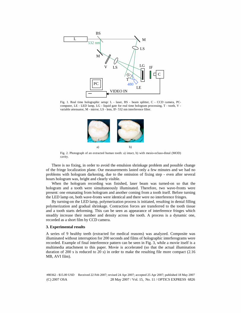
Fig. 1. Real time holographic setup: L - laser, BS - beam splitter, C - CCD camera, PC-computer, LE - LED lamp, LG - liquid gate for real time hologram processing, T - tooth, V - variable attenuator, M - mirror, LS - lens, IF- 532 nm interference filter.
a) b)
Fig. 2. Photograph of an extracted human tooth: a) intact, b) with mesio-ocluso-distal (MOD) cavity.
There is no fixing, in order to avoid the emulsion shrinkage problem and possible change
of the fringe localization plane. Our measurements lasted only a few minutes and we had no problems with hologram darkening, due to the omission of fixing step - even after several hours hologram was, bright and clearly visible.
When the hologram recording was finished, laser beam was turned-on so that the hologram and a tooth were simultaneously illuminated. Therefore, two wave-fronts were present: one emanating from hologram and another coming from a tooth itself. Before turning the LED lamp on, both wave-fronts were identical and there were no interference fringes.
By turning-on the LED lamp, polymerization process is initiated, resulting in dental filling polymerization and gradual shrinkage. Contraction forces are transferred to the tooth tissue and a tooth starts deforming. This can be seen as appearance of interference fringes which steadily increase their number and density across the tooth. A process is a dynamic one, recorded as a short film by CCD camera.
3. Experimental results
A series of 9 healthy teeth (extracted for medical reasons) was analyzed. Composite was illuminated without interruption for 200 seconds and films of holographic interferograms were recorded. Example of final interference pattern can be seen in Fig. 3, while a movie itself is a multimedia attachment to this paper. Movie is accelerated (so that the actual illumination duration of 200 s is reduced to 20 s) in order to make the resulting file more compact (2.16 MB, AVI film).
BS
L
LE
LS
LS
T
LG
M
M
PC
IF V
532 nm
480 nm
VIDEO IN
C
#80362 - $15.00 USD Received 22 Feb 2007; revised 24 Apr 2007; accepted 25 Apr 2007; published 18 May 2007
(C) 2007 OSA 28 May 2007 / Vol. 15, No. 11 / OPTICS EXPRESS 6826

Fig. 3. Interference pattern seen on the tooth surface. This is the final frame from a film (AVI file, 2.16 MB large).
All teeth exhibited almost the same behavior, independent on variation of their shape,
mechanical characteristics, and cavity size. In all instances, there is approximately 15 seconds period without any visible change in interference pattern. The first fringe appears at the tooth cusp and starts traveling down, towards a root. Other fringes follow with increasing speed up to a moment when the process starts slowing down.
We have analyzed teeth interferograms by counting fringes during illumination. Corresponding family of graphs is shown in Fig. 4. From the set of curves it can be seen that the maximum deformation is attained at the end of illumination process (after 200 s). On average, maximum deflection is 11.3 μm.
Fig. 4. Time dependence of fringe-count across the tooth. One fringe is equivalent to approximately 576 nm deformation.
4. Numerical calculation of internal stress
Based on holographic deformation measurements the internal stress of dental tissue is calculated. This is of utmost importance in dentistry, because too high pressure can lead to various adverse clinical effects.
It was impossible to measure individual characteristics of every tooth in our study (external shape, shape of dentino-enamel junction, shape of the pulpal region, cavity shape, mechanical properties – elasticity modulus and Poisson’s ratio of dentin and enamel). Therefore, we made a mechanical model corresponding to average tooth dimensions and mechanical characteristics [19] (see table I). Geometry of the model is shown in Fig. 5(a) and 5(b) so that overall shape and internal structure (dentin and enamel) can be seen. The same model with MOD cavity is shown in Fig. 5(c).
In order to calculate internal stress based on resulting deformation, Finite Element Analysis (FEA) was used. A tooth model with MOD cavity was meshed with tetrahedral elements having 168786 nodes and, thus, prepared for FEA. It was assumed that the contraction forces exert uniform pressure on cavity sides. Dentin and enamel were modeled as linear and isotropic.
0 50 100 150 2000
2
4
6
8
10
12
14LED turn off
Num
ber
of fr
inge
s
Time [s]
#80362 - $15.00 USD Received 22 Feb 2007; revised 24 Apr 2007; accepted 25 Apr 2007; published 18 May 2007
(C) 2007 OSA 28 May 2007 / Vol. 15, No. 11 / OPTICS EXPRESS 6827
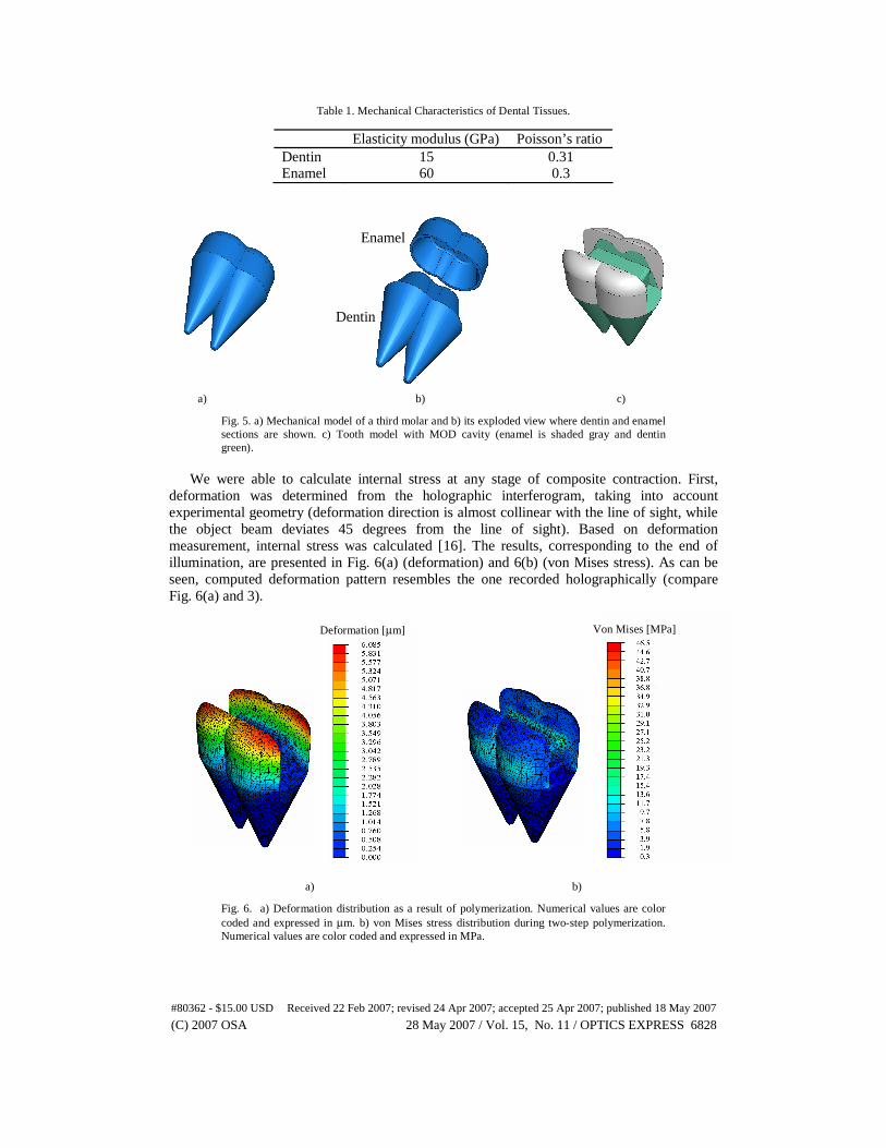
Table 1. Mechanical Characteristics of Dental Tissues.
Elasticity modulus (GPa) Poisson’s ratio Dentin 15 0.31 Enamel 60 0.3
a) b) c)
Fig. 5. a) Mechanical model of a third molar and b) its exploded view where dentin and enamel sections are shown. c) Tooth model with MOD cavity (enamel is shaded gray and dentin green).
We were able to calculate internal stress at any stage of composite contraction. First,
deformation was determined from the holographic interferogram, taking into account experimental geometry (deformation direction is almost collinear with the line of sight, while the object beam deviates 45 degrees from the line of sight). Based on deformation measurement, internal stress was calculated [16]. The results, corresponding to the end of illumination, are presented in Fig. 6(a) (deformation) and 6(b) (von Mises stress). As can be seen, computed deformation pattern resembles the one recorded holographically (compare Fig. 6(a) and 3).
a) b)
Fig. 6. a) Deformation distribution as a result of polymerization. Numerical values are color coded and expressed in μm. b) von Mises stress distribution during two-step polymerization. Numerical values are color coded and expressed in MPa.
Enamel
Dentin
Von Mises [MPa] Deformation [μm]
#80362 - $15.00 USD Received 22 Feb 2007; revised 24 Apr 2007; accepted 25 Apr 2007; published 18 May 2007
(C) 2007 OSA 28 May 2007 / Vol. 15, No. 11 / OPTICS EXPRESS 6828

While the stress is tensor, Von Mises stress is a scalar value calculated from three principal stresses and corresponds to mechanical deformation energy stored in particular area. We have found that its maximum is 46.5 MPa at the step-edge inside the cavity. While von Mises stress gives rough picture of stress distribution inside tooth cavity, maximum principal stress should be used as a measure of tooth susceptibility to mechanical damage (cracking). In our case it was 67.7 MPa.
5. Discussion
One peculiarity of the procedure was that teeth had to be painted with silver paint. The reason is that without paint interference fringes are barely visible or completely nonexistent. In a previous research [16], we attributed the disappearance of fringes to significant changes of internal structure of dentin and enamel, due to polymerization contraction. In short, dental tissues are highly translucent and scattering. Therefore, the beam reflected from a tooth is coming not only from its surface, but also from deeper internal layers. If internal structure changes significantly, reflected wave is considerably altered and interference fringes become extremely dense, beyond resolving power of a detection system.
Another important point is connected with the fact that only one side of a tooth could be observed holographically. Based on the fact that teeth are almost symmetrical with respect to MOD (mesio-ocluso-distal) line and a cavity is drilled symmetrically, we assume that the deformation on both sides is almost the same.
Graphs in Fig. 4 are significantly inclined at the end of illumination, indicating that polymerization is not fully completed. Additionally, there is an indication that dark polymerization reaction takes place even after the illumination is over. In one case, accidentally, we continued recording of interference picture for more that one minute after LEDs were turned off. Later analysis has shown that one additional fringe appeared, meaning that dental composite contracts, even after it is no more illuminated. Medical effects of post-illumination contraction could be important and should be further investigated.
In order to understand the significance of dental stress found in our research, it should be compared to mechanical strength of dental tissues. According to [20] strength of enamel ranges from 10.3 to 384 MPa, while for dentin it is between 98.7 and 297 MPa, depending on whether stress is compressive, tensile or shear. Our results indicate that internal stress is bellow limiting values almost everywhere, except at the edge of a step-like feature of the cavity. This may lead to localized cracking or debonding of the composite filling.
The final remark is that results of finite element calculations should be taken with caution – more as an estimate rather than an exact value. This is due to a lot of approximations connected with variation in teeth geometry, variability in properties of dental tissues, a tooth model and computational method.
6. Conclusions
We present results of holographic real-time measurements of dental structure deformation, due to composite contraction. It is shown that dental tissue gradually deforms, with maximum deflection of around 11.3 μm on top of a tooth cusp. Based on experimental results and tooth model, internal stress was calculated. It is distributed inside the cavity, being concentrated on edges, reaching up to 40.3 MPa von Mises stress (67.7 maximum value of principal stress).
Technique enables testing of various dental procedures and photo-sensitive composites, with the aim to reduce polymerization contraction. Also, there is a strong indication that there is a noticeable post-exposure contraction, which deserves further research. This all could lead to more permanent and durable dental filings.
#80362 - $15.00 USD Received 22 Feb 2007; revised 24 Apr 2007; accepted 25 Apr 2007; published 18 May 2007
(C) 2007 OSA 28 May 2007 / Vol. 15, No. 11 / OPTICS EXPRESS 6829

Acknowledgments
This work is realized by the support of the Serbian Ministry of Science and Environmental Protection, through contract No. 141003. We thank 3M Representative Office in Belgrade, Serbia, for providing the dental adhesive system and the composite.
#80362 - $15.00 USD Received 22 Feb 2007; revised 24 Apr 2007; accepted 25 Apr 2007; published 18 May 2007
(C) 2007 OSA 28 May 2007 / Vol. 15, No. 11 / OPTICS EXPRESS 6830