REAKSI HIPERSENSITIVITAS.ppt
55
Hypersensitivity Reactions
-
Upload
amanda-fairuz -
Category
Documents
-
view
318 -
download
4
Transcript of REAKSI HIPERSENSITIVITAS.ppt

Hypersensitivity Reactions

Hypersensitivity Reactions Outline
• Introduction

• Normal immune reactions do their job without hurting the host.
• Sometimes, immune reactions can be excessive, resulting in disease.
• People who mount normal immune responses are sensitized to that antigen.
• People who have excessive responses are hypersensitive.
Introduction

• Bugs• Environmental antigens• Self antigens
What antigens initiate these reactions?

• The immune response is triggered and maintained inappropriately.
• Hard to eliminate stimulus!• Hard to stop response once it starts!• …so hypersensitivity diseases are often
chronic, debilitating, hard to treat.
What happens in these reactions?

Four types of hypersensitivity reactions
• Type I Hypersensitivity• Type II Hypersensitivity• Type III Hypersensitivity• Type IV Hypersensitivity

Hypersensitivity Reactions Outline
• Introduction• Type I Hypersensitivity

• ALLERGY• “Immediate” hypersensitivity• Antigen (allergen) binds to IgE antibodies
on surface of mast cell• Mast cell releases nasty mediators• End result: vessels dilate, smooth muscle
contracts, inflammation persists
Type I Hypersensitivity

• Allergen is inhaled/eaten/injected
• Allergen stimulates TH2 production
• TH2 cell secretes cytokines:• IL-4 stimulates B cells to make IgE• IL-5 recruits eosinophils• IL-13 stimulates mucous secretion
• Mast cell binds IgE
• Allergen bridges IgE on mast cell
• Mast cell degranulates
Sequence of Events


Mast cells: Normal (left) and degranulated (right)

• Granule contents• histamine • some chemotactic factors
• Membrane phospholipid metabolites• prostaglandin D2
• leukotrienes • Cytokines
• TNF• interleukins• IL-13
What nasty stuff do mast cells release?

Reminder: Where do inflammatory mediators come from?
Complement proteins
Coagulation factors
Factors XII, XI, X, etc.


• Act on blood vessels, smooth muscle, and WBCs.• Immediate response (minutes)
• vasodilation, vascular leakage, smooth muscle spasm • granule contents, prostaglandin, leukotrienes
• Late phase reaction (hours)• inflammation, tissue destruction• cytokines
What do these nasty substances do?

• Local reactions• skin: itching, hives• GI: diarrhea• lung: bronchoconstriction
• Anaphylaxis• itching, hives, erythema• constriction of bronchioles, wheezing• laryngeal edema, hoarseness, obstruction• vomiting, cramps, diarrhea• shock• DEATH
What happens to the patient?


• “Atopy” – predisposition to react to allergens
• Atopic patients: higher IgE levels, more TH2 cells
• Candidate genes:• 5q31 (bunch of cytokine genes here)• 6p (close to HLA complex)
How come only some people have allergies?

Hypersensitivity Reactions Outline
• Introduction• Type I Hypersensitivity• Type II Hypersensitivity

• ANTIBODIES• “Antibody-mediated” hypersensitivity• Antibodies bind to antigens on cell surface• Macrophages eat up cells, complement gets
activated, inflammation comes in• End result: cells die, inflammation harms tissue
Type II Hypersensitivity

Disease Antigen Symptoms
Autoimmune hemolytic anemia
RBC antigens, drugs Hemolysis
Pemphigus vulgaris Proteins between epithelial cells Bullae
Goodpasture syndrome Proteins in glomeruli and alveoli Nephritis, lung hemorrhage
Myasthenia gravis Acetylcholine receptor Muscle weakness
Graves disease TSH receptor Hyperthyroidism
Which diseases involve type II hypersensitivity?

• Antibodies bind to cell-surface antigens• One of three things happens:
• Opsonization and phagocytosis• Inflammation• Cellular dysfunction
Sequence of Events

Opsonization and phagocytosis

Inflammation

Cellular dysfunction
Graves disease Myasthenia gravis

Hypersensitivity Reactions Outline
• Introduction• Type I Hypersensitivity• Type II Hypersensitivity• Type III Hypersensitivity

• IMMUNE COMPLEXES• “Immune complex-mediated” hypersensitivity• Antibodies bind to antigens, forming complexes• Complexes circulate, get stuck in vessels,
stimulate inflammation• End result: bad inflammation, necrotizing
vasculitis
Type III Hypersensitivity

Disease Antigen Symptoms
Systemic lupus erythematosus Nuclear antigens Nephritis, skin lesions,
arthritis…
Post-streptococcal glomerulonephritis
Streptococcal antigen Nephritis
Polyarteritis nodosa Hepatitis B antigen Systemic vasculitis
Serum sickness Foreign proteins Arthritis, vasculitis, nephritis
Arthus reaction Foreign proteins Cutaneous vasculitis
Which diseases involve type III hypersensitivity?

• Systemic immune complex disease• complexes formed in circulation • deposited in several organs• example: serum sickness
• Local immune complex disease• complexes formed at site of antigen injection • precipitated at injection site• example: Arthus reaction
Two Kinds of Type III Hypersensitivity Reactions

• In olden days: used horse serum for immunization• Inject foreign protein (antigen)• Antibodies are made; they form complexes with antigens• Complexes lodge in kidney, joints, small vessels• Inflammation causes fever, joint pain, proteinuria
Serum Sickness

• “Arthus reaction” = localized area of skin necrosis resulting from immune complex vasculitis
• Inject antigen into skin of previously-immunized person• Pre-existing antibodies form complexes with antigen• Complexes precipitate at site of infection• Inflammation causes edema, hemorrhage, ulceration
Arthus Reaction

• Immune complexes activate complement, which:• attracts and activates neutrophils and monocytes• makes vessels leaky
• Neutrophils and monocytes release bad stuff (PG, tissue-dissolving enzymes, etc.)
• Immune complexes also activate clotting, causing microthrombi
• Outcomes: vasculitis, glomerulonephritis, arthritis, other -itises
How do the complexes cause inflammation?


Immune-complex-mediated vasculitis

• C3b: promotes phagocytosis of complexes (and bugs!)• C3a, C5a (anaphylatoxins): increase permeability• C5a: chemotactic for neutrophils, monocytes• C5-9: membrane damage or cytolysis
What complement fractions are important to know?

Hypersensitivity Reactions Outline
• Introduction• Type I Hypersensitivity• Type II Hypersensitivity• Type III Hypersensitivity• Type IV Hypersensitivity

• T CELLS• “T-cell-mediated” hypersensitivity• Activated T cells do one of two things:
• release cytokines that activate macrophages, or• kill cells directly
• This process is normally useful against intracellular organisms (viruses, fungi, parasites)
• Here, it causes bad stuff: inflammation, cell destruction, granuloma formation
Type IV Hypersensitivity
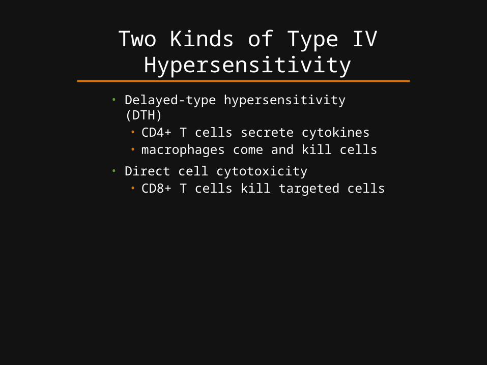
• Delayed-type hypersensitivity (DTH)• CD4+ T cells secrete cytokines• macrophages come and kill cells
• Direct cell cytotoxicity• CD8+ T cells kill targeted cells
Two Kinds of Type IV Hypersensitivity

• Patient exposed to antigen• APC presents antigen to CD4+ T cell• T cells differentiate into effector and memory TH1 cells
• Patient exposed to antigen again• TH1 cells come to site of antigen exposure• Release cytokines that activate macrophages, increase
inflammation
• Results• Macrophages eat antigen (good)• Lots of inflammation and tissue damage (bad)
Delayed-Type Hypersensitivity


Delayed-Type Hypersensitivity (DTH)

Perivascular cuffing by CD4+ cells

• Good example of DTH: positive Mantoux test• Patient previously exposed to TB• Inject (inactive) TB antigen into skin• See reddening, induration. Peaks in 1-3 days
Delayed-Type Hypersensitivity

• Prolonged DTH can lead to granulomatous inflammation• Perivascular CD4+ T cells replaced by macrophages
• Macrophages are activated, look “epithelioid”• Macrophages sometimes fuse into “giant cells”
• Granuloma = collection of epithelioid macrophages
Delayed-Type Hypersensitivity


Granuloma

• Why, yes it does. The same mechanisms underlie both.• Cell-mediated immunity is the major defense we have against
intracellular bugs (like TB and fungi).• Cell-mediated immunity (good) can coexist with DTH (bad)!• Patients with AIDS:
• Lack CD4+ cells• So have poor cell-mediated immune response!• Macrophages sit there unactivated; can’t kill bugs.
DTH sounds a lot like cell-mediated immunity!

• CD8+ T cells recognize antigens on the surface of cells• T cells differentiate into cytotoxic T lymphocytes (CTLs)
which kill antigen-bearing cells• CTLs normally kill viruses and tumor cells• In T-cell mediated cytotoxicity, CTLs kill other things:
• Transplanted organ cells• Pancreatic islet cells (Type I diabetes)
T-Cell Mediated Cytotoxcity

T-Cell-Mediated Cytotoxicity

Type I• Allergy• TH2 cells, IgE on mast cells, nasty mediators
Type II• Antibodies• Opsonization, complement activation, or cell dysfunction
Type III• Immune complexes• Lodge, cause inflammation, tissue injury
Type IV• CD4+ or CD8+ T cells• DTH or T-cell-mediated cytotoxicity
Summary