Readout No.39e 12 Feature Article...Feature Article 86 English Edition No.39 September 2012 Feature...
6
Feature Article 82 English Edition No.39 September 2012 Feature Article Biomarker Discovery using Surface Plasmon Resonance Imaging Elodie LY-MORIN, Sophie BELLON, Géraldine MÉLIZZI, Chiraz FRYDMAN Surface Plasmon Resonance (SPR) is an optical technique used to follow molecular interactions (binding) in real time without labeling. It provides information on kinetic processes (association and dissociation rates), binding affinity, analyte concentration and real time molecule detection. This article describes the principle of SPR imaging for high-throughput measurements. An example of a clinical application for the capture and characterization of a potential biomarker of breast cancer is illustrated. Introduction Surface Plasmon Resonance (SPR)-based biosensors are used to monitor biomolecular interactions in real time. SPR is an optical technique sensitive to variations of the refractive index near the surface of the sensor (or biochip), thus it does not require the use of labels (fluorescent tag for example) to follow the interaction between a molecule immobilized on the biochip (ligand) and one passed over the surface (analyte). Real time measurements give access to the evaluation of the kinetic parameters of the interaction (association rate, dissociation rate and affinity/ avidity). Physical Principle of Surface Plasmon Resonance The notion of evanescent wave is at the core of SPR. Let’s consider the interface between two media 1 and 2 (glass and liquid for example), with refractive indexes of n 1 and n 2 respectively (n 1 > n 2 ). If the incident angle of the light is superior to the critical angle, the incident light is totally reflected and no light is refracted in medium 2 (Snell − Descartes’ law). An evanescent wave is created at the interface. An evanescent wave is non-propagating and its intensity decays exponentially with the distance to the interface. This phenomenon is called total internal reflection. Because the evanescent wave remains confined to the interface of the two media, it is only sensitive to perturbations occurring at the vicinity of the interface. Evanescent waves are thus useful tools to measure in real time modifications of refractive index, layer thickness or molecular adsorption on a surface. If a golden surface is between the glass and liquid, surface plasmons (= collective oscillations of conduction electrons) of the metal can interact strongly with light to produce a surface plasmon wave. In a total internal reflection situation, surface plasmons can couple with the evanescent wave and resonate. The conditions to reach the resonance of surface plasmons depend on the wavelength, polarization and incident angle of the incoming light, but also on the properties of the metallic layer and the medium above the surface. How is the SPR Signal Measured? If the intensity of the reflected light after the biochip in a total internal reflection configuration is measured for different incident angles using a CCD camera for example, it will go through a minimum (Figure 1). This curve is called a plasmon curve. The angle at which the loss of intensity is maximum is called the resonance angle. It corresponds to the resonance of surface plasmons
Transcript of Readout No.39e 12 Feature Article...Feature Article 86 English Edition No.39 September 2012 Feature...

Fe
at
ur
e
Ar
ti
cl
e
82 English Edition No.39 September 2012
Feature Article
Biomarker Discovery using Surface Plasmon Resonance Imaging
Elodie LY-MORIN, Sophie BELLON, Géraldine MÉLIZZI,
Chiraz FRYDMAN
Surface Plasmon Resonance (SPR) is an optical technique used to follow molecular interactions (binding) in real time without labeling. It provides information on kinetic processes (association and dissociation rates), binding affinity, analyte concentration and real time molecule detection. This article describes the principle of SPR imaging for high-throughput measurements. An example of a clinical application for the capture and characterization of a potential biomarker of breast cancer is illustrated.
Introduction
Surface Plasmon Resonance (SPR)-based biosensors are used to monitor biomolecular interactions in real time. SPR is an optical technique sensitive to variations of the refractive index near the surface of the sensor (or biochip), thus it does not require the use of labels (f luorescent tag for example) to follow the interaction between a molecule immobilized on the biochip (ligand) and one passed over the surface (analyte). Real time measurements give access to the evaluat ion of the k inet ic parameters of the interaction (association rate, dissociation rate and affinity/avidity).
Physical Principle of Surface Plasmon Resonance
The notion of evanescent wave is at the core of SPR. Let’s consider the interface between two media 1 and 2 (glass and liquid for example), with refractive indexes of n1 and n2 respectively (n1 > n2). If the incident angle of the light is superior to the critical angle, the incident light is totally ref lected and no light is refracted in medium 2 (Snell − Descartes’ law). An evanescent wave is created at the interface. An evanescent wave is non-propagating and its intensity decays exponentially with the distance to the
interface. This phenomenon is called total internal reflection. Because the evanescent wave remains confined to the interface of the two media, it is only sensitive to perturbations occurring at the vicinity of the interface. Evanescent waves are thus useful tools to measure in real time modifications of refractive index, layer thickness or molecular adsorption on a surface.If a golden surface is between the glass and liquid, surface plasmons (= collect ive oscil lat ions of conduct ion electrons) of the metal can interact strongly with light to produce a surface plasmon wave. In a total internal reflection situation, surface plasmons can couple with the evanescent wave and resonate. The conditions to reach the resonance of surface plasmons depend on the wavelength, polarization and incident angle of the incoming light, but also on the proper ties of the metallic layer and the medium above the surface.
How is the SPR Signal Measured?
If the intensity of the reflected light after the biochip in a total internal ref lection configuration is measured for different incident angles using a CCD camera for example, it will go through a minimum (Figure 1). This curve is called a plasmon curve. The angle at which the loss of intensity is maximum is called the resonance angle. It corresponds to the resonance of surface plasmons

Technical Reports
83English Edition No.39 September 2012
of the gold layer. The position of the resonance angle is characteristic of the metal surface and the medium above it. For example, changes of refractive indexes of the medium above the biochip will induce changes of the position of the resonance angle.SPR can be used to monitor changes of the refractive index at the vicinity of the biochip surface. For example, a binding event (or mass accumulation) between ligands immobilized on the gold surface and analytes flowing on the surface will induce a change of refractive index and a shift of the plasmon curve, hence the position of the resonance angle. This shift can be monitored in real time. Most SPR-based biosensors retrieve the SPR signal by either following- the position of the resonance angle versus time (Δθ)- the variation of ref lectivity at a fixed angle position
versus time (ΔR)
Pr inc ip le of Sur face Plasmon Resonance Imaging
Surface Plasmon Resonance imaging (SPRi) is adding an imaging capacity to SPR. It combines the strength of SPR to monitor label-free biomolecular interactions to the throughput of microarrays. It is thus possible to measure several dozen to several hundred interactions in parallel (multiplexing). Imaging allows monitoring simultaneously the resonance conditions on the whole surface of the biochip. If the biochip contains different immobilized molecules (array of spots), potential interactions on those different molecules can be monitored in parallel. In addition, the real time image of the biochip surface is also retrieved. The “difference image” shows interacting spots when the analyte solution is injected. Our systems are
based on prism-coupled SPR (Figure 2). A collimated beam is sent towards the functionalized gold surface through the prism in order to illuminate the entire surface of the biochip (there is no need to perform a 2D scan). After ref lection on the biochip, the ref lected beam is collected by a CCD array (2D matrix) where each pixel corresponds to a given location on the biochip. The proprietary optical configuration enables a simple yet very precise imaging system without any moving part but a scanning mirror. The rotation of this mirror allows the selection of the working angle relatively to the resonance angle for kinetic measurements. The working angle (position at which kinetic curves will be recorded) is chosen at the highest slope of the plasmon curve. Kinetic measurements consist in monitoring reflectivity variations against time simultaneously on up to several hundred spots (regions of interest).SPRi is used to monitor changes of the refractive index occurring at the surface of the biochip. A binding event (or mass accumulation) will induce a change of refractive
Feature Article
Biomarker Discovery using Surface Plasmon Resonance Imaging
Elodie LY-MORIN, Sophie BELLON, Géraldine MÉLIZZI,
Chiraz FRYDMAN
Surface Plasmon Resonance (SPR) is an optical technique used to follow molecular interactions (binding) in real time without labeling. It provides information on kinetic processes (association and dissociation rates), binding affinity, analyte concentration and real time molecule detection. This article describes the principle of SPR imaging for high-throughput measurements. An example of a clinical application for the capture and characterization of a potential biomarker of breast cancer is illustrated.
Figure 1 The Dip of Reflectivity in the Plasmon Curve. The intensity of the reflected light reaches a minimum at the resonance angle.
Figure 2 SPRi System Configuration

Fe
at
ur
e
Ar
ti
cl
e
84 English Edition No.39 September 2012
Feature Article Biomarker Discovery using Surface Plasmon Resonance Imaging
index and a shift of the position of the resonance angle. SPRi follows the variations of reflectivity occurring at a fixed angle (working angle). The working angle is chosen at the highest slope of the plasmon curves. The principle is described in Figure 3:
- Step A: The ligands are immobilized in an array format on the biochip surface.
- Step B: When the sample solution enters the f low cell (interaction chamber), molecular binding can occur. This induces a shift of the plasmon curves and a variation of reflectivity. The kinetic curves show the variations of ref lectivity versus t ime. The process can also be monitored on the SPRi difference images. White spots correspond to interacting areas of the biochip.
- Step C: When the sample solution leaves the flow cell, the ligand-analyte complex dissociates. This induces a shift of the plasmon curves and a variation of reflectivity. The kinetic curves show the variations of ref lectivity versus time. The process can also be monitored on the SPRi difference images as interacting spots become darker.
- Step D: When the ligand-analyte complex is fully dissociated (using a regeneration solution sometimes), the plasmon curves and the kinetic curves return to the
initial state. The SPRi difference image is black.
Coupling of SPRi to Mass Spectrometry
The coupling of SPRi biosensors and matrix-assisted laser desorption ionization mass spectrometry (MALDI-MS) is an innovative approach for biomarker discovery in biological fluids. It permits analytes captured by SPRi to be ident if ied and character ized by MS f rom their molecular weight and peptide sequence. SPRi-MS opens a new method of detection, quantification and structural characterization of proteins of interest. In the future, it could help better discriminate between sub-species within a family of biomarkers. In this context, the complexity lies in the coupling of both techniques.[1] Most strategies require the elution of the bound analyte and its analysis by ESI- (electrospay ionisation) or MALDI-MS. This procedure has many drawbacks (analysis t ime, no multiplexing capabilities, decreased sensitivity, additional cross-contamination risks, etc…) which delayed the development of SPR-MS in the diagnostic field. The open format of our instruments makes MS coupling easier and faster. The possibility of direct MS analysis on the SPRi sensor was recently shown.[2] The biochip used for SPRi (SPRi-Sl ideT M) is d i rect ly t ransfer red to the MS
Figure 3 Monitoring of Biomolecular Interactions by SPRi
Feature Article Biomarker Discovery using Surface Plasmon Resonance Imaging
Technical Reports
85English Edition No.39 September 2012
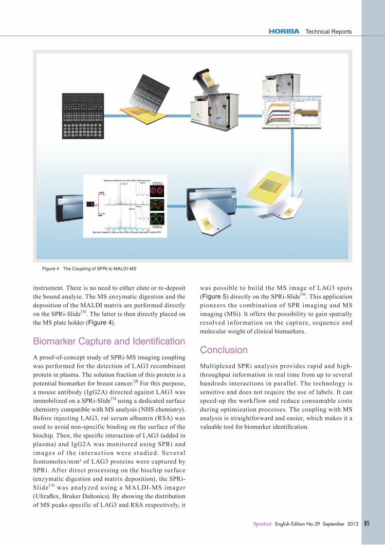
instrument. There is no need to either elute or re-deposit the bound analyte. The MS enzymatic digestion and the deposition of the MALDI matrix are performed directly on the SPRi-SlideTM. The latter is then directly placed on the MS plate holder (Figure 4).
Biomarker Capture and Identification
A proof-of-concept study of SPRi-MS imaging coupling was performed for the detection of LAG3 recombinant protein in plasma. The solution fraction of this protein is a potential biomarker for breast cancer.[3] For this purpose, a mouse antibody (IgG2A) directed against LAG3 was immobilized on a SPRi-SlideTM using a dedicated surface chemistry compatible with MS analysis (NHS chemistry). Before injecting LAG3, rat serum albumin (RSA) was used to avoid non-specific binding on the surface of the biochip. Then, the specific interaction of LAG3 (added in plasma) and IgG2A was monitored using SPRi and images of the inte ract ion were s t ud ied . Severa l femtomoles/mm² of LAG3 proteins were captured by SPRi. After direct processing on the biochip surface (enzymatic digestion and matrix deposition), the SPRi-SlideTM was analyzed using a MALDI-MS imager (Ultraflex, Bruker Daltonics). By showing the distribution of MS peaks specific of LAG3 and RSA respectively, it
was possible to build the MS image of LAG3 spots (Figure 5) directly on the SPRi-SlideTM. This application pioneers the combination of SPR imaging and MS imaging (MSi). It offers the possibility to gain spatially resolved information on the capture, sequence and molecular weight of clinical biomarkers.
Conclusion
Multiplexed SPRi analysis provides rapid and high-throughput information in real time from up to several hundreds interactions in parallel. The technology is sensitive and does not require the use of labels. It can speed-up the workf low and reduce consumable costs during optimization processes. The coupling with MS analysis is straightforward and easier, which makes it a valuable tool for biomarker identification.
Figure 4 The Coupling of SPRi to MALDI-MS

Fe
at
ur
e
Ar
ti
cl
e
86 English Edition No.39 September 2012
Feature Article Biomarker Discovery using Surface Plasmon Resonance Imaging
References
[ 1 ] Boireau and al. (2009) Revisited BIA-MS combination: Entire “on-a-chip” processing leading to the proteins identif ication at low femtomole to sub-femtomole leveks. Biosensors and Bioelectronics 24: 1121-1127
[ 2 ] Bellon and al (2009) Hyphenation of Surface Plasmon Resonance Imaging to Matrix-Assisted Laser Desorption Ionization Mass Spectrometry by On-Chip Mass Spectrometry and Tandem Mass Spectrometry Analysis. Anal. Chem. 81: 7695-7702
[ 3 ] Triebel and al (2006) A soluble lymphocyte activation gene-3 (sLAG-3) protein as a prognostic factor in human breast cancer expressing estrogen or progesterone receptors. Cancer Letters. 235(1):147-53.
Figure 5 On-a-chip detection, identification and imaging of LAG3 protein (potential marker of breast cancer) at 10 nM in human plasma

Feature Article Biomarker Discovery using Surface Plasmon Resonance Imaging
Technical Reports
87English Edition No.39 September 2012
Elodie LY-MORINBio Sales EngineerHORIBA Jobin Yvon SASPh. D
Sophie BELLONBio Application Lab ManagerHORIBA Jobin Yvon SASPh. D
Géraldine MÉLIZZIR&D Projet ManagerHORIBA Jobin Yvon SAS
Chiraz FRYDMANProduct ManagerHORIBA Jobin Yvon SASPh. D