Reading Ch 10 Waxman Dental Neuroanatomy Lecture Ch 10 Waxman Dental Neuroanatomy Lecture Suzanne...
43
Cerebral Cortex Structure, Function, Dysfunction Reading Ch 10 Waxman Dental Neuroanatomy Lecture Suzanne Stensaas, Ph.D. March 15, 2011
Transcript of Reading Ch 10 Waxman Dental Neuroanatomy Lecture Ch 10 Waxman Dental Neuroanatomy Lecture Suzanne...

Cerebral CortexStructure, Function, Dysfunction
Reading Ch 10 WaxmanDental Neuroanatomy Lecture
Suzanne Stensaas, Ph.D.March 15, 2011

Anatomy Review
• Lobes and layers• Brodmann’s areas• Vascular Supply• Major Neurological Findings
– Frontal, Parietal, Temporal, Occipital, Limbic• Quiz Questions?

Types of Cortex
• Sensory• Motor• Unimodal association• Multimodal association necessary for
language, reason, plan, imagine, create

Structure of neocortex (6 layers)

The general pattern of primary, association and mulimodalassociation cortex (Mesulam)

Brodmann, Lateral Left Hemisphere

MCA left hemispherefrom D.Haines


ACA and PCA -Haines

Issues of Functional Localization
• Earliest studies -Signs, symptoms and note location • Electrical discharge (epilepsy) suggested function• Ablation - deficit suggest function• Reappearance of infant functions suggest loss of inhibition
(disinhibition), i.e. grasp, suck, Babinski • Variabilities in case reports• Linked networks of afferent and efferent neurons in several
regions working to accomplish a task• Functional imaging does not always equate with abnormal
function associated with location of lesion• fMRI activation of several cortical regions• Same sign from lesions in different areas – i.e.paraphasias• Notion of the right hemisphere as "emotional" in contrast to the
left one as "logical" has no basis in fact.

Limbic System (not a true lobe)involves with cingulate gyrus and the
• Hippocampus- short term memory• Amygdala- fear, agression, mating• Fornix pathway to hypothalamus• Hypothalamus- ANS control• Prefrontal Cortex- appropriate behavior

Schematic Diagram of principal limbic areas
From College of DuPage Biology 1152 Syllabus

Amygdala and relationship to ventricle and hippocampus

Classic Hippocampal Circuit

Hippocampal Formation & Amygdala

Hippocampus

Lateral view gross brain. Left hemisphere Frontal Lobe

Frontal Lobe Motor areas
• Contralateral weakness or paralysis (area 4) body and CN’s
• Premotor planning of action (area 6)• Frontal eye fields for moving eyes to
opposite side (area 8)• Speech production (Broca’s area 44, 45)• e.g. Epileptic discharge

Frontal Lobe prefrontal association cortex
• Bilateral prefrontal damage– distractible, apathetic– lack foresight, abstract reasoning, initiative– stubborn, – perseverate, – lack ambition, responsibility, judgment or
social graces

Apraxia (Error in execution of learned movements without coexisting weakness)
• Damage to dominant parietal, premotor, and supplementary motor areas
• Dominant hemisphere association areas• Parietal - integrates motor sequences
with vision and somatic sensory info• Frontal lobe - execution of act

Language- Frontal-Parietal-Temporal Areas

Aphasias-Motor• Full Broca’s involves operculum, insula and
subjacent white matter with contralateral hemiparesis of face, arm
• Telegraphic speech• Agrammatism - syntax more affected than semantics• Usually agraphia too• Transcortical - interruption of inferred linkage paths
inward to Broca’s area

Aphasias-Sensory
• Wernicke’s• Dominant (left usually) hemisphere• Fluent, paraphasias, poor comprehension, • Naming, repetition, reading and writing
impaired• Less aware and less frustrated than motor
aphasias

Right hemisphere and aphasia
• Emotional tone modulation• Propositional prosody• Body language gestures

Anomia
• Anomia requires special testing• Seen in all language areas and outside
language cortex• Most severe in dominant temporal
lesions

Agnosia-impaired perception or recognition with OK vision, hearing, sensation , attention, intelligence• Visual: colors, faces, letters• Auditory: tunes, spoken words, pure word deafness• Somatosensory - stereognosis, graphesthesia• May not have other signs: aphasia, apraxia• Atrophy or metastatic disease• Disconnections of specific sensory association areas• Corpus callosum, deep white matter near main sensory areas

Global aphasia
• Dominant hemisphere• Frontal• Temporal• Parietal• Head of caudate associated with
language disorders• Internal carotid or proximal MCA,
hemorrhage, or large tumor

Temporal Lobe
• Association auditory cortex• Speech comprehension• Important in naming• Memory - bilateral medial temporal lobe
near hippocampus• Superior part of contralateral visual field

Temporal Lobe Functions• Wernicke speech comprehension - dominant side• Verbal learning- dominant• Inferior temporal gyrus naming and faces - dominant side• Upper homonymous quadrantanopia - (Meyers loop)• Hallucination incld gustatory, visual, auditory with emotion• Lyrics in dominant lobe• Harmony and melody is impaired by lesions of the
nondominant,• Visual learning- nondominant• Visual agnosia dominant, auditory agnosia nondominant
hemisphere• Bilateral: cortical deafness. Otherwise subtle• Bilateral: psychic blindness, Klüver-Bucy rarely full in
man.• Bilateral hippocampal formation : Amnesia

Parietal Lobe
•Somatosensory Cortex-paresthesias•Parietal lobe-reading, writing, naming
• Angular gyrus• Supramarginal gyrus• Multimodal cortex
•(Agraphia can be frontal or parietal)

Contralateral Neglect (asomatognosia)
• Right parietal• Right side is dominant for attention - do not attend to
opposite side, ie. Dressing apraxia• Severe - failure to recognize one’s opposite limb• Impaired visuospatial ability (drawing, copying, 3D,
manipulate objects in space• Fail to appreciate humor
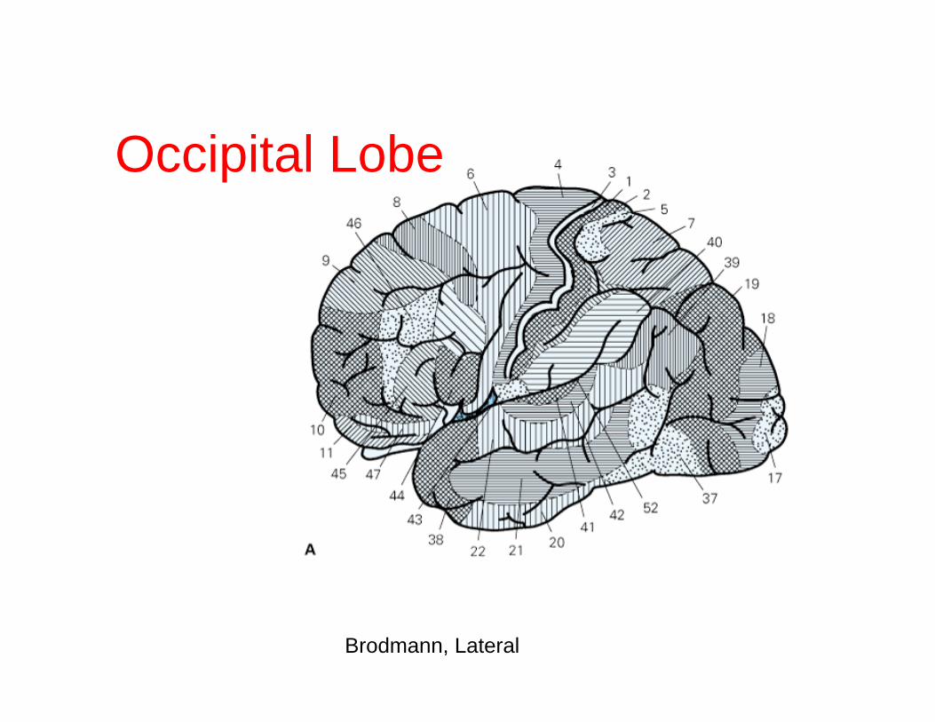
Brodmann, Lateral
Occipital Lobe

Ventral and dorsal Stream, MT

Brodmann, Medial

Medial Gross Brain

Ventral Gross Brain

PCA ventral view right hemisphere
from D.Haines

Visual Path

Write the patient historyPosterior view, angiogram
FIRST ANGIO SECOND ANGIO next day
R R LL

STROKE:Remember the 1st Four Letters...S.T.R.O.
If everyone can remember something this simple,
we could save some folks.
Doctors say a bystander can recognize a stroke by asking three simple questions:

Ask four simple questions:• S Ask the person to SMILE• T Ask the person to TALK and
SPEAK A SIMPLE SENTENCE (Coherently)(i.e. It is sunny out today)
• R Ask them to RAISE BOTH ARMS.• O Ask them to open their mouth
and STICK OUT your tongue. (Does it deviate to one side?)
• K Kall 911• E Every minute counts (180 mins)


End of Dental lecturego to ARS quiz
2011