Rational Drug Design Directed at Blocking the Initial ...
96
University of Missouri, St. Louis University of Missouri, St. Louis IRL @ UMSL IRL @ UMSL Theses UMSL Graduate Works 7-6-2017 Rational Drug Design Directed at Blocking the Initial Signaling Rational Drug Design Directed at Blocking the Initial Signaling Events in Lipopolysaccharide-Induced Sepsis. Events in Lipopolysaccharide-Induced Sepsis. Christopher A. Tipton University of Missouri - St. Louis, [email protected] Follow this and additional works at: https://irl.umsl.edu/thesis Part of the Biochemistry Commons, Biotechnology Commons, Immunoprophylaxis and Therapy Commons, Medicinal-Pharmaceutical Chemistry Commons, Molecular Biology Commons, and the Organic Chemistry Commons Recommended Citation Recommended Citation Tipton, Christopher A., "Rational Drug Design Directed at Blocking the Initial Signaling Events in Lipopolysaccharide-Induced Sepsis." (2017). Theses. 305. https://irl.umsl.edu/thesis/305 This Thesis is brought to you for free and open access by the UMSL Graduate Works at IRL @ UMSL. It has been accepted for inclusion in Theses by an authorized administrator of IRL @ UMSL. For more information, please contact [email protected].
Transcript of Rational Drug Design Directed at Blocking the Initial ...

University of Missouri, St. Louis University of Missouri, St. Louis
IRL @ UMSL IRL @ UMSL
Theses UMSL Graduate Works
7-6-2017
Rational Drug Design Directed at Blocking the Initial Signaling Rational Drug Design Directed at Blocking the Initial Signaling
Events in Lipopolysaccharide-Induced Sepsis. Events in Lipopolysaccharide-Induced Sepsis.
Christopher A. Tipton University of Missouri - St. Louis, [email protected]
Follow this and additional works at: https://irl.umsl.edu/thesis
Part of the Biochemistry Commons, Biotechnology Commons, Immunoprophylaxis and Therapy
Commons, Medicinal-Pharmaceutical Chemistry Commons, Molecular Biology Commons, and the
Organic Chemistry Commons
Recommended Citation Recommended Citation Tipton, Christopher A., "Rational Drug Design Directed at Blocking the Initial Signaling Events in Lipopolysaccharide-Induced Sepsis." (2017). Theses. 305. https://irl.umsl.edu/thesis/305
This Thesis is brought to you for free and open access by the UMSL Graduate Works at IRL @ UMSL. It has been accepted for inclusion in Theses by an authorized administrator of IRL @ UMSL. For more information, please contact [email protected].

1
Rational Drug Design Directed at Blocking the Initial Signaling Events in
Lipopolysaccharide-Induced Sepsis.
Christopher A. Tipton CCEMT-P NR-P Student Education/Degrees
B.S. Agriculture, University of Missouri-Columbia, 2007 Nationally Registered Paramedic, 2009
Critical Care, University of Maryland-Baltimore County, 2011 A Thesis Submitted to The Graduate School at the University
of Missouri-St. Louis in partial fulfillment of the requirements for the degree
Master of Science in Biochemistry & Biotechnology
August 2017
Month and Year of graduation
Advisory Committee
Michael Nichols, Ph.D. Chairperson
Christopher Spilling, Ph.D.
Co-Chair
James Bashkin D. Phil.
Keith Stine Ph.D.

2
Table of Contents i. List of Abbreviations
ii. Abstract
I. Introduction 1. Immunopathogenesis 2. Endotoxin 3. Sepsis 4. Current Interventions for Sepsis
II. Background A. Structure of LPS
1. The Core Region 2. The O-antigen Region 3. Lipid A
B. The LPS Receptor Complex 1. LBP & CD14 2. TLR4-MD-2 Complex 3. Signal Transduction Pathway & Mediators 4. Alternate TRIF-dependent Pathway 5. TNFα 6. Coagulation Cascade
C. Lipid A Analogues 1. Monosaccharide Mimetics 2. Antagonizing LPS-Induced Dimerization
D. Phosphonates 1. Phosphono-sugar analogues
III. Results & Discussion 1. Development of Lipid X Mimetics 2. Overall Synthesis 3. Protecting Group Strategy 4. Anomeric Hemiacetal Protection 5. Synthesis of Lactone Derivatives 6. Chemistry of Horner-Emmons Reaction 7. Biological Activity Assay 8. Biological Activity
IV. Conclusion & Future Directions V. Experimentals
VI. References

3
This page intentionally left blank

4
i. Abbreviations 2-Keto-3-Deoxyoctonic acid (Kdo); 3-O-desacyl-4’-Monophosphoryl Lipid A (MPL); Arterial Carbon Dioxide (PaCO2); Bacterial and Permeability-Increasing protein (BPI); Dimethyl Sulfoxide (DMSO); Disseminated Intravascular Coagulation (DIC); Endoplasmic Reticulum (ER); Enzyme-Linked Immunosorbent Assay (ELISA); Human Acute Monocytic Leukemia Cells (THP-1); Inhibitor of κB (IκB); Inhibitor of κB Kinase (IKK); Interleukin-1 Receptor-Associated Kinase 4 (IRAK4); Leucine-Rich Repeat Regions (LRR); Lipopolysaccharide (LPS); LPS Binding Protein (LBP); Membrane anchored CD14 (mCD14); Myeloid Differentiation primary response gene 88 (MyD88); Myeloid Differentiation protein-2 (MD-2); N-Methylmorpholine N-Oxide (NMO); Nitric Oxide (NO); Nuclear Factor κB (NFκB); Pathogen-Associated Molecular Patterns (PAMPs); Pattern Recognition Receptors (PRRs); Soluble CD14 (sCD14); Sterile α and HEAT-Armadillo Motif-containing protein (SARM); Systemic Inflammatory Response Syndrome (SIRS); Tetrapropylammonium Perruthenate (TPAP); TIR domain-containing adapter Inducing IFN-β (TRIF); TIR domain-containing Adapter Protein (TIRAP); Toll Interleukin-1 Receptor (TIR); Toll-Like Receptor 4 (TLR4); Toll-Like Receptors (TLR); TRIF-Related Adapter Molecule (TRAM); Trimethylsilyl Bromide (TMSBr); Tumor Necrosis Factor α (TNFα) ii. Abstract
Systemic Inflammatory Response Syndrome (SIRS) is
classified as an immune system response to an infectious
state. If left untreated, SIRS leads to sepsis, septic shock,
end-organ dysfunction, and death. As a patient progresses
through these stages, associations of acute respiratory
distress, disseminated intravascular coagulation, and acute
renal failure persist, resulting in millions of deaths
annually. Lipopolysaccharide (LPS), a bacterial endotoxin, is
5
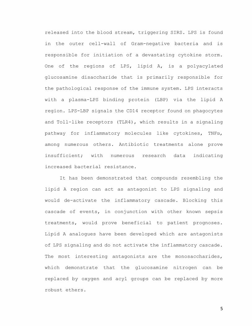
released into the blood stream, triggering SIRS. LPS is found
in the outer cell-wall of Gram-negative bacteria and is
responsible for initiation of a devastating cytokine storm.
One of the regions of LPS, lipid A, is a polyacylated
glucosamine disaccharide that is primarily responsible for
the pathological response of the immune system. LPS interacts
with a plasma-LPS binding protein (LBP) via the lipid A
region. LPS-LBP signals the CD14 receptor found on phagocytes
and Toll-like receptors (TLR4), which results in a signaling
pathway for inflammatory molecules like cytokines, TNFα,
among numerous others. Antibiotic treatments alone prove
insufficient; with numerous research data indicating
increased bacterial resistance.
It has been demonstrated that compounds resembling the
lipid A region can act as antagonist to LPS signaling and
would de-activate the inflammatory cascade. Blocking this
cascade of events, in conjunction with other known sepsis
treatments, would prove beneficial to patient prognoses.
Lipid A analogues have been developed which are antagonists
of LPS signaling and do not activate the inflammatory cascade.
The most interesting antagonists are the monosaccharides,
which demonstrate that the glucosamine nitrogen can be
replaced by oxygen and acyl groups can be replaced by more
robust ethers.

6
I. Introduction 1. Immunopathogenesis
Our innate immunity has evolved into a complex system
that elicits a response to pathogenic microbes to achieve a
survival advantage. The immune response attempts to localize
the infection and repair the damaged tissue. This is achieved
by activation of circulating and fixed phagocytic cells and
the production of pro-inflammatory and anti-inflammatory
mediators. The balance between these mediators facilitates
tissue repair, while simultaneously keeping the infection
from spreading. When the inflammatory response extends beyond
the infected tissues and becomes generalized, this balance is
lost. The process to control infection then becomes
uncontrolled and unregulated, leading to sepsis.
2. Endotoxin
Near the turn of the 20th century, it was discovered that
heat-killed Vibrio cholerae were intrinsically toxic as
opposed to producing toxicity by secretion of a product. To

7
differentiate, toxicity from a secreted product became
recognized as an exotoxin while the toxic components of
bacteria themselves were termed endotoxins. After further
characterizations, these heat-stable endotoxins became known
as lipopolysaccharide (LPS) and are localized to the cell-
wall of Gram-negative bacteria. Gram-negative bacteria
feature peptidoglycan that is encapsulated by two distinct
lipid membranes (Figure 11). The cytosolic bilayer consists
of conventional phosphoglycerides, whilst the outer membrane
is profoundly distinctive. The outer membrane is an
Figure 11 | Cell-wall of Gram-negative Bacteria. Organization of lipopolysaccharide, lipid A, lipoprotein, porins, peptidoglycan and phospholipid. The outer membrane is an asymmetric bilayer. The outer-leaflet of the outer membrane is highly distinctive due to the presence of lipopolysaccharide. The cytosolic bilayer consists of conventional phosphoglycerides.

8
asymmetric bilayer and LPS is the primary constituent of the
outer leaflet, of which lipid A is an integral component.
Pyrogenic bacteria generate endotoxins that stimulate
the release of inflammatory mediators, leading to fever and
systemic effects of inflammation advancing to septicemia. LPS
may be released from the membrane during bacterial growth or
during treatment with antibiotics. Intriguingly, relatively
low concentrations of LPS can act as an immune-modulator by
inducing non-specific resistance to both bacterial and viral
infections.2
3. Sepsis
Sepsis is a highly complex, variable and multifactorial
disease process caused by the over-exaggeration of the host’s
response to endotoxin.3 Predominantly responsible for Gram-
negative bacteremia are Enterobacteriaceae and Pseudomonas,
although other microorganisms can induce a similar response.
Clinicians consider Gram-negative bacteremia as an
idiosyncratic ailment due to its distinct clinical
manifestations, epidemiology, pathogenesis, and treatment.
Therefore, a consensus of the progression through stages of
the illness was adopted by physicians. To start, systemic
inflammatory response syndrome (SIRS) is classified as an
immune system response to an infectious state and is evident

9
with at least two of the following patient indicators: (1)
temperature greater than 38º C or less than 36º C; (2) heart
rate greater than 90 beats per min; (3) tachypnea, which is
defined as a respiratory rate greater than 20 breaths per
minute coupled with an arterial carbon dioxide (PaCO2) less
than 32 mmHg; (4) an alteration of white blood cell counts of
greater than 12,000 cells/mm3, less than 4,000 cells/mm3, or
greater than 10% immature neutrophils.3 Furthermore, severe
sepsis is defined as illness complicated by hypoperfusion
abnormalities like: lactic acidosis, oliguria, and/or mental
status changes, eventually leading to hypotension. Septic
shock is used to reference the illness when associated with
Table 1. The 20 most expensive conditions treated in U.S. hospitals, all payers, 2013
Rank CCS principal
diagnosis category
Aggregate hospital costs,
$ millions
National
costs, %
Number of
hospital stays,
thousands
Hospital
stays, %
1 Septicemia 23,663 6.2 1,297 3.6
2 Osteoarthritis 16,520 4.3 1,023 2.9
3 Liveborn 13,287 3.5 3,765 10.6
4 Device complications,
implant or graft
12,431 3.3 632 1.8
5 Acute myocardial
infarction
12,092 3.2 602 1.7
Table 15 | Epidemiology of Sepsis
Abbreviation: CCS, Clinical Classifications Software Sources include: Agency for Healthcare Research and Quality (AHRQ), Center for Delivery, Organization, and Markets, Healthcare Cost and Utilization Project (HCUP), National Inpatient Sample (NIS), 2013
10
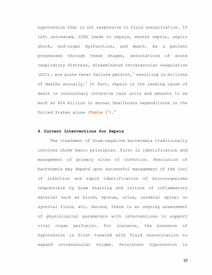
hypotension that is not responsive to fluid resuscitation. If
left untreated, SIRS leads to sepsis, severe sepsis, septic
shock, end-organ dysfunction, and death. As a patient
progresses through these stages, associations of acute
respiratory distress, disseminated intravascular coagulation
(DIC), and acute renal failure persist,3 resulting in millions
of deaths annually.4 In fact, sepsis is the leading cause of
death in noncoronary intensive care units and amounts to as
much as $24 billion in annual healthcare expenditures in the
United States alone (Table 15).6
4. Current Interventions for Sepsis
The treatment of Gram-negative bacteremia traditionally
involves three basic principles. First is identification and
management of primary sites of infection. Resolution of
bacteremia may depend upon successful management of the loci
of infection and rapid identification of microorganisms
responsible by Gram staining and culture of inflammatory
material such as blood, sputum, urine, cerebral spinal or
synovial fluid, etc. Second, there is an ongoing assessment
of physiological parameters with interventions to support
vital organ perfusion. For instance, the presence of
hypotension is first treated with fluid resuscitation to
expand intravascular volume. Persistent hypotension is

11
treated with sympathomimetic amines including dopamine,
dobutamine and isoproterenol. Levophed (norepinephrine) is an
intense vasoconstrictor considered if the previous
sympathomimetics are found ineffective. Third, the
administration of intravenous antibiotic therapy appropriate
for the spectrum of bacteria. However, this classical triad
of treatments is not geared towards blocking the toxic effects
of endotoxin, which are further exaggerated by bacterial
lysis from antibiotic therapy. Consequently, antibiotics
alone do not alleviate, but rather increase the toxic effects
of LPS in the septicemic patient.7 The ensuing fluid
administration and sympathomimetic treatments are merely
supportive measures aimed to combat subsequent hemodynamic
compromise from the overzealous host response. Whilst these
treatments are necessary, future sepsis treatments should be
spearheaded towards treating immunopathogenesis, not its
symptoms.
Clearly, the pathophysiology of sepsis is
extraordinarily complex. Exacerbating this complexity,
patients that are susceptible to infection have many other
medical conditions that affect their immune responsiveness
and contribute to mortality. A distinct combination of
therapies with a patient that is neutropenic (low neutrophils
in bloodstream)8 may differ for adjunctive therapies in an

12
elderly patient with a perforated diverticulum (bulging sac
on the colon wall).9 Presumably, an intervention with a single
agent at a single time point in the progression of sepsis is
unlikely to be effective. Advancement in treatments of sepsis
may depend upon disrupting underlying mechanisms of
immunopathogenesis responsible for tissue damage.
Corticosteroids have been researched as a potential
adjuvant therapy due to their ability to attenuate the
inflammatory response. However, in phase 3 clinical trials
mortality rates of patients receiving corticosteroid
treatment and a placebo were similar.10,11 Opioid receptor
blockers such as naloxone demonstrated improved survival in
animal studies, as did corticosteroids. Similarly, naloxone
failed to show any significant difference in mortality rates
in human trials.12
Neutralization of endotoxin could be an attractive
treatment against Gram-negative bacteremia induced sepsis.
Past studies in humans using antibodies to endotoxin by
administering polyclonal antiserum raised against core
polysaccharide and lipid A regions of LPS demonstrated
significantly reduced mortality rates.13 However, the
associated cost of producing antiserum coupled with the
potential for transmission of infection prevented the
widespread use of this treatment.

13
An approach that would circumvent the complications of
cost and transmission of infection could be the development
of nontoxic lipid A analogues. In animal models, enhanced
survival from Gram-negative bacteremia using lipid A
analogues has been demonstrated.14 An in-depth analysis of the
initial events in LPS-signaling is essential to develop a
rational therapy directed at blocking LPS-induced sepsis.
Additionally, illuminating the structural components of LPS
responsible for immunopathogenesis is vital to identify
therapeutic targets.
II. Background
A. Structure of LPS
Early attempts to elucidate the structure of LPS failed
for many reasons. LPS is highly amphipathic and has an
inherent tendency to aggregate through hydrophobic bonding or
by crosslinking via ionic interactions. Mildly acidic
conditions using trichloroacetic acid to extract and purify
LPS was first performed by Boivin et. al. in the 1930s.
However, LPS purified by the Boivin method was in effect a
crude fraction containing many cell-wall contaminants. It was

14
not until later that Westphal and Luderitz et. al. developed
an improved method for isolating endotoxin, which led to the
LPS nomenclature.15 Today, modern mass spectrometry with
matrix-assisted laser desorption and electrospray ionization
has been pivotal for characterizing intricate details of LPS
between species.16 Accordingly, LPS derived from all
characterized Gram-negative bacteria are composed of three
distinct regions, namely lipid A, core oligosaccharide and O-
antigen repeats (Figure 21). Lipid A contains a hydrophobic
region that anchors LPS to the outer leaflet of the outer
membrane. Distal to lipid A is a core oligosaccharide area
consisting of sugar residues with multiple phosphoryl
substituents, followed by a structurally diverse polymer
Figure 21 | Structure of LPS. The three major regions of LPS are: O-polysaccharide, Core oligosaccharide, and lipid A. O-polysaccharide is highly variable, but the Core and lipid A regions are more conserved between Bacteria. The lipid A portion of LPS is responsible for endotoxicity. Hep, heptose; Kdo, 2-keto-3-deoxyoctonate; GlcN, glucosamine; P, phosphate.
Repeating Subunitsn P
P
P
NH2 P
P
O-polysaccharide chain Core oligosaccharide Lipid A
Outer core Inner core
Hep
Hep Hep
Kdo
Kdo
Kdo
GlcN
GlcN

15
called O-antigen that is composed of repeat oligosaccharide
units.16 Both core oligosaccharide and O-antigen are displayed
on the surface of Gram-negative bacterial cells. The
remaining surface of the outer leaflet of the outer membrane
is taken up by proteins, while the inner leaflet contains
conventional phosphoglycerides, mostly phosphatidyl-
ethanolamine/glycerol and cardiolipin.16
1. The Core Region
The core region is more
architecturally uniform than the
outer O-antigenic region,
exhibiting moderate interbacterial
variability.17 The inner core
contains characteristic components
heptose and 2-keto-3-deoxyoctonic
acid (Kdo). Predominantly, the
inner core contains two or more Kdo
residues and two or three L-
glycero-D-manno-heptose residues
(Figure 316). The Kdo residue is
positioned at the reducing end of
the inner core and is linked to C-
Figure 316 | The Core region. The inner core usually consists of two Kdo and three L-glycero-D-manno-heptose residues. The outer core is composed of conventional sugars such as glucose and/or galactose.
16
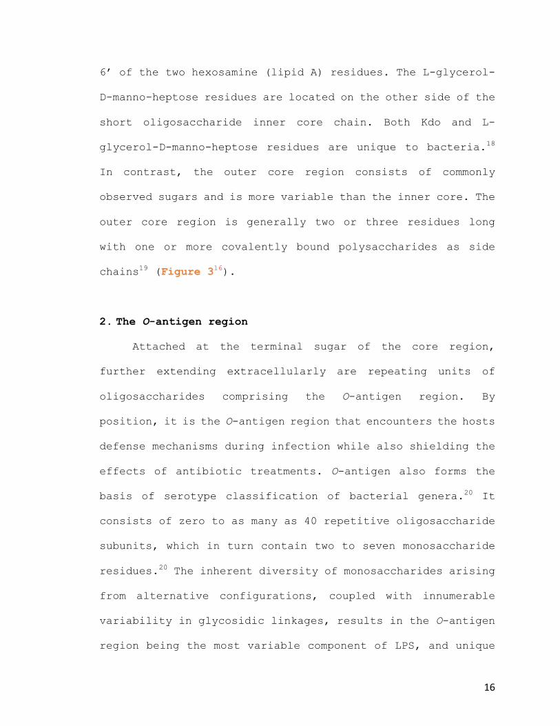
6’ of the two hexosamine (lipid A) residues. The L-glycerol-
D-manno-heptose residues are located on the other side of the
short oligosaccharide inner core chain. Both Kdo and L-
glycerol-D-manno-heptose residues are unique to bacteria.18
In contrast, the outer core region consists of commonly
observed sugars and is more variable than the inner core. The
outer core region is generally two or three residues long
with one or more covalently bound polysaccharides as side
chains19 (Figure 316).
2. The O-antigen region
Attached at the terminal sugar of the core region,
further extending extracellularly are repeating units of
oligosaccharides comprising the O-antigen region. By
position, it is the O-antigen region that encounters the hosts
defense mechanisms during infection while also shielding the
effects of antibiotic treatments. O-antigen also forms the
basis of serotype classification of bacterial genera.20 It
consists of zero to as many as 40 repetitive oligosaccharide
subunits, which in turn contain two to seven monosaccharide
residues.20 The inherent diversity of monosaccharides arising
from alternative configurations, coupled with innumerable
variability in glycosidic linkages, results in the O-antigen
region being the most variable component of LPS, and unique

17
to individual bacterial strains. Significantly, when
separated from the lipid A component of LPS, neither the O-
antigen nor the core polysaccharide exhibit endotoxic
activity.20
3. Lipid A
Lipid A is a distinct
phosphoglycolipid and the fundamental
backbone structure is highly conserved
amongst bacteria (Figure 421).22 All
contain D-gluco-configured pyranose
hexosamine residues that are β (1➝6)
linked dimers.16 Also, the disaccharide
component consists of α-glycosidic and
non-glycosidic phosphoryl substituents
located at C-1 and C-4’. The
phosphorylated disaccharide backbone
contains ester or amide linkages at
positions O-2, O-3, O-2’ and O-3’of
(R)-3-hydroxy fatty acids, of which
two are usually further acetylated.16
Figure 421 | Lipid A Lipid A functions as an anchor by binding LPS to peptidoglycan with fatty acid chains. Fatty acids widely recognized include: caporic (C6), lauric (C12), myristic (C14), palmitic (C16), and steric (C18) acids.

18
However, each Gram-negative bacterial species has unique
structural features and composition for lipid A (Figure 523).
Figure 523 | Lipid A from various bacterial species. Structural differences compared to the E. coli archetype arise from: the presence of phosphoryl substituents, degree of phosphorylation, and lipophilic chain lengths.

19
Fluctuations of the detailed structure emanate from: (1) the
presence of phosphoryl substituents, such as a 4-amino-deoxy-
L-arabinose and/or phosphoethanolamine linked axial at C-1 or
C-4’, (2) the degree of phosphorylation, (3) and importantly
the highly variable nature of the lipids, with lipophilic
chain lengths usually 6 to 18 carbon atoms in length. Also
important are the type and position of the acyl groups. The
acylation pattern of each hexosamine residue can have either
a symmetric (3+3) or an asymmetric (4+2) distribution.22
Functionally, lipid A provides the anchor that binds LPS
to the membrane with large numbers of saturated fatty acyl
groups. This generates a gelatinous barrier of low fluidity
and even impedes the infiltration of hydrophobic particles
into the membrane. The two polysaccharide components interact
with the extracellular environment and extend ~10 nm from the
surface of the outer membrane. These heteropolysaccharide
chains allow passage of small molecules for nutrient uptake,
but are impermeable to larger molecules like proteins. This
feature confines periplasmic proteins to prevent them from
diffusing away. The barrier is additionally stabilized by
LPS-associated cations that link adjacent molecules through
salt bridges. Taken as a whole, the highly oriented and
tightly cross-linked structure protects Gram-negative
bacteria from a variety of host-defense molecules, thereby

20
permitting growth and survival within harsh environments or
an infected host.
B. The LPS Receptor Complex
Accurate recognition of pathogen-associated molecular
patterns (PAMPs) by pattern recognition receptors (PRRs) is
the crux of the innate immune response.24 An important
receptor on the surface of immune cells such as
monocytes/macrophages, neutrophils, B lymphocytes, myeloid
dendritic and mast cells that recognizes LPS is toll-like
receptor 4 (TLR4). As a homodimer, TLR4 requires the small
myeloid differentiation protein-2 (MD-2) for recognition of
LPS.24 Other key proteins such as LPS binding protein (LBP)
and CD14 facilitate the presentation of LPS to the TLR4-MD-2
Figure 624 | LPS receptor complex. TLR4 recognizes LPS at the surface of immune cells. Recognition is facilitated by MD-2, CD14 and LBP. MD-2 associates with extracellular domains (A and B patches) of TLR4 and evokes sensitivity to LPS. LBP is a soluble shuttle protein that transfers LPS to the complex. CD14 exists as soluble (sCD14) or membrane bound (mCD14). CD14 is a co-receptor that binds LPS transported by LBP and in turn relocates LPS to the TLR4-MD-2 complex. LPB, lipid binding protein; TLR4, toll-like receptor 4; MD-2, myeloid differentiation protein 2.

21
complex (Figure 624).24 Once activated, TLR4 mobilizes adapter
molecules within the cytoplasm of cells to propagate a
signal.24 In turn, the adapter molecules activate molecules
within the cell to amplify the signal, which leads to the
induction of genes that orchestrate the inflammatory
response.
1. LBP & CD14
Above critical micellar concentrations, LPS forms large
aggregates in aqueous environments due to its inherent
amphipathic nature. LBP is a soluble shuttle protein that
avidly binds to LPS aggregates
and facilitates the association
between LPS and CD14.25 As a
complex, LBP-CD14 enhance the
detection of LPS by extracting
and monomerizing it prior to
presentation at the TLR4-MD-2
complex.
LBP belongs to the lipid
transfer family and to date its
structure has not been reported.
Bacterial and permeability-
increasing protein (BPI),
Figure 726 | BPI and CD14. (a) The crystal structure of BPI shares 48% sequence homology with LBP and has two phospholipid binding sites. (b) Crystal structure of CD14 showing two LPS binding pockets.
22
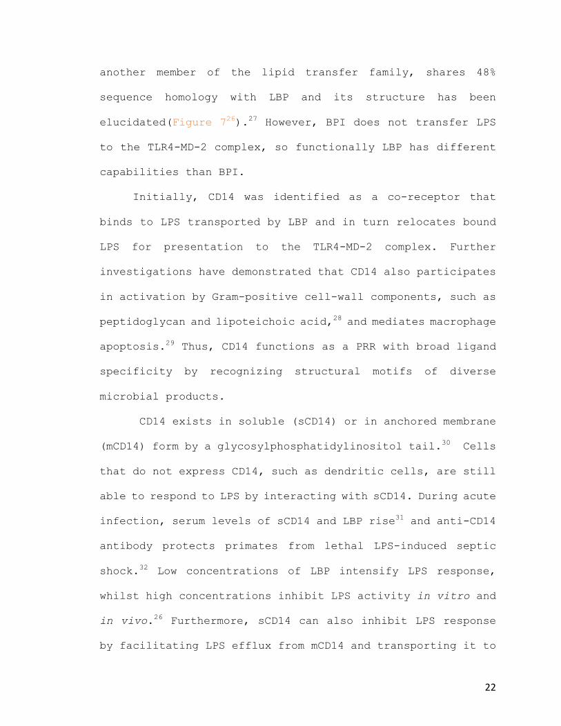
another member of the lipid transfer family, shares 48%
sequence homology with LBP and its structure has been
elucidated(Figure 726).27 However, BPI does not transfer LPS
to the TLR4-MD-2 complex, so functionally LBP has different
capabilities than BPI.
Initially, CD14 was identified as a co-receptor that
binds to LPS transported by LBP and in turn relocates bound
LPS for presentation to the TLR4-MD-2 complex. Further
investigations have demonstrated that CD14 also participates
in activation by Gram-positive cell-wall components, such as
peptidoglycan and lipoteichoic acid,28 and mediates macrophage
apoptosis.29 Thus, CD14 functions as a PRR with broad ligand
specificity by recognizing structural motifs of diverse
microbial products.
CD14 exists in soluble (sCD14) or in anchored membrane
(mCD14) form by a glycosylphosphatidylinositol tail.30 Cells
that do not express CD14, such as dendritic cells, are still
able to respond to LPS by interacting with sCD14. During acute
infection, serum levels of sCD14 and LBP rise31 and anti-CD14
antibody protects primates from lethal LPS-induced septic
shock.32 Low concentrations of LBP intensify LPS response,
whilst high concentrations inhibit LPS activity in vitro and
in vivo.26 Furthermore, sCD14 can also inhibit LPS response
by facilitating LPS efflux from mCD14 and transporting it to

23
serum lipoproteins.33 To sum, dual stimulatory and inhibitory
mechanisms of LBP and sCD14 afford systemic anti-inflammatory
effects, potentially hindering pathological systemic
responses.34 At the same time, LBP and sCD14 mechanisms can
promote pro-inflammatory effects at local sites of infection,
where they are needed.25
2. TLR4-MD-2 Complex
The transmembrane TLRs were first discovered in
Drosophila.35 In humans, a family of 10 genes encodes TLR1-
10, which are expressed by cells of the innate immune system.
The 10 human TLR genes encode distinctive TLR polypeptides.
Some TLRs are heterodimers of two polypeptides; others, such
as TLR4, are homodimers (Table 224). TLRs contain a variable
extracellular domain for detection of PAMPs from an array of
pathogens, including bacteria, viruses and fungi.36 Toll
Interleukin-1 Receptor (TIR) is the conserved cytoplasmic
domain that conveys signal transduction intracellularly. TIR
is critical for mediating protein-protein interactions
between TLRs and five signal transduction adapter proteins,
namely: myeloid differentiation primary response gene 88
(MyD88), TIR domain-containing adapter inducing IFN-β (TRIF),
TRIF-related adapter molecule (TRAM), TIR domain-containing
adapter protein (TIRAP), and sterile α and HEAT-Armadillo

24
motif-containing protein (SARM).30 Different combinations of
adapter proteins are utilized by distinct TLRs, which in turn
determines downstream signaling events. The signaling
pathways of TLRs are well defined, however the precise
mechanisms by which TLRs are activated upon ligand-binding
are not entirely understood. Interestingly, TLR4 is the only
recognized receptor that uses all five adapter proteins.30
The pathogen-recognition domains of TLRs consist of
hydrophobic leucine-rich repeat (LRR) regions, which are
Table 224 | Human TLRs recognize microbial ligands with pathogen associated molecular patterns (PAMPs). TLRs are encoded by 10 genes in humans from multiple chromosomes. TLRs acquired their nomenclature from the analogous receptor “Toll” found in the fruit fly Drosophila melanogaster. Differing PAMPs are found on distinct TLRs to confer variable ligand recognition. Some TLRs are heterodimers of two polypeptides and some exist solely as homodimers, such as TLR4.

25
responsible for receptor dimerization and the characteristic
horseshoe-like shape (Figure 837). Variation in the
composition and number of LRRs affords TLRs specificities for
different microbial ligands.37 LRR family proteins are
classified into 7 subfamilies, which are characterized by
conformations. Most LRR proteins contain uniform radii and β-
sheet angles. However, the structure of some TLRs, including
TLR4, substantially deviate from the consensus LRR
confirmations. They are divided by structural transitions
into three subdomains: N-terminal, central, and C-terminal
(Figure 837).37 Irregular LRR sequences in the central domain
cause the structural deviations from other LRR family
proteins. Furthermore, the subdomain boundaries of TLRs play
Figure 837 | Crystal structure of TLR4-MD-2 bound to LPS. (a) Top view of LPS bound to TLR4-MD-2 complex. The primary interface is formed prior to LPS binding and the dimerization interface is created after LPS binding. (b) Side view of receptor complex. Lipid A is colored red and inner core region of LPS is colored pink. TLR4 is divided into N- central and C-terminal domains. LRRNT and LRRCT, leucine-rich repeat regions N- C-terminus respectively.

26
key roles in ligand binding, demonstrated by the primary
contact interface of the TLR4-MD-2 complex.37 The N-terminal
and central domains of TLR4 provide charge complementary for
binding MD-2, forming a stable 1:1 heterodimer via two
distinct regions, the A and B patches respectively.37 Notably,
the TLR4-MD-2 complex is formed prior to binding LPS.
MD-2 is a soluble protein and can directly form a complex
with LPS, yet LPS-MD-2 binding is enhanced with TLR4
association. Importantly, there is no evidence to suggest
that TLR4 can independently bind LPS, emphasizing the
significance of LPS recognition by MD-2. MD-2 has a β-cup
fold structure formed by two anti-parallel β-sheets.37 The β-
sheets are separated from one another, which in turn exposes
the hydrophobic interior for ligand binding. This generates
a large internal pocket that is ideal for binding flat
hydrophobic ligands, such as lipid A. In fact, the interaction
between LPS and the TLR4-MD-2 complex occurs with high
affinity, and the KD is estimated to be 3-10 nM.38
3. Signal Transduction Pathway & Mediators
As a homodimer, TLR4 binds MD-2 to form two 1:1
complexes. Then, sCD14 or mCD14 present LPS to the TLR4-MD-2
complex, which in turn propagates the signal by dimerization
of the entire receptor complex. Such extracellular

27
recognition of LPS causes the cytoplasmic TIR domain of TLR4
to bind to a similar TIR domain of MyD88 (Figure 924). Once
TIR is bound to MyD88, a separate domain of MyD88 binds to
the protein kinase IRAK4, whereupon in auto-phosphorylates
itself and dissociates from the complex (Figure 924). IRAK4
propagates the signal by phosphorylating the adapter protein
Figure 924 | MyD88-dependent pathway induces NFκB to initiate transcription of cytokines upon LPS recognition by TLR4-MD-2 complex. Pathway from left to right: (1) LPS is detected by the TLR4-MD-2 complex. (2) Receptor complex dimerization causes cytoplasmic TIR domain to bind MyD88. (3) MyD88-associated IRAK4 auto-phosphorylates causing dissociation from MyD88. (4) Unbound IRAK4 phosphorylates TRAF6, which in turn induces a kinase cascade leading to activation of IKK. (5) Activated IKK phosphorylates IκB, leading to its degradation and subsequent release of transcription factor NFκB. (6) NFκB translocates into nucleus to initiate transcription of cytokine genes. (7) Cytokine mRNA is translated at ribosome and secreted extracellularly by the ER. Abbreviations: TIR, toll interleukin-1 receptor; IRAK4, interleukin-1 receptor-associated kinase 4; TRAF6, TNF receptor associated factor 6; IKK, inhibitor of κB kinase; IκB, inhibitor of κB; ER, endoplasmic reticulum.

28
TRAF6, which induces a cascade of events, eventually leading
to activation of the kinase complex IKK (inhibitor of κB
Kinase). IKK performs the critical function of activating a
transcription factor termed nuclear factor κB (NFκB), which
conducts significant operations to both innate and adaptive
immune responses.39 NFκB is held in an inactive state in a
cytosolic complex with inhibitor of κB (IκB). When IKK
phosphorylates IκB, it releases NFκB from inhibition.
Consequently, NFκB travels to the nucleus where it induces
the transcription of genes for cytokines and numerous other
proteins required to amplify the inflammatory response.39
4. TRIF-dependent
Alternatively, an MyD88-independent signaling pathway
can be triggered by the recruitment of TRIF and TRAM.30 These
adapter molecules play a key role in activating interferon
regulatory factor IRF3, which is essential for the expression
of type I interferons, like IFN-β.30 Also, exuberant nitric
oxide (NO) production results by activating the MyD88-
independent (TRIF-dependent) pathway.40 NO performs a major
role in inflammatory pathogenesis as a signaling molecule and
is thought to induce vasodilation.40 To add, NFκB can be
activated by the TRIF-dependent pathway, but in a later-
phase. It is speculated that subcellular localization of TLR4

29
distinguishes the activation of the two signaling pathways.
For instance, recognition of LPS at the plasma membrane
activates the MyD88-dependent pathway, but in contrast,
recognition of LPS at the endosome initiates the TRIF-
dependent pathway. Nonetheless, induction of either IRF3 or
NFκB activates the transcription of genes, which for the anti-
viral response are type I IFNs and in the pro-inflammatory
response, TNFα.
5. TNFα
NFκB induces the transcription of TNFα and other pro-
inflammatory cytokines including: IL-1β, IL-6, IL-8, IL-12
and IL-23.41 Of these, TNFα is one of the most important
soluble mediators of inflammation. It is responsible for an
array of signaling events and is mostly generated from
activated macrophages/monocytes.42 TNFα is produced rapidly
and can be detected within 15 minuets of LPS exposure in cell
culture and in vivo peaks at 1.5 hours.43 TNFα causes
contrasting effects to endothelial cells of blood vessels in
the infected tissues.44 In a systemic infection, macrophages
in the liver and spleen secrete TNFα into the bloodstream for
systemic circulation. The result is decreased blood flow from
vasodilation and diffuse leakage of plasma. TNFα released
into the bloodstream also causes the liver to secrete acute-

30
phase proteins, which include Mannan-binding lectin and C-
reactive protein. Both are critical to complement pathways
and exacerbate the inflammatory response.
6. The Coagulation Cascade
In localized infections, TNFα secretion causes blood
flow to increase and endothelia to produce platelet-
activating factor, which clots blood and blocks nearby
vessels.45 This induction of the coagulation cascade obstructs
pathogens from entering the bloodstream, thereby preventing
their spread. However, this process can impede delivery of
oxygen to the tissues and can induce further inflammatory
injury indirectly through the response to hypoxemia.
Usually, induction of the coagulation pathway induces
anticoagulant mechanisms to limit its progression. However,
when the infection is diffuse, as in sepsis, an imbalance of
the procoagulant and anticoagulant systems develop,
generating a sustained hypercoagulable state.46 Systemic
widespread clotting depletes coagulation proteins and
platelets from the blood. This process can lead to a bleeding
complication syndrome called DIC. Clinically, DIC is
increasingly common as a patient progresses from severe
sepsis to septic shock.46 Simultaneously, microvascular
thrombosis develops contributing to end-organ damage.46

31
Without intervention, lack of blood flow to the organs causes
multiple organ failure, which ultimately determines the
septic patients prognosis.
Overall, multiple and diverse pathways lead to a hyper-
activated immunological response that manifests end-organ
damage in sepsis. LPS itself is nontoxic but its adverse
effects emanate through systemic activation of host-derived
inflammatory mediators. Attempting to block a single
cytokine, like TNFα, would be inadequate due to the large
quantity and diversity of cytokines produced by activated
cells. Consequently, the path towards generating enhanced
therapeutics for the septicemic patient does not lie with
interrupting downstream incidents, but by blocking the
initial signaling events of the cascading inflammatory
response.
C. Lipid A Analogues
Synthesis of lipid A analogues first emerged with the
ambition to understand the chemical structures that were
principally responsible for endotoxic activity in sepsis.47
Some of the most informative studies of structure-activity
relationships of lipid A have utilized synthetic and natural
antagonist.48 In particular, the use of a naturally occurring
precursor from the constitutive biosynthesis pathway in E.

32
coli called lipid IVa (2) and a nonpathogenic lipid A molecule
from Rhodopseudomonas sphaeroides. The latter of which served
as the structural basis for the drug candidate Eritoran (3).
In phase III clinical trials, Eritoran (3) did not perform
better than existing treatments for sepsis.49 However, it did
demonstrate efficacy in combating cytokine storm induced from
strains of influenza in animal models.50 Another key
contributor to our continued understanding was isolation and
characterization of 3-O-desacyl-4’-monophosphoryl lipid A
(MPL)(4).51 Comparison of MPL derivatives provides clues for
Figure 10 | Structure of E. coli lipid A and derivatives. (1) Structure of agonistic E. coli lipid A. (2-5) Antagonist to LPS signaling at TLR4-MD-2 complex.
O
NH
OO
OH
OO
NH
HOO
O PO
OHOH
PO
HOHO
O
HO
O
HO
O
O
O
O
O
NH
OO
OH
OO
NH
HOO
O PO
OHOH
PO
HOHO
O
HO
O
HO
O
HO
O
HO
O
NH
OO
OH
OO
NH
HOO
O PO
OHOH
PO
HOHO
O
O
O
HO
OMeO
O
NH
HOO
O PO
OHOH
O
HO
O
HO
HOO
NH
OO
PO
HOHO
O
HO
O
O
OH
OH
1 E. coli lipid a 2 lipid IVa 3 Eritoran
5 Lipid X 6 GLA-60
O
NH
OO
OH
OO
NH
HOHO
OH
PO
HOHO
O
O
O
O
O
O
4 MPL
O
9
OO
OO
O

33
chemical alterations that direct endotoxicity and
adjuvanticity. Development of the less endotoxic MPL has led
to its widespread use as a component of numerous licensed
vaccines, including HBV and papilloma, and has been proven
both safe and effective.51 Detoxification was achieved by acid
hydrolysis of the 1-O-phosphono group followed by base
hydrolysis of the 3-hydroxytetradecanoly group to yield MPL
(4). In addition, studies revealed that lipid IVa (2) has
conflicting properties between human and murine cells.51 It
was shown to inhibit the induction of inflammatory cytokines
in human cells co-treated with LPS or lipid A. However, in
murine cell lines lipid IVa (2) was shown to be a potent
inducer of inflammatory cytokines. These species-specific
results were later explained by structural differences
between human and murine TLR4-MD-2 complexes. Specifically,
lipid IVa (2) caused contrasting effects on dimerization of
the TLR4-MD-2 receptor complex, which is required for
activation of downstream signaling. Expectedly, lipid IVa (2)
was shown to promote receptor dimerization in murine, but not
human TLR4-MD-2 complexes.
Activation of the TLR4-MD-2 complex can lead to distinct
signaling pathways namely; the MyD88-dependent or the TRIF-
dependent pathways. Induction of the MyD88-dependent pathway
causes production of proinflammatory cytokines such as TNFα,

34
whilst TRIF-dependent pathway activation leads to NO and IFN-
β production. Teasing apart the structural determinants of
lipid A that induces the former or the latter has implications
in determining subsequent endotoxicity or adjuvanticity.51
Previously stated, CD14 is a PRR that directly binds to LPS
and chaperones the formation of the dimerized TLR4-MD-2
complex. At low concentrations of LPS, CD14 plays an increased
role in formation and subsequent receptor dimerization
leading to induction of the MyD88-dependent pathway. Albeit,
CD14 is not essential for this induction when LPS is in higher
concentrations. CD14 is also required for TLR4 endocytosis
and internalization of the entire receptor complex into the
endosome, thereby inducing the TRIF-dependent pathway.51 It
has been demonstrated that TLR4 antagonist, such as lipid IVa
(2) and Eritoran (3), strongly interact with CD14, thereby
inhibiting re-localization of the receptor complex.52
1. Monosaccharide Mimetics
Researchers have attempted to separate beneficial
immunopharmacological attributes from adverse
pathophysiological endotoxic properties of lipid A by
performing structure-activity analyses of simplified
structures. Lipid A has a basic endotoxic structure of an
amphipathic molecule, with distinct hydrophilic and
35
hydrophobic domains. The association of a polar backbone
supporting an apolar region is vital for its antagonistic or
agonistic properties. However, the specific structure of the
glucosamine disaccharide backbone is seemingly not stringent
in directing which biological activity will result. Synthetic
lipid A derivatives with one of the glucosamine disaccharide
residues replaced with acyclic or pseudo-peptides have
contained similar activities of E. coli lipid A, whilst others
have demonstrated antagonistic properties.53 Interestingly,
both or at least one phosphate, or bioisotere of, substituents
at the anomeric or 4’-carbons seem to be a prerequisite for
substantial biological activity.
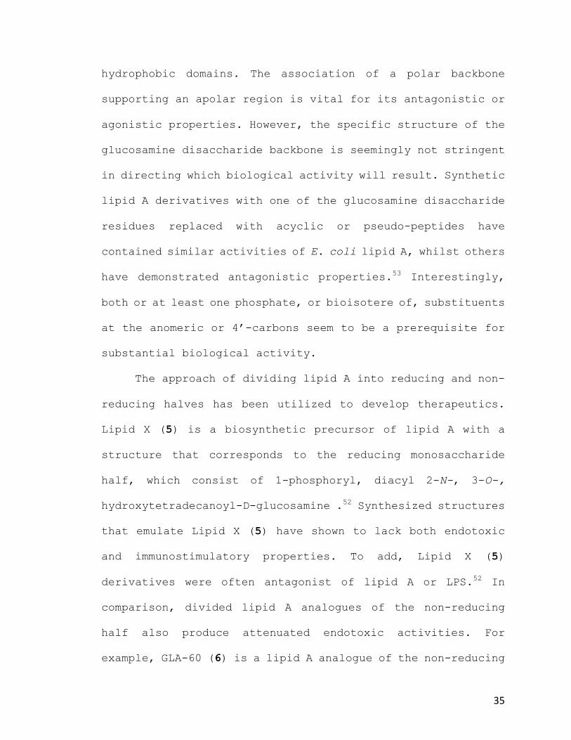
The approach of dividing lipid A into reducing and non-
reducing halves has been utilized to develop therapeutics.
Lipid X (5) is a biosynthetic precursor of lipid A with a
structure that corresponds to the reducing monosaccharide
half, which consist of 1-phosphoryl, diacyl 2-N-, 3-O-,
hydroxytetradecanoyl-D-glucosamine .52 Synthesized structures
that emulate Lipid X (5) have shown to lack both endotoxic
and immunostimulatory properties. To add, Lipid X (5)
derivatives were often antagonist of lipid A or LPS.52 In
comparison, divided lipid A analogues of the non-reducing
half also produce attenuated endotoxic activities. For
example, GLA-60 (6) is a lipid A analogue of the non-reducing
36
section and displays a 4-phosphoryl D-glucosamine
monosaccharide moiety with three acyl chains of 12-14 carbons
in length. Interestingly, GLA-60 (6) based compounds are
relatively potent adjuvants, provide non-specific protection
from bacterial and viral infections and are tumor
regressive.54 Furthermore, current studies have revealed
monosaccharide lipid A analogues as potential adjuvants for
cancer treatments with multiple phase 1 trials underway.55–65
2. Antagonizing LPS-Induced Dimerization
Despite substantial data on the activity of both
isolated and synthetic lipid A derivatives, there
unfortunately is no universal correlation between the
chemical structure of lipid A and its activity in the TLR4-
MD-2 complex. However, elucidation of the crystal structure
of the TLR4-MD-2 complex bound to antagonistic lipid IVa (2)
or Eritoran (3) and agonistic E. coli lipid A (1) has
contributed a better understanding of structural requirements
for the receptor complex (Figure 1137).37
Previously stated, TLR4 as a homodimer binds MD-2 to
form two 1:1 complexes that then dimerize upon ligand binding.
LPS binding induces dimerization by creating an additional
binding interface between TLR4 and MD-2. To distinguish the
secondary dimerized heterotetrameric TLR4-MD-2* complex from

37
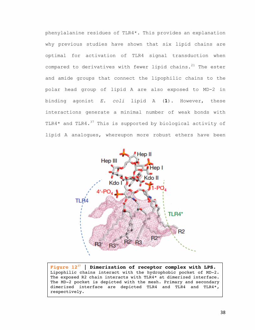
primary TLR4-MD-2, an asterisk is utilized. Accordingly, the
carbon chains of lipid A interact with the hydrophobic pocket
of MD-2. Five of the six lipid chains of agonistic LPS are
enclosed by the hydrophobic pocket and the sixth is uncovered
to the surface of MD-2 (Figure 1237), whilst all four lipid
chains of lipid IVa (2) are buried (Figure 1137).37 The sixth
lipid chain of LPS forms a hydrophobic interaction with
Figure 1137 | LPS antagonist shift the anomeric phosphate. The size of the MD-2 pocket is unchanged after binding agonistic or antagonistic lipid A. Added space for lipid binding displaces the anomeric phosphate upwards ~5Å, allowing interaction with nearby positive charges on TLR4 and TLR4*. (a) Comparison of LPS and Eritoran binding. (b) Comparison of LPS and lipid IVa binding.
38
phenylalanine residues of TLR4*. This provides an explanation
why previous studies have shown that six lipid chains are
optimal for activation of TLR4 signal transduction when
compared to derivatives with fewer lipid chains.21 The ester
and amide groups that connect the lipophilic chains to the
polar head group of lipid A are also exposed to MD-2 in
binding agonist E. coli lipid A (1). However, these
interactions generate a minimal number of weak bonds with
TLR4* and TLR4.37 This is supported by biological activity of
lipid A analogues, whereupon more robust ethers have been
Figure 1237 | Dimerization of receptor complex with LPS. Lipophilic chains interact with the hydrophobic pocket of MD-2. The exposed R2 chain interacts with TLR4* at dimerized interface. The MD-2 pocket is depicted with the mesh. Primary and secondary dimerized interface are depicted TLR4 and TLR4 and TLR4*, respectively.

39
substituted.21 The two phosphate groups bind to residues of
TLR4-MD-2 and at the dimerization interface of TLR4*, thus
supporting the formation of a stable (TLR4-MD-2)2 dimerized
complex. Both the 1-phosphate and 4’-phosphate of lipid A
bind to positives patches on TLR4 and TLR4* (Figure 1337). The
significance of these two interactions has been established.
As demonstrated with MPL (4), removal of one phosphate group
greatly reduces the endotoxicity of LPS. Secondly, when the
positively charged patch of TLR4* was mutated to an alanine
residue, NFκB and IFN-β activity was abolished.66 This finding
Figure 1337 | Both phosphates of LPS lipid A interact at dimerized receptor complex. The 1-phosphate and 4’-phosphate conduct dimerization by binding positively charged arginine and lysine residues of TLR4 and TLR4*. These two ionic interactions are critical elements for activation of TLR4-MD-2 complex. Removal of one phosphate group greatly reduces endotoxicity.

40
suggest that the 1-phosphate of lipid A is essential for
activation of both MyD88-dependent and TRIF-dependent
pathways. Thus, subtle structural changes at the anomeric
phosphate of lipid A could illuminate the path towards guided
activation between endotoxicity and adjuvanticity.
D. Phosphonates
The phosphate group [(HO)2P(=O)OR] is a fundamental
component of all living systems. It is essential for molecular
replication, cell biochemistry, signaling pathways, and
regulation of metabolic processes.67 Phosphonate analogs of
phosphates, wherein the phosphate ester bond has been
replaced with the hydrolytically stable phosphonate
[(HO)2P(O)R (R=carbon residue)], often contain enticing
physiological properties (Scheme 1).68–72 An alpha substituent
(X) can be used to return the pKa of the phosphonic acid to
the values typical of the corresponding phosphate ester. In
addition, the tetracordinate phosphoryl group is well
recognized as an excellent mimic for the tetrahedral
transition state of ester and amide hydrolysis.73 More

41
surprisingly, phosphonic acids can successfully act as non-
isosteric replacements for carboxylic acids.74
1. Phosphono-sugar Analogues
Phosphonates have become increasingly useful in the
development of tools for biological investigation and the
formulation of novel compounds for medicinal chemistry. For
instance, phosphonate derivatives that contain additional
functionality in the carbon chain are extremely versatile by
exhibiting activities as receptor agonists-antagonists75 and
herbicidal, antibacterial, and antiviral agents76–80, usually
through the inhibition of specific enzymes.81,82
There are many examples of phosphono-sugars where the
phosphonate is located on a ring substituent. Such compounds
are important in the development of non-hydrolysable
phosphonate mimics of bioactive carbohydrate phosphates, such
as nucleotides.83–85 Research in this area has resulted in
several examples of biologically relevant molecules.
42
III. Results & Discussion
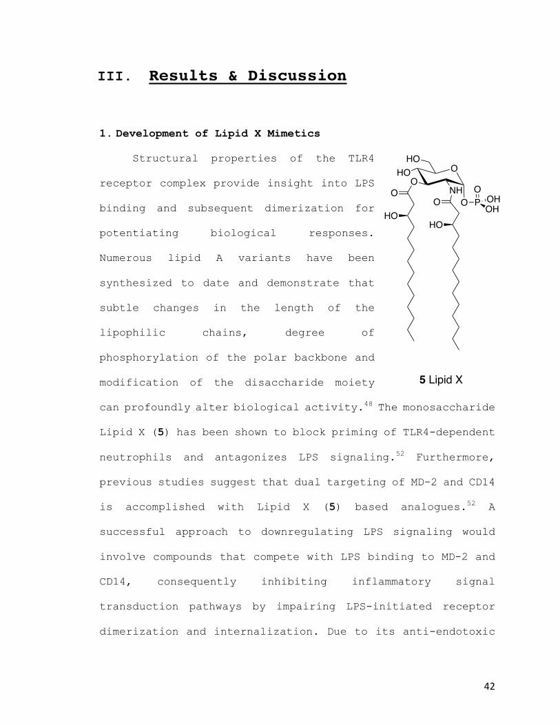
1. Development of Lipid X Mimetics
Structural properties of the TLR4
receptor complex provide insight into LPS
binding and subsequent dimerization for
potentiating biological responses.
Numerous lipid A variants have been
synthesized to date and demonstrate that
subtle changes in the length of the
lipophilic chains, degree of
phosphorylation of the polar backbone and
modification of the disaccharide moiety
can profoundly alter biological activity.48 The monosaccharide
Lipid X (5) has been shown to block priming of TLR4-dependent
neutrophils and antagonizes LPS signaling.52 Furthermore,
previous studies suggest that dual targeting of MD-2 and CD14
is accomplished with Lipid X (5) based analogues.52 A
successful approach to downregulating LPS signaling would
involve compounds that compete with LPS binding to MD-2 and
CD14, consequently inhibiting inflammatory signal
transduction pathways by impairing LPS-initiated receptor
dimerization and internalization. Due to its anti-endotoxic
O
NH
HOO
O PO
OHOH
O
HO
O
HO
HO
5 Lipid X

43
activity, coupled with a simplified monosaccharide moiety
that would prove more cost effective and more readily
scalable, we selected Lipid X (5) based derivatives to
synthesize.
Monosaccharide based TLR4 receptor targeting could also
afford compounds with enhanced water solubility. Previously
synthesized lipid A mimetics suffer from poor solubility in
aqueous media86, which is essential for improved
bioavailability and a more favorable pharmacokinetic profile.
Significantly, gaining structural insight to guide future
explorations in distinguishing endotoxicity and adjuvanticity
are paramount to progress lipid A
analogues. To date, C-glycosylated
phosphono-glucoside mimetics of Lipid X
have not been explored. Thus, we employed
traditional carbohydrate chemistry
techniques to develop a non-hydrolysable
phosphonate mimic of Lipid X (12α) and
assessed its biological activity for
antagonizing LPS in vitro.
O
O
HOHO
PO
OMeOMe
12-α
O

44
2. Overall Synthesis
Synthesizing TLR4-MD-2 receptor antagonists by
incorporating a phosphonate onto a ring substituent will
generate the non-hydrolysable Lipid X mimetic (Scheme 2). The
proposed synthesis begins with the formation of
peracytylated-thio-β-D glucoside (7) from penta-acylated
Scheme 2 | Reagents and Conditions (R = C14H29) (i) PhSh, BF3
.Et2O, DCM, rt, 4h, (78%); (ii) 1. NaOMe, MeOH/DCM 1:10, 0 ºC, 1h, (99%); 2. PhCH(OMe), pTsOH, DMF, 60 ºC, 4h, (80%); (iii) NaH, tetra-N-butylammonium iodide, Bromo-tetradecane, DMF/THF 2:3, 40ºC, 24h, (78%); (iv) 1. N-IS, 1.1 eq. TFA, DCM, rt, 1h; 2. Piperidene, rt, (84-94%); (v) THF, NaH, ((MeO)2(O)P)2CH2, rt, 2-24h, (48-54%); (vi) 15% TFA in wet DCM, rt, 1h, (77%); (vii) THF, NaCNBH3, 2 N HCl/Et2O, rt, 30 min., (84%).
O
OAcOAc
OAcO
OAcSPh
OAcO
OHSPh
OO O
ORSPh
OO
O
OR
OO OH
O
OR
OO
PO
OMeOMe
O
OR
BnOHO
PO
OMeOMe
O
OR
HOHO
PO
OMeOMe
12
11
10
96 7 8
13
RO
RO
RO
RO
RO
HOAcOAcOAcO
AcOPhPh
Ph
Ph
9
i ii iii
iv
vvi
vii

45
glucose (6) as depicted,87 deacetylation of the peracytylated-
thio-β-D-glucoside (7) will generate the tetrol, which will
then be protected as a benzylidene. The resulting diol (8) is
alkylated creating both lipophilic chains (9). Hydrolysis of
the thiophenol forms the anomeric hydroxy (10) that is
subjected to Horner–Wadsworth–Emmons reaction conditions to
introduce the phosphonate moiety (11). Hydrogenolysis or
selective C-4 ring cleavage of the benzylidene protecting
group will afford novel phosphono-sugar (12) and (13)
analogues of lipid X. The proposed route also allows for
divergent chemistry to produce a library of compounds (Scheme
3) to more fully explore structure activity relationships of
Lipid X.
O
OR
OO
9
O
OR
OO
21
O
OR
OO
18
O
OR
HOHO
14
O
OR
HOHO
22
O
OR
HOHO
19
SPh SPh
O O
SEt SEt
O
OR
BnOHO
15
O
OR
BnOHO
23
O
OR
BnOHO
20SEt
SPh
O
O
OR
OO
10
O
OR
HOHO
16
O
OR
BnOHO
17
OH
OH OH
RO RO RO RO
RORORORO
RO RO RO RO
Ph Ph
Ph
Ph
Scheme 3 | Our library of monosaccharide mimetic compounds synthesized. R = C14

46
3. Protecting Group Strategy
Strategical implementation of protecting groups is an
important component for any total synthesis of organic
molecules, but this is especially true in carbohydrate
chemistry. Carbohydrates present a large number of poly-
functionalized groups. Most of them are of the same sort,
that is in the form of free hydroxyls. Success depends upon
differentiating the relative reactivity of the hydroxyls,
which are reflected by electronic and conformational factors.
The different reactivity of the hydroxyls manifest from
namely: one primary (C-6), several secondary (C-2,-3, & -4),
and an acetalic group at the anomeric center. The most
reactive is the hydroxyl at the anomeric carbon followed by
the primary alcohol at C-6. The secondary hydroxyls contain
varying reactivity due to their equatorial or axial
orientations. This feature of carbohydrates necessitates
regioselective strategies, which can be arduous at times.
Protecting groups also impart other effects of the
compound. They can alter the reactivity of a molecule and can
also participate in the reaction itself, therefore affecting
the stereochemical outcomes. Ideally, it should be possible
to introduce and remove more permanent protecting groups with
regiocontrol and high efficiency. They should be stable under

47
conditions used for the addition and removal of temporary
protecting groups. Acetals confer this stability and in
addition contain efficient introduction and deprotection
properties, for instance simultaneous protection of C-4 and
C-6 hydroxyls. Of the acetals, we employed benzaldehyde
dimethyl acetal PhCH(OMe)2 under standard acetalization
conditions with pTsOH as the acid catalyst to produce
compounds (9) and (18) in good yields.
An added advantage of the benzylidene acetal as a
protecting group is the number of subsequent modifications
that can yield various protecting group patterns (Scheme 4).
Selective reduction to yield benzyl ethers and free hydroxyl
groups are readily utilized with a hydride reagent in
combination with a Lewis acid. Combination of LiAlH4/AlCl3
would afford 4-O-benzyl derivatives unveiling the primary
Scheme 4 | Examples of reductive cleavage of benzylidene acetals.
48
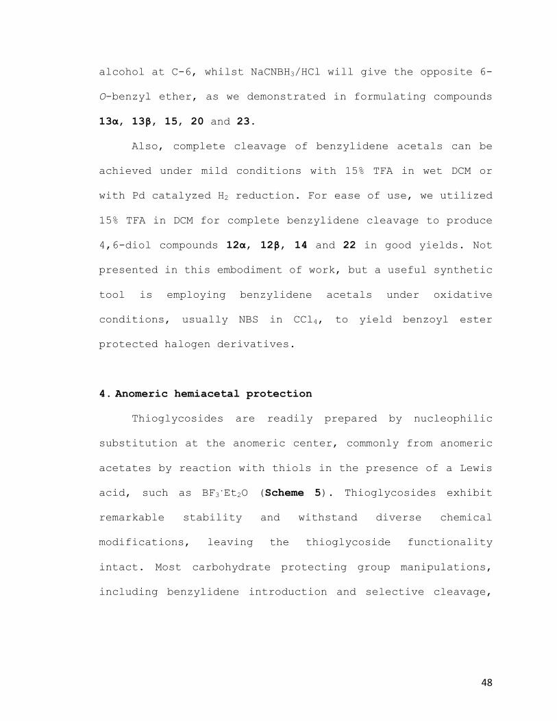
alcohol at C-6, whilst NaCNBH3/HCl will give the opposite 6-
O-benzyl ether, as we demonstrated in formulating compounds
13α, 13β, 15, 20 and 23.
Also, complete cleavage of benzylidene acetals can be
achieved under mild conditions with 15% TFA in wet DCM or
with Pd catalyzed H2 reduction. For ease of use, we utilized
15% TFA in DCM for complete benzylidene cleavage to produce
4,6-diol compounds 12α, 12β, 14 and 22 in good yields. Not
presented in this embodiment of work, but a useful synthetic
tool is employing benzylidene acetals under oxidative
conditions, usually NBS in CCl4, to yield benzoyl ester
protected halogen derivatives.
4. Anomeric hemiacetal protection
Thioglycosides are readily prepared by nucleophilic
substitution at the anomeric center, commonly from anomeric
acetates by reaction with thiols in the presence of a Lewis
acid, such as BF3.Et2O (Scheme 5). Thioglycosides exhibit
remarkable stability and withstand diverse chemical
modifications, leaving the thioglycoside functionality
intact. Most carbohydrate protecting group manipulations,
including benzylidene introduction and selective cleavage,

49
can be performed. In addition, thioglycosides can serve as
glycosyl acceptors to construct oligosaccharides.
Thus, we protected the anomeric carbon of (6) with
thiophenol to produce (7) prior to 4,6-O-benzylidene (8)
formation. However, in a subsequent step for our synthesis,
thiphenol proved its stability. To generate the phosphono-
sugar, the anomeric thio-protecting group needs to first be
hydrolyzed. We found this step to be difficult using commonly
applied methods. We observed the conventional strategies
using N-bromo- and N-iodo-succinimide, and N-iodosaccharin in
the presence of minute amounts of H2O to be unsuccessful. We
also tried catalytic auric chloride as a strategy to activate
thioglycoside donors, which had recently been reported.
O
AcOO
OAc
O
AcOSR
OAc
6
7α
AcO
AcOAcO
AcOiβ
O
BF3
O
AcO
OAc
6a
AcOAcO
O
AcO
OAc
AcOAcO
O
AcO
OAc
AcOAcO
S RH
SR
H
O
AcO
OAc
7β
AcOAcO
iα
SR
i
Scheme 5 | Thioglycoside Anomeric Protection. R = Ph or Et (i) Mechanism of to generate thioglycosides from penta-acylated-β-D-glucose, depicting the formation of the α or β anomers. We utilized BF3.Et2O in the presence of ethanethiol and thiophenol. Thiophenol was found to be β-selective, whilst ethanethiol generated a mixture of anomers.
50
Unfortunately, this method also proved unsuccessful. The
over-arching problem observed in these reactions were with
solubility. Addition of tiny amounts of H2O, required for
hydrolysis in these reaction methods, resulted in the
starting thioglucoside (9) to rapidly precipitate from
solution in a variety of solvents (DCM, Et2O, THF, dioxane,
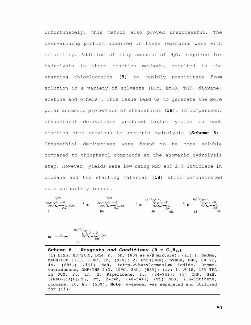
acetone and others). This issue lead us to generate the more
polar anomeric protection of ethanethiol (18). In comparison,
ethanethiol derivatives produced higher yields in each
reaction step previous to anomeric hydrolysis (Scheme 6).
Ethanethiol derivatives were found to be more soluble
compared to thiophenol compounds at the anomeric hydrolysis
step. However, yields were low using NBS and 2,6-lutidiene in
dioxane and the starting material (18) still demonstrated
some solubility issues.
Scheme 6 | Reagents and Conditions (R = C14H29) (i) EtSh, BF3.Et2O, DCM, rt, 6h, (83% as α/β mixture); (ii) 1. NaOMe, MeOH/DCM 1:10, 0 ºC, 1h, (99%); 2. PhCH(OMe), pTsOH, DMF, 60 ºC, 4h, (88%); (iii) NaH, tetra-N-butylammonium iodide, Bromo-tetradecane, DMF/THF 2:3, 40ºC, 24h, (83%); (iv) 1. N-IS, 15% TFA in DCM, rt, 1h; 2. Piperidene, rt, (84-94%); (v) THF, NaH, ((MeO)2(O)P)2CH2, rt, 2-24h, (48-54%); (vi) NBS, 2,6-lutidene, dioxane, rt, 6h, (53%). Note: α-anomer was seperated and utilized for (ii).
O
OAcOAc
OAcO
OAcSEt
OAcO
OHSEt
OO O
OR
OO
O
OR
OO OH
10
186 18a 18b
RO
RO
HOAcOAcOAcO
AcOPhPh
Ph
18
i ii iii
iv
SEt

51
Eventually, we did develop a method to alleviate the
solubility issues encountered by the particularly hydrophobic
compounds (9) and (18). To complete this step in the
synthesis, the thioacetal (9) was hydrolyzed by treatment
with NIS and dry TFA in DCM, followed by addition of
piperidine to cleave the intermediate triflouroacetate and
afford the hemiacetal (10) as a 1:1 anomeric mixture in high
yield (~90%). This step permitted hydrolysis without the use
of H2O, which evoked our problems with solubility.
5. Synthesis of lactone derivatives
Lipid A from the LPS of the nitrogen fixing bacterial
species Rhizobium sin-1 (24) is structurally distinct, in
comparison to endotoxic E. coli lipid A. It is completely
devoid of phosphates, has a very long chain fatty acid (27-
hydroxyoctacosonic acid), and contains a D-gluconolactone
moiety at the reducing end (Scheme 8). Interestingly,
compound (24) and a synthetic disaccharide derivative (25)
O
ORSPh
OO
9
RO
Ph1. N-IS , TFA
2. Piperidene
O
OR
OO OH
10
RO
Ph
(84-94%)
Scheme 7 | Deprotection of Thioglycoside. Reagents and Conditions: 1. N-IS, 1.1 eq. TFA, DCM, rt, 1h; 2. Piperidene, rt, (84-94%)

52
have shown to be potent in antagonizing LPS-induced cytokine
production in human macrophage cells by inhibiting both TRIF-
and MyD88-dependent pathways.21 Intriguingly, monosaccharide
lipid A derivatives containing a D-gluconolactone moiety have
not been investigated. Thus, we decided to also synthesize
Lipid X derivatives devoid of a phosphonate, yet containing
the D-gluconolactone moiety (21, 22, and 23).
Oxidation of carbohydrates is a widely-utilized
technique to attain derivatives with profoundly modified
reactivity and character. Mono-oxidation of carbohydrates at
the anomeric center produces aldonolactones with reactivity
unlike that of the corresponding aldoses.
Scheme 8 | Rhizobium sin-1 lipid A.

53
A facile synthetic technique we used to produce the
desired Lipid X D-gluconolactone derivative (21) was by
performing a Ley-Griffith oxidation to compound (10). This
technique utilizes the ruthenium based oxidant
tetrapropylammonium perruthenate N(C3H7)4RuO4 (TPAP) with 1.5
equivalents of co-oxidant N-methylmorpholine N-oxide (NMO) in
DCM as shown in Scheme 9. TPAP operates catalytically at room
temperature and is devoid of explosive side products. When
TPAP is used in the presence of NMO (Scheme 10), high yields
are usually observed and this was supported in the formation
of our D-gluconolactone derivative (21).
Scheme 9 | Ley-Griffith oxidation mechanism.

54
6. Chemistry of Horner-Emmons Reaction
This reaction makes use of phosphonate anions as the
nucleophilic species. This methodology is applicable to the
formation of C-glycosides (11) from sugars (Scheme 11).
However, considering the intermediate α, β-unsaturated ester
and the acidity of the proton at C-2, epimerization of the
stereocenter is possible. Indeed, the reaction conditions
that generated (11) did produce four diastereomers that were
Scheme 11 | Generated phosphono-sugar mimetics R = C14
O
OR
OO OHRO
Ph
TPAP
NMO , DCM O
OR
OORO
Ph
O
10 21(85%)
Scheme 10 | Anomeric oxidation produces D-glucono-δ-lactone.
O
OR
OO
PO
OMeOMe
O
OR
BnOHO
PO
OMeOMe
O
OR
HOHO
PO
OMeOMe
12-β11-β 13-β
O
OR
OO
PO
OMeOMe
O
OR
BnOHO
PO
OMeOMe
O
OR
HOHO
PO
OMeOMe
12-α11-α 13-α
RO
RO RO RO
ROROPh
Ph
(20%)
(16%) (84%)
(60%) (93%)
(92%)
55
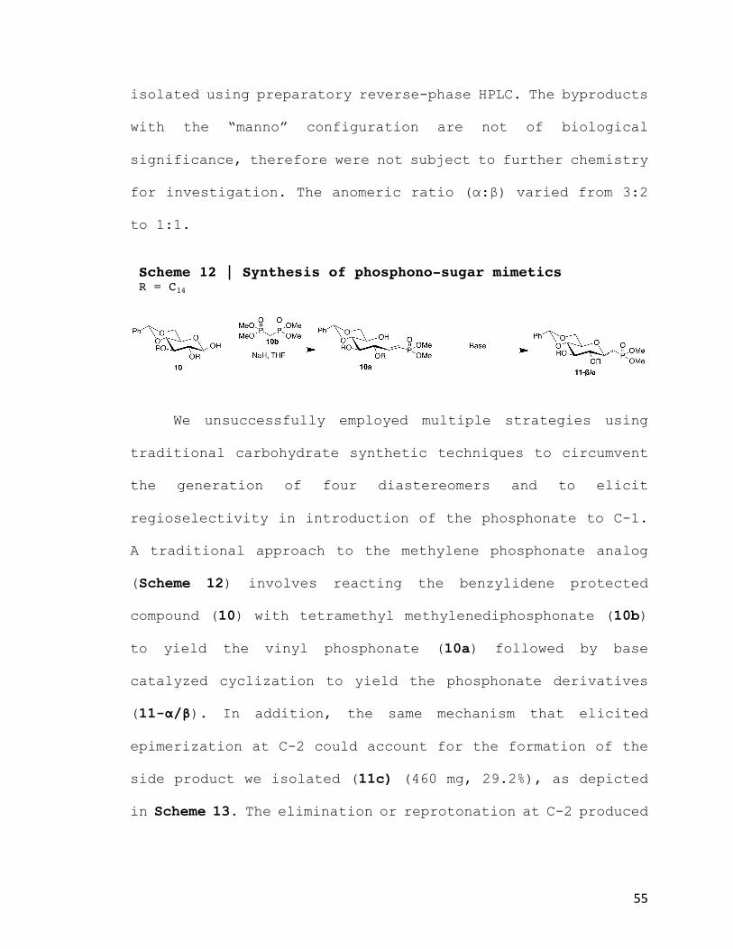
isolated using preparatory reverse-phase HPLC. The byproducts
with the “manno” configuration are not of biological
significance, therefore were not subject to further chemistry
for investigation. The anomeric ratio (α:β) varied from 3:2
to 1:1.
We unsuccessfully employed multiple strategies using
traditional carbohydrate synthetic techniques to circumvent
the generation of four diastereomers and to elicit
regioselectivity in introduction of the phosphonate to C-1.
A traditional approach to the methylene phosphonate analog
(Scheme 12) involves reacting the benzylidene protected
compound (10) with tetramethyl methylenediphosphonate (10b)
to yield the vinyl phosphonate (10a) followed by base
catalyzed cyclization to yield the phosphonate derivatives
(11-α/β). In addition, the same mechanism that elicited
epimerization at C-2 could account for the formation of the
side product we isolated (11c) (460 mg, 29.2%), as depicted
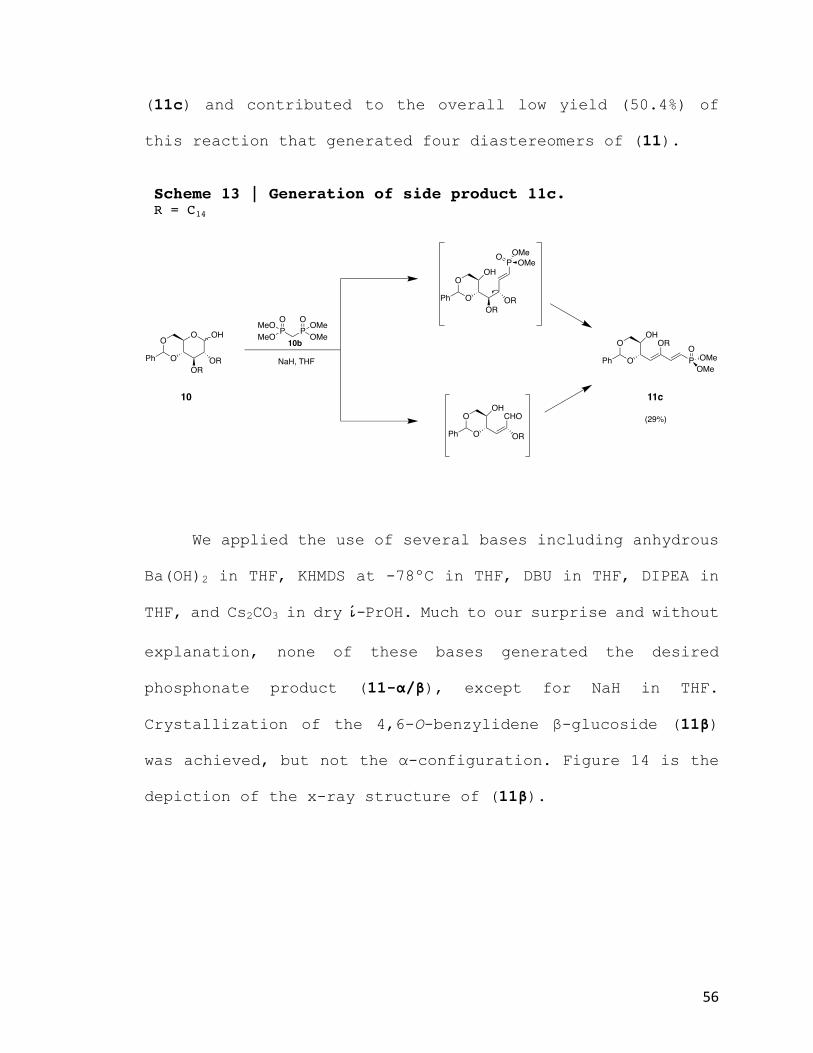
in Scheme 13. The elimination or reprotonation at C-2 produced
Scheme 12 | Synthesis of phosphono-sugar mimetics R = C14
56
(11c) and contributed to the overall low yield (50.4%) of
this reaction that generated four diastereomers of (11).
We applied the use of several bases including anhydrous
Ba(OH)2 in THF, KHMDS at -78ºC in THF, DBU in THF, DIPEA in
THF, and Cs2CO3 in dry i-PrOH. Much to our surprise and without
explanation, none of these bases generated the desired
phosphonate product (11-α/β), except for NaH in THF.
Crystallization of the 4,6-O-benzylidene β-glucoside (11β)
was achieved, but not the α-configuration. Figure 14 is the
depiction of the x-ray structure of (11β).
Scheme 13 | Generation of side product 11c. R = C14
O
OPh
O
OROR
OH
O
OPh
OH
OROR
PO OMeOMe
O
OPh
CHOOH
OR
O
OPh
OH
P
OR OOMe
OMe
P PO OMeO
MeOOMeOMe
NaH, THF
10b
10 11c
(29%)

57
At the reducing end of the sugar, the aldehyde is masked
in form of a hemiacetal. However, the equilibrium between the
hemiacetal and ring-opened form, especially considering ring
strain of the 4,6-O-benzylidene, is highly favored towards
the hemiacetal. Even so, the Wittig reaction can drive the
equilibrium entirely to the ring-opened form, producing the
newly formed olefin that can also be cyclized with addition
of a base. To this end, we attempted to utilize a Wittig
reagent that is readily available from Sigma Aldrich,
diphenyl(triphenylphosphoranylidenemethyl)phosphonate
Ph3P=CHPO(OBn)2, to introduce a phosphonate to (10) with
improved regioselectivity. This technique has been
demonstrated to produce C-glycosylated phosphonate analogues
with regioselectivity of the α-anomer. Moreover, the anomeric
Figure 14 | X-ray structure of 11. Only 4,6-O-benzylidene β-glucoside could be crystalized for x-ray crystallography.

58
ratio of the product mixture is dependent upon the time
exposure to the base. The Wittig method has been proven
valuable for the formation of olefins from aldehydes and
ketones using phosphorus ylides. Unfortunately, this method
too did not work on our compound (10).
7. Biological Activity Assay
Compounds (11α, 11β, 12α, 12β, 13α, 13β, & 22) were
selected to be evaluated for anti-inflammatory potential
against LPS-induced human acute monocytic leukemia (THP-1)
cells. THP-1 cells provide an ideal system for studying
inflammatory processes.88 They serve as a model for peripheral
monocytes/macrophages and their responsiveness to bacterial
infection.88 Unfortunately, benzylidene protected phosphono-
sugar mimetics 11α and 11β were not soluble in DMSO, therefore
were not suitable for cell culture studies. The same result
of insolubility was also observed for selectively ring-opened
6-O-benzyl ether compounds 13α and 13β. Albeit, they were
seemingly more soluble than the completely protected
benzylidene derivatives, more than likely due to the unveiled
hydroxy at C-4. However, both complete benzylidene cleaved
phosphono-mimetics 12α and 12β and lactone derivative 22 were
readily soluble in DMSO. Due to time constraints, we selected
to evaluate the targeted and more biologically relevant α-

59
anomer 12α and lactone derivative 22 by preincubating THP-1
cells with the compounds prior to cell stimulation with of E.
coli LPS as outlined in experimentals. Modulation of TNFα
production was analyzed in vitro and measured by ELISA.
8. Biological Activity
THP-1 cells were stimulated with LPS in the presence of
increasing concentrations of compounds 12α (Figure 14, red)
and 22 (Figure 15, red) to test antagonistic activity. THP-1
cells were also treated with increasing concentrations of
compounds 12α and 22 in the absence of LPS to examine
agonistic activity (Figure 14 & 15, blue). Neither
significantly demonstrated agonistic properties. The zero
concentration of compound represents a control treatment of
100 ng/mL LPS and the same percentage of DMSO included with
the compounds.

60
0
50
100
150
200
250
300
350
400
450
500
550
0 1 3 10 30 100
[TNFα](pg/mL)
Compound12α(µM)
Figure 14
[TNFα]w/LPS
[TNFα]w/oLPS
The targeted α-phosphonate 12α did not demonstrate
antagonistic properties. However, compound 12α appears to
elicit slight synergistic effects by increasing TNFα
production at 100 µM. This finding may be attributed to cell
toxicity.
Figure 14 | Effects to TNFα production in THP-1 cells inthepresenceofphosphono-sugar12α. Compound 12α does not elicit antagonistic effects. THP-1 cells were treated as described in experimentals. Y-axis shows TNFα concentration in pg/mL. X-axis displays increasing concentrations of 12α in the presence of LPS (red) and in absence of LPS (blue).

61
0
20
40
60
80
100
120
140
160
180
200
0 1 3 10 30 100
[TNF
α](pg/m
L)
Compound22(µM)
Figure 15
[TNFα]w/LPS
[TNFα]w/oLPS
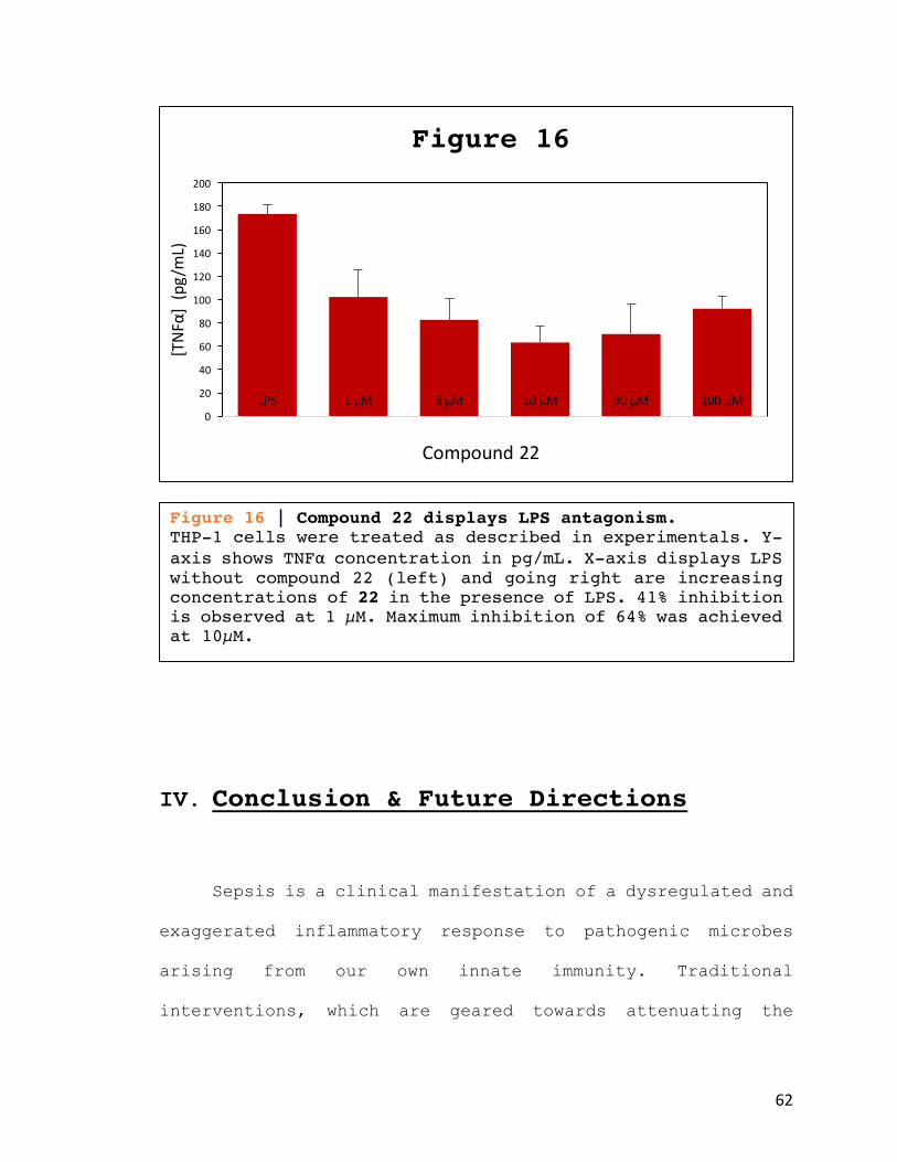
In contrast, D-glucono-δ-lactone derivative 22 did
demonstrate antagonism by reducing TNFα production by 41% at
the lowest tested concentration of 1 µM. At concentrations of
3 µM and 10 µM TNFα production was further decreased by 52%
and 64% respectively.
Figure 15 | Effects to TNFα production in THP-1 cells inthepresenceofD-glucono-δ-lactone derivative 22. Compound 22 displays antagonistic properties. THP-1 cells were treated as described in experimentals. Y-axis shows TNFα concentration in pg/mL. X-axis displays increasing concentrations of 22 in the presence of LPS (red) and in absence of LPS (blue).
62
IV. Conclusion & Future Directions
Sepsis is a clinical manifestation of a dysregulated and
exaggerated inflammatory response to pathogenic microbes
arising from our own innate immunity. Traditional
interventions, which are geared towards attenuating the
LPS 1 µM 3 µM 10 µM 30 µM 100 µM0
20
40
60
80
100
120
140
160
180
200[TNF
α](pg
/mL)
Compound22
Figure 16
Figure 16 | Compound 22 displays LPS antagonism. THP-1 cells were treated as described in experimentals. Y-axis shows TNFα concentration in pg/mL. X-axis displays LPS without compound 22 (left) and going right are increasing concentrations of 22 in the presence of LPS. 41% inhibition is observed at 1 µM. Maximum inhibition of 64% was achieved at 10µM.
63
symptoms of sepsis, have proved insufficient. This is
supported by rising annual healthcare expenditures and high
mortality rates in diagnosed patients. Moreover, antibiotic
resistant strains of bacteria are ever-growing and are a cause
for concern. Spearheaded efforts to enhance therapeutics by
disrupting underlying mechanisms of immunopathogenesis will
lead to improved patient outcomes.
The lipid A component of LPS causes immunopathogenesis
by initiating TLR4-MD-2 receptor dimerization, consequently
inducing the inflammatory pathway. Compounds that out-compete
LPS would disrupt TLR4-MD-2 signal transduction and
downregulate LPS signaling. Previously synthesized lipid A
analogues demonstrate that subtle structural changes
critically impact biological activity. Elucidating key
chemical structure-activity relationships between lipid A
variants will guide future explorations.
Our lipid A analogues were based upon the
monosaccharide Lipid X with the aim to produce TLR4-MD-2
antagonist with more robust, simpler, and readily scalable
chemical structures. Astute chemical modifications to
substituents were aimed at improving bioavailability and
solubility of lipid A analogues.
Synthesizing the C-glycosidic phosphono-sugar 12α
produced a non-hydrolysable analogue that could circumvent

64
inactivation by host phosphatases. Unfortunately, 12α did not
demonstrate LPS antagonism. Future attempts at synthesizing
a phosphonate derivative should cleave the methyl protecting
groups using TMSBr, which would yield the corresponding
phosphonic acid of 12α. In hind-site, this modification could
prove paramount in two ways. First, the anionic 1-phosphate
of lipid A binds to positive patches on TLR4. The significance
of this interaction has been established and is demonstrated
by the previously mentioned mutagenesis studies and the
attenuated endotoxicity of MPL (4). Second, the change in
ionic character between the methyl protected and de-protected
phosphonate substituent of 12α would potentially increase its
solubility profile in aqueous media.
D-glucono-δ-lactone derivative 22 evaded the solubility
issues compounds 11α, 11β, 12β, 13α, and 13β encountered from
their inherent amphipathic nature. More importantly, compound
22 demonstrated LPS antagonism in monocytic THP-1 cells,
representing the first lactone monosaccharide R. sin-1 lipid
A derivative to do so. Albeit, monosaccharide 22 does not
inhibit LPS signaling as much as some disaccharide compounds
like Eritoran (3). However, the comparative ease of synthesis
could outweigh their differences of inhibition.
Overall, this embodiment of work provides new
information regarding monosaccharide-based lipid A analogues.

65
Results from the studies herein provide a scientific basis
for future investigations of lipid A antagonist, with added
implications of inhibiting the adverse effects of septicemia.
It is my hope that this work reveals structural insights of
antagonistic properties to guide future explorations that
ultimately lead to improved patient outcomes.
V. Experimentals
1. General Procedures
Glassware used for all experiments were oven-dried and
all reactions were carried out under argon atmosphere unless
otherwise mentioned. All reaction solvents were purified
prior to use: CH2Cl2 was dried by distillation over calcium
hydride; THF was distilled over sodium. Reagent grade DMF and
HPLC grade MeOH was obtained from Sigma-Aldrich and used
without further purification.
NMR spectra were recorded at 600 MHz or 300 MHz. Chemical
shifts (δ) for 1H and 13C spectra are expressed in ppm relative
to internal standard (CDCl3: 7.26 for 1H and 77.23 for 13C).
Chemical shifts for 31P were referenced with phosphoric acid

66
and then back calculated. Signals were abbreviated as s,
singlet; d, doublet; dd, doublet of doublets; t, triplet; q,
quartet; m, multiplet. Thin layer chromatography (TLC) was
carried out with pre-coated Merck F254 silica gel plates.
Flash chromatography (FC) was carried out with Macherey-Nagel
silica gel 60-230 mesh. Automated preparative chromatography
was performed using a Biotage Isolera Prime or reverse phase
C18 HPLC preparatory system.
2. General Procedure for selective C4 ring opening of 4,6-O-
benzylidene
A stirred solution of 4,6-O-Benzylidene (0.1 mmol) in
dry THF (0.66 mL) containing 4 Å molecular sieves was added
NaCNBH3 (13.3 eq.). The reaction mixture was stirred for 5
min. and subsequent addition of 2 N HCl/Et2O (13.3 eq.) was
added in portions using a syringe over 10 min. The reaction
was monitored by TLC until complete disappearance of starting
material. The reaction mixture was then filtered through
celite, diluted with DCM, washed with H2O (x1), HCO3- (x1),
H2O (x1), dried over Na2SO4 and concentrated in vacuo. The
residue was separated by preparative HPLC.

67
3. General Procedure for C6/C4 complete ring cleavage of 4,6-
O-benzylidene
4,6-O-Benzylidene (0.05 mmol) was dissolved in TFA and
DCM (15% TFA in DCM, 0.7 mL) and stirred for 20 min. One drop
of H2O was added and the reaction was stirred for 1 h. Reaction
was monitored by TLC until complete and then concentrated in
vacuo. The residue was azeotroped with toluene (1 mL x 4) and
finally concentrated in vacuo. The residue was separated by
preparative HPLC.
Synthesis of 4,6-O-benzylidene-2,3-O-tetradecane-β-
thiophenol-D-glucopyranose (9)
A stirred solution of 4,6-O-benzylidene-2,3-diol-β-
thiophenol-D-glucopyranose 87 (8) (9.15 g, 25.25 mmol) in dry
DMF (18 mL) containing 4 Å molecular sieves was cooled to
0˚C. A suspension of NaH (1.4g, 55.55 mmol) mixed in dry THF
(12 mL) was added portion wise over 15 minutes. The reaction
mixture was allowed to warm up to 40˚C and was stirred for 30
minutes. A catalytic amount of tetra-N-butylammonium iodide
(0.933g, 2.52 mmol) was added to the mixture immediately
O
OHSPh
OO NaH, DMF, RBr
R = C14H29
8
HO
PhO
ORSPh
OO
9
RO
Ph

68
followed by dropwise addition of bromo-tetradecane (15.4 g,
55.55 mmol) over 30 minutes. Reaction was monitored by TLC.
After 24 hours, no further conversion of the starting material
could be detected. The reaction mixture was cooled to 0˚C,
diluted with CH2Cl2 (30 mL), quenched with NH4Cl, and washed
with H2O (x 3). The organic phase was dried over Na2SO4,
filtered and concentrated in vacuo. The residue was purified
by column chromatography on silica gel (ethyl acetate-hexane
gradient elution) to afford the title compound (9) (14.8 g,
78%) as a white solid. (9): 1H NMR (600 MHz Chloroform-d) δ
7.55 – 7.24 (m, 10H), 5.53 (s, 1H), 4.65 (d, J = 9.8 Hz, 1H),
4.34 (dd, J = 10.5, 5.0 Hz, 1H), 3.84 (m, 1H), 3.76 (m, 3H),
3.68 (m, 1H), 3.53 (m, 2H), 3.44 – 3.36 (m, 1H), 3.21 (dd, J
= 9.7, 8.0 Hz, 1H), 1.66 – 1.50 (m, 10H), 1.42 – 1.18 (m,
38H), 0.88 (t, J = 7.0 Hz, 6H).
Synthesis of 4,6-diol-2,3-O-tetradecane-β-thiophenol-D-
glucopyranose (14)
O
OR
HOHO
14
ROO
OR
OO
9
RO
Ph15% TFA in DCM
H2OSPh SPh
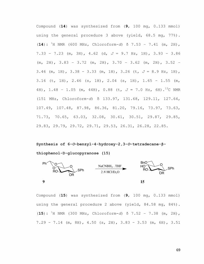
69
Compound (14) was synthesized from (9, 100 mg, 0.133 mmol)
using the general procedure 3 above (yield, 68.5 mg, 77%).
(14): 1H NMR (600 MHz, Chloroform-d) δ 7.53 – 7.41 (m, 2H),
7.33 – 7.23 (m, 3H), 4.62 (d, J = 9.7 Hz, 1H), 3.93 – 3.86
(m, 2H), 3.83 – 3.72 (m, 2H), 3.70 – 3.62 (m, 2H), 3.52 –
3.46 (m, 1H), 3.38 – 3.33 (m, 1H), 3.26 (t, J = 8.9 Hz, 1H),
3.16 (t, 1H), 2.46 (s, 1H), 2.04 (s, 1H), 1.65 – 1.55 (m,
4H), 1.48 – 1.05 (m, 44H), 0.88 (t, J = 7.0 Hz, 6H).13C NMR
(151 MHz, Chloroform-d) δ 133.97, 131.68, 129.11, 127.64,
107.49, 107.48, 87.98, 86.36, 81.20, 79.16, 73.97, 73.63,
71.73, 70.65, 63.03, 32.08, 30.61, 30.51, 29.87, 29.85,
29.83, 29.79, 29.72, 29.71, 29.53, 26.31, 26.28, 22.85.
Synthesis of 6-O-benzyl-4-hydroxy-2,3-O-tetradecane-β-
thiophenol-D-glucopyranose (15)
Compound (15) was synthesized from (9, 100 mg, 0.133 mmol)
using the general procedure 2 above (yield, 84.58 mg, 84%).
(15): 1H NMR (300 MHz, Chloroform-d) δ 7.52 – 7.38 (m, 2H),
7.29 – 7.14 (m, 8H), 4.50 (s, 2H), 3.83 – 3.53 (m, 6H), 3.51
O
OR
BnOHO
15
ROO
OR
OO
9
RO
PhNaCNBH3 , THF
2 N HCl/Et2OSPh SPh

70
– 3.33 (m, 2H), 3.26 – 3.01 (m, 2H), 2.60 (s, 1H), 1.61 –
1.46 (m, 5H), 1.31 – 1.14 (m, 44H), 0.82 (t, J = 6.5 Hz, 6H).
Synthesis of 4,6-O-benzylidene-2,3-O-tetradecane-α/β-D-
glucopyranose (10)
Thio-glucoside (9) (1.022 g, 1.357 mmol) was dissolved in
CH2Cl2 (14 mL) and the reaction mixture was stirred and cooled
to 0˚ C. NIS (335.83 mg, 1.493 mmol 1.1 equiv.) and TFA
(114.30 µL, 1.493 mmol, 1.1 equiv.) were added. The reaction
was allowed to warm to room temp. After 1 h, the reaction
mixture was cooled to 0˚ C, and piperidine (402.12 µL, 4.071
mmol, 3 equiv.) was added. After 30 min, TLC analysis showed
total conversion of the anomeric trifluoroacetate into a
compound with a lower Rf (DCM/Hexanes 5:3, v/v, Rf 0.10). The
reaction mixture was quenched by the subsequent addition of
triethylamine (the reaction turned from dark red to yellow)
and Na2S2O3 (aq., 20%, the yellow reaction mixture turned
colorless). The reaction mixture was diluted with CH2Cl2,
washed with H2O, dried with Na2SO4, filtered through celite,
and concentrated in vacuo. Flash chromatography
O
ORSPh
OO
9
RO
Ph1. N-IS , TFA
2. Piperidene
O
OR
OO OH
10
RO
Ph
(84-94%)

71
(EtOAc:Hexanes 1.25:10, v/v, isocratic) afforded hemiacetal
(10) as an anomeric mixture (α/β 1:1, 760.3 mg, 84%) as a
white solid. (10): 1H NMR (300 MHz, Chloroform-d) δ 7.53 –
7.43 (m, 2H), 7.42 – 7.30 (m, 3H), 5.54 (s, 1H), 5.30 (d, J
= 3.8 Hz, 0.5H), 4.73 (d, J = 7.7 Hz, 0.5H), 4.36 – 4.27 (m,
1H), 4.12 – 3.94 (m, 1H), 3.86 – 3.75 (m, 2H), 3.73 – 3.63
(m, 3H), 3.61 – 3.44 (m, 2H), 3.43 – 3.34 (m, 1H), 3.12 (t,
J = 8.0 Hz, 1H), 1.57 (s, 10H), 1.35 – 1.18 (m, 38H), 0.88
(t, J = 6.5 Hz, 6H).
Synthesis of 4,6-O-benzylidene-2,3-O-tetradecane-α/β-D-
methylene-dimethyl-phosphono-glucoside (11)
A stirred solution of dry CH2Cl2:THF (12 mL, 1:3 respectively)
and NaH (95% oil dispersion, 106.95 mg, 4.282 mmol, 1.5 eq.)
was cooled to 0˚C. ((MeO)2(O)P)2CH2 (633.6 µL, 3.426 mmol, 1.2
eq.) was added portion wise with a syringe over 15 minutes.
The reaction mixture was allowed to warm up to room
temperature and was stirred for 30 minutes. The reaction
mixture was again cooled to 0˚C and (10) (1.877 g, 2.855 mmol)
in dry CH2Cl2:THF (4 mL, 1:3 respectively) was added dropwise
O
OR
OO OH
10
RO
Ph((MeO)2(O)P)2CH2
NaH , THF
O
OR
OO
PO
OMeOMe
11
RO
Ph
72
via an addition funnel over 30 mins. The reaction mixture was
allowed to warm up to room temperature and was monitored via
HPLC. After 23 h, the reaction mixture was cooled to 0˚C,
quenched with NH4Cl, diluted with CH2Cl2, washed with H2O (x1),
HCO3- (aq. satd., x1), H2O (x1), dried with Na2SO4, filtered
through celite, and concentrated in vacuo. Flash
chromatography (EtOAc:Hexanes 1:4, v/v, isocratic) afforded
(11) (1.104 g, 50.4%) as a white solid mixture of four
diasteromers. The mixture was separated by preparative HPLC
to afford; (11c-manno, 24.34 mg, 1.11%, Rf 4.331,); (7a-9a,
351.11 mg, 16.03%, Rf 5.875); (11d-manno, 31.56 mg, 1.44%, Rf
8.182); (15a-20a, 427.32 mg, 19.51%, Rf 8.353) white solids.
(11c-manno): 1H NMR (600 MHz, Chloroform-d) δ 7.50 – 7.43 (m,
2H), 7.38 – 7.28 (m, 3H), 5.60 (s, 1H), 4.47 (q, J = 7.7 Hz,
1H), 4.20 (dd, J = 10.2, 4.6 Hz, 1H), 4.10 (t, J = 9.6 Hz,
1H), 3.85 – 3.54 (m, 14H), 2.31 – 2.22 (m, 1H), 2.13 – 2.04
(m, 1H), 1.66 – 1.55 (m, 5H), 1.38 – 1.22 (m, 45H), 0.88 (t,
J = 6.7 Hz, 6H). 13C NMR (151 MHz, Chloroform-d) δ 137.74,
128.87, 128.26, 126.10, 101.40, 79.06, 78.39, 78.32, 76.17,
72.64, 71.99, 71.29, 69.07, 66.22, 52.90, 52.86, 52.68,
52.64, 32.07, 30.18, 29.99, 29.86, 29.84, 29.82, 29.69,
29.66, 29.52, 26.59, 26.27, 26.18, 25.66, 22.84, 14.27. 31P
NMR (243 MHz, Chloroform-d) δ 30.32. (11α): 1H NMR (600 MHz,
Chloroform-d) δ 7.52 – 7.44 (m, 2H), 7.39 – 7.29 (m, 3H),
73
5.58 (s, 1H), 4.24 (dd, J = 10.3, 4.9 Hz, 1H), 4.03 – 3.95
(m, 2H), 3.92 – 3.87 (m, 1H), 3.81 (t, J = 10.3 Hz, 1H), 3.78
– 3.69 (m, 7H), 3.63 – 3.58 (m, 1H), 3.56 – 3.48 (m, 2H),
3.44 – 3.39 (m, 1H), 2.20 (ddd, J = 18.4, 6.3, 4.8 Hz, 2H),
1.64 – 1.56 (m, 4H), 1.42 – 1.19 (m, 44H), 0.88 (t, J = 7.0
Hz, 6H). 13C NMR (151 MHz, Chloroform-d) δ 137.77, 128.88,
128.25, 126.15, 101.38, 81.10, 78.77, 74.84, 74.41, 72.01,
71.71, 68.59, 52.87, 52.83, 52.29, 52.24, 32.08, 30.44,
30.28, 29.88, 29.85, 29.83, 29.72, 29.65, 29.53, 27.95,
27.01, 26.28, 26.25, 22.85, 14.28. 31P NMR (243 MHz,
Chloroform-d) δ 31.67. (11d-manno): 1H NMR (600 MHz,
Chloroform-d) δ 7.50 – 7.44 (m, 2H), 7.39 – 7.32 (m, 3H),
5.53 (s, 1H), 4.57 – 4.51 (m, 1H), 4.25 (dd, J = 9.8, 4.3 Hz,
1H), 3.80 – 3.70 (m, 7H), 3.69 – 3.60 (m, 4H), 3.58 – 3.52
(m, 2H), 3.51 – 3.48 (m, 1H), 3.47 – 3.44 (m, 1H), 2.28 –
2.18 (m, 2H), 1.65 (s, 1H), 1.59 – 1.51 (m, 4H), 1.37 – 1.22
(m, 44H), 0.88 (t, J = 7.0 Hz, 6H). 13C NMR (151 MHz,
Chloroform-d) δ 137.54, 128.99, 128.32, 126.10, 101.27,
82.31, 79.66, 79.57, 78.68, 73.33, 72.26, 71.14, 71.10,
69.45, 64.34, 52.60, 52.56, 52.52, 32.08, 30.47, 30.27,
29.87, 29.84, 29.82, 29.80, 29.71, 29.66, 29.53, 26.29,
26.26, 22.85, 14.28, 1.17. 31P NMR (243 MHz, Chloroform-d) δ
32.76. (11β): 1H NMR (600 MHz, Chloroform-d) δ 7.49 – 7.45
(m, 2H), 7.39 – 7.32 (m, 3H), 5.52 (s, 1H), 4.31 (dd, J =

74
10.3, 4.9 Hz, 1H), 3.91 – 3.85 (m, 2H), 3.76 – 3.71 (m, 6H),
3.70 – 3.68 (m, 1H), 3.68 – 3.66 (m, 1H), 3.66 – 3.63 (m,
1H), 3.55 – 3.48 (m, 3H), 3.46 – 3.42 (m, 1H), 3.05 (t, 1H),
2.40 – 2.33 (m, 1H), 1.97 – 1.90 (m, 1H), 1.59 – 1.54 (m,
4H), 1.31 – 1.20 (m, 44H), 0.88 (t, J = 7.0 Hz, 6H). 13C NMR
(151 MHz, Chloroform-d) δ 137.54, 129.01, 128.34, 126.12,
101.23, 83.19, 82.41, 82.32, 82.17, 75.39, 73.94, 73.47,
70.64, 68.86, 52.67, 52.31, 32.09, 30.58, 30.54, 29.87,
29.83, 29.72, 29.71, 29.53, 26.34, 26.32, 22.85, 14.29. 31P
NMR (243 MHz, Chloroform-d) δ 31.83.
Synthesis of 6-O-benzyl-4-hydroxy-2,3-O-tetradecane-α-D-
methylene-dimethyl-phosphono-glucoside (13α)
Compound (13α) was synthesized from (11α, 100 mg, 0.1304 mmol)
using the general procedure 2 above (yield, 92.27 mg, 92%).
(13α): 1H NMR (300 MHz, Chloroform-d) δ 7.35 – 7.26 (m, 5H),
4.89 (d, J = 10.8 Hz, 1H), 4.60 (d, J = 11.0 Hz, 1H), 3.98 –
3.87 (m, 1H), 3.84 – 3.60 (m, 10H), 3.52 (t, J = 7.9 Hz, 2H),
3.45 – 3.38 (m, 1H), 3.38 – 3.27 (m, 1H), 2.28 – 2.07 (m,
O
OR
BnOHO
13α
ROO
OR
OO
11α
RO
Ph
PPO O
OMe OMeOMeOMe
NaCNBH3 , THF2 N HCl/Et2O

75
2H), 1.71 – 1.51 (m, 9H), 1.39 – 1.18 (m, 42H), 0.88 (t, J =
6.5 Hz, 6H).
Synthesis of 6-O-benzyl-4-hydroxy-2,3-O-tetradecane-β-D-
methylene-dimethyl-phosphono-glucoside (13β)
Compound (13β) was synthesized from (11β, 35.6 mg, 0.0464
mmol) using the general procedure 2 above (yield, 32.8 mg,
92%).(13β): 1H NMR (600 MHz, Chloroform-d) δ 7.38 – 7.23 (m,
5H), 4.58 – 4.52 (m, 2H), 3.83 – 3.75 (m, 2H), 3.75 – 3.65
(m, 8H), 3.65 – 3.35 (m, 5H), 3.22 (t, J = 8.9 Hz, 1H), 2.96
(t, J = 9.2 Hz, 1H), 2.65 (s, 1H), 2.36 – 2.29 (m, 1H), 1.96
– 1.89 (m, 1H), 1.78 – 1.61 (m, 2H), 1.61 – 1.48 (m, 4H),
1.48 – 1.07 (m, 42H), 0.87 (t, J = 7.0 Hz, 6H). 13C NMR (151
MHz, Chloroform-d) δ 137.93, 128.54, 127.90, 86.43, 86.41,
82.18, 82.09, 78.03, 74.81, 74.76, 73.73, 73.50, 72.12,
70.45, 52.71, 52.67, 52.28, 52.24, 32.08, 30.65, 30.55,
29.87, 29.85, 29.82, 29.80, 29.73, 29.72, 29.52, 28.54,
27.59, 26.31, 22.85, 14.28. 31P NMR (243 MHz, Chloroform-d) δ
32.20.
O
OR
BnOHO
13β
ROO
OR
OO
11β
RO
PhNaCNBH3 , THF
2 N HCl/Et2OPP
O OOMe OMe
OMeOMe

76
Synthesis of 4,6-diol-2,3-O-tetradecane-α-D-methylene-
dimethyl-phosphono-glucoside (12α)
Compound (12α) was synthesized from (11α, 44 mg, 0.0574 mmol)
using the general procedure 3 above (yield, 32.7 mg, 84%).
(12α): 1H NMR (600 MHz, Chloroform-d) δ 3.91 – 3.58 (m, 13H),
3.47 (q, J = 6.9 Hz, 1H), 3.43 – 3.39 (m, 1H), 3.37 – 3.32
(m, 1H), 3.24 (dd, J = 9.4, 2.4 Hz, 1H), 2.49 (s, 1H), 2.27
– 2.21 (m, 1H), 2.15 – 2.09 (m, 1H), 1.84 (s, 1H), 1.63 –
1.54 (m, 4H), 1.44 – 1.09 (m, 44H), 0.87 (t, J = 7.0 Hz, 6H).
13C NMR (151 MHz, Chloroform-d) δ 84.76, 79.77, 75.37, 75.31,
73.68, 73.65, 70.24, 67.51, 63.28, 52.77, 52.72, 52.50,
52.46, 32.07, 30.41, 30.11, 29.85, 29.83, 29.81, 29.80,
29.77, 29.76, 29.65, 29.51, 28.03, 27.09, 26.29, 26.22,
22.84, 14.27. 31P NMR (243 MHz, Chloroform-d) δ 31.84.
O
OR
HOHO
12α
ROO
OR
OO
11α
RO
Ph
PPO O
OMe OMeOMeOMe
15% TFA in DCMH2O

77
Synthesis of 4,6-diol-2,3-O-tetradecane-β-D-methylene-
dimethyl-phosphono-glucoside (12β)
Compound (12β) was synthesized from (11β, 39 mg, 0.0508 mmol)
using the general procedure 3 above (yield, 20.8 mg, 60%).
(12β): 1H NMR (600 MHz, Chloroform-d) δ 3.84 – 3.69 (m, 10H),
3.58 – 3.44 (m, 4H), 3.33 (dd, J = 7.4, 4.7 Hz, 1H), 3.23 (t,
J = 9.0 Hz, 1H), 2.95 (t, J = 9.2 Hz, 1H), 2.35 – 2.28 (m,
1H), 2.00 – 1.91 (m, 2H), 1.65 – 1.47 (m, 5H), 1.38 – 1.06
(m, 44H), 0.93 – 0.79 (m, 6H). 13C NMR (151 MHz, Chloroform-
d) δ 86.43, 86.41, 82.62, 82.53, 79.71, 74.57, 74.52, 73.79,
73.55, 71.07, 62.62, 52.74, 52.69, 52.61, 52.57, 32.07,
30.65, 30.56, 29.86, 29.85, 29.82, 29.81, 29.79, 29.74,
29.72, 29.52, 28.63, 27.68, 26.31, 26.30, 22.84, 14.27. 31P
NMR (243 MHz, Chloroform-d) δ 32.47.
O
OR
HOHO
12β
ROO
OR
OO
11β
RO
Ph 15% TFA in DCM
PPO O
OMe OMeOMeOMe
H2O
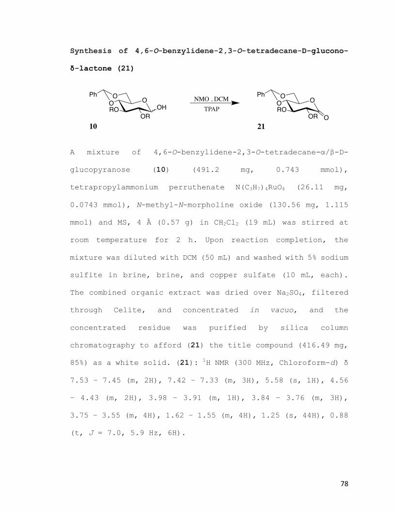
78
Synthesis of 4,6-O-benzylidene-2,3-O-tetradecane-D-glucono-
δ-lactone (21)
A mixture of 4,6-O-benzylidene-2,3-O-tetradecane-α/β-D-
glucopyranose (10) (491.2 mg, 0.743 mmol),
tetrapropylammonium perruthenate N(C3H7)4RuO4 (26.11 mg,
0.0743 mmol), N-methyl-N-morpholine oxide (130.56 mg, 1.115
mmol) and MS, 4 Å (0.57 g) in CH2Cl2 (19 mL) was stirred at
room temperature for 2 h. Upon reaction completion, the
mixture was diluted with DCM (50 mL) and washed with 5% sodium
sulfite in brine, brine, and copper sulfate (10 mL, each).
The combined organic extract was dried over Na2SO4, filtered
through Celite, and concentrated in vacuo, and the
concentrated residue was purified by silica column
chromatography to afford (21) the title compound (416.49 mg,
85%) as a white solid. (21): 1H NMR (300 MHz, Chloroform-d) δ
7.53 – 7.45 (m, 2H), 7.42 – 7.33 (m, 3H), 5.58 (s, 1H), 4.56
– 4.43 (m, 2H), 3.98 – 3.91 (m, 1H), 3.84 – 3.76 (m, 3H),
3.75 – 3.55 (m, 4H), 1.62 – 1.55 (m, 4H), 1.25 (s, 44H), 0.88
(t, J = 7.0, 5.9 Hz, 6H).
O
OR
OO OHRO
Ph
TPAPNMO , DCM O
OR
OORO
Ph
O10 21

79
Synthesis of 6-O-benzyl-4-hydroxy-2,3-O-tetradecane-D-
glucono-δ-lactone (23)
Compound (23) was synthesized from (21, 100 mg, 0.1517 mmol)
using the general procedure 2 above (yield, 78.66 mg, 78%).
(23): 1H NMR (300 MHz, Chloroform-d) δ 7.28 – 7.13 (m, 5H),
4.46 (d, J = 2.0 Hz, 2H), 3.73 – 3.38 (m, 11H), 2.81 (s, 1H),
2.53 (s, 1H), 1.54 – 1.38 (m, 4H), 1.14 (s, 42H), 0.76 (t, J
= 6.0 Hz, 6H).
Synthesis of 4,6-diol-2,3-O-tetradecane-D-glucono-δ-lactone
(22)
Compound (22) was synthesized from (21, 50 mg, 0.07587 mmol)
using the general procedure 3 above (yield, 35.08 mg, 81%).
(22): 1H NMR (300 MHz, Chloroform-d) δ 4.60 – 4.21 (m, 1H),
4.11 (dd, J = 13.8, 4.8 Hz, 1H), 4.03 – 3.96 (m, 1H), 3.91 –
3.60 (m, 6H), 3.60 – 3.32 (m, 2H), 3.15 (s, 1H), 2.18 (s,
O
OR
BnOHO
23
ROO
OR
OO
21
RO
Ph NaCNBH3 , THF2 N HCl/Et2O
OO
O
OR
HOHO
22
ROO
OR
OO
21
RO
Ph
OO
15% TFA in DCMH2O
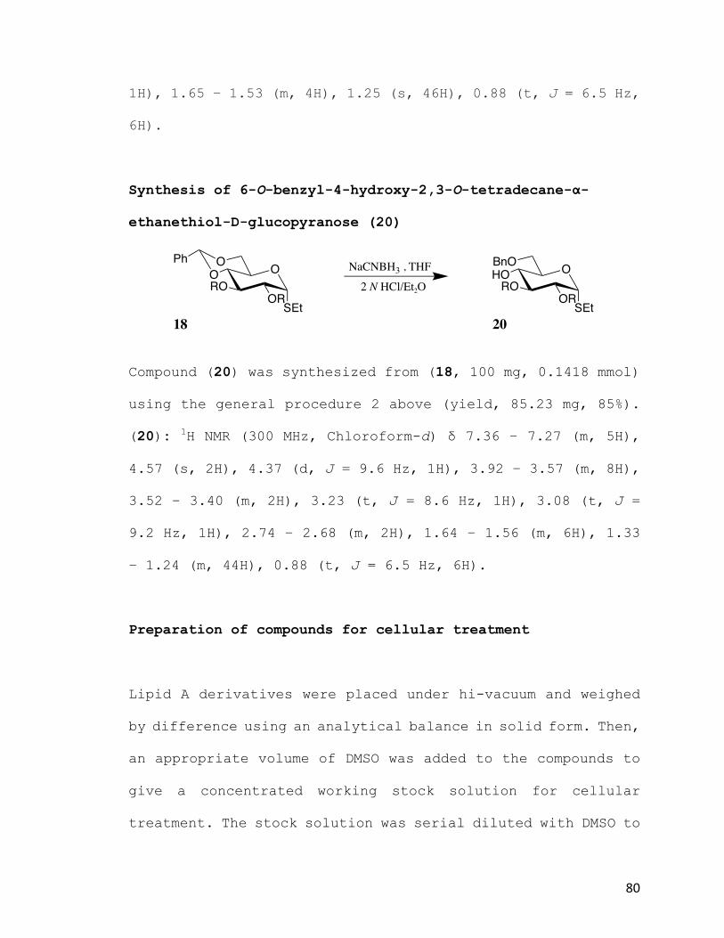
80
1H), 1.65 – 1.53 (m, 4H), 1.25 (s, 46H), 0.88 (t, J = 6.5 Hz,
6H).
Synthesis of 6-O-benzyl-4-hydroxy-2,3-O-tetradecane-α-
ethanethiol-D-glucopyranose (20)
Compound (20) was synthesized from (18, 100 mg, 0.1418 mmol)
using the general procedure 2 above (yield, 85.23 mg, 85%).
(20): 1H NMR (300 MHz, Chloroform-d) δ 7.36 – 7.27 (m, 5H),
4.57 (s, 2H), 4.37 (d, J = 9.6 Hz, 1H), 3.92 – 3.57 (m, 8H),
3.52 – 3.40 (m, 2H), 3.23 (t, J = 8.6 Hz, 1H), 3.08 (t, J =
9.2 Hz, 1H), 2.74 – 2.68 (m, 2H), 1.64 – 1.56 (m, 6H), 1.33
– 1.24 (m, 44H), 0.88 (t, J = 6.5 Hz, 6H).
Preparation of compounds for cellular treatment
Lipid A derivatives were placed under hi-vacuum and weighed
by difference using an analytical balance in solid form. Then,
an appropriate volume of DMSO was added to the compounds to
give a concentrated working stock solution for cellular
treatment. The stock solution was serial diluted with DMSO to
O
OR
BnOHO
20
ROO
OR
OO
18
RO
Ph NaCNBH3 , THF2 N HCl/Et2O
SEt SEt

81
afford the desired concentration range. The final DMSO
concentration was maintained at 0.5%.
LPS antagonistic activity assay and cell culture
Human THP-1 cells that were in 1 mL aliquots and were stored
in liquid nitrogen (Cane 4-level 1), were thawed and diluted
1:10 in fresh growth medium and cultured in a T-75 flask.
THP-1 growth medium was RPMI 1640 containing 10% FBS, 50 U/mL
penicillin, 50 µg/mL streptomycin, 50µM β-mercapto-ethanol.
THP-1 assay medium was the same as growth medium but with
only 2% FBS. For cellular assays, THP-1 monocytes were
centrifuged and resuspended to a cell concentration of 1*106
cells/mL. LPS dilutions were conducted from 1 mg/mL UP LPS
Invivogen Stock to achieve desired final concentration (see
plate arrangement below). 96-well plate was then treated as
follows: (1) Added 5µl of the LPS to some wells and added
sterile water to the control wells, with 3 replicates for
each concentration and 0.5% DMSO was used for control i.e. A
(DMSO only) is added to wells A1, A2, and A3 triplicates with
LPS and A4, A5, & A6 triplicates without LPS and so forth,
(2) cells were incubated at 37ºC for 6 hours. After 6 hours,
the entire solution from each well was removed, centrifuged
for 10 mins to remove cells and the supernatant was placed in
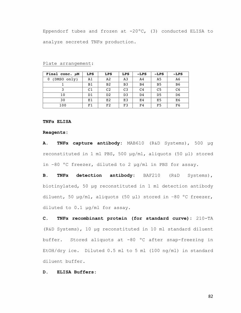
82
Eppendorf tubes and frozen at -20ºC, (3) conducted ELISA to
analyze secreted TNFα production.
Plate arrangement:
Final conc. µM LPS LPS LPS -LPS -LPS -LPS 0 (DMSO only) A1 A2 A3 A4 A5 A6
1 B1 B2 B3 B4 B5 B6 3 C1 C2 C3 C4 C5 C6 10 D1 D2 D3 D4 D5 D6 30 E1 E2 E3 E4 E5 E6 100 F1 F2 F3 F4 F5 F6
TNFα ELISA
Reagents:
A. TNFα capture antibody: MAB610 (R&D Systems), 500 µg
reconstituted in 1 ml PBS, 500 µg/ml, aliquots (50 µl) stored
in –80 ºC freezer, diluted to 2 µg/ml in PBS for assay.
B. TNFα detection antibody: BAF210 (R&D Systems),
biotinylated, 50 µg reconstituted in 1 ml detection antibody
diluent, 50 µg/ml, aliquots (50 µl) stored in –80 ºC freezer,
diluted to 0.1 µg/ml for assay.
C. TNFα recombinant protein (for standard curve): 210-TA
(R&D Systems), 10 µg reconstituted in 10 ml standard diluent
buffer. Stored aliquots at -80 ºC after snap-freezing in
EtOH/dry ice. Diluted 0.5 ml to 5 ml (100 ng/ml) in standard
diluent buffer.
D. ELISA Buffers:

83
Wash; PBS containing 0.05% Tween 20 (5 ml 5% Tween 20, dilute
with PBS to 500 ml); Sample Diluent, 20 mM Tris, pH 7.3
containing 150 mM NaCl, 0.1% BSA, and 0.05% Tween 20; Blocking
buffer, PBS containing 1% BSA, 5% sucrose, and 0.05% NaN3;
Detection Antibody Diluent, 20 mM Tris, pH 7.3 containing 150
mM NaCl and 0.1% BSA; Conjugate Diluent, PBS containing 1%
BSA; Stop Solution, 1 M H2SO4, prepared from 96% stock. Diluted
1:10 to make 1.8 M for assay; Standard Diluent, PBS containing
0.1% BSA.
Plate Preparation for ELISA
Applied 100 µl capture antibody (2 µg/ml) to required wells.
Sealed and incubated overnight at room temperature. Then
washed x3 with wash buffer and applied 300 µl blocking buffer.
Then incubated 1 h at room temperature followed by wash.
ELISA Assay Procedure:
Standard curve preparation – Diluted refrigerated standard
(0.1 µg/ml) to 2000 pg/ml and performed serial dilutions in
standard diluent buffer with 60 µl.

84
Then added 50 µl sample diluent buffer plus 50 µl standards
and samples. Used 50 µl H20 for 0 pg/ml standard and mixed
by gently tapping plate. Then sealed and incubated at room
temperature for 2 h. Then washed 3x with wash buffer and added
100 µl biotinylated detection antibody (0.1 µg/ml) and sealed
and incubated for 2 h at room temperature. Then washed 3x
with wash buffer and added 100 µl streptavidin HRP with 1/200
dilution into conjugate diluent, sealed and incubated for 20
min at room temperature. Then washed 3x with wash buffer and
prepared substrate solution by mixing equal volumes of color
reagent A & B within 15 minutes of use. Then added 100 µl of
substrate solution to each well and incubated for 30 minutes.
Then added 50 µl stop solution and mixed by gently tapping.
Then covered and read wells with absorbance plate reader at
450 nm and at 630 nm to subtract out optical differences
between wells.
1000 500 250 125 62.5 31.25 15.625stock
2000
60µl
2 3 4 5 6 71

85
VI. References
1.Madigan,M.T.,Martinko,J.M.,Bender,K.S.,Buckley,D.H.&Stahl,D.A.Brock
biologyofmicroorganisms.(2014).
2.PappaterraMendoza,G.J.etal.InVitroandInVivoEffectsofanImmunomodulator
ComposedofEscherichiaColiLipopolysaccharideandPropionibacteriumgranulosum-
InactivatedCellsinPigs.J.Vet.Med.Ser.B47,619–627(2000).
3.Nduka,O.O.&Parrillo,J.E.ThePathophysiologyofSepticShock.Sepsis23,41–66
(2011).
4.Novosad,S.&Sapiano,M.EpidemiologyofSepsis:PrevalenceofHealthCareFactors
andOpportunitiesforPrevention.Morb.Mortal.Wkly.Rep.MMWR65,864–869
(2016).
5.Motzkus,C.A.etal.ICUAdmissionSourceasaPredictorofMortalityforPatients
WithSepsis.J.IntensiveCareMed.885066617701904(2017).
doi:10.1177/0885066617701904
6.Torio,C.&Moore,B.NationalInpatientHospitalCosts:TheMostExpensive
ConditionsbyPayer,2013.(2016).
7.GINSBURG,I.Theroleofbacteriolysisinthepathophysiologyofinflammation,
infectionandpost-infectioussequelae.APMIS110,753–770(2002).

86
8.Penack,O.etal.Managementofsepsisinneutropenicpatients:2014updated
guidelinesfromtheInfectiousDiseasesWorkingPartyoftheGermanSocietyof
HematologyandMedicalOncology(AGIHO).Ann.Hematol.93,1083–1095(2014).
9.Schaffzin,D.M.&Wong,W.D.NonoperativeManagementofComplicated
DiverticularDisease.Clin.ColonRectalSurg.17,169–176(2004).
10. Annane,D.etal.Corticosteroidsforseveresepsisandsepticshock:asystematic
reviewandmeta-analysis.BMJ329,480(2004).
11. Salluh,J.I.F.&Póvoa,P.CorticosteroidsinSevereSepsisandSepticShock:A
ConciseReview.Shock47,(2017).
12. Canady,J.,Williams,W.,Thompson,I.,Vincent,G.S.&Hoover,E.Useof
naloxoneinsepticshock.J.Natl.Med.Assoc.81,669–673(1989).
13. Oettinger,C.W.&D’Souza,M.J.Microencapsulateddrugdelivery:anew
approachtopro-inflammatorycytokineinhibition.J.Microencapsul.29,455–462
(2012).
14. Werdan,K.PathophysiologyofSepticShockandMultipleOrganDysfunction
SyndromeandVariousTherapeuticApproacheswithSpecialEmphasison
Immunoglobulins.Ther.Apher.5,115(2001).
15. WESTPHAL,O.&LUDERITZ,O.Chemicalandbiologicalanalysisofhighlypurified
bacterialpolysaccharides.DtschMedWochenschr78,9–17(1953).
16. Christie,W.LipidAandbacteriallipopolysaccharides:structure,occurrenceand
biology.lipidlibrary.aocs.org(2014).Availableat:
http://lipidlibrary.aocs.org/Primer/content.cfm?ItemNumber=39339.

87
17. Heinrichs,D.E.,Yethon,J.A.&Whitfield,C.Molecularbasisforstructural
diversityinthecoreregionsofthelipopolysaccharidesofEscherichiacoliand
Salmonellaenterica.Mol.Microbiol.30,221–232(1998).
18. Tacken,A.,Rietschel,E.T.&Brade,H.Methylationanalysisoftheheptose/3-
deoxy-d-manno-2-octulosonicacidregion(innercore)ofthelipopolysaccharidefrom
Salmonellaminnesotaroughmutants.Carbohydr.Res.149,279–291(1986).
19. Sadovskaya,I.etal.StructuralcharacterizationoftheoutercoreandtheO-chain
linkageregionoflipopolysaccharidefromPseudomonasaeruginosaserotypeO5.Eur.
J.Biochem.267,1640–1650(2000).
20. Lerouge,I.&Vanderleyden,J.O-antigenstructuralvariation:mechanismsand
possiblerolesinanimal/plant–microbeinteractions.FEMSMicrobiol.Rev.26,17–47
(2002).
21. White,A.F.B.&Demchenko,A.V.Chapter5-ModulatingLPSSignal
TransductionattheLPSReceptorComplexwithSyntheticLipidAAnalogues.in
AdvancesinCarbohydrateChemistryandBiochemistry(ed.DerekHorton)Volume
71,339–389(AcademicPress,2014).
22. Wang,X.&Quinn,P.J.Lipopolysaccharide:Biosyntheticpathwayandstructure
modification.Prog.LipidRes.49,97–107(2010).
23. Molinaro,A.etal.ChemistryofLipidA:AttheHeartofInnateImmunity.Chem.–
Eur.J.21,500–519(2015).
24. Parham,P.TheImmuneSystem.(GarlandScience,2014).

88
25. Kitchens,R.L.&Thompson,P.A.ModulatoryeffectsofsCD14andLBPonLPS-
hostcellinteractions.J.EndotoxinRes.11,225–229(2005).
26. Park,B.S.&Lee,J.-O.RecognitionoflipopolysaccharidepatternbyTLR4
complexes.ExpMolMed45,e66(2013).
27. Beamer,L.J.,Carroll,S.F.&Eisenberg,D.TheBPI/LBPfamilyofproteins:a
structuralanalysisofconservedregions.ProteinSci.Publ.ProteinSoc.7,906–914
(1998).
28. Pugin,J.CD14isapatternrecognitionreceptor.Immunity1,509–516(1994).
29. Devitt,A.HumanCD14mediatesrecognitionandphagocytosisofapoptoticcells.
Nature392,505–509(1998).
30. Lu,Y.-C.,Yeh,W.-C.&Ohashi,P.S.LPS/TLR4signaltransductionpathway.
Cytokine42,145–151(2008).
31. Landmann,R.IncreasedcirculatingsolubleCD14isassociatedwithhigh
mortalityinGram-negativesepticshock.JInfectDis171,639–644(1995).
32. Leturcq,D.J.AntibodiesagainstCD14protectprimatesfromendotoxin-induced
shock.JClinInvest98,1533–1538(1996).
33. Yu,B.,Hailman,E.&Wright,S.D.Lipopolysaccharidebindingproteinandsoluble
CD14catalyzeexchangeofphospholipids.JClinInvest99,315–324(1997).
34. Wright,S.D.,Ramos,R.A.,Tobias,P.S.,Ulevitch,R.J.&Mathison,J.C.CD14,a
receptorforcomplexesoflipopolysaccharide(LPS)andLPSbindingprotein.Science
249,1431–1433(1990).

89
35. Werner,T.Afamilyofpeptidoglycanrecognitionproteinsinthefruitfly
Drosophilamelanogaster.ProcNatlAcadSciUSA97,13772–13777(2000).
36. Vasselon,T.&Detmers,P.A.Tollreceptors:acentralelementininnateimmune
responses.InfectImmun70,1033–1033(2002).
37. Park,B.S.etal.Thestructuralbasisoflipopolysacchariderecognitionbythe
TLR4–MD-2complex.Nature458,1191–1195(2009).
38. Shimazu,R.MD-2,amoleculethatconferslipopolysaccharideresponsivenesson
Toll-likereceptor4.JExpMed189,1777–1782(1999).
39. Liu,S.F.&Malik,A.B.NF-κBactivationasapathologicalmechanismofseptic
shockandinflammation.Am.J.Physiol.-LungCell.Mol.Physiol.290,L622(2006).
40. Brieger,A.,Rink,L.&Haase,H.DifferentialRegulationofTLR-DependentMyD88
andTRIFSignalingPathwaysbyFreeZincIons.J.Immunol.191,1808(2013).
41. Dinarello,C.A.Proinflammatoryandanti-inflammatorycytokinesasmediatorsin
thepathogenesisofsepticshock.Chest112,321S–329S(1997).
42. Cabal-Hierro,L.&Lazo,P.S.Signaltransductionbytumornecrosisfactor
receptors.Cell.Signal.24,1297–1305(2012).
43. daSilva,A.M.T.etal.ShockandMultiple-OrganDysfunctionafterSelf-
AdministrationofSalmonellaEndotoxin.N.Engl.J.Med.328,1457–1460(1993).
44. Pfeffer,K.Biologicalfunctionsoftumornecrosisfactorcytokinesandtheir
receptors.TNFSuperfamily14,185–191(2003).
45. vanderPoll,T.etal.ActivationofCoagulationafterAdministrationofTumor
NecrosisFactortoNormalSubjects.N.Engl.J.Med.322,1622–1627(1990).

90
46. Aderka,D.Roleoftumornecrosisfactorinthepathogenesisofintravascular
coagulopathyofsepsis:potentialnewtherapeuticimplications.Isr.J.Med.Sci.27,
52–60(1991).
47. Kotani,S.etal.ImmunobiologicallyactivelipidAanalogssynthesizedaccording
toarevisedstructuralmodelofnaturallipidA.Infect.Immun.45,293–296(1984).
48. Bowen,W.S.,Gandhapudi,S.K.,Kolb,J.P.&Mitchell,T.C.
ImmunopharmacologyofLipidAMimetics.Immunopharmacology66,81–128(2013).
49. OpalSM,LaterreP,FrancoisB&etal.Effectoferitoran,anantagonistofmd2-
tlr4,onmortalityinpatientswithseveresepsis:Theaccessrandomizedtrial.JAMA
309,1154–1162(2013).
50. Shirey,K.A.etal.TheTLR4antagonistEritoranprotectsmicefromlethal
influenzainfection.Nature497,498–502(2013).
51. Casella,C.R.&Mitchell,T.C.Puttingendotoxintoworkforus:monophosphoryl
lipidAasasafeandeffectivevaccineadjuvant.Cell.Mol.LifeSci.CMLS65,3231–
3240(2008).
52. Cighetti,R.etal.ModulationofCD14andTLR4.MD-2activitiesbyasynthetic
lipidAmimetic.ChembiochemEur.J.Chem.Biol.15,250–258(2014).
53. Khalaf,J.K.etal.CharacterizationofTRIFSelectivityintheAGPClassofLipidA
Mimetics:RoleofSecondaryLipidChains.Bioorg.Med.Chem.Lett.25,547–553
(2015).

91
54. Matsuura,M.,Kiso,M.&Hasegawa,A.ActivityofMonosaccharideLipidA
AnaloguesinHumanMonocyticCellsasAgonistsorAntagonistsofBacterial
Lipopolysaccharide.Infect.Immun.67,6286–6292(1999).
55. Inagawa,H.etal.Anti-tumoreffectoflipopolysaccharidebyintradermal
administrationasanoveldrugdeliverysystem.AnticancerRes17,(1997).
56. D’Agostini,C.etal.AntitumoureffectofOM-174andcyclophosphamideon
murineB16melanomaindifferentexperimentalconditions.IntImmunopharmacol5,
(2005).
57. Garay,R.P.etal.Cancerrelapseunderchemotherapy:whyTLR2/4receptor
agonistscanhelp.EurJPharmacol563,(2007).
58. Kiani,A.etal.Downregulationoftheproinflammatorycytokineresponseto
endotoxinbypretreatmentwiththenontoxiclipidAanalogSDZMRL953incancer
patients.Blood90,(1997).
59. Jeannin,J.F.&Onier,N.LagadecP,vonJN,StutzP,LiehlE:Antitumoreffectof
syntheticderivativesoflipidAinanexperimentalmodelofcoloncancerintherat.
Gastroenterology101,(1991).
60. Reisser,D.,Pance,A.&Jeannin,J.F.Mechanismsoftheantitumoraleffectof
lipidA.Bioessays24,(2002).
61. Vosika,G.J.,Barr,C.&Gilbertson,D.Phase-Istudyofintravenousmodifiedlipid
A.CancerImmunolImmunother18,(1984).

92
62. Isambert,N.etal.PhaseIstudyofOM-174,alipidAanalogue,withassessment
ofimmunologicalresponse,inpatientswithrefractorysolidtumors.BMCCancer13,
172(2013).
63. deBono,J.S.etal.PhaseIstudyofONO-4007,asyntheticanalogueofthelipid
Amoietyofbacteriallipopolysaccharide.ClinCancerRes6,(2000).
64. Engelhardt,R.,Mackensen,A.&Galanos,C.PhaseItrialofintravenously
administeredendotoxin(Salmonellaabortusequi)incancerpatients.CancerRes51,
(1991).
65. Evans,C.,Dalgleish,A.G.&Kumar,D.Reviewarticle:immunesuppressionand
colorectalcancer.AlimentPharmacolTher24,(2006).
66. Meng,J.,Gong,M.,Björkbacka,H.&Golenbock,D.T.Genome-WideExpression
ProfilingandMutagenesisStudiesRevealthatLipopolysaccharideResponsiveness
AppearsToBeAbsolutelyDependentonTLR4andMD-2ExpressionandIsDependent
uponIntermolecularIonicInteractions.J.Immunol.187,3683(2011).
67. Voet,D.&Voet,J.FundamentalsofBiochemistry:LifeattheMolecularLevel.
(WileyandSons,2015).
68. Hilderbrand,R.TheRoleofphosphonatesinlivingsystems.(CRCPress,1983).
69. Engel,R.Phosphonatesasanaloguesofnaturalphosphates.Chem.Rev.77,349–
367(1977).
70. Pompliano,D.L.etal.Steady-statekineticmechanismofrasfarnesyl:protein
transferase.Biochemistry(Mosc.)31,3800–3807(1992).

93
71. Bandyopadhyay,S.,Dutta,S.,Spilling,C.D.,Dupureur,C.M.&Rath,N.P.
SynthesisandBiologicalEvaluationofaPhosphonateAnalogoftheNaturalAcetyl
CholinesteraseInhibitorCyclophostin.J.Org.Chem.73,8386–8391(2008).
72. Magnin,D.R.etal.α-PhosphonosulfonicAcids: PotentandSelectiveInhibitors
ofSqualeneSynthase.J.Med.Chem.39,657–660(1996).
73. Zapata,A.J.,Gu,Y.&Hammond,G.B.TheFirstα-Fluoroallenylphosphonate,the
SynthesisofConjugatedFluoroenynes,andtheStereoselectiveSynthesisof
VinylfluorophosphonatesUsingaNewMultifunctionalFluorine-ContainingBuilding
Block.J.Org.Chem.65,227–234(2000).
74. Bartlett,P.A.&Giangiordano,M.A.TransitionStateAnalogyofPhosphonicAcid
PeptideInhibitorsofPepsin.J.Org.Chem.61,3433–3438(1996).
75. Dappen,M.S.etal.Synthesisandbiologicalevaluationofcyclopropylanalogsof
2-amino-5-phosphonopentanoicacid.J.Med.Chem.34,161–168(1991).
76. Walker,D.M.,McDonald,J.F.,Franz,J.E.&Logusch,E.W.Designandsynthesis
of[gamma]-oxygenatedphosphinothricinsasinhibitorsofglutaminesynthetase.J.
Chem.Soc.[Perkin1]659–666(1990).doi:10.1039/P19900000659
77. Sikorski,J.A.etal.EPSPSynthase:TheDesignandSynthesisofBisubstrate
InhibitorsIncorporatingNovel3-PhosphateMimics.PhosphorusSulfurSiliconRelat.
Elem.76,115–118(1993).
78. Glabe,A.R.,Sturgeon,K.L.,Ghizzoni,S.B.,Musker,W.K.&Takahashi,J.N.
NovelFunctionalizedAcylphosphonatesasPhosphonoformateAnalogs.J.Org.Chem.
61,7212–7216(1996).
94
79. Atherton,F.R.,Hali,M.J.,Hassall,C.H.,Lambert,R.W.&Ringrose,P.S.
PhosphonopeptidesasAntibacterialAgents:Rationale,Chemistry,andStructure-
ActivityRelationships.Antimicrob.AgentsChemother.15,677–683(1979).
80. Kim,C.U.,Luh,B.Y.&Martin,J.C.Regiospecificandhighlystereoselective
electrophilicadditiontofuranoidglycals:synthesisofphosphonatenucleotide
analogswithpotentactivityagainstHIV.J.Org.Chem.56,2642–2647(1991).
81. Rijkers,D.S.,Wielders,S.J.H.,Tesser,G.I.&Hemker,H.C.Designandsynthesis
ofthrombinsubstrateswithmodifiedkineticparameters.Thromb.Res.79,491–499
82. Dixon,H.B.F.&Sparkes,M.J.Phosphonomethylanaloguesofphosphateester
glycolyticintermediates.Biochem.J.141,715–719(1974).
83. McEldoon,W.L.,Lee,K.&Wiemer,D.F.Synthesisofnucleosideα-hydroxy
phosphonates.Int.J.RapidPubl.Prelim.Commun.Org.Chem.34,5843–5846(1993).
84. Serra,C.,Dewynter,G.,Montero,J.-L.&Imbach,J.-L.3ʹ-C-phosphonatesas
nucleotidesanaloguessynthesisstartingfromoriginalC-phosphonosugars(inribo-
anddeoxyribo-series).Int.J.RapidPubl.Crit.50,8427–8444(1994).
85. Chen,X.,Wiemer,A.J.,Hohl,R.J.&Wiemer,D.F.StereoselectiveSynthesisof
the5‘-Hydroxy-5‘-phosphonateDerivativesofCytidineandCytosineArabinoside.J.
Org.Chem.67,9331–9339(2002).
86. Nijland,R.,Hofland,T.&vanStrijp,J.A.G.RecognitionofLPSbyTLR4:Potential
forAnti-InflammatoryTherapies.Mar.Drugs12,4260–4273(2014).

95
87. Boulineau,F.P.&Wei,A.Mirror-ImageCarbohydrates:Synthesisofthe
UnnaturalEnantiomerofaBloodGroupTrisaccharide.J.Org.Chem.69,3391–3399
(2004).
88. Kaeothip,S.etal.DevelopmentofLPSantagonistictherapeutics:synthesisand
evaluationofglucopyranoside-spacer-aminoacidmotifs.RSCAdv.1,83–92(2011).