Synaptic Transmission. Electrical & Chemical Synapses Electrical & Chemical Synapses.

LETTERS
Rapid formation and selective stabilization ofsynapses for enduring motor memoriesTonghui Xu1*, Xinzhu Yu1*, Andrew J. Perlik1, Willie F. Tobin1, Jonathan A. Zweig1, Kelly Tennant2, Theresa Jones2
& Yi Zuo1
Novel motor skills are learned through repetitive practice and,once acquired, persist long after training stops1,2. Earlier studieshave shown that such learning induces an increase in the efficacyof synapses in the primary motor cortex, the persistence of whichis associated with retention of the task3–5. However, how motorlearning affects neuronal circuitry at the level of individualsynapses and how long-lasting memory is structurally encodedin the intact brain remain unknown. Here we show that synapticconnections in the living mouse brain rapidly respond to motor-skill learning and permanently rewire. Training in a forelimbreaching task leads to rapid (within an hour) formation of post-synaptic dendritic spines on the output pyramidal neurons in thecontralateral motor cortex. Although selective elimination ofspines that existed before training gradually returns the overallspine density back to the original level, the new spines inducedduring learning are preferentially stabilized during subsequenttraining and endure long after training stops. Furthermore, weshow that different motor skills are encoded by different sets ofsynapses. Practice of novel, but not previously learned, tasksfurther promotes dendritic spine formation in adulthood. Ourfindings reveal that rapid, but long-lasting, synaptic reorganiza-tion is closely associated with motor learning. The data also sug-gest that stabilized neuronal connections are the foundation ofdurable motor memory.
Fine motor movements require accurate muscle synergies that relyon coordinated recruitment of intracortical synapses onto corticospinalneurons6,7. Obtaining new motor skills has been shown to strengthenthe horizontal cortical connections in the primary motor cortex4,5. Inthis study, we taught mice a single-seed reaching task (SupplementaryMovie 1). The majority of 1-month-old mice that underwent traininggradually increased their reaching success rates during the initial 4 days,and then levelled off (n 5 42, Fig. 1a, b). There were a few mice (n 5 5)that engaged in extensive reaching, but continually failed to grasp theseeds. These mice normally gave up reaching after 4–8 days (Fig. 1b). Toinvestigate the process of learning-induced synaptic remodelling in theintact motor cortex, we repeatedly imaged the same apical dendrites oflayer V pyramidal neurons marked by the transgenic expression ofyellow fluorescent protein (YFP-H line) in various cortical regionsduring and after motor learning, using transcranial two-photon micro-scopy8 (Supplementary Fig. 1). Dendritic spines are the postsynapticsites of most excitatory synapses in the brain and changes in spinemorphology and dynamism serve as good indicators of synapticplasticity9,10. Spines that were formed and eliminated were identifiedby comparing images from two time points, and then normalized to theinitial images. Imaged regions were guided by stereotaxic measure-ments, ensuring the imaged neurons resided in the primary motor
cortex. In several experiments, intracortical microstimulation wasperformed at the end of repetitive imaging to confirm that images weretaken from the functionally responding motor cortex (Fig. 1c, Sup-plementary Notes and Supplementary Fig. 2).
*These authors contributed equally to this work.
1Department of Molecular, Cell and Developmental Biology, University of California Santa Cruz, Santa Cruz, California 95064, USA. 2Institute for Neuroscience, Department ofPsychology, University of Texas at Austin, Austin, Texas 78712, USA.
0
5
10
15
0 10 20
Sp
ine
form
atio
n (%
)
Successful reaches
0
5
10
15
Formation Elimination
Sp
ines
(%)
General controlShaping controlActivity controlTraining (>10 successes)
Food reward
***
a b
d
c
e
Motor response
Imagingregion
No response
Bregma
Anterior
d0 d0
d1 d1
gf
Control Training
Lateral
0
10
20
30
40
50
0 4 8 12 16
Suc
cess
rat
e (%
)
Days
Training (learner)Training (no learner)
Figure 1 | Motor skill learning in adolescent mice promotes immediatespine formation in the contralateral motor cortex. a, A cartoon of motortraining. b, Average success rates during training for learning and non-learning mice (mean 6 s.e.m., 42 learners and 5 no learners). c, Anintracortical microstimulation map indicates that the imaged region iswithin the motor cortex. Scale bar, 1 mm. d, e, Repeated imaging of the samedendritic branches over one-day intervals reveals spine elimination (arrows)and formation (arrowheads), and filopodia (asterisks) in a general control(d) and a trained (e) mouse. Scale bar, 2mm. f, Percentage of spines formedand eliminated under various control and training conditions immediatelyfollowing the first training session (mean 6 s.d., ***P , 0.001). g, Thedegree of spine formation observed following the first training session islinearly correlated with the number of successful reaches during this session(r2 5 0.77).
Vol 462 | 17 December 2009 | doi:10.1038/nature08389
915 Macmillan Publishers Limited. All rights reserved©2009
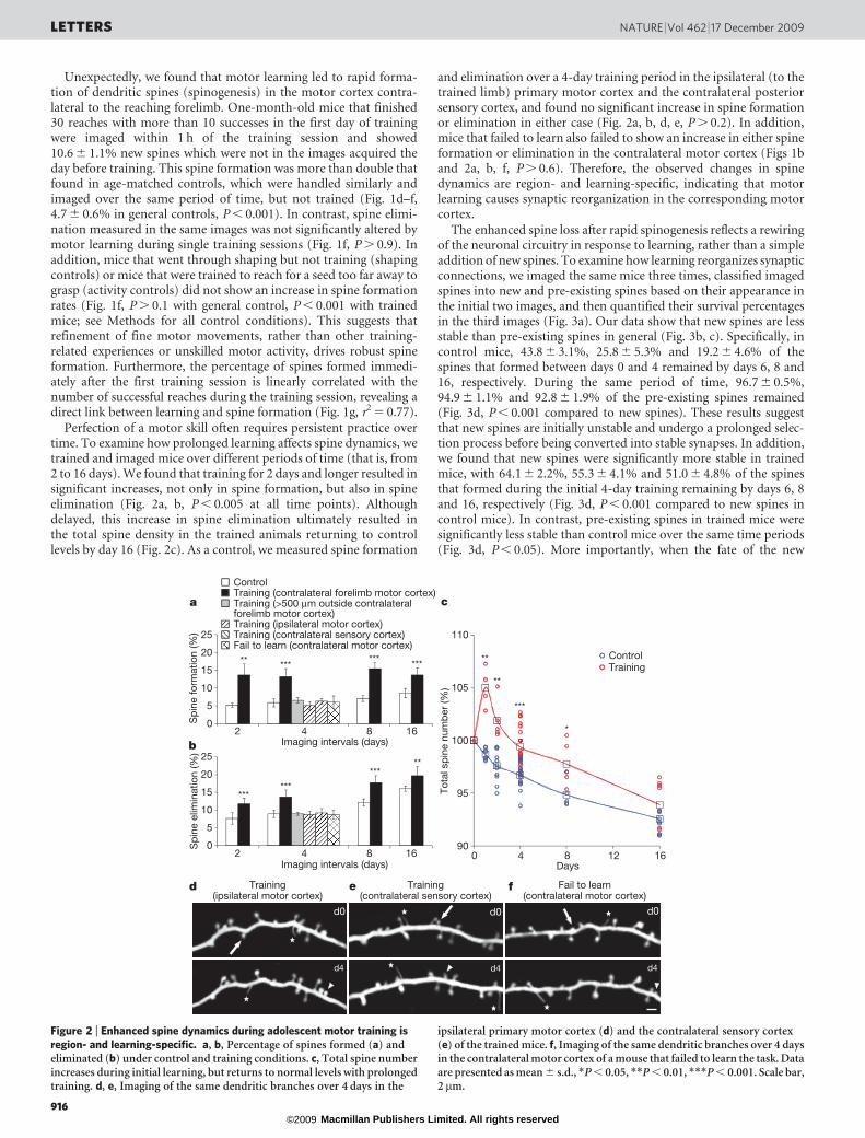
Unexpectedly, we found that motor learning led to rapid forma-tion of dendritic spines (spinogenesis) in the motor cortex contra-lateral to the reaching forelimb. One-month-old mice that finished30 reaches with more than 10 successes in the first day of trainingwere imaged within 1 h of the training session and showed10.6 6 1.1% new spines which were not in the images acquired theday before training. This spine formation was more than double thatfound in age-matched controls, which were handled similarly andimaged over the same period of time, but not trained (Fig. 1d–f,4.7 6 0.6% in general controls, P , 0.001). In contrast, spine elimi-nation measured in the same images was not significantly altered bymotor learning during single training sessions (Fig. 1f, P . 0.9). Inaddition, mice that went through shaping but not training (shapingcontrols) or mice that were trained to reach for a seed too far away tograsp (activity controls) did not show an increase in spine formationrates (Fig. 1f, P . 0.1 with general control, P , 0.001 with trainedmice; see Methods for all control conditions). This suggests thatrefinement of fine motor movements, rather than other training-related experiences or unskilled motor activity, drives robust spineformation. Furthermore, the percentage of spines formed immedi-ately after the first training session is linearly correlated with thenumber of successful reaches during the training session, revealing adirect link between learning and spine formation (Fig. 1g, r2 5 0.77).
Perfection of a motor skill often requires persistent practice overtime. To examine how prolonged learning affects spine dynamics, wetrained and imaged mice over different periods of time (that is, from2 to 16 days). We found that training for 2 days and longer resulted insignificant increases, not only in spine formation, but also in spineelimination (Fig. 2a, b, P , 0.005 at all time points). Althoughdelayed, this increase in spine elimination ultimately resulted inthe total spine density in the trained animals returning to controllevels by day 16 (Fig. 2c). As a control, we measured spine formation
and elimination over a 4-day training period in the ipsilateral (to thetrained limb) primary motor cortex and the contralateral posteriorsensory cortex, and found no significant increase in spine formationor elimination in either case (Fig. 2a, b, d, e, P . 0.2). In addition,mice that failed to learn also failed to show an increase in either spineformation or elimination in the contralateral motor cortex (Figs 1band 2a, b, f, P . 0.6). Therefore, the observed changes in spinedynamics are region- and learning-specific, indicating that motorlearning causes synaptic reorganization in the corresponding motorcortex.
The enhanced spine loss after rapid spinogenesis reflects a rewiringof the neuronal circuitry in response to learning, rather than a simpleaddition of new spines. To examine how learning reorganizes synapticconnections, we imaged the same mice three times, classified imagedspines into new and pre-existing spines based on their appearance inthe initial two images, and then quantified their survival percentagesin the third images (Fig. 3a). Our data show that new spines are lessstable than pre-existing spines in general (Fig. 3b, c). Specifically, incontrol mice, 43.8 6 3.1%, 25.8 6 5.3% and 19.2 6 4.6% of thespines that formed between days 0 and 4 remained by days 6, 8 and16, respectively. During the same period of time, 96.7 6 0.5%,94.9 6 1.1% and 92.8 6 1.9% of the pre-existing spines remained(Fig. 3d, P , 0.001 compared to new spines). These results suggestthat new spines are initially unstable and undergo a prolonged selec-tion process before being converted into stable synapses. In addition,we found that new spines were significantly more stable in trainedmice, with 64.1 6 2.2%, 55.3 6 4.1% and 51.0 6 4.8% of the spinesthat formed during the initial 4-day training remaining by days 6, 8and 16, respectively (Fig. 3d, P , 0.001 compared to new spines incontrol mice). In contrast, pre-existing spines in trained mice weresignificantly less stable than control mice over the same time periods(Fig. 3d, P , 0.05). More importantly, when the fate of the new
b
a c
****** *****
******
*****
**
***
**
*
d e
d0
d4
Training(ipsilateral motor cortex)
Training(contralateral sensory cortex)
Fail to learn(contralateral motor cortex)
f
d0d0
d4 d4
90
95
100
105
110
0 4 8 12 16
Tota
l sp
ine
num
ber
(%)
Days
TrainingControl
0
5
10
15
20
25
0
5
10
15
20
25
2 4 8 16
Sp
ine
form
atio
n (%
)
Imaging intervals (days)
ControlTraining (contralateral forelimb motor cortex)Training (>500 μm outside contralateral forelimb motor cortex)Training (ipsilateral motor cortex)Training (contralateral sensory cortex)Fail to learn (contralateral motor cortex)
2 4 8 16
Sp
ine
elim
inat
ion
(%)
Imaging intervals (days)
Figure 2 | Enhanced spine dynamics during adolescent motor training isregion- and learning-specific. a, b, Percentage of spines formed (a) andeliminated (b) under control and training conditions. c, Total spine numberincreases during initial learning, but returns to normal levels with prolongedtraining. d, e, Imaging of the same dendritic branches over 4 days in the
ipsilateral primary motor cortex (d) and the contralateral sensory cortex(e) of the trained mice. f, Imaging of the same dendritic branches over 4 daysin the contralateral motor cortex of a mouse that failed to learn the task. Dataare presented as mean 6 s.d., *P , 0.05, **P , 0.01, ***P , 0.001. Scale bar,2 mm.
LETTERS NATURE | Vol 462 | 17 December 2009
916 Macmillan Publishers Limited. All rights reserved©2009
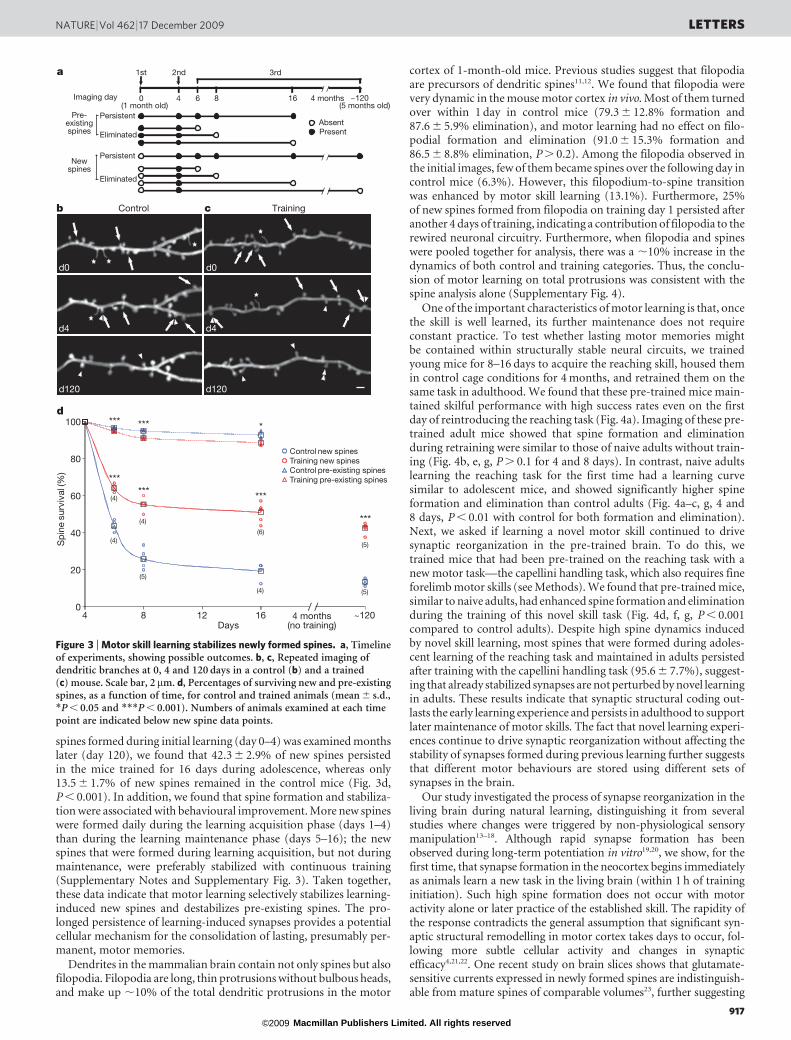
spines formed during initial learning (day 0–4) was examined monthslater (day 120), we found that 42.3 6 2.9% of new spines persistedin the mice trained for 16 days during adolescence, whereas only13.5 6 1.7% of new spines remained in the control mice (Fig. 3d,P , 0.001). In addition, we found that spine formation and stabiliza-tion were associated with behavioural improvement. More new spineswere formed daily during the learning acquisition phase (days 1–4)than during the learning maintenance phase (days 5–16); the newspines that were formed during learning acquisition, but not duringmaintenance, were preferably stabilized with continuous training(Supplementary Notes and Supplementary Fig. 3). Taken together,these data indicate that motor learning selectively stabilizes learning-induced new spines and destabilizes pre-existing spines. The pro-longed persistence of learning-induced synapses provides a potentialcellular mechanism for the consolidation of lasting, presumably per-manent, motor memories.
Dendrites in the mammalian brain contain not only spines but alsofilopodia. Filopodia are long, thin protrusions without bulbous heads,and make up ,10% of the total dendritic protrusions in the motor
cortex of 1-month-old mice. Previous studies suggest that filopodiaare precursors of dendritic spines11,12. We found that filopodia werevery dynamic in the mouse motor cortex in vivo. Most of them turnedover within 1 day in control mice (79.3 6 12.8% formation and87.6 6 5.9% elimination), and motor learning had no effect on filo-podial formation and elimination (91.0 6 15.3% formation and86.5 6 8.8% elimination, P . 0.2). Among the filopodia observed inthe initial images, few of them became spines over the following day incontrol mice (6.3%). However, this filopodium-to-spine transitionwas enhanced by motor skill learning (13.1%). Furthermore, 25%of new spines formed from filopodia on training day 1 persisted afteranother 4 days of training, indicating a contribution of filopodia to therewired neuronal circuitry. Furthermore, when filopodia and spineswere pooled together for analysis, there was a ,10% increase in thedynamics of both control and training categories. Thus, the conclu-sion of motor learning on total protrusions was consistent with thespine analysis alone (Supplementary Fig. 4).
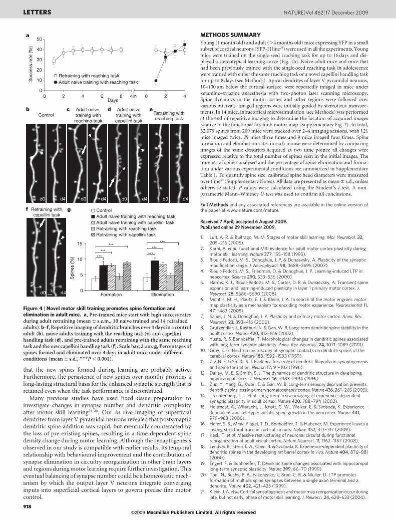
One of the important characteristics of motor learning is that, oncethe skill is well learned, its further maintenance does not requireconstant practice. To test whether lasting motor memories mightbe contained within structurally stable neural circuits, we trainedyoung mice for 8–16 days to acquire the reaching skill, housed themin control cage conditions for 4 months, and retrained them on thesame task in adulthood. We found that these pre-trained mice main-tained skilful performance with high success rates even on the firstday of reintroducing the reaching task (Fig. 4a). Imaging of these pre-trained adult mice showed that spine formation and eliminationduring retraining were similar to those of naive adults without train-ing (Fig. 4b, e, g, P . 0.1 for 4 and 8 days). In contrast, naive adultslearning the reaching task for the first time had a learning curvesimilar to adolescent mice, and showed significantly higher spineformation and elimination than control adults (Fig. 4a–c, g, 4 and8 days, P , 0.01 with control for both formation and elimination).Next, we asked if learning a novel motor skill continued to drivesynaptic reorganization in the pre-trained brain. To do this, wetrained mice that had been pre-trained on the reaching task with anew motor task—the capellini handling task, which also requires fineforelimb motor skills (see Methods). We found that pre-trained mice,similar to naive adults, had enhanced spine formation and eliminationduring the training of this novel skill task (Fig. 4d, f, g, P , 0.001compared to control adults). Despite high spine dynamics inducedby novel skill learning, most spines that were formed during adoles-cent learning of the reaching task and maintained in adults persistedafter training with the capellini handling task (95.6 6 7.7%), suggest-ing that already stabilized synapses are not perturbed by novel learningin adults. These results indicate that synaptic structural coding out-lasts the early learning experience and persists in adulthood to supportlater maintenance of motor skills. The fact that novel learning experi-ences continue to drive synaptic reorganization without affecting thestability of synapses formed during previous learning further suggeststhat different motor behaviours are stored using different sets ofsynapses in the brain.
Our study investigated the process of synapse reorganization in theliving brain during natural learning, distinguishing it from severalstudies where changes were triggered by non-physiological sensorymanipulation13–18. Although rapid synapse formation has beenobserved during long-term potentiation in vitro19,20, we show, for thefirst time, that synapse formation in the neocortex begins immediatelyas animals learn a new task in the living brain (within 1 h of traininginitiation). Such high spine formation does not occur with motoractivity alone or later practice of the established skill. The rapidity ofthe response contradicts the general assumption that significant syn-aptic structural remodelling in motor cortex takes days to occur, fol-lowing more subtle cellular activity and changes in synapticefficacy4,21,22. One recent study on brain slices shows that glutamate-sensitive currents expressed in newly formed spines are indistinguish-able from mature spines of comparable volumes23, further suggesting
0 4 6 8 16Imaging day
Pre-existingspines
Newspines
Persistent
Persistent
Eliminated
Eliminated
1st 2nd 3rda
(1 month old)
AbsentPresent
~120(5 months old)
/ /4 months
b c
d0 d0
d4 d4
d120 d120
Control Training
/ /
/ /
d***
****** ***
****
***
(4)
(4)
(6)
(5)(4)
(5)
(4) (5)
0
20
40
60
80
100
4 8 12 16
Sp
ine
surv
ival
(%)
Days
Control new spinesTraining new spinesControl pre-existing spinesTraining pre-existing spines
~120/ /
4 months(no training)
Figure 3 | Motor skill learning stabilizes newly formed spines. a, Timelineof experiments, showing possible outcomes. b, c, Repeated imaging ofdendritic branches at 0, 4 and 120 days in a control (b) and a trained(c) mouse. Scale bar, 2mm. d, Percentages of surviving new and pre-existingspines, as a function of time, for control and trained animals (mean 6 s.d.,*P , 0.05 and ***P , 0.001). Numbers of animals examined at each timepoint are indicated below new spine data points.
NATURE | Vol 462 | 17 December 2009 LETTERS
917 Macmillan Publishers Limited. All rights reserved©2009
that the new spines formed during learning are probably active.Furthermore, the persistence of new spines over months provides along-lasting structural basis for the enhanced synaptic strength that isretained even when the task performance is discontinued.
Many previous studies have used fixed tissue preparation toinvestigate changes in synapse number and dendritic complexityafter motor skill learning24–28. Our in vivo imaging of superficialdendrites from layer V pyramidal neurons revealed that postsynapticdendritic spine addition was rapid, but eventually counteracted bythe loss of pre-existing spines, resulting in a time-dependent spinedensity change during motor learning. Although the synaptogenesisobserved in our study is compatible with earlier results, its temporalrelationship with behavioural improvement and the contribution ofsynapse elimination in circuitry reorganization in other brain layersand regions during motor learning require further investigation. Thiseventual balancing of synapse number could be a homeostatic mech-anism by which the output layer V neurons integrate converginginputs into superficial cortical layers to govern precise fine motorcontrol.
METHODS SUMMARYYoung (1 month old) and adult (.4 months old) mice expressing YFP in a smallsubset of cortical neurons (YFP-H line29) were used in all the experiments. Young
mice were trained on the single-seed reaching task for up to 16 days and dis-
played a stereotypical learning curve (Fig. 1b). Naive adult mice and mice that
had been previously trained with the single-seed reaching task in adolescence
were trained with either the same reaching task or a novel capellini handling task
for up to 8 days (see Methods). Apical dendrites of layer V pyramidal neurons,
10–100mm below the cortical surface, were repeatedly imaged in mice under
ketamine–xylazine anaesthesia with two-photon laser scanning microscopy.
Spine dynamics in the motor cortex and other regions were followed over
various intervals. Imaged regions were initially guided by stereotaxic measure-
ments. In 14 mice, intracortical microstimulation (see Methods) was performed
at the end of repetitive imaging to determine the location of acquired images
relative to the functional forelimb motor map (Supplementary Fig. 2). In total,
32,079 spines from 209 mice were tracked over 2–4 imaging sessions, with 121
mice imaged twice, 79 mice three times and 9 mice imaged four times. Spine
formation and elimination rates in each mouse were determined by comparing
images of the same dendrites acquired at two time points; all changes were
expressed relative to the total number of spines seen in the initial images. Thenumber of spines analysed and the percentage of spine elimination and forma-
tion under various experimental conditions are summarized in Supplementary
Table 1. To quantify spine size, calibrated spine head diameters were measured
over time30 (Supplementary Notes). All data are presented as mean 6 s.d., unless
otherwise stated. P-values were calculated using the Student’s t-test. A non-
parametric Mann–Whitney U-test was used to confirm all conclusions.
Full Methods and any associated references are available in the online version ofthe paper at www.nature.com/nature.
Received 7 April; accepted 6 August 2009.Published online 29 November 2009.
1. Luft, A. R. & Buitrago, M. M. Stages of motor skill learning. Mol. Neurobiol. 32,205–216 (2005).
2. Karni, A. et al. Functional MRI evidence for adult motor cortex plasticity duringmotor skill learning. Nature 377, 155–158 (1995).
3. Rioult-Pedotti, M. S., Donoghue, J. P. & Dunaevsky, A. Plasticity of the synapticmodification range. J. Neurophysiol. 98, 3688–3695 (2007).
4. Rioult-Pedotti, M. S., Friedman, D. & Donoghue, J. P. Learning-induced LTP inneocortex. Science 290, 533–536 (2000).
5. Harms, K. J., Rioult-Pedotti, M. S., Carter, D. R. & Dunaevsky, A. Transient spineexpansion and learning-induced plasticity in layer 1 primary motor cortex. J.Neurosci. 28, 5686–5690 (2008).
6. Monfils, M. H., Plautz, E. J. & Kleim, J. A. In search of the motor engram: motormap plasticity as a mechanism for encoding motor experience. Neuroscientist 11,471–483 (2005).
7. Sanes, J. N. & Donoghue, J. P. Plasticity and primary motor cortex. Annu. Rev.Neurosci. 23, 393–415 (2000).
8. Grutzendler, J., Kasthuri, N. & Gan, W. B. Long-term dendritic spine stability in theadult cortex. Nature 420, 812–816 (2002).
9. Yuste, R. & Bonhoeffer, T. Morphological changes in dendritic spines associatedwith long-term synaptic plasticity. Annu. Rev. Neurosci. 24, 1071–1089 (2001).
10. Gray, E. G. Electron microscopy of synaptic contacts on dendrite spines of thecerebral cortex. Nature 183, 1592–1593 (1959).
11. Ziv, N. E. & Smith, S. J. Evidence for a role of dendritic filopodia in synaptogenesisand spine formation. Neuron 17, 91–102 (1996).
12. Dailey, M. E. & Smith, S. J. The dynamics of dendritic structure in developinghippocampal slices. J. Neurosci. 16, 2983–2994 (1996).
13. Zuo, Y., Yang, G., Kwon, E. & Gan, W. B. Long-term sensory deprivation preventsdendritic spine loss in primary somatosensory cortex. Nature 436, 261–265 (2005).
14. Trachtenberg, J. T. et al. Long-term in vivo imaging of experience-dependentsynaptic plasticity in adult cortex. Nature 420, 788–794 (2002).
15. Holtmaat, A., Wilbrecht, L., Knott, G. W., Welker, E. & Svoboda, K. Experience-dependent and cell-type-specific spine growth in the neocortex. Nature 441,979–983 (2006).
16. Hofer, S. B., Mrsic-Flogel, T. D., Bonhoeffer, T. & Hubener, M. Experience leaves alasting structural trace in cortical circuits. Nature 457, 313–317 (2009).
17. Keck, T. et al. Massive restructuring of neuronal circuits during functionalreorganization of adult visual cortex. Nature Neurosci. 11, 1162–1167 (2008).
18. Lendvai, B., Stern, E. A., Chen, B. & Svoboda, K. Experience-dependent plasticity ofdendritic spines in the developing rat barrel cortex in vivo. Nature 404, 876–881(2000).
19. Engert, F. & Bonhoeffer, T. Dendritic spine changes associated with hippocampallong-term synaptic plasticity. Nature 399, 66–70 (1999).
20. Toni, N., Buchs, P. A., Nikonenko, I., Bron, C. R. & Muller, D. LTP promotesformation of multiple spine synapses between a single axon terminal and adendrite. Nature 402, 421–425 (1999).
21. Kleim, J. A. et al. Cortical synaptogenesis and motor map reorganization occur duringlate, but not early, phase of motor skill learning. J. Neurosci. 24, 628–633 (2004).
0
5
10
15
Formation Elimination
Sp
ines
(%)
ControlAdult naive training with reaching taskAdult naive training with capellini taskRetraining with reaching taskRetraining with capellini task
/ /4m
ControlAdult naive training with
reaching task
Retraining withreaching task
Retraining withcapellini task
a
b c d
f
***
***
******
******
Adult naivetraining withcapellini task
e
d0 d0 d0 d0
d0 d4
d4 d4 d4 d4
g
0
10
20
30
40
50
0 2 4 6 8
Suc
cess
rat
e (%
)
Days0 2 4
Retraining with reaching taskAdult naive training with reaching task
Figure 4 | Novel motor skill training promotes spine formation andelimination in adult mice. a, Pre-trained mice start with high success ratesduring adult retraining (mean 6 s.e.m., 10 naive trained and 14 retrainedadults). b–f, Repetitive imaging of dendritic branches over 4 days in a controladult (b), naive adults training with the reaching task (c) and capellinihandling task (d), and pre-trained adults retraining with the same reachingtask and the new capellini handling task (f). Scale bar, 2 mm. g, Percentages ofspines formed and eliminated over 4 days in adult mice under differentconditions (mean 6 s.d., ***P , 0.001).
LETTERS NATURE | Vol 462 | 17 December 2009
918 Macmillan Publishers Limited. All rights reserved©2009

22. Adkins, D. L., Boychuk, J., Remple, M. S. & Kleim, J. A. Motor training inducesexperience-specific patterns of plasticity across motor cortex and spinal cord.J. Appl. Physiol. 101, 1776–1782 (2006).
23. Zito, K., Scheuss, V., Knott, G., Hill, T. & Svoboda, K. Rapid functional maturationof nascent dendritic spines. Neuron 61, 247–258 (2009).
24. Kleim, J. A., Vij, K., Ballard, D. H. & Greenough, W. T. Learning-dependent synapticmodifications in the cerebellar cortex of the adult rat persist for at least fourweeks. J. Neurosci. 17, 717–721 (1997).
25. Greenough, W. T., Larson, J. R. & Withers, G. S. Effects of unilateral and bilateraltraining in a reaching task on dendritic branching of neurons in the rat motor-sensory forelimb cortex. Behav. Neural Biol. 44, 301–314 (1985).
26. Withers, G. S. & Greenough, W. T. Reach training selectively alters dendriticbranching in subpopulations of layer II–III pyramids in rat motor-somatosensoryforelimb cortex. Neuropsychologia 27, 61–69 (1989).
27. Kleim, J. A. et al. Motor learning-dependent synaptogenesis is localized tofunctionally reorganized motor cortex. Neurobiol. Learn. Mem. 77, 63–77 (2002).
28. Kolb, B., Cioe, J. & Comeau, W. Contrasting effects of motor and visual spatiallearning tasks on dendritic arborization and spine density in rats. Neurobiol. Learn.Mem. 90, 295–300 (2008).
29. Feng, G. et al. Imaging neuronal subsets in transgenic mice expressing multiplespectral variants of GFP. Neuron 28, 41–51 (2000).
30. Zuo, Y., Lin, A., Chang, P. & Gan, W. B. Development of long-term dendritic spinestability in diverse regions of cerebral cortex. Neuron 46, 181–189 (2005).
Supplementary Information is linked to the online version of the paper atwww.nature.com/nature.
Acknowledgements We thank D. States, W. Thompson, L. Hinck, D. Feldheim,J. Ding, X. Li, A. Lin and C. Cirelli for critical comments on this manuscript; A. Sitkofor her pilot studies of skilled reaching in mice, and D. Adkins, J. Kleim andN. Thomas for their assistance with intracortical microstimulation procedures. Thiswork was supported by grants from the Ellison Medical Foundation, the DANAFoundation, and the National Institute on Aging to Y.Z.
Author Contributions T.X. and X.Y. contributed equally to this work. Both of themperformed in vivo imaging, analysed the data, made figures and participated in thediscussion. A.J.P., W.F.T. and J.A.Z. trained all the mice used in the experiments.K.T. and T.J. developed behavioural methods, performed the intracorticalmicrostimulation experiments, and provided comments for the manuscript. Y.Z.initiated the project, did data analysis and wrote the manuscript.
Author Information Reprints and permissions information is available atwww.nature.com/reprints. Correspondence and requests for materials should beaddressed to Y.Z. ([email protected]).
NATURE | Vol 462 | 17 December 2009 LETTERS
919 Macmillan Publishers Limited. All rights reserved©2009
METHODSSingle-seed reaching task. Mice were food-restricted to maintain 90% of free
feeding weight before the start of training. The training chamber was constructed
as a clear Plexiglas box 20 cm tall, 15 cm deep and 8.5 cm wide into which each
individual mouse was placed. Three vertical slits 0.5 cm wide and 13 cm high were
located on the front wall of the box: in the centre, on the left side, and on the right
side (Supplementary Fig. 5). A 1.25-cm-tall exterior shelf was affixed to the wall in
front of the slits to hold millet seeds for food reward. The training included two
phases: ‘shaping’ and ‘training’. The shaping phase (2–5 days in duration) was used
to familiarize mice with the training chamber and task requirements and also to
determine their preferred limbs. During the shaping phase millet seeds were placedin front of the centre slit and mice used both paws to reach for them. Shaping was
considered finished when 20 reach attempts were achieved within 20 min, and the
mouse showed .70% limb preference. Training started the day after shaping, and
each training day consisted of one session of 30 trials with preferred limb or 20 min
(whichever occurred first). Seeds were presented individually in front of the slit on
the side of preferred limb. Occasionally a mouse used the non-preferred limb;
however, because of the difficulties presented by reaching angle, such reaches
usually were unsuccessful. Mice displayed three reach attempt types: fail, drop
and success (Supplementary Movie 1). A ‘fail’ was scored as a reach in which the
mouse failed to touch the seed or knocked it away. A ‘drop’ was a reach in which the
mouse retrieved the seed, but dropped it before putting into its mouth. A ‘success’
was a reach in which the mouse successfully retrieved the seed and put it into its
mouth. Success rates were calculated as the percentage of successful reaches over
total reach attempts. About half of the mice in our experiments were right handed
(55 right handed out of a total of 109 mice, 50.6%). All data collected from both
left- and right-handed mice were pooled for analysis in this study. No significant
difference was found in the reaching performance of left- and right-handed mice.
All our control mice were littermates that underwent the same food restriction.
All mice were handled (that is, removed from their cages and placed temporarilyin the training chamber into which some seeds were dropped) by the same
experimenters. To ensure that the increase seen in spine dynamics was learning
specific, three different controls were used in our study. The first control group
was general controls comprising mice with neither training nor shaping, but with
food restriction, food reward and handling. The second was shaping controls in
which mice received similar shaping as trained mice. During training, they were
placed into the training chamber for 20 min daily, with ,15 seeds periodically
dropped into the training chamber. This control group was used to determine
whether the shaping period and/or experience of the training environment had
any effect on spine dynamics. The third control group was activity controls in
which mice were given similar shaping as trained mice. During training, mice were
placed into the training chamber and trained to reach for a seed placed outside the
slit for 20 min daily. However, the seed was placed out of reach, so that they could
never obtain it and, therefore, did not learn skilful reaching movements (as shown
by testing their performance occasionally). Thus, both trained mice and activity
control mice experienced similar amounts of forelimb activity, but only trained
mice developed the motor skill. The activity control was used to determine
whether enhanced spine dynamics were caused by increased motor activity orwere specific to motor skill learning. Our results indicate that there is no difference
in the spine dynamics between the activity controls and general controls.
Capellini handling task. This task was similar to the vermicelli handling tasks
previously described for rats31. Mice were food-restricted to maintain 90% of free
feeding weight before training began. A daily training session consisted of 10 trials
with uncooked capellini pasta pieces (2.5 cm), given one piece per trial. Mice
learned to use coordinated forepaw movements to eat the pasta. The average con-
sumption time for one piece of capellini pasta decreased from 3.44 6 0.18 min on
day 1 to 1.98 6 0.29 min on day 4 (mean 6 s.e.m., P , 0.005, 7 mice). There was no
significant behavioural difference in the capellini handling task between naive adults
and adults pre-trained in the reaching task in adolescence.
In vivo imaging of superficial dendrites. The procedure for transcranial two-
photon imaging has been described previously8. Mice aged 1–6 months were
anaesthetized with an intraperitoneal injection (5.0 ml per kg body weight) of
17 mg ml21 ketamine and 1.7 mg ml21 xylazine in 0.9% NaCl. The skull was
exposed with a midline scalp incision and imaged regions were located based on
stereotactic coordinates. A small region of skull (,300mm in diameter) was
manually thinned down to ,20mm in thickness using both a high-speed drill
and a microknife. To reduce respiration-induced movements, the skull wasglued to a 400-mm-thick stainless steel plate with a central opening for skull
access. The plate was screwed to two lateral bars located on either side of the
head and fixed to a metal base. The brain of the mouse was then imaged through
the thinned skull using a Prairie Ultima IV multi-photon microscope with a Ti-
sapphire laser tuned to the excitation wavelength for YFP (925 nm). Stacks of
image planes were acquired with a step size of 0.70mm using a water-immersion
objective (360, NA 1.1 infrared Olympus objective) at a zoom of 3.0. Forrelocation of the same dendrites at subsequent imaging times, an image stack
containing the dendritic structures of interest was taken without zoom with a
step size of 2.0 mm and the surrounding blood vessels were imaged with a CCD
camera. The patterns of blood vessels and neuronal processes in this low-reso-
lution image stack were used for relocating the same dendrites at each sub-
sequent imaging session (Supplementary Fig. 1). After imaging, the plate was
detached from the skull, the scalp sutured, and the animal was returned to its
home cage until the next imaging session.
Spine and filopodium identification. All analysis of spine dynamics was done
manually using ImageJ software, blind with regard to experimental conditions.
Briefly, the same dendritic segments (,5–20mm in length) were identified from
three-dimensional image stacks selected from all views having high image quality
(signal-to-background-noise ratio .4-fold). Individual dendritic protrusions
were tracked manually along dendrites. Three-dimensional stacks, instead of
two-dimensional projections, were used for analysis to ensure that tissue move-
ments and rotation between imaging intervals did not influence identification of
dendritic protrusions. The number and location of dendritic protrusions (protru-
sion length .1/3 dendritic shaft diameter) were identified in each view. Filopodiawere identified as long thin structures with head diameter/neck diameter ,1.2 and
length/neck diameter .3. The remaining protrusions were classified as spines.
Analysis of spine and filopodial dynamics. Notations of the formation and
elimination of spines and filopodia were based on comparison of the images
collected at two different time points. Spines or filopodia were considered the same
between two views if they were within 0.7mm of their expected positions, based on
their spatial relationship to adjacent landmarks and/or their position relative to
immediately adjacent spines. A stable spine is a spine that was present in both
images. An eliminated spine is a spine that appeared in the initial image, but not
the second image. A newly formed spine is a spine that appeared in the second
image, but was absent from the initial image. Percentages of stable, eliminated and
formed spines were all normalized to the initial image. Percentage changes in the
total spine number over a given interval were relative to the first view and calculated
as percentage of formation minus percentage of elimination measured over that
interval. Data on spine dynamics are presented as mean 6 s.d..
Image processing and presentation. Two-dimensional projections of three-
dimensional image stacks containing in-focus dendritic segments of interest
were used for all figures. We chose very sparsely labelled regions as examplesand maximum projections were made from images from 2–4 focus planes. There
were normally few crossing structures in the projected images from such a
shallow stack, and the presented branches could be clearly isolated. Finally,
images were thresholded, Gaussian filtered and contrasted for presentation.
Mapping of motor cortex by intracortical microstimulation. This method was
adapted from those used in rat experiments21. Mice were anaesthetized with an
initial cocktail of ketamine (150 mg kg21, intraperitoneal) and xylazine
(10 mg kg21, intraperitoneal) and supplemented with additional ketamine and
isofluorane (0.5–1% in oxygen) as necessary. The mouse was placed into a mouse
stereotaxic frame (Stoelting), lidocaine (2 mg kg21, subcutaneous) was injected
into the scalp, and a midline incision was made. The cisterna magna was drained
to prevent cortical swelling and the skull and dura overlying the motor cortex
were removed. The craniotomy was then filled with warm (37 uC) silicone oil to
prevent drying. A picture of the cortical surface was taken and overlaid with a
250mm square grid in Canvas software.
Intracortical penetrations of a glass microelectrode (diameter of 20-25mm) with
a platinum wire were made at 250mm intervals in a systematic order throughout
the cortex at a depth of 790–800mm (corresponding to deep layer V/shallow layerVI) with a hydraulic micropositioner until the entire extent of the forelimb rep-
resentation was resolved. A 40-ms train of 13 200-ms monophasic cathodal pulses
was delivered at 350 Hz from an electrically isolated, constant current stimulator at
a rate of 1 Hz stimulation and current was increased to a maximum of 60 mA until a
visible movement was evoked. If a movement was evoked at or below 60mA, the
threshold current was determined by gradually decreasing the stimulation until
the movement stopped. The lowest current that evoked a movement was taken as
the threshold current. If no movement was seen at 60mA, the site was considered
non-responsive. In cases where stimulation evoked more than one movement, the
site was considered responsive to the movement that was determined to have the
lowest threshold. To verify that the stimulation position was located within layer V,
we injected DiI in seven mice at the end of the experiments and found that all
injections left deposits extending through mid layer V to mid layer VI. In addition,
penetrating electrode tracts could be observed in Nissl-stained coronal sections in
most mice. Most (81.3 6 4.7%) of these tracts terminated in layer V at a measured
depth of 782 6 137 mm, with the remainder terminated in upper layer VI.
31. Allred, R. P. et al. The vermicelli handling test: a simple quantitative measure ofdexterous forepaw function in rats. J. Neurosci. Methods 170, 229–244 (2008).
doi:10.1038/nature08389
Macmillan Publishers Limited. All rights reserved©2009