Rapid Analysis of Synthetic mRNA Cap Structure Using Ion ...
15
Application Note Rapid Analysis of Synthetic mRNA Cap Structure Using Ion-Pairing RPLC with the BioAccord LC-MS System Jennifer M. Nguyen, Siu-Hong Chan, Bijoyita Roy, Martin Gilar, Brett Robb, Weibin Chen, Matthew A. Lauber Waters Corporation, New England Biolabs Inc. Abstract
Transcript of Rapid Analysis of Synthetic mRNA Cap Structure Using Ion ...
Application Note
Rapid Analysis of Synthetic mRNA Cap
Structure Using Ion-Pairing RPLC with the
BioAccord LC-MS System
Jennifer M. Nguyen, Siu-Hong Chan, Bijoyita Roy, Martin Gilar, Brett Robb, Weibin Chen, Matthew A. Lauber
Waters Corporation, New England Biolabs Inc.
Abstract
Due to the crucial need to fight the SARS-CoV-2 pandemic, the development of mRNA vaccines progressed rapidly
throughout 2020. This has led to the emergency use authorization of two highly efficacious mRNA vaccines.1
These vaccines contain synthetic mRNA encapsulated in a lipid nanoparticle for delivery, and they use the
recipient’s body to conduct in vivo translation of a stabilized version of the viral spike protein and elicit the
immune response.
The mRNA in these vaccines is produced by an enzymatic process known as in vitro transcription (IVT). Much like
cellular mRNAs, synthetic mRNAs also need to be modified on their 5’ ends to include a 7-methyl guanosine
nucleotide in a 5’-5’-linkage that is referred to as a 5’ cap. The vaccine mRNA is also modified to include a chain
of adenosines on the 3’ end, called the poly(A) tail (typically ~120 nucleotides). These modifications are critical
features of mRNA structure, playing important roles in recognition of the mRNA by cellular factors, stability of the
synthetic mRNA, and translational efficiency of the synthetic mRNA molecule.2 As such, determining the nature of
the 5’ cap and the measurement of the poly(A) tail length by LC-MS analysis is vital to understand the product
quality and ensure the safety and efficacy of these new vaccines.3
In this application note, we developed a rapid and sensitive LC-MS method applicable to synthetic mRNA capping
analysis. Previously, a LC-MS method has been reported to analyze pre-defined 5’ fragments of synthetic mRNA.4
Here, we demonstrate that ACQUITY Premier Columns and their MaxPeak High Performance Surfaces (HPS),
previously shown to enhance oligonucleotide recovery, can also improve the performance of mRNA fragment
analysis. This, in combination with an easy-to-use, compliance-ready BioAccord System, provides a fit-for-purpose
platform for the rapid analysis of 5’ capping of IVT mRNA preparations. A less than 5-minute method was
established, and linearity was demonstrated to detecting product-related impurities down to less than 0.1% of the
target 5’ capped fragment.
Benefits
Fast, quantitative analysis of synthetic mRNA 5’ analysis ■
Minimal to no column conditioning required for ACQUITY Premier Columns with MaxPeak High Performance
Surfaces
■
Linearity, reproducibility, and robustness suitable in both development and QC testing■
High quality MS spectra produced with DIPEA (diisopropylethylamine) mobile phase■
Introduction
The success of the Pfizer-BioNTech and Moderna SARS-CoV-2 vaccines has brought a surge of interest to mRNA
molecules while also placing new demands on analytics to better support the development and manufacturing of a
new modality. Among critical quality attributes (CQAs) such as percentage of full-length mRNA, untranslated
regions (UTRs), 3’ poly(A) tail length, the sequence, structure, and chemical modifications of the mRNA,5 the
presence of a proper 5’ cap structure is important to ensuring maximal gene expression, to evading innate
recognition mechanisms against foreign RNA, and to increasing resistance towards exonuclease degradation.6
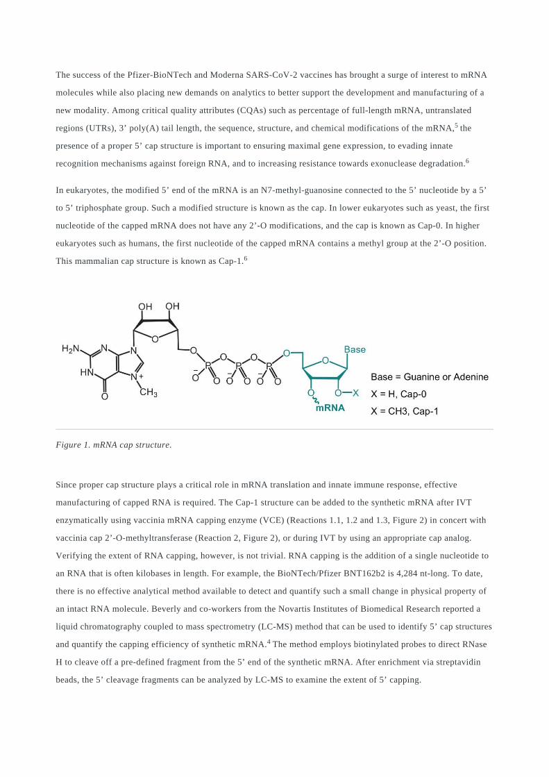
In eukaryotes, the modified 5’ end of the mRNA is an N7-methyl-guanosine connected to the 5’ nucleotide by a 5’
to 5’ triphosphate group. Such a modified structure is known as the cap. In lower eukaryotes such as yeast, the first
nucleotide of the capped mRNA does not have any 2’-O modifications, and the cap is known as Cap-0. In higher
eukaryotes such as humans, the first nucleotide of the capped mRNA contains a methyl group at the 2’-O position.
This mammalian cap structure is known as Cap-1.6
Figure 1. mRNA cap structure.
Since proper cap structure plays a critical role in mRNA translation and innate immune response, effective
manufacturing of capped RNA is required. The Cap-1 structure can be added to the synthetic mRNA after IVT
enzymatically using vaccinia mRNA capping enzyme (VCE) (Reactions 1.1, 1.2 and 1.3, Figure 2) in concert with
vaccinia cap 2’-O-methyltransferase (Reaction 2, Figure 2), or during IVT by using an appropriate cap analog.
Verifying the extent of RNA capping, however, is not trivial. RNA capping is the addition of a single nucleotide to
an RNA that is often kilobases in length. For example, the BioNTech/Pfizer BNT162b2 is 4,284 nt-long. To date,
there is no effective analytical method available to detect and quantify such a small change in physical property of
an intact RNA molecule. Beverly and co-workers from the Novartis Institutes of Biomedical Research reported a
liquid chromatography coupled to mass spectrometry (LC-MS) method that can be used to identify 5’ cap structures
and quantify the capping efficiency of synthetic mRNA.4 The method employs biotinylated probes to direct RNase
H to cleave off a pre-defined fragment from the 5’ end of the synthetic mRNA. After enrichment via streptavidin
beads, the 5’ cleavage fragments can be analyzed by LC-MS to examine the extent of 5’ capping.
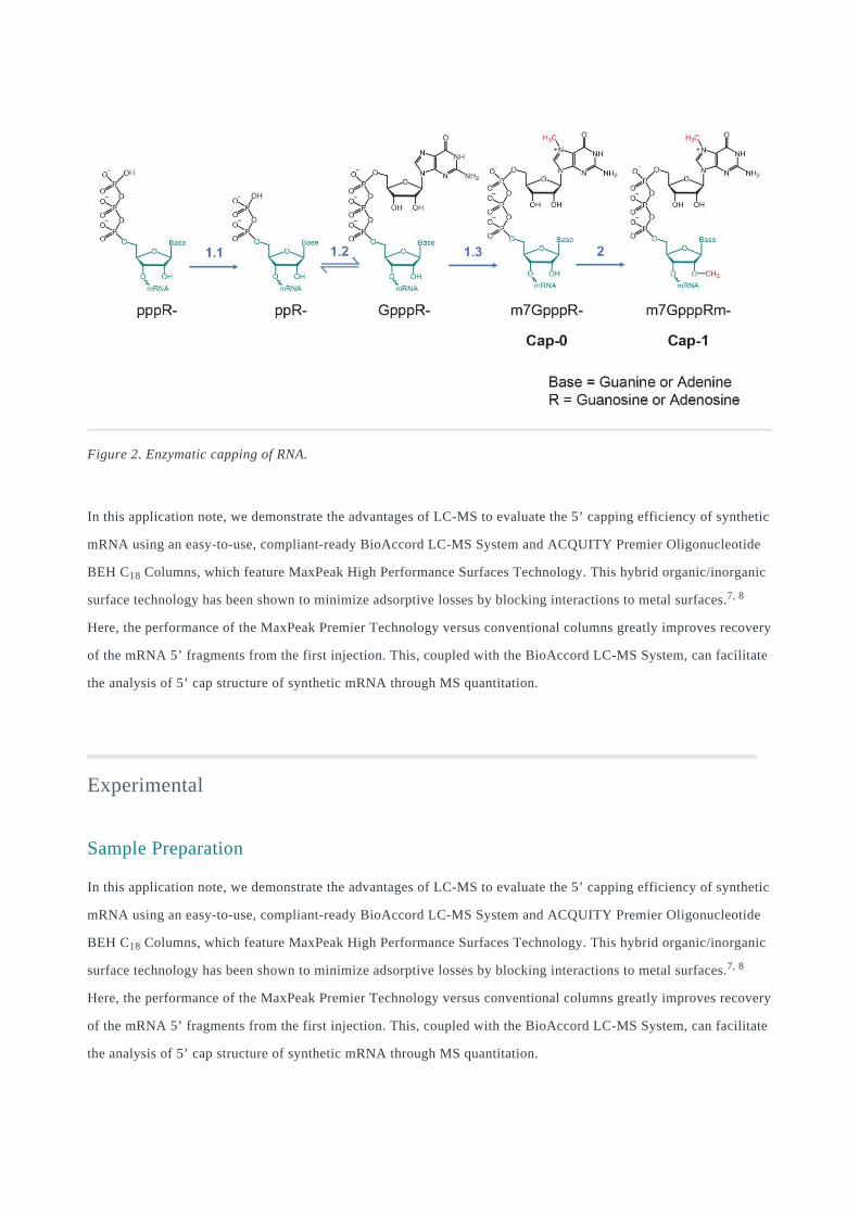
Figure 2. Enzymatic capping of RNA.
In this application note, we demonstrate the advantages of LC-MS to evaluate the 5’ capping efficiency of synthetic
mRNA using an easy-to-use, compliant-ready BioAccord LC-MS System and ACQUITY Premier Oligonucleotide
BEH C18 Columns, which feature MaxPeak High Performance Surfaces Technology. This hybrid organic/inorganic
surface technology has been shown to minimize adsorptive losses by blocking interactions to metal surfaces.7, 8
Here, the performance of the MaxPeak Premier Technology versus conventional columns greatly improves recovery
of the mRNA 5’ fragments from the first injection. This, coupled with the BioAccord LC-MS System, can facilitate
the analysis of 5’ cap structure of synthetic mRNA through MS quantitation.
Experimental
Sample Preparation
In this application note, we demonstrate the advantages of LC-MS to evaluate the 5’ capping efficiency of synthetic
mRNA using an easy-to-use, compliant-ready BioAccord LC-MS System and ACQUITY Premier Oligonucleotide
BEH C18 Columns, which feature MaxPeak High Performance Surfaces Technology. This hybrid organic/inorganic
surface technology has been shown to minimize adsorptive losses by blocking interactions to metal surfaces.7, 8
Here, the performance of the MaxPeak Premier Technology versus conventional columns greatly improves recovery
of the mRNA 5’ fragments from the first injection. This, coupled with the BioAccord LC-MS System, can facilitate
the analysis of 5’ cap structure of synthetic mRNA through MS quantitation.
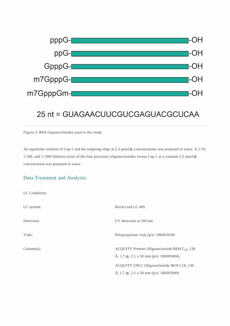
Figure 3. RNA oligonucleotides used in this study.
An equimolar solution of Cap-1 and the targeting oligo at 2.5 pmol/µL concentrations was prepared in water. A 1:10,
1:100, and 1:1000 dilution series of the four precursor oligonucleotides versus Cap-1 at a constant 2.5 pmol/µL
concentration was prepared in water.
Data Treatment and Analysis:
LC Conditions
LC system: BioAccord LC-MS
Detection: UV detection at 260 nm
Vials: Polypropylene vials (p/n: 186002639)
Column(s): ACQUITY Premier Oligonucleotide BEH C18, 130
Å, 1.7 µm, 2.1 x 50 mm (p/n: 186009484);
ACQUITY UPLC Oligonucleotide BEH C18, 130
Å, 1.7 µm, 2.1 x 50 mm (p/n: 186003949)
Column temp.: 60 °C
Sample temp.: 4 °C
Injection volume: 5.0 µL (sample)
Flow rate: 0.3 mL/min
Mobile phase A: 1.0% hexafluoroisopropanol (HFIP), 0.1% N,N-
diisopropylethylamine (DIPEA) in water
Mobile phase B: 0.75% HFIP, 0.0375% DIPEA in 65:35
acetonitrile:water
Gradient: 5–25% B in 5, 10, or 20 min
MS Conditions
MS system: BioAccord LC-MS
Ionization mode: Full scan, ESI negative
Acquisition range: 400–5000 m/z
Capillary voltage: 0.8 kV
Cone voltage: 40 V
Desolvation temp.: 400 °C
Data Management
Chromatography and MS software: waters_connect with UNIFI v1.9.12
Results and Discussion
Initial Column Performance
Before developing a LC-MS method for 5’ capping efficiency, we evaluated the chromatographic recovery of the
mRNA fragments from a LC-MS analysis. As described in literature and prior application notes, we have
demonstrated that oligonucleotides as well as phosphorylated compounds can adsorb to metal surfaces such as
stainless steel. This unwanted effect can be mitigated with the use of MaxPeak Premier Columns.9-11 For RNA 5’
cap analysis, we investigated the LC-UV-MS separations of the Cap-1 species, consisting of the m7GpppGm group
on the 5’ end, using either a conventional ACQUITY UPLC Oligonucleotide BEH C18 Column or an ACQUITY
Premier Oligonucleotide BEH C18 Column packed with the same lot of stationary phase. It was confirmed that the
ACQUITY Premier Column with its MaxPeak High Performance Surfaces confers benefits like improved
recoveries and out-of-the-box performance without the need for conditioning or passivation.
For oligonucleotide LC-MS analysis, ion-pairing reversed-phase (IP-RP) separations with amine mobile phase
additives is the preferred mode of chromatography given their resolving power and amenability to MS. However,
amines with acidic counter ions, such as acetate or bicarbonate salts, produce too much ion suppression at the
concentrations required for effective separations, though reducing the concentration of IP reagent can help. Since
its introduction in 1997 by Apffel and co-authors, hexafluoroisopropanol (HFIP) has been preferred for IP-RP
separations that are hyphenated to MS technology.12 HFIP is a weak acid that can help buffer amine containing
mobile phases without significantly impairing ionization efficiency. Additionally, it has been theorized that
alkylamines are more likely to adsorb onto the stationary phase to act as a better IP system in the presence of HFIP
and that it positively affects the desolvation of oligonucleotides in ESI-MS.13 Here, we used the amine N,N-
diisopropylethylamine (DIPEA) buffered with HFIP, as it has been shown to greatly increase MS signal intensity
and peak shape versus triethylamine.14
Recovery of the Cap-1 fragment as based on peak area from the first injections were evaluated for the conventional
ACQUITY UPLC Column versus the ACQUITY Premier Column. Representative UV chromatograms resulting
from the first injection on the ACQUITY Premier Column and from the first to fifth injection of the conventional
ACQUITY UPLC Column are shown in Figure 4. No peak representing the Cap-1 species was seen upon the initial
use of the conventional column (Figure 4). In contrast, the ACQUITY Premier Column gave a high intensity peak
on its first injection and reproducible peak areas from subsequent injections. Even after successive injections of
Cap-1 on the conventional column, by injection 5, the peak area of the Cap-1 fragment was still only 37% of that of
the ACQUITY Premier Column.
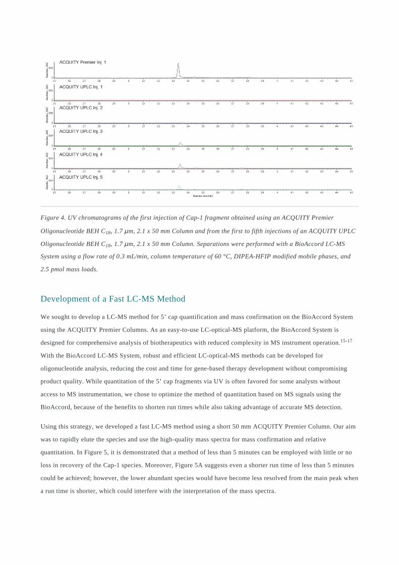
Figure 4. UV chromatograms of the first injection of Cap-1 fragment obtained using an ACQUITY Premier
Oligonucleotide BEH C18, 1.7 μm, 2.1 x 50 mm Column and from the first to fifth injections of an ACQUITY UPLC
Oligonucleotide BEH C18, 1.7 μm, 2.1 x 50 mm Column. Separations were performed with a BioAccord LC-MS
System using a flow rate of 0.3 mL/min, column temperature of 60 °C, DIPEA-HFIP modified mobile phases, and
2.5 pmol mass loads.
Development of a Fast LC-MS Method
We sought to develop a LC-MS method for 5’ cap quantification and mass confirmation on the BioAccord System
using the ACQUITY Premier Columns. As an easy-to-use LC-optical-MS platform, the BioAccord System is
designed for comprehensive analysis of biotherapeutics with reduced complexity in MS instrument operation.15-17
With the BioAccord LC-MS System, robust and efficient LC-optical-MS methods can be developed for
oligonucleotide analysis, reducing the cost and time for gene-based therapy development without compromising
product quality. While quantitation of the 5’ cap fragments via UV is often favored for some analysts without
access to MS instrumentation, we chose to optimize the method of quantitation based on MS signals using the
BioAccord, because of the benefits to shorten run times while also taking advantage of accurate MS detection.
Using this strategy, we developed a fast LC-MS method using a short 50 mm ACQUITY Premier Column. Our aim
was to rapidly elute the species and use the high-quality mass spectra for mass confirmation and relative
quantitation. In Figure 5, it is demonstrated that a method of less than 5 minutes can be employed with little or no
loss in recovery of the Cap-1 species. Moreover, Figure 5A suggests even a shorter run time of less than 5 minutes
could be achieved; however, the lower abundant species would have become less resolved from the main peak when
a run time is shorter, which could interfere with the interpretation of the mass spectra.
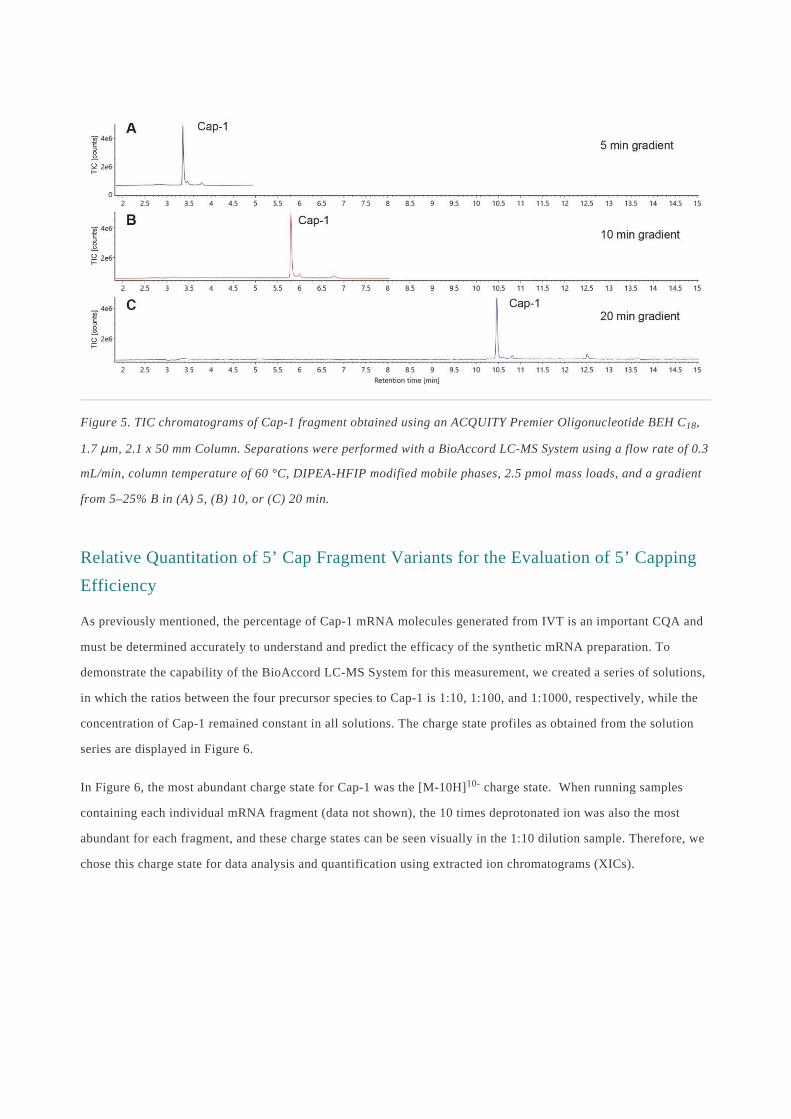
Figure 5. TIC chromatograms of Cap-1 fragment obtained using an ACQUITY Premier Oligonucleotide BEH C18,
1.7 μm, 2.1 x 50 mm Column. Separations were performed with a BioAccord LC-MS System using a flow rate of 0.3
mL/min, column temperature of 60 °C, DIPEA-HFIP modified mobile phases, 2.5 pmol mass loads, and a gradient
from 5–25% B in (A) 5, (B) 10, or (C) 20 min.
Relative Quantitation of 5’ Cap Fragment Variants for the Evaluation of 5’ Capping
Efficiency
As previously mentioned, the percentage of Cap-1 mRNA molecules generated from IVT is an important CQA and
must be determined accurately to understand and predict the efficacy of the synthetic mRNA preparation. To
demonstrate the capability of the BioAccord LC-MS System for this measurement, we created a series of solutions,
in which the ratios between the four precursor species to Cap-1 is 1:10, 1:100, and 1:1000, respectively, while the
concentration of Cap-1 remained constant in all solutions. The charge state profiles as obtained from the solution
series are displayed in Figure 6.
In Figure 6, the most abundant charge state for Cap-1 was the [M-10H]10- charge state. When running samples
containing each individual mRNA fragment (data not shown), the 10 times deprotonated ion was also the most
abundant for each fragment, and these charge states can be seen visually in the 1:10 dilution sample. Therefore, we
chose this charge state for data analysis and quantification using extracted ion chromatograms (XICs).
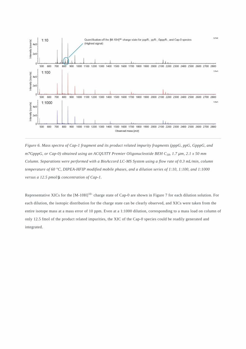
Figure 6. Mass spectra of Cap-1 fragment and its product related impurity fragments (pppG, ppG, GpppG, and
m7GpppG, or Cap-0) obtained using an ACQUITY Premier Oligonucleotide BEH C18, 1.7 μm, 2.1 x 50 mm
Column. Separations were performed with a BioAccord LC-MS System using a flow rate of 0.3 mL/min, column
temperature of 60 °C, DIPEA-HFIP modified mobile phases, and a dilution series of 1:10, 1:100, and 1:1000
versus a 12.5 pmol/µL concentration of Cap-1.
Representative XICs for the [M-10H]10- charge state of Cap-0 are shown in Figure 7 for each dilution solution. For
each dilution, the isotopic distribution for the charge state can be clearly observed, and XICs were taken from the
entire isotope mass at a mass error of 10 ppm. Even at a 1:1000 dilution, corresponding to a mass load on column of
only 12.5 fmol of the product related impurities, the XIC of the Cap-0 species could be readily generated and
integrated.
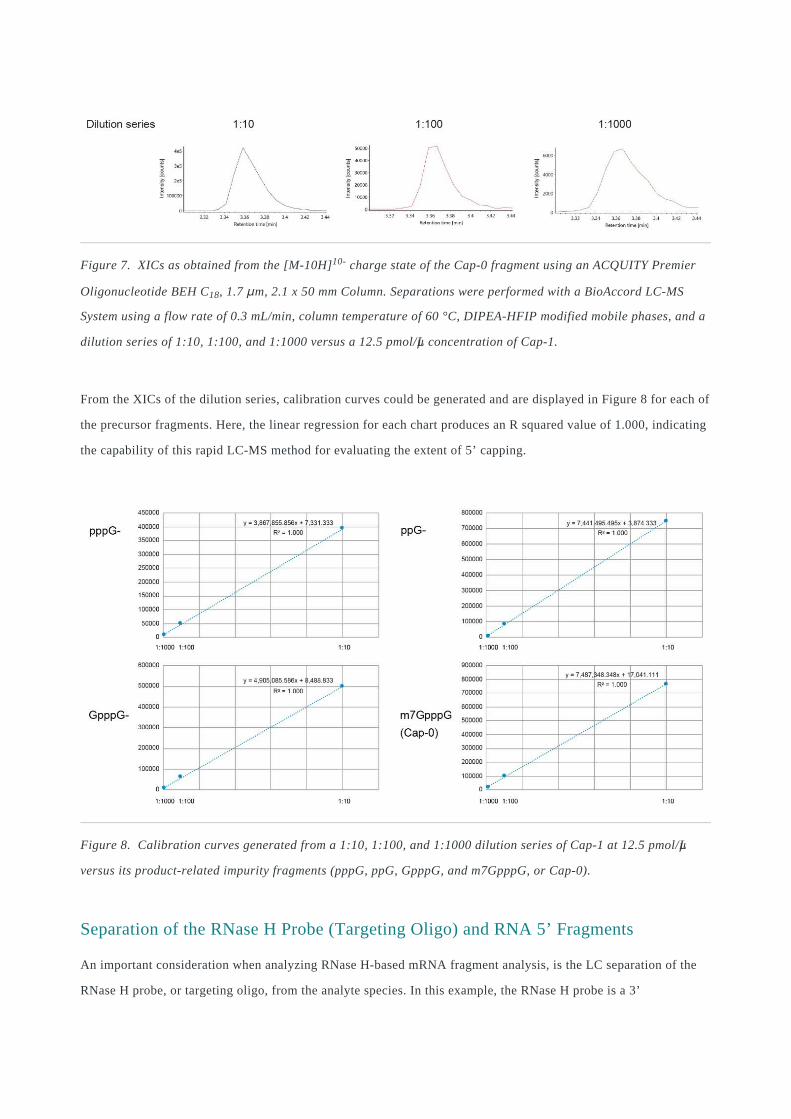
Figure 7. XICs as obtained from the [M-10H]10- charge state of the Cap-0 fragment using an ACQUITY Premier
Oligonucleotide BEH C18, 1.7 μm, 2.1 x 50 mm Column. Separations were performed with a BioAccord LC-MS
System using a flow rate of 0.3 mL/min, column temperature of 60 °C, DIPEA-HFIP modified mobile phases, and a
dilution series of 1:10, 1:100, and 1:1000 versus a 12.5 pmol/µL concentration of Cap-1.
From the XICs of the dilution series, calibration curves could be generated and are displayed in Figure 8 for each of
the precursor fragments. Here, the linear regression for each chart produces an R squared value of 1.000, indicating
the capability of this rapid LC-MS method for evaluating the extent of 5’ capping.
Figure 8. Calibration curves generated from a 1:10, 1:100, and 1:1000 dilution series of Cap-1 at 12.5 pmol/µL
versus its product-related impurity fragments (pppG, ppG, GpppG, and m7GpppG, or Cap-0).
Separation of the RNase H Probe (Targeting Oligo) and RNA 5’ Fragments
An important consideration when analyzing RNase H-based mRNA fragment analysis, is the LC separation of the
RNase H probe, or targeting oligo, from the analyte species. In this example, the RNase H probe is a 3’
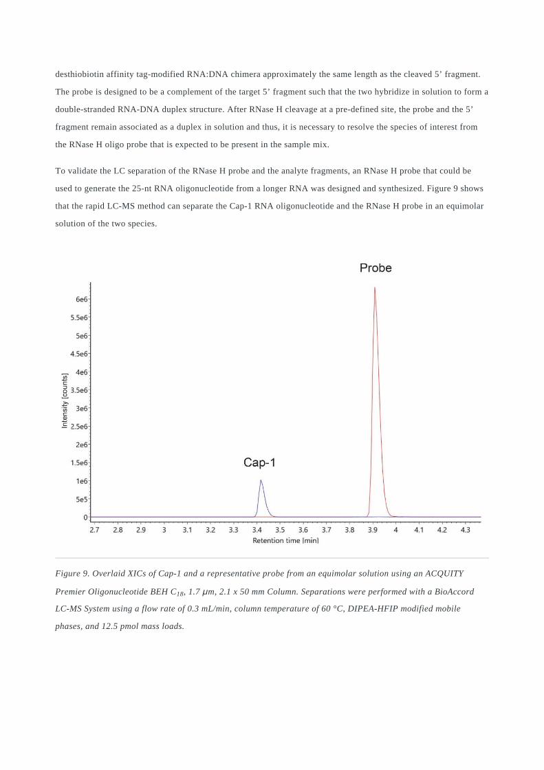
desthiobiotin affinity tag-modified RNA:DNA chimera approximately the same length as the cleaved 5’ fragment.
The probe is designed to be a complement of the target 5’ fragment such that the two hybridize in solution to form a
double-stranded RNA-DNA duplex structure. After RNase H cleavage at a pre-defined site, the probe and the 5’
fragment remain associated as a duplex in solution and thus, it is necessary to resolve the species of interest from
the RNase H oligo probe that is expected to be present in the sample mix.
To validate the LC separation of the RNase H probe and the analyte fragments, an RNase H probe that could be
used to generate the 25-nt RNA oligonucleotide from a longer RNA was designed and synthesized. Figure 9 shows
that the rapid LC-MS method can separate the Cap-1 RNA oligonucleotide and the RNase H probe in an equimolar
solution of the two species.
Figure 9. Overlaid XICs of Cap-1 and a representative probe from an equimolar solution using an ACQUITY
Premier Oligonucleotide BEH C18, 1.7 μm, 2.1 x 50 mm Column. Separations were performed with a BioAccord
LC-MS System using a flow rate of 0.3 mL/min, column temperature of 60 °C, DIPEA-HFIP modified mobile
phases, and 12.5 pmol mass loads.
Conclusion
The value of mRNA as a vaccine modality is now proven by the high effectiveness of the mRNA-based vaccines
against COVID-19 that have now been administered to over 25% of the world population. Analytical techniques to
ensure their proper design, development, and reproducible manufacture are important. In this application note, we
demonstrate that a rapid LC-MS method applicable to evaluating the extent of 5’ capping of synthetic mRNA, an
important CQA for synthetic mRNAs, using an ACQUITY Premier Oligonucleotide BEH C18 Column couple with
a BioAccord LC-MS System. The ACQUITY Premier Column with MaxPeak High Performance Surfaces
Technology could provide considerable improvements in RNA recovery upon first injection. Paired with the
BioAccord LC-MS, this combination of technologies allows for accurate quantitation, even at low limits of
detection, such that it is possible to validate the manufacturing of Cap-1 mRNA molecules, and potential presence
of product precursor related impurities. Moreover, these results highlight the potential of MS-based quantitation for
high throughput assays, which could help accelerate the development of mRNA modalities.
References
Padda, I. S.; Parmar, M. COVID (SARS-COV-2) Vaccine. StatPearls. Treasure Island (FL), 2021.1.
Jackson, N. A. C.; Kester, K. E.; Casimiro, D.; Gurunathan, S.; DeRosa, F. The Promise of mRNA Vaccines: A
Biotech and Industrial Perspective. NPJ Vaccines 2020, 5:11. doi:10.1038/s41541-020-0159-8.
2.
Muttach, F.; Muthmann, N.; Rentmeister, A. Synthetic mRNA Capping. Beilstein J. Org. Chem. 2017, 13,
2819–32. doi:10.3762/bjoc.13.274.
3.
Beverly, M.; Dell, A.; Parmar, P.; Houghton, L. Label-Free Analysis of mRNA Capping Efficiency Using
RNase H Probes and LC-MS. Anal. Bioanal. Chem. 2016, 408, 5021–30. doi:10.1007/s00216-016-9605-x.
4.
Alelyunas, Y.; Shion, H.; Wrona, M. High Sensitivity Intact Monoclonal Antibody (mAb) HRMS
Quantification. Waters Application Note, 720006222 <
https://www.waters.com/nextgen/us/en/library/application-notes/2018/high-sensitivity-intact-monoclonal-
antibody-hrms-quantification.html> , 2018.
5.
Ramanathan, A.; Robb, G. B.; Chan, S. H. mRNA Capping: Biological Functions and Applications. Nucleic
Acids Research 2016, 44, 7511–26. doi:10.1093/nar/gkw551.
6.

Lauber. M.; Walter, T. H.; Gilar, M.; DeLano, M.; Boissel, C. A.; Smith, K.; et al. Low Adsorption HPLC
Columns Based on MaxPeak High Performance Surfaces. Waters White Paper, 720006930EN <
https://www.waters.com/waters/library.htm?cid=511436&lid=135074404&lcid=135074403> , 2020.
7.
Smith, K. M.; Wilson, I. D.; Rainville, P. D. Sensitive and Reproducible Mass Spectrometry-Compatible RP-
UHPLC Analysis of Tricarboxylic Acid Cycle and Related Metabolites in Biological Fluids: Application to
Human Urine. Anal. Chem. 2020. doi:10.1021/acs.analchem.0c03863.
8.
DeLoffi, M.; Nguyen, J. M.; Izzo, G. S.; Lauber, M.; Savaria, M. Improved Chromatographic Performance with
a Premier Peptide C18 Column Versus a Titanium-Lined C18 Column Technology. Waters Application Brief,
720007022 <https://www.waters.com/nextgen/us/en/library/application-notes/2020/improved-chromatographic-
performance-with-a-premier-peptide-c18-column-versus-a-titanium-lined-c18-column-technology.html> , 2020.
9.
DeLano, M.; Walter, T. H.; Lauber, M. A.; Gilar, M.; Jung, M. C.; Nguyen, J. M.; et al. Using Hybrid Organic-
Inorganic Surface Technology to Mitigate Analyte Interactions with Metal Surfaces in UHPLC. Anal. Chem.
2021. doi:10.1021/acs.analchem.0c05203.
10.
Gilar, M.; DeLano, M.; Gritti, F. Mitigation of Analyte Loss on Metal Surfaces in Liquid Chromatography. J.
Chromatogr., A 2021; 1650, 462247. doi:10.1016/j.chroma.2021.462247.
11.
Apffel, A.; Chakel, J. A.; Fischer, S.; Lichtenwalter, K.; Hancock, W. S. Analysis of Oligonucleotides by
HPLC-Electrospray Ionization Mass Spectrometry. Anal. Chem. 1997, 69, 1320–5. doi:10.1021/ac960916h.
12.
Gilar, M.; Fountain, K. J.; Budman, Y.; Holyoke, J. L.; Davoudi, H.; Gebler, J. C. Characterization of
Therapeutic Oligonucleotides Using Liquid Chromatography with On-Line Mass Spectrometry Detection.
Oligonucleotides 2003, 13, 229–43. doi:10.1089/154545703322460612.
13.
Gong. L.; McCullagh, J. S. Comparing Ion-Pairing Reagents and Sample Dissolution Solvents for Ion-Pairing
Reversed-Phase Liquid Chromatography/Electrospray Ionization Mass Spectrometry Analysis of
Oligonucleotides. Rapid Commun. Mass Spectrom. 2014, 28, 339–50. doi:10.1002/rcm.6773.
14.
Li, W.; Lauber, M. Comprehending COVID-19: Preliminary Examination of the SARS-CoV-2 Spike Protein by
Peptide Mapping. Waters Application Note, 720006909 <
https://www.waters.com/nextgen/us/en/library/application-notes/2020/comprehending-covid-19-preliminary-
examination-of-the-sars-cov-2-spike-protein-by-peptide-mapping.html> , 2020.
15.
Doneanu, C.; Fox, J.; Harry, E.; Knowles, C.; Yu, Y. Q.; Fredette, J.; et al. Intact Mass Confirmation Analysis
on the BioAccord LC-MS System for a Variety of Extensively Modified Oligonucleotides. Waters Application
Note, 720007028 <https://www.waters.com/nextgen/us/en/library/application-notes/2020/intact-mass-
confirmation-analysis-on-the-bioaccord-lc-ms-system-for-a-variety-of-extensively-modified-
16.
oligonucleotides.html> , 2020.
Shion, H.; Berger, S. J.; Yu, Y. Q. Application of a Mass Confirmation Workflow for Biotherapeutics
Screening. Waters Application Note, 720007027 <https://www.waters.com/nextgen/us/en/library/application-
notes/2020/application-of-a-mass-confirmation-workflow-for-biotherapeutics-screening.html> , 2020.
17.
Acknowledgments
Jennifer Nguyen, Weibin Chen, Matthew Lauber, Martin Gilar (Waters Corporation, Milford, MA, USA).
Siu-Hong Chan, Bijoyita Roy, Brett Robb (New England Biolabs Inc., Ipswich, MA, USA).
Featured Products
ACQUITY UPLC I-Class PLUS System <https://www.waters.com/134613317>
ACQUITY UPLC Tunable UV Detector <https://www.waters.com/514228>
BioAccord LC-MS System for Biopharmaceuticals <https://www.waters.com/waters/nav.htm?cid=135005818>
UNIFI Scientific Information System <https://www.waters.com/134801648>
ACQUITY Premier Columns <https://www.waters.com/waters/nav.htm?&cid=513206>
720007329, August 2021
© 2021 Waters Corporation. All Rights Reserved.