RADY 401 Case Presentation Kristen Blanton June 2019
15
RADY 401 Case Presentation Kristen Blanton June 2019
Transcript of RADY 401 Case Presentation Kristen Blanton June 2019

RADY 401 Case Presentation
Kristen BlantonJune 2019

Focused patient history and workup
Presents with vomiting, lethargy, and 4 lb weight loss→ Admitted for dehydration and hypokalemia → Abdominal x-ray shows dilated bowel→ Contrast enema yields distended colon without fecal material.
Rectal biopsy negative for Hirschsprung disease
Initial Presentation
1 Week later
2-year-old male presents to PCP with abdominal distension and 6-8 episodes of diarrhea daily for the last year. No significant medical history.

Continued patient workup
Continues to have problems feeding and failure to thrive → GI surgery team attempts to place G-tube and discovers “peach”-sized mass in the retroperitoneum. → CT Abdomen Pelvis with IV contrast
→ FDG-PET scan with CT
→Bone marrow and mass biopsy negative
3 Weeks later
4 Weekslater Suspect neuroblastoma
→Biopsy confirms diagnosis (but it is not neuroblastoma)

List of imaging studies
• Abdominal radiograph
• Contrast enema (not pictured)
• CT Abdomen and Pelvis with IV Contrast
• FDG-PET Scan with CT

Abdominal radiograph
Dilated Bowel Loops
Note: We can see contrast in the descending colon and sigmoid colon. This is likely
from a prior study.
Calcifications

CT Abdomen and Pelvis with IV Contrast
Retroperitoneal Mass
G tubeDilated Bowel Loops
Calcifications
Dilated bowel loops are anterior to the
retroperitoneal mass. Calcifications are more readily seen (than on the plain radiograph).

PET with CT Scan
Sparing of the vessels Adrenals
with low uptake
Avid uptake of the retroperitoneal mass with sparing of the SMA and descending aorta together suggest that the mass may be folding
around important structures as opposed to invasive growth.
Intact adrenal glands with low uptake bilaterally
suggest that this is NOT the primary tumor source.
Uptake of the kidneys, bladder, brain and liver are all normal in this case.
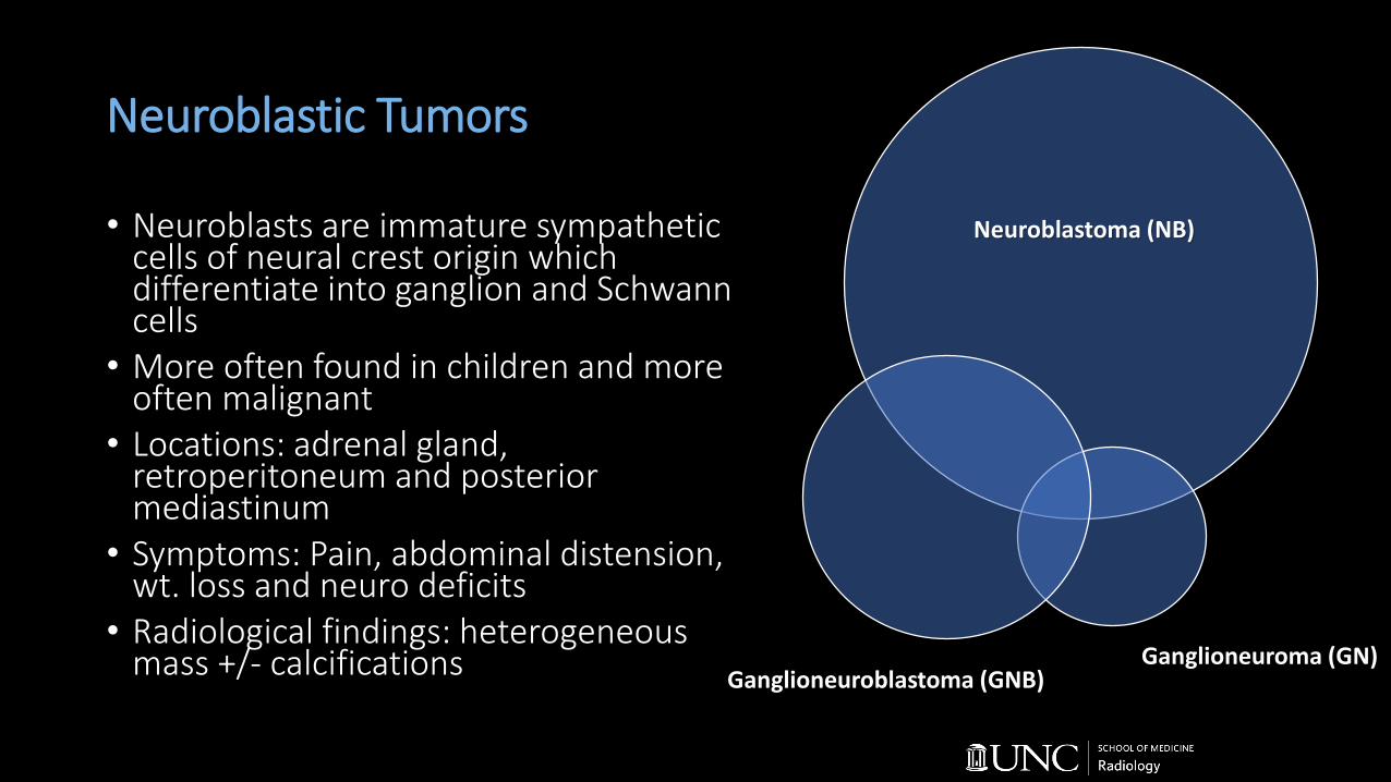
Neuroblastic Tumors
• Neuroblasts are immature sympathetic cells of neural crest origin which differentiate into ganglion and Schwann cells
• More often found in children and more often malignant
• Locations: adrenal gland, retroperitoneum and posterior mediastinum
• Symptoms: Pain, abdominal distension, wt. loss and neuro deficits
• Radiological findings: heterogeneous mass +/- calcifications
Neuroblastoma (NB)
Ganglioneuroblastoma (GNB)Ganglioneuroma (GN)
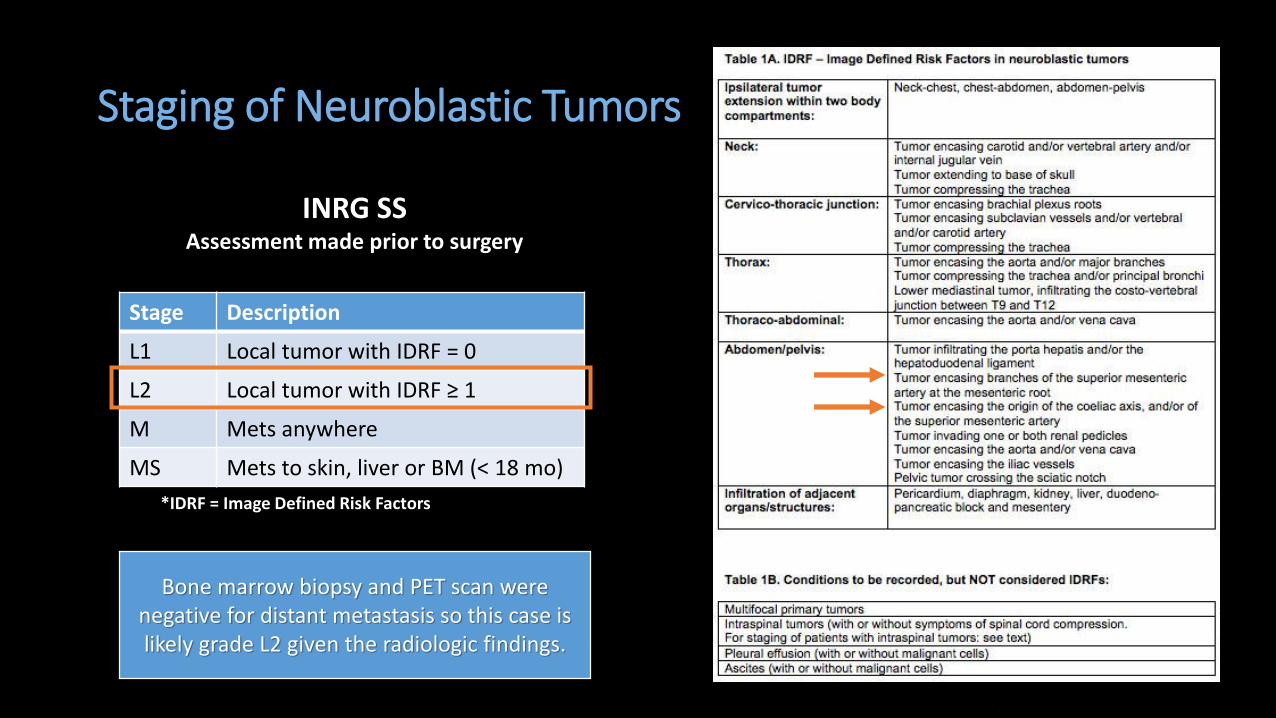
Staging of Neuroblastic Tumors
Stage Description
L1 Local tumor with IDRF = 0
L2 Local tumor with IDRF ≥ 1
M Mets anywhere
MS Mets to skin, liver or BM (< 18 mo)
INRG SSAssessment made prior to surgery
*IDRF = Image Defined Risk Factors
Bone marrow biopsy and PET scan were negative for distant metastasis so this case is likely grade L2 given the radiologic findings.

Patient treatment
• Biopsy confirms ganglioneuroma
• Patient awaits excision of mass
Standard Treatment and Prognosis for Neuroblastic Tumors
Neuroblastic Tumor Subtype Cells Prognosis Treatment
Neuroblastoma Immature (malignant) Poor Surgery and chemo +/-bone marrow transplant
Ganglioneuroblastoma Immature and mature (malignant)
Intermediate Surgery and chemo
Ganglioneuroma Mature (benign) Excellent Surgery
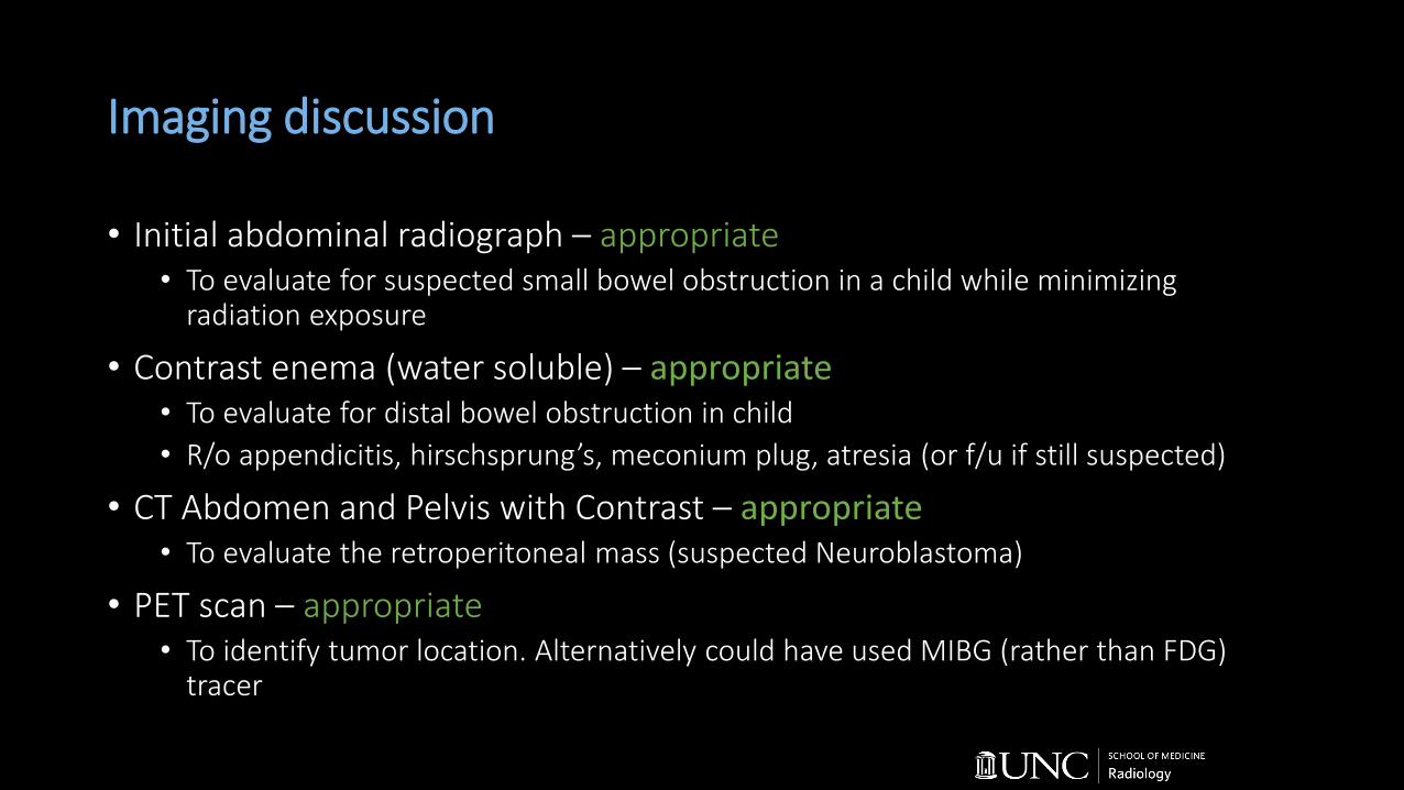
Imaging discussion
• Initial abdominal radiograph – appropriate• To evaluate for suspected small bowel obstruction in a child while minimizing
radiation exposure
• Contrast enema (water soluble) – appropriate• To evaluate for distal bowel obstruction in child
• R/o appendicitis, hirschsprung’s, meconium plug, atresia (or f/u if still suspected)
• CT Abdomen and Pelvis with Contrast – appropriate• To evaluate the retroperitoneal mass (suspected Neuroblastoma)
• PET scan – appropriate• To identify tumor location. Alternatively could have used MIBG (rather than FDG)
tracer

Approximate Cost of Imaging Studies
Imaging Radiation exposure* Cost
Abdominal x-ray 0.7 mSv $23 - $380
Contrast enema with Fluoro 8 mSv $242 - $620
CT Abdomen and Pelvis 10 mSv $889 - $4,050
PET scan with CT up to 25 mSv $1,526 - $3,738
*Exposure is approximated for an adult.

Imaging discussion for MIBG-PET Scan
• ¹²³Iodine metaiodobenzyl guanidine (¹²³I-MIBG) is used for diagnostic workup of neuroblastoma• MIBG, an analog of norepinephrine (NE), is taken up by NE transporters and
accumulates in cells. • > 90% of NB are MIBG avid; however, other tumors can have this uptake
pattern (e.g. NB, GNB, GN, pheo, carcinoid, medullary thyroid). This means MIBG cannot definitively differentiate among these tumors.
• Sensitivity (90%) and Specificity (88%) for neuroblastoma, but MIBG uptake is variable for ganglioneuroma.
• Regardless of the tracer used, confirm via primary biopsy, bone marrow biopsy or catecholamine metabolites.
UNC Top Three
• With the presence of heterogeneous mass in the adrenal glands, retroperitoneum, or posterior mediastinum (especially in a child), suspect neuroblastic tumor.
• Ganglioneuroma is a rare but benign tumor that presents similarly to neuroblastoma.
• ¹²³I-MIBG can be used to identify various tumors of neural crest origin (NE uptake), but particularly neuroblastoma.

ReferencesMonclair T, Brodeur GM, Ambros PF, et al. The International Neuroblastoma Risk Group (INRG) staging system: An INRG Task Force report. J Clin Oncol. 2009;27(2):298-303. doi:10.1200/JCO.2008.16.6876
G.M. B, J. P, F. B, et al. Revisions of the international criteria for neuroblastoma diagnosis, staging, and response to treatment. J Clin Oncol. 1993;11(8):1466-1477.
Murphey MD, Carroll JF, Flemming DJ, Kransdorf MJ. From the Archives of the AFIP OBJECTIVES. RadioGraphics. 2004;24:1433-1466.
Decarolis B, Simon T, Krug B, et al. Treatment and outcome of Ganglioneuroma and Ganglioneuroblastoma intermixed. BMC Cancer. 2016;16(1):1-12. doi:10.1186/s12885-016-2513-9
College of Radiology A. ACR-SPR practice guideline for the performance of pediatric fluoroscopic contrast enema examinations. In: Practice guidelines & technical standards 1997. Revision 2016. 2016;1076(Revised 2008):1-14.
Shohet, MD,PhD, Nuchtern, MD. Clinical Presentation, Diagnosis and Staging Evaluation of Neuroblastoma. Post TW, ed. UpToDate. Waltham, MA: UpToDate Inc. https://www.uptodate.com/contents/epidemiology-pathogenesis-and-pathology-of-neuroblastoma?search=neuroblastoma&source=search_result&selectedTitle=3~122&usage_type=default&display_rank=3 Accessed on June 16, 2017.
Consumer Fair Price Search. Healthcare Bluebook website. https://www.healthcarebluebook.com/ui/consumerfront. Accessed June 16, 2019.
Radiation Exposure from Medical Exams and Procedures. Health Physics Society, Specialists in Radiation Safety website. http://hps.org/documents/Medical_Exposures_Fact_Sheet.pdf. Updated January 2010. Accessed June 16, 2019.