Radiology/Ultrasound Phantoms ―Tradition and … · high quality phantoms to the field of...
23
Transcript of Radiology/Ultrasound Phantoms ―Tradition and … · high quality phantoms to the field of...
1 2
Kyoto Kagaku Co., Ltd has developed an extensive history and tradition in providing high quality phantoms to the field of radiology. Kyoto Kagaku Co., Ltd continues with its challenge in offering innovative, state-of-the-art solutions for this rapidly developing industry.
Founded during post-war Japan in 1948, Kyoto Kagaku Co., Ltd originates from the Shimadzu Corporation. Early days of the company began with the production of scientific specimens, anatomical models and skeletons. Its first radiology phantom was developed in the early 1960’s with the collaboration of Shimadzu. Original human tissue substitute materials for diagnostic energy range and therapeutic energy range were also invented. Tissue substitute materials with ultrasound compatibility were a recent development that opened doors to a variety of QA and training phantoms in the field of sonography.
Through anatomical research and model crafting breakthroughs such as our long sought human tissue substitutes, Kyoto Kagaku strives to perfect the production of anthropomorphic phantoms for medical imaging. Kyoto Kagaku will continue to uphold high standards in developing products that effectively contribute to patient safety and training of healthcare professionals.
Radiology/Ultrasound Phantoms ―Tradition and Innovation―
Shimadzu anatomical human model with muscles wins the Gold prize at the International exhibition in Alaska-Yukon Pacific Ocean
The educational scientific division of Shimadzu Corp is established.
The educational scientific specimens division begins to manufacture of anatomical models
Kyoto Kagaku is established.
First resinous skeleton models
First resinous anatomical models
Development of Stomach Phantom in partnership with Shimadzu Corp.
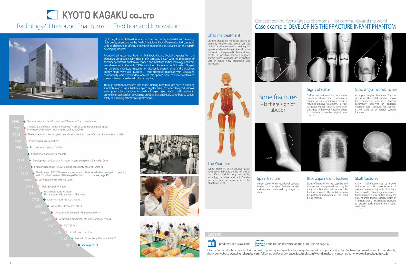
Concept behind Kyoto Kagaku phantoms ―for community and the world―Case example: DEVELOPING THE FRACTURE INFANT PHANTOM
Bone fractures- is there sign of
abuse?
Child maltreatment
The Phantom
Signs of callus
Spiral fracture Back, scapula and rib fracturesSigns of fractures on the scapulae and ribs are to be inspected not only by their form, but also their location. Rib fractures close to the vertebrae may be potential indicators of the child being thrown.
Skull fracturesA linear skull fracture may be another indication of child maltreatment. A common cause of injury is blunt force trauma, in which the energy from a blow is transferred over a wide surface area of the skull. At times, fractures display better on x-ray scans than CT imaging and it is crucial to prevent such fractures from being overlooked.
Supracondylar humerus fractureA supracondylar humerus fracture occurs on the distal humerus above the epicondyles and is a fracture commonly observed in children. Pediatric cases account for approxi-mately 20% of all lateral condyle fractures.
Lateral
Typical fractures of an abusive attack have been replicated on the left side of the infant model's body and limbs, including the ulnae and radii. Smaller fractures can be seen around the phantom's wrist.
Calluses on their own are not definite proofs of abuse cases. However, a number of callus examples can be a result of abusive treatment. For this particular model, calluses have been simulated in the wound healing phase 5 of remodeling to the original bone contour.
Certain causes of non-accidental pediatric injuries, such as spiral fractures, include maltreatment stimulated by anger or distress.
: product video is available : publication reference on the product is on page 40
Information on this brochure is of at the time of printing and specifications may change without prior notice. For the latest information and further details, check our website www.kyotokagaku.com, follow us on Facebook www.facebook.com/kyotokagaku or contact us at [email protected].
Children around the world are victims to domestic violence and abuse, yet the problem is often overlooked. Noticing the signs of an abusive fracture of a child is the first step to putting an end to these maltreat-ments. This phantom has been designed and developed to cultivate such observation skills in future X-ray radiologists and technicians.
Legend
First participation in RSNA (Radiological Society of North America)
Development of TOUGH serious, human tissue substitute for radiotherapy range in cooperating with the National Institute of Radiological Sciences see page 25
Development of Synthetic Bones
Multi-Slice CT PhantomFirst Whole Body PhantomFirst ultrasound Examination PhantomChest Phantom N1 "LUNGMAN"
Whole Body Phantom PBU-50
Ultrasound Examination Phantom ABDFAN
Awarded "Grand Prize" of Good Company Award
FAST/ER FAN
Dental Head Phantom
Pediatric Whole Body Phantom PBU-70
See Page 05-11 !

3 43 4 Index Index
TABLE OF CONTENTS
25
25
25
25
25
26
26
26
26
27
27
27
27
28
28
28
28
29
29
30
30
30
30
30
31
31
31
31
32
32
33
33
34
34
35
35
36
36
37
37
37
37
38
38
39
39
40
41
41
42
PH-40
PH-41
PH-42
PH-37
PH-38
PH-31
PH-26
PH-32
PH-32B
PH-33
PH-34
PH-53
PH-27
PH-24
PH-29
PH-28
PH-30
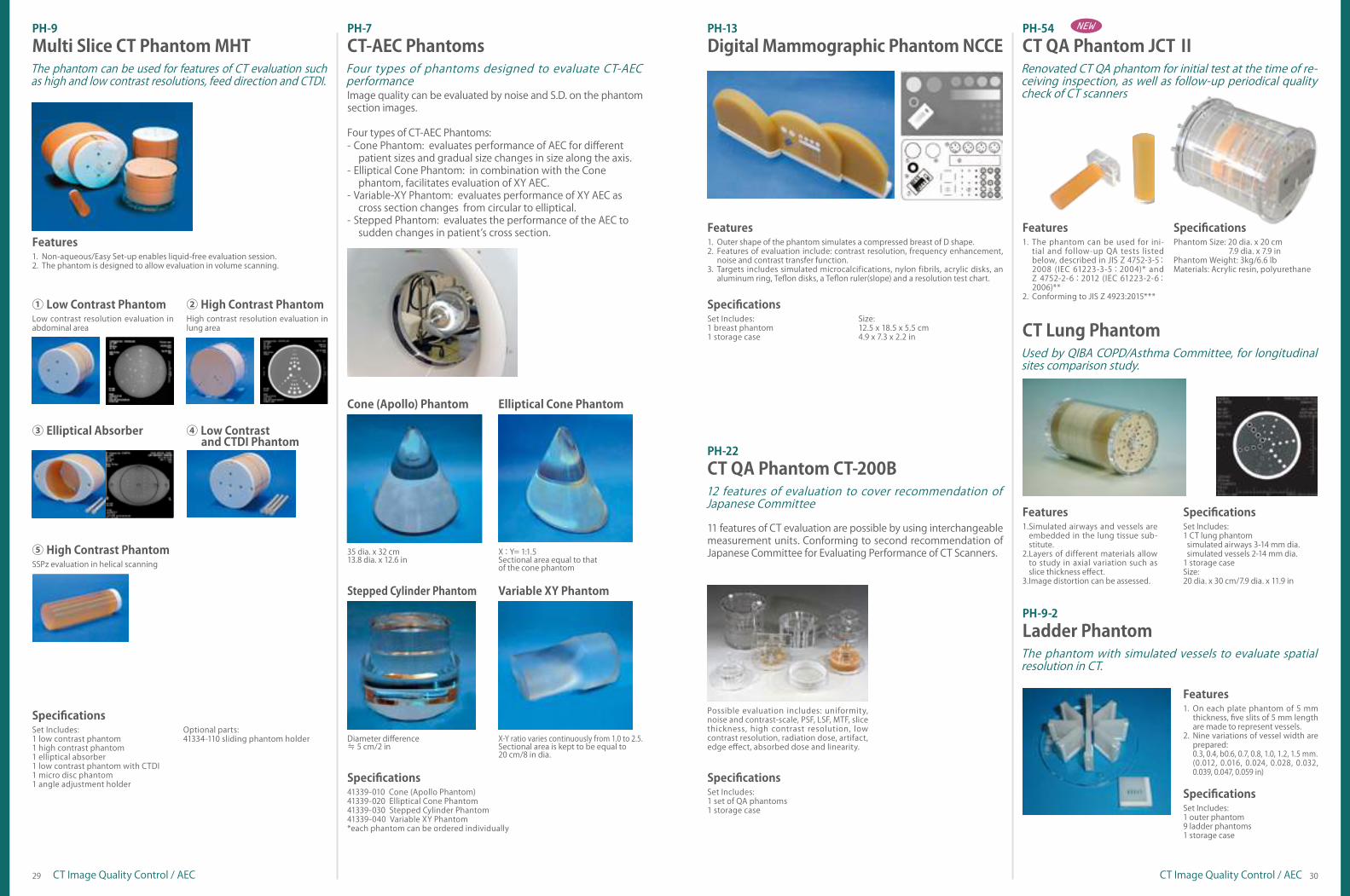
PH-9
PH-7
PH-13
PH-22
PH-54
PH-9-2
PH-10
PH-16
PH-17
PH-14
US-1
US-1B
US-5
US-8
US-7α
US-6
US-3
US-13
US-10
US-11
US-9
US-2
US-4
M93UB
M93C
MW18
M43E
05-06
07
08
09
09
10
11
11
12
12
13
13
13
14
14
15
16
17
17
17
17
17
17
18
19
19
20
20
20
21
21
21
21
22
23
24
24
24
PH-61
PH-60
PH-59
PH-55
PH-56
PH-57
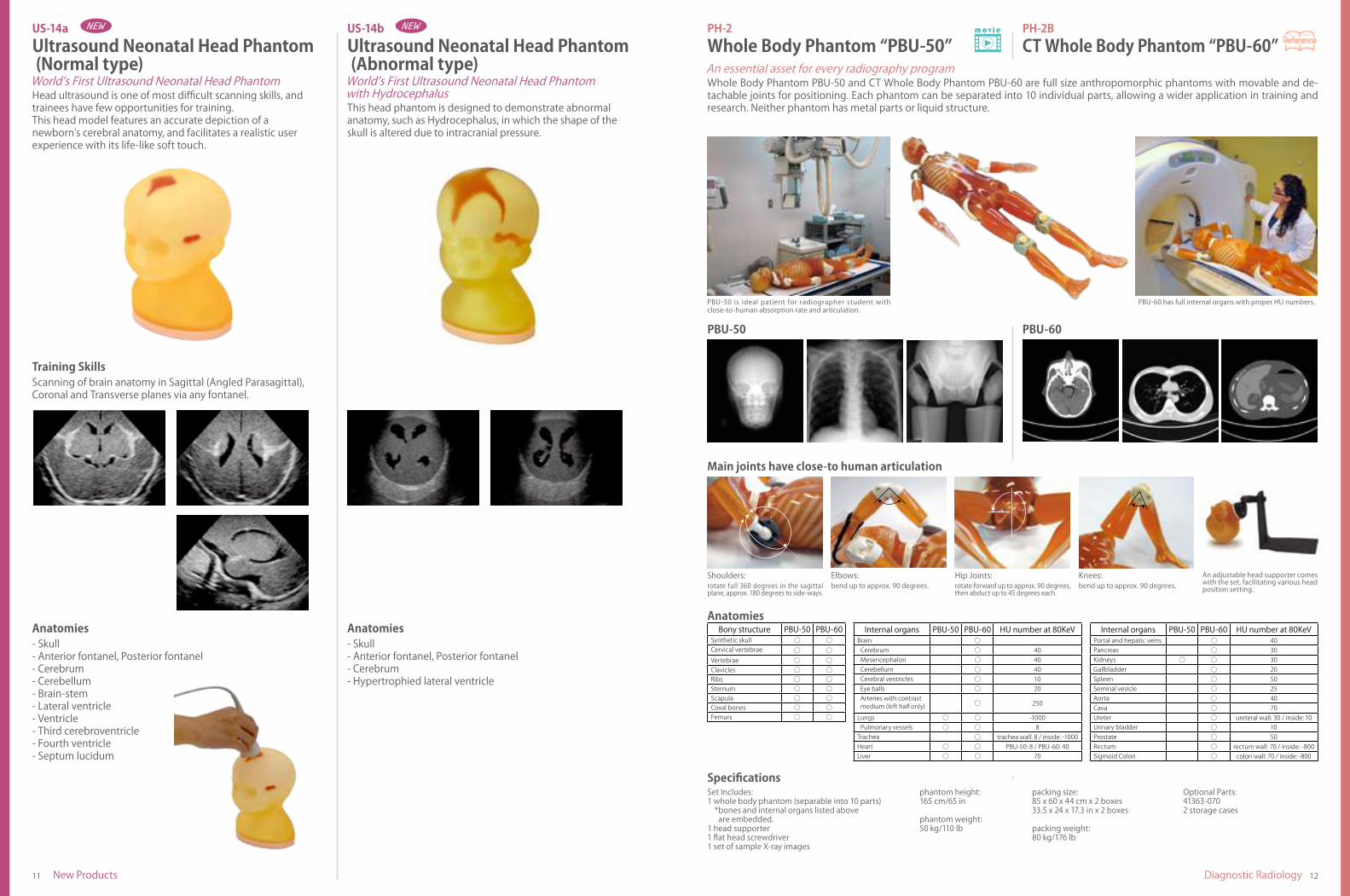
US-14a
US-14b
PH-2
PH-2B
PH-2C
PH-2D
PH-50
PH-1
PH-58
PH-1C
PH-8
PH-4
PH-47
PH-62
PH-3
PH-5
PH-19
PH-46
PH-18
PH-49
PH-51
PH-48
PH-39
PH-6B
11347-210
Ultrasound Examination Training Phantom "ECHOZY"
Ultrasound Examination Training Phantom "ABDFAN"
FAST/Acute Abdomen Phantom "FAST/ER FAN"
Pediatric FAST/Acute Abdomen Phantom
Fetus Ultrasound Examination Phantom "SPACE FAN-ST"
Breast Ultrasound Examination Phantom "BREAST-FAN"
Abdominal Intraoperative & Laparoscopic Ultrasound Phantom "IOUS-FAN"
Infant Hip Sonography Training Phantom
Female Pelvic Ultrasound Phantom
Scrotal Ultrasound Phantom
Ultrasound Guided Breast Biopsy Phantom
Ultrasound Quality Assurance Phantoms
Introductory Ultrasound Training Block "REAL VESSEL"
Breast Ultrasound QA Phantom
CVC Insertion Simulator II
CVC Insertion Simulator III
Ultrasound-Guided PICC Training Simulator
Ultrasound Compatible Lumbar Puncture/Epidural Simulator
References
Custom order example
Common Specifications
Product Supervision
Tough Water Phantom WD
Tough Bone Phantom BE-T, BE-H, BE-N
Tough Lung Phantom LP
Therapy Body Phantom THRA-1
Pediatric Therapy Body Phantom THRA-2
MRI Quality Assurance Phantom MHR
ORINS Thyroid Phantom ITS
MRI Quality Assurance Phantom JMR
MRI Quality Assurance Phantom JMR II
MRI Head Phantom NH
MRI/NM Head Phantom BHC
Brain Phantom IB-20 advanced
Brain Phantom IB-10
Myocardial Phantom HL
ECT Hot Cold Phantom SP-6
SPECT QA Phantom JSP
SPECT QA Phantom JS-10
Multi Slice CT Phantom MHT
CT-AEC Phantoms
Digital Mammographic Phantom NCCE
CT QA Phantom CT-200B
CT QA Phantom JCT II
CT Lung Phantom
Ladder Phantom
BMD Chart Phantom UHA
Contrast Detail Phantom
Water Body Phantom WAC
Acrylic Phantom XAC
Sectional Phantoms
Tough Whole Body Phantom "PBU-90 RUGGED"
CT-DI Phantom
CT ERF Phantom HIT
Tomosynthesis Phantom NS
Thorax Low Contrast Phantom ODA-LC
Ultrasound Neonatal Head Phantom(Normal type)
Ultrasound Neonatal Head Phantom(Abnormal type)
Whole Body Phantom "PBU-50"
CT Whole Body Phantom "PBU-60"
Fractured Hand/Forearm Phantom
Body Plates for PH-2/2B (BMI 32/40)
CT Whole Body Phantom with Pathologies
Pediatric Whole Body Phantom "PBU-70"
Bone Fracture Pediatric Phnatom "PBU-70B"
Newborn Whole Body Phantom
Multipurpose Chest Phantom N1 "LUNGMAN"
Optional and replacement parts
-Chest Plates
-Storage Case
-Simulated Tumors
-Custom Order Simulated Tumors
-Components for Radioisotope
GGO Tumor Phantom
Pediatric Chest Phantom
Lung Cancer Screening CT Phantom LSCT001
CT Torso Phantom CTU-41
Dental Radiography Head Phantom (mouth closed)
Dental Radiology Head Phantom (mouth open)
Angiographic CT Head Phantom ACS
CT Abdomen Phantom
Rotation Stomach Phantom TMP-R
CT Prostate Phantom
Stomach Phantom BMU-1
CT Colonography Phantom NCCS
Lumbar Spine Fluoroscopy Training Phantom
Dynamic Heart and Lung Phantom
Dynamic Thorax Phantom
Dynamic Cardiac CT Phantom MD-CT
Ultrasound Phantoms
CT Image Quality Control / AEC
MRI / Nuclear Medicine
Dosimetry and RadiotherapyNew Products
Dynamic Phantoms
Diagnostic Radiology

5 6New Products New Products
Sectional Phantoms allow for imaging of individual anatomy as needed.Sectional Phantom Series (Plain X-rays)PH-61
Leg
Arm
* Opaque model shown.
* Opaque model shown.
* Transparent model shown.
* Transparent model shown.
* Transparent model shown.
* Transparent model shown.
Right Elbow
Right Knee
Right Hand
Right Foot
Left Hand
Left Foot
(Transparent)
(Transparent)
(Transparent)
(Transparent)
(Transparent)
(Transparent)
Right Elbow
Right Knee
Right Hand
Right Foot
Left Hand
Left Foot
(Opaque)
(Opaque)
(Opaque)
(Opaque)
(Opaque)
(Opaque)
41926-150
41926-190
41926-030
41926-110
41926-050
41926-130
41926-140
41926-180
41926-020
41926-100
41926-040
41926-120
Normal flexion range allows for AP/lateral and partial flexion views with one phantom.
Freely movable patella and joint allows for realistic positioning of the knee for AP/lateral, oblique, sunrise and tunnel views.
Includes thoracic skeletal system with embedded mediast inal space and bronchus to provide realistic imaging. The scapulae are rotated outside of the lung fields for proper PA chest imaging.
Body
* Opaque model shown.
Head
Head (Transparent)
Thorax (Transparent)
Head (Opaque)
Thorax (Opaque)
Pelvic (Opaque)
41926-010
41926-070
41926-000
41926-060
41926-080
* Opaque model shown.
Stand-alone design can be used with the adjustable head positioning stand to demonstrate accurate skull positioning.
Includes lumbar/sacral spine, pelvic bony anatomy and proximal femurs.
Optional parts:Adjustable head supporter

7 8New Products New Products
Open hand position
Durability testing
Shoulders: rotate full 360 degrees in the sagittal plane, approx. 180 degrees to side-ways.
Elbows: bend up to approx. 90 degrees.
Hip Joints: rotate forward up to approx. 90 degrees, then abduct up to 45 degrees each.
Knees: bend up to approx. 90 degrees.
An adjustable head supporter comeswith the set, facilitating various headposition setting.
Main joints have close-to human articulation
Phantom size and weightBody phantom: 32 dia. x 15 cm, 15kg 12.6 dia. x 5.9 in, 33lb Head phantom: 16 x 15 cm, 4kg 6.3 x 5.9 in, 8.8lb
Materials: Acrylic resin
Accessory: Storage case
Specifications
A set of phantoms for CTDI-100, conforming to requirements described in JIS Z 4752-3-5:2008 (IEC 61223-3-5:2004) as acceptance test, and JIS Z 4752-2-6:2012 (IEC 61223-2-6:2006) as consistency test.
CT-DI Phantom (Head and Body Phantom)
Skeletal system with embedded lungs, heart, liver and kidneys. PBU-90 includes a whole body phantom (separable into 10 parts) and head supporter.Freely articulating joints allow for realistic positioning.
New phantom material is designed for rough handling, improved durability and low maintenance. TOUGH Whole Body Phantom“PBU-90 RUGGED”PH-60 PH-59
Storage Case II for PH-2/2B/6041363-070
(a pair)
Features
The set of head and body phantom made of PH-40Custom order example: head and body phantom in different types of tissue substitutes
*Custom order example

9 10New ProductsNew Products
Modality CT tomosynthesis
Sectional Image
Absorbed Dose 11.9mGy(CTDlvol) 0.4mGy(CTDlvol) 2.18mGyTube Voltage 120kv 120kv 100kv
Hole: 1.0 mm/0.04 in dia.Aluminum plate: 0.5mm/0.02 in thickAcrylic plate 5mm/0.2 in thick.*the aluminum plate is sandwiched between layers of Acrylic70 x 150 mm/2.8 x 5.9 in
For calculation of slice thickness using FWHM
20mm thick
Test units can be set in the alu-minum supporting box at 10, 15 or 20 mm (0.4,0.6 or 0.79 in) height
For verification of the special positioning in reconstructionStainless steel line: 0.1 mm/0.004 in dia.
For evaluation of uni-formity and tilting of the examination table70 x 150 mm/2.8 x 5.9 in
Upper side: tree parallel lines
Lower side: cross lines
Specific gravity of simulated nodules *Specific gravity of the background is 0.14
() : CT
specific gravity
1st layer 1.06(0)2nd layer 0.47(-550)3rd layer 0.35(-640)4th layer 0.26(-730)
specific gravity
5th layer 0.24(-780)6th layer 0.15(-825)7th layer 0.75(-250)8th layer 0.64(-375)
Sagittal section
*For the radiation absorber in different sizes can be requested as a custom order
1st layer
2nd layer
3rd layer
4th layer
5thlayer
6thlayer
7thlayer
8thlayer
Materials: PP, polyurethane foam, polyurethaneSimulated nodules: Sizes: 2,3,4,5, 7 and 10 mm (0.08, 0.12, 0.16, 0.2, 0.28 and 0.4) dia. in HU number: 0, -250, -375, -550, -640, -730, -780, -825
Specifications
Comparison between CT images and tomosynthesis images.Evaluation of images in three different planes with one scan.A variety of nodules that simulates GGOQuantitative evaluation of image quality, using CNR of simu-lated nodulesVisual evaluation of image quality of simulated nodules, using contrast detail diagram for contrast resolutionOptimization of radiation dose in CT scanningOptimization of radiation dose in tomosynthesisElliptical radiation absorber that simulate human body, to study scattering effect of soft tissue
1.2.3.4.
5.
6.7.8.
Features
For image evaluation of low contrast targets with CT, tomosynthesis as well as cone beam CT.Thorax Low Contrast Phantom ODA-LC
Set Includes:1 Reconstruction positioning unit1 Slice thickness unit1 Uniformity unit
41919-010Angle adjustment holder (table-top type)
Compatible with PH-9
SpecificationsOptional part
Verification of slice thickness in reconstructionMeasurement of slice thicknessVerification of uniformity
1.2.3.
Evaluation Features
The phantom is designed for daily quality control of Tomosyn-thesis, allowing evaluation of reconstruction slices and unifor-mity in the measurement of slice thickness through showing the images numerically and graphically. It is also useful in evaluation of image quality that varies depending on reconstruction func-tion.
Tomosynthesis Phantom NS
Size:phantom size: 20 dia. x 25 cm 7.9 dia x 9.8 inphantom weight: 4.5 kg 9.9 lb
Optional part:41919-010Angle adjustment holder (table-top type)
Set Includes:Cylindrical container (200 mm dia.)Measurement plates: 5 variations; HU20, 50, 100, 200 and 500Rotation holderFixture for the cylindrical container
Materials:Acrylic resin, polyurethane
Specifications
The phantom is designed to physically and quantitatively evaluate interactively reconstructed images in the low CNR area, such as abdomen, where MTF of PSF is less useful com-paring to high CNR area.The phantom uses edge spread function (ESF) to calculate MTF of the low CNR images, which facilitate assessing perfor-mance properties of iteratively reconstructed images under low CNR.
1.
2.
Features
A phantom designed for physical evaluation of iteratively reconstructed images under low CNR.
Slice thickness unitReconstruction positioning unit
Uniformity unit Height setting rack
CT ERF Phantom HITPH-57PH-56PH-55
11 12Diagnostic RadiologyNew Products
PBU-60PBU-50
PBU-50 is ideal patient for radiographer student with close-to-human absorption rate and articulation.
PBU-60 has full internal organs with proper HU numbers.
Shoulders: rotate full 360 degrees in the sagittal plane, approx. 180 degrees to side-ways.
Elbows: bend up to approx. 90 degrees.
Hip Joints: rotate forward up to approx. 90 degrees, then abduct up to 45 degrees each.
Knees: bend up to approx. 90 degrees.
An adjustable head supporter comeswith the set, facilitating various headposition setting.
Bony structure PBU-50 PBU-60Synthetic skull ○ ○Cervical vertebrae ○ ○Vertebrae ○ ○Clavicles ○ ○Ribs ○ ○Sternum ○ ○Scapula ○ ○Coxal bones ○ ○Femurs ○ ○
Internal organs PBU-50 PBU-60 HU number at 80KeVBrain ○
Cerebrum ○ 40Mesencephalon ○ 40Cerebellum ○ 40Cerebral ventricles ○ 10Eye balls ○ 20Arteries with contrastmedium (left half only) ○ 250
Lungs ○ ○ -1000Pulmonary vessels ○ ○ 8
Trachea ○ trachea wall: 8 / inside: -1000Heart ○ ○ PBU-50: 8 / PBU-60: 40Liver ○ ○ 70
Internal organs PBU-50 PBU-60 HU number at 80KeVPortal and hepatic veins ○ 40Pancreas ○ 30Kidneys ○ ○ 30Gallbladder ○ 20Spleen ○ 50Seminal vesicle ○ 25Aorta ○ 40Cava ○ 70Ureter ○ ureteral wall: 30 / inside: 10Urinary bladder ○ 10Prostate ○ 50Rectum ○ rectum wall: 70 / inside: -800Sigmoid Colon ○ colon wall: 70 / inside: -800
US-14bUltrasound Neonatal Head Phantom (Abnormal type)
Ultrasound Neonatal Head Phantom (Normal type)
This head phantom is designed to demonstrate abnormalanatomy, such as Hydrocephalus, in which the shape of theskull is altered due to intracranial pressure.
World’s First Ultrasound Neonatal Head Phantomwith HydrocephalusHead ultrasound is one of most difficult scanning skills, and
trainees have few opportunities for training.This head model features an accurate depiction of anewborn’s cerebral anatomy, and facilitates a realistic userexperience with its life-like soft touch.
World’s First Ultrasound Neonatal Head Phantom
- Skull- Anterior fontanel, Posterior fontanel- Cerebrum- Hypertrophied lateral ventricle
Anatomies
US-14a
- Skull- Anterior fontanel, Posterior fontanel- Cerebrum- Cerebellum- Brain-stem- Lateral ventricle- Ventricle- Third cerebroventricle- Fourth ventricle- Septum lucidum
Anatomies
Scanning of brain anatomy in Sagittal (Angled Parasagittal),Coronal and Transverse planes via any fontanel.
Training Skills
packing size: 85 x 60 x 44 cm x 2 boxes33.5 x 24 x 17.3 in x 2 boxes packing weight:80 kg/176 lb
Optional Parts:41363-0702 storage cases
Set Includes:1 whole body phantom (separable into 10 parts) *bones and internal organs listed above are embedded.1 head supporter1 flat head screwdriver1 set of sample X-ray images
phantom height:165 cm/65 in
phantom weight:50 kg/110 lb
Specifications
Whole Body Phantom “PBU-50” CT Whole Body Phantom “PBU-60”
Whole Body Phantom PBU-50 and CT Whole Body Phantom PBU-60 are full size anthropomorphic phantoms with movable and de-tachable joints for positioning. Each phantom can be separated into 10 individual parts, allowing a wider application in training and research. Neither phantom has metal parts or liquid structure.
An essential asset for every radiography program
Anatomies
Main joints have close-to human articulation
PH-2BPH-2

13 14Diagnostic RadiologyDiagnostic Radiology
Spiral Fracture of the Distal Tibia
All fractures are prepared on the left side of the phantom.
* Specify with or without adult teeth at the time of order.
Forearm Shaft Fractures
a
b
c
d
e
fg
h
h
f
i
g
e
c
ba
d
HU number
a Brain tumor 130
b Subarachnoid bleeding 190
c Pulmonary tumor surface:30inside 130
d Hepatic tumor 10
e Pancreatitis 30
f Gall stone 170
g Kidney stone
h Appendicitis
70
i Spondylolisthesis -
Skull Fracture Supracondylar Fracture of the Humerus
41350-000-11
41363-070
Fractured Hand/Forearm Phantom
Body plates for PH-2/2B
Storage case Ⅱ
Optional Parts for PH-2/2B/60
X-ray phantom for trauma evaluation
Optional Parts for PH-2/2B/60
Optional Parts for PH-2/2B/60
Bone fractures:ulna, radius, first metacarpal, middle phalanx of the index finger, distal phalanx of the first finger (compressed fracture),fifth metacarpal
Set Includes:1 fractured hand/forearm phantom
Specifications
Customized PH-2BCT Whole Body Phantom with Pathologies
Pathological findings in the phantom expand possibilities in training application.
OOOOXOOOOXOOOOXOOOOXOOOOXOOOOXOOOOX
The body plate provides the phantom with a variety of body shapes.
Body plates to simulate a body of BMI30
PH-2DBone Fracture Pediatric Phantom “PBU-70B”Training in pediatric radiography can be enriched with clear and subtle bone fractures. Typical fractures resulting from child abuse are also included.Improve your skills in detecting bone fractures in children.
PH-2CPediatric Whole Body Phantom “PBU-70”
Main joints have life-like articulation, allowing various posi-tioning for plain X-ray.Training and research applications can be enriched by disas-sembling the phantom into 10 individual parts (head, limbs and trunk). The phantom has no metal parts or liquid structures.No defect in bone images of joints.
1.
2.
3.4.
Features
Pediatric Whole Body Phantom is modeled after a 5-year-old child of 105cm (43”) in height. This is a life-size, full body anthropomorphic phantom with a state-of-the-art synthetic skeleton, lungs, liver, mediastinum and kidneys. Its movable and detachable joints allow various positioning.
This Phantom is easy to handle positioning, and provides complete bone images for every joint.
- Full synthetic skeleton- Main pulmonary vessels, mediastinum, liver, kidneys
Anatomies
Set Includes:1 pediatric whole body phantom: life-size, 5-year-old consists of 10 parts1 head supporter1 hand fixture belt1 set of sample X-ray images
Size:phantom height: 110 cm/43.3 in
phantom weight: 20 kg/44 lb
Optional Parts:41363-080storage case for PH-2C
Specifications
- Plain X-ray photography and basic CT scanning- Basic patient positioning for X-ray and CT
Training Skills
Subarachnoid bleeding
Pulmonary tumor
Pancreatitis
Kidney stone
Spondylolisthesis
Brain tumor
Hepatic tumor
Gall stone
Appendicitis
Fifth Costal Bone
Fracture Callus of the FemurForearm Shaft Fractures
Spiral Fracture of the Distal Tibia
Scapula
(a pair)
surface:70inside 40

15 16Diagnostic Radiology Diagnostic Radiology
– 800
– 630
+ 100
Set Includes:1 newborn whole body phantom1 storage case1 set of sample X-ray images1 instruction manual
Size:phantom size: 42 cm (represent a baby of 50 cm tall)/16.5 inphantom weight: 2.8 kg /6.2 lb
Specifications
- Skull, spine, ribs, pelvis, scapulae, clavicles, humeri, radius,ulnae, bones of hands, femora, fibulae, tibiae and bones of feet- Lungs and mediastinum
Anatomies
Newborn Whole Body PhantomThe world's first full body phantom for neonatal radiographyNewborn Whole Body Phantom is the world's first full body phantom for neonatal radiography with correct anatomical structure and movable limbs. Neonatal radiography is an important tool in NICU (Neonatal Intensive Care Unit). Patient positioning and immobilization are essential features. This phantom provides opportunities for hands-on training and experiments to minimize radiation exposure to newborn babies.
PH-50
1.2.3.4.5.6.
Limbs rotate 360 degrees at shoulders and hip joints.Left hand is clenched and right hand is open.Life size whole body newborn baby.Original human tissue substitute.No metal parts or liquid structures.Meconium aspiration syndrome can be made per custom order.
Features
-Immobilization - Manual immobilization - Immobilization with fixtures
-Autopsy imaging
Training Skills-Radiography - Upright AP (anteroposterior) - Supine AP - Upright lateral - Supine lateral
Set Includes:1male chest torso main body: synthetic bones are embedded mediastinum: heart, trachea pulmonary vessels (right and left) abdomen (diaphragm) block: no internal structure15 simulated tumors (15 variations 1 piece each) 3 varieties of Hounsfield number: approx. -800, -630, +100 5 sizes for each type: diameters 0.3, 0.5, 0.8, 1.0, 1.2 cm diameters 0.12, 0.2, 0.32, 0.39, 0.47 in
Size:phantom size: 43 x 20 x 48 cm, chest girth 94 cm 17 x 8 x 18 in, chest girth 37 inphantom weight: 18 kg/39.6 lbpacking size: 65 x 55 x 29 cm, 25 kg 26 x 22 x 11 in, 55.1 lb
Specifications
Applicable for both plain radiography and CT scanning.Simulated tumors and other targets can be attached at any points in the lung field.Wide variety of uses in interpretation training, anatomical education, evalu-ation and assessment of devices and other research.Accurate anatomy and high quality substitute materialsArms-abducted position of the torso suits the CT scanning. The pulmonary vessels are spatially traceable. Assessment of computer-aided de-tection systems is possible.
1.
2.
3.
4.
5.
6.
7.
Features
The phantom provides life-like radiographs very close to actual clinical images. The three-dimensional structure allows both PA and LATERAL images to be obtained. The phantom bones and vessels show life-like contrast gradations on the image along with tube voltages.PH-1 is used in a study by the FDA to create a database of CT scans with different scanners and protocols, as a resource for assessment of lung nodule size estimation method.
Broad range of possible applications in research and trainingMultipurpose Chest Phantom N1 “LUNGMAN”PH-1
“LUNGMAN” Training Skills
CT PLAIN X-RAY
Attach the simulated tumors
Plain radiography
Improve interpretation skills
Computed tomography
Simulated tumors in five-size and three-HU-number variations can be attached to arbitrary position in the lung field.
Radiograph trainingInterpretation trainingAssessment of tube voltages, films and other devices
Comparison
Review the plain X-ray
Simulated tumors (HU# 100)
Comparison between Plain X-ray and CT, as well as between these images and the direct observation of the phantom, helps trainees to have three dimensional understanding and to improve X-ray in-terpretation skills.
CT scan trainingInterpretation trainingAssessment of computer-aided detection systems

17 18Diagnostic RadiologyDiagnostic Radiology
41337-020 Lungs of urethane41337-030 Liver RI container41337-040 Gallbladder RI container41337-050 Pulmonary nodule RI container41337-060 Mediastinum with left myocardium RI container
41923-000 No.1-7 41923-100 No.8-10
CT
PET
fusion
Item No. GGO field Solid field TypeDiameter HU Diameter HU1
1.5 cm/0.59 in
-650
0.5 cm/0.20 in
-50
Concentric
2 0
3 50
4
2.0 cm/0.79 in
0.3 cm/0.12 in
05 0.5 cm/0.20 in
6 0.7 cm/0.28 in
7 0.9 cm/0.35 in
8
1.5 cm/0.59 in 0.5 cm/0.20 in
-50 Eccentric
9 0
10 50
11
2.0 cm/0.79 in
0.3 cm/0.12 in0.5 cm/0.20 in 0
Eccentric
12 0.5 cm/0.20 in0.7 cm/0.28 in 0
a
1.5 cm/0.59 in
-750 - -
b -650 - -
c -550 - -
d -450 - -
e -350 - -
f -250 - -
g -150 - -
h -50 - -
3D-GGO 1.5 x 1.5 cm/0.59 x 0.59 in -590 - - -
41923-200 No.11, 12 41923-300 No. a-h 3D GGO
-800
-630
+100
41337-030 41337-040 41337-020
41337-050 41337-060
PH-58Subsolid Nodules Phantom
Subsolid Nodules Phantom is a set of simulated lesions designed for study and training in Grand-Glass Opacity (GGO) detection and interpretation. Both mixed and pure GGO are provided in variety of sizes and HU numbers. The set also includes 3-D GGO modeled on clinical CT data. The simulated lesions can be attached to the pulmonary vessels of the Chest Phantom N1 “LUNGMAN” or in the CT Lung Phantom.
Both mixed and pure GGO are provided in variety of sizes and HU numbers.
41337-010
41337-070
41363-020Chest Plates
Simulated Tumors (standard set)
Components for Radioisotope
Storage Case
Custom order simulated tumors
The set of RI container inserts can be set in the chest phantom in place of standard inserts allowing wider research applications including PET/CT fusion evaluation.The lungs of urethane foam can be worked easily to accommo-date simulated nodules or other inserts.
Set Includes:1 five-year-old chest torso main body: synthetic bones are embedded thyroid block diaphragm block1 lung vasculature insert: mediastinum with pulmonary vessels1 lung density insert: mediastinum, lung fields (L・R)1 set of sample images1 instruction manual
Size:phantom size: 32 x 17 x 38 cm/12.6 x 6.7 x 15 inweight: 6 kg/13.3 lb
Specifications
Rib, clavicle, spine, mediastinum, scapula, sternum and *pulmonary vessel *lung vascular insert only
Anatomies
Two types of lung inserts
Pediatric Chest X-rayPediatric Chest CTDosimetry
Applications
Two types of interchangeable lung inserts are available. –lung vascular insert and lung density insert.Pencil-shaped ion chamber for CTDI can be set in the medias-tinum.TLD or RPL dosimeters can be set in the thyroid block and the lung density insert.Lung vascular inserts with pulmonary vessels provide life-like radiographs.Detachable internal structure allows insertion of variety of pathologies and targets.Simulates a life-size chest of 5- year-old.
1.
2.
3.
4.
5.
6.
Features
Imaging and dosimetry for radiosensitive 5-year-oldfor PH-1 for PH-1 LUNGMAN (p.16) and CT Lung Phantom (p.30)Chest X-ray is one of the most common examinations in pediatric radiography. This Pediatric Chest Phantom is designed to find out optimal parameters and protocols to minimize radiation exposure to children. The phantom has two kinds of interchangeable lung inserts. The lung vascular insert can be used to study image quality in relation to CT/X-ray protocols. The lung density insert allows users to evaluate dosage distribution in the lung field.
PH-1CPediatric Chest PhantomOptional and replacement parts
TLD or RPL dosimeters can be set in the thyroid block.

19 20Diagnostic RadiologyDiagnostic Radiology
Virtual Bronchoscopic View
Angio Type
CT Type
Bifurcation
Base of Lungs
Apical portion
Dosimeter Hole
Apical portion
Bifurcation
Base of Lungs
Lung Cancer Screening CT PhantomLSCT001
LSCT001 is a unique phantom dedicated for optimizing lung cancer CT screening conditions for early cancer detection, as well as setting standard conditions across multiple systems or facilities for mass screening. Anthropomorphic structure of the phantom provides life-like images allowing operators visual evaluation. Quantitative evaluation on radiation dose and densi-ty curve of the image can be done simultaneously with a single scanning.
Chest phantom for standardization studies in low dose lung cancer CT screening.
-Synthetic bones with cartilage: artificial skull, vertebrae, clavicles, ribs, sternum, scapula, coxal bones, femurs-Brain with cerebral ventricles-Eye balls-Lungs with pulmonary vessels -Trachea (up to the third bifurcations)-Liver with portal and hepatic veins-Kidneys, gallbladder, pancreas, spleen, aorta, cava, ureter, urinary bladder, prostate, rectum, sigmoid colon and ascites
Anatomies
Set Includes:1 CT Torso Phantom: life size, male1 storage case
Size:phantom height: 100 cm/39.4 inphantom weight: 45 kg/99 lbpacking size: 106 x 58 x 62 cm 42 x 23 x 24 inpacking weight: 52 kg/114 lb
Specifications
CT Torso Phantom CTU-41
Original human tissue substitute material creates life-like arti-fact under CT scanning.Simulated GGO type tumors with different sizes and HU num-bers are prepared in the vicinity of three main sections of bi-lateral lungs.Dosimeter holder on the central axis of the phantom allows housing a pencil type ion chamber. 8-step cylindrical linearity phantom to control density curve as a scale can be attached to the chest phantom base.
1.
2.
3.
Features
A one-piece anthropomorphic torso phantom with anatomical structures allows various CT approaches in-cluding helical scanning.
Set Includes:1 chest phantom: life size torso with arm up position internal structures: bones simulated tumors at three lung areas apical portion of the lungs bifurcation of the trachea base of lungs dosimeter hole (1.3 cm / 0.5 in dia., on the central axis of the phantom)1 8-step linearity phantom 8 steps of 3 cm / 1.2 in dia. density samples are embedded1 adjustment base
Size: chest phantom chest girth 93 cm/36.6 in height 45 cm/17.7 in weight 18 kg/40 lb linearity phantom diameter 20 cm/7.9 in height 10 cm/3.9 in
Specifications
PH-47, PH-62Dental Radiography Head Phantom
Each tooth is individually modeled and has a three-layer structure of enamel, dentin and pulp cavity.Each hard tissue (enamel, dentin, cortical bone and cancellous bone) has a par-ticular HU number and X-ray absorption rate.Jaws and tongue are detachable to allow access to the oral cavity, pharyngeal cavity and maxillary sinus. Censors, simulated lesions, or residue can be set in these cavities.Carotid arteries are prepared as lumens to accommodate simulated calcifications.
1.
2.
3.
4.
Features
Removable jaws and tongue allow a variety of application for training and research.
- Synthetic skull with nasal cavity, maxillary sinus, mandible alveolar, and maxillary alveolar; cervical vertebrae and hyoid bone, teeth with enamel, dentin and pulp cavity.- Tongue, oral cavity, pharyngeal cavity and carotid arteries
Anatomies and Pathologies
Set Includes:1 main head unit 1 upper jaw (alveolar bone)1 lower jaw (alveolar bone)
1 tongue 1 fixation base (including screws)1 tripod 1 storage case
Specifications
PH-4PH-8 PH-3
41309-100
41309-200
Angiographic CT Head Phantom ACS
Contrast-enhanced left cerebral arteries are three dimension-ally embedded in the brain. Diameters of arteries range from 0.5 to 4.0 mm / 0.02 in to 0.16 in.
1.
2.
Features
Kyoto Kagaku’s best-selling CT head phantom.
Left anterior cerebral artery, left middle cerebral artery, cere-brum, mesencephalon, cerebellum, ventricles, eye balls, synthet-ic skull and cervical vertebrae (C1-C7).
Anatomies
Set Includes:1 head phantom1 storage case
Size:Packing size:49 x 33 x 35 cm19.3 x 13 x 13.8 inpacking weight:9.5 kg/21 lb
Specifications
Mouth OpenPH-62
Mouth ClosedPH-47

21 22Diagnostic RadiologyDiagnostic Radiology
sagittal
axial
coronal
a: Outer diameter
b: Inner diameter c: Height
1.0 cm/0.39 in 0.5 cm/0.2 in
0.1 cm/0.03 in
0.7 cm/0.27 in 0.35 cm/0.13 in0.5 cm/0.20 in 0.25 cm/0.1 in0.3 cm/0.11in 0.15 cm/0.06 in0.2 cm/0.07 in 0.1 cm/0.03 in0.1 cm/0.03 in 0.05 cm/0.02 in
a: Diameter b: Height
1 cm/0.4 inch 1.0 cm/0.39 in 0.7 cm/0.27 in 0.7 cm/0.27 in0.5 cm/0.2 in 0.5 cm/0.20 in0.3 cm/0.11in 0.3 cm/0.11in0.2 cm/0.07 in 0.2 cm/0.07 in0.1 cm/0.03 in 0.1 cm/0.03 in
a: Diameter b: Height
1.0 cm/0.4 inch
0.7cm/0.27 in0.5 cm/0.20 in0.3 cm/0.11in0.2 cm/0.07 in0.1 cm/0.03 in0.05 cm/0.02 in
a: Outer diameter
b: Inner diameter c: Height
0.7 cm/0.27 in 0.35 cm/0.13 in
0.2 cm/0.07 in0.15 cm/0.06 in0.1 cm/0.03 in0.05 cm/0.02 in0.025 cm/0.01 in0.015 cm/0.005 in
aa
b
bc
-Virtual colonography-Visualization and detection of targets-Study on optimal dose for low dose CT colonography-Evaluation of accuracy of measurement (size, volume)-Study on optimal density of contrast media
Applications
Spine, pelvis, femursAnatomies
Set Includes:1 lower torso phantom (with three holes for colon units and one hole for ion chamber)3 plugs for colon unit hole1 plug for ion chamber hole4 types of colon units (depressed I, depressed II, projection I and projection II)1 acrylic container1 storage case
Specifications
CT Colonography Phantom NCCS
Depressed type -2 variations-
Projection type -2 variations-
Virtual Colonoscopy with CT colonography is an invasive and demanding examination for patients and people who undergo screening for polyps. CT Colonography Phantom NCCS provides ideal tools to evaluate preparation, including tagging and cleansing, protocol for CT scanning, and software for interpretation.
Cylindrical colon units with targets that represent polyps can be set at the position of ascending colon, descending colon and rectum in the life-size lower torso phantom.Four types of colon units are included for evaluation. Each unit has six targets lining in sequence on the inner wall of the unit. Depressed types are to evaluate tumor detection sensitivity, and projection types can be used to evaluate volume measure-ment accuracy. Depressed I: circle targets with fixed diameter Depressed II: circle targets with fixed height Projection I: half-ellipsoid sphere targets with fixed diameter Projection II: half-ellipsoid sphere targets with fixed ratioContrast agent can be poured into the colon units for tagging.Pencil shaped ion chambers can be inserted in the center of the phantom for CTDI measurement.
1.
2.
3.4.
Features
Innovative study tool for safe and effective CT Colon screening
PH-46PH-5
PH-18PH-19
CT Prostate PhantomCT Abdomen Phantom
Stomach Phantom BMU-1Rotation Stomach Phantom TMP-R
Life-size distended stomach with lesions modeled from real specimens.Barium can be poured in the stomach for imaging.Pathology includes early cancer and gastric ulcer.
1.2.3.
FeaturesRotation system to simulate the movement of patient.Life-size distended stomach with lesions modeled from real specimens.Barium can be poured in the stomach for imaging.Pathology includes early cancer and gastric ulcer.Sample model of lesions are included.
1.2.3.4.5.
Features
Resourceful model for therapy planning for prostate cancer.CT and ultrasound fusion experiments are possible with combination of the US-1 Echozy.
Stomach phantom for double contrast gastrography.Rotational phantom to simulate double contrast gastrography.
Set includes:1 stomach phantom1 storage case
Size:phantom size: 30 x 20 x 33 cm 11.8 x 7.9 x 13 inphantom weight: 16 kg 35.3 lb
SpecificationsSet includes:1 stomach phantom1 rotation unit1 controller
1 phantom holder1 model of lesions1 storage case
Specifications
Organs:prostate, urinal bladder with simulated internal fluid, seminal vesicles and rectum.Bones: L4, L5, pelvis and femurs (partial).
Anatomies
Virtual Endoscope View
Depressed I: fixed diameter
Depressed II: fixed height
Virtual Endoscope View
Projection I: fixed diameter
Projection II: fixed ratio
Air Image View Virtual Gross Pathology View
Set Includes:1 prostate phantom1 storage case
Size:35 cm H/13.8 in H
Specifications
PH-49
Set Includes:1 abdomen phantom1 storage case
SpecificationsSize:phantom size: 25 x 18 x 28 cm 9.8 x 7.1 x 11 in
Anatomieslungs (no internal structure)heart (no internal structure)liverportal veingallbladder
hepatic veinhepatic arterykidneyspancreasspleen
aortaIVCspinal columnribs

23 24Dynamic PhantomsDiagnostic Radiology
Beam pitch and image quality
Vertebroplasty Block
Anesthesia Block
Set Includes:1 lumbar torso1 vertebroplasty block1 anesthesia block1 skin cover 1 syringe1 irrigation bag1 instruction manual1 storage case
Replacement Parts:41913-000-01 anesthesia block41913-000-02 vertebroplasty block11348-150 skin cover
Specifications
Lumbar Spine Fluoroscopy Training Phantom
Lumbar Spine Fluoroscopy Training Phantom allows various training methods of fluoroscopy guided procedures in pain relief of the lumbar area. The phantom has two types of interchangeable and replaceable inserts with radio-opaque lumbar spine.
Two types of replaceable training block : vertebroplasty block and anesthesia blockLumbar spine L2-L5 can be visualized under X-ray.True-to-life resistance to the needle
1.
2.3.
Features
“I have tested the final product with various different manufacturing kits and would have no hesitation in recommending these phantoms to clinicians who wish to teach any of the technical vertebroplasty procedures.” DR DAVID J WILSON MBBS BSc MFSEM FRCP FRCRCONSULTANT MUSCULOSKELETAL INTERVENTIONAL RADIOLOGIST
Ideal training tool for hands-on workshop
Lumbar spine (L2-L5), spinal canal, epidural space (anesthesia block only)
Anatomies
-Recognition of fluoroscopic anatomy and landmarks-Vertebroplasty-Fluoroscopy guided epidural anesthesia: needle placement in facet joint injection, root block and dis-cogram.
Training Skills
PH-51
Set Includes:1 drive unit3 heart phantoms1 protective cover1 set of simulated coronary arteries 1 controller1 storage case
Heart phantom: materials: polyurethane based resin HU value: approx.40 volume: ESV=approx.47.5mlMotion parameters: pulse rate: 30-120 beats/min ejection volume: 60-100 ml ejection fraction: 30-60%
Specifications
The heart phantom is made of human tissue substitute.Simulated coronary arteries including stenosis can be attached to the wall of the phantom heart.The phantom generates pulses that are synchronized with the cardiac move-ment for ECG gating.Controllable parameters include pulse rate, ejection volume and ejection fraction.Operation with the touch panel controller is simple and easy.
1. 2.
3.
4.5.
Features
TLD can be inserted to simulate the nodule.Six preset respiratory patterns are prepared.Respiratory patterns can be modified and saved.Up to three different respiratory patterns can be run in sequence.
1. 2. 3.4.
Features
Set Includes:1 drive unit1 chest phantom1 mediastinum phantom with right pulmonary vessels1 nodule rotation unit1 diaphragm block1 set of simulated nodules1 controller1 storage case
Controllable Parameters:respiratory rate: 6-24 cycles/min.movement of diaphragm: 0-38 mm/0-1.5 inlinearly movement of nodule unit: 38-64mm/1.5-2.5 inrotation of nodule unit: 50-70 degrees
Specifications
Respiratory gating CT, dosimetry and radiation therapy. Evaluation Applications
Anthropomorphic chest phantom for respiratory gating.Dynamic Thorax Phantom
Set Includes:1 drive unit1 nodule rotation unit1 diaphragm block1 chest phantom3 types of heart unit1 set of simulated tumors (15 types)1 tablet PC1 storage case
Controllable Parameters:heart rate: 30-120 times/minejection volume: 60, 70, 80, 90, 100mlef rate: 30%, 35%, 40%, 45%, 50%, 55%, 60%respiratory rate: 6-24 cycles/minlinear movement of nodule unit: 8-64mm / 0-1.5 inrotation range of nodule unit: 50-70 degrees
Specifications
Pulmonary nodule, stenosis of coronary arteriesPathologies
Respiratory gating chest CTTumor tracking in radiotherapyECG gating cardiac CT
Applications
Synthetic bones of the chestHeart with coronary artery, diaphragm
Anatomies
ECG-gated
Non ECG-gated
Measurement of the left ventricle ejection fraction (EF)Image quality evaluation of coronary arteries
Evaluation Applications
The phantom represents movement of the heart, lungs and pulmonary nodule.The pulmonary nodule and diaphragm moves independently with the respiratory cycle. -Three dimensional movement of the pulmonary nodule (linearly and rotationally) -Motion disc represents respiratory movement of abdomen.The elastic heart represents systolic and diastolic motion. The coronary arteries including stenotic examples are shown. -The phantom can be connected to ECG for ECG gating.Simple operation with wireless tablet
1.
2.
3.
4.
Features
The motion of diaphragm and tumor, and the realistic heart motions provide various solutions for clinical research.
Dynamic Heart and Lung Phantom
For evaluation and research in ECG gating cardiac and thoracic CT.Dynamic Cardiac CT Phantom MD-CTPH-6B
PH-39PH-48
25 26MRI / Nuclear MedicineDosimetry and Radiotherapy
Conforming to JIS Z 4924
MRI Quality Assurance Phantom JMRPH-32
SN ratio, uniformity, slice thickness, spatial resolution, geometric distortion can be evaluated.
Set includes:1 storage case
Size:12.5 (dia) x 12.5 (H) cm/4.9 x 4.9 (H) in.
Specifications
PH-31MRI Quality Assurance Phantom MHRThis QA phantom for MRI allows to evaluate the slice thickness, spatial resolution, uniformity and geometric distortion as well as contrast. Complies with NEMA standards.
PH-26ORINS Thyroid Phantom ITSThis phantom is Oak Ridge Institute for Nuclear Studies (ORINS).For measurement of RI thyroid uptake.
Set Includes:1 phantom unit A1 phantom unit B1 set of nickel chloride solution1 storage case
Size:22 dia. x 14cm/8.7 dia. x 5.5 in
SpecificationsSet Includes:1 phantom unit A1 phantom unit B1 set of nickel chloride solution1 storage case
Size:22 dia. x 14cm/8.7 dia. x 5.5 in
Specifications
Tough series phantoms can be ordered with cavities and plugs.Easy-to-work
* Slice thickness and dosimeter holes can also be custom ordered.
Set Includes:1 torso phantom1 pair of breast phantom1 supporting frame1 storage case
Size:phantom size: 90 cm/35.5 inslice thickness: 3 cm/1.2 in
dosimeter holes: in lattice-like pattern of 3x3 cm/1.2 x 1.2 in
Specifications
A stable, high quality and shatter-free phantom for radiotherapy. THRA-1 is an anthropomorphic, cross sectional dosimetry for therapeutic energy range.
Therapy Body Phantom THRA-1
300 x 300 x 2 mm12 x 12 x 0.08 in300 x 300 x 3 mm12 x 12 x 0.12 in300 x 300 x 5 mm12 x 12 x 0.2 in300 x 300 x 10 mm12 x 12 x 0.4 in300 x 300 x 15 mm12 x 12 x 0.6 in300 x 300 x 20 mm12 x 12 x 0.8 in 300 x 300 x 25 mm12 x 12 x 1.0 in300 x 300 x 30 mm12 x 12 x 1.2 in300 x 300 x 40 mm12 x 12 x 1.6 in300 x 300 x 50 mm12 x 12 x 2.0 in400 x 400 x 2 mm16 x 16 x 0.08 in
WD-3002
WD-3003
WD-3005
WD-3010
WD-3015
WD-3020
WD-3025
WD-3030
WD-3040
WD-3050
WD-4002
Compact Bone200 x 200 x 5 mm / 8 x 8 x 0.2 inCompact Bone200 x 200 x 10 mm / 8 x 8 x 0.4 inCompact Bone200 x 200 x 20 mm / 8 x 8 x 0.8 inCortical Bone200 x 200 x 5 mm / 8 x 8 x 0.2 inCortical Bone 200 x 200 x 10 mm / 8 x 8 x 0.4 inCortical Bone200 x 200 x 20 mm / 8 x 8 x 0.8 inInner Bone 200 x 200 x 5 mm / 8 x 8 x 0.2 inInner Bone 200 x 200 x 10 mm / 8 x 8 x 0.4 inInner Bone 200 x 200 x 20 mm / 8 x 8 x 0.8 in
BE-T-2005
BE-T-2010
BE-T-2020
BE-H-2005
BE-H-2010
BE-H-2020
BE-N-2005
BE-N-2010
BE-N-2020
300 x 300 x 10 mm / 12 x 12 x 0.4 in300 x 300 x 20 mm / 12 x 12 x 0.8 in300 x 300 x 30 mm / 12 x 12 x 1.2 in300 x 300 x 50 mm / 12 x 12 x 2.0 in
LP-3010LP-3020LP-3030LP-3050
400 x 400 x 3 mm16 x 16 x 0.12 in400 x 400 x 5 mm16 x 16 x 0.2 in400 x 400 x 10 mm16 x 16 x 0.4 in400 x 400 x 15 mm16 x 16 x 0.6 in400 x 400 x 20 mm16 x 16 x 0.8 in400 x 400 x 25 mm16 x 16 x 1.0 in400 x 400 x 30 mm16 x 16 x 1.2 in400 x 400 x 40 mm16 x 16 x 1.6 in400 x 400 x 50 mm16 x 16 x 2.0 in
WD-4003
WD-4005
WD-4010
WD-4015
WD-4020
WD-4025
WD-4030
WD-4040
WD-4050
Cortical Bone300 x 300 x 5 mm / 12 x 12 x 0.2 inCortical Bone300 x 300 x 10 mm / 12 x 12 x 0.4 inCortical Bone300 x 300 x 20 mm / 12 x 12 x 0.8 inInner Bone300 x 300 x 5 mm / 12 x 12 x 0.2 in Inner Bone300 x 300 x 10 mm / 12 x 12 x 0.4 inInner Bone300 x 300 x 20 mm / 12 x 12 x 0.8 in
BE-H-3005
BE-H-3010
BE-H-3020
BE-N-3005
BE-N-3010
BE-N-3020
Tough Phantom Series
Tough Water PhantomWD
PH-40
Tough Bone PhantomBE-T, BE-H, BE-N
PH-41
Tough Lung PhantomLP
PH-42
PH-37
Set includes:1 torso phantom1 supporting frame1 storage case
Size:phantom size: 60 cm/23.6 in
Specifications
Pediatric Therapy Body Phantom THRA-2PH-38
PH-40/41/42
PH-32BMRI Quality Assurance Phantom JMR IIThis phantom was developed corresponding to JIS Z 4924 revised in 2016.Both measurement of the ghost and evaluation of the im-age contrast are added to test items. The size of this phan-tom is modified to the human head size.
Set includes;1 phantom1 storage case
SizeSize: 180 (dia) X 130(H)
Specifications

27 28MRI / Nuclear Medicine MRI / Nuclear Medicine
Conforming to JIS Z 4924
Conforming to JIS Z 4922
Features
Allows the study of RI liver intake and its effect on the myocardial SPECT.Cold defect can be set in the left cardiac muscle. Background can be set individually in lung field, mediastinum and right ventricle.
1.
2.3.
Features
Specifications
Size:32 x 22 x 31 cm12.6 x 8.7 x 12.2 in
Specifications
SPECT QA Phantom JSP
OX-00Optional parts for PH-28 and PH-30(Holder and accessories)
PH-30SPECT QA Phantom JS-10For daily quality control in SPECT and PET imaging
PH-28
For daily quality control in SPECT and PET imaging
PH-24Myocardial Phantom HL
Five sphere containers with different sizes can be filled with RI solution.Volume of sphere phantoms are:50 mm/2 in (100%), 80%, 60%, 40% and 20%.
1.2.
Features
For the study of high radio accumulation interference in the liver with the myocardial SPECT images.
PH-29ECT Hot Cold Phantom SP-6Volumetric measurement phantom for PET/SPECT
Set Includes:1 outer phantom1 line source phantom1 cold spot phantom1 hot spot phantom1 dose linearity phantom1 geometric distortion phantom1 phantom holder1 storage case
Size:22dia. x 22 cm8.7dia. x 8.2 in
Specifications
Set Includes:1 outer phantom5 sphere phantoms1 storage case
Size:21 dia. x 16 cm8.3 dia. x 6.3 in
Specifications
Set Includes:1 outer phantom1 slice thickness phantom1 spatial resolution phantom1 bar phantom
1 hot/cold spot phantom1 scatter radiation phantom1 phantom holder1 storage case
Specifications
PH-34MRI/NM Head Phantom BHCSimulate life-size head images in nuclear medicine and MRI.
Set Includes:1 head phantom2 simulated tumor (1 cm dia., 2 cm dia. each) (0.4 in dia., 0.79 in dia. each)1 nickel chloride solution1 storage case
Size:33 cm height12.9 in height
Specifications
Brain Phantom IB-20 advanced
PH-27Brain Phantom IB-10A simulated skull section contains a brain slice. The brain comprises artificial grey and white matter, ventricular cav-ities and orbits. Radioactive solutions may be added to the phantom components. One version of the phantom con-tains geometrical test pieces.
PH-53
This brain phantom of the striatal region with replicated skull densities of a male and female is useful for uptake ratio calibrations and studying the I-123 DaTSCAN scatter correction techniques.
PH-33MRI Head Phantom NHLife-size head phantom to assess uniformity.
Set Includes:1 head phantom1 nickel chloride solution1 storage case
SpecificationsSize:inside dimensions13.5 x 18.5 cm/5.3 x 7.3 inheight8.8 cm/3.5 in
Specifications
Size:Skull Cross-section: 20.5 cm x 15.5 cmBrain Slice Thickness: 5cm
SpecificationsSize:Skull Cross-section: 20.5 cm x 15.5 cmBrain Slice Thickness: 5cm
Specifications
29 30CT Image Quality Control / AEC CT Image Quality Control / AEC
② High Contrast PhantomHigh contrast resolution evaluation in lung area
① Low Contrast PhantomLow contrast resolution evaluation in abdominal area
③ Elliptical Absorber ④ Low Contrast and CTDI Phantom
⑤ High Contrast PhantomSSPz evaluation in helical scanning
Set Includes:1 CT lung phantom simulated airways 3-14 mm dia. simulated vessels 2-14 mm dia.1 storage caseSize:20 dia. x 30 cm/7.9 dia. x 11.9 in
SpecificationsSimulated airways and vessels are embedded in the lung tissue sub-stitute.Layers of different materials allow to study in axial variation such as slice thickness effect.Image distortion can be assessed.
1.
2.
3.
Features
CT Lung PhantomUsed by QIBA COPD/Asthma Committee, for longitudinal sites comparison study.
PH-9-2Ladder PhantomThe phantom with simulated vessels to evaluate spatial resolution in CT.
Set Includes:1 outer phantom9 ladder phantoms1 storage case
Specifications
On each plate phantom of 5 mm thickness, five slits of 5 mm length are made to represent vessels.Nine variations of vessel width are prepared: 0.3, 0.4, b0.6, 0.7, 0.8, 1.0, 1.2, 1.5 mm.(0.012, 0.016, 0.024, 0.028, 0.032, 0.039, 0.047, 0.059 in)
1.
2.
Features
The phantom can be used for ini-tial and follow-up QA tests listed below, described in JIS Z 4752-3-5:2008 (IEC 61223-3-5:2004)* and Z 4752-2-6:2012 (IEC 61223-2-6:2006)**Conforming to JIS Z 4923:2015***
1.
2.
FeaturesPhantom Size: 20 dia. x 20 cm 7.9 dia. x 7.9 inPhantom Weight: 3kg/6.6 lbMaterials: Acrylic resin, polyurethane
Specifications
CT QA Phantom JCT ⅡPH-54
Renovated CT QA phantom for initial test at the time of re-ceiving inspection, as well as follow-up periodical quality check of CT scanners
Set Includes:1 breast phantom1 storage case
Size:12.5 x 18.5 x 5.5 cm4.9 x 7.3 x 2.2 in
Specifications
Digital Mammographic Phantom NCCE
PH-22CT QA Phantom CT-200B12 features of evaluation to cover recommendation of Japanese Committee
PH-13
Set Includes:1 set of QA phantoms1 storage case
Specifications
CT-AEC PhantomsPH-7
Four types of phantoms designed to evaluate CT-AEC performance
PH-9Multi Slice CT Phantom MHT
Non-aqueous/Easy Set-up enables liquid-free evaluation session.The phantom is designed to allow evaluation in volume scanning.
1.2.
Features
The phantom can be used for features of CT evaluation such as high and low contrast resolutions, feed direction and CTDI.
Set Includes:1 low contrast phantom1 high contrast phantom1 elliptical absorber1 low contrast phantom with CTDI1 micro disc phantom1 angle adjustment holder
Optional parts: 41334-110 sliding phantom holder
Specifications
41339-010 Cone (Apollo Phantom) 41339-020 Elliptical Cone Phantom41339-030 Stepped Cylinder Phantom41339-040 Variable XY Phantom *each phantom can be ordered individually
Specifications
Possible evaluation includes: uniformity, noise and contrast-scale, PSF, LSF, MTF, slice thickness, high contrast resolution, low contrast resolution, radiation dose, artifact, edge effect, absorbed dose and linearity.
Image quality can be evaluated by noise and S.D. on the phantom section images.
Four types of CT-AEC Phantoms:
11 features of CT evaluation are possible by using interchangeable measurement units. Conforming to second recommendation of Japanese Committee for Evaluating Performance of CT Scanners.
Cone Phantom: evaluates performance of AEC for different patient sizes and gradual size changes in size along the axis.Elliptical Cone Phantom: in combination with the Cone phantom, facilitates evaluation of XY AEC.Variable-XY Phantom: evaluates performance of XY AEC as cross section changes from circular to elliptical.Stepped Phantom: evaluates the performance of the AEC to sudden changes in patient’s cross section.
-
-
-
-
Cone (Apollo) Phantom
35 dia. x 32 cm13.8 dia. x 12.6 in
Elliptical Cone Phantom
X:Y= 1:1.5Sectional area equal to that of the cone phantom
Variable XY PhantomStepped Cylinder Phantom
Diameter difference≒ 5 cm/2 in
X-Y ratio varies continuously from 1.0 to 2.5.Sectional area is kept to be equal to 20 cm/8 in dia.
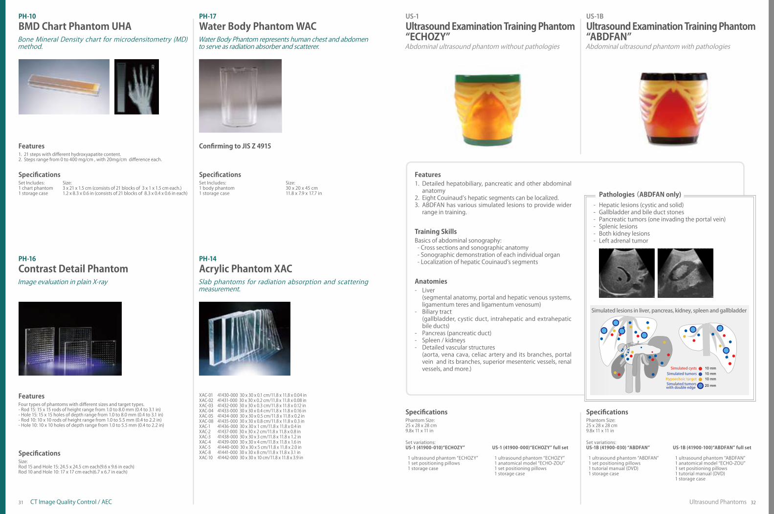
Outer shape of the phantom simulates a compressed breast of D shape.Features of evaluation include: contrast resolution, frequency enhancement, noise and contrast transfer function.Targets includes simulated microcalcifications, nylon fibrils, acrylic disks, an aluminum ring, Teflon disks, a Teflon ruler(slope) and a resolution test chart.
1.2.
3.
Features
31 32Ultrasound PhantomsCT Image Quality Control / AEC
PH-10 PH-17BMD Chart Phantom UHA Water Body Phantom WAC
Four types of phantoms with different sizes and target types.- Rod 15: 15 x 15 rods of height range from 1.0 to 8.0 mm (0.4 to 3.1 in)- Hole 15: 15 x 15 holes of depth range from 1.0 to 8.0 mm (0.4 to 3.1 in)- Rod 10: 10 x 10 rods of height range from 1.0 to 5.5 mm (0.4 to 2.2 in)- Hole 10: 10 x 10 holes of depth range from 1.0 to 5.5 mm (0.4 to 2.2 in)
Features
21 steps with different hydroxyapatite content.Steps range from 0 to 400 mg/cm , with 20mg/cm difference each.
1.2.
Features
Bone Mineral Density chart for microdensitometry (MD) method.
Water Body Phantom represents human chest and abdomen to serve as radiation absorber and scatterer.
PH-16 PH-14Contrast Detail Phantom Acrylic Phantom XACImage evaluation in plain X-ray Slab phantoms for radiation absorption and scattering
measurement.
Set Includes:1 chart phantom1 storage case
Size:3 x 21 x 1.5 cm (consists of 21 blocks of 3 x 1 x 1.5 cm each.)1.2 x 8.3 x 0.6 in (consists of 21 blocks of 8.3 x 0.4 x 0.6 in each)
SpecificationsSet Includes:1 body phantom1 storage case
Size:30 x 20 x 45 cm11.8 x 7.9 x 17.7 in
Specifications
Size:Rod 15 and Hole 15: 24.5 x 24.5 cm each(9.6 x 9.6 in each)Rod 10 and Hole 10: 17 x 17 cm each(6.7 x 6.7 in each)
Phantom Size:25 x 28 x 28 cm 9.8x 11 x 11 in
Phantom Size:25 x 28 x 28 cm 9.8x 11 x 11 in
SpecificationsSet variations:US-1B (41900-030) “ABDFAN” 1 ultrasound phantom “ABDFAN” 1 set positioning pillows 1 tutorial manual (DVD) 1 storage case
US-1B (41900-100)“ABDFAN” full set
1 ultrasound phantom “ABDFAN” 1 anatomical model “ECHO-ZOU” 1 set positioning pillows 1 tutorial manual (DVD) 1 storage case
Specifications
Set variations:US-1 (41900-010)“ECHOZY”
1 ultrasound phantom “ECHOZY” 1 set positioning pillows 1 storage case
US-1 (41900-000)“ECHOZY” full set
1 ultrasound phantom “ECHOZY” 1 anatomical model “ECHO-ZOU” 1 set positioning pillows 1 storage case
Specifications
Liver(segmental anatomy, portal and hepatic venous systems, ligamentum teres and ligamentum venosum)Biliary tract(gallbladder, cystic duct, intrahepatic and extrahepatic bile ducts)Pancreas (pancreatic duct)Spleen / kidneys Detailed vascular structures (aorta, vena cava, celiac artery and its branches, portal vein and its branches, superior mesenteric vessels, renal vessels, and more.)
-
-
- - -
Anatomies
Basics of abdominal sonography: - Cross sections and sonographic anatomy - Sonographic demonstration of each individual organ - Localization of hepatic Couinaud’s segments
Training Skills
Detailed hepatobiliary, pancreatic and other abdominal anatomyEight Couinaud’s hepatic segments can be localized.ABDFAN has various simulated lesions to provide wider range in training.
1.
2.3.
Features
Abdominal ultrasound phantom with pathologiesAbdominal ultrasound phantom without pathologies
US-1BUltrasound Examination Training Phantom “ABDFAN”
Ultrasound Examination Training Phantom “ECHOZY”
US-1
Confirming to JIS Z 4915
41430-000 30 x 30 x 0.1 cm/11.8 x 11.8 x 0.04 in 41431-000 30 x 30 x 0.2 cm/11.8 x 11.8 x 0.08 in41432-000 30 x 30 x 0.3 cm/11.8 x 11.8 x 0.12 in41433-000 30 x 30 x 0.4 cm/11.8 x 11.8 x 0.16 in41434-000 30 x 30 x 0.5 cm/11.8 x 11.8 x 0.2 in41435-000 30 x 30 x 0.8 cm/11.8 x 11.8 x 0.3 in41436-000 30 x 30 x 1 cm/11.8 x 11.8 x 0.4 in41437-000 30 x 30 x 2 cm/11.8 x 11.8 x 0.8 in 41438-000 30 x 30 x 3 cm/11.8 x 11.8 x 1.2 in41439-000 30 x 30 x 4 cm/11.8 x 11.8 x 1.6 in41440-000 30 x 30 x 5 cm/11.8 x 11.8 x 2.0 in41441-000 30 x 30 x 8 cm/11.8 x 11.8 x 3.1 in41442-000 30 x 30 x 10 cm/11.8 x 11.8 x 3.9 in
XAC-01XAC-02XAC-03XAC-04XAC-05XAC-08XAC-1XAC-2XAC-3XAC-4XAC-5XAC-8XAC-10
Pathologies (ABDFAN only)Hepatic lesions (cystic and solid)Gallbladder and bile duct stonesPancreatic tumors (one invading the portal vein)Splenic lesionsBoth kidney lesionsLeft adrenal tumor
- - - - - -

33 34Ultrasound Phantoms Ultrasound Phantoms
Brain with Septum Lucidum,Lateral Ventricles and Cerebellum UV Cyst Malignant Tumor
Cardiac Tamponade
Right Upper Abdominal Bleeding
Pelvic Bleeding
Pleural Hemorrhage
Hydronephrosis
Appendicitis
Set Includes:1 ultrasound phantom with storage case1 tutorial manual (DVD)
Size: 62 x 30 x 24 cm21 x 12 x 9 in
SpecificationsSet Includes:1 ultrasound phantom1 storage case1 tutorial manual (DVD)
Size:41 x 15 x 15 cm16 x 6 x 6 in
Specifications
Anatomies and Pathologies Anatomies and Pathologies
The phantom includes life-size 2-year-old thoracoabdominal organs, a bone structure, free fluid to learn FAST procedures and pathologies that are commonly seen in pediatrics.With this phantom, trainees can acquire skills in basics of pedi-atric abdominal ultrasound.
1.
2.
FeaturesAn innovative phantom for repetitive training of FAST as an adjunct to the ATLS primary surveyPathologies including cholecystitis, an aortic aneurysm, lesion on the colon
1.
2.
Features
US-8Pediatric FAST/Acute Abdomen Phantom
FAST/Acute Abdomen Phantom “FAST/ER FAN”
This abdominal ultrasound phantom includes life-size anatomies of a 2 years old with internal hemorrhage and other conditions commonly found in acute pediatric patients.
The world’s first pediatric ultrasound torso phantomFAST/ER FAN provides training to detect the presence of free intraperitoneal or pericardial fluid in patients experiencing trauma.
Best tool for workshops in emergency ultrasound
US-5
Set Includes:1 breast phantom1 storage case1 tutorial manual (DVD)
Size:26 x 38 x 11 cm7.6 x 8.8 x 2.8 in
Specifications
Subcutaneous adipose, mammary gland, galactophore, Cooper’s ligament, retromammary adipose, costae, clavicle, pectoralis major, lung , and lymph nodes at axilla.
Anatomies
- Skills to scan full area of breast systematically- Visualization of key anatomical landmarks- Tracking galactphore- Visualization and differentiation of typical pathologies- Localization and measurement of cyst and tumors
Training Skills
Cyst, mammary ductal ectasia, malignant tumor, benign tumorPathologies
State-of-the-art breast phantom with ultrasound anatomySkills required for ultrasound breast screening can be greatly advanced with practice.
1.2.
Features
US-6
BREASTFAN is a unique phantom for training in basic breast ul-trasound examination. Simulated targets with different echoge-nicities are embedded in the mammary gland.
Training in ultrasound breast cancer screening with detailed anatomySPACEFAN-ST provides high quality training for second trimester
screening in pregnancy. A 23-week fetus is included with detailed anatomies which are essential for the assessment at the period.
Fetus ultrasound phantom with a full skeletal structure
Breast Ultrasound Examination Phantom”BREAST FAN”
Fetus Ultrasound Examination Phantom“SPACE FAN-ST”
Set Includes:1 mother body torso1 ultrasound pregnant uterus phantom1 fetus demonstration model1 storage case1 tutorial manual (DVD)
Size:40 x 29 x 22 cm16 x 11.6 x 8.8 in
Specifications
Uterus: amniotic fluid, placenta, umbilical cord, and a 23-week fetus (10.2 in)Fetus: skeletal structure, brain with septum lucidum, lateral ventricles
and cerebellum, heart with four chambers, lungs, spleen, kid-neys, aorta, UV, UA, and the external genital
Anatomies and Pathologies
- Fetal size assessment: BPD, AD, AC and FL- Measurement of amniotic fluid volume- Determination of fetus position- Assessment of each body part
-Head: skull and brain-Spine and limbs-Cardiac chambers, blood vessels, and lungs
- Assessment of umbilical cord and placenta- Determination of sex (fetus is a male)
Training Skills
SPACE FAN-ST provides high quality training for routine sec-ond trimester screening.The oval shape phantom abdomen can be set in four different positions
1.
2.
Features
US-7α
The oval shape phantom abdomen can set in four different positions.
fetus demonstration model
35 36Ultrasound Phantoms Ultrasound Phantoms
Femoralhead
bony rim
(plane)
great trochanter
cartilage acetabular roof
labrum lower limb
ilium
Pathological unit- endometrial cancer, uterine fibroid, dermoid cyst of ovary, bleeding at Douglas pouch
Ectopic pregnancy unit- ectopic pregnancy in a fallopian tube, bleeding at Douglas pouch
Set includes:1 ultrasound infant phantom1 instruction manual1 storage case
Size:55 x 25 x 13 cm21.6 x 9.8 x 5.1 in
Specifications
Anatomies
-Setting and preparation for hip sonography-Changing the position of the infant-Communication and interaction with the infant’s guardian-Correct positioning and use of the transducer-Recognition of ultrasonic landmarks for hip sonography-Visualization of standard, anterior and posterior planes-Interpretation and morphological classification of the sonogram
Training Skills
The market’s only training model for hip sonography on a full body manikin of 6-week-old infantBilateral hips for examinationKey landmarks that can be recognized under ultrasound in-clude:
- chondro-osseous junction (bony part of femoral neck),- femoral head, synovial fold, joint capsule, labrum,- hyaline cartilage preformed acetabular roof,- bony part of acetabular roof, bony rim (check list I),- lower limb of os ilium, correct plane, labrum (check list II).
Facilitate anatomical understandingThe full body manikin with movable arms allows training in supporting and changing the position of the infant.
1.
2.3.
4.5.
Features
US-13Infant HipSonography Training PhantomBest tool to teach Graf’s method
Abdominal Intraoperative & Laparoscopic Ultrasound Phantom“IOUS FAN”
Set includes:1 upper abdomen ultrasound phantom1 stomach ultrasound phantom1 phantom container1 tutorial manual (DVD)
Manikin Size:30 x 38 x 17.5 cmW12 x D15 x H7 in
Specifications
- Liver(segmental anatomy, portal and hepatic venous systems,ligamentum teres and ligament venosum)
- Biliary tract(gallbladder, cystic duct, intrahepatic and extrahepaticbile ducts)
- Pancreas (pancreatic duct)- Spleen / kidneys- Detailed vascular structures
(aorta, vena cava, celiac artery and its branches, portal vein andits branches, superior mesenteric vessels, renal vessels, etc.)
Anatomies
- Abdominal intraoperative ultrasound examination- Laparoscopic ultrasound examination
Training Skills
Soft phantom materials allow realistic probe manipulation.Various simulated lesions including biliary stones and cysts,solid tumors (hypoechoic, hyperechoic and target-appearance)in the liver, pancreas, spleen and kidneysDetachable stomach and duodenum allows various scanningmethods of the bile duct and pancreas.
1.2.
3.
Features
US-3
Effective training tool for abdominal intraoperative ultrasound examinationInnovative phantom simulating abdominal open intraoperativeand laparoscopic ultrasound examination
Set includes:1 lower torso manikin 1 ultrasound ectopic pregnancy unit1 ultrasound pathology unit 1 storage case
Set Variations:
(US-10a) Female Pelvic Ultrasound Phantom (includes a normal unit, a ectopic pregnancy unit and a pathology unit)
SpecificationsSet includes:1 lower torso manikin 1 normal scrotal unit1 pathological scrotal unit 1 storage case
Size:34 x 33 x 24 cm13.4 x 13 x 9.5 in
Specifications
Anatomies and Pathologies
-Scrotal ultrasound screening-Visualization of testicular cancer
Training Skills
Excellent ultrasound image qualityNormal and pathological unit provides differing case typesExchangeable scrotal phantoms with easy cleaning
1.2.3.
Features
US-11Scrotal Ultrasound Phantom
Anatomies and Pathologies
-Transvaginal and transabdominal screenings-Localization of pathologies-3D ultrasound imaging restructuring
Training Skills
Realistic pathology for transvaginal ultrasound training as well as transabdominal procedureExcellent ultrasound image qualityAnatomically correct and life-like imagesCompatible with any ultrasound machine2 kinds of exchangeable phantoms for differing pathologies
1.
2.3.4.5.
Features
Female pelvic phantom with 2 screening methodsFemale Pelvic Ultrasound PhantomUS-10
The two phantoms, normal and pathological, facilitate a thorough anatomical understanding as well as a clear visualization of scrotal pathologies.
Excellent visualization of scrotal pathologies
Size: 34 x 33 x 24 cm13.4 x 13 x 9.5 in

37 38Ultrasound PhantomsUltrasound Phantoms
Gray Scale String target
Probe Skin
CarotidInternal jugular vein
Transparent Anatomical Block
Landmark puncture pad
Set includes:1 phantom1 storage case
Phantom size:19 x 22 x 7 cmW7.6 x D8.8 x H2.8 in
SpecificationsSet includes:1 mass targets blocks1 dot targets blocks1 thermometer1 storage case
Phantom size:mass targets block phantom size: W18.2 x D7.6 x H11 cm W7.2 x D3 x H4.4 indot targets block phantom size: W13.7 x D7.6 x H11 cm W5.4 x D3 x H4.4 in
Specifications
Four kinds of targets, gray scale, cyst targets, dot targets and 45 degrees line target at 2 different depth, 10mm (0.4 in) and 20mm (0.8 in).Background of each phantom block is of different attenuation rate and speed of sound.Detailed spatial resolution as minute as 0.5mm (0.02 in) can be assessed.Comes with a thermometer to measure inner temperature of the phantom.
1.
2.3. 4.
Features
Compatible for FNAB, CNB and mammotome biopsy with ultrasound guidanceColored targets embedded in three levelsRealistic representation of the mammary glandAn inexpensive and disposable phantom that provides many numbers of trials.The opaque phantom includes 2 types of targets; Hyperechoic and Hypoechoic.
1.
2.3.4.
5.
Features
Set Variations:Duo set: (11387-000) transparent + opaque typeTransparent set: (11387-100) 1 pair of transparent typeOpaque set: (11387-200) 1 pair of opaque type)
Size:16 dia. x 8cm6.3 dia x 3.2 in
Specifications
-Hand-eye coordination in ultrasound biopsy-Localization of targets under ultrasound guidance-Sampling of target
Training Skills
2 simulated vessel lines: straight and curve.Lines have slope to represent vessels with different depth.Vessel wall yields under pressure of a needle tip.
1.2.3.
Features
Set includes:REAL VESSEL Introductory ultrasound training block (a set of 2)
Specifications
- Visualization and localization of the vessels.- Transducer manipulation.- Basics for ultrasound-guided vascular access.
Training Skills
Provides step by training in ultrasound guided breast biopsy
Ultrasound Quality Assurance PhantomDurable and stable.
US-2
Ensure highly detailed images to ensure reliable breast cancer examinations
Breast Ultrasound QA PhantomUS-4
Provides training in hands-eye coordination and basic skills in ultrasound-guided venous access.
Introductory Ultrasound Training Block“REAL VESSEL”
Ultrasound Guided Breast Biopsy Phantom11347-210US-9
N-365 Multipurpose Phantom
Set includes:1 male upper torso manikin 1 introductory ultrasound training block1 landmark puncture pad 1 skin for cannulation training1 ultrasound puncture pad 1 red coloring powder1 transparent anatomical block 1 blue coloring powder
Size: 34 x 40 x 20 cm16 x 18 x 13 in
SpecificationsSet includes:2 CVC placement pads 1 vein pipe2 artery tubes 2 vein tubes1 instruction manual
Size: 34 x 40 x 20 cm16 x 18 x 13 in
Specifications
Ultrasound-guided CVCLandmark guided CVCUltrasound-guided venous accessPrevention of mechanical complications
Training Skills
- Ultrasound-guided CVC- Landmark guided CVC- Ultrasound-guided venous access- Prevention of mechanical complications
Training Skills
Internal jugular vein & carotid arterySubclavian vein & arterySuperior vena cavaRibsSternumLung
Anatomies
Repeated insertion: Improved frictionless tissue of the pad allows Seldinger technique and repeated insertion and removable of the
catheter with less needle marks left on the surface of the pad.Both Landmark and ultrasound-guided CVC: Anatomically correct structure facilitates training in both landmark and ultrasound-guided CVC.Mechanical complications, such as arterial puncture and pneumothorax can be simulated for training.New Material Close to human tissue material of the pad provides true-to-life sensation to the catheter.Realistic venous collapse
1.
2.
3.
4.
5.
Features
CVC Insertion Simulator II offers training in both landmark and ultrasound-guided central venous catheterization. Landmark puncture pad with anatomically correct vein bifur-cations simulates mechanical complications including pneu-mothorax, mislodging and artery puncture.Introductory ultrasound training block to acquire basics of ultrasound guided venous access.Transparent anatomical block for anatomical understanding and guide wire manipulation.Both internal jugular and subclavian (axillary) veins are acces-sible.
1.
2.
3.
4.
5.
Features
CVC Insertion Simulator III provides training in a sequence of procedural skills from the needle insertion to catheter placement, including Seldinger technique.
Great practice simulator for CVC catheter insertion with a variety of methods
CVC Insertion Simulator IIICVC Insertion Simulator IIM93CM93UB

39 4040Ultrasound Phantoms
Cannulation
Cannulation
Publication References
Set includes:1 lumbar region model1 ultrasound lumbar puncture/epidural block1 ultrasound lumbar region skin cover2 lumbar region support bases1 irrigator bag/ tube/ support base and syringe
Replacement parts:(11348-190) 1 ultrasound lumbar puncture/epidural block(11348-230) 1 ultrasound lumbar region skin cover for M43E
Manikin Size:33 x 21 x 30 cmW13 x D8.3 x H11.8 in
Specifications
- Lumbar spine (L2-L5) including spinous process and transverse process- Spinal canal, epidural space
Anatomies
- Ultrasound-guided lumbar puncture- Ultrasound-guided epidural anesthesia- CSF collection and CSF pressure measurement
Training Skills
Set includes:1 male upper torso with the right arm 1 syringe2 PICC puncture pad 1 instruction manual10 simulated blood (swab type) 1 storage case1 jar
Size: 40 x 15 x 60 cm15.7 x 5.9 x 23.7 in
Replacement part:(11398-010) 2 PICC puncture pads
Specifications
- Correct needle insertion, PICC, manipulation and catheter tip placement- Finding a puncture site under ultrasound guidance- The Seldinger technique- Technique for the peel-away sheath- Advancing of the cannula into the SVC
Training Skills
Ultrasonic landmarks of lumbar spine can be visualized.Skin cover allows marking with a pen.Both upright and lateral positions are possible for training.Translucent blocks allow users to see the needle pathway un-der direct vision.
1.2.3.4.
FeaturesExcellent image quality and visualization of the needle tip for ultrasound guided venous accessMovable shoulder to demonstrate positioningRealistic flashback in needle provides confirmation for suc-cessful venous accessRibs and right clavicle provide anatomical understanding of correct PICC placementAnatomically correct bifurcation of the veinSimulation of cannula malposition
1.
2.3.
4.
5.6.
Features
Ultrasound compatible puncture block is anatomically correct and offers realistic image of ultrasound. Both epidural space and subarachnoid space are accessible for training.
Ultrasonic anatomy and needle access trainingThis model provides training in full procedural skills from the needle insertion, manipulation of the PICC to the placement of the distal tip in the SVC. Both basilic and cephalic veins are ac-cessible for different levels of challenges in cannulation. The re-placeable puncture pad is ultrasound-compatible. Anatomically correct bifurcation of the vein provides realistic resistance on its outer walls and simulates complications such as catheter malpo-sitioning.
Comprehensive, hands-on training for PICC insertion
Ultrasound CompatibleLumbar Puncture/Epidural Simulator
Ultrasound-GuidedPICC Training Simulator
M43EMW18
Gomi T, Nakajima M, Fujiwara H, Umeda T. Comparison of Chest Dual-en-ergy Subtraction Digital Tomosynthesis Imaging and Dual-energy Sub-traction Radiography to Detect Simulated Pulmonary Nodules with and without Calcifications. Academic Radiology, February 2011; 18(2): 191–196.Read more: http://www.ncbi.nlm.nih.gov/pubmed/21232683
Norihisa Nitta et al. Nodule detection by chest X-ray and evaluation of computer-aided detection (CAD) software using an originally developed phantom for instructional purposes. Japanese Journal of Clinical Radiology. 2006; 51(1): 61-70.
Lynne M. Hurwitz, Terry T. Yoshizumi, Philip C. Goodman, Rendon C. Nelson, Greta Toncheva, Giao B. Nguyen, Carolyn Lowry, Colin Anderson-Evans. Radiation Dose Savings for Adult Pulmonary Embolus 64-MDCT Using Bismuth Breast Shields, Lower Peak Kilovoltage, and Automatic Tube Current Modulation. American Journal of Roentgenology. 2009; 192: 244-253. Read More: http://www.ajronline.org/doi/full/10.2214/AJR.08.1066
Gavrielides MA, Kinnard LM, Myers KJ, Peregoy J, Pritchard WF, Zeng R, Esparza J, Karanian J, Petrick N. A resource for the assessment of lung nodule size estimation methods: database of thoracic CT scans of an an-thropomorphic phantom. Optics Express. 2010; Jul 5; 18(14): 15244-55 Read more: http://www.ncbi.nlm.nih.gov/pmc/articles/PMC3408907/
Ushio N, Nitta N, Maumi M, Miyata K, Ueda J, Sonoda A, Takahashi M, Imai M, Murata K. Which methods do you use to reduce radiation exposure dose of chest CT? ECR2011 Poster No. 2011.Read more: http://posterng.netkey.at/esr/viewing/index.php?module=viewing_poster&-pi=105274
PH-2B CT Whole Body Phantom “PBU-60”Seon Chil Kim, Hong-Moon Jung. A Study on Performance of Low-Dose Medical Radiation Shielding Fiber (RSF) in CT Scans. International Journal of Technology. 2013: 2; 178-187.Read more: http://www.ijtech.eng.ui.ac.id/index.php/journal/article/view/107
PH-2C Pediatric Whole Body Phantom “PBU-70”Marcus Soderberg, Sonny La. Evaluation of adaptation strengths of CARE Dose 4D in pediatric CT. SPIE Medical Imaging. February 2013: 9-14.Read more: http://dx.doi.org/10.1117/12.2001694
Hsieh CY, Gladish G, Willis CE. Evaluation of commercial cardiac motion phantom for dual-energy chest radiography. Journal of Applied Clinical Medical Physics, 2014; 15, No 2Read more: http://www.ncbi.nlm.nih.gov/pubmed/24710435
PH-8 Lung Cancer Screening Phantom LSCT 001Muramatsu Y, Tsuda Y, Nakamura Y, Kubo M, Takayama T, Hanai K, The development and use of a chest phantom for optimizing scanning tech-niques on a variety of low-dose helical computed tomography devices. Japan Computer Assisted Tomography. May-Jun 2003; 27(3): 364-74.Read more: http://www.ncbi.nlm.nih.gov/pubmed/12794601
PH-47 Dental Radiography Head PhantomKitai N, Mukai Y, Murabayashi M, Kawabata A, Washino K, Matsuoka M, Shi-mizu I, Katsumata A. Measurement accuracy with a new dental panoram-ic radiographic technique based on tomosynthesis. Angle Orthodontist. 2013; 83, No 4. Read more: http://www.ncbi.nlm.nih.gov/pubmed/22612390
PH-6 Dynamic Cardiac CT PhantomUshio N, Nagatani Y, Sakurai K, Takazakura R, Miyata K, Takahashi M, Imai M, Murata K. The temporal resolution of CT coronary Angiography: Ro-tation time and temporal resolution. RSNA 2007 Poster.
Nitta N, Takahashi M, Takazakura R, Nagatani Y, Murata K, Ishimoi K, Kimura M. Efficacy of ECG gating for lung CT imaging in evaluating pulmonary nodule: Fundamental experiment with a newly developed pulsating car-diac phantom. ECR 2006, Poster C-207
PH-7 CT-AEC PhantomsMuramatsu Y, Ikeda S, Osawa K, Sekine R, Niwa N, Terada M, Keat N, Mi-yazaki S. Performance Evaluation for CT-AEC CT Automatic Exposure Control Systems. Japanese Journal of Radiological Technology, 2007-05-20; 63(5): 534-545.
US-2 Ultrasound Quality Assurance PhantomsNatori H, Igarashi T, Arakawa M. Durable fine resolution test phantom for diagnostic ultrasound system. ECR 2013 Poster C-1765.
US-4 Breast Ultrasound QA Phantom Sakurai M, Fukuda M, Imamura K, Tsujimoto F, Endo T, Morita T, Nakajima K, Quality Assurance of Ultrasonography Scanner Using a Phantom Designed for Quality Control and Teaching, Journal of Japan Association of Breast Can-cer Screening, 2008, 17(1), 52-59
PH-48 Dynamic Heart and Lung PhantomPH-1 Multipurpose Chest Phantom N1 “Lungman”Xueqian Xie, Yingru Zhao, Roland A. Snijder, Peter M. A. van Ooijen, Pim A. de Jong, Matthijs Oudkerk, Geertruida H. de Bock, Rozemarijn Vliegenthart, Marcel J. W. Greuter. Sensitivity and accuracy of volumetry of pulmonary nodules on low-dose 16- and 64-row multi-detector CT: an anthropomor-phic phantom study. European Radiology. 2013 : 139-147; 23: 139-147Read more: http://www.ncbi.nlm.nih.gov/pmc/articles/PMC3517707/

41 4242
Body Plate for PBU-60Iodinated Vessel Phantom Custom order simulated tumorsfor N1 “LUNGMAN”
PH-1 Multipurpose Chest Phantom N1 “LUNGMAN”Kiyoshi Murata, Ph.D. Professor Norihisa Nitta, Ph.D.Shiga University of Medical Science
US-5 FAST/Acute Abdomen Phantom “FAST/ER FAN”Junji Machi, MD, PhDUniversity of Hawaii at Manoa and Kuakini Medical Center
US-8 Pediatric FAST/Acute Abdomen Phantom Shunsuke Nosaka, MD, PhDDirector of RadiologyNational Medical Center for Children and Mothers
US-7a Fetus Ultrasound Examination Phantom “SPACE FAN-ST”Kiyoko Kabeyama RN, RM, PhDProfessor Midwifery & Women’s Health, School of Human Health Science,Graduate School of Medicine, Kyoto UniversityHaruto Egawa, PhDNational Hospital Organization Kyoto Medical CenterDepartment of Obstetrics, Head Physician or Medical Director
US-6 Breast Ultrasound Examination Phantom ”BREAST FAN”Tokiko Endo, MD, PhDDirector of Department of Advanced Diagnosis,Clinical Research Center,National Hospital Organization Nagoya Medical CenterDirector of Department of Radiology,National Hospital Organization Nagoya Medical Center
US-4 Breast Ultrasound QA PhantomJapan Association of Breast and Thyroid Sonology,Quality Assurance Committee Working Team.
Recommendation from:Japan Radiology Society, Imaging CommitteeBreast Imaging Group
M93UB CVC Insertion Simulator IIMasahiro Tanabe, M.D., Ph.D., Professor Director General Medical Education Center Chiba University School of MedicineKinya Sando, M.D., Ph.D., Professor Director Department of Human Dietetics Graduate School of Human Science Osaka Shoin Women’s UniversityMasanori Hoki, M.D., Ph.D., Professor Department of Human Dietetics Graduate School of Human Science Osaka Shoin Women’s UniversityJoho Tokumine, M.D.Ph.D., Department of Anesthesia, Kawatetsu Chiba Hospital
Custom Orders example Product Supervision
Let us know your thoughtsWe believe in the importance of providing phantoms that meet your needs and listening to your voice to find a solution.If you would like to suggest any additional features for our phantoms, please do not hesitate to contact Kyoto Kagaku Co., Ltd. Innovation is our tradition.
PH-47, PH-62 Dental Radiography Head PhantomAkitoshi Katsumata, D.D.S., Ph.D.ProfessorAsahi University, School of Dentistry
PH-49 CT Colonography Phantom NCCSNational Cancer Center (Japan)
PH-51 Lumbar Spine Fluoroscopy Training PhantomDr. David Wilson MBBS, BSc, MFSEM, FRCP, FRCR Consultant Radiologist St Luke’s Hospital Oxford Senior Clinical Lecturer University of Oxford
US-1 Ultrasound Examination Training Phantom “ECHOZY”Dr. Hitoshi Asai, DirectorOsaka Kyoiku University, Health Administration CenterDr. Shigeru NakamuraNagayoshi General Hospital, Clinical Examination Department
US-1B Ultrasound Examination Training Phantom “ABDFAN”Junji Machi, MD, PhDUniversity of Hawaii at Manoa and Kuakini Medical Center
US-3 Abdominal Intraoperative & Laparoscopic Ultrasound Phantom“IOUS FAN”Junji Machi, MD,PhDUniversity of Hawaii at Manoa and Kuakini Medical Center
US-13 Infant Hip Sonography Training PhantomUniv. Prof., Prof. hc. Reinhard Graf, M.D.
US-10 Female Pelvic Ultrasound PhantomCharlotte Henningsen, MS, RT(R), RDMS, RVT, FSDMS, FAIUMChair & Professor - Sonography DepartmentAdventist University of Health Sciences
US-11 Scrotal Ultrasound PhantomCharlotte Henningsen, MS, RT(R), RDMS, RVT, FSDMS, FAIUMChair & Professor - Sonography DepartmentAdventist University of Health Sciences
P.16 P.34
P.35
P.35
P.36
P.36
P.37
P.38
P.20
P.22
P.23
P.32
P.32
P.33
P.33
P.34
Common Specifications
All KYOTO KAGAKU phantoms are made in JapanAll KYOTO KAGAKU phantoms are latex free
PH-1, PH-1C, PH-2B, PH-2C, PH-2D, PH-3, PH-4, PH-5, PH-9, PH-15, PH-39, PH-45, PH-46, PH-49, PH-50 Tissue substitute materials: polyurethane based resin Synthetic bones: epoxy based resinPH-6, PH-48 Tissue substitute materials: polyurethane based resin Synthetic bones: epoxy based resin Heart Phantom: siliconPH-8, PH-10, PH-15 Polyurethane based resinPH-37, PH-40, PH-41 Epoxy based resinPH-9-2 Epoxy based resin, acrylic resin
Specifications printed in this catalogue are of the time of printing. Since products are continuously improved and updated, specifications are subject to change without prior notice.
PH-7, PH-14, PH-17, PH-24, PH-26, PH-28, PH-29, PH-31, PH-33, PH-34 Acrylic resin Synthetic bones: epoxy based resinPH-13 Acrylic resin, nylon, aluminum, aluminum oxide, TeflonPH-40 Phenol based resinPH-9, PH-45 Urethane based resin, Acrylic resinUS-1, US-2, US-4, US-5, US-6, US-7a, US-8, PH-12, Subsolid Nodules Phantom Urethane based resinUS-9 Hydrogel

43