RADIOLOGY (SURGERY) - · PDF file6cm for the colon and 9cm for the caecum ... has been...
89
RADIOLOGY (SURGERY) BY MARYAM MALIK Rawalpindi Medical College
Transcript of RADIOLOGY (SURGERY) - · PDF file6cm for the colon and 9cm for the caecum ... has been...

RADIOLOGY (SURGERY)
BY MARYAM MALIK
Rawalpindi Medical College
NORMAL BOWEL GAS PATTERN
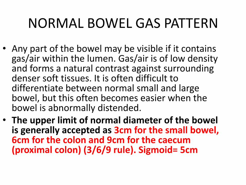
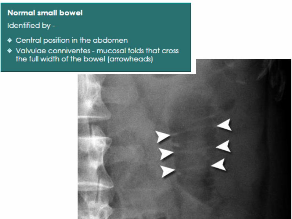
bull Any part of the bowel may be visible if it contains gasair within the lumen Gasair is of low density and forms a natural contrast against surrounding denser soft tissues It is often difficult to differentiate between normal small and large bowel but this often becomes easier when the bowel is abnormally distended
bull The upper limit of normal diameter of the bowel is generally accepted as 3cm for the small bowel 6cm for the colon and 9cm for the caecum (proximal colon) (369 rule) Sigmoid= 5cm
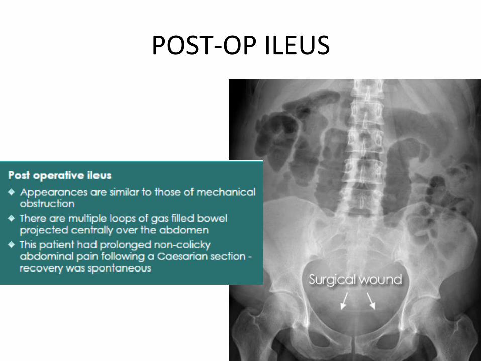
POST-OP ILEUS
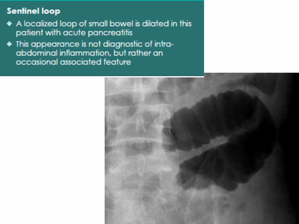
Sentinel loop
bull Intra-abdominal inflammation such as with pancreatitis can lead to a localized ileus This may appear as a single loop of dilated bowel known as a sentinel loop
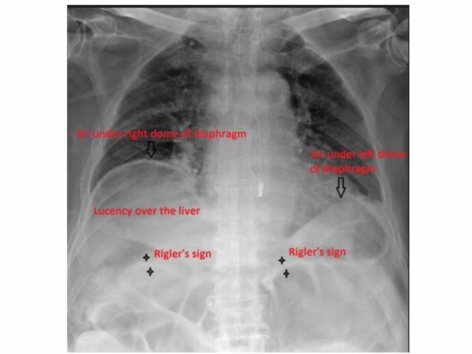
Rigler sign
bull The Rigler sign also known as the double wall sign is seen on an x-ray of the abdomen when air is present on both sides of the intestine ie when there is air on both the luminal and peritoneal side of the bowel wall
bull Pneumoperitoneum may be a result of perforation or from recent instrumentation or surgery A false double wall sign can result from two loops of bowel being in contact with one another
SIGMOID VOLVULUS
bull Sigmoid volvulus is a cause of large bowel obstruction and occurs when the sigmoid colon twists on the sigmoid mesocolon
bull COFFEE BEAN SIGN on X-ray
CAECAL VOLVULUS
bull Caecal volvulus describes torsion of the caecum around its own mesentery which often results in obstruction If unrecognised can result in bowel perforation and faecal peritonitis
Esophageal atresia
bull may show a dilated pharyngeal pouch
bull the presence of air in the stomach and bowel in the setting of oesophageal atresia implies that there is a distal fistula
bull if an oesophago-gastric (feeding) tube insertion has been attempted this may show the tube blind looping and turning back at the upper thoracic part of the oesophagus or heading into the trachea andor bronchial tree
bull Oesophageal atresia without fistula The Replogle tube (arrow) is coiled in the dilated blind ending oesophageal pouch AXR demonstrates absence of bowel gas indicating that there is no distal tracheoesophageal fistula
Double bubble sign (duedenal atresia)
Perforated duedenal ulcer
Foreign body rectum
Achalasia birdrsquos beak appearance on braium swallow
ULCERATIVE COLITIS barium enema showing a lead pipe colon
Lead pipe appearance of colon is the classical barium enema finding in chroniculcerative colitis There is complete loss of haustral markings in the diseased section of colon and the organ appears smooth walled and cylindrical
Carcinoma esophagus
Epiphrenic diverticula
Gastric ulcer
Gastric ulcer
Pyloric obstruction
The shoulder sign the impression of the hypertrophied pyloric muscle on the distended gastric antrum
MRCP
Magnetic Resonance Cholangiopancreatography
Normal hepatic ductal anatomy Coronal oblique MIP reformat image reveals the confluence (circle) between
the right posterior duct (RPD) and the right anterior duct (RAD) originating the right hepatic duct (RHD) Note that the RPD has a more horizontal route while
the RAD is more vertical By its turn the RHD joins the left hepatic duct (LHD) originating the common hepatic duct The LHD results from the confluence of the ducts
of the left hepatic lobe segments here only represented by segments II (S2) and III (S3) Cystic duct
(CD) common bile duct (CBD) main pancreatic duct (MPD) gallbladder (GB)
Normal pancreatic ductal anatomyrdquo Coronal oblique reformat shows the main pancreatic duct (MPD) crossing the whole pancreas and continuing as the duct of Wirsung (DW) at the pancreatic head At its distal portion the DW joins the common bile duct (CBD) draining into the major papilla (pointing triangle) The duct of Santorini is not demonstrated
MRCP showing stone in lower part of CBD
MRCP image shows a dilated bile duct with a dark stone (arrow) in its distal end
ERCP
Endoscopic Retrograde Cholangiopancreatography
ERCP showing normal CBD amp pancreatic duct
These two fluorospot images taken during an ERCP demonstrates stones in the common bile duct on the left radiograph and cystic
duct on the right radiograph
PTC (Percutaneous transhepatic cholangiography)
PTC (PercutaneousTranshepatic Cholangiography)
It is a radiologic techniqueused to visualize the
anatomy of the biliary tract
T-tube cholangiogramsT-tube cholangiograms are a fluroscopic study performed in the setting of hepatobiliary disease
Peroperative cholangiogram during cholecystectomy
XRAY KUB showing radio-opaque shadows consistent with renal stone
XRAY KUB showing staghorn (phosphate) stones
XRAY KUB showing bladder stone
A plain KUB showing alarge smooth ovalradio-opaque masssuggesting a calcified bladder stone
Normal intravenous urograms
IVU showing bilateral double ureters
Ascending cystography showing urinary bladder diverticuli
A cystogram showing marked diverticulation with a large superior diverticulum
Retrograde Urethrogram showing urethral stricture
Normal mammogram
Mammographic images showing well-circumscribed masses with features of
fibroadenomacyst
Mammography showing cancer
Xray showing right clavicular fracture
Xray showing right shoulder dislocation
Xray showing mid shaft fracture of humerus
Xray showing posterior elbow dislocation
Xray showing the fracture of lower end of radius amp ulna
Xray showing Undisplaced Inter-Trochanteric Femoral Fracture
Xray showing neck of left femur fracture
Xray showing left hip dislocation
Xray showing Austin Moore Prosthesis for femur neck fracture
Xray showing total hip replacement
Xray showing dynamic hip screw transfixing an inter-trochanteric
fracture of right femur
Xray showing knee dislocation
Xray showing fracture of lower part of tibia amp fibula
Xray showing lateral malleolar fracture
Xray showing bi-malleolar fracture (pott fracture) with distortion of ankle
joint
Bimalleolar fracture and right ankle dislocation on X-ray (anteroposterior) Both the end of the fibula (1) and the tibia (2) are broken and the malleolar fragments (arrow medial malleolus arrowhead lateral malleolus) are displaced
Xray showing fracture of 1st metatarsal bone
Preoperative radiographs showing the dorsal dislocation at the MTP joint and fracture of the base of the metatarsal bone
CT Scan showing multiple metastasis in brain
NORMAL CT-SCAN BRAIN
CT Scan showing subdural hematoma
CT Scan showing epidural hematoma
CT Scan showing hydrocephalus
CT Scan showing multiple fractures in frontal part of skull
MRI showing intrameduallary tumor of spinal cord
Magnetic resonance angiogram (MRA) showing normal anatomy
CT angiogram of abdomen amp lower limb showing normal vasculature
Angiogram showing abdominal aortic aneurysm
This magnetic resonance angiogram (MRA) of the lower extremities was obtained by using the bolus-chase technique A short-segment high-grade stenosis is present in the middle of the left superficial femoral artery Note
the collateral arterial supply
Magnetic resonance angiogram showing narrowing of left popliteal artery
CT scan showing tumor in head of pancreas
The arrow indicates the superior mesenteric artery
CT scan showing tumor in tail of pancreas
CT scan showing tumor in right kidney
CT SCAN showing bilateral kidney cysts
CT scan showing normal liver
Liver metastasis Multiple hypodense lesions seen in the liver with no significant contrast enhancement
LIVER CYST
bullOval well definedbullImperceptible or thin wallbullWater density
CT scan showing tumor in right lobe of liver (arrowheads)
NORMAL BOWEL GAS PATTERN
bull Any part of the bowel may be visible if it contains gasair within the lumen Gasair is of low density and forms a natural contrast against surrounding denser soft tissues It is often difficult to differentiate between normal small and large bowel but this often becomes easier when the bowel is abnormally distended
bull The upper limit of normal diameter of the bowel is generally accepted as 3cm for the small bowel 6cm for the colon and 9cm for the caecum (proximal colon) (369 rule) Sigmoid= 5cm
POST-OP ILEUS
Sentinel loop
bull Intra-abdominal inflammation such as with pancreatitis can lead to a localized ileus This may appear as a single loop of dilated bowel known as a sentinel loop
Rigler sign
bull The Rigler sign also known as the double wall sign is seen on an x-ray of the abdomen when air is present on both sides of the intestine ie when there is air on both the luminal and peritoneal side of the bowel wall
bull Pneumoperitoneum may be a result of perforation or from recent instrumentation or surgery A false double wall sign can result from two loops of bowel being in contact with one another
SIGMOID VOLVULUS
bull Sigmoid volvulus is a cause of large bowel obstruction and occurs when the sigmoid colon twists on the sigmoid mesocolon
bull COFFEE BEAN SIGN on X-ray
CAECAL VOLVULUS
bull Caecal volvulus describes torsion of the caecum around its own mesentery which often results in obstruction If unrecognised can result in bowel perforation and faecal peritonitis
Esophageal atresia
bull may show a dilated pharyngeal pouch
bull the presence of air in the stomach and bowel in the setting of oesophageal atresia implies that there is a distal fistula
bull if an oesophago-gastric (feeding) tube insertion has been attempted this may show the tube blind looping and turning back at the upper thoracic part of the oesophagus or heading into the trachea andor bronchial tree
bull Oesophageal atresia without fistula The Replogle tube (arrow) is coiled in the dilated blind ending oesophageal pouch AXR demonstrates absence of bowel gas indicating that there is no distal tracheoesophageal fistula
Double bubble sign (duedenal atresia)
Perforated duedenal ulcer
Foreign body rectum
Achalasia birdrsquos beak appearance on braium swallow
ULCERATIVE COLITIS barium enema showing a lead pipe colon
Lead pipe appearance of colon is the classical barium enema finding in chroniculcerative colitis There is complete loss of haustral markings in the diseased section of colon and the organ appears smooth walled and cylindrical
Carcinoma esophagus
Epiphrenic diverticula
Gastric ulcer
Gastric ulcer
Pyloric obstruction
The shoulder sign the impression of the hypertrophied pyloric muscle on the distended gastric antrum
MRCP
Magnetic Resonance Cholangiopancreatography
Normal hepatic ductal anatomy Coronal oblique MIP reformat image reveals the confluence (circle) between
the right posterior duct (RPD) and the right anterior duct (RAD) originating the right hepatic duct (RHD) Note that the RPD has a more horizontal route while
the RAD is more vertical By its turn the RHD joins the left hepatic duct (LHD) originating the common hepatic duct The LHD results from the confluence of the ducts
of the left hepatic lobe segments here only represented by segments II (S2) and III (S3) Cystic duct
(CD) common bile duct (CBD) main pancreatic duct (MPD) gallbladder (GB)
Normal pancreatic ductal anatomyrdquo Coronal oblique reformat shows the main pancreatic duct (MPD) crossing the whole pancreas and continuing as the duct of Wirsung (DW) at the pancreatic head At its distal portion the DW joins the common bile duct (CBD) draining into the major papilla (pointing triangle) The duct of Santorini is not demonstrated
MRCP showing stone in lower part of CBD
MRCP image shows a dilated bile duct with a dark stone (arrow) in its distal end
ERCP
Endoscopic Retrograde Cholangiopancreatography
ERCP showing normal CBD amp pancreatic duct
These two fluorospot images taken during an ERCP demonstrates stones in the common bile duct on the left radiograph and cystic
duct on the right radiograph
PTC (Percutaneous transhepatic cholangiography)
PTC (PercutaneousTranshepatic Cholangiography)
It is a radiologic techniqueused to visualize the
anatomy of the biliary tract
T-tube cholangiogramsT-tube cholangiograms are a fluroscopic study performed in the setting of hepatobiliary disease
Peroperative cholangiogram during cholecystectomy
XRAY KUB showing radio-opaque shadows consistent with renal stone
XRAY KUB showing staghorn (phosphate) stones
XRAY KUB showing bladder stone
A plain KUB showing alarge smooth ovalradio-opaque masssuggesting a calcified bladder stone
Normal intravenous urograms
IVU showing bilateral double ureters
Ascending cystography showing urinary bladder diverticuli
A cystogram showing marked diverticulation with a large superior diverticulum
Retrograde Urethrogram showing urethral stricture
Normal mammogram
Mammographic images showing well-circumscribed masses with features of
fibroadenomacyst
Mammography showing cancer
Xray showing right clavicular fracture
Xray showing right shoulder dislocation
Xray showing mid shaft fracture of humerus
Xray showing posterior elbow dislocation
Xray showing the fracture of lower end of radius amp ulna
Xray showing Undisplaced Inter-Trochanteric Femoral Fracture
Xray showing neck of left femur fracture
Xray showing left hip dislocation
Xray showing Austin Moore Prosthesis for femur neck fracture
Xray showing total hip replacement
Xray showing dynamic hip screw transfixing an inter-trochanteric
fracture of right femur
Xray showing knee dislocation
Xray showing fracture of lower part of tibia amp fibula
Xray showing lateral malleolar fracture
Xray showing bi-malleolar fracture (pott fracture) with distortion of ankle
joint
Bimalleolar fracture and right ankle dislocation on X-ray (anteroposterior) Both the end of the fibula (1) and the tibia (2) are broken and the malleolar fragments (arrow medial malleolus arrowhead lateral malleolus) are displaced
Xray showing fracture of 1st metatarsal bone
Preoperative radiographs showing the dorsal dislocation at the MTP joint and fracture of the base of the metatarsal bone
CT Scan showing multiple metastasis in brain
NORMAL CT-SCAN BRAIN
CT Scan showing subdural hematoma
CT Scan showing epidural hematoma
CT Scan showing hydrocephalus
CT Scan showing multiple fractures in frontal part of skull
MRI showing intrameduallary tumor of spinal cord
Magnetic resonance angiogram (MRA) showing normal anatomy
CT angiogram of abdomen amp lower limb showing normal vasculature
Angiogram showing abdominal aortic aneurysm
This magnetic resonance angiogram (MRA) of the lower extremities was obtained by using the bolus-chase technique A short-segment high-grade stenosis is present in the middle of the left superficial femoral artery Note
the collateral arterial supply
Magnetic resonance angiogram showing narrowing of left popliteal artery
CT scan showing tumor in head of pancreas
The arrow indicates the superior mesenteric artery
CT scan showing tumor in tail of pancreas
CT scan showing tumor in right kidney
CT SCAN showing bilateral kidney cysts
CT scan showing normal liver
Liver metastasis Multiple hypodense lesions seen in the liver with no significant contrast enhancement
LIVER CYST
bullOval well definedbullImperceptible or thin wallbullWater density
CT scan showing tumor in right lobe of liver (arrowheads)
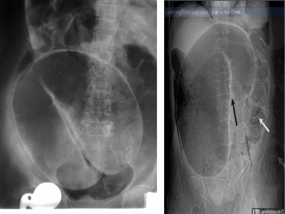
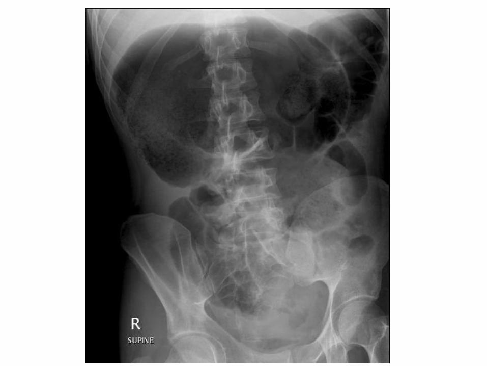

Gastric ulcer
Gastric ulcer
Pyloric obstruction
The shoulder sign the impression of the hypertrophied pyloric muscle on the distended gastric antrum
MRCP
Magnetic Resonance Cholangiopancreatography
Normal hepatic ductal anatomy Coronal oblique MIP reformat image reveals the confluence (circle) between
the right posterior duct (RPD) and the right anterior duct (RAD) originating the right hepatic duct (RHD) Note that the RPD has a more horizontal route while
the RAD is more vertical By its turn the RHD joins the left hepatic duct (LHD) originating the common hepatic duct The LHD results from the confluence of the ducts
of the left hepatic lobe segments here only represented by segments II (S2) and III (S3) Cystic duct
(CD) common bile duct (CBD) main pancreatic duct (MPD) gallbladder (GB)
Normal pancreatic ductal anatomyrdquo Coronal oblique reformat shows the main pancreatic duct (MPD) crossing the whole pancreas and continuing as the duct of Wirsung (DW) at the pancreatic head At its distal portion the DW joins the common bile duct (CBD) draining into the major papilla (pointing triangle) The duct of Santorini is not demonstrated
MRCP showing stone in lower part of CBD
MRCP image shows a dilated bile duct with a dark stone (arrow) in its distal end
ERCP
Endoscopic Retrograde Cholangiopancreatography
ERCP showing normal CBD amp pancreatic duct
These two fluorospot images taken during an ERCP demonstrates stones in the common bile duct on the left radiograph and cystic
duct on the right radiograph
PTC (Percutaneous transhepatic cholangiography)
PTC (PercutaneousTranshepatic Cholangiography)
It is a radiologic techniqueused to visualize the
anatomy of the biliary tract
T-tube cholangiogramsT-tube cholangiograms are a fluroscopic study performed in the setting of hepatobiliary disease
Peroperative cholangiogram during cholecystectomy
XRAY KUB showing radio-opaque shadows consistent with renal stone
XRAY KUB showing staghorn (phosphate) stones
XRAY KUB showing bladder stone
A plain KUB showing alarge smooth ovalradio-opaque masssuggesting a calcified bladder stone
Normal intravenous urograms
IVU showing bilateral double ureters
Ascending cystography showing urinary bladder diverticuli
A cystogram showing marked diverticulation with a large superior diverticulum
Retrograde Urethrogram showing urethral stricture
Normal mammogram
Mammographic images showing well-circumscribed masses with features of
fibroadenomacyst
Mammography showing cancer
Xray showing right clavicular fracture
Xray showing right shoulder dislocation
Xray showing mid shaft fracture of humerus
Xray showing posterior elbow dislocation
Xray showing the fracture of lower end of radius amp ulna
Xray showing Undisplaced Inter-Trochanteric Femoral Fracture
Xray showing neck of left femur fracture
Xray showing left hip dislocation
Xray showing Austin Moore Prosthesis for femur neck fracture
Xray showing total hip replacement
Xray showing dynamic hip screw transfixing an inter-trochanteric
fracture of right femur
Xray showing knee dislocation
Xray showing fracture of lower part of tibia amp fibula
Xray showing lateral malleolar fracture
Xray showing bi-malleolar fracture (pott fracture) with distortion of ankle
joint
Bimalleolar fracture and right ankle dislocation on X-ray (anteroposterior) Both the end of the fibula (1) and the tibia (2) are broken and the malleolar fragments (arrow medial malleolus arrowhead lateral malleolus) are displaced
Xray showing fracture of 1st metatarsal bone
Preoperative radiographs showing the dorsal dislocation at the MTP joint and fracture of the base of the metatarsal bone
CT Scan showing multiple metastasis in brain
NORMAL CT-SCAN BRAIN
CT Scan showing subdural hematoma
CT Scan showing epidural hematoma
CT Scan showing hydrocephalus
CT Scan showing multiple fractures in frontal part of skull
MRI showing intrameduallary tumor of spinal cord
Magnetic resonance angiogram (MRA) showing normal anatomy
CT angiogram of abdomen amp lower limb showing normal vasculature
Angiogram showing abdominal aortic aneurysm
This magnetic resonance angiogram (MRA) of the lower extremities was obtained by using the bolus-chase technique A short-segment high-grade stenosis is present in the middle of the left superficial femoral artery Note
the collateral arterial supply
Magnetic resonance angiogram showing narrowing of left popliteal artery
CT scan showing tumor in head of pancreas
The arrow indicates the superior mesenteric artery
CT scan showing tumor in tail of pancreas
CT scan showing tumor in right kidney
CT SCAN showing bilateral kidney cysts
CT scan showing normal liver
Liver metastasis Multiple hypodense lesions seen in the liver with no significant contrast enhancement
LIVER CYST
bullOval well definedbullImperceptible or thin wallbullWater density
CT scan showing tumor in right lobe of liver (arrowheads)

Gastric ulcer
Pyloric obstruction
The shoulder sign the impression of the hypertrophied pyloric muscle on the distended gastric antrum
MRCP
Magnetic Resonance Cholangiopancreatography
Normal hepatic ductal anatomy Coronal oblique MIP reformat image reveals the confluence (circle) between
the right posterior duct (RPD) and the right anterior duct (RAD) originating the right hepatic duct (RHD) Note that the RPD has a more horizontal route while
the RAD is more vertical By its turn the RHD joins the left hepatic duct (LHD) originating the common hepatic duct The LHD results from the confluence of the ducts
of the left hepatic lobe segments here only represented by segments II (S2) and III (S3) Cystic duct
(CD) common bile duct (CBD) main pancreatic duct (MPD) gallbladder (GB)
Normal pancreatic ductal anatomyrdquo Coronal oblique reformat shows the main pancreatic duct (MPD) crossing the whole pancreas and continuing as the duct of Wirsung (DW) at the pancreatic head At its distal portion the DW joins the common bile duct (CBD) draining into the major papilla (pointing triangle) The duct of Santorini is not demonstrated
MRCP showing stone in lower part of CBD
MRCP image shows a dilated bile duct with a dark stone (arrow) in its distal end
ERCP
Endoscopic Retrograde Cholangiopancreatography
ERCP showing normal CBD amp pancreatic duct
These two fluorospot images taken during an ERCP demonstrates stones in the common bile duct on the left radiograph and cystic
duct on the right radiograph
PTC (Percutaneous transhepatic cholangiography)
PTC (PercutaneousTranshepatic Cholangiography)
It is a radiologic techniqueused to visualize the
anatomy of the biliary tract
T-tube cholangiogramsT-tube cholangiograms are a fluroscopic study performed in the setting of hepatobiliary disease
Peroperative cholangiogram during cholecystectomy
XRAY KUB showing radio-opaque shadows consistent with renal stone
XRAY KUB showing staghorn (phosphate) stones
XRAY KUB showing bladder stone
A plain KUB showing alarge smooth ovalradio-opaque masssuggesting a calcified bladder stone
Normal intravenous urograms
IVU showing bilateral double ureters
Ascending cystography showing urinary bladder diverticuli
A cystogram showing marked diverticulation with a large superior diverticulum
Retrograde Urethrogram showing urethral stricture
Normal mammogram
Mammographic images showing well-circumscribed masses with features of
fibroadenomacyst
Mammography showing cancer
Xray showing right clavicular fracture
Xray showing right shoulder dislocation
Xray showing mid shaft fracture of humerus
Xray showing posterior elbow dislocation
Xray showing the fracture of lower end of radius amp ulna
Xray showing Undisplaced Inter-Trochanteric Femoral Fracture
Xray showing neck of left femur fracture
Xray showing left hip dislocation
Xray showing Austin Moore Prosthesis for femur neck fracture
Xray showing total hip replacement
Xray showing dynamic hip screw transfixing an inter-trochanteric
fracture of right femur
Xray showing knee dislocation
Xray showing fracture of lower part of tibia amp fibula
Xray showing lateral malleolar fracture
Xray showing bi-malleolar fracture (pott fracture) with distortion of ankle
joint
Bimalleolar fracture and right ankle dislocation on X-ray (anteroposterior) Both the end of the fibula (1) and the tibia (2) are broken and the malleolar fragments (arrow medial malleolus arrowhead lateral malleolus) are displaced
Xray showing fracture of 1st metatarsal bone
Preoperative radiographs showing the dorsal dislocation at the MTP joint and fracture of the base of the metatarsal bone
CT Scan showing multiple metastasis in brain
NORMAL CT-SCAN BRAIN
CT Scan showing subdural hematoma
CT Scan showing epidural hematoma
CT Scan showing hydrocephalus
CT Scan showing multiple fractures in frontal part of skull
MRI showing intrameduallary tumor of spinal cord
Magnetic resonance angiogram (MRA) showing normal anatomy
CT angiogram of abdomen amp lower limb showing normal vasculature
Angiogram showing abdominal aortic aneurysm
This magnetic resonance angiogram (MRA) of the lower extremities was obtained by using the bolus-chase technique A short-segment high-grade stenosis is present in the middle of the left superficial femoral artery Note
the collateral arterial supply
Magnetic resonance angiogram showing narrowing of left popliteal artery
CT scan showing tumor in head of pancreas
The arrow indicates the superior mesenteric artery
CT scan showing tumor in tail of pancreas
CT scan showing tumor in right kidney
CT SCAN showing bilateral kidney cysts
CT scan showing normal liver
Liver metastasis Multiple hypodense lesions seen in the liver with no significant contrast enhancement
LIVER CYST
bullOval well definedbullImperceptible or thin wallbullWater density
CT scan showing tumor in right lobe of liver (arrowheads)
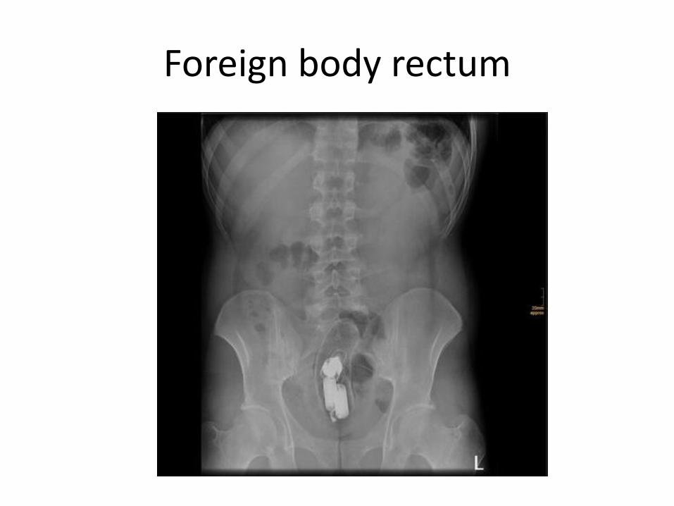
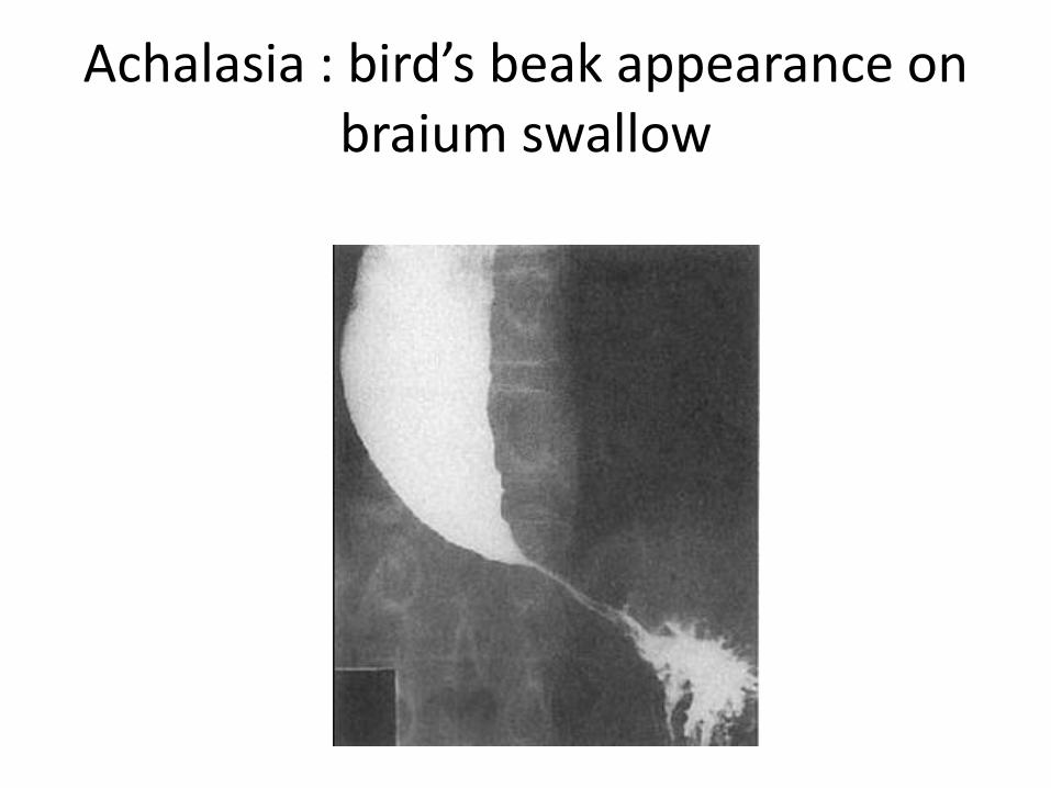
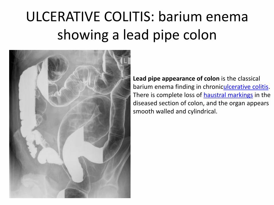
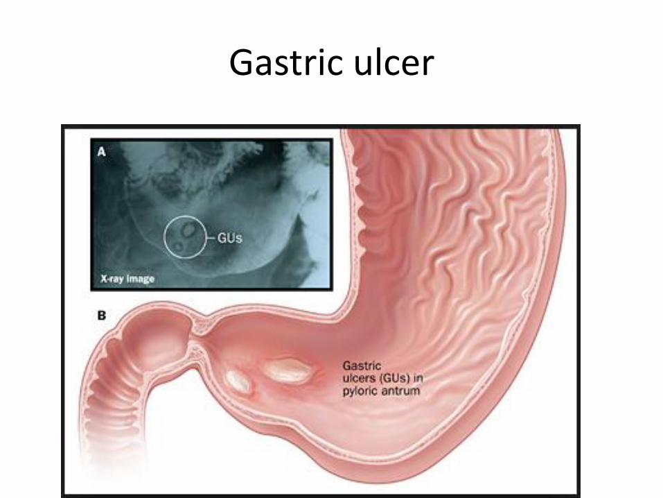
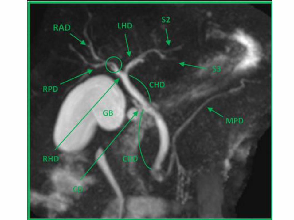
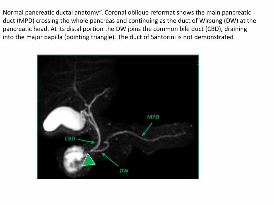
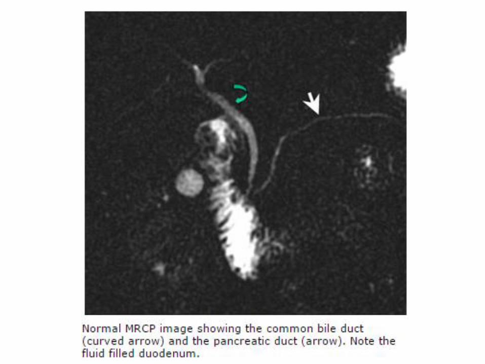
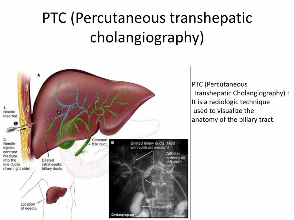
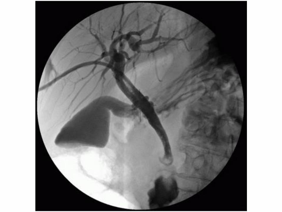
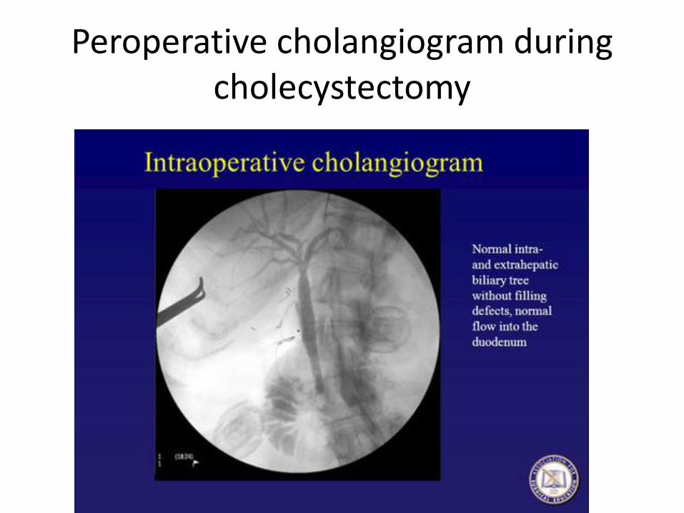
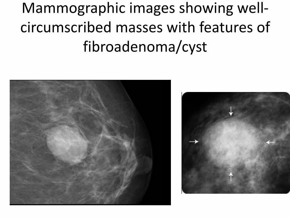
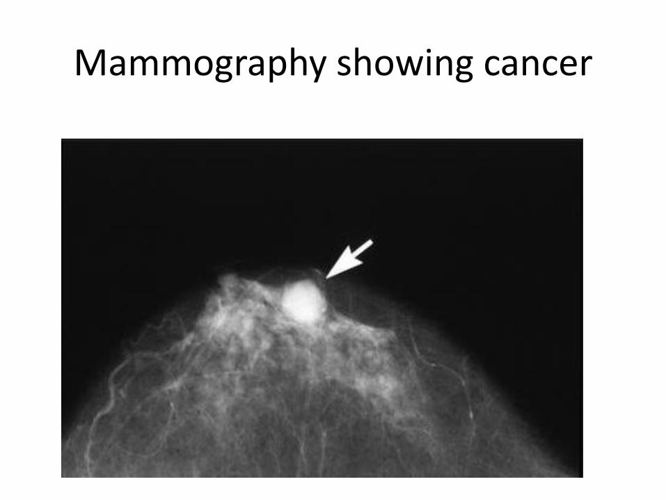
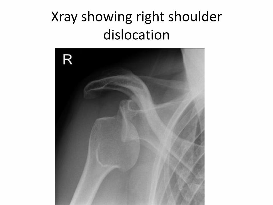
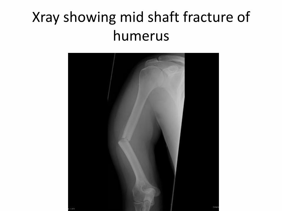
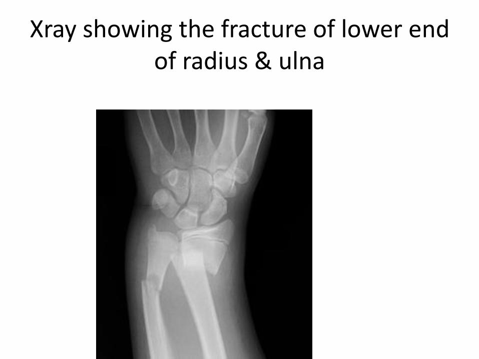
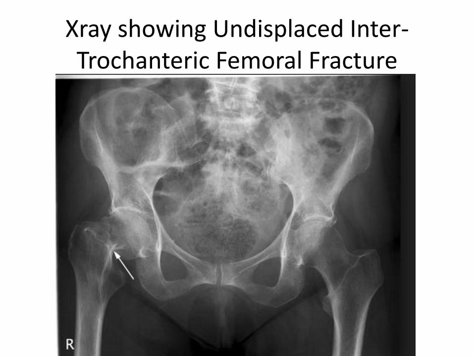
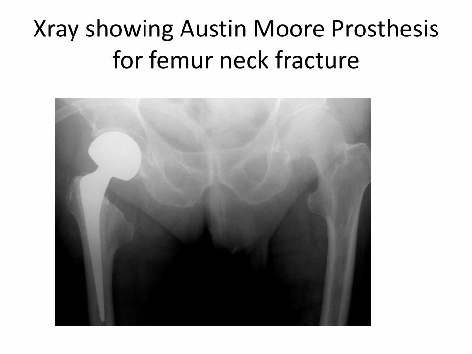
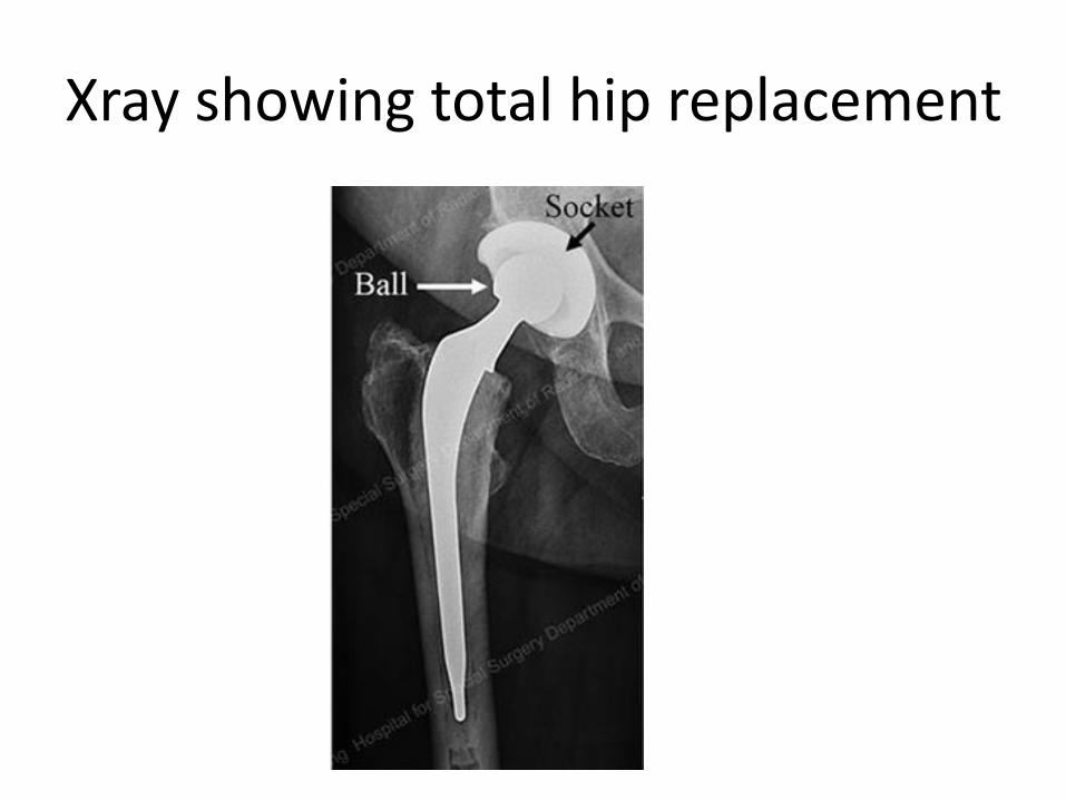
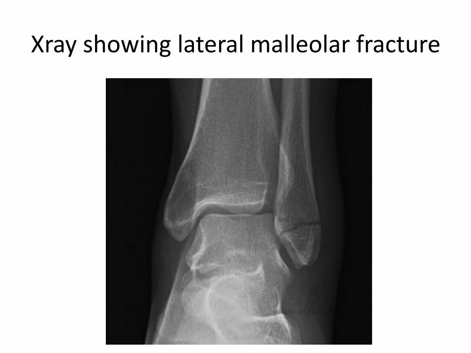
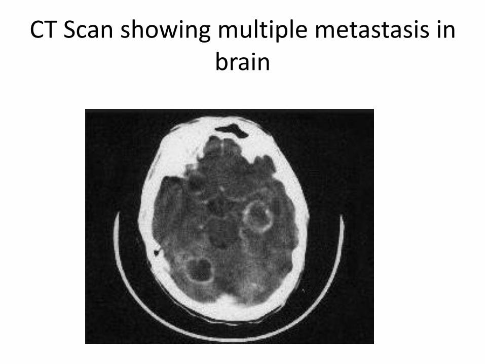
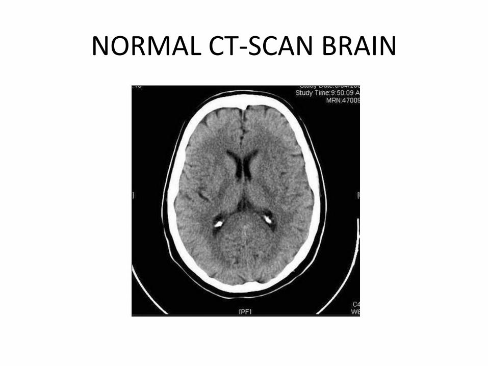
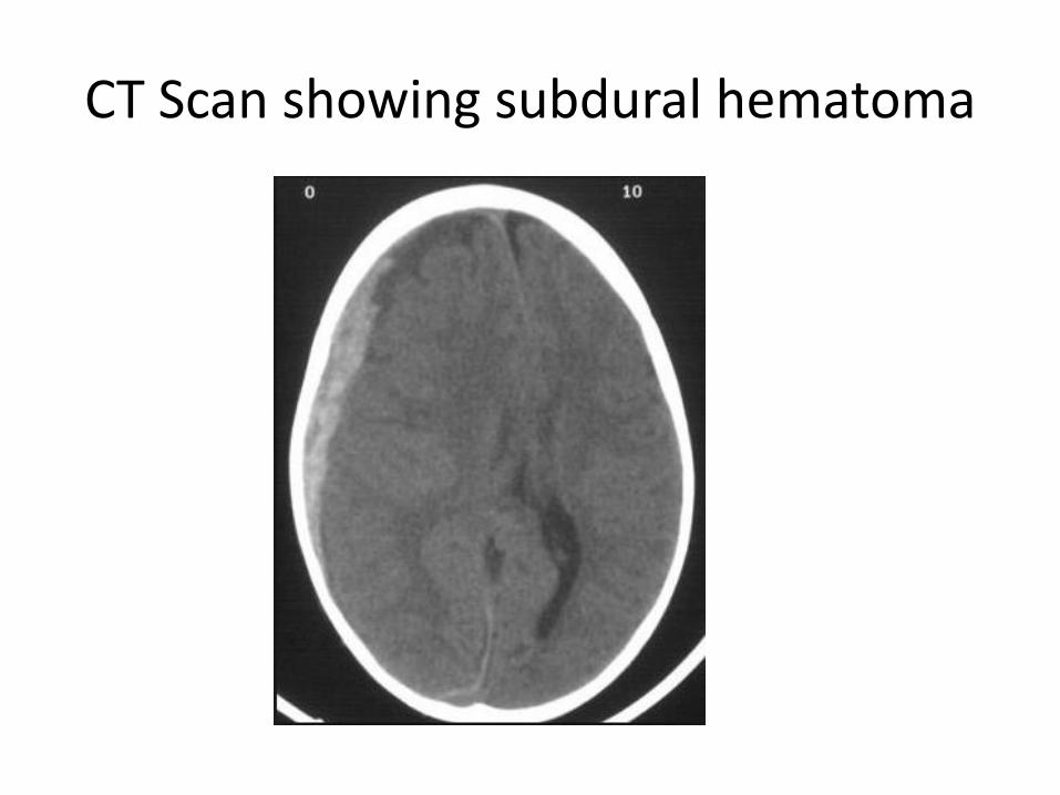
CT scan showing normal liver
Liver metastasis Multiple hypodense lesions seen in the liver with no significant contrast enhancement
LIVER CYST
bullOval well definedbullImperceptible or thin wallbullWater density
CT scan showing tumor in right lobe of liver (arrowheads)
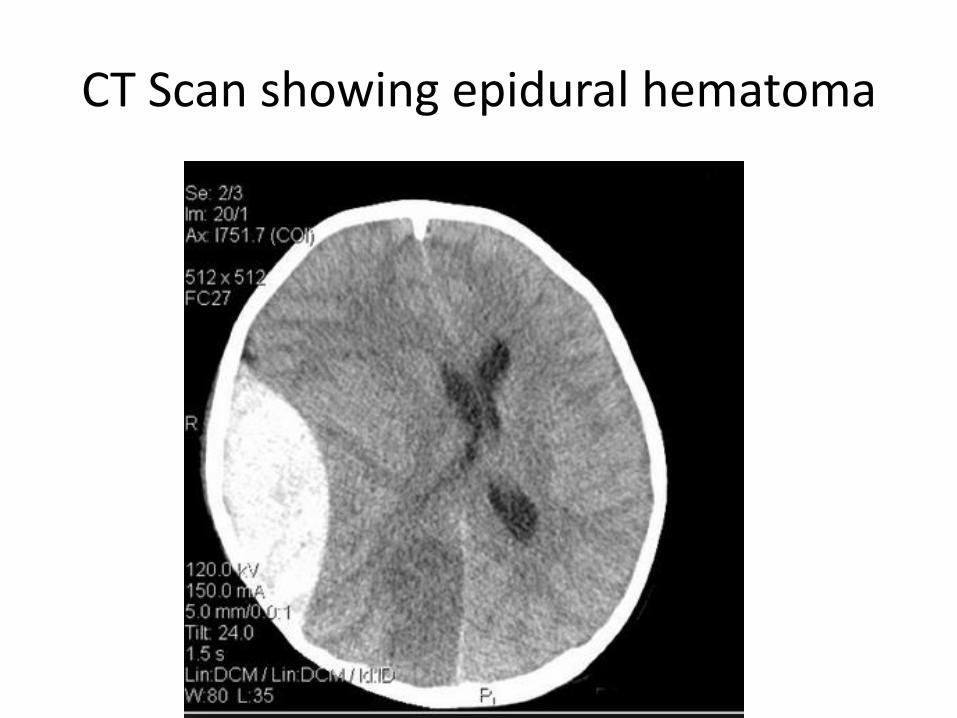
Liver metastasis Multiple hypodense lesions seen in the liver with no significant contrast enhancement
LIVER CYST
bullOval well definedbullImperceptible or thin wallbullWater density
CT scan showing tumor in right lobe of liver (arrowheads)
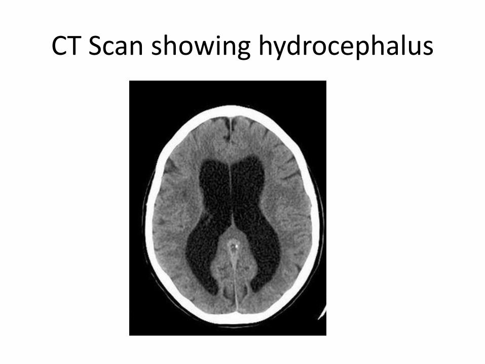
LIVER CYST
bullOval well definedbullImperceptible or thin wallbullWater density
CT scan showing tumor in right lobe of liver (arrowheads)
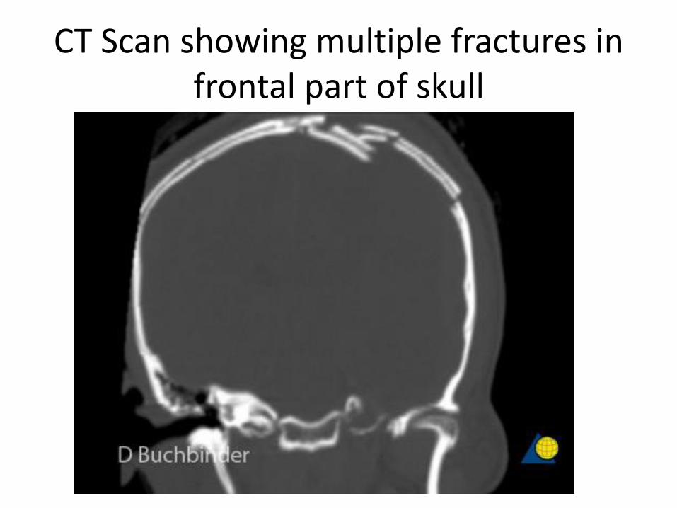
CT scan showing tumor in right lobe of liver (arrowheads)