Radiology guided treatment for spinal pain -...
76
Radiology guided treatment for spinal pain Dr Paul O’Connell
Transcript of Radiology guided treatment for spinal pain -...

Radiology guided treatment for spinal pain
Dr Paul O’Connell

• Management of
– Acute radicular pain
– Chronic low back pain
• Facetal
• Sacro-iliac joint
– Cervicogenic headaches
– Osteoporotic crush fracture
– Hypotension headaches
Image guided treatment for back pain

• pain radiating into the limb in dermatome
– lancinating, burning, stabbing, or electric quality
• limitation of straight-leg-raise to <300
• CT or MRI to assess for disc herniation
– at segmental level consistent with clinical features
Acute radicular pain

Acute radicular pain

Disc protrusionSubarticular (posterolateral)

Perineural injection L5 Perineural injection S1
Transforaminal nerve root block

Lumbar epidural steroidIndications – radicular pain with
• Lumbosacral disk herniation (if transforaminal not possible)
• Spinal stenosis
• Facetal cyst
• Compression fracture lumbar spine
Contraindication
• Acute spinal cord compression

Peripheral nerve fibre typesPain
• Type A & B
– Myelinated
– Quick shallow pain specific to an area (first pain)
• Type C
– Unmyelinated - slow conduction
– Remak bundles
– Slow, deep, spread out, non specific pain (second pain)

Local anaesthetic
• Sodium channel blocker
• Affects
– Unmyelinated fibres generally
– Myelinated nerves at the nodes of Ranvier

The nodes of Ranvier span from 1–2 µm
The internode distances varies with nerve type & size:
Pain smallest
Sensory intermediate
Motor largest
Local anaesthetic

Pain nerve
Motor nerve
Local anaesthetic needs to cover ≥3 nodes of Ranvier

• Acute disc protrusion
→annular nerve ending pain - stretch
• Mechanical compression / stretch of spinal root
→weakness and numbness• uninflamed nerve compressed will stop functioning
• 20 inflammation
→radicular pain
Radiculopathy usually recovers 2-6 weeks but disc resorption takes months
Surgical decompression does not always relieve radiculopathyMcLain et al
Cleveland Clinic J Medicine 71:12 Dec 2004
Disc Protrusion

Steroid effect
• Inhibits neural transmission in nociceptive C fibers
• ↓Phospholipase A2
• ↓ Arachidonic acid
• ↓ capillary permeability and endothelial reaction– ↓ Acute polymorphonuclear leukocyte (PML) inflammatory response
– ↓ Endothelial cytokine release
– ↓ Late monocyte and macrophage
– ↓ Capillary and fibroblast proliferation scar collagen

Injury
Phospholipase A2
Arachidonic acid
Cyclooxygenase Lipooxygenase
Hyperalgaesic Prostoglandins Hyperalgaesic leukotrines
& thromboxanes
Inflammation & pain
Inflammatory cascade
STEROID

• Systemic infection or local infection at site
• Bleeding disorder or fully anticoagulated
• History of significant allergic reactions to injectates
• Patient refusal
Steroid injectionContraindications

• Pregnancy
– to avoid exposing the fetus to ionizing radiation
• Poorly controlled diabetes
– may transiently, but significantly, ↑ blood glucose
• Immunosuppression
– may require additional precautions
• Congestive heart failure
– potential for steroid-induced fluid retention (Not Celestone)
Steroid injectionRelative contraindications

Celestone chronodose
– Betamethasone sodium phosphate and acetate
– Does not precipitate with local anaesthetic
– Does not cause water retention
– Crosses placenta
– Particulate

Cause of cord infarcts
• Particle embolisation• Benzyl alcohol
• Needle induced vasospasm
• Disruption of artery
• Haematoma compressing artery
• Infarcts have only been reported with particulates Particulate versus non-particulate steroids for lumbar transforaminal or interlaminar epidural steroid injections: an update
Skeletal Radiology, Nov 2014Tobias J. Dietrich, Reto Sutter, Johannes M. Froehlich, Christian W. A. Pfirrmann
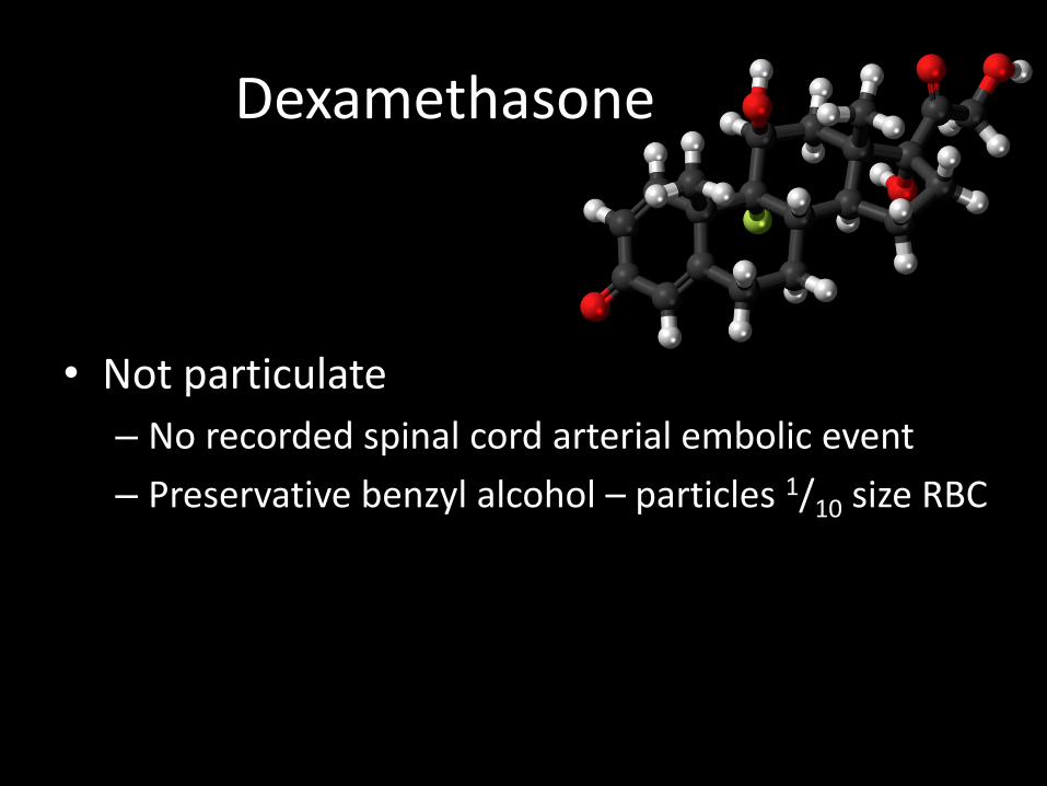
• Not particulate
– No recorded spinal cord arterial embolic event
– Preservative benzyl alcohol – particles 1/10 size RBC
Dexamethasone

• High solubility and negligible particle size– Particles significantly smaller than red blood cells
– Least tendency to aggregation
– Lowest density
Size and Aggregation of Corticosteroids Used for Epidural InjectionsRichard Derby MDPain Medicine March 2008 Volume 9, Issue 2 Pages 147–265
Dexamethasone


Sciatica from disc herniation -conservative versus surgery
• Outcomes at 1 and 2 years were similar for 283 patients• No clinically significant difference 8 wks & 6 months’ follow-up• 56% of patients did not require surgery for recovery
• Early surgery roughly doubled the speed of recovery• Delayed surgery might result in some extra weeks of discomfort• Major advantage of early surgery
– more rapid relief of pain– reassurance about recovery– earlier return to normal activities
Sciatica caused by lumbar disc herniation conservative care versus early surgery2 year results of a randomised controlled trial
BMJ ; June 2008
Department of Neurosurgery, Leiden University Netherlands.

Pain Medicine article 2010
• 150 patients randomised into test groups
• 54% relief at 1 month (>50% reduction of pain)
• 25% relief at 1 year (after 1 injection)
• Acute & chronic radiculopathy
• Transforaminal injection of steroids a viable alternative to surgery for lumbar radicular pain due to disc herniation.
Transforaminal Steroid Injection for Lumbar Radicular Pain Superior to Placebo
Pain Medicine August 2010 Bogduk et al

• Management of
– Acute radicular pain
– Chronic low back pain
• Facetal
• Sacro-iliac joint
– Cervicogenic headaches
– Osteoporotic crush fracture
– Hypotension headaches
Image guided treatment for back / neck pain

Chronic spinal painnon radicular
• Low back pain– Facet pain 40%– Disc pain 40%– Sacro-iliac pain 20%
• Chronic cervical pain– Facet pain (zygo-apophyseal pain) 60%
• Malignant pain– Tumours and infection <1%

Facet Syndrome
• LBP with
– Buttock pain
– Pseudo-radiculopathy to inguinal or posterior thigh
– Paravertebral tenderness
– Transitional movement aggravation (from sitting)

Facet pain
• Animal studies show nociceptors in joint capsule & adjacent muscles and tendons.
• Joint capsule pain may be from pinching, compression, stretching or strain.
The Journal of Bone and Joint Surgery. April 2006. Vol. 88-A.
Cavanaugh et al. Pain Generation in Lumbar and Cervical Facet Joints.

Facet pain
Unlike elsewhere in the body a normalappearing facet joint may be a pain generator
• Plain Xrays & CT
• Nuclear medicine
• Facet joint injection
• Medial branch block

SPECT nuclear medicine

Facet injection

Medial Branch Block Radiofrequency Ablation

Medial BranchLumbar
• Junction of the superior articular process and transverse process – under the mamilloaccessory ligament of the vertebra below
– L3 medial branch lies on the L4 vertebra
• EXCEPTION over S1• L5 posterior 10 ramus sited junction sacral ala and the S1 superior articular process
• Each facet joint innervated by 2 medial branches

• Mamillary process of sup. art. process
• Accessory tubercle of transverse process
• Encloses the medial branch in fibro-osseous tunnel.
• Reliable course relative to bone.
• Ossified in over 10%
• May be site of entrapment causing low-back pain.
Mamillo-accessory ligament (MAL)
Bogduk N. Spine (Phila Pa 1976). 1981 Mar-Apr;6(2):162-7.
The lumbar mamillo--accessory ligament. Its anatomical and neurosurgical significance.
T
T T
T

RF technique
• Preceding MBB with >50% relief– Small volume (1/2 ml Marcaine 0.5%)
• Pre RF stimulation– 50 Hz to ensure proximity of electrode to sensory fibers threshold 0.3 - 0.9 V
– 2 Hz stimulation to detect muscle contractions in the multifidus muscle threshold within 1.5 times of sensory stimulation.
– Ensure not stimulating anterior ramus
• ≥ 20 G needle (curved tip)


Radiofrequency ablation
• Thermal radiofrequency
– Oscillating electrical pulse at microwave frequency
– Causes tissue heating (70-850)
– Denatures protein, disrupting nerves
• Pulsed radiofrequency
– Electromagnetic pulses
– Does not exceed 420
– No definable lesion

Complications
• No pain relief
• Anaesthesia
• Multifidus wasting
• Charcot joint
Presented at SMISS 2013 Annual Conference By Farhan Siddiqi MD et al

Complications
• No pain relief
• Anaesthesia
• Multifidus wasting
• Charcot joint

Complications
• No pain relief
• Anaesthesia
• Multifidus wasting
• Charcot joint

Multifidus
Latissimus dorsi
Quadratus
lumborum
Psoas Major
Transversus abdominus
Internal oblique
External oblique
Anterior layer
Middle layer
Posterior layer
Erector spinaeIliocostalis
Longissimus
Lateral raphe
Thoracolumbar
fascia

• Case study single patient post lumbar RF
• Pain decreased by 60% at 2 weeks, 92% by 4 weeks
• Immediate and sustained ↓ EMG activity over the multifidus and erector spinae muscles
• Gradual positive changes in gait kinematics across all sessions
Changes in gait kinematics and lower back muscle activity post RF denervation of the zygapophysial joint: a case study.
Stegemöller EL1, Roper J, Hass CJ, Kennedy DJ.
Spine J. 2013 Oct 10. pii: S1529-9430(13)00747-X.
doi: 10.1016/j.spinee.2013.06.061. [Epub ahead of print]
Multifidus wasting

Multifidus wasting
Retrospective study 27 patients
No statistical difference in size or morphology of Multifidus
↑Disc degeneration following RF (14.9% vs 4.6%)
Morphologic changes in the lumbar spine after lumbar medial branch radiofrequency neurotomy: a quantitative radiological study.
Smuck M1, Crisostomo RA, Demirjian R, Fitch DS, Kennedy DJ, Geisser ME.
Spine J. 2013 Nov 14: S1529-9430(13)01218-7.

Complications
• No pain relief
• Anaesthesia
• Multifidus wasting
• Charcot joint
No published reports directly attributed to RFMerril D G et al, Reg Anesth Pain Med
2003;28:547-560

Sacro–iliac joint injection

Sacroiliac joint RF denervation
L4
L5
S3
S2
S1

Sacroiliac joint RF denervationL5 posterior 10 ramus S1 lateral branch
S2 lateral branchS3 lateral branch


• Management of
– Acute radicular pain
– Chronic low back pain
• Facetal
• Sacro-iliac joint
– Cervicogenic headaches
– Osteoporotic crush fracture
– Hypotension headaches
Image guided treatment for back / neck pain

Facet injection - cervical

Whiplash
Cervical RF of 53 chronic whiplash patients:
significant early (within 1 month) and sustained (3 months) improvements in pain, disability, local and widespread hyperalgesia
Cervical radiofrequency neurotomy reduces central hyperexcitability and improves neck movement in individuals with chronic whiplash.
Smith AD1, Jull G, Schneider G, Frizzell B, Hooper RA, Sterling M.
Pain Med. 2014 Jan;15(1):128-41.
The Journal of Bone and Joint Surgery. April 2006. Vol. 88-A.
Cavanaugh et al. Pain Generation in Lumbar and Cervical Facet Joints.
Whiplash injuries pain generators are facet joints Facet joints in the cervical spine more sensitive than lumbar
Probably overstretched joint capsule

Medial BranchCervical
• Waist of the articular pillars – C3–T1
– eg C5 & C6 medial branches → C5/6 facet
EXCEPTION– C0-C2 joints by C1 & C2 ventral rami
– C2/3 facet by TON

Third occipital nerve
On the concept of third occipital headache
NIKOLAI BOGDUK, ANTHONY MARSLAND
Journal of Neurology, Neurosurgery, and Psychiatry 1986;49:775-780
TON
C3
Semispinalis capitis

Radiofrequency ablation C2/3 & C3/4

Medial Branch Block /Radiofrequency ablation

Third occipital nerve (TON) RFcomplications
• Numbness
– larger area & more constant than other cervical MBB
• Head neck proprioception sense (HPNS)
– Semispinalis capitus
– temporary
• Neuritis

Greater occipital nerve
Lesser occipital nerve
Third occipital nerve
• Pain (burning) and hypersensitivity (2-4 weeks)
• Highest incidence C2/3 & C3/4
Radiofrequency neuritis

Greater occipital nerve block

Greater occipital nerve block
UltrasoundCT

Assessment of the escalating growth of facet joint interventions in the medicare population in the United States from 2000 to 2011.
Pain Physician. 2013 Jul-Aug;16(4):E365-78.
• All facet joint interventions ↑ 308%
– Lumbosacral facet blocks ↑ 228% (990,449)
– Lumbosacral RF ↑ 662% (406,378)
– Cerv. & thoracic facet blocks ↑ 359% (317,220)
– Cervical & thoracic RF ↑ 836% (97,526)
Usage (USA 2011)

Evidence based medicinePain Physician. 2013 Apr;16(2 Suppl):S49-283.
An update of comprehensive evidence-based guidelines
for interventional techniques in chronic spinal pain.
Part II: guidance and recommendations.
Manchikanti L1, Abdi S, Atluri S, Benyamin RM, Boswell MV, Buenaventura RM, Bryce DA, Burks PA, Caraway DL, Calodney AK, Cash KA, Christo PJ, Cohen SP, Colson J, Conn A, Cordner H, CoubarousS, Datta S, Deer TR, Diwan S, Falco FJ, Fellows B, Geffert S, Grider JS, Gupta S, Hameed H, HameedM, Hansen H,Helm S 2nd, Janata JW, Justiz R, Kaye AD, Lee M, Manchikanti KN, McManus CD, Onyewu O, Parr AT, Patel VB, Racz GB, Sehgal N, Sharma ML, Simopoulos TT, Singh V, Smith HS, Snook LT, Swicegood JR, Vallejo R, Ward SP, Wargo BW, Zhu J, Hirsch JA.
OBJECTIVE:
To develop evidence-based clinical practice guidelines for interventional techniques in the diagnosis and treatment of chronic spinal pain.
METHODOLOGY:
Systematic assessment of the literature.

EvidenceGrade
Definition
Good Lumbar CRF*Lumbar MBB (therapeutic)
Fair Cervical CRF*Cervical MBB (therapeutic)
Thoracic MBB (therapeutic)
Limited or Poor
Thoracic CRF
PRF all medial branchesFacet blocks
Diagnostic lumbar nerve root block
* Preceded by successful MBB

2011 RF / MBB audit Brisbane Private Imaging
• 5 month audit - 82 patients
• 50% MBB’s did not proceed to RF (30/58)
• 75% improvement if +ve MBB (21/28) - all regions*
• 50% improvement if no preceding MBB (12/24)

• Management of
– Acute radicular pain
– Chronic low back pain
• Facetal
• Sacro-iliac joint
– Cervicogenic headaches
– Osteoporotic crush fracture
– Hypotension headaches
Image guided treatment for back / neck pain

Osteoporotic crush fractures

Vertebroplasty

VertebroplastyMedical Journal of Australia 2010 – Clarke et al
Persisting vertebral body fracture (Kummell’s disease)

Vertebroplasty
• Osteoporotic crush fractures
• Trans pedicular approach
• Polymethylmethacrylate (PMMA) + barium
• Internally fixates fracture
• Heats to 82-860 during polymerization

VertebroplastyThe New England Journal of Medicine in 2009
• 2 studies found no benefit for compression fractures compared to sham procedure
– Kallimes University of Washington• multicenter, prospective double-blinded randomized trial
• 131 participants
• vertebroplasty had no detectable benefit from sham procedures.
– Buchbinder trial• funded by the Australian government and Cook Medical Inc
• Multicenter, randomized, double-blind, placebo-controlled trial
• 78 participants with osteoporotic vertebral compression fractures
• vertebroplasty and sham procedures nearly identical pain relief

Medicare response to NEJM articles
• USA 20/6/2011 in order to be reimbursable
– 1) detailed medical record showing pain caused by fracture
– 2) radiographic confirmation of a fracture
– 3) other treatment plans attempted for a reasonable time
– 4) procedure not performed in the emergency department
– 5) that at least 1 year of follow-up
• Australia 1st November 2011
– Removes vertebroplasty from MBS
Vertebroplasty

• Osteoporotic crush fractures usually heal 6-12 weeks
• Suggest perform vertebroplasty < 6 weeks– Or if fracture / fluid filled cleft persists > 6weeks
• Buchbinder study– trial average 9.5 weeks (up to 12 months)
– MRI oedema = fracture (may persist for months after union)
• Kallimes study– 18 weeks average
– No MRI or nuclear medicine required
VertebroplastyMedical Journal of Australia 2010 – reply Clarke et al

• Management of
– Acute radicular pain
– Chronic low back pain
• Facetal
• Sacro-iliac joint
– Cervicogenic headaches
– Osteoporotic crush fracture
– Hypotension headaches
Image guided treatment for back / neck pain

Pachymeningeal enhancement
CSF Hypotension Headache

CSF Hypotension Headache