Radiology Corner - Defense Technical Information Center · desert ground in Iraq when an AK-47...
8
Venous Fragment Embolism to the Pulmonary Artery: A Rare Occurrence Military Medicine Radiology Corner, 174, September 2009 Radiology Corner Venous Fragment Embolism to the Pulmonary Artery: A Rare Occurrence—Case Report and Literature Review of Venous Fragment Embolization to the Pulmonary Artery. Guarantor: LCDR Christopher M. Andrews, USN, MC* Contributors: LCDR Christopher M. Andrews, USN, MC*; CPT Kristine E. Andrews, USAF, MC*; LT Roger Boodoo, USN, MC † ; COL Les Folio, USAF, MC, SFS* Note: This is the full text version of the radiology corner question published in the August 2009 issue, with the abbreviated answer in the September 2009 issue. 1 Despite the large number of gunshot wounds treated in civilian and military practice, embolization of missiles to the pulmonary artery is relatively uncommon. In a review of 7,500 medical reports from casualties from the Vietnam Vascular Registry, only 22 patients with known vascular trauma had missile emboli (0.3%), and of those, only 4 (.0005%) were to the pulmonary artery. 1 Causative agents of missile embolism are numerous and are often a considerable diagnostic and therapeutic challenge. We believe this is the first reported case clearly demonstrating venous fragment embolus to the pulmonary artery from the lower extremity, specifically the popliteal vein using x-rays pre- and post-migration. The fragment was created when a ground-fired rifle bullet shattered the inferior nose bubble of a military helicopter, and a mixture of material was subsequently introduced into the popliteal fossa of the navigator. We also offer a review of the literature and introduce an algorithm for the workup and management options of missile emboli. Introduction A 24 year-old helicopter navigator was flying low above the desert ground in Iraq when an AK-47 round penetrated their helicopter nose bubble before lodging in the patients left leg. The extremity sustained a vascular injury, so a tourniquet was immediately placed and still airborne he was rushed to a nearby combat support hospital. At initial presentation, he was found to have intact distal pulses and parasthesias of the left foot in the tibial nerve distribution. Surgical exploration and irrigation of his wounds occurred within 20 minutes of injury. Multiple metal and plastic fragments were removed from his leg. Five hours after injury he became hypoxic and had increasing oxygen requirements. Imaging studies revealed a rare phenomenon: one of the fragments initially seen on x-ray in the popliteal fossa was now absent, and a previously clear chest x-ray showed what appeared to be the same missing *School of Medicine, Uniformed Services University of the Health Sciences, 4301 Jones Bridge Road, Bethesda, Maryland 20814. † National Naval Medical Center, 8901 Rockville Pike, Bethesda, MD 20889. Reprint & Copyright © by Association of Military Surgeons of U.S., 2006. fragment lodged in the right lower pulmonary lobe region (see Figures 1 and 2). See CT for further localization of fragment (figure 3). Fig. 1A: Initial chest radiograph (left image) and frontal view of the left knee (right image) at 1420 hours on the day of injury in Iraq. Chest radiograph demonstrates no acute cardiopulmonary abnormality. A large metallic fragment projects over the popliteal fossa on the knee radiograph. Fig. 1B Chest radiograph (left image) and frontal view of the knee (right image) taken at 1900 hours on the same day. The previously seen metallic fragment in the region of the popliteal fossa demonstrates interval migration to the rightly lower lobe. Also noted is mild hypoinflation with associated vascular crowding. . Patient discussion: The patient was immediately started on systemic anticoagulation with IV Heparin drip and stabilized for transport to Germany on post-injury day 1 where he underwent LLE irrigation and debridement and a four- compartment fasciotomy due to increasing compartment pressures. Anticoagulation was continued, 4 units of blood
Transcript of Radiology Corner - Defense Technical Information Center · desert ground in Iraq when an AK-47...

Venous Fragment Embolism to the Pulmonary Artery: A Rare Occurrence
Military Medicine Radiology Corner, 174, September 2009
Radiology Corner
Venous Fragment Embolism to the Pulmonary Artery: A Rare Occurrence—Case Report and Literature Review of Venous Fragment Embolization to the Pulmonary Artery.
Guarantor: LCDR Christopher M. Andrews, USN, MC* Contributors: LCDR Christopher M. Andrews, USN, MC*; CPT Kristine E. Andrews, USAF, MC*; LT Roger Boodoo, USN, MC†; COL Les Folio, USAF, MC, SFS*
Note: This is the full text version of the radiology corner question published in the August 2009 issue, with the abbreviated answer in the September 2009 issue. 1
Despite the large number of gunshot wounds treated in civilian and military practice, embolization of missiles to the pulmonary artery is relatively uncommon. In a review of 7,500 medical reports from casualties from the Vietnam Vascular Registry, only 22 patients with known vascular trauma had missile emboli (0.3%), and of those, only 4 (.0005%) were to the pulmonary artery.1 Causative agents of missile embolism are numerous and are often a considerable diagnostic and therapeutic challenge. We believe this is the first reported case clearly demonstrating venous fragment embolus to the pulmonary artery from the lower extremity, specifically the popliteal vein using x-rays pre- and post-migration. The fragment was created when a ground-fired rifle bullet shattered the inferior nose bubble of a military helicopter, and a mixture of material was subsequently introduced into the popliteal fossa of the navigator. We also offer a review of the literature and introduce an algorithm for the workup and management options of missile emboli.
Introduction
A 24 year-old helicopter navigator was flying low above the desert ground in Iraq when an AK-47 round penetrated their helicopter nose bubble before lodging in the patients left leg. The extremity sustained a vascular injury, so a tourniquet was immediately placed and still airborne he was rushed to a nearby combat support hospital. At initial presentation, he was found to have intact distal pulses and parasthesias of the left foot in the tibial nerve distribution. Surgical exploration and irrigation of his wounds occurred within 20 minutes of injury. Multiple metal and plastic fragments were removed from his leg. Five hours after injury he became hypoxic and had increasing oxygen requirements. Imaging studies revealed a rare phenomenon: one of the fragments initially seen on x-ray in the popliteal fossa was now absent, and a previously clear chest x-ray showed what appeared to be the same missing
*School of Medicine, Uniformed Services University of the Health Sciences, 4301 Jones Bridge Road, Bethesda, Maryland 20814. †National Naval Medical Center, 8901 Rockville Pike, Bethesda, MD 20889.
Reprint & Copyright © by Association of Military Surgeons of U.S., 2006.
fragment lodged in the right lower pulmonary lobe region (see Figures 1 and 2). See CT for further localization of fragment (figure 3).
Fig. 1A: Initial chest radiograph (left image) and frontal view of the left
knee (right image) at 1420 hours on the day of injury in Iraq. Chest radiograph demonstrates no acute cardiopulmonary abnormality. A large metallic fragment projects over the popliteal fossa on the knee radiograph.
Fig. 1B Chest radiograph (left image) and frontal view of the knee (right
image) taken at 1900 hours on the same day. The previously seen metallic fragment in the region of the popliteal fossa demonstrates interval migration to the rightly lower lobe. Also noted is mild hypoinflation with associated vascular crowding.
.
Patient discussion:
The patient was immediately started on systemic anticoagulation with IV Heparin drip and stabilized for transport to Germany on post-injury day 1 where he underwent LLE irrigation and debridement and a four-compartment fasciotomy due to increasing compartment pressures. Anticoagulation was continued, 4 units of blood

Report Documentation Page Form ApprovedOMB No. 0704-0188
Public reporting burden for the collection of information is estimated to average 1 hour per response, including the time for reviewing instructions, searching existing data sources, gathering andmaintaining the data needed, and completing and reviewing the collection of information. Send comments regarding this burden estimate or any other aspect of this collection of information,including suggestions for reducing this burden, to Washington Headquarters Services, Directorate for Information Operations and Reports, 1215 Jefferson Davis Highway, Suite 1204, ArlingtonVA 22202-4302. Respondents should be aware that notwithstanding any other provision of law, no person shall be subject to a penalty for failing to comply with a collection of information if itdoes not display a currently valid OMB control number.
1. REPORT DATE SEP 2009 2. REPORT TYPE
3. DATES COVERED 00-00-2009 to 00-00-2009
4. TITLE AND SUBTITLE Venous Fragment Embolism to the Pulmonary Artery: A RareOccurrence -Case Report and Literature Review of Venous FragmentEmbolization to the Pulmonary Artery.
5a. CONTRACT NUMBER
5b. GRANT NUMBER
5c. PROGRAM ELEMENT NUMBER
6. AUTHOR(S) 5d. PROJECT NUMBER
5e. TASK NUMBER
5f. WORK UNIT NUMBER
7. PERFORMING ORGANIZATION NAME(S) AND ADDRESS(ES) Uniformed Services University of the Health Sciences,Department ofRadiology and Radiological Sciences,4301 Jones Bridge Road,Bethesda,MD,20814
8. PERFORMING ORGANIZATIONREPORT NUMBER
9. SPONSORING/MONITORING AGENCY NAME(S) AND ADDRESS(ES) 10. SPONSOR/MONITOR’S ACRONYM(S)
11. SPONSOR/MONITOR’S REPORT NUMBER(S)
12. DISTRIBUTION/AVAILABILITY STATEMENT Approved for public release; distribution unlimited
13. SUPPLEMENTARY NOTES
14. ABSTRACT
15. SUBJECT TERMS
16. SECURITY CLASSIFICATION OF: 17. LIMITATION OF ABSTRACT Same as
Report (SAR)
18. NUMBEROF PAGES
7
19a. NAME OFRESPONSIBLE PERSON
a. REPORT unclassified
b. ABSTRACT unclassified
c. THIS PAGE unclassified
Standard Form 298 (Rev. 8-98) Prescribed by ANSI Std Z39-18
Venous Fragment Embolism to the Pulmonary Artery: A Rare Occurrence
Military Medicine Radiology Corner, 174, September 2009
were transfused, and bilateral distal pulses remained intact throughout his stay.
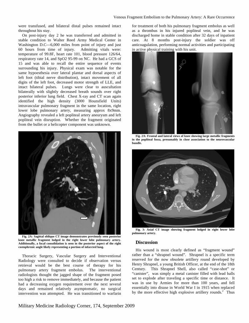
On post-injury day 2 he was transferred and admitted in stable condition to Walter Reed Army Medical Center in Washington D.C—6,000 miles from point of injury and just 60 hours from time of injury. Admitting vitals were: temperature of 99.8F, heart rate 101, blood pressure 126/64, respiratory rate 14, and SpO2 95-99 on NC. He had a GCS of 15 and was able to recall the entire sequence of events surrounding his injury. Physical exam was notable for the same hypoesthesia over lateral plantar and dorsal aspects of left foot (tibial nerve distribution), intact movement of all digits of the left foot, decreased motor strength of LLE, and intact bilateral pulses. Lungs were clear to auscultation bilaterally with slightly decreased breath sounds over right posterior inferior lung field. Chest X-ray and CT scan again identified the high density (3000 Hounsfield Units) intravascular pulmonary fragment in the same location, right lower lobe pulmonary artery, measuring approx 8x9mm. Angiography revealed a left popliteal artery aneurysm and left popliteal vein disruption. Whether the fragment originated from the bullet or a helicopter component was unknown.
Fig. 2A: Sagittal oblique CT image demonstrates previously seen posterior
knee metallic fragment lodged in the right lower lobe pulmonary artery. Additionally, a focal consolidation is seen in the posterior aspect of the right costophrenic angle likely representing a portion of infarcted lung.
Thoracic Surgery, Vascular Surgery and Interventional
Radiology were consulted to decide if observation versus retrieval would be the best course of therapy for his pulmonary artery fragment embolus. The interventional radiologists thought the jagged shape of the fragment posed too high a risk to remove immediately, and because the patient had a decreasing oxygen requirement over the next several days and remained relatively asymptomatic, no surgical intervention was attempted. He was transitioned to warfarin
for treatment of both his pulmonary fragment embolus as well as a thrombus in his injured popliteal vein, and he was discharged home in stable condition after 32 days of inpatient care. At 8 months post-injury the soldier was off anticoagulation, performing normal activities and participating in active physical training with his unit.
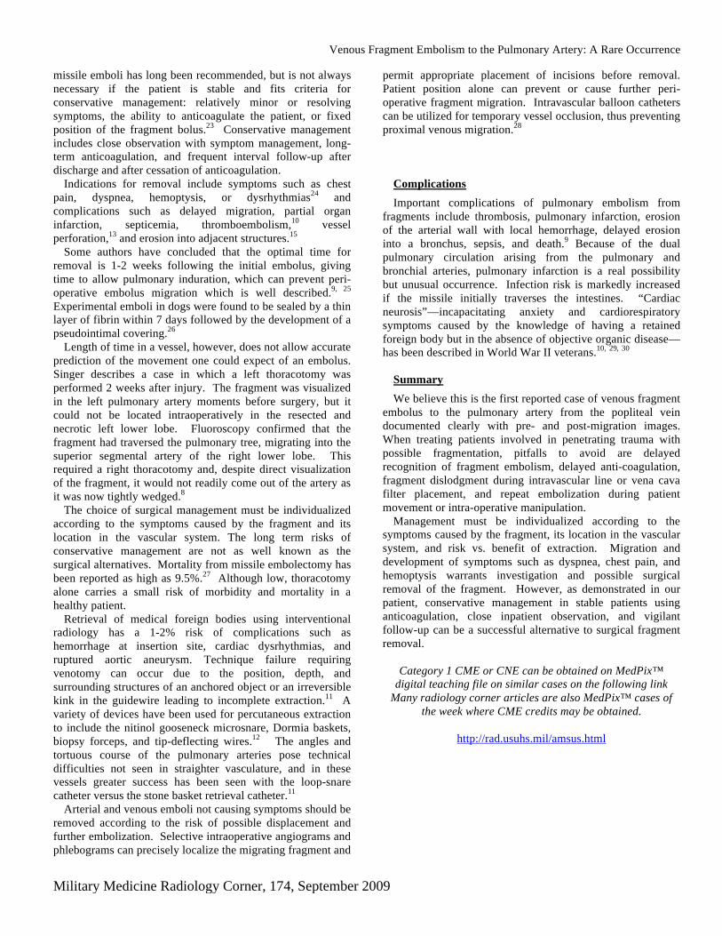
Fig. 2A: Frontal and lateral views of knee showing large metallic fragments
in the popliteal fossa, presumably in close association to the neurovascular bundle.
Fig. 3: Axial CT image showing fragment lodged in right lower lobe
pulmonary artery. Discussion
His wound is most clearly defined as “fragment wound” rather than a “shrapnel wound”. Shrapnel is a specific term reserved for the now obsolete artillery round developed by Henry Shrapnel, a young British Officer, at the end of the 18th Century. This Shrapnel Shell, also called “case-shot” or “canister”, was simply a metal canister filled with lead balls set to explode after traveling a specific time or distance. It was in use by Armies for more than 100 years, and fell essentially into disuse in World War I in 1915 when replaced by the more effective high explosive artillery rounds.2 Thus

Venous Fragment Embolism to the Pulmonary Artery: A Rare Occurrence
Military Medicine Radiology Corner, 174, September 2009
our patient’s embolus is more accurately classified as a “fragment” or “missile” embolus.
Missile Ballistics
In this patient, several elements of ballistics converged allowing the embolus to occur. The most likely rifle used, an AK-47, is a high-velocity weapon that fires an 8g projectile at 710 meters/second with an effective range of 350 meters. The fragments created from the bullet upon impact with the helicopter outer skin, cockpit components, thick aviation uniform, and body tissue each had less mass and/or were less streamlined than the original pointed aerodynamic bullet. With small caliber rounded bullets, fragment bluntness determines the initial value of the area of interface between the bullet and the tissue and thus influences the frictional drag on the bullet. The fragment presented in this case had a shorter period of gyration, tumbling much sooner than an intact high-velocity rifle round, and having much less kinetic energy as it passed through the patient’s dense body tissue.3
Large, high-velocity bullets have rarely been reported to cause embolism, because their increased kinetic energy provides sufficient residual energy to allow full through-and-through perforation of the blood vessels and heart chambers.4 Smaller low-velocity missiles, however, have a greater chance of perforating only one wall of the vessel, thus remaining within the vessel and increasing the possibility of embolization. Once intravascular, any migration of the bullet/fragment will be influenced by position, respiratory motion, blood flow, and gravitational forces.5, 6, 7, 8, 9 Operative manipulation alone has been described in causing fragment migration.10 In this soldier’s dynamic evolution of care, blood loss compounded with the dilution of clotting factors with crystalloid resuscitation, restoration of blood flow with tourniquet removal, manual manipulation during surgery, and peri-operative repositioning would all have had varying degrees of influence in the migration of the popliteal fossa fragment.
Other Migrating Foreign Bodies – Literature Review
The first report of a foreign body embolus is from 1834 involving a wooden missile fragment traveling to the right heart of a 10 year old boy.10 Other unusual objects reportedly found in the pulmonary artery include a piece of an axe, acupuncture needles, as well as pill fragments and needle fragment injected intravenously by drug abusers.
Reports of numerous types of implantable medical devices embolizing to pulmonary vasculature—catheters, coils, filters, stents, shunts, guidewires,11 cardiac valves,12 pacemaker leads,13 ultrasound lithotripter probes,14 acrylic cement,15 and surgical sponges. Retrieval of medically-induced foreign bodies is the current treatment of choice using percutaneous extraction with interventional radiology.16 Success rates have been reported from 71 to 100%.11-13
Symptoms
The spectrum of symptoms from intravascular missile embolus is wide: ranging from completely asymptomatic in two-thirds of patients,6 , 17 to others experiencing chest pain, dyspnea, and hemoptysis.18 While in some cases these symptoms can be attributed to chest wall injury and hypotension from blood loss, in the absence of chest trauma or marked exsanguination they are more assuredly caused by the embolus. Resuscitation is paramount, and a high index of suspicion must be maintained for diagnosis and prevention/management of complications.
Workup
In the absence of pulmonary symptoms, isolated fragment wounds to distal extremities do not always prompt a chest radiograph, therefore asymptomatic patients may experience a delay in diagnosis and subsequent treatment. Fragment embolus must always be considered in asymptomatic patients in which no exit wound is present and the missile is not immediately localized via radiography in the area of injury. A fragment overlying the cardiac silhouette would be suspicious for being intracardiac if motion artifact of the fragment is present and the remainder of the film is free of the motion artifact.19 An abnormal ventilation-perfusion scan due to a pulmonary artery fragment would show normal ventilation but a perfusion defect distal to the fragment. High resolution and reformatted CT images can be extremely reliable in demonstrating fragment trajectories, peritoneal/retroperitoneal violation, and extent of organ injury.20 Multi Detector CT can also determine precise wound tract and trajectory in many cases.21 This modality alone can demonstrate whether retrieval will be necessary and can help identify patients with injuries that may be safely treated without surgery. It is important to note that intravascular fragments may be obscured by IV contrast, and therefore misinterpreted as "radiographic artifact" on x-ray images.
The workup of an individual who has sustained any number of gunshot wounds is greatly aided by a systematic approach. Folio describes an “Even Number Guide” in the assessment of these wounds.22 The guide is effective in aiding efficient initial workup and enhancing communication between the various levels throughout the medical disciplines. The guide calls for counting the entrance and exit wounds and fragments found on clinical examination and radiographic images. Ideally, the total should be an even number. Odd number deviation from the even-number guide should then prompt the trauma team to initiate further investigation to locate a yet undiscovered fragment, exit wound or explanation for exam findings. The fragments may have embolized (arterial or venous) or in the case of multiple gunshot wounds there may be a shared exit wound, as exit wounds are typically larger than entrance wounds and vary in size depending on the distance from which the patient was shot.
Management (See Figure 4 for management algorithm)
Early diagnosis allows time to formulate a proper management plan. Early removal of the pulmonary artery
Venous Fragment Embolism to the Pulmonary Artery: A Rare Occurrence
Military Medicine Radiology Corner, 174, September 2009
missile emboli has long been recommended, but is not always necessary if the patient is stable and fits criteria for conservative management: relatively minor or resolving symptoms, the ability to anticoagulate the patient, or fixed position of the fragment bolus.23 Conservative management includes close observation with symptom management, long-term anticoagulation, and frequent interval follow-up after discharge and after cessation of anticoagulation.
Indications for removal include symptoms such as chest pain, dyspnea, hemoptysis, or dysrhythmias24 and complications such as delayed migration, partial organ infarction, septicemia, thromboembolism,10 vessel perforation,13 and erosion into adjacent structures.15
Some authors have concluded that the optimal time for removal is 1-2 weeks following the initial embolus, giving time to allow pulmonary induration, which can prevent peri-operative embolus migration which is well described.9, 25 Experimental emboli in dogs were found to be sealed by a thin layer of fibrin within 7 days followed by the development of a pseudointimal covering.26
Length of time in a vessel, however, does not allow accurate prediction of the movement one could expect of an embolus. Singer describes a case in which a left thoracotomy was performed 2 weeks after injury. The fragment was visualized in the left pulmonary artery moments before surgery, but it could not be located intraoperatively in the resected and necrotic left lower lobe. Fluoroscopy confirmed that the fragment had traversed the pulmonary tree, migrating into the superior segmental artery of the right lower lobe. This required a right thoracotomy and, despite direct visualization of the fragment, it would not readily come out of the artery as it was now tightly wedged.8
The choice of surgical management must be individualized according to the symptoms caused by the fragment and its location in the vascular system. The long term risks of conservative management are not as well known as the surgical alternatives. Mortality from missile embolectomy has been reported as high as 9.5%.27 Although low, thoracotomy alone carries a small risk of morbidity and mortality in a healthy patient.
Retrieval of medical foreign bodies using interventional radiology has a 1-2% risk of complications such as hemorrhage at insertion site, cardiac dysrhythmias, and ruptured aortic aneurysm. Technique failure requiring venotomy can occur due to the position, depth, and surrounding structures of an anchored object or an irreversible kink in the guidewire leading to incomplete extraction.11 A variety of devices have been used for percutaneous extraction to include the nitinol gooseneck microsnare, Dormia baskets, biopsy forceps, and tip-deflecting wires.12 The angles and tortuous course of the pulmonary arteries pose technical difficulties not seen in straighter vasculature, and in these vessels greater success has been seen with the loop-snare catheter versus the stone basket retrieval catheter.11
Arterial and venous emboli not causing symptoms should be removed according to the risk of possible displacement and further embolization. Selective intraoperative angiograms and phlebograms can precisely localize the migrating fragment and
permit appropriate placement of incisions before removal. Patient position alone can prevent or cause further peri-operative fragment migration. Intravascular balloon catheters can be utilized for temporary vessel occlusion, thus preventing proximal venous migration.28
Complications
Important complications of pulmonary embolism from fragments include thrombosis, pulmonary infarction, erosion of the arterial wall with local hemorrhage, delayed erosion into a bronchus, sepsis, and death.9 Because of the dual pulmonary circulation arising from the pulmonary and bronchial arteries, pulmonary infarction is a real possibility but unusual occurrence. Infection risk is markedly increased if the missile initially traverses the intestines. “Cardiac neurosis”—incapacitating anxiety and cardiorespiratory symptoms caused by the knowledge of having a retained foreign body but in the absence of objective organic disease—has been described in World War II veterans.10, 29, 30
Summary
We believe this is the first reported case of venous fragment embolus to the pulmonary artery from the popliteal vein documented clearly with pre- and post-migration images. When treating patients involved in penetrating trauma with possible fragmentation, pitfalls to avoid are delayed recognition of fragment embolism, delayed anti-coagulation, fragment dislodgment during intravascular line or vena cava filter placement, and repeat embolization during patient movement or intra-operative manipulation.
Management must be individualized according to the symptoms caused by the fragment, its location in the vascular system, and risk vs. benefit of extraction. Migration and development of symptoms such as dyspnea, chest pain, and hemoptysis warrants investigation and possible surgical removal of the fragment. However, as demonstrated in our patient, conservative management in stable patients using anticoagulation, close inpatient observation, and vigilant follow-up can be a successful alternative to surgical fragment removal.
Category 1 CME or CNE can be obtained on MedPix™
digital teaching file on similar cases on the following link Many radiology corner articles are also MedPix™ cases of
the week where CME credits may be obtained.
http://rad.usuhs.mil/amsus.html

Venous Fragment Embolism to the Pulmonary Artery: A Rare Occurrence
Military Medicine Radiology Corner, 174, September 2009
High Index of Suspicion
-No exit wound -Fragment locally absent by exam or X-ray -Symptoms incongruent with injury
Stable Unstable
Initiate Embolus Workup • Full Body X-ray
(Statscan) • CT Scan • Angiography • Fluoroscopy
Resuscitate Patient • Control Bleeding, • Stabilize Vital signs
Fragment Found No
Yes
Consider Embolectomy If: • Easily Retrievable • Likely Delayed Migration • Organ Infarction • Erosion Into Bronchus • Erosion Into Arterial Wall • Erosion Into Adjacent Structures • Clot Propagation • Thromboemoblism • Dyspnea • Hemoptysis • Fragment Traversed
Gastrointestinal Organs • Abscess • Septicemia • Unrelenting Chest Pain • Dysrhythmias • Located in Left Heart • Valve Dysfunction • Endocarditis • Somatoform Symptoms -
“Cardiac Neurosis”
Consider Observation If: • Remains Asymptomatic • Symptoms Are Relatively Minor Or
Resolving • Fragment Is Fixed In Position • Patient Can Be Anticoagulated
Long-term Management: • Long-term anticoagulation- Low molecular
weight heparin or coumadin x6 months • Frequent interval follow-up after discharge
then again after anticoagulation is discontinued
• Re-evaluate need for re-initiation of anticoagulation vs emoblectomy if symptoms recur
Figure 4: Algorithm for the Diagnosis and Management of Intravascular Fragments and Fragment Emboli

Venous Fragment Embolism to the Pulmonary Artery: A Rare Occurrence
Military Medicine Radiology Corner, 174, September 2009
References
1 Rich NM, Collins GJ Jr, Anderson CA, et al. Missile emboli. J Trauma. 1978 Apr;18(4):236-9.
2 Rich N, Burris D. Letters to the editor. J Trauma. 2006 Oct;61(4):1024.
3 Di Maio VJM. Gunshot Wounds: Practical Aspects of Firearms, Ballistics, and Forensic Techniques. Amsterdam: Elsevier; 1985.
4 Jones AM, Graham NJ, Looney JR. Arterial embolism of a high-velocity rifle bullet after a hunting accident: case report and literature
review. Am J Forensic Med Pathol 1983 Sep;4(3):259–64.
5 Bland EF, Beebe GW. Missiles in the heart: a twenty-year follow-up report of World War II cases. N Engl J Med 1966
May;274(19):1039-46.
6 Bernini CO, Junqueira AR Jr, Horita LT, et al. Pulmonary embolism from gunshot missiles. Surg Gynecol Obstet 1983 May;156(5):615-
9.
7 Nehme AE. Intracranial bullet migrating to pulmonary artery. J Trauma. 1980 Apr;20(4):344-6.
8 Singer RL, Dangleben DA, Salim A, Kurek SJ, Shah KT, Goodreau JJ, Shaff DI, Szydlowski GW. Missile embolism to the pulmonary
artery: case report and pitfalls of management. Ann Thorac Surg. 2003 Nov;76(5):1722-5.
9 Stephenson LW, Workman RB, Aldrete JS, Karp RB. Bullet emboli to the pulmonary artery: a report of 2 patients and review of the
literature. Ann Thorac Surg. 1976 Apr;21(4):333-6.
10 Bertoldo U, Enrichens F, Comba A, Ghiselli G, et al. Retrograde venous bullet embolism: a rare occurrence—case report and literature
review. J Trauma, Injury, Infection and Critical Care. 2004 Jul;57(1):187-92.
11 Yang FS, Ohta I, Chiang HJ, Lin JC, Shih SL, Ma YC. Non-surgical retrieval of intravascular foreign body: experience of 12 cases. Eur
J Radiol. 1994 Feb;18(1):1-5.
12 Gabelmann A, Kramer S, Gorich J. Percutaneous retrieval of lost or misplaced intravascular objects. Am J Roentgenol. 2001 Jun;
176(6):1509-13.
13 Sheth R, Someshwar V, Warawdekar G. Percutaneous retrieval of misplaced intravascular foreign objects with the Dormia basket: an
effective solution. Cardiovasc Intervent Radiol. 2007 Jan-Feb;30(1):48-53.
14 Lin CH, Chen CH, Tzeng WS, Cheng BC, Chiu AW. Migration of a dislodged tip of an ultrasound lithotripter probe to the pulmonary
artery: a rare complication of percutaneous nephrolithotomy. Urology. 2003 Dec;62(6):1121-2.

Military Medicine Radiology Corner, 174, September 2009
2
15 MacTaggart JN, Pipinos II, Johanning JM, Lynch TG. Acrylic cement pulmonary embolus masquerading as an embolized central
venous catheter fragment. J Vascular Surgery. 2006 Jan; 43(1):180-3.
16 Pelage JP, Hajjam ME, Lagrange C, et al. Pulmonary artery interventions: an overview. Radiographics. 2005 Nov-Dec;25(6):1653-67.
17 Michelassi F. Bullet emboli to the systemic and venous circulation. Surgery. 1990 Mar;107(3):239-45.
18 Symbas PN, Harlaftis N. Bullet emboli in the pulmonary and systemic arteries. Ann Surg. 1977 Mar;185(3):318-20.
19 Nguyen V. Plain film of intracardiac foreign bodies: the blurring effect. South Med J. 1991 May;84(5):651-3.
20 Munera F. Gunshot wounds of abdomen: evaluation of stable patients with triple-contrast helical CT. Radiology. 2004
May;231(2):399-405.
21 Backus C, Folio L. Lung laceration with active bleeding, contusion and hemothorax. Military Medicine. 2008 Aug;173(8): xv, xvi 22 Folio LR, McHugh, Hoffman M. “The Even Number Guide: A Method for Accounting for Ballistic Injuries.” Radiol Technol. 2007 Jan/Feb; 78(3):197-203. 23 Kortbeek JB, Clark JA, Carraway RC. Conservative management of a pulmonary artery bullet embolism: case report and review of the
literature. J Trauma. 1992 Dec;33(6):906-8.
24 Nagy KK, Massad M, Fildes J, Reyes H. Missile embolization revisited: a rationale for selective management. Am Surg. 1994
Dec;60(12):975-9.
25 John LC, Edmund SJ. Bullet pulmonary embolus and the role of surgery. Thorac Cardiovasc Surg. 1991 Dec;39(6):386-8.
26 Fritz JM, Newman MM, et al. Fate of cardiac foreign bodies. Surgery. 1949 Jun;25(6): 869-79.
27 Massad M, Slim MS. Intravascular missile embolization in childhood: report of a case, literature review, and recommendations for
management. J Pediatr Surg. 1990 Dec;25(12):1292-4.
28 Shannon FL, McCroskey BL, Moore EE, Moore FA. Venous bullet embolism: rationale for mandatory extraction. J Trauma. 1987
Oct;27(10):1118-22.
29 Abbott JA, Cousineau M, Cheitlin, M, et al. Late sequelae of penetrating cardiac wounds. J Thorac Cardiovasc Surg. 1978 Apr;75(4): 510-8. 30 Keiser, L. The Traumatic Neurosis. Philadelphia: J B Lippincott; 1968: 35.