RADIOLOGIC STAGING OF ESOPHAGEAL AND … Hendrik van.pdf · RADIOLOGIC STAGING OF ESOPHAGEAL AND...
108
RADIOLOGIC STAGING OF ESOPHAGEAL AND GASTROESOPHAGEAL JUNCTION CARCINOMA
Transcript of RADIOLOGIC STAGING OF ESOPHAGEAL AND … Hendrik van.pdf · RADIOLOGIC STAGING OF ESOPHAGEAL AND...

RADIOLOGIC STAGING OF ESOPHAGEAL AND GASTROESOPHAGEAL JUNCTION CARCINOMA


RADIOLOGIC STAGING OF ESOPHAGEAL AND GASTROESOPHAGEAL JUNCTION
CARCINOMA
Radiologische stagering van oesofagus- en cardiacarcinoom
PROEFSCHRIFf
ter verkrijging van de graad van doctor aan de Erasmus Universiteit Rotterdam
op gezag van de rector magnificus Prof. Dr. C.J. RIJNVOS
en volgens besluit van het College van Dekanen. De openbare verdediging zal plaatsvinden op
woensdag 30 juni 1993 om 15.45 uur door
HENDRIK VAN OVERHAGEN geboren te Leiden
1993 PASMANS OFFSETDRUKKERU B.V., DEN HAAG

Promotiecomm.issie
Promotor: Prof. Dr. H.E. Schutte
Co-promotor: Dr. J.S. Lameris
Overige !eden: Prof. Dr. H. Obertop Prof. Dr. C.D.A. Verwoerd Prof. J.H.P. Wilson

aan Bettina en mijn ouders
CIP-GEGEVENS KONINKLJJKE BIBLIOTHEEK, DEN HAAG
Overhagen, Hans van
Radiologic Staging of Esophageal and Gastroesophageal Junction Carcinoma I Hans van Overhagen ; [ilL A.W. Zwamborn ... et aL]. - [S.L:s.n.].- Ill Proefschrift Rotterdam. - Met lit. opg. - Met samenvatting in het Nederlands. ISBN 90-9006099-5 Trefw.: darmkanker I maagkanker I radiologie.
Type-setting: J.W. Kuiper, M.G.A.M. van der Lee, A.W. Zwamborn " !993, H. van Overhagen ISBN 90-9006099-5


CONTENTS
Chapter 1 Introduction 11
L 1 Anatomy 11 1.2 Pathogenesis 12 1.3 Pathology 13 1.4 Routes of spread 13 1.5 Treatment and Prognosis 15 1.6 Staging 16 L 7 Purpose of the study 18
Chapter 2 Ultrasound and ultrasound-guided fme-needle aspiration biopsy 25 of supraclavicular lymph nodes in patients with esophageal carcinoma. Cancer 1991; 67: 585-587.
Chapter 3 Supraclavicular lymph node metastases in carcinoma of the 31 esophagus and gastroesophageal junction: Assessment with CT, US and US-guided fine-needle aspiration biopsy. Radiology 1991; 179: 155-158.
Chapter 4 Improved assessment of supraclavicular and abdominal 41 metastases in oesophageal and gastro-oesophageal junction carcinoma with the combination of ultrasound and computed tomography. Br J Radio! 1993; 66: 203-208.
Chapter 5 Assessment of distant metastases with ultrasound-guided fine- 53 needle aspiration biopsy and cytologic study in carcinoma of the esophagus and gastroesophageal junction. Gastrointest Radio! 1992; 17: 305-310.
Chapter 6 CT assessment of resectability in patients undergoing transhiatal 63 esophagectomy for esophageal and gastroesophageal junction carcinoma. J Comput Assist Tomogr. in press.
Chapter 7 Influence of radiologically and cytologically assessed distant 75 metastases on the survival of patients with esophageal and gastroesophageal junction carcinoma. Cancer: in press.
Chapter 8 Final remarks: A radiologic staging strategy 89

Summary 97
Sarnenvatting 101
Epiloog 105
Curriculum Vitae 107


11
Chapter 1
Introduction
Esophageal and Gastroesophageal Junction Carcinoma
1.1 Anatomy
The esophagus is a muscular tube that connects the pharynx to the stomach. In
craniocaudad direction it passes through the neck, the posterior mediastinum and the
diaphragm, to terminate in the gastroesophageal junction. Here the esophageal tube
transforms into the pouch of the stomach [1-3].
The relationship of the esophagus to adjacent anatomical structures is as follows. In the
neck, the esophagus lies anterior to the prevertebral fascia and posterior to the trachea.
In the thorax, the esophagus is bounded on the right side by the mediastinal pleura, lung
and azygos arch, and on the left side by mediastinal pleura, lung, aorta and pulmonary
artery. In the upper mediastinum, the vertebral bodies lie posterior to the esophagus,
anterior are the trachea and left recurrent laryngeal nerve. In the middle mediastinum,
the thoracic duct and azygos vein lie posteriorly and to the right of the esophagus, the
spine lies posteriorly and the carina and bronchi lie anteriorly. In the lower
mediastinum, the aorta lies between the esophagus and vertebral column, anterior to the
esvphagus is the pericardium. At the esophageal hiatus, the esophagus is most
commonly surrounded by the right diaphragmatic crus. In the abdomen, the left lobe of
the liver lies anterior to the esophagus and gastroesophageal junction. The caudate lobe
of the liver lies to the right and the fundus of the stomach to the left [1-3].
The esophageal and gastroesophageal wall consists of the following layers (Fig L 1): the
mucosa, lined with a thick layer of nonkeratinizing stratified squamous epithelium in the
esophagus. The gastroesophageal junction is marked by an irregular boundary between
this epithelium and simple columnar epithelium. The submucosa, a layer of loose
connective tissue. The muscularis propria ( extema). The serosa is the outer layer of the
wall of the intraabdominal esophagus and gastroesophageal junction. The wall of the
intrathoracic esophagus does not contain a serosa but is covered by an adventitia, loose
connective tissue of the mediastinum [1-3].

12
Gastroesophageal Junction
Epithelium
&;-----Lamina propria
Sorosa
Figure 1.1 Layers of the wall of the esophagus and gastroesophageal junction.
1.2 Pathogenesis
Chapter 1
In western countries the incidence of esophageal carcinoma is relatively low [4]. France
has the highest incidence rate in Europe [4,5]. In the Netherlands, esophageal
carcinoma comprise about 1.5% of all malignant tumors [6]. The estimated annual
incidence of esophageal carcinoma in the Netherlands is 4.4 per 100,000 men and 2.5
per 100,000 women [6]. The true incidence of gastroesophageal junction carcinoma is
less well known because it is frequently impossible to determine the exact site of origin
of tumors in this area, and many authors do not distinguish tumors of the
gastroesophageal junction from those of the esophagus and other parts of the stomach in
their reports [7]. In a recent study from the south part of the Netherlands the incidence
of gastroesophageal junction carcinoma was reported to be slightly higher than for
esophageal carcinoma [6].
Introduction 13
The risk of developing esophageal and gastroesophageal junction carcinoma increases
with age and the majority of patients are in the sixth and seventh decade [6-9]. In
France and other European countries, alcohol and tobacco are important factors in
developing esophageal carcinoma. Dietary factors seem to play an important role in
Iran, Northern China and the Transkei region of South Africa where a high incidence of
esophageal carcinoma is observed [8,9]. Also associated with an increased risk of
esophageal carcinoma is Barrett's esophagus. The estimated risk of esophageal cancer in such patients is 30-40 fold above that in the general population [10]. Other factors
possibly associated with esophageal and gastroesophageal cancer are achalasia and lye
strictures [8,9].
1.3 Pathology
The majority of malignant tumors in the esophagus are squamous cell carcinomas.
Adenocarcinomas are reported to account for 1-24% of all esophageal cancers [11-14],
an important number of these tumors arise in patients with Barrett's esophagus [10].
The great majority of gastroesophageal junction tumors are adenocarcinomas. Other
tumors such as undifferentiated carcinoma, leiomyosarcoma, rhabdomyosarcoma and
carcinosarcoma of the esophagus and gastroesophageal junction are rare [11].
1.4 Routes of spread
Spread of esophageal and gastroesophageal junction carcinoma may occur via direct
extension, lymphatic spread or hematogenic metastases.
Direct extension
Carcinomas of the esophagus and gastroesophageal junction are usually well advanced at
the time of diagnosis. Of 224 esophageal and gastroesophageal junction carcinomas
resected in Rotterdam during the period 1978-1984, only 82 (37%) were confined to
either the mucosa or submucosa [15].
According to some authors, spread of esophageal carcinoma into the surrounding
mediastinum is facilitated by the fact that the esophagus lacks a serosa and is separated
from neighbouring structures by only a loose adventitia [8,16]. Autopsy studies report
invasion of the trachea in 13-32%, the bronchi in 16-17%, the pleura in 4-17%, the
pericardium in 1-13%, the aorta in 3-18% and the diaphragm in 19% of patients,
respectively [5,9,17].

14 Chapter 1
Lymphatic spread
The lymphatic network of the esophagus is primarily longitudinally. Sakata was one of
the first to report this and to describe lymphatic vessels in the mucosa and muscularis
that run long longitudinal courses. He also reported that lymph drainage occurred in a
cranial direction in the upper and middle part of the esophagus, some of these vessels
went directly to nodes in the supraclavicular fossa. In the lower part of the esophagus,
lymph drainage occurred in a caudad direction [18].
McCort pointed out that drainage to unexpected sites may occur when normal pathways
become blocked and collateral circulation opens up [19]. Groups of lymph nodes
draining the esophagus and gastroesophageal junction are illustrated in Figure 1.2. Due
to the rich distribution of lymph vessels and their nonsegmental type of drainage, spread
by way of the lymphatics is frequent and the site is unpredictable in esophageal and
gastroesophageal junction carcinoma. Lymph node metastases are found in 59-65% of
patients with esophageal carcinoma [19-21] and 70-80% of patients with
gastroesophageal junction carcinoma [22-24] who undergo surgery. In one series,
supraclavicular metastases were found in 27% of patients with esophageal carcinoma
who underwent dissections of these nodes [25].
Figure 1.2 Lymph nodes drai.niDg the esophagus and gastroesophageal junction.

Introduction 15
Hematogenic spread The venous drainage of the upper two-thirds of the esophagus is to the systemic veins,
that of the lower one-third and of the gastroesophageal junction is to the portal system.
However, numerous areas of anastomosis exist both on the surface and within the wall
of the lower esophagus [3]. Hematogenic metastases are often found in patients with
advanced carcinomas. The most frequently involved sites at autopsy are the liver
(21-47%), the lung (8-52%), the adrenals (3-20%), the kidney (5-13%), the peritoneum
(4-5%) and bone (5-14%) [5,9,17].
1.5 Treatment and Prognosis.
At present, surgical resection of the tumor is generally accepted as the standard
treatment for esophageal and gastroesophageal junction carcinoma. There is no
unanimous opinion regarding the type and extent of resection to be performed [26].
Whereas some authors recommend radical resections and extensive lymph node
dissections [20,21,27], others advocate more limited surgical procedures [22,28,29].
During the last 15 years, an increasing number of surgeons have favoured transhiatal
resection without thoracotomy of the esophagus and gastroesophageal junction because
this procedure theoretically reduces the operative risk for the patient [30]. Since 1986
this approach has become the standard surgical procedure for esophageal and
gastroesophageal junction carcinoma in our institution [31]. The long-term survival of
surgically treated patients with esophageal and gastroesophageal junction carcinoma is
poor [26,32]. Due to the late onset of symptoms, many of these patients have well
advanced disease at the time of diagnosis and survival rates after resection are
negatively affected by an advanced tumor stage [15,21,22,24,27-29]. Involvement of
lymph nodes is especially associated with a poor prognosis [20,21], in one series the
5-year survival rates following resection dropped from 48% to 6% in patients with
metastatic lymph nodes [33]. A critical review of the results of surgical treatment of
esophageal carcinoma was published by Earlarn and Cunba-Melo who analyzed the
literature on esophageal carcinoma during the period 1953-1978 [32]. They estimated
that of 100 patients with the disease, 58 were surgically explored, 39 of 58 (67%) had
their tumor resected and 19 (33 %) had an irresectable tumor at surgery. Forty-five
percent of those resected and leaving the hospital survived for I year, 29% for 2 years and 18% for 5 years. The postoperative mortality was 29% for those patients in whom
a resection was performed. Miiller et al reviewed the literature on esophageal carcinoma
during the period 1980-1988 [26]. They estimated that of 100 patients, 56 had their

16 Chnpter 1
tumor resected. Fifty-six percent of all resected patients survived for 1 year, 34% for 2
years and 20% for 5 years. The postoperative mortality in their review was 13%. They
concluded that since the report of Earlam and Cunha-Melo 10 years previously, the
postoperative mortality had been reduced by half but that efforts to improve long-term
survival had failed [26]. Furthermore, they did not find evidence that adjuvant radiation
therapy or chemotherapy improved either tumor resectability or long-term survival.
Although it is the current opinion that adenocarcinomas of the gastroesophageal junction
are unsuitable for radiation therapy, there is no consensus regarding the role of primary
radiation therapy in squamous cell carcinoma of the esophagus [34,35]. Pearson et al
reported promising results with radiation therapy in these patients, but there has never
been a controlled randomized trial of radiation therapy versus surgery [35,36]. Several
chemotherapeutic drugs have shown some demonstrable activity in squamous cell
carcinomas of the esophagus but their definitive role needs to be determined [37]. At
present the prognosis of patients with symptomatic esophageal and gastroesophageal
junction carcinoma continues to be poor, regardless of the therapy employed.
1.6 Staging
The goal of preoperative staging in esophageal and gastroesophageal junction carcinoma
is twofold. The depth of invasion of the primary tumor and the local resectability should
be assessed. Presence or absence of metastases must be evaluated.
N onradiologic staging
Physical examination is performed to detect metastases. Esophagoscopy is used not only
to obtain a tissue diagnosis, but also to deterutine the upper and lower limits of the
tumor and to detect ulceration, fistula and submucosal metastases [9]. Bronchoscopy is
used to exclude tracheobronchial invasion [9,38]. Some authors advocate mediastino
scopy to assess mediastinal lymph nodes [39].
Radiologic staging
Lindell et al analyzed the influence of radiographic chest film abnormalities, such as an
abnormal azygoesophageal line or a widened mediastinum, on the resectability rate in
patients with carcinoma of the esophagus but did not find any of these to be of
prognostic value [40). Esophageal axis deviation on barium studies has been used by
Akiyama et al and others to assess resectability [41,42]. Furthermore, craniocaudad
extent of the tumor, submucosal metastases and esophageal-airway fistula can be
demonstrated on these studies [43]. McCort described the identification of metastatic
lymph nodes on barium studies in patients with esophageal carcinoma. He also noted

Introduction 17
that metastatic nodes should enlarge before they can be detected on these studies and
that enlargement is not always due to metastases [19]. Other techniques that have been
used selectively to assess tumor resectability and metastases are scintigraphy [38],
azygos venography [42] and lymphography [44].
Computed Tomography
The appearance of the normal esophagus and of esophageal carcinoma on computed
tomographic (CT) studies was first described in detail by Halber and Daffner in 1979
[45,46] and was followed by a description of the gastroesophageal junction on CT in
1981 by Marks et al [47]. CT seemed to be an ideal method of assessing the extent of
mediastinal invasion by esophageal carcinoma because it was the first noninvasive
technique that separately delineated· the esophageal wall and the surrounding structures
[45]. The gastroesophageal junction was slightly more difficult to evaluate because focal
thickening or a pseudomass, due to absence of gastric distension and the transverse
plane of CT sections through the gastroesophageal junction, were observed in a
significant number of normal individuals [47,48]. Enlarged metastatic lymph nodes and
hepatic metastases were also demonstrated on CT [46].
Earlier reports evaluating the ability of CT to stage esophageal carcinoma were
enthusiastic [46,49-51]. Moss et al even reported an accuracy of 100% in assessing
invasion of adjacent mediastinal structures and mediastinal lymph node involvement
[49]. Others, however, reported CT to be of limited value in staging [52-56]. The
accuracy of CT to assess either invasion or lymph node metastases was as low as 66%
and 39% in these series [52,53]. The accuracy of CT to stage gastroesophageal junction
carcinomas is also disputed [48,57,58]. These conflicting conclusions in the literature
can be explained by the following reasons: differences in patient populations, lack of
adequate correlation in some studies, different scanning techniques and different criteria
used on CT studies [59]. At present controversy persists regarding the role and accuracy
of CT in staging esophageal and gastroesophageal junction carcinoma [59].
Ultrasound
Only few reports are published that concern the use of transcutaneous ultrasound (US)
for staging esophageal and gastroesophageal junction carcinoma [60]: the main reason is
probably due to the fact that the primary tumor and the mediastinal lymph nodes
generally cannot be displayed on these studies. US is an established technique for
detection of hepatic metastases [61-63] and has also proved to be a valuable modality
for assessment of cervical lymph node metastases in patients with head and neck tumors
[64,65]. In one study US was clearly more sensitive than palpation in detecting these
nodes [64].

18 Chapter I
US-guided Fme-Needle Aspiration Biopsy
CT- and US-guided biopsies are widely accepted techniques to obtain material for
cytologic and histologic studies [66-69]. Recent studies have shown that the combination
of US-guided fine-needle aspiration biopsy (FNAB) and cytologic study is a useful
method to diagnose metastases in patients with carcinomas. Most importantly, false
positive diagnoses are extremely rare and are reported in only 0-2% of biopsies [69-71].
There are, however, no reports concerning the utility of US-guided biopsies in esophageal and gastroesophageal junction carcinoma.
Maguetic Resonance Imaging and Endosonography
A few studies have been published that concern the use of magnetic resonance (MR)
imaging, using spin-echo techniques and obtaining Tl-weighted and T2-weighted
images, for staging esophageal carcinoma [72-74], Preliminary results of these studies
indicate that MR is not superior to CT in predicting tumor resectability or assessing
metastases.
Endosonography (ES) is a relatively new imaging modality to stage esophageal and
gastroesophageal junction carcinoma that became available at our institution only during
the late period of this study [75-77]. With this technique, an echoprobe is introduced
endoscopically to and beyond the level of the tumor. The main advantage of ES
compared to CT is that the different layers of the esophageal wall can be displayed
separately. A major limitation of the technique is that in an important number of
patients (16-50%) accurate staging is not possible because the echoendoscope cannot
pass through the tumor [76,77]. Preliminary results indicate that ES is more accurate
than CT to assess the depth of invasion of the primary tumor in those cases in which the
tumor stenosis can be passed by the echoendoscope [75, 77]. ES is reported to be more
sensitive but less specific than CT in assessing regional lymph node metastases which
carries the risk of overstaging [75,76] andES is limited in detecting distant lymph node
metastases [76]. The liver cannot be accurately exantined with ES because of the limited
penetration depth of the ES probe [76].
1. 7 Purpose of the study
Pretreatment radiologic staging can, theoretically, improve the effectiveness and results
of surgical treatment in esophageal and gastroesophageal junction carcinoma. Ideally, on
these studies it is possible to select only patients with limited local disease for surgery,
whereas those with nonresecta.ble tumors or metastases to distant sites are excluded
from surgery and submitted to other treatment modalities. The purpose of this study was

Introduction 19
to evaluate the utility of CT, US and US-guided FNAB for pretreatment staging of
esophageal and gastroesophageal junction carcinoma. In assessing distant metastases,
these techniques were evaluated at different sites, Because little has been published
regarding the examination of supraclavicular lymph nodes in esophageal and
gastroesophageal junction carcinoma, a retrospective stndy was first performed to
determine the number of patients with squamous cell carcinoma of the intrathoracic
esophagus in whom supraclavicular metastases could be demonstrated with us and usguided FNAB (Chapter 2), Subsequently, palpation, CT and US were prospectively
evaluated for assessing supraclavicular metastases in patients with either esophageal or
gastroesophageal junction carcinoma (Chapter 3). Accuracy of either CT, US, or a
combination of both studies, to assess distant metastases in general and at the various
sites was determined (Chapter 4). The utility of US-guided FNAB for diagnosing
metastases was evaluated (Chapter 5). Assessment of resectability of the primary tnmor
was analyzed on CT stndies alone because, generally, this cannot be displayed on US
stndies (Chapter 6). Finally a survival analysis was performed to estimate the influence
on survival of distant metastases, assessed on US or CT studies, or diagnosed by means
of US-guided FNAB and cytologic stndy (Chapter 7).

20 Clulpter I
1.8 References
1. Halvorsen RA, Thompson WM. CT and MRI of the esophagus. In: Levine MS ed. Radiology of
the esophagus. Philadelphia: WB Saunders Company, 1989:291-309.
2. Postlethwait RW. Surgery of the esophagus. 2nd ed. Norwalk: Appleton-Century-Crofts. 1986;
563-587.
3. Skandalakis JE, Gray SW, Skandalakis U. Surgical anatomy of the oesophagus. In: Jamieson
GG, ed. Surgery of the oesophagus. Melbourne: Churchill Livingstone, 1988; 19-35.
4. Meller Jensen 0, Esteve J, Meller H, Renard H. Cancer in the European community and its
member states. Eur J Cancer 1990; 26: 1167-1256.
5. Mandard AM, Cbasle J, Marnay J et al. Autopsy findings in 111 cases of esophageal cancer. Cancer 1981; 48: 329-335.
6. Damhuis RAM:, Coebergh JWW. Epidemiologische aspecten van slokdarmkanker in Zuid
Nederland. IKR bulletin 1991; 15: 3-5.
7. Carter DC, Anderson JR. Surgery for adenocarcinoma of the cardia- an overview. In: Jamieson
GG, ed. Surgery of the oesophagus. Melbourne: Churchill Livingstone, 1988; 597-604.
8. Devitt PG, Iyer PV, Rowland R. Pathogenesis and clinical features of cancer of the oesophagus.
In: Jamieson GG, ed. Surgery of the oesophagus. Melbourne: Churchill Livingstone, 1988; 551-
558.
9. Postlethwait RW. Surgery of the esophagus. 2nd ed Norwalk: Appleton-Century-Crofts, 1986:
369-442. 10. Specbler SJ, Goyal RK. Barrett's Esophagus. N Engl J Med 1986; 315: 362-371.
11. Postlethwait RW. Surgery of the esophagus. 2nd ed Norwalk: Appleton-Century-Crofts. 1986:
443-448.
12. Wang HH, Antonioli DA. Goldman H. Comparative features of esophageal and gastric adenocarcinomas: Recent changes in type and frequency. Hum Pathol1986; 17: 482-487.
13. Sons HU, Bochard F. Esophageal cancer. Autopsy findings in 171 cases. Arch Pathol Lab Med 1984; 108; 983-988.
14. Bosch A, Frias Z, Caldwell WL. Adenocarcinoma of the esophagus. Cancer 1979: 43: 1557-
1561.
15. Eeftinck Schattenkerk M, Obertop H, Mud ID, Eijkenboom WMH. van Andel JG, van Houten
H. Survival after resection for carcinoma of the oesophagus. Br J Surg 1987: 74: 165-168.
16. Iyer PV, Rowland R. The pathology of oesophageal cancer. In: Jamieson GG, ed. Surgery of the oesophagus. Melbourne: Churchill Livingstone, 1988; 559-570
17. Anderson LL. Lad TE. Autopsy findings in squamous cell carcinoma of the esophagus. Cancer
1982; 50; 1587-1590.
18. Sakata K. Ueber die lymphgeflisse des oesophagus und iiber seine regionfuen lymphdriisen mit
beriicksichtigung der verbreitung des carcinoms. Mitt Grenz Geb Med Chir 1903; 11: 634-656.
19. McCort JJ. Radiographic identification of lymph node metastases from carcinoma of the
esophagus. Radiology 1952; 59: 694-711.
20. Akiyama H, Tsurumaru M, Kawamura T, Ono Y. Principles of surgical treatment for carcinoma of the esophagus. Ann Surg 1981; 194: 438-446.
21. Skinner DB. En bloc resection for neoplasms of the esophagus and cardia. J Thorac Cardiovasc
Surg 1983; 85; 59-71.
22. Finley RJ, Inculet RI. The results of esophagogastrectomy without thoracotomy for adeno
carcinoma of the esophagogastric junction. Ann Surg 1989; 210: 535-543.

Introduction 21
23. Husemann B. Cardia carcinoma considered as a distinct clinical entity. Br J Surg 1989; 76: 136-
139. 24. Okamura T, Tsujitani S, Marin P et al. Adenocarcinoma in the upper third part of the stomach.
Surg Gynecol Obstet 1987; 165: 247-250. 25. Sannohe Y, Hiratsuka R, Doki K. Lymph node metastases in cancer of the thoracic esophagus.
Am J Surg 1981; 141: 216-218. 26. MUller JM, Erasmi H, Stelzner M, Zieren U, Pichlmaier H. Surgical therapy of oesophageal
carcinoma. Br J Surg 1990; 77: 845-857. 27. Paolini A, Tosato F, Cassese M et al. Total gastrectomy in the treatment of adenocarcinoma of
the cardia. Am J Surg 1986; 151: 238-243. 28. Ellis FH, Gibb SP, Watkins E. Limited esophagogastrectomy for carcinoma of the cardia. Ann
Surg 1988; 208: 354-361. 29. Orringer MB. Trnnshiatal esophagectomy without thoracotomy for carcinoma of the thoracic
esophagus. Ann Surg 1984; 200: 282-288.
30. Orringer MB, Sloan H. Esophagectomy without thoracotomy. J Thorac Cardiovasc Surg 1978; 76: 643-654.
31. Tilanus HW, Langenhorst BLAM. Oesophagusresectie en reconstructie zonder thoracotomie; eerste ervaringen. Ned Tijdschr Geneeskd 1990; 134: 2237-2240.
32. Earlam R, Cunha-Melo JR. Oesophageal squamous cell carcinoma: A critical review on surgery. Brl Surg 1980; 67: 381-390.
33. Lu YK, Li YM. Gu YZ. Cancer of esophagus and esophagogastric junction: Analysis of results of 1025 resections after 5 to 20 years. Ann Thorac Surg 1987; 43: 176-181.
34. Earlam R, Cunha-Melo JR. Oesophageal squamous cell carcinoma: A critical review of radiotherapy. Br J Surg 1980; 67: 457-461.
35. Earlam R. Radiotherapy in the management of oesophageal cancer - an overview. In Jamieson GG, eel. Surgery of the oesophagus. Melbourne: Churchill Livingstone 1988; 579-583.
36. Pearson JG. The value of radiotherapy in the management of squamous oesophageal cancer. Br J Surg 1971; 58: 794-798.
37. Gill PG. Chemotherapy in the management of oesophageal cancer - an overview. In Jamieson GG, ed. Surgery of the oesophagus. Melbourne: Churchill Livingstone 1988; 571-578.
38. Inculet RI, Keller SM, Dwyer A, Roth JA. Evaluation of noninvasive tests for the preoperative staging of carcinoma of the esophagus: A prospective study. Ann Thorac Surg 1985; 40: 561-
565. 39. Murray GF, Wilcox BR, Starek PJK. The assessment of operability of esophageal carcinoma.
Ann Thorac Surg 1977; 23: 393-399. 40. Lindell MM, Hill CA, Libschitz HI. Esophageal cancer: Radiographic chest findings and their
prognostic significance. AIR 1979; 133: 461-465. 41. Akiyama H, Kogure T, Itai Y. The esophageal axis and its relationship to the resectability of
carcinoma of the esophagus. Ann Surg 1971; 176: 30-36. 42. Mori S, Kasai M, Watanabe I, Sbibuya I. Preoperative assessment of resectability for carcinoma
of the thoracic esophagus. Part I. Esophagogram and azygogram. Ann Surg 1979; 190: 100-105. 43. Levine MS. Radiology of the esophagus. Philadelphia: \VB Saunders Company, 1989; 131-168.
44. Sugimachi K, Okudaira Y, Ueo H, Ikeda M, Inokuchi K. Transtracheal mediastinal lymphography for visualization of metastatic lymph nodes in carcinoma of the esophagus. Surg
Gynecol Obstet 1982; 154: 34-38.

22 Chapter I
45. Halber MD, Daffner RH, Thompson WM. CT of the esophagus. I. Normal appearance. AJR
1979; 133: 1047-1050.
46. Daffner RH, Halber MD, Postlethwait RW, Korobkin M, Thompson WM. CT of the esophagus. n. Carcinoma. AJR 1979; 133: 1051-1055.
47. Marks WM, Callen PW, Moss AA. Gastroesophageal region: Source of confusion on CT. AJR
1981; 136: 359-362.
48. Thompson WM, Halvorsen RA, Williford ME, Foster WL, Kofobkin M. Computed tomography
of the gastroesophageal junction. Radiographies 1982; 2: 179-193.
49. Moss AA, Schnyder P, Thoeni RF, Margulis AR. Esophageal carcinoma: Pretherapy staging by
computed tomography. AJR 1981; 136: 1051-1056.
50. Picus D, Balfe DM, Koehler RE, Roper CL, Owen JW. Computed tomography in the staging of
esophageal carcinoma. Radiology 1983; 146: 433-438.
51. Thompson Wl\.1, Halvorsen RA, Foster WL, Williford ME, Postlethwait RW. Korobkin M. Computed tomography for staging esophageal and gastroesophageal cancer: Reevaluation. AJR
1983; 141: 951-958.
52. Lea JW, Prager RL, Bender HW. The questionable role of computed tomography in
preoperative staging of esophageal cancer. Ann Thorac Surg 1984; 38: 479-481.
53. Samuelsson L, Hambraeus GM, Mercke CE, Tylen U. CT staging of oesophageal carcinoma.
Acta Radio! Diagn 1984; 25: 7-11.
54. Quint LE, Glazer GM, Orringer MB, Gross BH. Esophageal carcinoma: CT findings. Radiology
1985; 155: 171-175.
55. Salonen 0, Kivisaari L, StandertskjOld-Nordenstam CG, Somer K, Virkkunen P. Computed
tomography in staging of oesophageal carcinoma. Scand J Gastroenterol 1987; 22: 65-68.
56. Becker CD, Barbier P, Porcellini B. CT evaluation of patients undergoing. transhiatal
esophagectomy for cancer. J Comput Assist Tomogr 1986; 10: 607-611.
57. Freeny PC, Marks WM:. Adenocarcinoma of the gastroesophageal junction: Barium and Cf
examination . .AJR 1982; 138: 1077-1084.
58. Terrier, Schapira C, Fuchs A. cr assessment of operability in carcinoma of the
oesophagogastric junction. Eur J Radio I 1984; 4: 114-117.
59. Halvorsen RA., Thompson WM:. cr of esophageal neoplasms. Radio! Clin North Am 1989; 27:
667-685.
60. Tohnosu N, Onoda S, Isono K. Ultrasonograph.ic evaluation of cervical lymph node metastases
in esophageal cancer with special reference to the relationship bet\.Veen the short to long axis
ratio (S/L) and the cancer content. JCU 1989; 17: 101-106.
61. Snow JH, Goldstein HM, Wallace S. Comparison of scintigraphy, sonography, and computed
tomography in the evaluation of hepatic neoplasms . .AJR 1979; 132: 915-918.
62. Alderson PO, Adams DF, McNeil BJ et al. Computed tomography, ultrasound and scintigraphy
of the liver in patients with colon or breast carcinoma: A prospective comparison. Radiology
1983;149: 225-230.
63. Ferrucci IT. Liver tumor imaging. Current concepts. AJR 1990; 155: 473-484.
64. Baatenburg de Jong RJ, Rongen RJ, Lameris JS, Hartboom M, Vexwoerd CDA, Knegt P.
Metastatic neck disease: palpation vs ultrasound examination. Arch Otol Head Neck Surg 1989;
115: 689-690.
65. Hajek PC, Salomonowitz E, Turk R, Tscholak:off D, Kumpan W, Czembirek H. Lymph nodes
of the neck: Evaluation with US. Radiology 1986; 158: 739-742.
66. Zomoza J. Needle biopsy of metastases. Radio! Clin North Am 1982; 20: 569-590.

Introduction 23
67. Palaez JC, Hill MC, Dach JL, Isikoff MB, Morse B. Abdominal aspiration biopsies.
Sonographic v computed tomographic guidance. JAMA 1983; 250: 2663-2666.
68. Charboneau JW, Reading CC, Welch TJ. CT and sonographically guided needle biopsy: Current
techniques and new innovations. AJR 1990; 154: 1-10.
69. Reading CC, Charboneau JW, Meredith James E, Hurt MR. Sonographically guided
percutaneous biopsy of small (3 em or less) masses. AJR 1988; 151: 189-192.
70. van der Brekel MWM, Castelijns JA, Stel HV et al. Occult metastatic neck disease: Detection
with US and US-guided fine-needle aspiration cytology. Radiology 1991; 180: 457-461.
71. Baatenburg de Jong RJ, Rongen RJ, Verwoerd CDA, van Overhagen H, Lam6ris JS, K.negt P.
Ultrasound-guided fine-needle aspiration biopsy of neck nodes. A.rch Otolaryngol Head Neck
Surg 1991; 117: 402-404.
72. Quint LE, Glazer GM, Orringer MB. Esophageal imaging by MR and CT: Study of normal
anatomy and neoplasms. Radiology 1985; 156: 727-731.
73. Lehr L. Rupp N, Siewert JR. Assessment of resectability of esophageal cancer by computed
tomography and magnetic resonance imaging. Surgery 1988; 103: 344-350.
74. Takashima S, Takeuchi N, Schiozaki H et al. Carcinoma of the esophagus: CT vs MR imaging
in determining resectability. AJR 1991; 156: 297-302.
75. Tio TL, Cohen P, Coene PP, Udding J, den Hartog FCA. Tytgat GNJ. Endosonography and
computed tomography of esophageal carcinoma. Gastroenterology 1989; 96: 1478-1486.
76. Tio TL, Coene PP, Schouwink MH, Tytgat GNJ. Esophagogastric carcinoma: Preoperative
TNM classification with endosonography. Radiology 1989; 173: 411-417.
77. Vilgrain V, Mompoint D, Palazzo L. Staging of esophageal carcinoma: Comparison of results
with endoscopic sonography and cr. AJR 1990; 155: 277-281.

24

25
Chapter 2
Ultrasound and Ultrasound-guided Fine-Needle Aspiration Biopsy of Supraclavicular Lymph Nodes in Patients with Esophageal Carcinoma
(Supraclavicular US and US-guided FNAB)
2.1 Abstract
The use of ultrasound (US) combined with US-guided fine-needle aspiration biopsy (FNAB)
of supraclavicular lymph nodes in the pretreatment staging of 37 patients with squamous
cell carcinoma of the esophagus is described. All patients underwent computed tomography
of the chest and the abdomen and US of the abdomen and supraclavicular regions.
Supraclavicular lymph node metastases (Stage N disease according to the tumor nodes
metastasis (TNM) classification) were cytologically diagnosed in seven (18.9 %) of the 37
patients. 1n two of these patients, no other metastases were found. In the other five
patients, US-guided FNAB replaced more invasive diagnostic procedures. Due to their
superficial location, US and US-guided FNAB of the supraclavicular lymph nodes was
relatively simple to perform, and contributed to an improved staging of squamous cell
carcinoma of the esophagns.
2.2 Introduction
Carcinoma of the esophagus has a poor prognosis. Due to the advanced stage of disease
frequently present at the time of diagnosis, survival rates remain low. Surgical treatment
is generally associated with a high postoperative morbidity and mortality and does not seem
justified in patients with distant metastases [1-3]. Therefore, accurate pretreatment staging
is of great importance to select the subgroup of patients who can benefit from an operation.
Published as: Hans van Overhagen, MD, Johan S. Lameri.s, MD, Harmine M. Zonderland, MD, Hugo W. Tilanus, MD, Ren6e van Pel, MD, Henri E. SchUtte, MD. ULTRASOUND AND ULTRASOUND-GUIDED FINE-NEEDLEASPIRATIONBIOPSYOFSUPRACLAVICULARLYMPHNODESINPATIENTSWITH ESOPHAGEAL CARCINOMA. Cancer 1991; 67:585-587

26 Clwpter 2
Computed tomography (CT) of the chest and abdomen is generally accepted as the single
most accurate noninvasive study for the pretreatment staging of carcinoma of the esophagus
[4]. Some authors claim that CT is accurate in demonstrating local invasion of the tumor
[5-7], although opposing opinions have been reported [8-9]. CT however, is not a sensitive
predictor of metastatic involvement of periesophageal lymph nodes [6,8-10] and also seems
limited in its ability to detect involvement of abdominal nodal areas [8-11]. Patients are
usually not excluded from operative treatment on the basis of CT findings alone, and more
invasive procedures are necessary to obtain cytologic or histologic evidence of metastatic
spread.
According to the tumor node metastasis (TNM) classification, metastatic involvement of
the supraclavicular lymph nodes in patients with carcinoma of the intrathoracic esophagus
is associated with Stage IV disease (distant metastases) [12]. Due to their superficial
location, the supraclavicular lymph nodes are accessible to ultrasound (US) and US-guided
fine-needle aspiration biopsy (FNAB). In a previous study from our department the
accuracy of US in combination with US-guided FNAB was good in detecting nonpalpable
lymph node metastases in the neck [13]. This encouraged us to use US and US-guided
FNAB of the supraclavicular lymph nodes when staging patients with squamous cell
carcinoma of the esophagus.
2.3 Patients and methods Thirty-seven patients with histologically proven primary squamous cell carcinoma of the
intrathoracic esophagus who were referred to the Rotterdam Esophageal Tumor Study
Group and underwent US of the supraclavicular lymph nodes in the period January 1987 -
December 1988 were retrospectively studied. The patients, 26 men and 11 women, ranged
from 42 to 77 years (mean, 59 years). Three patients had carcinoma of the upper
esophagus, 16 had carcinoma of the middle esophagus, and 18 had carcinoma of the lower
esophagus. Twenty-two patients had already undergone CT or US in other hospitals to rule
out metastases to the chest and the abdomen. CT scans performed elsewhere were
reevaluated at our department. In seven patients they were found to be adequate. The
remaining 30 patients underwent CT scanning using a Tomoscan 350 (Philips Medical
Systems, Eindhoven, The Netherlands) total body scanner with 1 em thick contiguous slices
through the chest and the abdomen, including the entire liver. Mediastinal and abdominal
lymph node metastases were suspected if lymph nodes were greater than I em in diameter.
All patients underwent US of the neck and upper abdomen using a 7.5-MHz and a 3.5-
MHz transducer (Aloka SSD-650). US-guided FNAB with a 23-gauge needle was done in

Supraclavicular US & US-guided FNAB 27
supraclavicular lymph nodes larger than 5 mm in diameter. In cases of multiple unilateral
nodes, the largest node was aspirated. In cases of bilateral nodes, the largest nodes on both
sides were aspirated. A negative outcome of the aspiration biopsy was accepted when the
smears showed lymphocytes and no malignant cells.
2.4 Results
CT and US findings in the 37 patients are summarized in Table 2. L Ten patients had
supraclavicular lymph nodes larger than 5 mm in diameter on US and underwent US-guided
FNAB. In two of these patients, enlarged, suspiciously firm lymph nodes were also
palpable.
Table 2.1 CT and US findings in 37 patients with esophageal carcinoma
Findings
Supraclavicular lymphadenopathy~ (US) Mediastinallymphadenopathyo (CT) Abdominal lymphadenopathy<> (CT and US) Liver metastasis (CT and US) Adrenal metastasis (CT and US) Pleural metastasis (CT)
US: ultrasound; CT: computed tomography. • Lymph nodes larger than 5 mm in diameter. 6 In r.vo patients, enlarged lymph nodes were also palpable. <> Lymph nodes larger than 1 em in diameter.
No. patients
10°/37 5/37 7/37 1137 2/37 1/37
%
(27) (14) (19)
(3) (5) (3)
Cytologic exantination revealed metastatic involvement in seven patients, two patients with
palpable nodes and five with nonpalpable nodes. In four patients, the left side, in two, the
right side, and in one, both sides of the neck were involved. Of these seven patients, four
had a carcinoma in the middle third of the esophagus and three in the lower third. Two
patients with supraclavicular metastases did not show metastatic spread to the chest or the
abdomen on CT and US. Abdominal adenopathy was found in the other five patients and
mediastinal adenopathy in one. Two of these patients were also suspected of having distant
metastases elsewhere (Table 2.2).
Cytologic examination of the supraclavicular lymph nodes did not show malignant
involvement in three patients, nor were they suspected of metastatic spread to the chest or

28 Chapter 2
abdomen on CT and US.
On the basis of US-guided FNAB of the supraclavicular lymph nodes, Stage N disease was
diagnosed in seven patients who were subsequently excluded from operative resection.
Table 2.2 CT and US findings in seven patients with esophageal carcinoma and cytologically proven supraclavicular metastases
Findings
Mediastinal lymphadenopathy (CT) Abdominal lymphadenopathy (CT)
Abdomina! lymphadenopathy (US) Liver metastasis (CT and US) Adrenal metastasis (CT and US) Pleural metastasis (CT)
+
CT: computed tomography; US: ultrasound.
2.5 Discussion
2 3
+ + +
Patient no.
4 5 6 7
+ + +
+ +
+ +
Carcinoma of the esophagus has a poor prognosis because the diagnosis is rarely made
before the development of local invasion and distant metastases. This explains why the 5-
year survival remains low in western countries despite the recent advances in surgery and
radiation therapy [1,3].
Spread by way of the lymphatics occurs frequently: at surgery, lymph node metastases are
found in 4!.3% to 59%, [14,15] and at autopsy, in 74.5% of cases [16]. The presence of
lymph node metastases suggests an omnious prognosis [10,14,15], and in case of distant
nodal metastases, resection of the tumor is at least questionable as an operation is generally
associated with high postoperative morbidity and mortality rate of 14% to 29% [1,3].
In the past 10 years, CT has improved the accuracy of preoperative staging. A 1intitation
of CT scanning is its inability to detect tumor in lymph nodes that are of normal size. Also,
enlarged lymph nodes may not contain tumor [10]. Therefore CT is insensitive in
predicting involvement of the periesophageal lymph nodes [6,8-10] and, to a lesser degree,
in predicting the involvement of abdominal lymph nodes [8-l!]. More invasive procedures
are usually necessary to obtain cytologic or histologic evidence of tumor spread.
The incidence of metastatic spread of esophageal carcinoma to the supraclavicular lymph

Supraclavicular US & US-guided FNAB 29
nodes is often underestimated. Reports on pretreatment staging by CT are invariably limited to the chest and abdomen. To our knowledge there has been only one report in articles
published in English that concerns sonographic evaluation of cervical lymph node
metastases in esophageal carcinoma [17]. At surgery, metastatic involvement of the supra
clavicular lymph nodes has been reported in 18% to 27% of cases [18]. We were able to
detect supraclavicular metastases in 18.9% of patients, a finding that correlates well with
these data. The 37 patients were not referred at random. Most had been selected elsewhere
by excluding thoracic and abdominal metastases before their referral. In an unselected
group of patients, the incidence of supraclavicular metastases may be even higher. Due to the superficial location of the supraclavicular lymph nodes, US-guided FNAB is a
relatively simple procedure with only little patient discomfort. The ease with which the
exact nature of adenopathy can be established is a major advantage. According to the TNM
classification all seven patients with supraclavicular metastases had Stage IV disease [12],
and it is the policy of our Tumor Study Group that operation is not indicated in this group
of patients with distant metastases.
On the basis of CT findings alone, Patients 3, 5 and 7 were suspected of having Stage IV
disease, and, on the basis of abdominal US, Patients 1 and 6 were also suspected of having
Stage IV disease (Table 2.2). Without supraclavicular US-guided FNAB, abdominal US
guided FNAB and/ or surgical exploration would have been necessary in these 5 patients
for correct assessment of distant metastases. Supraclavicular US-guided FNAB prevented
an extensive operation in Patients 2 and 4 as distant metastases were not found on CT and
US of the chest and the abdomen.
We recommend supraclavicular ultrasound and ultrasound-guided fine-needle aspiration
biopsy in addition to CT of the chest and abdomen and ultrasound of the abdomen to
improve the staging of squamous cell carcinoma of the esophagus.

30 Chapter 2
2.6 Fteferences
1. Ear lam R, Cunha- Melo JR. Oesophageal squamous cell carcinoma: I. A critical review of surgery.
BrJ Surg 1980; 67; 381-390.
2. van Andel JG, Dees J, Dijkhuis CM et al. Carcinoma of the esophagus: Results of treatment. Ann
Surg 1979; 190; 684-689.
3. Eeftinck Schattenkerk M, Obertop H, Mud HJ, Eijkenboom WMH, van Andel JG, van Houten H.
Survival after resection for carcinoma of the oesophagus. Br J Surg 1987; 74: 165-168.
4. Inculet RI, Keller SM, Dwyer A, Roth JA. Evaluation of noninvasive tests for the preoperative
staging of carcinoma of the esophagus: A prospective study. Ann Thorac Surg 1985; 40: 561-565. 5. Moss AA, Schnyder P, Thoeni RF, Margulis AR. Esophageal carcinoma: Pretherapy staging by
computed-tomography. AJR 1981; 136: 1051-1056.
6. Picus D, Balfe DM, Koehler RE, Roper CL, Owen JW. Computed tomography in the staging of
esophageal carcinoma. Radiology 1983; 146: 433-438.
7. Thompson WM, Halvorsen RA, Foster WL Jr., Williford ME, Postlethwait RW, Korobkin M.
Computed tomography for staging esophageal and gastroesophageal cancer: Reevalution. AJR 1983; 141; 951-958.
8. Quint LE, Glazer GM, Orringer MB, Gross BH. Esophageal carcinoma: CT findings. Radiology
1985; 155; 171-175.
9. Salonen 0, Kivisaari L, StandertskjOld-Nordenstam CG, Somer K, Virkkunen P. Computed
tomography in staging of oesophageal carcinoma. Scand J. Gastroenterol1987; 22: 65-68.
10. Halvorsen RA, Magruder-Habib K, Foster WL, Roberts L, Postlethwait RW, Thompson WM.
Esophageal cancer staging by CT: Long-term follow-up study. Radiology 1986; 161: 147-151.
11. Lea JW. Prager RL, Bender H\V Jr. The questionable role of computed tomography in preoperative
staging of esophageal cancer. Ann Thorac Surg 1984; 38: 479481.
12. Beabrs OH, Henson DE, Hutter RVP, Myers MH. Manual for staging of cancer. 3rd ed.
Philadelphia: JB Lippincott, 1988
13. Baatenburg de Jong RJ, Rongen RJ, Lam6ris JS, Hartboom M, V erwoerd CDA, Knegt P. Metastatic
neck disease: Palpation vs ultrasound examination . .Arch Otolaryngol Head Neck Surg 1989; 115:
689-690.
14. Akiyama H, Tsurumaru M, Kawamura T, Ono Y. Principles of surgical treatment for carcinoma
of the esophagus. Ann Surg 1981; 194: 438-446
15. Lu YK, Li YM, Gu YZ. Cancer of esophagus and esophagogastric junction: Analysis of results of
1,025 resections after 5 to 20 years. Ann Thorac Surg 1987; 43: 176-181
16. Mandard AM, Chasle J, Marnay Jet al. Autopsy findings in 111 cases of esophageal cancer. Cancer
1981; 48; 329-335.
17. Tobnosu N, Onoda S, Isono K. Ultrasonograpbic evaluation of cervical lymph node metastases in esophageal cancer with special reference to the relationship between the short to long axis ratio (SIL)
and the cancer content. JCU 1989; 17: 101-106.
18. Sannohe Y, Hiratsuka R, Doki K. Lymph node metastases in cancer of the thoracic esophagus. Am
J Surg 1981; 141: 216-218.

31
Chapter 3
Supraclavicular Lymph Node Metastases in Carcinoma of the Esophagus and Gastroesophageal Junction: Assessment with CT, US, and US-guided Fine-Needle Aspiration Biopsy
(Supraclavicular Metastases: CT, US and US-guided FNAB)
3.1 Abstract
The preoperative assessment of supraclavicular lymph node metastases was
prospectively studied in one hundred patients with carcinoma of the esophagus and
gastroesophageal junction. Findings at computed tomography (CT), ultrasound (US),
and palpation were compared, and US-guided fine-needle aspiration biopsy of nodes
with a small axis of 5 rom or greater was performed. Supraclavicular metastases were
detected on CT scans in 11 of 13 patients (85%) and on US scans in 14 of 16 patients
(88%) but were palpable in only three of the 16 patients (19%). The predictive value of
a supraclavicular node indicating metastases was 0.74 at US and 0.85 at CT. Metastases
were diagnosed in 10 of 46 patients with squamous cell carcinoma (22%) and five of 50 patients (10%) with adenocarcinoma. Nodes with metastases had a round configuration,
with a statistically significant greater short-axis to long-axis ratio i:han that of benign
nodes (0.89 vs 0.54; p = 0.05). ln four of 16 patients (25%) with supraclavicular
metastases proved with cytologic examination, neither CT nor US of the mediastinum
and abdomen showed enlarged nodes.
3.2 Introduction
Lymph node metastases in carcinoma of the esophagus and gastroesophageal junction
are associated with a poor prognosis. The 5-year survival rate after resection of
esophageal carcinoma is as high as 47.9% - 53.8% in patients without lymph node
metastases but only 6.3% - 15.3% in patients with them [1,2].
Published as: Hans van Overhagen, MD, Johan S. Lameris, MD, Mrujolein Y. Berger, MD, Frans van der Voorde, MD, Hugo W. Tilanus, MD, Abraham I.J. Klooswijk, MD, Harmine M. Zonderland, MD, Renee van Pel. MD. SUPRACLAVICULAR LYMPH NODE METASTASES IN CARCINOMA OF THE ESOPHAGUS AND GASTROESOPHAGEAL JUNCTION' ASSESSMENT WITH CT. US AND US-GUIDED FINE-NEEDLE ASPIRATION BIOPSY. Radiology !991' 179,!55-!58

32 Chapter 3
The survival rates after resection of gastroesophageal junction carcinoma also drop
significantly when lymph node metastases are found [3,4].
Preoperative assessment of lymph node metastases is therefore one of the most
important factors in selecting therapy and estimating the prognosis. Many reports
concern assessment of mediastinal and abdominal lymph node metastases with computed
tomography (CT) [5-12]. Although CT evaluation of supraclavicular adenopathy in
general has been reported by Goldberg and Austin [13], we are unaware of any study in
which CT has been systematically used to determine supraclavicular lymph node
metastases in esophageal and gastroesophageal junction carcinoma. In addition, few
articles report the use of ultrasound (US) for detection of supraclavicular metastases in
these cases [14,15]. Supraclavicular metastases are often detected with palpation only,
despite the fact that palpation is an unreliable method [16,17].
The use of US and US-guided fine-needle aspiration biopsy (FNAB) to diagnose
supraclavicular metastases in carcinoma of the thoracic esophagus was discus:;ed by us
in a previous report [15]. This study was undertaken to compare palpation, US and CT
findings and to reevaluate the use of US-guided FNAB. Morphologic characteristics of
malignant and benign nodes were studied, and the relative frequency of supraclavicular
metastases was established in relation to the location of and histologic fmdings in the
primary tumor. In cases of supraclavicular metastases the presence of radiologically
detectable mediastinal and abdominal adenopathy was evaluated.
3.3 Patients and Methods
One hundred consecutive patients, 81 men and 19 women aged 40-85 years (mean, 63
years), were prospectively studied between January 1989 and June 1990. All patients
had a primary malignant tumor of the thoracic esophagus and/or gastroesophageal
junction that had been proved with histologic exantination, and all were referred to the
Rotterdam Esophageal Tumor Study Group for treatment. Forty-six patients had
squamous cell carcinoma (carcinoma of the upper thoracic esophagus, n = 3; middle
esophagus, n = 23; lower esophagus, n = 20), which was staged according to the classification put forth by the American Joint Committee on Cancer [ 18]. Fifty patients
had adenocarcinoma of the middle thoracic esophagus (n = 2) or lower esophagus and
gastroesophageal junction (n = 48). Four patients had undifferentiated carcinoma.
Palpation of the supraclavicular regions of all patients was done by members of the
surgical and gastroenterologic departments. All patients underwent CT scanning of the
chest and upper abdomen, including the entire liver, in 8- or 12-mm-thick contiguous

Supraclavicular Metastases: CT, US & US-guided FNAB 33
sections by means of a Somatom Plus Scanner (Siemens, Erlangen, Federal Republic of
Germany) or a Tomoscan 350 (Philips Medical Systems, Eindhoven, The Netherlands),
Ninety patients also underwent CT scanning of the neck from the cricoid cartilage to the
sternal angle, with 5- or 6-mm-thick contiguous sections. CT scanning of the neck was
not done in lO patients during the initial period of the study. Contrast material was
administered intravenously in all patients to better define the vascular structures, and a
swallow of contrast material was routinely given. All patients underwent US of the neck
and upper abdomen by means of a 7.5-MHz and a 3.5-MHz transducer, respectively
(SSD-650; Aloka, Tokyo, Japan). The short and long axes of the supraclavicular nodes
were measured at CT and US. Measurements on CT scans were taken in the axial plane
and were considered representative of the true values because they did not differ
significantly from the values obtained with US when nodes were detected with both
modalities. Short-axis to long-axis ratios (S/Ls) were calculated with the mean of the
US and CT values. When nodes were detected with either CT or US only, we used the
values obtained with this modality. Supraclavicular lymph nodes were considered
abnormal when they had a short axis of 5mm or more.
Palpation, CT and US of the supraclavicular regions were initially performed
independently without knowledge of the other examiners' findings, and the results were
documented. However, in patients with nodes seen on CT scans only, the US
examination was repeated to confirm the CT finding before US-guided FNAB was
performed.
US-guided FNAB with a 23-gauge needle was done in all but three patients with
supraclavicular lymph nodes with a short axis of 5 mm of more. In cases of multiple
nodes the largest node was aspirated. Mediastinal and abdominal lymph nodes were
considered abnormal when they had a diameter of l em or more. Patients with
metastases proved by means of cytologic study were excluded from surgery. All patients
in whom the tumor was resected underwent transhiatal esophagectomy with gastric
interposition and cervical gastroesophageal anastomosis. This rype of surgery does not
include dissection of the supraclavicular lymph nodes, and therefore, further verification
of the status of nodes with a short axis of less than 5 mm at US and CT was not
possible. Thus, false-negative fmdings cannot be reported.
The comparisons between US and CT are based on the predictive value of a positive
test finding (a lymph node with a short axis of 5 mm or more indicated metastases).
The Student t-test was used for statistical analysis of the S/Ls. Test-based 95% (a =
0.05) confidence intervals (Cis) of relative risks (RRs) and exact Cis of the predictive
values of positive test results were calculated.

34 Chapter 3
Table 3.1 Supraclavicular lymph nodes and supraclavicular lymph node metastases in 100 patients with carcinoma of the esophagus and gastroesophageal junction: CT and US findings
Lymph nodes at CT Lymph nodes at US Total no. of lymph nodes at CT
Present Absent
Present II (9) 4 (2) 15 (11) Absent 7 (2) 68 (0) 75 (2) Examination not done 4 (3) 6 (0) 10 (3)
Total (US) 22 (14) 78 (2) 100 (16)
Note: Numbers are the number of patients with supraclavicular lymph nodes. Numbers in parentheses are the number of patients with supraclavicular metastases proved by means of cytologic examination.
3.4 Results
Supraclavicular lymph nodes were detected in a total of 26 patients, on the initial US
scan in 22 of the 100 patients, and on CT scans in 15 of the 90 patients who underwent
supraclavicular CT. In seven patients, nodes were present on the initial US scan but
absent on CT scans. In four patients, nodes wer~ present on CT scans but absent on the
initial US scans; however, in two of these four patients, nodes were found on a second
US scan with the aid of the positive CT finding, and US-guided FNAB was
subsequently performed (Table 3.1). Twenty-three of the 26 patients with supra
clavicular nodes underwent US-guided FNAB. This procedure was not done in three
patients; two patients already had liver metastases proved with cytologic examination,
and in one patient it was impossible to avoid major blood vessels. Cytologic
examination revealed metastases in 16 patients and benign cells in five patients. In two
patients it was not possible to obtain adequate material for cytologic examination by
means of repeat biopsies. No complications were observed in patients who underwent
US-guided FNAB. Metastatic nodes were detected at the initial US examination in 14 of
the 16 patients (88%) and at CT in 11 of the 13 patients (85%) but were palpable in
only three of the 16 patients (19%). Benign nodes were found on US scans in five
patients and on CT scans in two of these patients and were not found on clinical
examination.
On the basis of the initial US examination, 14 of the 19 cytologically tested lymph
nodes were metastatic. The predictive value ~f a lymph node on a US scan that
indicated metastasis was 0.74 (Cl, 0.49-0.91). Eleven of the 13 lymph nodes studied
with cytologic tests and seen on CT scans were metastatic. The predictive value of a

Supraclavicular Metastases: CT, US & US-guided FNAB 35
Table 3.2 Location of and histologic findings in the primary tumor in 16 patients with supraclavicular metastases proved by means of cytologic examination
Histologic findings
Location of Squamous cell Undifferentiated primary tumor carcinoma Adenocarcinoma carcinoma Total
Upper esopbagus1 113 (33) 0 (0) 011 (0) 1/4 (25) Middle esophagui 7/23 (30) 0/2 (0) Oil (0) 7/26 (27) Lower esophaguS~ and gastroesophageal junction 2/20 (10) 5/48 (10) 112 (50) 8/70 (11)
Total 10/46 (22) 5150 (10) 1/4 (25) 16/100 (16)
Note: Data represent number of patients with supraclavicular metastases per number of patients. Numbers in parentheses are percentages. 1Thoracic inlet to tracheal bifurcation. ::Proximal half of the esophagus bet\.Veen the tracheal bifurcation and the gastroesophageal junction. 3Distal half of the esophagus between the tracheal bifurcation and the gastroesophageal junction.
lymph node on a CT scan indicating metastasis was 0.85 (CI, 0.55-0.98).
The short axis of the metastatic nodes ranged from 9 mm to 23 mm (mean, 12 mm),
and of benign nodes, from 5 mm to 8 mm (mean, 6 mm). The S/L of malignant nodes
(0.89) was significantly greater than that of benign nodes (0.54) (p=0.05). Lymph
nodes with a hypoattenuated necrotic center were observed in five cases of metastatic
enlarged nodes with short axes of 10 mm or longer, but such a center was not seen in
cases of benign nodes.
The relationship between the percentage of patients with supraclavicular metastases and
the location of and histologic findings in the primary tumor is shown in Table 3.2.
Supraclavicular metastases were found more frequently in patients with squamous cell
carcinoma (22%) than in patients with adenocarcinoma (10%) (RR, 2.2; CI, 0.83-5.7).
A relationship exists between the location of the primary tumor and the frequency of
supraclavicular metastases, which were mainly found in patients with carcinoma of the
upper third (25%) and middle third (27%) of the esophagus. However, even in patients
with carcinoma of the lower esophagus and gastroesophageal junction, supraclavicular
metastases were proved in 11% of cases. The RR of the prevalence of supraclavicular
metastases in carcinoma of the upper and middle thirds of the esophagus compared with
that of the lower third and gastroesophageal junction is 2.3 (CI, 0.97-5.60).
On the basis of CT and US examinations, mediastinal lymph node metastases were also
suspected in 50% and abdominal lymph node metastases in 35% of patients with

36 Chapter 3
supraclavicular metastases (Table 3.3). In 25% of patients with supraclavicular
metastases, radiologically detectable mediastinal and abdominal adenopathy was absent.
Table 3.3 Mediastinal and abdominal adenopathy: CT and US findi~s in 16 patients with supraclavicular metastases proved by means of cytologic examination
Adenopathy/Modality
Mediastinal/CT Abdorolllal/CT Abdorolllal/US Mediastinal and abdominal/CT and US None/CT and US
Note: All lymph nodes had a diameter of 1 em or greater.
3.5 Discussion
No. of patients
8 6 5 6 4
%
so 35 31 35 25
The longitudioal orientation of the lymphatic network of the esophagus was reported by
Sakata io 1903 [19]. Sakata noticed that many lymphatics of the middle esophagus
draioed ioto the neck nodes and that lymphatics of the lower esophagus drained into
cardiac nodes. He expressed strong indications for the existence of connections between the deep layers of the esophagus and nodes in the supraclavicular fossa.
This nonsegmental type of draioage is responsible for metastases to lymph nodes far
from the primary tumor [20]. It was observed by Akiyama et a! [1]. who performed
extended lymph node dissections, and it explaios the high percentage (22%) of
supraclavicular metastases we found in patients with squamous cell carcinoma of the
esophagus. This percentage does not differ significantly from the 18.9% that. we
reported previously io another group of patients [15]. Sannohe et a! even found
supraclavicular metastases in 27% of patients with esophageal carcinoma when these
nodes were systematically dissected [21], but their selection criteria for performing neck
dissections were not specified. In our study, supraclavicular metastases were found most
commonly in patients with squamous cell carcinoma of the upper and middle esophagus,
a finding consistent with the fmdiogs of Sakata et a! [19]. The lower percentage of
supraclavicular metastases in patients with adenocarcinoma is probably due to the more
distal location of the primary tumor in these cases (Table 3.2). In patients with
carcinoma of the lower esophagus and gastroesophageal junction, supraclavicular
metastases might spread through collateral circulation when normal pathways to the
stomach become blocked [22]. Although supraclavicular metastases are less frequent io
these patients, we were able to detect them in a significant number of cases.

Supraclavicular Metastases: CT, US & US-guided FNAB 37
Figure 3.1 Axial sonogram of the right supraclavicular fossa in a 58-year-old woman with squamous cell carcinoma of the middle thoracic esophagus. Round, hypoechoic lymph node (short solid arrow) in the angle of the jugular vein (long solid arrow) and subclavian vein {open arrow). The lymph node was also detected at CT. US-guided FNAB revealed metastases.
Supraclavicular lymph nodes in general were found slightly more often with US than
with CT, and neither modality was superior in enabling prediction of the metastatic
status of the nodes. We prefer US as the initial exantination to enable US-guided
FNAB. In cases of a negative initial US exantination, an additional CT examination can
be performed. With the aid of the CT findings we were able to locate metastatic nodes
at a second US examination and subsequently perform US-guided FNAB in two patients
with previously negative US findings.
Unlike abdominal US-guided FNAB, supraclavicular US-guided FNAB is not hindered
by respiratory movements or overlying bowel shadow, and the superficial location of
the nodes also contributes to the easiness of the procedure. The direct visualization of
the needle point enables precise puncturing of the nodes while one avoids the blood
vessels. We found supraclavicular US-guided FNAB to be a safe technique that caused
little discomfort to the patient. Supraclavicular lymph nodes were invariably found
around the angle of the jugular and subclavian veins (Fig.3.1) and between the carotid
artery and the thyroid gland or the trachea (Fig.3.2), and these areas are not
systematically exantined during transhiatal esophagectomy. Therefore, we are unable to
report the percentage of cases in which supraclavicular metastases were falsely excluded

38 Chapter 3
at US and CT. Metastatic nodes were palpable in only a minority of patients, and
therefore examination of the supraclavicular regions should not be confined to palpation
only (Fig.3.3). Sonographic examination of the shape of cervical lymph nodes in
patients with carcinoma of the esophagus was done by Tohnosu et al [14]. Metastatic
nodes tended to be round and had an increased SIL, exceeding 0.5 when compared with
that of benign nodes. The average S/L of metastatic riodes in our series was also
significantly greater than that of benign nodes. The greater size of benign nodes in our
series is caused by the selection of only lymph nodes with a short axis of 5 mm or
greater for US-guided FNAB. We agree with Tohnosu et al that round nodes are more
likely to contain metastases, but because lymph nodes may also enlarge as a result of
inflammatory processes [9], the diagnosis of metastases should not be confirmed with
CT or US findings alone but should be confirmed with either cytologic or histologic
examination. This is probably also true in cases of enlarged lymph nodes with necrotic
Figure 3.2 Axial contrast material-enhanced CT scan from a 67-year-old man with squamous cell carcinoma of the middle thoracic esophagus. An enhancing lymph node with a hypoattenuated, necrotic cente:r (short solid arrow) is localized between the right carotid artery (long solid arrow) and the trachea (open arrow). Metastasis was proved at cytologic examination.
Supraclavicular Metastases: CT, US & US-guided FNAB 39
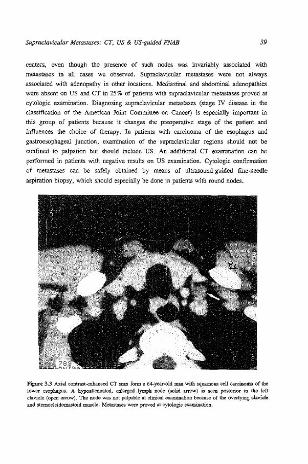
centers, even though the presence of such nodes was invariably associated with
metastases in all cases we observed. Supraclavicular metastases were not always
associated with adenopathy in other locations. Mediastinal and abdominal adenopathies
were absent on US and CT in 25% of patients with supraclavicular metastases proved at
cytologic examination. Diagnosing supraclavicular metastases (stage IV disease in the
classification of the American Joint Committee on Cancer) is especially important in
this group of patients because it changes the preoperative stage of the patient and
influences the choice of therapy. In patients with carcinoma of the esophagus and
gastroesophageal junction, examination of the supraclavicular regions should not be
confined to palpation but should include US. An additional CT examination can be
performed in patients with negative results on US examination. Cytologic confirmation
of metastases can be safely obtained by means of ultrasound-guided fine-needle
aspiration biopsy, which should especially be done in patients with round nodes .
• Figure 3.3 Axial contrast-enhanced CT scan form a 64-year-old man with squamous cell carcinoma of the lower esophagus. A hypoattenuated, enlarged lymph node (solid arrow) is seen posterior to the left clavicle (open arrow). The node was not palpable at clinical examination because of the overlying clavicle
and sternocleidomastoid muscle. Metastases were proved at cytologic examination.
40 Chllpter 3
3.6 References
1. Akiyama H, Tsurumaru M, Kawamura T, Ooo Y. Principles of surgical treatment for carcinoma of the esophagus. Ann Surg 1981; 194: 438-446.
2. Wu YK, Huang KC. Chinese experience in the surgical treatment of carcinoma of the esophagus. Ann Surg 1979; 190: 361-365.
3. Finley RJ, Inculet RI. The results of esophagogastrectomy without thoracotomy for adenocarcinoma of the esopbagogastric junction. Ann Surg 1989; 210: 535-543.
4. Paolini A, Tosato F, Cassese M et al. Total gastrectomy in the treatment of adenocarcinoma of the cardia. Am J Surg 1986; 151: 238-243.
5. Picus D, Balfe DM, Koehler RE. Roper CL, Owen JW. Computed tomography in the staging of esophageal carcinoma. Radiology 1983; 146: 433-438.
6. Thompson \VM, Halvorsen RA, Foster \VL, Williford ME. Postlethwait RW. Korobkin M. Computed tomography for staging esophageal and gastroesophageal cancer: re-evaluation. A.JR 1983; 141: 951-958.
7. Quint LE, Glazer GM, Orringer MB, Gross BH. Esophageal carcinoma: CT findings. Radiology 1985; 155: 171-175.
8. Halvorsen RA. Magruder-Habib K, Foster WL. Roberts L, Postlethwait RW, Thompson WM. Esophageal cancer staging by CT: long-term follow-up study. Radiology 1986: 161: 147-151.
9. Halvorsen RA, Thompson WM. CT of esophageal neoplasms. Radial Clin North Am 1989; 27: 667-685.
10. Inculet RI, Keller SM, Dwyer A, Roth JA. Evaluation of noninvasive tests for the preoperative staging of carcinoma of the esophagus: a prospective study. Ann Thorac Surg 1985; 40: 561-565.
ll. Lea JW, Prager RL, Bender HW. The questionable role of computed tomography in preoperative staging of esophageal cancer. Ann Thorac Surg 1984; 38: 479-481.
12. Becker CD, Barbier P, Porcellini B. CT evaluation of patients undergoing transhiatal esophagectomy for cancer. J Comput Assist Tomogr 1986; 10: 607-611.
13. Goldberg RP. Austin RM:. Computed tomography of axillary and supraclavicular adenopathy. Clin Radio! 1985; 36: 593-596.
14. Tohnosu N, Onoda S, Isono K. Ultrasonographic evaluation of cervical lymph node metastases in esophageal cancer with special reference to the relationship between the short to long axis ratio (SIL) and the cancer content. JCU 1989; 17: 101-106.
15. van Overhagen H, Lamt:ris JS, Zonderland HM. Tilanus HW. van Pel R. SchUtte HE. Ultrasound and ultrasound-guided fine-needle aspiration biopsy of supraclavicular lymph nodes in patients with esophageal carcinoma. Cancer 1991; 67: 585-587.
16. Baatenburg de Jong RJ, Rongen RJ, Lam6ris JS, HartboOrn M, Verwoerd CDA, Knegt P. Metastatic neck disease. Palpation vs ultrasound examination. Arch Otol Head Neck Surg 1989; 115: 689-690.
17. Johnson JT. A surgeon looks at cervical lymph nodes. Radiology 1990; 175: 607-610. 18. Beah.rs OH. Henson DE, Hutter RVP, Myers MH. Manual for staging of cancer. 3rd ed.
Philadelphia: JB Lippincott, 1988; 63-65, 69-71. 19. Sakata K. Ueber die lymphgefisse des oesophagus und iiber seine regioniren lymphdrii.sen m.it
beriicksichtigung der verbreitung des carcinoms. Mitt Grenz Geb Med Chir 1903; 11: 634-656. 20. Postlethwait RW. Surgery of the esophagus. 2nd ed. Norwalk. Va: Appleton-Century-Crofts,
1986; 404-405. 576-577. 21. Sannohe Y, Hiratsuka R, Doki K. Lymph node metastases in cancer of the thoracic esophagus.
Am J Surg 1981; 141: 216-218. 22. McCort JJ. Radiographic identification of lymph node metastases from carcinoma of the
esophagus. Radiology 1952; 59: 694-711.
41
Chapter 4
Improved Assessment of Supraclavicular and Abdominal Metastases in Oesophageal and Gastro-oesophageal Junction Carcinoma with the Combination of Ultrasound and Computed Tomography
(Assessment of Distant Metastases with US and CT)
4.1 Abstract
The purpose of the study was to evaluate ultrasound and computed tomography in the
assessment of distant metastases, supraclavicular and abdominal, in 113 patients with
carcinoma of the oesophagus and gastro-oesophageal junction. Ultrasound and computed
tomographic findings were compared with the cytological data in 29 patients and with
the surgical data in 84 patients. In assessing distant metastases, ultrasound and
computed tomography had a sensitivity of 61% and 70%, and a specificity of 93% and
85%, respectively (p = 1.0). When ultrasound and computed tomography were
combined the sensitivity increased to 83% and the specificity decreased to 81%. There
was no significant difference in the assessment of supraclavicular metastases (p = 0.8),
coeliac metastases (p = 1.0) or liver and other nonlymphatic abdontinal metastases (p
= 1. 0) on ultrasound or computed tomography. The results show that both ultrasound
and computed tomography should be used for assessment of distant metastases and
abnormalities confirmed by imaging guided biopsy.
4.2 Introduction
In patients with carcinoma of the oesophagus and gastro-oesophageal junction,
pretreatment staging is required to select the optimal therapeutic approach.
Assessment of distant metastases is of great importance because metastatic spread to
distant sites either obviates the need for surgery or limits it to palliative resection.
Published as: Hans van Overhagen, MD, Johan S. Lam6ris, MD, Marjolein Y. Berger, MD, Hugo W. Tilanus, MD, Ren6e van Pel, MD, Abraham l.J. Klooswijk, MD, Henri E. Schiitte. MD. IMPROVED ASSESSMENT OF SUPRACLAVICULAR AND ABDOMINAL METASTASES IN OESOPHAGEAL AND GASTRO-OESOPHAGEAL JUNCTION CARCINOMA WITH THE COMBINATION OF ULTRASOUND AND COMPUTED TOMOGRAPHY. Br J Radiol1993;66;203-208

42 Chapter 4
The longitudinal orientation of the lymphatic network of the oesophagus is responsible
for early metastases to the supraclavicular and coeliac lymph nodes in these tumours.
In assessing distant metastases both computed tomography (CT) and ultrasound (US)
have certain advantages. On CT scans the primary tumour, the mediastinum and the
lungs can be examined during the same session that is used for examining the
supraclavicular nodes and the abdomen [1-3]. US scans can be performed more quickly
than CT scans and a cytological diagnosis can be obtained almost immediately by means
of US-guided fme-needle aspiration biopsy (FNAB) from any masses detected [4-6].
The purpose of this study was to evaluate the assessment of distant metastases with US,
CT. and a combination of both techniques by comparing US and CT findings with
cytological and surgical data.
4.3 Patients and Methods
Between January 1989 and December 1990 113 patients, 88 men and 25 women, aged
42-76 years (mean 61 years) with oesophageal (92 patients) and gastro-oesophageal
junction (21 patients) carcinoma were studied prospectively. Histology of the primary
tumour was squamous cell carcinoma in 57, adenocarcinoma in 50 and undifferentiated
carcinoma in ~ix patients. Staging was performed according to the classification of the
American Joint Committee on Cancer (AJCC) [7] with one exception: whereas the
AJCC considers coeliac nodes as regional nodes (N2) in carcinoma of the gastro
oesophageal junction and as distant nodes (MI) in carcinoma of the oesophagus, in our
study they were considered distant in all cases.
Palpation, US and CT
Palpation of the supraclavicular regions of all patients was performed by physicians of
the surgical and gastroenterological departments. Patients underwent US of the
supraclavicular regions and the abdomen by means of a 7.5-MHz linear array transducer
and a 3.5-MHz curvilinear transducer, respectively (SSD-650; Aloka, Tokyo, Japan).
All patients underwent CT scanning of the supraclavicular regions from the cricoid
cartilage to the sternal angle with 5-or 6-mm-thick contiguous sections and contiguous
CT scanning of the chest and upper abdomen, in 8-12-mm-thick sections by means of a
Somatom Plus Scanner (Siemens, Erlangen, Germany) or a Tomoscan 350 (Philips
Medical Systems, Eindhoven, The Netherlands). Contrast material was routinely
administered intravenously by means of a drip infusion to better define the vascular
structures. The supraclavicular lymph nodes were bilaterally studied on US and CT
scans and were considered abnormal when they had a short axis of 5 mm or more. US

Assessment of Distant Metastases with US and CT 43
and CT ~cans of the abdomen included examination of the coeliac lymph nodes which
were considered abnormal when they had a short axis of 10 mm or more, the liver and
other nonlymphatic abdominal sites; the peritoneum, the kidneys and the adrenal glands.
Palpation of the supraclavicular regions and CT and US of the supraclavicular regions
and the abdomen were performed independently without knowledge of the other
examiners' findings and the results were documented. In patients with enlarged
supraclavicular nodes and nonlymphatic abdominal lesions present on CT but absent on
US, US studies were repeated and US-guided FNAB performed in those cases when the
lesions could now be detected.
US-guided FNAB
All but two patients with US and/or CT findings indicating supraclavicular, liver and
other nonlymphatic abdominal metastases underwent US-guided FNAB of one suspect
lesion. Biopsies were not performed in two patients with enlarged supraclavicular nodes
present on CT scans but absent on US scans because the nodes could not be found on
repeated US scans and in our experience it is not possible to perform CT -guided
biopsies of these small ( < I em) and often mobile nodes. FNABs were only
occasionally performed of enlarged coeliac nodes, because these nodes are usually
removed at surgery when the primary tumour is resectable. In patients with multiple
lesions biopsies were preferentially performed of supraclavicular nodes because these
are easier to perform and are less distressing and invasive for the patient compared with
abdominal biopsies. FNABs of supraclavicular lymph nodes were performed with a 23-
gauge needle in a "free-hand" method and of abdominal lesions with a 22-gauge needle
usually with an attachable needle guide. Patients with distant metastases proved by
means of cytologic studies were excluded from surgery. The remaining patients,
including those with negative cytological examinations, underwent surgery.
Surgery
At surgery laparotomy was performed as the first step of a planned resection; the liver
was bimanually palpated, the coeliac lymph nodes were removed for histological
examination and resectability of the tumour was determined. Patients with a surgically
resectable tumour underwent transhiatal oesophagectomy (THE). In these patients the
lower oesophagus was dissected transhiatally and the cervical oesophagus was dissected
through a right sided neck incision. Enlarged lymph nodes on the right side of the neck
were removed for histological examination. The intrathoracic oesophagus and the
gastro-oesophageal junction were resected, a stomach tube was brought up to the neck
and an anastomosis was made with the cervical oesophagus. Patients with an
unresectable tumour underwent laparotomy only.

44 Chapter 4
Data Analysis
Sensitivity and specificity of US and CT were estimated and are reported with 95%
exact confidence intervals, by using the cytological data as standard of reference in the
patients with cytologically proven metastases, and by using the data of the surgically
assessed sites as the standard of reference in the patients who underwent surgery. The
sensitivity was the chance of demonstrating positive findings on US or CT in a patient
with metastases, whereas the specificity was the chance of demonstrating negative
findings when no metastases were present. For the combination of US and CT the
sensitivity was the chance of demonstrating positive findings on either US or CT in a
patient with metastases whereas the specificity was the chance of demonstrating negative
findings on both studies when no metastases were present. Statistical analysis of false
positive and false negative findings on US and CT was performed in a patient-by-patient
comparison by means of McNemar's test for paired data.
4.4 Results
Thirty-nine of 113 patients underwent US-guided FNAB. Distant metastases were
proven by means of cytologic studies in 29 of these patients who were subsequently
excluded from ~urgery. The sites of the cytologically proven metastases ill these 29
patients are shown in Table 4.1.
Table 4.1 Number of patieots with cytologically or surgically assessed sites
No surgery Surgery Total
Cytologically proven Metastases No metastases Total Distant sites metastases
Supraclavicular nodes 19 0 70 70' 89 Coeliac nodes 2 25 59 84 86 Liver and other non-lymphatic
abdominal sites 8' 2 82 84 92
Total 29 25" 59 84' 113
• Supraclavicular nodes: surgically assessed in 70 patients at THE. Not assessed in 14 patients who undet\Vent laparotomy alone.
b Liver metastases in 5 patients, peritoneal carcinomatosis in 2, renal metastases in 1. c 2/25 patients had both liver metastases and coeliac lymph node metastases at surgery. d 10/84 patients had negative cytology studies prior to surgery (supraclavicular lymph nodes, 8; adrenal
glands, 2).

Assessment of Distant Metastases with US and CT 45
Metastases were not found in the cytological smears of 10 other patients who underwent
US-guided FNAB; of the supraclavicular nodes in eight patients and of the adrenal
glands in two patients. These patients subsequently underwent surgery at which
metastases were not found at the cytologically tested sites.
Eighty-four patients underwent surgery. Seventy patients underwent THE. The
remaining 14 patients underwent explorative laparotomy only, due to advanced local
spread of the primary tumour (10 patients), the presence of multiple abdominal
metastases (two patients) or other reasons (two patients).
At surgery distant metastases were found and proved by histopathological examination
at 27 sites in 25 of 84 patients (Table 4.1). Twenty-five patients had coeliac lymph node
metastases and two of these patients also had liver metastases. Supraclavicular
metastases were not found on the right side in the 70 patients who underwent THE. In
the remaining 14 patients surgery was abandoned after laparotomy and the
supraclavicular nodes were not surgically assessed. Supraclavicular metastases
Nineteen patients had supraclavicular metastases. In four of 19 (21%) patients with
metastatic nodes, enlarged nodes were palpable. Metastatic nodes were detected on US
scans in 15 of 19 (79%) patients (Fig. 4.1) and on CT scans in 16 of 19 (84%) patients
(Fig. 4.2) (Table 4.2).
Table 4.2 Correlation of US and CT f'mdings with cytological and surgical data in assessing supraclavicular metastases
Results of US and CT studies Supraclavicular lymph nodes: numbers of patients (n = 89)
us CT
True positive 15 !6 False negative 4 3 False positive 6 5 True negative 64 65 Sensitivity" (%) 79' (54-94) 84 (60-97) Specificity (%) 91 (82-97) 93 (84-98)
US and CT findings were correlated with the cytological data in 19 patients with supraclavicular metastases cytologically proven and were correlated with the surgical data in 70 patients who underwent THE, 8 of whom had negative cytology. ~ Sensitivity = true positive/(true positive + false negative); specificity = true negative/(true negative + false positive). b 95% confidence interval in parentheses.

46 Chapter 4
There was no significant difference between US and CT in assessing supraclavicular
metastases (p = 0.8), but both techniques were superior to palpation in detecting
metastatic nodes. A false negative diagnosis was made in four patients on US scans; in
these patients nodes were absent on initial US scans but present on CT scans, however,
enlarged nodes were found on repeated US scans with the aid of the positive CT
findings and metastases were subsequently proven by US-guided FNAB and cytological
examination. In six patients metastases were predicted on US studies, but proved
incorrect by cytological study and at THE. A false negative diagnosis was made on CT
scans in three patients with metastatic nodes, absent on CT scans but present on US
scans. In five patients supraclavicular metastases were predicted on CT scans, but
proved incorrect at THE; three of these patients also had previously negative cytological
studies.
Figure 4.1 Axial sonogram of the right supraclavicular fossa in a patient with squamous cell carcinoma of the lower oesophagus. A round hypoechoic lymph node (solid arrow) is seen posterior to the sterno-cleidomastoid muscle and the carotid artery (open arrow). US-guided FNAB revealed metastases.
Figure 4.2 Axial contrast material-enhanced CT scan in a patient with squamous cell carcinoma of the middle thoracic oesophagus. A hypoattenuated, enlarged supraclavicular node (arrow) is localized posterior to the thyroid gland and lateral to the left carotid artery. Metastases were proven at cytologic examination.

Assessmem of Distant Metastases with US and CT 47
Abdominal metastases
Table 4.3 summarizes the results of US and CT studies in assessing distant abdominal
metastases.
Coeliac lymph node metastases
There was no significant difference in assessing coeliac metastases on US or CT studies
(p = 1.0). Metastases were correctly predicted on US scans in eight of 27 (30%)
patients (Fig. 4.3) and on CT scans in 13 of 27 (48%) patients (Fig. 4.4). Nineteen
patients had metastatic nodes at surgery that were not detected on US scans. Metastatic
nodes were not detected on CT scans in 14 patients but were proved cytologically in
one patient and at surgery in 13 patients. Metastases were incorrectly predicted on CT
in four patients without metastases at surgery.
US scans.
Figure 4.3 Saggital sonogram of the upper abdomen in a patient with squamous cell carcinoma of the middle third of the oesophagus shows an enlarged, bypoechoic. metastatic lymph node (arrow) posterior to the left hepatic lobe.
There were no false positive findings on
Figure 4.4 Axial contrast material-enhanced CT scan in a patient with squamous cell carcinoma of the lower oesophagus shows an enlarged metastatic coeliac lymph node between the aorta and the stomach.
48 Chapter 4
Liver and other nonlymphatic metastases
There was no significant difference between the assessment of these metastases by US
or CT (p = 1.0). Metastases were correctly predicted on US and CT scans in eight of
10 patients (80%) (liver metastases in five, peritoneal carcinomatosis in two and renal
metastasis in one) (Figs. 4.5 and 4.6). Liver metastases were not detected on either US
or CT scans in the two patients with small ( < 1.0 em) metastases at surgery. Adrenal
metastases were incorrectly predicted on CT scans in two other patients and on US
scans in one of these patients, but proved incorrect by cytologic study and subsequently
at surgery. Distant metastases
Table 4.4 summarizes the results of US and CT studies in assessing the presence or
absence of distant metastases in each of 113 patients with cytological (29 patients) or
surgical (84 patients) correlation. Fifty-four patients had distant metastases, either
supraclavicular or abdominal. There was no significant difference between US or CT
scans in assessing distant metastases (p = 1.0). Distant metastases were correctly
predicted on US scans in 33 of 54 (61 %) patients and on CT scans in 38 of 54 (70%)
patients. The combination of US and CT correctly predicted distant metastases in 45 of
54 (83%) patients. A false positive diagnosis was made in four patients on US scans, in
nine patients on CT scans and in 11 patients when both techniques were combined,
respectively.
Table 4.3 Correlation of US and CT findings with cytological and surgical data in assessing distant abdominal metastases
Coeliac nodes\ n = 86 Liver and other non-lymphatic sitesb, n = 92 Results of us and CT studies us CT us CT
True positive 8 13 8 8 False negative 19 14 2 2 False positive 0 4 2 True negative 59 55 81 80 Sensitivity(%) 300 (14- 50) 48 (29-68) 80 (44- 98) 80 (44- 98) Specificity (%) 100 (94-100) 93 (84-98) 99 (93-100) 98 (92-100)
a Correlated in 86 patients; by cytology in 2, at surgery in 84. b Correlated in 92 patients; by cytology in 8, at surgery in 84, including 2 with negative cytology of the
adrenal glands prior to surgery. " 95% confidence inteiYal in parentheses.
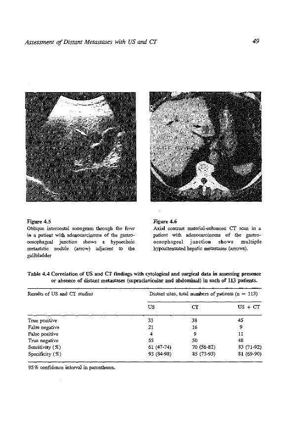
Assessment of Distant Metastases with US and CT 49
Figure 4.5 Oblique intercostal sonogram through the liver in a patient with adenocarcinoma of the gastrooesophageal junction shows a hypoechoic metastatic nodule (arrow) adjacent to the gallbladder
F;gure 4.6 Axial contrast material-enhanced CT scan in a patient with adenocarcinoma of the gastrooesophageal junction shows multiple hypoattenuated hepatic metastases (arrows).
Table 4.4 Correlation of US and CT f'mdings "With cytological and surgical data in assessing presence or absence of distant metastases (supraclavicular and abdominal} in each of 113 patients.
Results of US and cr studies
True positive False negative False positive True negative Sensitivity(%) Specificity (%)
95% confidence interval in parentheses.
Distant sites. total numbers of patients (n = 113)
us CT US+ CT
33 38 45 21 16 9 4 9 11
55 50 48 61 (47-74) 70 (56-82) 83 (71-92) 93 (84-98) 85 (73-93) 81 (69-90)

50 Chapter 4
4S Discussion Carcinoma of the oesophagus and gastro-oesophageal junction has a poor prognosis
because many patients have advanced widespread disease at the time of clinical
presentation and are not curable by surgical resection [8-11]. This is reflected by the
fact that 54 of 113 ( 48%) patients who were staged had distant metastases proven by
either cytological or histopathological examination. In our institution THE is the standard procedure for resection of oesophageal and
gastro-oesophageal junction carcinoma because the postoperative morbidity and
mortality are less than with transpleural oesophagectomy including thoracotomy [12].
However, the supraclavicular lymph nodes are not systematically dissected in patients
who undergo THE and enlarged neck nodes are removed and examined only
unilaterally.
This limited verification of the state of the supraclavicular nodes in the present study
means that the false negative findings and the speclficity of the US and CT studies
should be interpreted with caution. In order to achieve the best available nonsurgical
estimate of the numbers of false negative findings on CT and US studies, these studies
were reevaluated in cases of conflicting results. Furthermore, biopsies were performed
in cases when nodes could be detected on repeated US studies after they had initially
been detected on CT studies alone. Optimal verification of the state of the
supraclavicular nodes can only be obtained when these nodes are bilaterally dissected at
surgery. However, it was not considered ethical to extend the surgical procedure by performing these dissections.
The significance of examining the supraclavicular lymph nodes radiologically in patients
with carcinoma of the oesophagus and gastro-oesophageal junction has been reported
previously and is confirmed in the present study [6]. Both US and CT were clearly
superior to palpation in the assessment of supraclavicular metastases. The relatively high
numbers of false positive diagnoses on US and CT studies of the supraclavicular lymph
nodes were due to the fact that these lymph nodes were considered abnormal when they
had a short axis of 5 mm or more. When supraclavicular nodes were considered
abnormal only when they had a short axis of 10 mm or more the number of false
positive diagnoses decreased from six to one on US studies and from five to one on CT
studies. However, the numbers of false negative findings increased from four to eight
on US studies and from three to six on CT studies with consequent decreases in
sensitivity to 58% and 68%, respectively (results not shown). Therefore, we prefer to
perform FNABs of supraclavicular nodes with a short axis of 5 mm or more to confirm
or exclude the presence of metastases by cytology.

Assessment of Distant Metastases with US and CT 51
US and to a lesser degree CT were limited in the assessment of coeliac lymph node
metastases. Only a minority of metastatic nodes were detected on US scans and normal
sized nodes were invariably not seen. The relatively low sensitivity of CT in detecting
coeliac metastases has been reported by others [2,3] and is explained by the fact that
normal sized lymph nodes may harbor metastases. The number of false positive findings
on CT scans confirms the reports of other authors that lymph nodes may enlarge
because of inflammatory processes [3].
Our results in assessing liver metastases with US and CT are comparable to the CT
results reported by Halvorsen and Thompson [3]. ln the present study neither US nor
CT was superior in detecting metastatic nodules. However, at CT contrast was
administered with a drip infusion, arid better results of CT scanning of the liver may be
obtained by dynamic bolus injection of contrast and rapid table incrementation during
injection, but this technique is rather demanding to use for screening purposes [13].
Although peritoneal carcinomatosis was correctly predicted on US and CT studies in
two patients with ascites, it can be very difficult to diagnose in the absence of ascites.
However, because of the low prevalence of peritoneal carcinomatosis in the present
study, the use of intraperitoneal contrast for better CT detection of peritoneal metastases
is not advocated in patients with oesophageal and gastro-oesophageal junction carcinoma
[ 14]. ln assessing distant metastases US and to a lesser degree CT were characterized
by a relatively low sensitivity and relatively higb specificity. Although CT was slightly
more sensitive than US in detecting distant metastases the difference was not statistically
significant. This was not altered by including pulmonary findings on CT, as although
one patient had pulmonary metastases, coeliac lymph node metastases were correctly
predicted by both scanning techniques in this patient (results not shown).
With the combination of US and CT the sensitivity increased to an acceptable level.
Although this was accompanied by a decrease in specificity this is considered to be less
important because FNABs can be performed to enable cytological examination. ln our
study US guidance was used for biopsies. However, CT guidance is a well established
alternative, especially for biopsies of abdominal lesions and can be performed ln
patients with abdominal lesions only detected on CT scans [15].
ln conclusion, the combination of US and CT should be used for the assessment of
distant metastases in carcinoma of the oesophagus and gastro-oesophageal junction. For
logistic and cost-effective reasons we recommend US as the initial examination and the
use of US-guided FNAB to confirm positive findings. Patients without lesions on US
scans should undergo CT. When lesions are detected on CT scans either US-guided or
CT -guided biopsies can be performed.
52 Chapter 4
4.6 References
1. Terrier F. Schapira C, Fuchs WA. CT assessment of operability in carcinoma of the
oesophagogastricjunction. Eur J Radioll984: 4: 114-117.
2. Becker CD, Barbier P, Porcellini B. CT evaluation of patients undergoing transhiatal
esophagectomy for cancer. J Comput Assist Tomogr 1986; 10: 607-611.
3. Halvorsen RA, Thompson \VM. CT of esophageal neoplasms. Radial Clin North Am 1989; 27:
667-685.
4. Reading CC, Charboneau JW. Meredith James E, Hurt MR. Sonographically guided
percutaneous biopsy of small (3 em or less) masses. AJR 1988; 151: 189-192.
5. van Overbagen H. Lam.:ris JS. Zonderland HM:, Tilanus H\V, van Pel R. SchUtte HE.
Ultrasound and ultrasound-guided fine-needle aspiration biopsy of supraclavicular lymph nodes in patients with esophageal carcinoma. Cancer 1991; 67: 585-587.
6. van Overbagen H. Lam&is JS, Berger MY, et al. Supraclavicular lymph node metastases in carcinoma of the esophagus and gastroesophageal junction: Assessment with CT. US, and USguided fine-needle aspiration biopsy. Radiology 1991; 179: 155-158.
7. Beahrs OH, Henson DE, Hutter RVP, Myers MH. Manual for staging of cancer. 3rd ed. Philadelphia: Lippincott, 1988: 63-65, 69-71.
8. Earlam R, Cunha-Melo JR. Oesophageal squamous cell carcinoma: A critical review of surgery. Br J Surg 1980; 67: 381-390.
9. Eeftinck Schattenkerk M, Obertop H, Mud HJ. Eijkenboom WMH, van Andel JG, van Houten
H. Survival after resection for carcinoma of the oesophagus. Br J Surg 1987; 74: 165-168. 10. Finley RJ, Inculet RI. The results of esophagogastrectomy without thoracotomy for
adenocarcinoma of the esophagogastric junction. Ann Surg 1989; 210: 535-543. 11. Mii.ller J1...1, Erasmi H, Stelzner M, Zieren H. Pichlmaier H. Surgical therapy of oesophageal
carcinoma. Br J Surg 1990; 77: 845-857.
12. Orri.nger MB. Transhiatal esophagectomy without thoracotomy for carcinoma of the thoracic esophagus. Ann Surg 1984; 200; 282- 288.
13. Ferrucci IT. Liver tumor imaging: Current concepts. AJR 1990; 155: 473-484. 14. Halvorsen RA. Panushka C, Oakley GJ, Letourneau JG, Adcock LL. Intraperitoneal contrast
material improves the CT detection of peritoneal metastases. AJR 1991; 157: 37-40. 15. Charboneau JW, Reading CC, Welch TJ. CT and sonographically guided needle biopsy: Current
techniques and new innovations. AJR 1990; 154: 1-10.

53
Chapter 5
Assessment of Distant Metastases with Ultrasound-guided Fine-Needle Aspiration Biopsy and Cytologic Study in Carcinoma of the Esophagus and Gastroesophageal Junction
(Assessment of Metastases with US-guided FNAB)
5.1 Abstract
The use of ultrasound (US)·guided fine-needle aspiration biopsy (FNAB) for the
assessment of distant metastases was prospectively studied in 135 consecutive patients
with carcinoma of the esophagus and gastroesophageal junction. Patients with accessible
lesions on US and computed tomographic studies of the supraclavicular regions and the
abdomen underwent US-guided FNAB. In patients with multiple lesions biopsies were
preferentially performed on enlarged supraclavicular nodes. Forty-nine patients
underwent US-guided FN ABs of 53 lesions. A cytologic diagnosis was established in 46
of 53 (87%) biopsies. Seven of 53 (13%) biopsies were nondiagnostic. Distant
metastases were diagnosed by means of cytologic study in 33 of 135 (24%) patients.
Supraclavicular metastases were diagnosed in 22 patients and abdominal metastases
were diagnosed in 12 patients, including one patient who also had supraclavicular metastases. US-guided FNAB can improve the selection of patients for surgical and
nonsurgical treatment by diagnosing distant metastases in an important number of
patients.
5.2 Introduction
Carcinoma of the esophagus and gastroesophageal junction has a poor prognosis [1-5].
Surgical treatment is associated with a high postoperative morbidity and mortality, and the long-term survival of surgically treated patients is negatively affected by an
advanced stage of disease [2-4].
Published as: Hans van Overhagen, MD, Johan S. I.am6ris, MD, Matjolein Y. Berger, MD, Ren6e van Pel, MD. Hugo W. TilanU5, MD, Abraham I.J. Klooswijk, MD, Henri E. SchUtte, MD. ASSESSMENT OF DISTANT METASTASES WITH ULTRASOUND-GUIDED FINE-NEEDLE ASPIRATION BIOPSY AND CYTOLOGIC STUDY IN CARCINOMA OF THE ESOPHAGUS AND GASTROESOPHAGEAL JUNCTION. Gastromtest Radiol1992; 17:305-310

54 Chapter 5
Preoperative assessment of distant metastases is of great importance, because in cases of
metastatic spread to distant sites surgery can be refrained from, or can be limited to, a
palliative resection. Search for metastases is usually performed with ultrasound (US),
computed tomography (CT), and endosonography. Recent reports [6-8], however, show
that none of these techniques is reliable enough to correctly predict the presence of
metastases and, thus, in case of a suspect lesion, cytologic or histologic proof of
metastases is mandatory to avoid overstaging. Diagnosing metastases by means of
cytologic study will prevent unnecessary surgical procedures in some patients, and will improve selection of patients that can benefit from surgery. The purpose of this study
was to evaluate the use of ultrasound (US)-guided fine-needle aspiration biopsy (FNAB)
for the assessment of distant metastases in carcinoma of the esophagus and gastro
esophageal junction. The utility of US-guided FNAB was determined by the number of
biopsies in which adequate material for cytologic examination could be obtained and by
the number of patients in which distant metastases could be proved by means of
cytologic study.
5.3 Materials and methods
Between January 1989 and December 1990, 135 consecutive patients with a biopsy·
proven carcinoma of the intrathoracic esophagus and/or gastroesophageal junction were
referred to the Rotterdam Esophageal Tumor Study Group for treatment. Sixty-six
patients had squamous cell carcinoma, 63 patients had adenocarcinoma and six patients
had undifferentiated carcinoma. Staging was performed according to the classification of
the American Joint Committee on Cancer [9]. Forty-nine patients underwent US-guided FNABs of accessible lesions on US and/or CT
studies to confirm or exclude the presence of distant metastases (stage IV disease) by
means of cytologic study. In patients with multiple lesions, biopsies were preferentially
performed of supraclavicular nodes. Forty-five patients underwent biopsies of one lesion
and four patients underwent biopsies of two lesions. An average of two needle passes
was made in each biopsy session. All biopsies were performed by using real-time
sonographic guidance (Aloka SSD-630 and Aloka SSD-650, Tokyo, Japan). US-guided
FNAB of supraclavicular lymph nodes with a short axis that ranged from 0.5 - 2.0 em
was performed in 34 patients. US-guided FNAB of hepatic lesions with a diameter that
ranged from LO - 6.0 em was performed in seven patients and of celiac lymph nodes
with a short axis that ranged from LO - 4.0 em in seven patients. US-guided FNAB of
a solid renal mass was performed in one patient, of enlarged adrenal glands in two
patients and of ascites in two patients. Supraclavicular biopsies were performed in a

Assessment of Metastases with US-guided FNAB 55
"free-hand" manner using a 7.5-MHz linear array transducer and a 23-gauge needle
(Sherwood Medical, Ballymoney, Northern Ireland). Aspiration of ascites was done in a
"free-hand" manner using a 5.0-MHz linear array transducer. All other abdominal
biopsies were performed using a 3.5-tvrnz sector scanner with an attachable needle
guide and a 22-gauge needle (Cook, Spencer, Indiana, USA). In all cases the needle
was connected with a 24-cm hollow flexible cannula (Polystan, Almere, The
Netherlands) which was connected with an empty 12-ml syringe held by an assistant who applied suction after the lesion had been punctured (Fig. 5.1).
Figure 5.1 US-guided FNAB of a supraclavicular lymph node with a
23-gauge needle and an interconnecting cannula.
Smears obtained from the aspirated material were air dried for 10 minutes, stained with
May-Griinwald-Giemsa staining methods, and examined by an experienced cytopatholo
gist. Biopsies were considered diagnostic when the specimens contained either
metastatic cells or benign cells of the biopsied tissue in an adequate amount to be
classified as negative for malignancy. Biopsies were considered nondiagnostic in cases
of bloody, necrotic, or hypocellular specimens.
Patients with distant metastases proved by means of cytologic study were excluded from
surgery. The remaining patients, with the exception of II patients who had a poor
general condition, underwent surgery. Patients with a resectable tumor at surgery
underwent transhiatal esophagectomy (THE) with gastric interposition and cervical
gastroesophageal anastomosis [10]. In patients with an irresectable tumor surgery was
abandoned after laparotomy.

56 Chapter 5
5.4 Results Table 5.1 summarizes the results of US-guided FNAB and cytologic studies in the
assessment of distant metastases.
Supraclavicular metastases
Thirty-four patients underwent US-guided FNAB of supraclavicular lymph nodes with a
short axis of 5 mm or more. A cytologic diaguosis was established in 30 of 34 (88%)
biopsies. Four of 34 (12%) biopsies were nondiaguostic.
Supraclavicular metastases were diaguosed in 22 of 135 (16%) patients; 15 of 66 (23%)
patients with squamous cell carcinoma, six of 63 (10%) patients with adenocarcinoma
and one of six (17%) patients with undifferentiated carcinoma. In only five of 22 (23%)
patients with supraclavicular metastases, enlarged nodes were also palpable.
Metastatic nodes were not found at THE in six of eight patients with benign cells at
cytologic examination and in three of four patients with nondiagnostic biopsies. In the
other three patients, the supraclavicular regions were not surgically assessed because the
tumor was considered irresectable at surgery.
Table 5.1 Results of US-guided FNAB and cytologic studies in assessing distant metastases
US-guided Cytologic diagnosis FNAB
Metastases Benign cells Nondiagnostic
Supraclavicular nodes, total 34 22 8 4 Squamous cell carcinoma 19 IS""" 3 I Adenocarcinoma 13 6 5 2 Undifferentiated carcinoma 2 I 0 I
Abdominal lesions, total 19 12 4 3 Squamous cell carcinoma 9 4. 3 ' 20
Adenocarcinoma 10 8' 1 '
Distant metastases, total 53 34 12 7
Squamous cell carcinoma 28 19 6 3
Adenocarcinoma 23 14 6 3 Undifferentiated carcinoma 2 0
Numbers are numbers of biopsies. Forty-nine patients undenvent 53 biopsies; 45 patients underwent one biopsy and four patients underwent two biopsies. • Supraclavicular and liver metastases cytologically proven in one patient. b Supraclavicular metastases cytologically proven in one patient with benign results on cytologic studies of
the celiac nodes. c Supraclavicular metastases cytologically proven in one patient with a nondiagnostic biopsy of the celiac
nodes. 4 Peritoneal carcinomatosis cytologically proven in one patient with a nond.iagnostic biopsy of the liver.

Assessment of Metastases with US-guided FNAB 57
Abdominal metastases
US-guided FNAB of 19 abdominal lesions was performed in 18 patients. A cytologic
diagnosis was established in 16 of 19 (84%) biopsies. Three of 19 (16%) biopsies were nondiagnostic.
Abdominal metastases were diagnosed in 12 of 135 (9%) patients; four of 66 (6%)
patients witb squamous cell carcinoma and eight of 63 (13%) patients witb
adenocarcinoma. The sites where abdominal metastases were cytologically proven are
shown in Table 5.2. Three of four patients with benign results on cytologic studies of
abdominal lesions underwent surgery. Benign results on cytologic studies of tbe adrenal
glands were confirmed at surgery in two of tbese patients.
Table 5.2 Results of US-guided FNAB and cytologic studies of abdominal metastases
US-guided Cytologic diagnosis
FNAB Metastases Benign cells Nondiagnostic
Abdominal lesions, total 19 12 4
Liver 7 6 0 Squamous cell carcinoma I I • 0 Adenocarcinoma 6 5 0
Celiac nodes 7 3 2 Squamous cell carcinoma 6 2 2 >
Adenocarcinoma 0
Adrenal glands 2 0 2 Squamous cell carcinoma I 0 I Adenocarcinoma I 0 I
Peritoneum (ascites) Adenocarcinoma 2 2' 0
Kidney Squamous cell carcinoma 0
Numbers are numbers of biopsies. Eighteen patients underwent 19 biopsies of abdominal lesions. • Supraclavicular metastases and liver metastases cytologically proven in one patient.
3
I 0 I'
2 2" 0
0 0 0
0
0
b One of two patients with benign results on cytologic studies of the celiac nodes also had cytologically proven supraclavicular metastases.
~ One of two patients with nondiagnostic biopsies of the celiac nodes also had cytologically proven
supraclavicular metastases. d Peritoneal carcinomatosis was cytologically proven in one patient with a nondiagnostic biopsy of the
liver.

58 Chapter 5
In the third patient cytologic studies of the celiac nodes showed benign cells only, however, celiac metastases were nevertheless found at surgery. Surgery was not performed in another patient with a benign cytologic result of a celiac biopsy because the patient also had cytologically proven supraclavicular metastases. One of three patients with nondiagnostic biopsies of abdominal lesions underwent surgery; metastases were not found at surgery in this patient with a nondiagnostic biopsy of the celiac nodes. Two other patients with nondiagnostic biopsies of abdominal lesions were excluded from surgery because of cytologically proven metastases at other sites; one patient with a nondiagnostic biopsy of the celiac nodes also had supraclavicular metastases and one patient with a nondiagnostic biopsy of the liver also had peritoneal carcinomatosis.
Distant metastases In total, US-guided FNAB of 53 lesions was performed in 49 patients. A cytologic diagnosis was established in 46 of 53 (87%) biopsies. Seven of 53 (13%) biopsies were nondiagnostic. Distant metastases were diagnosed by means of cytologic study in 33 of 135 (24%) patients; 18 of 66 (27%) patients with squamous cell carcinoma, 14 of 63 (22%) patients with adenocarcinoma, and one of six (17%) patients with undifferentiated carcinoma. In one patient with squamous cell carcinoma, both supraclavicular and hepatic metastases were cytologically proven. No complications, other than localized mild discomfort, were observed in patients undergoing US-guided FNAB.
Figure 5.2 Needle aspirate of a metastatic lymph node in a patient with adenocarcinoma of the gastroesophageal junction shows atypical epithelial cells with finely vacuolated cytoplasm in acinar arrangement (x400, May-Griinwald-Giemsa stain).

Assessment of Metastases with US-guided FNAB 59
5.5 DiscllSsion In most institutions the presence of cytologically proven distant metastases will importantly affect the therapeutic approach in carcinoma of the esophagus and gastroesophageal junction. The number of diagnostic biopsies in the present series shows the value of US-guided FNAB in obtaining material for cytologic examination (Figs. 5.2 and 5.3). CT guidance is a well-established alternative for US guidance, but it is more time consuming and expensive [11,12]. Furthermore we believe that real time visualization of lesions provided by US guidance is an advantage in cases of mobile and superficial lesions [II]. We agree with Reading et al. that clear visualization of the biospy needle is the key element in a successful biopsy [13]. With small target lesions, such as supraclavicular lymph nodes, there is a risk of displacing the needle tip out of the lesion during suction required to obtain cells. Therefore, in cases of FNABs with small-caliber needles for cytologic studies we use an interconnecting cannula which allows the operator to hold both the US probe and the needle during the whole procedure. Thus, subtle adjustments can be made while puncturing the lesion and the tip of the needle can be kept inside the lesion while it is moved up and down under direct US visualization during the appliance of suction. US-guided FNABs of enlarged supraclavicular lymph nodes were preferentially performed. Supraclavicular biopsies cause less patient discomfort compared with abdominal biopsies, do not require the use of local anesthetics and are not hindered by respiratory movements or overlying bowel shadow.
Fi.gure 5.3 Needle aspirate of a metastatic lymph node in a patient with squamous cell carcinoma of the esophagus shows a cluster of, and some dispersed, atypical epithelial cells with hyperchromatic nuclei and keratinized cytoplasm (x400, May-Griinwald-Giemsa stain).
60 Chapter 5
The superficial location of the nodes contributes to the ease of the procedure [8].
Rizzatto et al. reported successful US-guided FNABs of superficial lesions in 97% of
cases [14]. In our series a cytologic diagnosis was established in 88% of supraclavicular
biopsies. The greater number of nondiagnostic biopsies in our series is probably due to
the small size of the nodes in these cases (5-10 mm).
A cytologic diagnosis was established in 84% of abdominal biopsies. US-guided FNAB of celiac lymph .nodes was occasionally hindered by respiratory movements and
overlying bowel shadow, which is reflected in the lower percentage of celiac biopsies
(72%) that provided a cytologic diagnosis, compared to supraclavicular biopsies (88%),
even though the supraclavicular nodes were usually smaller in size.
Distant metastases were diagnosed by means of cytologic study in 24% of patients who
were subsequently excluded from surgery. By excluding these patients from surgery we
hope to improve the selection of patients who will benefit from surgery and thus
improve the long-term survival in this group of surgically treated patients.
Supraclavicular metastases were diagnosed more often in patients with squamous cell
carcinoma than in patients with adenocarcinoma. The prevalence of supraclavicular
metastases in both groups of patients, however, underlines the importance of examining
these nodes thoroughly. Abdominal metastases were diagnosed more often in patients
with adenocarcinoma, which is probably due to the more caudal location of adeno
carcinomas compared to squamous cell carcinomas. In patients with benign "results on cytologic studies, metastatic involvement of the cytologically assessed sites was
nevertheless proven at surgery in one patient with a celiac biopsy. Probably cells were
obtained from the nonmalignant part of the node. This stresses the importance of
repeating biopsies in cases of unexpected negative cytologic findings. In the remaining
patients with either benign results on cytologic studies or nondiagnostic biopsies,
metastatic involvement of the cytologically assessed sites was not found at surgery.
However, because surgery was not performed in all of these patients and because the
supraclavicular nodes are not systematically dissected, but are only removed unilaterally
when enlarged at THE, these results should be interpreted with caution.
In conclusion, distant metastases were proven by means of US-guided FNAB and
cytologic study in an important number of patients. It is our opinion that cytologic proof
of distant metastases will help to select patients for surgical and nonsurgical treatment.
Assessment of Metastases with US-guided FNAB 61
5.6 References
1. Earlam R, Cun.ha-Melo JR. Oesophageal squamous cell carcinoma: I. A critical review of surgery. Br 1 Surg 1980; 67: 381-390.
2. Eeftinck Schattenkerk M, Obertop H, Mud ID, Eijkenboom w:M:H. van Andel JG, van Houten H. Survival after resection for carcinoma of the oesophagus. Br J Surg 1987; 74: 165-168.
3. Finley RJ, Inculet RI. The results of esophagogastrectomy without thoracotomy for adenocarcinoma of the esophagogastric junction. Ann Surg 1989; 210: 535-543.
4. Ellis FH, Gibb SP, Watkins E. Limited esopbagogastrectomy for carcinoma of the cardia. Ann
Surg !988; 208; 354-360.
5. MUller JM, Erasmi H. Stelzner M, Zieren U, Picblmaier H. Surgical therapy of oesophageal
carcinoma. Br J Surg 1990: 71: 845-857.
6. Halvorsen RA, Thompson WM:. CT of esophageal neoplasms. Radial Clin North Am 1989; 27:
667-685.
7. Tio TL. Coene PP, Schouwink MH, Tytgat GNJ. Esophagogastric carcinoma: pre-operative
TNM classification with endosonography. Radiology 1989; 173: 411-417.
8. van Overhagen H. Lameris JS. Berger MY, et al. Supraclavicular lymph node metastases in carcinoma of the esophagus and gastroesophageal junction: assessment with CT, US and US
guided fine-needle aspiration biopsy. Radiology 1991; 179: 155-158.
9. Beahrs OH. Henson DE, Hutter RVP. Myers MH. Manual for staging of cancer. 3rd ed.
Philadelphia: Lippincott 1988; 63-65, 69-71.
10. Orringer MB. Transhiatal esophagectomy without thoracotomy for carcinoma of the thoracic
esophagus. Ann Surg 1984; 200: 282-288.
11. Charboneau JW. Reading CC, Welch TJ. CT and sonographically guided needle biopsy: current
techniques and new innovations. AJR 1990; 154: 1-10.
12. Pelaez JC, Hill MC, Dach JL, Isikoff MB. Morse B. Abdominal aspiration biopsies.
Sonographic v computed tomographic guidance. JAMA 1983; 250: 2663-2666.
13. Reading CC, Charboneau JW, James EM, Hurt MR. Sonographically guided percutaneous
biopsy of small (3 em or less) masses. AJR 1988; 151: 189-192.
14. Rizzatto G. Solbiati L. Croce F. Derchi LE. Aspiration biopsy of superficial lesions: ultrasonic
guidance with a linear-array probe. AJR 1987; 148: 623-625.

62 Chapter 5

63
Chapter 6
CT Assessment of Resectability in Patients Undergoing Transhiatal Esophagectomy for Esophageal and Gastroesophageal Junction Carcinoma
(CT Assessment of Resectability)
6.1 Abstract
The ability of preoperative computed tomography (CT) to assess resectability and to
stage carcinoma of the esophagus and gastroesophageal junction was studied in 71
patients who underwent transhiatal esophagectomy. Patients with preoperatively proven
distant metastases who did not have surgery were not included in the present study. At
surgery the tumor invaded adjacent mediastinal or abdominal structures in 18 patients
(prevalence 25%), but was nonresectable in only seven of these 18 (39%) patients.
Invasion of the tracheobronchial tree, the aorta and the diaphragm were correctly
detected on CT in 5 of 6, 1 of 2 and 2 of 10 patients. There were four false positive
results on CT; tracheobronchial invasion and pericardia! invasion were incorrectly
predicted in one and three patients, respectively. Invasion of adjacent structures was
correctly assessed on CT in 58 (82%) patients and the depth of tumor invasion was
correctly determined in 49 (69%) patients. CT correctly staged 57% of patients
according to the classification of the American Joint Committee on Cancer.
Understaging (31 %) occurred more often than overstaging (11 %). 1n the present study,
CT was not effective in assessing nonresectability by diagnosing invasion because of the
relatively low prevalence of invasion of adjacent structures and the fact that invasion
was often not associated with nonresectability. In assessing invasion itself, CT was
accurate in diagnosing tracheobronchial involvement, but was limited in diagnosing
invasion of other adjacent structures. In assessing stage grouping, CT was limited in detecting either diaphragmatic invasion or lymph node involvement.
Published as: Hans van Overhagen, MD, Johan S. Lam6ris, MD, Matjolein Y. Berger, MD, Abraham I.J. Klooswijk, MD, Hugo W. Tilanus, MD, Renee van Pel, MD, Henri E. SchUtte. MD. CT ASSESSMENT OF RESECTABILITY IN PATIENTS UNDERGOING TRANSHIATAL ESOPHAGECTOMY FOR ESOPHAGEAL AND GASTROESOPHAGEAL JUNCTION CARCINOMA. J Comput Assist Tomogr: in press.

64 Chapter 6
6.2 Introduction In patients with carcinoma of the esophagus and gastroesophageal junction, pretreatment
staging is used for planning the therapeutic management. Assessment of resectability is
of great importance because in locally advanced, nonresectable tumors, patients can be
spared unnecessary surgical procedures. At present, computed tomography (CT) is used
for staging esophageal and gastroesophageal junction carcinoma in many institutions,
but controversy persists regarding the accuracy of CT in preoperative staging [1-6]. At
our institution, transhiatal esophagectomy with gastric interposition and cervical
gastroesophageal anastomosis (THE) is performed for resection of both esophageal and
gastroesophageal junction carcinoma [7-9]. This study was performed to define the role
of CT in assessing resectability of these tumors and to determine the accuracy of CT in
classifying esophageal and gastroesophageal junction carcinoma according to the tumor
node metastasis classification of the American Joint Committee on Cancer (AJCC) [10].
6.3 Patients and Methods
Seventy-one patients, 59 men and 12 women, 43-76 years old (mean, 61.8 years), with
a histologically proven primary malignant tumor of the esophagus and gastroesophageal
junction, who underwent both complete CT examinations and surgery at our institution
between January 1989 and December 1990, were studied. Eleven of 71 patients had
received either radiation therapy (one patient) or chemotherapy (10 patients) prior to CT
scanning and surgery.
Not included in the present study are 32 patients with distant metastases detected on
ultrasound (n=27) and CT (n=29) studies and proven by cytologic biopsy (n=29) and
exploratory laparotomy (n=3), without surgical assessment of tumor resectability.
The tumors were classified by anatomic region as defined by the AJCC, as follows [10].
The upper thoracic esophagus extends from the thoracic inlet to the level of the tracheal
bifurcation. The middle thoracic esophagus is the proximal half and the lower thoracic
esophagus is the distal half of the esophagus between the tracheal bifurcation and the
gastroesophageal junction.
All patients underwent CT scanning of the supraclavicular regions with 5-or 6-mm-thick
contiguous sections and contiguous CT scanning of the chest and upper abdomen with
8-12-mm-thick contiguous sections by means of a Somatom Plus Scanner (Siemens,
Erlangen, Germany) or a Tomoscan 350 (Philips Medical Systems, Eindhoven, the
Netherlands). Additional 5-or 6-mm-thick contiguous sections were obtained at the level
of the primary tumor in cases of an uncertain relationship between the tumor and the

CT Assessment of Resectability 65
surrounding structures. Contrast material was administered intravenously in all patients
to better define the vascular structures and a swallow of diluted (I :20) contrast material
(Meglumine-ioxithalamate; Telebrix, Laboratoires Guerbet, France) was routinely
given. The CT scans were reviewed independently of the surgical records by two
radiologists. Evaluation of invasion of adjacent structures was based on the following criteria. In
tumors of the upper and middle thoracic esophagus, involvement of the tracheobronchial tree was predicted when an esophageal tumor mass either caused inward bowing of the
posterior tracheal or bronchial wall or displaced the trachea and/or bronchi away from
the spine. In tumors of the middle and lower thoracic esophagus, involvement of the
pericardium was predicted when the tumor extended directly to the pericardium,
obliterating the fat planes at this level when normal fat planes were present immediately
above and below the tumor mass. In tumors of the lower esophagus and gastroesophageal junction, involvement of the diaphragm was predicted when the crura were
surrounded by tumor mass. In all regions, involvement of the aorta was predicted when the tumor was in contact with the aorta, obliterating the periaortic fat in an arc greater
than 90°. Anatomical extent of the primary tumor and involvement of lymph nodes
were determined for all tumors on CT. Increased radiodensity of periesophageal or peri
gastroesophageal fat or ill defined outlines of the tumor indicated extramural tumor
growth (pT3). Lymph nodes were considered abnormal when they had a shon axis of 1
em or more. Each tumor was classified into the stage groups designated by the AJCC
[10]. The accuracy of CT for staging was determined by comparing the CT findings
with the surgical and histopathologic findings. Patients underwent surgery within a mean
of 20 days after CT scanning. Invasion of adjacent structures (pT4) was considered
present when the tumor was seen macroscopically to have invaded these structures at
surgery.
6.4 Results Two patients had squamous cell carcinoma of the upper thoracic esophagus, 16 patients
had carcinoma of the middle esophagus (squamous cell carcinoma, n=15;
undifferentiated carcinoma, n=l), 37 patients had carcinoma of the lower esophagus
(squamous cell carcinoma, n=14; adenocarcinoma, n=22; undifferentiated carcinoma,
n=l) and 16 patients had carcinoma of the gastroesophageal junction (adenocarcinoma,
n=l4; undifferentiated carcinoma, n=2).
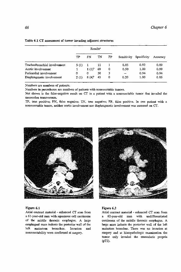
66 Chapter 6
Table 6.1 CT assessment of twnor invading adjacent structures
Results"
TP FN TN FP Sensitivity Specificity Accuracy
Tracheobronchial involvement 5 (I) II I 0.83 0.92 0.89 Aortic involvement I I (!)' 69 0 0.50 1.00 0.99 Pericardia! involvement 0 0 50 3 0.94 0.94 Diaphragmatic involvement 2 (I) 8 (4)' 43 0 0.20 1.00 0.85
Numbers are numbers of patients. Numbers in parentheses are numbers of patients with nonresectable tumors. Not shO"Wn is the false-negative result on CT in a patient with a nonresectable tumor that invaded the mesocolon transversum. TP, true positive; FN, false negative; TN, true negative; FP, false positive. In one patient with a nonresectable tumor, neither aortic involvement nor diaphragmatic involvement was assessed on CT.
F.gure 6.1 AXial contrast material - enhanced CT scan from a 61-year-old man with squamous cell carcinoma of the middle thoracic esophagus. A large esophageal mass indents the posterior wall of the left mainstem bronchus. Invasion and nonresectability were confirmed at surgery.
F;gure 6.2 Axial contrast material - enhanced CT scan from a 62-year-old man with undifferentiated carcinoma of the middle thoracic esophagus. A large mass indents the posterior wall of the left mainstem bronchus. There was no invasion at surgery and at histopathologic examination the tumor only invaded the muscuJ.aris propria (pT2).

CT Assessmem of Resectability
CT Assessment of Resectability
67
Fogure 6.3 Axial contrast material-enhanced CT scan from a 74-year old man with squamous cell carcinoma of the lower thoracic esophagus. Fat planes are absent between the tumor mass and the
pericardium. At surgery, however, pericardia} invasion was absent.
In 53 of 71 (75%) patients, the tumor was not adherent to the adjacent mediastinal and
abdominal structures at surgery and could be removed without difficulty. The tumor
invaded adjacent structures in the remaining 18 patients (prevalence 25%) (Table 6.1).
In seven (39%) of these patients the tumor was nonresectable. In the other 11 (61 %)
patients, the tumor could be resected. However, laceration of the left mainstem
bronchus occurred in one of 11 patients and the surgical procedure had to be extended
in six other patients: laryngectomy in one patient and partial resection of the diaphragm
in five patients. Involvement of the tracheobronchial tree could be identified on CT in
all but one patient (Fig. 6.1). In this patient the tumor was adherent to the left mainstem
bronchus, but could nevertheless be resected. There was one false positive result in a
patient with a large undifferentiated carcinoma that indented the posterior wall of the
left main stem bronchus on CT. However, at histopathologic examination the tumor
invaded only the muscularis propria (pT2) (Fig. 6.2). Involvement of the aorta was
correctly predicted on CT in one of two patients. In the other patient who had been
preoperatively treated with chemotherapy, 60° of the aortic circumference was in
contact with the tumor. None of the tumors was adherent to the pericardium at surgery.
However, in three patients pericardia! invasion was incorrectly predicted on CT (Figs.
6.3 and 6.4). Involvement of the diaphragm was correctly identified on CT in two of 10
patients. In one of these patients, the tumor was nonresectable, in the other the tumor
could be resected by resecting a part of the diaphragm (Fig. 6.5).

68
Figure 6.4 a Axial contrast material - enhanced CT scan from a 72-year-old woman with squamous cell carcinoma of the lower thoracic esophagus. At this level fat planes are absent bet\Veen the tumor mass and the pericardium.
Chapter 6
Figure 6.4 b Axial CT scan from the same patient shows fat planes present at a lower level. At surgery, pericardia! invasion was absent.
Figure 6.5 Axial contrast material - enhanced CT scan from a 76-year-old man with adenocarcinoma of the gastroesophageal junction. The left diaphragmatic crus is surrounded by tumor. The CT findings were confirmed at surgery, the tumor was nonresectable.

CT Assessment of Resectability 69
False negative findings occurred in eight patients. In four of these patients, the tumor
was considered nonresectable at surgery; due to invasion of the diaphragm and the
pancreas in one patient, the diaphragm and the aorta in another patient, and the
diaphragm alone in two patients. In another patient with a nonresectable tumor of the
gastroesophageal junction, invasion of the mesocolon transversum was not assessed on
CT studies.
CT Assessment of Depth of Invasion of Primary Tumor (pT)
At surgical/histopathological examination, the depth of invasion of the primary tumor
was pTis - p12 in 16 patients, pT3 in 37 patients and pT4 in 18 patients. The depth of
invasion was correctly determined on CT in 49 of 71 (69%) patients. Thirteen of 71
(18%) patients were understaged and nine of 71 (13%) patients were overstaged.
When patients were excluded who had received chemotherapy or radiotherapy
preoperatively, CT correctly staged 42 of 60 (70%) patients, understaged 12 of 60
(20%) patients and overstaged six of 60 (10%) patients. Preoperative CT could
determine the absence (pTis-pT3) or presence (pT4) of invasion of adjacent structures
with an accuracy of 82% (58 of 71 patients).
For those 10 patients who received chemotherapy preoperatively, CT findings (with a
mean 56 day interval) before and after chemotherapeutic treatment were compared. A
significant decrease in tumor size (~ 50% reduction of tumor size on axial CT scans)
was observed in three patients. In addition, in one of these patients, one of two enlarged
lymph nodes returned to normal size. A nonsignificant decrease in tumor size (<50%)
was observed in two patients and a nonsignificant increase was observed in one patient.
In four patients the tumor size remained unchanged. However, in all 10 patients, the
depth of tumor invasion on CT scans before and after chemotherapy was identical. In
the patient who received radiotherapy preoperatively, CT scans prior to radiation
therapy were not available for comparison.
CT Assessment of Lymph Node Metastases
Mediastinal lymph nodes were surgically and histopathologically assessed in 64 of 71
patients and were not assessed in the remaining seven patients with a nonresectable
tumor. Mediastinal lymph node metastases were found in the resected specimen of 10 of
64 (16%) patients. In assessing mediastina! lymph node metastases, CT had a sensitivity
of 30%, a specificity of 85% and an accuracy of 77%.
Abdominal lymph nodes were surgica!Iy and histopathologica!Iy assessed in all 71
patients. Involvement of abdominal lymph nodes was found in 32 (45%) patients;
perigastric nodes alone were found in 19 patients, celiac nodes alone were found in six
patients and both perigastric and celiac nodes were found in seven patients.

70 Chapter 6
CT had a sensitivity of 28%, a specificity of 90% and an accuracy of 62% in assessing
abdominal lymph node metastases.
CT Assessment of Tumor Stage
Twenty-nine patients had stage I-ll disease, 28 had stage ill disease and 14 had stage N
disease. CT correctly staged 40 (57%) patients, understaged 23 (32%) patients and
overstaged eight (11 %) patients. Carcinomas of the esophagus were correctly staged in
30 of 55 (55%) patients and gastroesophageal junction carcinomas were correctly staged
in 10 of 16 (63%) patients. When patients with preoperative chemotherapy or
radiotherapy were excluded, CT correctly staged 37 (61 %), understaged 19 (32%) and
overstaged four (7%) of 60 patients. For the 10 patients who received chemotherapy
preoperatively, tumor stages on CT scans before and after chemotherapeutic treatment
were identicaL
6.5 Discussion
Whether surgical resection for esophageal and gastroesophageal junction carcinoma
should be regarded as a potentially curative or a palliative procedure remains a matter
of controversy, but surgery is generally not recommended in patients with a
nonresectable tumor. Thus, at our institution where THE is performed· for both
esophageal and gastroesophageal junction carcinoma, a major role of preoperative CT
should be to determine suitability for resection.
Conflicting results are reported regarding the value of preoperative CT in staging
esophageal and gastroesophageal junction carcinoma, partly because of the different
prevalences of invasion of adjacent structures that are reported by different authors
[2,3,11,12]. The prevalence of invasion will be determined by the selection of patients
who have surgery. Whereas at some institutions patients have surgery regardless the
presence of distant metastases [13], at our institution patients with cytologically proven
distant metastases detected on ultrasound or CT studies are excluded from surgery
[14,15]. The prevalence of invasion in our series was relatively low and in more than
half the cases of invasion, the tumor could nevertheless be resected. This limited the
value of CT to assess nonresectability by diagnosing invasion. Furthermore, CT was
limited in assessing invasion itself.
CT was most accurate in assessing tracheobronchial involvement. Although this did not
preclude resection in all patients, it occasionally complicated surgery, which is
demonstrated by the accidental laceration of the bronchial tree in one of the patients.

CF Assessment of Resectability 71
F;gure 6.6 Axial contrast material - enhanced CT scan from a 67-year-old man with squamous cell carcinoma of the middle thoracic esophagus shows contact of 70° between the aorta and the tumor. A small atheromatous plaque is seen in the aortic wall. At surgery invasion was absent and the tumor could be resected without difficulty.
Our results in assessing tracheobronchial involvement on CT are comparable with those
reported by Becker et al [11]. In their series, tracheobronchial involvement at THE was
identified in all cases on CT. The false positive result in our series was caused by an
undifferentiated carcinoma of the esophagus, and it has been suggested previously that
CT is less accurate in staging these tumors [16].
The therapeutic management in patients with esophageal and gastroesophageal junction
carcinoma will rarely be affected by aortic or pericardia! invasion because these are
only reported at autopsy in 3% and I% of patients, respectively [17]. This is consistent
with the results of the present study; aortic involvement was observed in only two of 71
patients and pericardial involvement was not found at surgery.
For the assessment of aortic involvement, we modified the CT criteria of Picus et al.
[12]. According to their criteria, aortic invasion would have been considered
indeterminate in eight patients with contact of 45-90° between the aorta and the tumor
(Fig. 6.6). With the modified CT criteria(> 90° contact) there was one false negative
result, but there were no indeterminate results. As reported by others, CT was of little
help in assessing pericardia! involvement [11,13]. However, pericardia! involvement
was occasionally simulated by motion artefacts and lack of periesophageal fat, which
may be caused by the often poor nutritional status of patients with esophageal and
gastroesophageal tumors [3]. With use of the criteria suggested by Halvorsen and
Thompson, five patients with negative findings at surgery would have indeterminate
findings on CT because of the lack of fat planes [1,16].

72 Chapter 6
Similar to the reported experience, CT was inaccurate in predicting diaphragmatic
invasion [6,11-13]. In our study, diaphragmatic involvement was considered present on
CT scans only in cases in which the crura were surrounded by tumor mass. However,
when invasion was predicted on the basis of broad contact between the tumor and the
diaphragmatic crura over a length of 1 em or more, there were no false negative findings, but 19 false positive findings on CT. Thus, the close relationship of the
esophagus and the gastroesophageal junction with the diaphragm and the absence of
intervening fat planes at this level cause great difficulty in assessing diaphragmatic
involvement on CT scans (Figs. 6.7 and 6.8). Moreover, diaphragmatic involvement
does not necessarily preclude resection, in our present study 50% of tumors with
diaphragmatic invasion could be resected. The different layers of the esophageal wall are not separately displayed on CT scans.
Therefore, differentiation was only attempted among tumors within the wall (pT2 and
less), tumors with extramural extension (pTI) and tumors invading the adjacent
structures (pT4) [10]. Understaging was caused mainly by not recognizing
diaphragmatic involvement. Overstaging may have been caused by inflammatory and
desmoplastic reactions around the tumor that are often found at histopathologic
examination. Excluding patients who received either chemotherapy or radiotherapy
preoperatively did not improve the results.
CT was limited in assessing lymph node metastases and was characterized by a low
sensitivity and a relatively high specificity, which has been reported previously by
others [1,11,12]. This is explained because normal-sized nodes may harbor metastases
[1]. The lower sensitivity of CT in assessing lymph node metastases in the present
series compared to other series [1,6,11] is explained by the fact that our results concern
only patients who underwent surgery. Patients with cytologically proven supraclavicular
and celiac lymph node metastases detected on ultrasound and CT studies were excluded
from surgery and these results are not included in the present report [14,15]. The false
positive results of CT in assessing mediastinal lymph node metastases should be
interpreted with caution, because the mediastinal nodes are not systematically dissected
at THE [7-9]. This can also explain the lower prevalence of mediastinal lymph node
metastases compared with abdominal lymph node metastases in our series.
In assessing stage grouping on CT, understaging occurred more often than overstaging.
This was caused mainly by not detecting either diaphragmatic involvement or regional
lymph node metastases in patients with a stage III tumor and by not detecting distant
lymph node metastases in patients with a stage IV tumor. Excluding patients with
preoperative chemotherapy or radiotherapy slightly decreased the number of understaged

CT Assessment of Resecrahiliry 73
patients, but the numbers of these patients are too small to draw any final conclusions.
For those patients who received chemotherapy, similar results in assessing stage grouping on CT were obtained prior to and after chemotherapeutic treatment. However, only a limited number of patients were studied and the definitive role of CT in the
follow-up of chemotherapeutically treated patients remains to be determined.
In conclusion, CT was not effective in assessing resectability of esophageal and
gastroesophageal junction carcinoma in patients undergoing THE. This was caused by
the relatively low prevalence of invasion of adjacent structures at surgery and the fact
that invasion was often not associated with nonresecta.bility. Furthermore, in assessing
invasion, CT was accurate only in assessing tracheobronchial involvement.
In assessing stage grouping in surgically treated patients, CT had a tendency to understage rather than to overstage because of its limitation in detecting either
diaphragmatic invasion or lymph node involvement.
F;gure 6.7 Axial contrast material - enhanced CT scan from a 67-year-old man with squamous cell carcinoma of the lower esophagus. Broad contact between the tumor and the diaphragmatic crura. However, at surgery invasion was absent (pT3).
Figure 6.8 Axial contrast material - enhanced CT scan from a 44-year-old man with adenocarcinoma of the gastroesophageal junction. Broad contact between the tumor and the diaphragmatic crura. At surgery invasion was present and the tumor could not be resected (pT4).

74 Chapter 6
6.6 References
1. Halvorsen RA, Thompson WM. CT of esophageal neoplasms. Radial Clin North Am 1989; 27: 667-685.
2. Quint LE, Glazer GM, Orringer MB, Gross BH. Esophageal carcinoma: CT findings. Radiology 1985; 155; 171-175.
3. Thompson WM, Halvorsen RA, Foster WL, Williford ME, Postlethwait RW, Korobkin M.
Computed tomography for staging esophageal and gastroesophageal cancer. Reevaluation. A1R 1983; 141; 951-958.
4. Salonen 0, Kivisaari L, StandertskjOld-Nordenstam CG, Somer K, Virkkunen P. Computed
tomography in staging of oesophageal carcinoma. Scand J Gastroenterol 1987; 22: 65-68.
5. Freeny PC, Marks WM. Adenocarcinoma of the gastroesophageal junction: Barium and CT
examination. A1R 1982; 138: 1077-1084.
6. Terrier F, Schapira C, Fuchs A. cr assessment of operability in carcinoma of the oesophagogastricjunction. Eur J Radiol1984; 4: 114-117.
7. Oninger MB, Sloan H. Esophagectomy without thoracotomy. J Thorac Card.iovasc Surg 1978;
76; 643-654.
8. Finley RJ, Inculet RI. The results of esophagogastrectomy without thoracotomy for
adenocarcinoma of the esophagogastric junction. Ann Surg 1989; 210: 535-542. 9. Riiedi TP. Non-thoracotomy oesophageal resection. In: Jamieson GG, ed. Surgery of the
Oesophagus. 1st ed. Melbourne: Churchill Livingstone, 1988; 639-654.
10. Beabrs OH, Henson DE, Hutter RVP, Myers MH. Manual for staging of cancer. 3rd ed.
Pbiladelp~: JB Lippincott, 1988. 11. Becker CD, Barbier P, Porcellini B. CT evaluation of patients undergoing transbiatal
esophagectomy for cancer. J Comput Assist Tomogr 1986; 10: 607-611.
12. Picus D, Balfe DM, Koehler RE, Roper CL, Owen JW. Computed tomography in the staging of
esophageal carcinoma. Radiology 1983; 146: 433-438.
13. Takashima S, Takeuchi N, Shiozaki H, et al. Carcinoma of the esophagus: CT vs MR imaging
determining resectability. AJR 1991; 156: 297-302.
14. van Overhagen H, Lameris JS, Berger MY, et al. Supraclavicular lymph node metastases in carcinoma of the esophagus and gastroesophageal junction. Assessment with CT, US and USguided fine-needle aspiration biopsy. Radiology 1991; 179: 155-158.
15. van Overhagen H, Lam.eris JS, Berger MY, et al. Assessment of distant metastases with ultrasound-guided fine-needle aspiration biopsy and cytologic study in carcinoma of the esophagus
and gastroesophageal junction. Gastrointest Radiol1992; 17: 305-310. 16. Halvorsen RA, Thompson WM. Computed tomographic staging of gastrointestinal tract
malignancies. Part 1: Esophagus and stomach. Invest Radiol 1987; 22: 2-16. 17. Postlethwait RW. Surgery of the Esophagus. 2nd ed. Not\Valk, Va: Appleton-Century-Crofts,
1986; 369-442.

75
Chapter 7
Influence of Radiologically and Cytologically Assessed Distant Metastases on the Survival of Patients with Esophageal and Gastroesophageal Junction Carcinoma
(Survival in Esophageal and Gastroesophageal Cancer)
7.1 Abstract
Distant metastasis in carcinoma of the esophagus and gastroesophageal junction is
associated with a poor survival following resection. In order to improve the selection of patients for surgical and nonsurgical treatment, the influence on survival of distant
metastases, assessed on radiologic studies or proven on cytologic studies was deterntined. During the period 1989-1990, 135 patients were referred to our institution.
On ultrasound or computed tomographic studies distant metastases were suspected in 62
patients and were absent in 73 patients. 1n 33 patients metastases were cytologically
proven, 32 of these patients were subsequently excluded from surgery. Twelve other
patients were unfit for surgery due to their general condition. The remaining 91 patients
underwent surgery; 77 patients underwent transhiatal esophagectomy, the tumor was
irresectable in 14 patients.
The 2-year survival for all patients suspected of distant metastases on radiologic studies
was 11.2% and was 44.3% for patients without metastases on these studies (p < 0.001). For patients with cytologically proven metastases the 2-year survival (3%) was
shorter than for patients suspected of distant metastases on radiologic studies that were
not cytologically confirmed (21.1%) (p < 0.001). There was no statistically significant
difference in survival between this last group of patients and those without metastases
on radiologic studies (p = 0.87). Following resection the 2-year survival dropped from
53.9% to 0% when distant metastases were present on histopathologic studies of the
resected specimen (p = 0.04). Following resection there was no significant difference
in survival between patients with distant metastases suspected or absent on preoperative
Published as: Hans van Overhagen, MD, Matjolein Y. Berger, MD, Hjalmar Meijers, MD, Hugo W. Tilanus, MD, Tjebbe C. Kok, MD, Theo Stijnen, MSc, Johan S. Lam6ris, MD. INFLUENCE OF RADIOLOGICALLY AND CYTOLOGICALLY ASSESSED DISTANT METASTASES ON THE SURVNAL OF PATIENTS WITH ESOPHAGEAL AND GASTROESOPHAGEAL WNCTION CARCINOMA. Cancer: in press.

76 Chapter 7
radiologic studies (p = 0.47). Surgery should be avoided in patients with cytologically
proven distant metastases because the expected survival is low and surgery does not
seem a life prolonging procedure in these patients. However, patients should not be
excluded from surgery on the basis of metastases on radiologic studies alone.
7.2 Introduction
Carcinoma of the esophagus and gastroesophageal junction has a poor prognosis because
many patients have disseminated disease at first presentation. When metastases are
found at surgery, survival rates following resection drop significantly [1-5]. This
explains the persisting low survival of surgically treated patients despite the decrease in
postoperative mortality during the past 10 years [6,7].
In a previous report by our study group in 1987 it was concluded that effort should be
directed to better selection of patients for surgery in order to improve the long-term
survival of surgically treated patients and to avoid unnecessary surgery in patients with
disseminated disease [1].
Radiologic studies such as computed tomography (CT) and ultrasound (US) have
improved the pretreatment staging of esophageal and gastroesophageal junction
carcinoma, but cpnflicting results are reported regarding the accuracy of these studies
[8]. Thus, the decision to refrain from surgery in patients on the basis of radiologic
studies alone remains controversial and some authors have stated that "no noninvasive
test is of sufficient reliabiliry to exclude patients from operative resection" [9].
US-guided fine-needle aspiration biopsy (FNAB) is a safe and minimal invasive
technique to provide cytologic specimen of lesions suspected of metastases on radiologic
studies. During the last 3 years we have systematically included this technique in the
pretreatment staging of esophageal and gastroesophageal junction carcinoma [10].
Patients suspected of having distant metastases on US or CT studies underwent US
guided FNAB when these lesions were accessible to biopsies. With the exception of one
patient, patients with cytologically proven distant metastases were excluded from
surgery whereas those without cytologically proven metastases underwent surgery.
This study was performed to evaluate the survival of patients after having been selected
for surgery according to our staging strategy. Furthermore, the influence on survival of
distant metastases assessed on radiologic studies or diagnosed on cytologic studies was
determined.

Survival in Esophageal and Gastroesophageal Cancer 77
7.3 Patients and methods
Between January 1989 and December 1990, 135 patients, aged 40-84 years (mean 63
years) with a biopsy proven carcinoma of the intrathoracic esophagus or
gastroesophageal junction were referred to the Rotterdam Esophageal Tumor Study
Group for staging and treatment. Patient characteristics are shown in Table 7 .1. Staging was performed according to the classification of the American Joint Committee
on Cancer (AJCC) [11]. In the present report, however, malignant involvement of N2
nodes in carcinoma of the gastroesophageal junction is also considered as distant
metastasis. All patients underwent US of the supraclavicular regions and the abdomen
by means of a 7.5-MHz linear array transducer and a 3.5-MHz curvilinear transducer,
respectively (SSD-650; Aloka, Tokyo, Japan). All patients underwent CT scanning of
the chest and upper abdomen including the entire liver in 8-12-mm-thick contiguous
sections and all but II patients also underwent CT scanning of the supraclavicular
regions from the cricoid cartilage to the sternal angle in 5- or 6-mm-thick contiguous
sections by means of a Somatom Plus Scanner (Siemens, Erlangen, Germany) or a
Tomoscan 350 (Philips Medical Systems, Eindhoven, The Netherlands). Distant sites
systematically examined on US and CT scans were the lungs, the liver, the peritoneum,
the kidneys and the adrenal glands (visceral sites), the supraclavicular nodes and the
celiac nodes which were considered enlarged when they had a short axis of 5 mm and
10 mm or more, respectively. Patients with US or CT fmdings indicating distant
metastases that were accessible to biopsies underwent US-guided FNAB of one suspect
lesion. In patients with multiple lesions biopsies were preferentially performed of
supraclavicular nodes because supraclavicular biopsies are easier to perform and are less
invasive for the patients compared with abdominal biopsies [12,13]. FNABs of
supraclavicular lymph nodes were performed with a 23-gauge needle and of abdominal
lesions with a 22-gauge needle. With the exception of one patient, those patients who
had distant metastases proven by means of cytologic study were excluded from surgery.
Also excluded from surgery were patients who had a poor general condition. The
remaining patients, including patients with US or CT findings indicating distant
metastases that were not accessible to biopsies or patients who had negative results on
cytologic studies of biopsies, underwent surgery.
At surgery laparotomy was performed as the first step of planned resection. Patients
with a surgically resectable tumor underwent transhiatal esophagectomy (THE) with
gastric interposition and cervical gastroesophageal anastomosis [14]. Survival rates
following resection with this technique are almost identical to those obtained with a
transthoracic approach [7].

78 Chapter 7
In patients with an irresectable tumor surgery was abandoned after laparotomy.
Data on squamous cell carcinomas and adenocarcinomas were combined because the
radiologic staging and the surgical approach in these tumors are similar in our institution and recent reports do not show significant differences in survival [15,16].
Survival was measured from the date of radiologic staging. Survival curves were
derived by the Kaplan-Meier method and comparisons of survival rates of different
subgroups of patients were made by using the log-rank test. In addition, a multivariate
analysis was separately performed for both the surgically treated and nonsurgically
treated groups by using the Cox proportional hazards model. Results are reported with
P values and associated 95% confidence intervals (Cis). Operative mortality rate was
defined as the in-hospital death after surgery. Overall mortality rate represents death
due to any cause during follow up.
Table 7.1 Characteristics and two year survival of 135 patients with esophageal and gastroesophageal junction carcinoma
Characteristics No.(%) of patients 2-year survival*
All patients 135 28.9 (21.1-37.2)
Sex: Male !05 (78) 27.5 (!8.8-36.9) Female 30 (22) 34.1 (17.4-51.6)
Age: < 60 45 (33) 27.5 (15.1-41.4)
"' 60 90 (67) 29.5 (19.7-39.9)
Tumor type: squamous cell carcinoma 66 (49) 20.9 (11.6-32.0) adenocarcinoma 63 (47) 37.3 (24.6-49.9) undifferentiated carcinoma 6 ( 4) 33.3 ( 4.6-67.6)
Tumor location: upper esophagus 5 ( 4) 40.0 ( 0.8-58.2) middle esophagus 38 (28) 27.1 (13.8-42.4) lower esophagus 66 (49) 30.0 (18.8-42.0) gastroesophageal junction 26 (19) 27.5 (11.3-46.5)
Distant metastases on US and CT: present 62 (46) I 1.2 ( 4.5-21.3) - cytologically proven 33 (24) 3.0 ( 0.2-13.4) - negative cytology
or cytology not done 29 (22) 21.1 ( 7 .4-39.4) absent 73 (54) 44.3 (32.5-55.8)
Surgery: not performed 44 (33) 8.2 ( 2.3-19.1) performed 91 (67) 39.5 (28.8-50.0)
Note: * 95% confidence interval in parentheses.

Survival in Esophageal and Gastroesophageal Cancer 79
7.4 Results
Sixty-two patients were suspected of having distant metastases on US and/or CT studies
(Tables 7.1 and 7.2). Distant metastases were proven by meaos of US-guided FNAB
and cytologic study in 33 patients; 32 of these patients were excluded from surgery, in
another patient surgical resection was attempted despite the presence of cytologically
proven celiac metastases but it was abandoned after these metastases were confirmed
histopathologically. In 29 patients the cytologic smears did not show malignant cells (17
patients) or US-guided FNAB could not be performed (12 patients). Two of these
patients were excluded from surgery because they had a poor general condition, 27
patients had surgery at which the tumor was resected in 20 patients (resection rate
74%). Seventy-three patients did not have metastases on US and/or CT studies; 10 of
these patients were excluded from surgery because they had a poor general condition,
the remaining 63 patients had surgery with resection of the tumor in 57 patients
(resection rate 90%). In total, 44 patients were excluded from surgery; because of
cytologically proven distant metastases in 32 patients and because of a poor general
condition in 12 patients. These patients were treated with radiation therapy (n = 7),
chemotherapy (n = 6), tube insertion (n = 8), laser therapy (n = 3), a combination of
radiation therapy and chemotherapy (n = 3), chemotherapy and tube insertion (n = 14)
or radiation therapy, chemotherapy and tube insertion (n = 1). Two patients did not
receive any treatment. Ninety-one patients had surgery; 14 patients underwent
laparotomy only because of major abdominal metastases and irresectability of the
primary tumor, the other 77 patients underwent THE.
Table 7.2 Patients suspected of distant metastases on US and CT studies and patients with distant metastases proven on cytologic studies
No. (%) of patients
Location of distant metastases Distant metastases Distant metastases on US and/or CT cytologically proven
Lungs 2 ( 1) 0 ( 0) Liver 10 ( 7) 6 ( 4) Peritoneum 2 ( 1) 2 ( 1) Kidneys 1 ( 1) ( 1) Adrenal glands 2 ( 1) 0 ( 0) Supraclavicular lymph nodes 38 (28) 22 (16) Celiac lymph nodes 30 (22) 3 ( 2)
Total 62 (46) 33 (24)
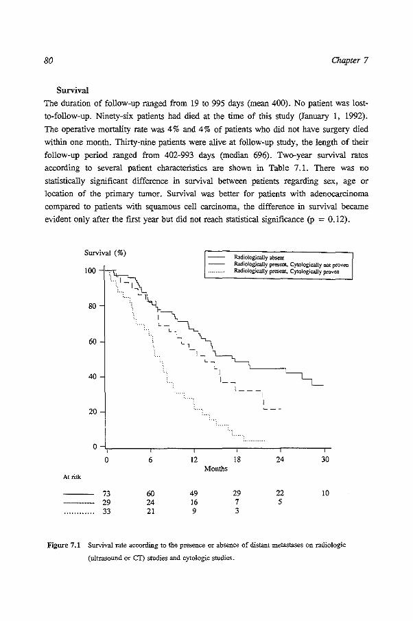
80 Chapter 7
Survival
The duration of follow-up ranged from 19 to 995 days (mean 400). No patient was lost
to-follow-up. Ninety-six patients had died at the time of this study (January 1, 1992).
The operative mortality rate was 4% and 4% of patients who did not have surgery died
within one month. Thirty-nine patients were alive at follow-up study, the length of their
follow-up period ranged from 402-993 days (median 696). Two-year survival rates
according to several patient characteristics are shown in Table 7. L There was no
statistically significant difference in survival between patients regarding sex, age or
location of the primary tumor. Survival was better for patients with adenocarcinoma
compared to patients with squamous cell carcinoma, the difference in survival became
evident only after the first year but did not reach statistical significance (p = 0.12).
Survival (%)
100
80
60
40
20
0
0 6
At risk
73 60 -------- 29 24 ............ 33 21
12 Months
49 16 9
Radiologically absent Radiologically present. CytOlogically not proven Radiologically present, CytOlogically proven
[ __ _
18
29 7 3
I
I
24 30
22 10 5
Figure 7.1 Survival rate according to the presence or absence of distant metastases on radiologic
(ultrasound or CT) studies and cytologic studies.

Survival in Esophageal and Gastroesophageal Cancer 81
When the patients were subdivided on the basis of their radiologic findings, patients
suspected of distant metastases on US and CT studies had a 2-year survival of 11.2%
whereas patients without metastases on these studies had a 2-year survival of 44.3% (p
< 0.001). Patients with cytologically proven metastases had a shorter survival than
either patients suspected of metastases on radiologic studies that were not cytologically
proven (p < 0.001) or patients without metastases on radiologic studies (p < 0.001)
(Table 7.1, Fig.7.1). The difference in survival between these last two groups of
patients was not statistically significant (p = 0.87). Within the group of patients
suspected of metastases on radiologic studies that were not cytologically proven, there
was no significant difference in survival between patients without metastases on
cytologic studies and those who did not undergo US-guided FNAB (p = 0.11).
SurviYdl (%)
100 ..,-,..,, -~~
80
60
40
20
0
0
At risk
77 --- 44 ·····-·-···· 14
c :.1,
:1 'I . 1
c l
6
69 27 9
-,_ ·t:···
\
12
58 12 4
18 Months
32 6
Figure 7.2 Survival rate according to the treatment patients received.
THE No surgery Laparotomy
24 30
24 12 4

82 Chapter 7
Table 7.3 One year and two year survival of 44 patients excluded from surgery
Characteristics
Poor general health
Cytologically proven metastases - Cytologic or radiologic evidence
of visceral metastases - Cytologic or radiologic evidence
of lymphatic metastases only
Note: 95% confidence interval in parentheses.
Survival (%)
40
20
No. of patients
12
32
12
20
1-year survival 2-year survival
33.3 (10.3-58.8) 22.2 (4.1-49.2)
25.0 (11.8-40.7) 3.1 (0.2-13.7)
8.3 ( 0.5-31.1) 0
35.0 (15. 7-55.2) 5.0 (0.3-20.5)
I== Metastases absent I Metastases presem:
L __ _
I
0~-----.-----.------.-----.-----.-
At risk
0
57 20
6
50 19
12
43 15
Months 18
25 7
24
20 4
30
10
Figure 7.3 Survival rate according to the presence or absence of distant metastases on radiologic studies,
within the group of patients who underw'ent transhiatal esophagectomy (THE).

Survival in Esophageal and Gastroesophageal COJICer 83
When the patients were subdivided on the basis of the treatment they received,
surgically treated patients bad a 2-year survival of 39.5% whereas patients excluded
from surgery bad a 2-year survival of 8.2% (p < 0.001) (Table 7.1, Fig. 7.2). Further
statistical analysis of survival was performed separately for surgically and nonsurgically
treated patients. For the group of patients excluded from surgery, patients excluded
because of cytologically proven distant metastases had a worse prognosis than those
with a poor general health, but the difference did not reach statistical significance (p
0.13) (Table 7.3).
Survival (%)
80
60
40
20
0
0 6
At risk
61 55 ~---------- 16 14
,_
12 18 Months
48 28 10 4
24
24
Metastases absent Metasta.~ pres<::nt
30
II
Figure 7.4 Survival rate according to the presence or absence of distant metastases on histopathologic
studies of the resected specimen within the group of patients who underwent transhiatal
esophagectomy (THE).
84 Chapter 7
Within the group of patients with cytologically proven distant metastases, those with
cytologic or radiologic evidence of visceral metastases had a worse prognosis than those
with evidence of lymphatic metastases alone (p = 0.024) (Table 7 .3). For the group of
surgically treated patients, those who underwent THE had a statistically better survival
than those who underwent laparotomy only (p < 0.001) (Table 7.4, Fig. 7.2). There
was no statistically significant difference in survival between patients who were radiologically suspected of distant metastases and those without distant metastases on
radiologic studies within the groups of patients who underwent either THE (p = 0.47)
or laparotomy (p = 0.52) (Table 7.4, Fig. 7.3). For patients who underwent THE,
those with distant metastases on histopathologic study had a worse survival compared to
those without distant metastases (p = 0.04) (Table 7.4, Fig. 7.4). A multivariate model
using patient characteristics as variables did not show any independent effect on survival
within the group of surgically treated patients. Within the group of nonsurgically treated
patients, for those patients with cytologically proven distant metastases, the presence of
visceral metastases was associated with a hazard ratio of 2.7 (95% Cl, 1.2-6.1)
compared to the presence of lymphatic metastases alone.
Table 7.4 One year and two year survival of 91 surgically treated patients
Characteristics No. of patients 1-year survival 2-year survival
Laparotomy 14 28.6 ( 8.8-52.4) 0 - Radiologic evidence
of distant metastases: present 8 25.0 ( 3. 7-55.8) 0 absent 6 33.3 ( 4.6-67.6) 0
Transhiatal esophagectomy 77 75.3 (64.1-83.5) 46.8 {34.5-58.2) - Radiologic evidence
of distant metastases: present 20 75.0 (50.0-88.7) 31.0 {10.7-54.1) absent 57 75.4 (62.1-84.7) 52.1 (37.7-64.6)
- Histopathologically proven distant metastases: present 16 62.5 {34.9-81.!) 0
absent 61 78.7 (66.1-87.0) 53.9 {40.1-65.8)
Note: 95% confidence interval in parentheses.

Survival in Esophageal and Gastroesophageal Cancer 85
7.5 Discussion
While numerous articles have been published that evaluate the accuracy of CT in staging esophageal and gastroesophageal junction carcinoma, there are only few articles
that have correlated the findings on CT or US studies with the length of patient survival
[17,18]. Halvorsen et al who compared the length of patient survival with pretherapy
CT findings in 89 patients with esophageal carcinoma reported that regardless of
therapy, evidence of abdominal metastases on CT studies decreased the mean patient
survival of 320.6 days to 102.4 days [17]. Similar observations are shown in our study,
the 2-year survival of patients decreased significantly when distant metastases were
suspected on US or CT studies. However, within this group of patients the worst
survival was observed in patients with distant metastases proven on cytologic studies,
these patients had a significantly shorter survival than those without cytologic proof of
metastases. This can be explained by the limited specificity of US and CT in diagnosing
metastases, in our study for example 13 of 62 patients suspected of distant metastases
on radiologic studies eventually had their tumor resected without evidence of distant
metastases on histopathologic studies of the resected specimen (results not shown).
Consequently, although the group of patients suspected of metastases on US and CT has
a poor prognosis, by excluding all these patients from surgery some patients without
metastases will be withheld their optimal treatment. By obtaining cytologic proof of
metastases the risk of overstaging is minimized which is reflected in the significantly
worse prognosis in cases metastases were cytologically proven. A critical remark may
be that bias was introduced regarding survival because patients were excluded from
surgery in cases when metastases were cytologically proven. However, the 2-year
survival rate of these patients is comparable to the 2-year survival rate of patients with
distant metastases following resection in our study and reported by others [4,5,14].
Naturally, in some patients correctly suspected of distant metastases on US and CT, the
cytologic examination will be false negative or cytologic biopsies cannot be performed.
This can explain the lower 2-year survival and the lower resection rate at surgery in
patients without cytologic confirmation of lesions suspected of metastases on US and CT
studies compared to patients not suspected of metastases on these studies. However,
because the difference in survival is not statistically significant and because suboptimal
treatment should be avoided in individual patients without metastases, we recommend
not to exclude patients from surgery on the basis of metastases on radiologic studies
alone. In patients with cytologically proven distant metastases surgery does not seem a
life prolonging procedure. The 2-year survival of these patients who were excluded
from surgery in our study was 3% whereas following resection there were no survivers
86 Chapter 7
after 2 years in patients with distant metastases in our study and in studies of other
authors [4,5,19], fu our study 75% of patients with cytologically proven metastases had
died within one year, thus if surgery is considered a palliative procedure in patients
with distant metastases, maximally 25% of these patients can be palliated for one year
including the duration of their hospital admission. Since surgery is a formidable
procedure with a postoperative morbidity of at least a few months [1], we do not
consider palliative surgery indicated in this group of patients although opposite opinions
have been reported in selected cases [15]. For patients with cytologically proven
metastases, survival was worst for those patients who had cytologic or radiologic
evidence of visceral metastases. However, patients with evidence of lymphatic
metastases alone also had a very poor prognosis with only 5% of patients alive after 2
years. For patients who underwent surgery, as expected, the survival rates were best for
those patients who had their tumor resected. Patients with irresectable tumors at surgery
had a poor prognosis, none of these patients was alive after 2 years and continuous
effort should be made to keep the numbers of patients that suffer from unnecessary
surgery as low as possible. Although the resection rate at surgery at our institution
increased from 81% in 1987 to 85% at present, the postoperative mortality rate
remained at the same level during this period which indicates that the higher resection
rate is not the result of a more agressive surgical approach with the goal to achieve
resection, but is more likely the result of better patient selection [!]. This is supported
by the fact that the 2-year survival following resection in our study is relatively high
compared to the survival after resection reported by others [5,7,19]. For patients who
underwent resection those suspected of distant metastases on US or CT did not have a
significantly worse prognosis compared to those without metastases on these studies
which is consistent with our current belief that patients should not be excluded from
surgery on the basis of metastases on radiologic studies alone. However, when distant
metastases were proven at histopathologic examination of. the resected specimen the
survival following resection deteriorated, none of these patients was alive after 2 years.
In conclusion, in patients with carcinoma of the esophagus and gastroesophageal
junction, pretreatment assessment of distant metastases is of great importance to select
the optimal treatment. Surgery should be avoided in cases of distant metastases because
the expected survival is very poor and surgical treatment does not seem life prolonging
in these patients. However, metastases should be proven by means of cytologic studies
and patients should not be excluded from surgery on the basis of radiologic findings
alone in order to avoid suboptimal treatment in individual patients with potentially
curable disease.
Survival in Esophageal and Gastroesophageal Cancer 87
7.6 Refe.rences
1. Eeftinck Schattenkerk M, Obertop H, Mud HJ, Eijkenboom WMH, van Andel JG, van Houten H. Survival after resection for carcinoma of the oesophagus. Br J Surg 1987; 74: 165-168.
2. Akiyama H. Tsurumaru M, Kawamura T, Ono Y. Principles of surgical treatment for carcinoma
of the esophagus. Ann Surg 1981; 194: 438-446.
3. Lu YK, Li YM, Gu YZ. Cancer of esophagus and esophagogastric junction: Analysis of results
of 1025 resections after 5 to 20 years. Ann Thorac Surg 1987; 43: 176-181.
4. Ellis FH, Gibb SP, Watlcins E. Limited esopbagogastrectomy for carcinoma of the cardia. Indications, technique and results. Ann Surg 1988; 208: 354-361.
5. Finley RJ, Inculet RI. The results of esophagogastrectomy without thoracotomy for
adenocarcinoma of the esophagogastric junction. Ann Surg 1989; 210: 535-543.
6. Earlam R, Cun.ha-Melo JR. Oesophageal squamous cell carcinoma: I. A critical review of surgery. Br J Surg 1980; 67: 381-390.
7. MUller JM. Erasmi H, Stelzner M. Zieren U, Pichlmaier H. Surgical therapy of oesophageal carcinoma. Br J Surg 1990; 77: 845-857.
8. Halvorsen RA, Thompson WM. CT of esophageal neoplasms. Radio! Clin North Am 1989; 27: 667-685.
9. Inculet RI, Keller SM, Dwyer A, Roth JA. Evaluation of noninvasive tests for the preoperative staging of carcinoma of the esophagus: A prospective study. Ann Thorac Surg 1985; 40: 561-
565.
10. van Overhagen H. Lameris JS, Berger MY, et al. Assessment of distant metastases with
ultrasound-guided fine-needle aspiration biopsy and cytologic study in carcinoma of the
esophagus and gastroesophageal junction. Gastrointest Radiol1992; 17: 305-310.
11. Beahrs OH, Henson DE, Hutter RVP, Myers MH. Manual for staging of cancer, 3rd ed.
Philadelphia: JB Lippincot, 1988.
12. van Overhagen H, Lam&is JS, Zonderland HM, Til anus H\V, van Pel R, SchUtte HE.
Ultrasound and ultrasound-guided fine-needle aspiration biopsy of supraclavicular lymph nodes
in patients with esophageal carcinoma. Cancer 1991; 67: 585-587.
13. van Overhagen H, Lamtiris JS, Berger MY, et al. Supraclavicular lymph node metastases in
carcinoma of the esophagus and gastroesophageal junction: Assessment with CT. US and USguided fine-needle aspiration biopsy. Radiology 1991; 179: 155-158.
14. Orringer MB. Sloan H. Esophagectomy without thoracotomy. J Thorac Cardiovasc Surg 1978; 76, 643-654.
15. Ellis FH. Treatment of carcinoma of the esophagus or cardia. Mayo Clin Proc 1989; 64: 945-
955.
16 Oliver SE, Robertson CS, Logan RFA. Oesophageal cancer: a population- based study of suvival
after treatment. Br J Surg 1992: 79: 1321-1325.
17. Halvorsen RA, Magruder-Habib K, Foster WL, Roberts L, Postlethwait RW, Thompson WM.
Esophageal cancer staging by CT: Long-term follow-up study. Radiology 1986; 161: 147-151.
18. Unger EC, Coia L, Gatenby R, et al. CT staging of esophageal carcinoma in patients treated by
primary radiation therapy and chemotherapy. J Comput Assist Tomogr 1992; 16: 235-239.
19. Orringer MB. Transhiatal esophagectomy without thoracotomy for carcinoma of the thoracic
esophagus. Ann Surg 1984; 200: 282-288.

88
89
Chapter 8
FINAL REMARKS
A Radiologic Staging Strategy
When surgery is considered, radiologic staging of esophageal and gastroesophageal
junction carcinoma should begin with the assessment of distant metastases. The presence
of distant metastases is associated with a poor prognosis (Chapter 7) [1-4] and most
surgeons do not consider resection in these patients [5-7]. The combination of ultrasound (US) and computed tomography (CT) is the best choice for noninvasive
assessment of distant metastases (Chapter 4) and should include examination of the
supraclavicular lymph nodes, since supraclavicular metastases are such a common
finding in esophageal and gastroesophageal junction carcinoma (Chapters 2 and 3). For
logistic and cost -effective reasons we recommend US as the initial examination and the
use of US-guided fine-needle aspiration biopsy (FNAB) when metastases are suspected.
Patients without metastases on US should undergo CT and US- or CT -guided biopsies in
those cases when lesions are detected. Distant metastases can thus be proven in 24% of
patients (Chapter 5) in whom surgery can be avoided (Chapter 7).
Presence or absence of regional lymph node metastases cannot be determined accurately
(Chapter 6) and there is no consensus regarding the therapeutic consequences in those
patients who have regional lymph node metastases [5,7].
Assessment of the invasion depth of the primary tumor is unsatisfactory. Furthermore,
assessment of resectability of the primary tumor by determining invasion of adjacent
structures on radiologic studies may be hazardous, because invasion does not necessarily
preclude resection (Chapter 6). A flow chart for a radiologic staging strategy is presented (Fig 8.1).
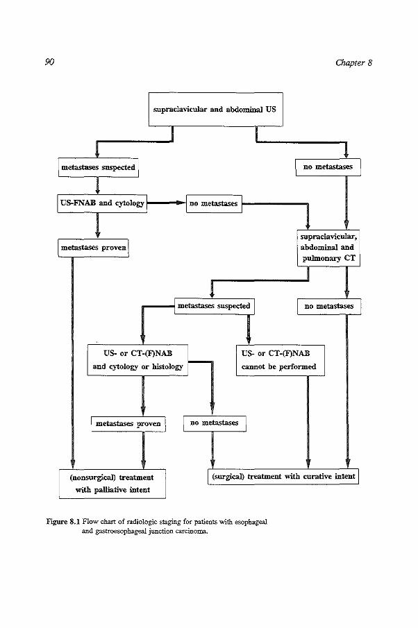
90 Chapter 8
supraclavicular and abdominal US
I I l 1
metastases suspected \ no metastases ·1
! US-FNAB and cytology I I no metastases
supraclavicular,
metastases proven ! abdominal and pulmonary CT
I 1
metastases suspected \ no metastases
US- or CT-(F)NAB US- or CT-(F)NAB
and cytology or histology f--- cannot be performed
\ metastases proven \ no metastases \
(nonsurgicaJ) treatment \ (surgical) treatment with curative intent
with palliative intent
Figure 8.1 Flow chart of radiologic staging for patients with esophageal and gastroesophageal junction carcinoma.

Final Remarks 91
References
1. Lu YK, Li YM. Gu YZ. Cancer of esophagus and esophagogastric junction: Analysis of results of 1025 resections after 5 to 20 years. Ann Thorac Surg 1987; 43: 176-181.
2. F:inley RJ, Inculet Rl. The results of esophagogastrectomy without thoracotomy for adenocarcinoma of the esophagogastric junction. Ann Surg 1989; 210: 535-543.
3. Ellis FH, Gibb SP, Watkins E. Limited esophagogastrectomy for carcinoma of the cardia. Ann
Surg 1988; 208: 354-361.
4. Paolini A, Tosato F, Cassese M, et al. Total gastrectomy in the treatment of adenocarcinoma of
the cardia. Am J Surg 1986; 151: 238-243.
5. Miiller JM, Erasmi H, Steimer M, Zieren U, Pichlmaier H. Surgical therapy of oesophageal carcinoma. Br J Surg 1990; 77: 845-857.
6. Akiyama H, Tsuromaru M, Kawamura T, Ono Y. Principles of surgical treatment for carcinoma
of the esophagus. Ann Surg 1981; 194: 348-446.
7. Earlam R, Cunba-Melo JR. Oesophageal squamous cell carcinoma: A critical review of surgery.
Br J Surg 1980; 67: 381-390.

92

93
APPENDIX
TNM classification and Stage Grouping (AJCC 1988)
Tumor Nodes Metastasis (TNM) classification for tumors of the esophagus
Primary Tumor (T)
T0 No evidence of primary tumor
T u. Carcinoma in situ T 1 Tumor invades lamina propria or submucosa
T 2 Tumor invades muscularis propria T3 Tumor invades adventitia
T4 Tumor invades adjacent structures
Regional Lymph Nodes (N)*
N0 No regional lymph node metastasis
N 1 Regional lymph node metastasis
Distant metastasis (M)
M0 No distant metastasis
M1 Distant metastasis
* For the upper and middle thoracic esophagus the mediastinal lymph nodes are
considered regional lymph nodes.
For the lower thoracic esophagus the mediastinal and perigastric lymph nodes are
considered regional lymph nodes.
Involvement of more distant nodes including the supraclavicular and celiac nodes is
considered distant metastasis.

94 Appendix
Tumor Nodes Metastasis (TNM) classification for tumors of the gastroesophageal
junction
Primary Tumor (T)
T0 No evidence of primary tumor
T m Carcinoma in situ
T 1 Tumor invades lamina propria or submucosa
T2 Tumor invades the muscularis propria or the subserosa
T 3 Tumor penetrates the serosa (visceral J;>eritoneum) without invasion of adjacent
structures
T 4 Tumor invades adjacent structures
Regional Lymph Nodes (N)*
N0 No regional lymph node metastasis
N1 Metastasis in perigastric lymph nodes within 3 em of the edge of the primary
tumor N2 Metastasis in perigastric lymph nodes more than 3 em from the edge of the
primary tumor, or in lymph nodes along the left gastric, common hepatic,
splenic, or celiac arteries
Distant Metastasis (M)
M0 No distant metastasis
M1 Distant metastasis
* Involvement of more distant nodes including the supraclavicular nodes is considered
distant metastasis.

Appendix 95
Stage Grouping for tumors of the esophagus
Stage 0 Ti• N, M, Stage I T, N, M, Stage ITA T, N, M,
T, No M, Stage liB T, N, Mo
T, N, Mo Stage III T, N, M,
T, AnyN Mo Stage N AnyT AhyN M,
Stage Grouping for tumors of the gastroesophageal junction
Stage 0 T;s No M, Stage IA T, No M, Stage m T, N, Mo
T, No Mo Stage II T, N, Mo
T, N, Mo T, No M,
Stage lilA T, N, Mo T, N, Mo T, N, Mo
Stage IIIB T, N, Mo T, N, M,
Stage IV T, N, M,
AnyT AnyN M,

96

97
SUMMARY
The purpose of this thesis was to evaluate the utility of computed tomography (CT),
ultrasound (US) and US-guided fine-needle aspiration biopsy (FNAB) for pretreatment
staging of esophageal and gastroesophageal junction carcinoma. Aspects of assessing the
depth of invasion of the primary tumor and the presence or absence of metastases are
discussed.
In Chapter 1, general information on esophageal and gastroesophageal junction carci
noma is provided.
Chapter 2 presents a retrospective pilot study that was performed to determine the
prevalence of demonstrable supraclavicular metastases with US and US-guided FNAB in
patients with squamous cell carcinoma of the intrathoracic esophagus. Supraclavicular
metastases were cytologically proven in 19% of patients. In these patients US-guided
FNAB of supraclavicular nodes replaced more invasive diagnostic procedures or
avoided extensive surgical procedures.
In Chapter 3, the assessment of supraclavicular metastases with palpation, US and CT
is compared in patients with carcinoma of the esophagus and gastroesophageal junction. Morphologic characteristics of benign and malignant nodes are studied. The relative
frequency of supraclavicular metastases is related to the location of and histologic
findings in the primary tumor. CT and US were equally sensitive and both modalities
were superior to palpation in detecting supraclavicular metastases. Metastases were
found more frequently in patients with squamous cell carcinoma compared to
adenocarcinoma and were most frequently observed in those patients who had
carcinoma of the upper and middle thoracic esophagus. Metastatic nodes tended to have
a round configuration. Neither CT nor US was superior in predicting the metastatic
status of nodes and in cases of enlarged nodes on either study, US-guided FNAB should
be performed to obtain material for cytologic examination.

98 SLlliVrlfjry
Chapter 4 evaluates US and CT in assessing distant metastases in carcinoma of the
esophagus and gastroesophageal junction by comparing these findings with cytological
and surgical data. In assessing distant metastases US, and to a lesser degree, CT were
characterized by a relatively low sensitivity and a relatively high specificity. With the
combination of US and CT, the sensitivity increased to an acceptable level of 83%.
Although this was accompanied by a decrease in specificity it was considered to be less
important because FNAB can be performed to enable cytological examination.
Chapter 5 describes the utility of US-guided FNAB for diagnosing distant metastases in
esophageal and gastroesophageal junction carcinoma by determining the number of
biopsies in which adequate material for cytologic study could be obtained and the
number of patients in whom distant metastases could be proven. A cytologic diagnosis
was established in 87% of biopsies, 13% of biopsies were nondiagnostic. Distant
metastases were diagnosed by means of cytologic study in 24% of patients. No
complication, other than localized mild discomfort was observed.
Chapter 6 discusses the ability of preoperative CT to assess resectability and to stage
carcinomas in patients who undergo transhiatal esophagectomy. CT was not effective to
assess nonresectability by diagnosing invasion; this was due to the relatively low
prevalence of invasion of adjacent structures and the fact that invasion was often not
associated with nonresectability. In assessing invasion itself, CT was accurate in
diagnosing tracheobronchial involvement, but was limited in diagnosing invasion of
other structures. In assessing stage grouping, CT was limited in detecting either
diaphragmatic invasion or lymph node involvement.
In Chapter 7, the influence on survival of distant metastases, assessed on US or CT
studies or proven on cytologic studies is evaluated. Patients with cytologically proven
distant metastases and those unfit to be operated were excluded from surgery, which
was performed in the remaining patients. The 2-year survival for patients with
cytologically proven metastases (3%) was shorter than for patients suspected of
metastases on US and CT studies that were not cytologically confirmed. There was no
significant difference in survival between this last group of patients and those without
metastases on US and CT studies. Following resection the 2-year survival dropped from
53.9% to 0% when distant metastases were present on histopathologic studies of the
resected specimen. Because the expected survival of patients with distant metastases is
low, it is recommended to avoid surgery when these are cytologically proven. Due to

Summary 99
the limited specificity of US and CT, patients should not be excluded from surgery on
the basis of metastases suspected on these studies alone,
In Chaprer 8, final remarks are given within the scope of a pretreatment staging
strategy for patients with esophageal and gastroesophageal junction carcinoma. A flow
chart for a radiologic staging strategy is presented.

100

101
SAMENVATTING
Doe! van dit proefschrift was
tomografie (CT), echografie
pretherapeutische stagering van
het bepalen van de gebruikswaarde van computer
en echogeleide cytologische punctie voor de
oesofagus- en cardiacarcinoom. Aspecten van bet
bepalen van de lokale uitbreiding van de primaire tumor en de aanwezigheid van metastasen worden behandeld.
In Hoofdstuk 1 worden enkele algemene aspecten van het oesofagus- en cardiacarcinoom besproken.
Hoofdstuk 2 geeft de resultaten weer van een retrospectief vooronderzoek met als doe!
het bepalen van de prevalentie van met echografie en echogeleide cytologische punctie
aantoonbare supraclaviculaire lymfkliermetastasen in patienten met een
plaveiselcelcarcinoom van de thoracale oesofagus. Supraclaviculaire metastasen waren in
19% van de patienten cytologisch aantoonbaar. Echogeleide cytologische punctie van
supraclaviculaire lymfklieren verving in deze patienten meer invasieve diagnostische procedures of voorkwam een uitgebreide operatie.
Hoofdstuk 3 geeft de resultaten weer van het vaststellen van supraclaviculaire
lymfkliermetastasen met pa!patie, echografie en CT in patienten met een oesofagus- en
cardiacarcinoom. Morfologische kenmerken van benigne en maligne lymfklieren worden
besproken. Het relatief voorkomen van supraclaviculaire metastasen wordt gerelateerd aan de lokatie en het histologisch type van de primaire tumor. CT en echografie waren even gevoelig en beide technieken waren superieur aan palpatie in de detectie van
supraclaviculaire metastasen.
Metastasen werden vaker gevonden in pati.enten met een plaveiselcelcarcinoom dan in
patienten met een adenocarcinoom en werden bet meest frequent aangetroffen in
patienten met een carcinoom van bet bovenste en middelste e6nderde deel van de
oesofagus. Door metastasen aangedane Iymfklieren badden vaker een ronde
configuratie. CT en echografie waren gelijkelijk beperkt in het voorspellen van
metastasen wanneer vergrote l)rmfklieren werden waa.rgenomen. Wanneer vergrote
lymfklieren met deze onderzoeken worden gevonden moet aansluitend echogeleide
punctie worden verricht om materiaal te verkrijgen voor cytologisch onderzoek.
102 Samenvatring
Hoofdstuk 4 bebandelt de betrouwbaarbeid van ecbografie en CT voor bet vaststellen
van metastasen op afstand door de bevindingen bij deze onderzoeken te vergelijken met
de cytologische en operatieve resultaten. Echografie en in mindere mate CT werden
gekenmerkt door een relatief lage sensitiviteit en een relatief hoge specificiteit in bet
vaststellen van deze metastasen. Wanneer de bevindingen van beide onderzoeken
samengevoegd werden bereikte de sensitiviteit een acceptabel niveau van 83%. De
hiermee gepaard gaande daling in specificiteit wordt van minder belang geacbt, omdat
met cytologische punctie nadere diagnostiek kan worden verricht.
Hoofdstuk 5 beschrijft de bruikbaarbeid van ecbogeleide cytologiscbe punctie voor bet
diagnostiseren van metastasen op afstand in patienten met een oesofagus- en
cardiacarcinoom. Graadmeters voor de bruikbaarbeid waren bet percentage
diagnostiscbe biopsieen en bet aantal patienten bij wie metastasen op afstand cytologiscb
konden worden aangetoond. Van de biopsieen was 87% diagnostisch, in 13% van de
biopsieen werd geen cytologische diagnose verkregen. In 24% van de patienten konden
metastasen op afstand worden aangetoond. Complicaties anders dan lokale gevoeligbeid werden niet waargenomen.
In Hoofdstuk 6 wordt de mogelijkheid onderzocbt om met preoperatief CT on<lerzoek de
resectabiliteit van de tumor te bepalen en deze te stageren in patienten die transbiatale
oesofagusresectie ondergaan. Het voorspellen van irresectabiliteit van de tumor door
middel van het vaststellen van ingroei in naburige organen met CT bleek niet
doeltreffend. Dit werd enerzijds veroorzaakt door de !age prevalentie van ingroei en
anderzijds doordat ingroei vaak geen irresectabiliteit ten gevolge had. CT was
betrouwbaar in het vaststellen van ingroei in de trachea en de bronchi, maar was weinig
betrouwbaar in het voorspellen van ingroei in andere organen. Ten aanzien van de
stagering was CT weinig gevoelig in bet diagnostiseren van diafragma-ingroei en het
detecteren van lymfklierrnetastasen.
Hoofdstuk 7 bespreekt de invloed van metastasen op afstand, gedetecteerd met
ecbografie en CT of bewezen met cytologiscb onderzoek, op de over!eving van
patienten met een oesofagus- en cardiacarcinoom. Patienten met cytologisch bewezen
metastasen op afstand en patienten ongeschikt vanwege andere redenen werden
uitgesloten van operatie; de overige patienten werden geopereerd.
De 2-jaars overleving was lager voor patienten met cytologiscb bewezen metastasen
(3%) dan voor patienten verdacbt van metastasen op echo en CT onderzoeken, die niet

Samenvatting 103
cytologisch waren aangetoond. Er was geen significant verschil in overleving tussen
deze laatste groep patienten en patienten zonder metastasen op echo en CT onderzoeken.
Na operatieve resectie van de tumor daalde de 2-jaars overleving van 53,9% tot 0%
voor de patienten bij wie metastasen op afstand in het resectie preparaat werden
aangetroffen. Omdat de verwachte overleving van patienten met metastasen op afstand
gering is, wordt gea.dviseerd niet te opereren wanneer deze cytologisch worden
aangetoond. Patienten behoren niet te worden uitgesloten van operatie aileen op grand
van voor metastasen verdachte afwijk.ingen op echo en CT onderzoeken, omdat de
specificiteit van deze onderzoeken beperkt is.
Hoofdstuk 8 bevat enkele laatste opmerkingen in het kader van een radiologisch protocol
ten behoeve van de stagering van patienten met een oesofagus- en cardiacarcinoom. Het
protocol wordt in een stroomschema weergegeven.

104
105
EPILOOG
Dit onderzoek werd verricht binnen de afdeling Radiodiagnostiek in samenwerking met
de afdelingen Algemene Heelkunde, Klinische Pathologie (AZR Dijkzigt) en de afdeling
Klinische Besliskunde (Erasmus Universiteit Rotterdam).
Velen hebben bijgedragen aan het totstand komen van dit proefschrift, In het bijzonder
gaat mijn dank uit naar:
Prof. Dr, H,E, Schutte, wiens deur voor mij altijd open stond, voor zijn steun en
opbouwende kritiek
Dr, J,S, Lameris voor het initieren en ten aile tijde stimulerend begeleiden van het
onderzoek.
- Dr. H.W. Tilanus voor zijn waardevolle adviezen en niet aflatend enthousiasme.
Drs. M. Y. Berger voor de intensieve en prettige samenwerking gedurende de laatste
vier jaar.
Drs. R. van Pel voor het beoordelen van de cytologische preparaten en het geven
van feed-back.
Drs. A.I.J. Klooswijk voor het beoordelen van de CT onderzoeken.
Aile leden van de oesofagus werkgroep Rotterdam voor de leerzame bijeen
komsten.
Laboranten van de afdeling Radiodiagnostiek, met name de heer C. Verlooy en
mevrouw E. Nieukoop, voor het verrichten van de CT onderzoeken.
Mevrouw K. Schoenmaker, mevrouw J. Beyl, mevrouw S. van Eysden en mevrouw
E. van Gelder voor het uitwerken van de diverse publicaties.
De heer T. Rij sdijk voor de fotografie.
Drs. LW. Kuiper, mevrouw M.G.A.M. van der Lee en de heer A.W. Zwambom
voor het vervaardigen van de lay-out.
Dr. K. Brake! voor het kritisch doorlezen van het manuscript.

106

CURRICULUM VITAE
HANS VAN OVERHAGEN
1959
1977
1978
1985
1985-1986
1986-1991
1991-heden
4 september, geboren te Lei den
eindexamen Atheneum B, Rijnlands Lyceum
te Oegstgeest
kandidaatexarnen I Nederlands Recht, Rijksuniversiteit te Leiden
artsexamen, Rijksuniversiteit te Leiden
onderzoeker, afdelingen Klinische Immunologie en
Chirurgische Oncologie, Academisch Ziekenhuis te Groningen
arts-assistent in opleiding tot radiodiagnost,
Academisch Ziekenbuis Rotterdam
stafmedewerker afdeling Radiodiagnostiek,
Academisch Ziekenhuis Rotterdam
107

![Typical and atypical radiologic manifestations of ......of radiographic staging [1,2,6]. Middle mediastinal nodes (at the left paratracheal level, subcarinal level, and level of the](https://static.fdocuments.us/doc/165x107/5f7d2ff2ec543436a327439a/typical-and-atypical-radiologic-manifestations-of-of-radiographic-staging.jpg)