
Radiochemistry.ppt
80
Supplementary Course Topic 1: uclear and Radiation Chemistry • Nucleons, nuclides and isotopes • Nuclear fusion and stellar nucleogenesis • Natural Radioactivity • Which nuclides are stable and why • Where decay mechanisms come from • How fast does an unstable nucleus decay? Half-lives • Radiocarbon Dating • How does radiation interact with (biological) matter? • How is radiation exposure measured? • What are common sources of radioactivity? • Medical Imaging
-
Upload
brucelee55 -
Category
Documents
-
view
1.647 -
download
0
Transcript of Radiochemistry.ppt

Supplementary Course Topic 1:
Nuclear and Radiation Chemistry
• Nucleons, nuclides and isotopes
• Nuclear fusion and stellar nucleogenesis
• Natural Radioactivity
• Which nuclides are stable and why
• Where decay mechanisms come from
• How fast does an unstable nucleus decay? Half-lives
• Radiocarbon Dating
• How does radiation interact with (biological) matter?
• How is radiation exposure measured?
• What are common sources of radioactivity?
• Medical Imaging

Nucleons - The Sub-Atomic Particles
Particle Symbol Charge Mass (a.m.u.)
proton p +1 1.007276
neutron n 0 1.008665
electron e- -1 0.000549
positron e+ +1 0.000549
The unit of mass is atomic mass units (a.m.u.), defined by setting the mass of the isotope to exactly 12. 1 a.m.u. = 1.66 x 10⁻²⁷ kg.
126C
Not present in stable atoms.

Nuclides and Isotopes
The composition of any nucleus is defined by two numbers.• The atomic number is the number of protons in the nucleus.
• This defines the chemical nature of the atom.• It is equal to the total charge on the nucleus.
• The mass number is the total number of nucleons (protons and neutrons) in the nucleus.
E.g. has an atomic number of 6 and a mass number of 12.
• A nuclide is an atom with a particular mass number and atomic number.
• Nuclei with the same atomic number but different mass numbers are called isotopes.
126C

Nuclides and Isotopes
Nuclei with the same atomic number but different mass numbers are called isotopes.
E.g. Carbon may exist as a number of isotopes
116C 12
6C 136C 14
6C
Stable nucleus;
accounts for 98.89% of
natural carbon.
Stable nucleus;
accounts for 1.11% of natural carbon.
Unstable nucleus.
Unstable nucleus;
prepared by nuclear
reaction in a cyclotron.
156C
Unstable nucleus; trace
amounts present in
living matter.

How Mass Spectrometry Works
In a mass spectrometer, the atoms or molecules to be studied are vapourised and then ionised, usually by an electrical discharge.
In the conventional design of a mass spectrometer, ions follow a curved path and their deflection depends on the mass-to-charge ratio, m/z (sometimes denoted m/e). This deflection was originally recorded as impact on a strip of photographic film, but now use digital current or luminescence detectors.

Mass Spectrometry
Aston’s results established the existence of isotopes. (They were already known for radioactive elements, but never shown for stable elements.)
1920 - Aston measured two isotopes of Ne (20 and 22), three of S (32, 33, 34), three of Si (28, 29, 30), six of Kr (78, 80, 82, 83, 84, 86), and many others

Nuclides and Isotopes
The atomic mass of an element is the average of the atomic masses and abundances of each of the naturally-occurring isotopes.
E.g. The atomic mass of carbon is 12.01.
That is (12.0000x98.89 + 13.00335x1.11)/100
126C 13
6C
Mass of nuclide taken from a
reference table
Mass of nuclide is the reference for a.m.u scale.

Nucleogenesis
Where do the elements come from?
How are atoms (nuclei) formed?
All atoms are generated from the simplest element, hydrogen , by nuclear reactions.
Clouds of atomic hydrogen are pulled together by gravity and begin to heat as they are compressed. Eventually high enough temperatures for nuclear fusion are achieved and the cloud ignites as a star.
11 H
Nucleogenesis
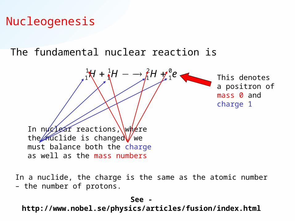
The fundamental nuclear reaction is
1 1 2 01 1 1 1H H H e This denotes a
positron of mass 0 and charge 1
In nuclear reactions, where the nuclide is changed, we must balance both the charge as well as the mass numbers
In a nuclide, the charge is the same as the atomic number – the number of protons.
See - http://www.nobel.se/physics/articles/fusion/index.html
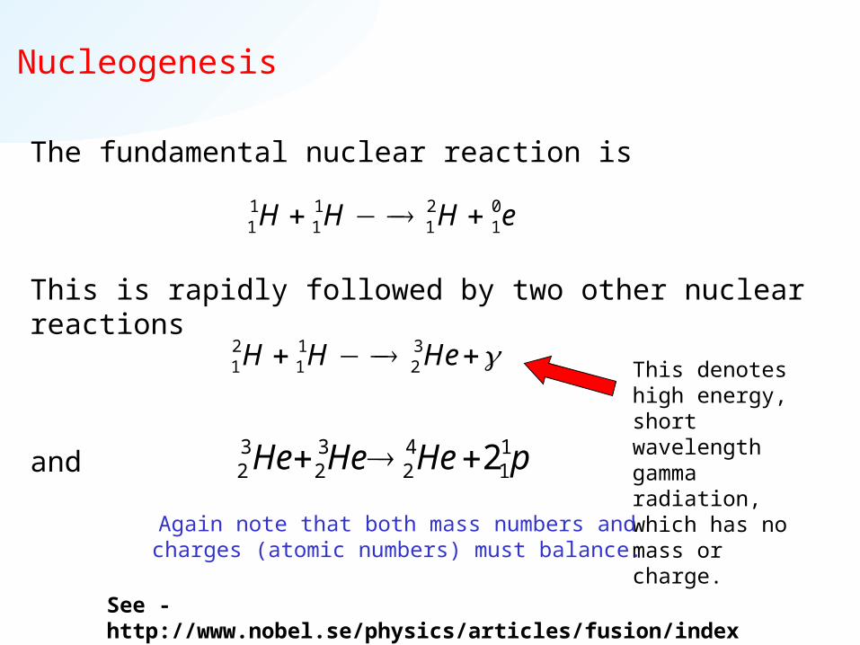
Nucleogenesis
The fundamental nuclear reaction is
This is rapidly followed by two other nuclear reactions
and
1 1 2 01 1 1 1H H H e
2 1 31 1 2H H He This denotes high
energy, short wavelength gamma radiation, which has no mass or charge.
Again note that both mass numbers and charges (atomic numbers) must balance.
pHeHeHe 11
42
32
32 2
See - http://www.nobel.se/physics/articles/fusion/index.html

Nucleogenesis
The overall “hydrogen burning reaction”
releases energy into the surroundings as heat (exothermic) and radiation (also releases neutrinos ).
As the star exhausts its hydrogen, it begins helium burning to fuse heavier nuclei to form increasingly larger atoms.
E.g.
1 4 01 2 14 2H He e
Heavier nuclei like 13C, 13N, 14N, 15N, 15O... are produced by red giant stars, still heavier nuclei in supergiants, and true heavy elements form in supernovae.
BeHeHe 74
42
32
BpBe 85
11
74

Life Cycle of Stars
Hydrogen Hydrogen burningburning
T ~ 107KHeavy Heavy
elementselements4040Ca…Ca…5858Ni formedNi formed
(C and O burning)T < 3 x 10⁹K
Hydrogen burningHydrogen burning
T ~ 107K
Helium burningHelium burning
T < 2 x 10⁸K
Helium Helium burningburning
T < 2 x 10⁸K
Carbon coreCarbon core
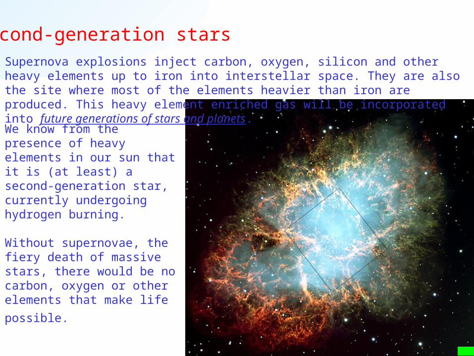
Second-generation starsSupernova explosions inject carbon, oxygen, silicon and other heavy elements up to iron into interstellar space. They are also the site where most of the elements heavier than iron are produced. This heavy element enriched gas will be incorporated into future generations of stars and planets.
We know from the presence of heavy elements in our sun that it is (at least) a second-generation star, currently undergoing hydrogen burning.
Without supernovae, the fiery death of massive stars, there would be no carbon, oxygen or other elements that make life
possible.

Nucleogenesis …and the periodic table
H burningH burning
He burning. Star expands to red giantC burning. Core
of red giant
Red supergiant core.
Supernova (everything heavier)Supernova (everything heavier)

Natural Radioactivity
Nucleogenesis produces nuclides that can be stable or unstable. Unstable nuclei decay through a range of mechanisms involving the release of particles with high kinetic energy or of -radiation. These high-energy products are collectively known as radioactivity.
"in recognition of the extraordinary services they have rendered by their joint researches on the radiation phenomena discovered by Professor Henri Becquerel"
"in recognition of the extraordinary services he has rendered by his discovery of spontaneous radioactivity"
Henri Becquerel Pierre Curie
Marie Curie

Natural Radioactivity
The four most important radioactive decay mechanisms are
1. decay
e.g.
2. decay
e.g.
212 208 483 81 2Bi Tl
12 12 05 6 1B C e
The particle is simply a helium nucleus with mass 4 and charge 2+.
As with all nuclear reactions, both mass and charge are balanced.
(or -) is an electron ejected from the nucleus.
One neutron is changed into a proton in this nuclear reaction to balance the charge.
Natural Radioactivity
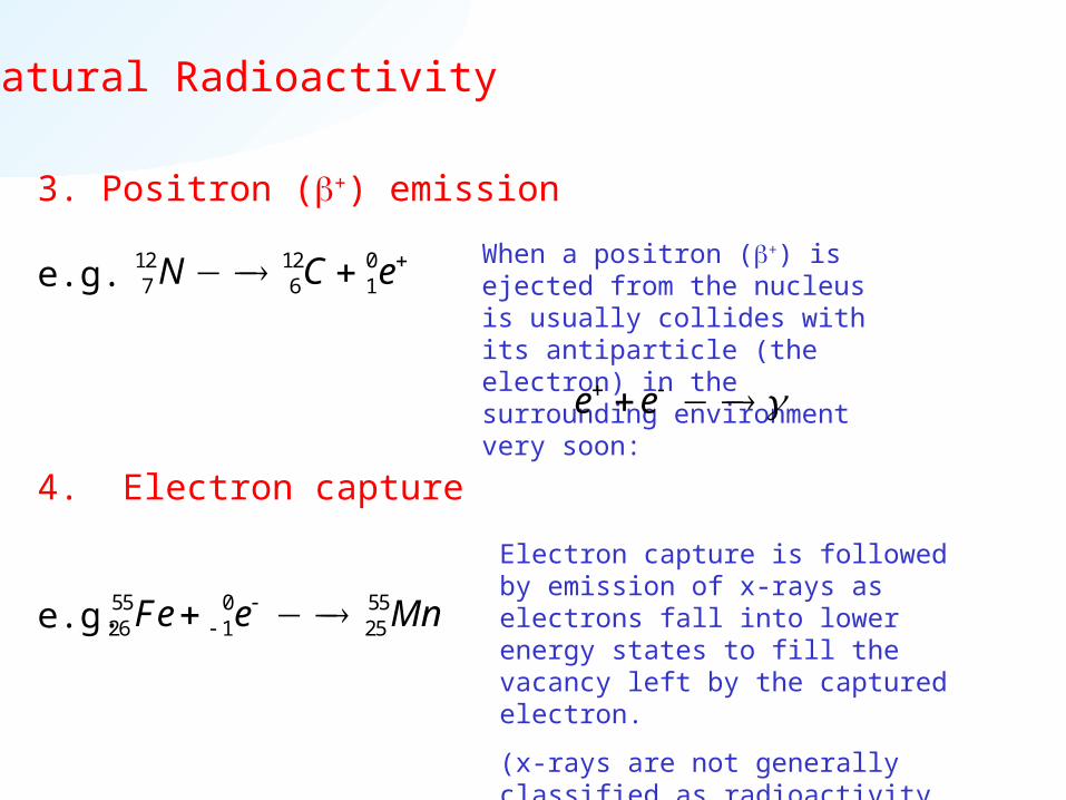
3. Positron (+) emission
e.g.
4. Electron capture
e.g. 55 0 5526 1 25Fe e Mn
12 12 07 6 1N C e
Electron capture is followed by emission of x-rays as electrons fall into lower energy states to fill the vacancy left by the captured electron.
(x-rays are not generally classified as radioactivity, although they can cause radiation damage.)
When a positron (+) is ejected from the nucleus is usually collides with its antiparticle (the electron) in the surrounding environment very soon:
e e

Natural Radioactivity - worked example
Balance the following nuclear decay reactions and identify the emitted particle where appropriate.
1.
2.
3.
63 028 1 Ni e
234 23092 90U Th
36 3617 16 Cl S
4 42 2 or He
6329Cu
01e

Nuclear Reactions - worked example
Nuclear reactions are balanced in the same way, but may involve more than one reactant. Balance the following nuclear reactions and identify the missing nuclide or particle.
1.
2.
3.
239 4 194 2 0 Pu He n
14 4 177 2 8N He O
28 2 2914 1 15Si H P
1 11 1 or H p
24296Cm
10 n

Natural Radioactivity - and x-rays
x-rays have shorter wavelengths than visible or ultraviolet light - between 0.01nm and 10nm.
rays have very short wavelengths - less than
0.01nm or 0.1Å
Both x-rays and radiation are high energy (= high frequency or short wavelength) forms of light.

Natural Radioactivity
Unstable heavy nuclei decay spontaneously by a series of steps through unstable intermediates. Over time, unstable nuclei give rise to a family of decay products in a decay series.
E.g. 238U decays into…
…etc, etc,...
238 234 492 90 2U Th
234 234 090 91 1Th Pa
234 234 091 92 1Pa U 234 230 492 90 2U Th
230 226 490 88 2Th Ra

Natural Radioactivity
A radioactive decay sequence (e.g. of 238U) can be represented more concisely as a graph of atomic number versus neutron number.
decay is shown as a decrease of two protons (Z) and two neutrons (N).
decay is shown as a decrease of one neutron and an increase of one proton.
Isotopes (same Z, different N) lie along vertical lines in this graph.

Natural Radioactivity
A radioactive isotope like 238U thus generates a family of daughter isotopes in a decay series.Naturally-occurring uranium contains 238U, and so will also contain components of the decay series.

238238UU
206206PbPb
Radioactive Decay Series …and the periodic table

Marie Curie
•Born Maria Sklodowska in Warsaw, Poland on November 7, 1867.
•In 1891 at 24, Sklodowska went to Paris to study mathematics, physics and chemistry at the Sorbonne.
•July 25, 1895 married Pierre Curie
•1903 Nobel Prize for Physics
•1911 Nobel Prize for Chemistry
•Discovered Radium and Polonium, coined term “Radio-activity”.
•In 1906, Pierre Curie, was hit by a horse-drawn carriage and killed.
•Marie Curie died at the age of 67 in 1934 of leukemia. Her cremated remains are kept in the Pantheon in Paris
•In 1935, the Curie's daughter, Irene Joliot-Curie won a Nobel Prize for Chemistry in 1935, making them the first mother and daughter to share this honor.

Nuclear Stability
What factors determine whether a nucleus is stable or unstable?
If we look at the range of stable nuclides that exist in nature, then there are two main observations
1. The size of the nucleus.
2. The composition of the nucleus (proton:neutron)

Nuclear stability 1. Nuclear size
There are no stable nuclei heavier thanThere are no stable nuclei heavier than20983 Bi

Nuclear Stability 2. neutron:proton (N:Z)
All known stable nuclides fall inside the zone of stability.This zone has a N:Z ratio near to 1, but “bends” towards more neutrons per proton as the nucleus gets larger.
These two observations are enough to give us a “rule” for nuclear stability that goes something like
“Unstable isotopes must decay towards the zone of stability, finally falling below 209Bi.”
Nuclear Stability and Decay Mechanisms
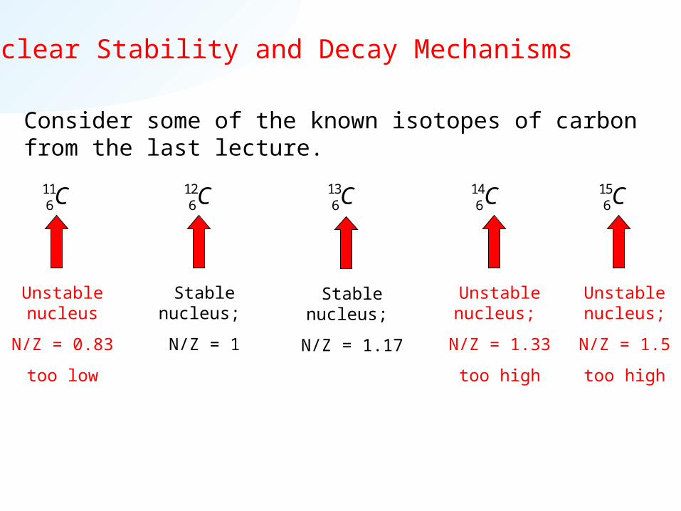
Consider some of the known isotopes of carbon from the last lecture.
116C 12
6C 136C 14
6C
Stable nucleus;
N/Z = 1
Stable nucleus;
N/Z = 1.17
Unstable nucleus;
N/Z = 1.5
too high
Unstable nucleus
N/Z = 0.83
too low
156C
Unstable nucleus;
N/Z = 1.33
too high

Nuclear Stability and Decay Mechanisms
Each nuclide decays towards the zone of stability by changing its N/Z ratio at constant mass number.
11 11 06 5 1C B e
N/Z = 0.83 N/Z = 1.2
14 14 06 7 1C N e
N/Z = 1.33 N/Z = 1.0
15 15 06 7 1C N e
N/Z = 1.5 N/Z = 1.14
N/Z too low gives + decay. N/Z too high gives - decay.
N/Z = 1.11 N/Z = 1.2
Or equivalently by electron capture.
55 0 5526 1 25Fe e Mn

Nuclear Stability
The “rule” for nuclear stability:
“Unstable isotopes must decay towards the zone of stability, finally falling below 209Bi.”
N/Z too low + decay
or electron capture.
N/Z too high - decay.
Heavier nuclides than 209Bi decay by a combination of mechanisms, using decay to reduce mass (with N/Z = 1) and the other mechanisms to change N/Z.
Mass too high decay.

Nuclear Stability
E.g. Here is how the 238U decay sequence looks on our zone of stability graph.

Nuclear Stability - Origin of Decay Mechanisms
The nuclear stability “rule” is empirical, based on the simple experimental observation of which nuclides are stable and which are not.
We can apply it like an algorithm to solve some nuclear decay problems, without understanding the reasons for nuclear stability.
To understand the reasons, the rule, and the observations, we need to consider the forces between nucleons within the nucleus.

Nuclear Stability - Origin of Decay Mechanisms
The stability of a nucleus involves the competition between two forces.
1. Coulomb or electrostatic repulsion between protons acts to push these nucleons apart over a long range.
2. The strong nuclear force is a short range attraction between all nucleons.
This is the main function of neutrons in the nucleus. They contribute to the binding of the nucleus without also contributing to the electrostatic destabilisation.

Nuclear Stability - Origin of Decay Mechanisms
How does this explain our observations?
1. In nuclides with too few neutrons, the electrostatic repulsions overwhelm the strong nuclear attractions.
2. As the nucleus gets larger, the long-range electrostatic repulsion between protons accumulates and eventually overwhelms the strong nuclear attraction, even if N/Z is optimised.
This microscopic model does not explain how nuclides with too many neutrons can be unstable. To do so will involve quantum mechanics.

Rate of Nuclear Decay
Unstable nuclides are present in nature for two reasons.
• Some unstable nuclides have long half-lives, so they simply haven’t decayed yet.
• Some unstable nuclides continue to be formed by nuclear reactions.
The decay of an unstable nuclide is characterised by a half-life. This is the time required for half of the nuclei present to undergo a decay event.
For each parent nuclide that decays, one daughter nuclide is produced and a particle or is emitted. e.g.
212 208 483 81 2Bi Tl 12 12 05 6 1B C e
Parent nuclide
Daughter nuclide
Emitted Particle

Rate of Nuclear Decay - Half-Life
E.g. 32P decays into 32S with a half-life of 14.7 days.
The number of 32P nuclei halves in 14.7 days, and halves again after a further 14.7 days...
32 32 015 16 1P S e
So, after 14.7 days, half of an initial 10g of 32P will have decayed, leaving 5g. At the same time 5g of 32S will have formed.
After a further 14.7 days, only 2.5g of 32P will remain, and 7.5g of 32S will be present…
This also tells us that the rate of decay, the number of nuclei that disintegrate each second, also halves every 14.7 days.
The rate of decay halves after every half-life.

0
0.25
0.5
0.75
1
0 25 50 75 100
time (years)
N/N
0
The Rate of Nuclear Decay
The disappearance of a radionuclide by radioactive decay is described by
The number of nuclei remaining
after time t.
0( ) exp( )N t N t
The number of nuclei present at the beginning.
The decay constant.
E.g. Decay curve for 3H, T1/2 = 12.26 years.
The number if nuclei halves every 12.26 years.

The half-life is the time required for half of the nuclides to decay (and for half to be left). So if we let N = N0/2
and solve, this gives
So finally we can write the decay in terms of half-life as
The Rate of Nuclear Decay
00 1 2exp( )
2
NN t
1 2
ln(2) 0.693t
0 1 2( ) exp( 0.693 / )N t N t t

Activity is the Rate of Nuclear Decay
The activity, A, of a radionuclide is simply the rate of emission, or minus the rate of disappearance of the nuclide. i.e.
0
0
exp( )
exp( )
dN dA N t
dt dtN t
N
The activity (rate of decomposition) of a sample is proportional to the number of nuclei present.
i.e. when the number of nuclei present is halved, the activity is also halved.

Units of Activity
• Fundamental unit of activity - Disintegrations per second, also known as the becquerel (Bq)
• Curie (Ci) – 1 Ci equals the number of nuclei disintegrating each second in 1g of 226Ra.
= 3.70 x 10¹⁰ counts per second (or Bq).

Activity and Half-Life
• Activity and Half-Life are related.• Low activity (few disintegrations per second) = long half-life.• High activity = short half-life
• Molar Activity = Activity/mole
• Specific Activity = Activity/gram
M = atomic mass
1 2
0.693A N N
t
1 2
0.693M A AA N N
t
Molar Activity, Specific Activity and Half-Life are both independent of the amount of radioactive material present in the sample.
1 2
0.693A AS
N NA
M t M

Activity and Half-Life - worked example
What is the molar activity of 13N, which has a half life of 9.96
minutes?
Answer.
9.96 minutes = 598s
AS = 0.693 x 6.022 x 1023/598
= 6.98 x 10²⁰ disintegrations mol-1 s-1 (or Bq mol-1)
or 6.98 x 10²⁰/3.70 x 1010 = 1.88 x 10¹⁰ Ci mol-1
1 2
0.693M A AA N N
t

Rate of Nuclear Decay - Decay Series
In a decay series, each step in the mechanism has its own half-life.
Notice that the half-life of 238U is 4.5x10⁹ y, so that many of the atoms present when the earth was formed still have not decayed. However the daughter nuclides decay much more quickly.
Notice also that half lives can be as long as billions of years or as short as a ms or less.

Radiocarbon Dating
99.9% of the naturally abundant 99.9% of the naturally abundant 1414C is C is produced in the upper atmosphere by produced in the upper atmosphere by neutrons reacting with neutrons reacting with 1414N, which then N, which then enters the carbon cycle. enters the carbon cycle.
The production rate is 2.5 atom cmThe production rate is 2.5 atom cm-2-2 s s-1-1 with a global inventory of 3 x 10with a global inventory of 3 x 103030 1414C C atoms (90% oceans, 8% biosphere and atoms (90% oceans, 8% biosphere and soils, 2% atmosphere). Typical soils, 2% atmosphere). Typical 1414C C concentration in sea waters is concentration in sea waters is 1.2 x 101.2 x 1099 1414C atoms/L (2 x 10C atoms/L (2 x 10-15 -15 M). M).
www.ansto.gov.au/ansto/environment1/ams/ams_14c.htmwww.ansto.gov.au/ansto/environment1/ams/ams_14c.htm
14 1 14 17 0 6 1N n C p
14C is an example of an isotope that is continuously produced in our environment.

Radiocarbon Dating
Other Sources of 14C:
In-situ (0.1%): 14C is produced by spallation reactions induced by neutrons and muons incident on the surface of the Earth. The 14C production rate from quartz (at sea-level) is 20 atoms g-1 yr-1 and varies with elevation and geomagnetic latitude. A wide range of geophysical problems can be studied by this method including erosion histories, uplift rates, glacial histories, eruption ages, rates of movements of sand dunes, accumulation and ablation rates of ice and climatic change.
Radiogenic: 14C can be produced underground, directly or indirectly, by the decay of uranium and thorium series. An estimate of this 14C can be useful in the study of hydrological environments where uranium and thorium contents are high.
Anthropogenic: 14C levels in the atmosphere show a major peak in 1963 (with about 100% increase in 14C concentration) because of contributions from nuclear weapons testing and a slow drop since then. This bomb pulse is useful in the study of environmental problems such as the air enclosure process in ice and the circulation of groundwaters.
http://www.ansto.gov.au/ansto/environment1/ams/ams_14c.htm

How Radiocarbon Dating Works
All organic living matter contains a fixed fraction of 14C amongst all its carbon. This comes mostly (from atmospherically generated) material taken up by biochemical paths.
After death, 14C no longer accumulates, and decays into
with a half-life of 5730 y.
Thus comparing the concentration of 14C in dead and comparable living matter can tell us how long since the sample died.
14 14 06 7 1C N e

Radiocarbon Dating
How is the amount of 14C determined?
As noted before, 14C undergoes decay.
Radiocarbon dating can be achieved either
• by measuring the concentration of 14C present in a sample, or
• by measuring the activity due to emission. (Recall that activity is
proportional to the number of decaying nuclei: )
http://www.c14dating.com/
14 14 06 7 1C N e
1 2
0.693A N N
t

Radiocarbon Dating
Measuring Activity - scintillation counter.
Scintillation is the emission of light when exposed to ionizing radiation. Scintillation counters simply measure the intensity of light emitted as a result of exposure to a radiation source. By calibration against a standard, this can read activity directly.
Measuring 14C - accelerator mass spectrometry (AMS)
AMS is a high precision mass spectrometry technique that can measure small amounts of sample and resolve isotopic composition. In 14C dating, AMS is used to measure the ratio of 14C/13C and 14C/12C. As 13C and 12C are stable, these ratios can be used directly to obtain radiocarbon age. As carbon from different sources can have slightly different isotopic ratios due to various chemical processes, the ratio of 13C/12C is measured directly in AMS as an additional calibration correction.

Radiocarbon Dating - Activity Ratio
The activity is proportional to the number of nuclei present. A NThus the ratio of the activity after death to activity while alive is equal to
the ratio of the number of 14C nuclides.
0
0 0 0
exp( )A N NdNdN
tdt dtA N N
To determine the age of a sample we compare the activity A with the activity of a still-living (or recently dead) sample, A0, and use the half-life or decay constant.
0
0.25
0.5
0.75
1
0 25 50 75 100
time (years)
N/N
0
0
0.25
0.5
0.75
1
0 25 50 75 100
time (years)
A/A
0

Radiocarbon Dating
This method assumes that the concentration of 14C in living matter has been constant over the dating period.
This assumption is known not to be exactly true, so a number of qualifications and corrections are applied to 14C dates, and a standard method is always used to report radiocarbon age.
1. The age of a sample is reported as its radiocarbon age. This may be reported as “years BP” (before present, where present = AD1950 when radiocarbon dating was invented).
2. An uncertainty or “error range” is often reported based on known changes in 14C levels as well as on experimental uncertainty.
3. The radiocarbon age may be corrected using a calibration graph obtained from independent data.
4. Variations in natural isotopic ratios between sources are also corrected.

Calibration of Radiocarbon DatingWillard Libby, who invented 14C dating in 1946 (Nobel Prize, 1960), prepared a primary calibration graph, shown below, using samples with independently-determined ages.
The curve shows the “Libby half-life” of 5568y, which is used to determine the radiocarbon age of materials and effectively assumes a constant rate of 14C production.
Note that all the independent data is <5000 years old.

Radiocarbon Dating - worked example
E.g. A one gram sample of carbon from peat moss has an activity of 0.350mCi. A reference or modern standard sample yields 0.446mCi. What is the radiocarbon age of the sample?
08033ln
0.4468033ln
0.350
1950
At
A
Years BP (rounded up from 1947)
The units of time are determined by the units of the pre-factor or half-life.
This is a ratio. The units of activity must be the same in the numerator & denominator.
This pre-factor is obtained from the “Libby half-life” and is equal to5568/ln(2)

Radiocarbon Dating by AMS
AMS has two major advantages over activity measurements.
1. Sensitivity. AMS can measure samples as small as a few mg of carbon, or much older samples in which the fraction of the original 14C remaining is very small. AMS has an effective limit of around 26,000 years, whereas activity ages are limited to about 10,000y.
2. Internal Calibration. AMS can measure the ratios between all carbon isotopes directly. This means that local variations in isotopic composition (fractionation) for stable (12C and 13C) and unstable 14C isotopes are determined in situ.
In AMS, ratios are often expressed as deviations from a standard. For the stable isotopes the National Institute of Standards and Technology (NIST) gives the value Rstd=13C/12C = 0.011237. Deviation from this value for different materials is expressed in parts per thousand (per mille, or ‰)
1000std
std
R R
R

Radiocarbon Dating by AMS - The Shroud of Turin
AMS was used to determine the age of the Shroud of Turin by radiocarbon dating in 1989. Each sample investigated consisted of 50mg of cloth, which was analysed independently by three different laboratories.
13C was measured directly, and gave results around -25‰, consistent with calibration standards for such fibres (independent of age).
Read the original scientific article at http://www.shroud.com/nature.htm
Radiocarbon age (corrected for 13C) was determined from the 14C/13C ratio to be 690±30 years BP. Three similar references samples were also dated:
• 11-12th century linen dated at 940y BP• Linen from the mummy of Cleopatra dated at
1960y BP• Threads independently dated to 1300AD, 14C
dated at 724y BP.
Conclusion: “...the linen of the shroud of Turin is mediaeval.”

The “Bomb Pulse”
The ambient The ambient 1414C level increased due to atmospheric nuclear testing, C level increased due to atmospheric nuclear testing, peaking in 1962 - known as the “Bomb Pulse.”peaking in 1962 - known as the “Bomb Pulse.”
This has been accurately tracked over time as the deviation from the pre-bomb isotope ratio, 14C, and can be used to accurately determine the (recent) age of carbon-containing materials.
E.g. • wine dating & detecting false labels or blends.• dating drug crops.
U.S. Department of Energy photograph http://www.nv.doe.gov/news&pubs/photos&films/atm.htm

Biological Effects of Radiation
How do various forms of radiation interact with (biological) matter?
The basic characteristic of radiation produced by radioactivity is that it is high energy, and causes the ionization of matter by ejecting an electron from an atom. (It’s generally called ionizing radiation.)
When radiation is stopped my matter, it has interacted with it and therefore caused ionization.
Highly penetrating radiation passes through matter without ionizing it.
Radiation protection and biological effects are the concern of Health Physicists. Nuclear facilities employ health physicists to train staff, monitor activites, and develop safety protocols.
Biological Effects of Radiation
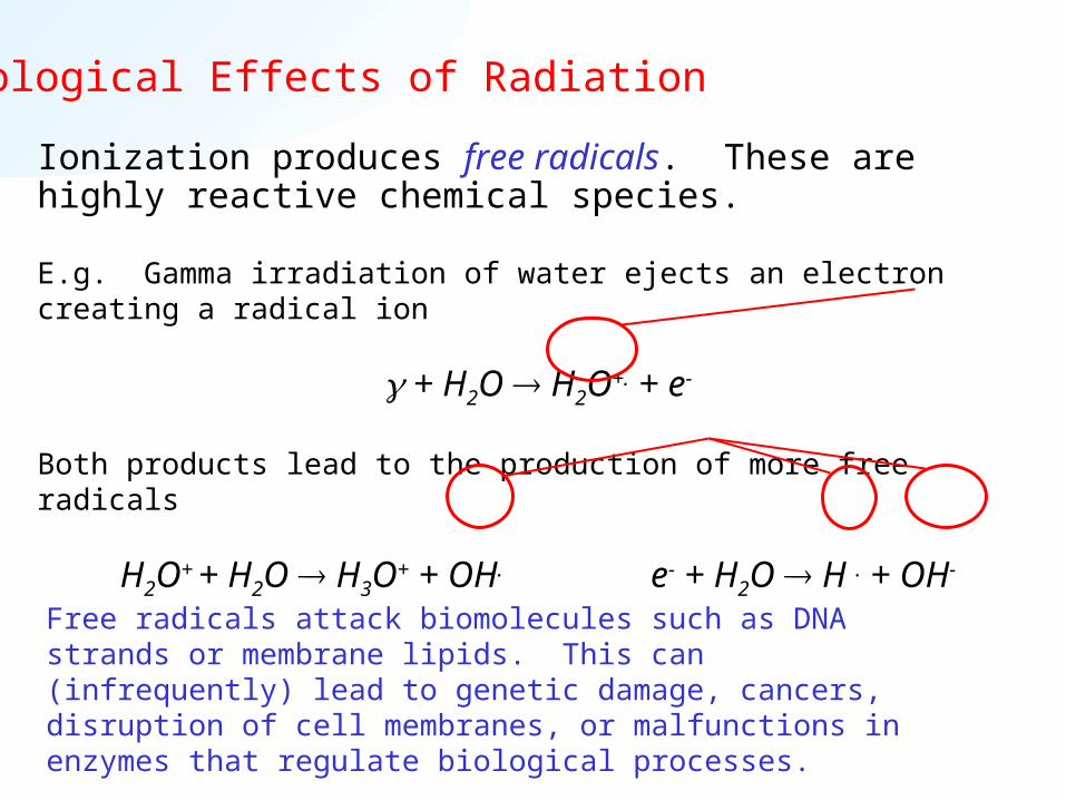
Ionization produces free radicals. These are highly reactive chemical species.
E.g. Gamma irradiation of water ejects an electron creating a radical ion
+ H2O H2O+. + e-
Both products lead to the production of more free radicals
H2O+ + H2O H3O+ + OH. e- + H2O H . + OH-
Free radicals attack biomolecules such as DNA strands or membrane lipids. This can (infrequently) lead to genetic damage, cancers, disruption of cell membranes, or malfunctions in enzymes that regulate biological processes.
Biological Effects of Radiation
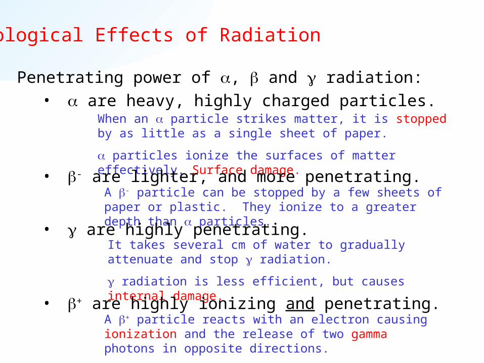
Penetrating power of , and radiation:• are heavy, highly charged particles.
When an particle strikes matter, it is stopped by as little as a single sheet of paper.
particles ionize the surfaces of matter effectively. Surface damage.
A - particle can be stopped by a few sheets of paper or plastic. They ionize to a greater depth than particles.
• - are lighter, and more penetrating.
It takes several cm of water to gradually attenuate and stop radiation.
radiation is less efficient, but causes internal damage.
• are highly penetrating.
A + particle reacts with an electron causing ionization and the release of two gamma photons in opposite directions.
• + are highly ionizing and penetrating.

Radiation Exposure
Biological exposure arises through a variety of mechanisms.
E.g. Direct exposure to a radioactive source (external)
Solid or liquid: Localised source. Radiation can be shielded.
Gas or vapour: Diffuse source. May diffuse around shielding.
E.g. Contamination (external and internal)
External: Picking up radioactive material on hands, shoes, etc…
Internal: Ingestion or inhalation.
The risk of damage from particles through internal contamination is much higher than external, where they are quite well shielded by your skin.For more details, you can install and run the U.S. National Centre for Neutron Research safety training programme available on the First Year Chemistry web site. This is the same programme used to train visitors, temporary and permanent workers in the nuclear reactor at the National Institute of Standards and Technology.

Biological Effects of Radiation
Half-Life and Radiation damage
Nuclides with longer half-lives disintegrate at lower frequency.
That is, longer half lives equals lower (molar) activity, so lower potential for ionization and radiation damage.
From this point of view 238U, with a half-life of 4.5 billion years, or 230Th (T1/2= 83,000 y) is less damaging than 3H (T1/2= 12 y) or 234Th (T1/2= 24.5 days).
1 2
0.693A N N
t
Units of Radiation Dosage
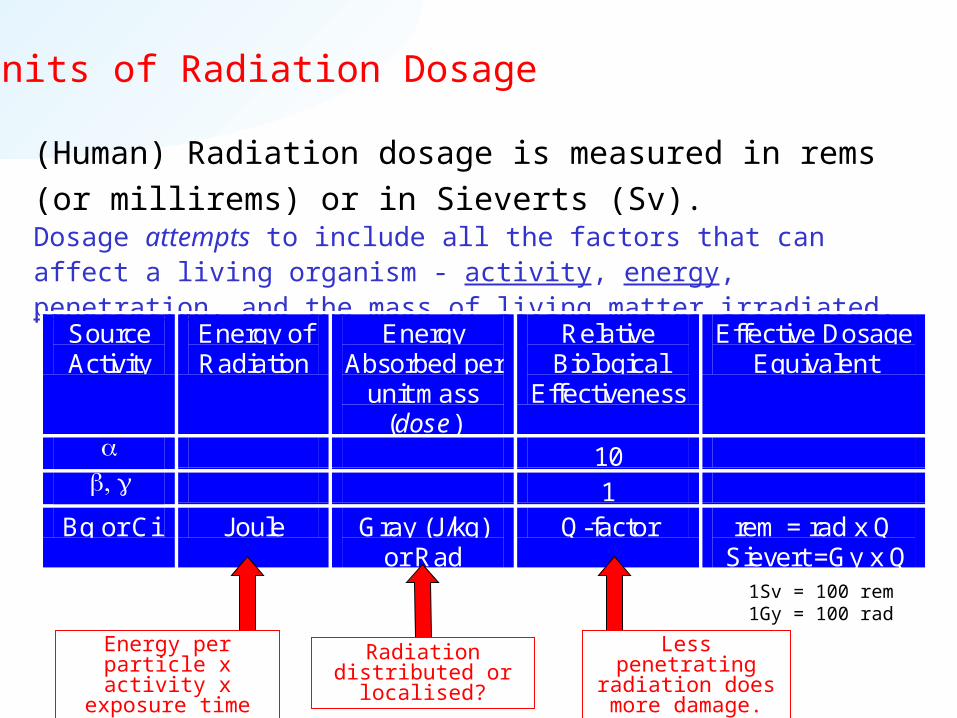
(Human) Radiation dosage is measured in rems (or millirems)
or in Sieverts (Sv).Dosage attempts to include all the factors that can affect a living organism - activity, energy, penetration, and the mass of living matter irradiated.
Source Activity
Energy of Radiation
Energy Absorbed per
unit mass (dose)
Relative Biological
Effectiveness
Effective Dosage Equivalent
10 1
Bq or Ci Joule Gray (J/kg) or Rad
Q-factor rem = rad x Q Sievert =Gy x Q
Energy per particle x activity x exposure
time
Less penetrating radiation does more
damage.
Radiation distributed or localised?
1Sv = 100 rem1Gy = 100 rad

Dosages and Their Effects
The total expected dosage for an average person is about 360 mrem/year.
What are the short-term effects of radiation dosage?
25,000 mrem in 24h No detectable effects
50,000 mrem in 24h Slight temporary blood change
100,000 mrem in 24h Nausea & fatigue
200,000 mrem in 24h First death (no medical intervention)
500,000 mrem in 24h LD50 (50% of humans exposed die.)
N.B. The probability of longer-term effects increases with dose. Most health physicists use a linear no-threshold model. That is, they assume that there is no level of exposure that is free from effects. However the time-scale and statistical nature of the effects make low-dose response hard to determine.

Common Sources of Radioactivity
We are exposed to several common natural sources of radioactivity. These account for about 300mrem/year. The most common is radon, which is part of the decay series of 238U and other heavy elements, and decays into polonium with a half-life of 3.82 days.
decay of radon gas causes damage to lungs and is thoughtto be responsible for up to 10% of lung cancers.
Other ambient isotopes include
as dissolved potassium ions, and
in CO2 and organic compounds.
222 218 486 84 2Rn Po He
40 40 019 18 1K Ar e
14 14 06 7 1C N e

Other Common Sources of Radioactivity
• Other common sources of radiation include cosmic rays in the upper atmosphere. Average annual exposure from this source at ground level is ~26mrem.
• A 4h plane flight increases dosage by a few mrem.
• Medical exposure (x-rays and nuclear medicine) is around 40mrem/year
• Consumer products ~10mrem/y
• Nuclear fallout <1mrem/y
• Nuclear power ~.05mrem/y

Medical Imaging
Basic principles of medical imaging.
• Use a radioisotope to specifically target a chemical agent, organ or
process in the body with high selectivity.
• Isotope should emit low-energy, highly-penetrating radiation to minimise
effective dosage equivalent to patient. In practice this usually means .
• Image distribution of radioisotope (by its activity) using scintillation
counting • gamma camera (planar image like an x-ray) or • computer-assisted tomography (CAT or CT scan - cross section or
three-dimensional reconstruction)
• Images may be a simple gray scale density or pseudo-colour signal.
Pseudo colour is especially common in computer-reconstructed imaging.

Gamma camera CT scanner
E.g. -camera image of 131I (from NaI solution) uptake in a normal and diseased thyroid gland, showing localisation of iodine.
E.g. tomographic image of a single anatomical level of the brain using 18F-labelled glucose.

Medical Imaging - 99mTc
Technetium-99m (99mTc) is used in about 85% of radionuclear
chemistry. It is formed by the decay of 99Mo by
Tc is a wholly synthetic element and is unknown in nature. It has no stable isotopes.
The first isotope prepared was 98Tcobtained in 1937 by the nuclear reaction
99 99 042 43 1
mMo Tc e
N/Z = 1.36 N/Z = 1.30
98 2 98 142 1 43 02Mo H Tc n

Medical Imaging - 99mTc
Technetium-99m a metastable isomer that decays into 99Tc by
emission with a half-life of 6h.
Then decays into ruthenium by emission, but with a half-life
of 2.1 x 10⁵ years.
N.B. As a gamma emitter, 99mTc remains the same element during its residence in the body so it doesn’t change its chemistry when it decays.
99 9943 43mTc Tc
99 99 043 44 1Tc Ru e
Highly penetrating radiation.
Long half-life = low activity.

Chemical Generation of 99mTc from 99Mo
• 99Mo has a half-life of 67h. It can be used for ~1 week as a continuous source of 99mTc, which it replenishes after extraction. (Commonly referred to as a “Molly cow” periodically “milked for” Tc.)
• Extraction is based on the differential solubility of molybdate (MoO42-,
insoluble) and pertechnetate ions (TcO4-, soluble) in NaCl solution.
99MoO42- is supplied in a cartridge, precipitated or adsorbed onto the
surface of a support like alumina (Al2O3). This is flushed with saline
solution to extract the daughter isotope 99mTc for use.• Pertechnetate ion may be used directly for imaging, e.g. thyroid or blood
circulation, or it may be chemically transformed to target other sites.
Several other generators are also used, exploiting chemical changes accompanying radioactive decay to form and separate short half-life nuclides for imaging. E.g.
t1/2(82Rb) = 76 s82 82 038 37 1Sr Rb e

Medical Imaging
How is 99mTc used?
99mTc is incorporated into a wide variety of compounds that are used to specifically target sites in the body.
E.g.1. Technetium pyrophosphate is used to target bones and identify bone cancer, as in this gamma camera image.
E.g.2. Real-time imaging of technetium penetrate is used to monitor kidney function.
http://www.nuclearonline.org/PIbyGeneric2.htm

Medical Imaging - Positron Emission Tomography
Unlike 99mTc and other direct gamma emitters, positron emitters undergo a nuclear transformation when they decay.
e.g.
This means that chemical reactions may ensue from both the nuclear change and the reaction with an electron that produces the two ’s for tomographic scanning.
11 11 06 5 1C B e 18 18 0
9 8 1F O e
2e e
The annihilation of the positron by its antiparticle produces energy in the form of two ’s. Conservation of momentum ensures that they travel in exactly opposite directions, so the tomographic detector gets two signals from each decay event.

Medical Imaging - Positron Emission Tomography
Positron Emitting Isotopes are generally formed in a cyclotron, which bombards a stable sample with protons or deuterons.Charged protons or deuterons are generated and accelerated in electric field and magnetic fields along a spiral path until they strike their target (stable) nuclide.
14 1 11 47 1 6 2N H C He
16 1 13 48 1 7 2O H N He
14 2 15 17 1 8 0N H O n
13 1 13 16 1 7 0C H N n
20 2 18 410 1 9 2Ne H F He
t1/2 = 20.3 min
t1/2 = 9.97 min
t1/2 = 2.07 min
t1/2 = 109.7 min
The short half-life of these radionuclides means that cyclotrons need to be on-site at hospitals. E.g. The Australian Medical Cyclotron is located next door to RPAH.

Medical Imaging - Positron Emission Tomography
The nuclear reactions that generate PET radioisotopes transform atoms within stable molecules. The desired products then need to be chemically isolated from the product mixture.
E.g 14N in target N2 is transformed by 1H into 11C, producing CN.
• Target N2 mixed with H2 leads to H11CN formation, which is
an important reagent used to produce many radiolabelled
organic molecules.
• Target N2 mixed with O2 yields a mixture of products
including 11CO2 and 11CO.
14 1 11 47 1 6 2N H C He
Medical Imaging - Positron Emission Tomography
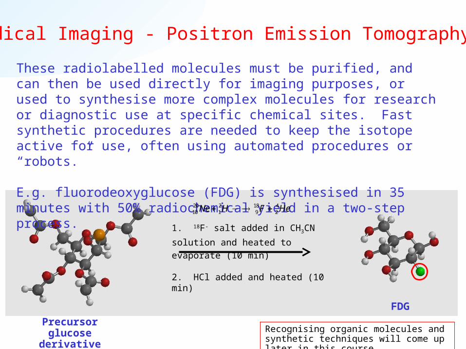
These radiolabelled molecules must be purified, and can then be used directly for imaging purposes, or used to synthesise more complex molecules for research or diagnostic use at specific chemical sites. Fast synthetic procedures are needed to keep the isotope active for use, often using automated procedures or “robots.”
E.g. fluorodeoxyglucose (FDG) is synthesised in 35 minutes with 50% radiochemical yield in a two-step process.
1. 18F- salt added in CH3CN solution
and heated to evaporate (10 min)
2. HCl added and heated (10 min)
FDG
Precursor glucose derivative reagent Recognising organic molecules and synthetic
techniques will come up later in this course.
20 2 18 410 1 9 2Ne H F He

Medical Imaging - Positron Emission Tomography
Fast synthesis of radiolabelled molecules allows them to be used to study specific biochemical processes and responses.
E.g.1. FDG is absorbed by the brain like glucose, but the fluorine substitution stops it from being completely metabolised. 18FDG It can be used to examine the reaction mechanism, and changes in uptake & metabolism accompanying various diseases.
These PET scans of a single anatomical level in the brain show changes in the brain chemistry and glucose metabolism by 18FDG with the progress of AIDS Dementia Complex.
Colour scale increases:black-blue-yellow-red-white

Medical Imaging - SummaryNuclear imaging is useful because it allows us to radiolabel molecules that specifically target organs, molecules or chemical processes for diagnosis or biochemical research.
The synthetic chemistry to design these target molecules differs widely:-
• Cyclotron-produced PET isotopes (11C, 18F…) are often exploited in the synthesis of organic molecules (drugs, peptides, carbohydrates, steroids, vitamins…)
• Metals (99mTc, 82Rb,…) may be used as soluble or insoluble salts, or as co-ordination compounds, to mimic biological molecules, toxins, as heavy heavy metal tracers,…
Footnote: Magnetic Resonance Imaging (MRI) is not a radiochemical technique.
Although based on the technique of nuclear magnetic resonance (NMR), MRI uses the quantum mechanical properties of 1H and other stable nuclei (e.g. 13C) to measure and map the concentration of chemical species at different positions in an organ or whole body.
There are no nuclear reactions or decay processes involved.
Summary I
You should now be able to
• Recognise nuclear reactions, including the major
spontaneous decay mechanisms.
• Define and distinguish between nucleons, nuclides &
isotopes, x-rays & gamma rays, decay series and
daughter isotopes.
• Explain stellar nucleogenesis.
• Calculate the average atomic mass from isotope
information.
• Balance nuclear reactions.

Summary II
You should now be able to
• Recognise stable and unstable nuclides.
• Predict the decay mechanism for an unstable isotope.
• Calculate the activity or half-life of an unstable nuclide
from appropriate data.
• Calculate the age of a sample using the carbon-14
method, and know the underlying assumptions and
appropriate timescale for its application.

Summary III
You should now be able to• Explain the main factors that contributes to effective
radiation dose, including penetrating power, activity, energy.
• Explain the main mechanism of biological damage by ionizing radiation.
• Explain the use of radioactive isotopes in medical imaging, and distinguish the information obtained from x-rays.
• Explain how isotope generators produce e.g. 99mTc for medical imaging, and give some examples of its use.
• Explain PET, the generation of radioisotopes by a cyclotron, and the know the kinds of isotopes produced.


