gene expression changes shared by DRPLA and Huntington's ...

Biochimica et Biophysica Acta 1832 (2013) 421–430
Contents lists available at SciVerse ScienceDirect
Biochimica et Biophysica Acta
j ourna l homepage: www.e lsev ie r .com/ locate /bbad is
Quercetin supplementation is effective in improving mitochondrial dysfunctionsinduced by 3-nitropropionic acid: Implications in Huntington's disease
Rajat Sandhir ⁎, Arpit MehrotraDepartment of Biochemistry, Basic Medical Science Building, Panjab University, Chandigarh, 160014, India
Abbreviations: 3-NP, 3-nitropropionic acid; CAT,2-nitrobenzoic acid; ETC, electron transport chain; HD, Hlow molecular weight thiols; MDA, malondialdehyde; Nprotein thiols; ROS/RNS, reactive oxygen and nitrogdismutase; SDH, succinate dehydrogenase; TSH, total thio⁎ Corresponding author at: Department of Biochemistry
160014, India. Tel.: +91 172 2534131/38.E-mail address: [email protected] (R. Sandhir).
0925-4439/$ – see front matter © 2012 Elsevier B.V. Allhttp://dx.doi.org/10.1016/j.bbadis.2012.11.018
a b s t r a c t
a r t i c l e i n f oArticle history:Received 3 March 2012Received in revised form 21 November 2012Accepted 27 November 2012Available online 4 December 2012
Keywords:FlavonoidHuntington's diseaseMitochondrionNeurodegeneration3-Nitropropionic acidQuercetin
The studywas designed to investigate the beneficial effect of quercetin supplementation in 3-nitropropionic acid(3-NP) induced model of Huntington's disease (HD). HDwas induced in rats by administering sub-chronic doseof 3-NP, intraperitoneally, twice daily for 17 days. Quercetin was supplemented at a dose of 25 mg/kg bodyweight by oral gavage for 21 days. At the end of treatment, mitochondrial bioenergetics, mitochondrial swelling,oxidative stress, neurobehavioral deficits and histopathological changes were analyzed. Quercetin supplementa-tion was able to reverse 3-NP induced inhibition of respiratory chain complexes, restore ATP levels, attenuatemitochondrial oxidative stress in terms of lipid peroxidation and prevent mitochondrial swelling. Quercetinadministration also restored the activities of superoxide dismutase and catalase along with thiol content in3-NP treated animals. Beneficial effect of quercetin administration was observed on 3-NP inducedmotor deficitsanalyzed by narrow beam walk and footprint analysis. Histopathological analysis of 3-NP treated rats revealedpyknotic nuclei and astrogliosis in striatum, which were reduced or absent in quercetin supplemented animals.Altogether, our results show that quercetin supplementation to 3-NP induced HD animals ameliorated mito-chondrial dysfunctions, oxidative stress and neurobehavioral deficits in rats showing potential of this flavonoidin maintaining mitochondrial functions, suggesting a putative role of quercetin in HD management.
© 2012 Elsevier B.V. All rights reserved.
1. Introduction
Huntington's disease (HD) is a progressive, fatal, neurodegenerativedisorder caused by an expanded CAG repeat in the huntingtin gene(Htt), that encodes for an abnormally long polyglutamine tract in aprotein termed huntingtin (htt) with a molecular weight of approxi-mately 350 kDa [1]. The disease is inherited in an autosomal dominantmannerwith age-dependent penetrance. Clinical features of HD includeprogressive motor dysfunctions, cognitive decline, and psychiatricdisturbances, including both neuronal dysfunctions and neuronal celldeath [2]. Although, mutation in Htt gene was discovered more than17 years ago, the role of Htt in the physiology and the pathophysiol-ogy is still under investigation [3]. Recent data indicates that the trans-location of mHtt into nucleus and transcriptional dysregulation likelyplay an important role in the pathogenic process and more specificallythese events have a significant impact on mitochondrial functionssuch as electron transport chain (ETC) and reactive oxygen species(ROS) generation leading to bioenergetic failure [4]. Numerous studies
catalase; DTNB, 5,5-dithiobis-untington's disease; LMW-SH,BT, nitroblue tetrazolium; PSH,en species; SOD, superoxidels, PanjabUniversity, Chandigarh,
rights reserved.
in cell and mouse models of HD have revealed mitochondrial impair-ment [5].
The hypothesis that mitochondrial dysfunctions contribute tothe pathogenesis of HD was first tested pharmacologically by using3-nitropropionic acid (3-NP), an irreversible inhibitor of succinate de-hydrogenase [6]. One of themechanisms following 3-NP administrationis the development ofmitochondrial dysfunctions leading to generationof a bioenergetic defect which involves three interacting processes suchas: energy impairment, oxidative stress and excitotoxicity [7]. 3-NPinduced HD model replicates most of the clinical and pathophysiologi-cal hallmarks of HD, including spontaneous choreiform and dystonicmovements, frontal-type cognitive deficits, and progressive striatalneuronal degeneration [8]. 3-NP administration also results in ATPdepletion, which impairs intracellular calcium buffering therebyleading to production of damaging ROS [9].
At present, there are no effective treatments against HD. Currenttherapies for treating HD are symptomatic; focusing on neurologicaland psychiatric symptoms that aim at improving the quality of life[10]. Attention has been given on the influence of phytochemicaltherapeutics on health and mental well-being. Evidence has indicatedthat a group of plant-derived compounds known as flavonoids exertsparticularly powerful action as cardioprotective, chemopreventiveand neuroprotective agents [11]. The biological activities of flavo-noids have been attributed to their antioxidant, anti-inflammatoryand their property to modulate signaling cascades [12]. Within theflavonoid family, quercetin is the most potent scavenger of ROS and
422 R. Sandhir, A. Mehrotra / Biochimica et Biophysica Acta 1832 (2013) 421–430
reactive nitrogen species (RNS). This can be explained by the pres-ence of two antioxidant pharmacophores within the molecule thathave the optimal configuration for free radical scavenging [13]. Querce-tin has been shown to easily traverse the blood–brain-barrier and actsas promising agents for intervention in neurodegenerative conditionslike ischemia [14], Alzheimer's disease [15] and Parkinson's disease[16]. Use of neuroprotective antioxidants is being considered as a prom-ising approach to slow down the disease progression and to limit theextent of functional neuronal loss in chronic neurodegeneration aswell as after acute lesions of the brain. However, only a few studies onthe use of antioxidants in themanagement of neurodegenerative condi-tions have so far been undertaken.Methodological inconsistencies, poorpermeation of the blood–brain barrier and lower efficacy per dose aresome reasons for the lack of studies in this area [17]. Quercetin exertsits beneficial effect in brain primarily through its antioxidant action.Quercetin has been shown to improve mitochondrial functions inbrain by increasingmitochondrial biogenesis [18]. Within the subcellu-lar compartment quercetin shows preferential accumulation in mito-chondria [19]. Based on the information in the literature, quercetinappears to be promising agent against HD. Therefore, in the presentstudy, we have evaluated the neuroprotective potential of quercetinagainst 3-NP induced mitochondrial oxidative stress, mitochondrialdysfunctions and neurobehavioral deficits.
2. Experimental procedures
2.1. Chemicals
All the chemicals used in the present study were of analytical gradeand were purchased from Sigma Chemical Co. (St. Louis, USA), Merck(Mumbai, India) and Sisco Research Laboratories Pvt. Ltd. (Mumbai,India). Quercetin was purchased from Himedia Laboratories Pvt. Ltd.(Mumbai, India).
2.2. Animals and treatment schedule
Female Wistar rats aged 9–10 weeks, weighing between 200 and250 g were procured from the Central Animal House facility of PanjabUniversity, Chandigarh, India. The animals were allowed to acclimatizeto the local vivarium for 7 days. All the experiments were carried outbetween 09:00 and 15:00 h. The protocols followed were approved bythe Institutional Animal Ethics Committee of the University and werein accordancewith the guidelines for humaneuse and care of laboratoryanimals. The animals were randomly segregated into the following fourgroups with each group having 6–8 animals.
Control (vehicle): animals were given vehicle.Control+quercetin treated: animalswere administeredwith vehiclefor 3-NP and quercetin at a dose of 25 mg/kg through oral gavage for21 days.3-NP treated: animals were administered 3-NP at a sub-chronic dosetwice a day intraperitoneally for 17 days. Twice a day injectionswere: 7.5 mg/kg for the first 2 days, followed by 3.75 mg/kg fornext 7 days, finally a dosage of 2 mg/kg for the last 8 days. Thedose of 3-NP used in the study is based on the doses reported in lit-erature and were standardized in our laboratory [20].3-NP+quercetin treated: One hour before sub-chronic 3-NP treat-ment, animals were administered with quercetin at a dose of25 mg/kg by oral gavage for 21 days.
2.3. Mitochondrial respiratory chain enzymes
2.3.1. Isolation of rat brain mitochondriaOn day 21, animals were sacrificed by decapitation under mild ether
anesthesia. Mitochondria were isolated from striatum by the method
described by Puka-Sundvall [21]. Briefly, corpus striatum was dissected,rinsed in ice-cold isotonic saline and homogenized in ice-cold extractionbuffer (10 mM Tris–HCl, pH 7.4, 0.44 M sucrose, 10 mM EDTA and 0.1%BSA). The homogenate was centrifuged at 2100 g for 15 min at 4 °C.The pellet was discarded and the supernatant re-centrifuged at 14,000 gfor 15 min at 4 °C. The crudemitochondrial pelletwas separated, washedwith extraction buffer and centrifuged at 7000 g for 15 min at 4 °C. Thefinal mitochondrial pellet was re-suspended in buffer containing 0.44 Msucrose in 10 mM Tris–HCl, pH 7.4.
2.3.2. NADH dehydrogenase (complex I)Activity of NADH dehydrogenase was measured as described by
King and Howard [22]. Requisite amount of mitochondrial preparationwas added to the reaction mixture containing 0.2 M glycyl-glycine(pH 8.5), 6 mM NADH, 1 mM oxidized cytochrome c and 0.02 MNaHCO3. NADH dehydrogenase catalyzed reduction of cytochrome cand the increase in absorbance was followed spectrophotometricallyat 550 nm for 3 min. Results were expressed as nmol NADH oxidized/min/mg protein. The enzyme activity was normalized to citratesynthase activity.
2.3.3. Succinate dehydrogenase (complex II)Activity of succinate dehydrogenase was assayed according to the
method of King et al. [23]. The reaction mixture contained 0.2 M sodi-um phosphate buffer (pH 7.8), 1% (w/v) BSA, 0.6 M succinate and0.03 M potassium ferricyanide. The reaction was initiated by additionof requisite amount of mitochondrial preparation. Succinate dehydro-genase catalyzes the oxidation of succinate to fumarate by potassiumferricyanide, which was measured spectrophotometrically by decreasein absorbance at 420 nm for 3 min. Results were expressed as nmolsuccinate oxidized/min/mg protein. The enzyme activity was normal-ized to citrate synthase activity.
2.3.4. Cytochrome oxidase (complex IV)Activity of cytochrome oxidase was assayed according to the
method described by Sottocasa et al. [24]. Oxidized cytochrome cwas reduced by adding few crystals of sodium borohydride and thenneutralized to pH 7.0 by 0.1 M HCl. 0.3 mM of reduced cytochrome cwas added to 0.075 M phosphate buffer and the reaction was initiatedby mixing appropriate amount of mitochondrial suspension. Thereduced cytochrome c is oxidized in the reaction mixture containingcytochromeoxidasewhich ismeasured spectrophotometrically by a de-crease in absorbance at 550 nm for 3 min. Results were expressed asnmol cytochrome c oxidized/min/mg protein. The enzyme activitywas normalized to citrate synthase activity.
2.3.5. F1F0 synthase (complex V)Mitochondrial F1F0 synthase activity was measured as described
by Griffiths and Houghton [25]. Reaction was started by adding appro-priate amount of mitochondrial suspension in ATPase buffer [50 mMTris and 5 mM MgCl2, pH 7.5] at 37 °C with 5 mM ATP for 10 min.The reaction was stopped by adding 10% (w/v) trichloroacetic acid.The contents were centrifuged at 3000 g for 20 min, and an appropriatevolume of supernatantwasmixedwithwater. Phosphate producedwasmeasured by the method of Fiske and Subbarow [26]. Results wereexpressed as nmol of ATP hydrolyzed/min/mg protein. The enzymeactivity was normalized to citrate synthase activity.
2.3.6. MTT reductionThe reduction of MTT to blue formazan by dehydrogenases present
in the mitochondrial suspension was also monitored to assess mito-chondrial functions [27]. To appropriate mitochondrial pellet, MTT(0.1 mg/ml) was added, mixed and incubated at 37 °C for 30 min andthen centrifuged to obtain formazan pellet. The pellet was dissolved inabsolute ethanol and the mixture was re-centrifuged at 2000 g for10 min. The absorbance of the supernatant was measured at 595 nm.
423R. Sandhir, A. Mehrotra / Biochimica et Biophysica Acta 1832 (2013) 421–430
Results were expressed as μg formazan formed/min/mg protein and thevalues were normalized to citrate synthase activity.
2.3.7. Citrate synthase assayCitrate synthase (CS) was assayed as described by Coore et al. [28],
wherein the reduction of 0.2 mM 5,5′-dithio-bis(2-nitrobenzoic acid)in the presence of 0.2 mM acetyl-CoA and 0.1 mM oxaloacetate in amedium with 100 mM Tris–HCl, pH 8.0 and 0.1% Triton X-100 wasperformed at 412 nm. The CS activity was expressed as nmol/min/mgprotein.
2.3.8. Mitochondrial swellingMitochondrial swelling and contraction by measurement of light
scattering in a spectrophotometer were used as a functional test ofmitochondrial membrane integrity as described by Tedeshi andHarris [29]. This method is based upon the increased absorbance ofmitochondria in a contracted state and decreased density in a swollenor orthodox configuration due to cation influx that can be detected bymeasurement of light scattering at 520 nm. To appropriate mitochon-drial suspension, 0.12 M KCl in 0.02 M Tris–HCl was added and mito-chondrial swelling was measured at 520 nm for 6 min. Contraction ofmitochondria was initiated after 6 min by adding Mg2+-ATP. Changein absorbance at 520 nmwas normalized by the protein concentration.
2.3.9. Adenine nucleotide levelsATP and ADP levels were determined by the method of Victor et al.
[30]. Frozen tissues were transferred to a 1.5-ml micro-centrifuge tubeand added with 0.4 M perchloric acid. The tissue was immediatelyhomogenized with a pestle and the homogenate was kept on ice for30 min, and then centrifuged at 14,000 rpm at 4 °C. The supernatantwas neutralized with 4 M K2CO3, kept on ice for 10 min and at −80 °Cto promote precipitation of the perchlorate, and then centrifuged again.Supernatants were then stored at −80 °C until HPLC assay. Aliquotsof the extract and standard were applied separately to the columnand the eluate was monitored at 254 nm, at a flow rate of 2 ml/minusing supelcosil LC-18, 5 μm (15 cm∗0.46 cm) reversed phase column(Supelco, Crans, Switzerland). An isocratic elution of the sampleswas performed with 0.52 μm potassium phosphate buffer (KH2PO4;pH 4.0), containing 0.04% tetrabutylammonium phosphate and 1.25%(v/v) methanol. Peaks were identified by their retention times andby using cochromatography with standards. Results were expressed asnmol/mg protein.
2.4. Mitochondrial oxidative stress
2.4.1. Lipid peroxidationMalondialdehyde (MDA), a measure of lipid peroxidation was
quantified by reaction with thiobarbituric acid at 532 nm by themethod of Ohkawa et al. [31]. The values were expressed as nmolMDA/mg protein, using molar extinction coefficient of chromophore(1.56×10⁵M−1cm−1).
2.4.2. Superoxide dismutase (SOD)SOD activity was assayed inmitochondrial preparation, wherein the
inhibition of nitrozo blue tetrazolium (NBT) reductionwasmeasured at560 nm [32]. Briefly, the reaction was initiated by the addition ofhydroxylamine hydrochloride to the reaction mixture containing NBTand sample. The results were expressed as Units/mg protein, whereone unit of enzyme is defined as the amount of enzyme inhibiting therate of reaction by 50%.
2.4.3. CatalaseCatalase (CAT) activity was assayed in mitochondrial suspension
by the method of Aebi [33]. The decomposition of hydrogen peroxide(H2O2) by CAT was monitored spectrophotometrically by followingthe decrease in absorbance at 240 nm. The activity of enzyme was
expressed as nmol of H2O2 decomposed/min/mg protein, using molarextinction coefficient of H2O2 (71 M−1cm−1).
2.4.4. Thiols
2.4.4.1. Total thiols. Total thiols (TSH) were quantified in themitochon-drial preparation according to the method of Sedlak and Hanus [34]. Inthis method, DTNB is reduced by protein and non-protein free \SHgroups to form 1 mol of 5-mercapto-2-nitrobenzoate per mol of \SH.Absorbance was read at 412 nm and the results were expressed asnmol TSH/mg protein.
2.4.4.2. Low molecular weight thiols (LMW-SHs). LMW-SHs (probablyGSH) were estimated in the mitochondrial preparation by the methodof Roberts and Francetic [35]. Absorbance was measured at 412 nmand results were expressed as nmol LMW-SH/mg protein.
2.4.4.3. Protein thiols. Levels of protein thiols (P-SHs) were calculatedfrom the difference between the values of total thiols and LMW-SHsand expressed as nmol P-SH/mg protein.
2.5. Estimation of protein
The protein content was estimated according to the method ofLowry et al. [36].
2.6. Neurobehavioral studies
2.6.1. Locomotor activityThe locomotor activity was measured using actophotometer [37].
The interruption of a beam of light falling on a photocell followingthe movement of the animal was recorded. Each rat was placed indi-vidually in the actophotometer for 3 min and numbers of counts wererecorded.
2.6.2. Narrow beam walk testThis behavioral test was used to evaluate motor performance in the
treated animals vs. the controls, by progressively increasing the difficul-ty in the execution of the task as described by Henderson et al. [38]. Theanimals were trained in crossing a 150 cm long wooden beam, dividedinto three 50 cm segments, from a platform at one end to the animal'shome cage at the other end, placed horizontally 60 cm above thefloor. The number of paw slips onto an under-hanging ledge and thetime taken to traverse the beam was recorded. The maximum timeallowed for the task was 2 min. Occurrence of bradykinesia was quanti-fied by calculating the average velocity of walking for treated and con-trol animals.
2.6.3. Footprint analysisThis test was used to assess gait abnormalities in 3-NP treated an-
imals [20]. After coating the hind feet with a non-toxic green dye andforepaws with a non-toxic red dye, rats were allowed to walk on abeam (100 cm length, 12 cm breadth and 10 cm high walls with aninclination of 30°) leading to a darkened enclosure. The gangwaywas lined with white paper for recording the feet impressions.Animals in all the groups were tested for footprint length, footprintbreadth, and footprint stride length for both left and right paws.Specifically, footprint stride length was quantified as the distancebetween two subsequent feet. Paws overlap analysis was carried outby measuring the center distance between the anterior paw andrear paw footprints.
2.7. Histochemical and histological analysis
The frozen brain sections were processed for SDH histochemicalstaining [39]. Animals were anesthetized and transcardially perfused

Table 1Effect of quercetin administration on the activity of mitochondrial complexes in striatum of 3-NP treated rats.
Complex I (nmol NADHoxidized/min/mg protein)
Complex II (nmol succinateoxidized/min//mg protein)
Complex IV (nmol cytochromeC oxidized/min/mg protein)
F1F0 ATPase (nmol ATPhydrolyzed/min//mg protein)
MTT reduction (μg formazanformed/min/mg protein)
Control 109.23±13.32 147.61±4.54 162.58±6.08 17.69±1.69 5.46±0.2(131.6±8.49) (177.74±4.38) (195.82±8.19) (21.22±3.78) (6.57±0.5)
Quercetin 106.9±16.19 145.9±8.42 145.57±2.77 17.45±1.2 4.56±0.38(116.9±11.84) (159.4±14.11) (158.65±8.5) (19.0±3.55) (4.97±0.7)
3-NP 60.05±7.83⁎ 71.37±3.33⁎ 112.52±3.28⁎ 11.7±0.53⁎ 1.64±0.17⁎
(15.6±1.04)⁎ (18.47±1.95)⁎ (29.39±2.24)⁎ (3.06±0.7)⁎ (0.42±0.02)⁎
3-NP+quercetin 99.11±10.25† 91.56±1.05† 126.96±3.25† 16.96±0.96† 2.7±0.1†
(38.91±2.08)† (36.0±2.33)† (49.86±3.76)† (6.47±1.38)† (1.05±0.12)†
Values in parenthesis are normalized to citrate synthase activity. All values are expressed as mean±SEM; n=6/group.⁎ Significantly different from control group (pb0.05).† Significantly different from 3-NP treated group (pb0.05).
424 R. Sandhir, A. Mehrotra / Biochimica et Biophysica Acta 1832 (2013) 421–430
with cold 0.1 M PBS, pH 7.4, followed by cold 10% (v/v) glycerol in PBS.Twentymicron thick frozen sections of the brain were dried for 30 min,activated in PBS at 37 °C for 10 min and then incubated with reactionmixture containing 0.3 M NBT, 0.05 M phosphate buffer, pH7.4, and0.05 M sodium succinate at 37 °C in dark for 30 min. At the end of thereaction, sections were extensively washed with the reaction buffer,examined under the microscope and photographed.
Animals for histology were perfused transcardially with cold sa-line followed by phosphate buffered 4% paraformaldehyde. The brainswere then post-fixed overnight in 4% paraformaldehyde and laterbrain sections were processed for routine hematoxylin and eosinstaining [40].
2.8. Statistical analysis
All values are expressed as mean±standard error of mean (SEM)of six animals per group. Data was analyzed using one way analysisof variance (ANOVA) followed by Newman–Keuls test for multiplepair-wise comparisons between the various treated groups usingSPSS 14 software. Values with pb0.05 were considered as statisticallysignificant.
Fig. 1. Effect of quercetin supplementation on SDH histochemical staining in frozen brain secat a magnification of 40×. Control sections show dense staining for normal SDH functioningalone shows SDH functioning similar to that of control animals (B & II). SDH functioning wshowing improved SDH functioning (D & IV) (scale bar—40 μm).
3. Results
3.1. Effect of quercetin on mitochondrial electron transport chainenzymes, MTT reduction and mitochondrial swelling
Activities of mitochondrial electron transport chain (ETC) enzymeswere found to be significantly inhibited by 3-NP administration(Table 1). NADH dehydrogenase activity was inhibited by 45.01% in3-NP treated animals as compared to control animals. The activity wasincreased by 39.4% on quercetin supplementation to 3-NP treatedanimals. Activity of succinate dehydrogenase (complex-II) was foundto be significantly lowered by 51.64% in 3-NP treated group as com-pared to control group. Supplementation with quercetin increasedcomplex-II activity by 22.05% in 3-NP treated animals. Histochemicalstaining for SDH activity revealed a significant decrease of the enzymereaction in the striatum of 3-NP treated animals (Fig. 1, C & III) as com-pared to the control animals (Fig. 1, A & I). Quercetin supplementationalone had SDH staining comparable to that of control striatal tissuesection (Fig. 1, B & II). The SDH staining recovered in brain sections onquercetin supplementation to 3-NP treated animals (Fig. 1, D & IV).Activity of terminal enzyme of mitochondrial ETC, cytochrome oxidasewas inhibited by 30.79% in 3-NP treated group as compared to control
tions of 3-NP treated rats. Frozen brain sections were visualized under light microscope(A & I). Histological appearance of striatal tissue from rats supplemented with quercetinas compromised in 3-NP induced rats (C & III). Quercetin+3-NP administered groups

Fig. 2. Effect of quercetin supplementation on mitochondrial swelling in 3-NP treatedrats. Values are expressed as mean±SEM; n=6/group. * Significantly different fromcontrol group (pb0.05), † significantly different from 3-NP treated group (pb0.05).
Table 3Effect of quercetin administration on lipid peroxidation, superoxide dismutase and cata-lase activity in mitochondria isolated from striatum of 3-NP treated rats.
Lipid peroxidation(nmol MDA/mgprotein)
Superoxidedismutase(Units/mgprotein)
Catalase (μmol of H2O2
decomposed/min/mgprotein)
Control 4.09±0.37 4.42±0.21 2.07±0.17Quercetin 4.21±0.48 3.81±0.13 1.69±0.063-NP 7.91±0.82⁎ 2.99±0.1⁎ 0.96±0.03⁎
3-NP+quercetin 5.93±0.31† 3.51±0.07† 1.34±0.11†
Values are expressed as mean±SEM; n=6.⁎ Significantly different from control group (pb0.05).† Significantly different from 3-NP treated group (pb0.05).
425R. Sandhir, A. Mehrotra / Biochimica et Biophysica Acta 1832 (2013) 421–430
group. The activity was increased by 11.37% in 3-NP treated animalssupplemented with quercetin. Activity of mitochondrial F1–F0 synthase(complex-V) was assessed by hydrolysis of ATP to ADP and was foundto be inhibited by 33.86% in 3-NP treated group as compared to controlgroup. On quercetin supplementation, the activity was increased by31.01% in 3-NP treated animals. Mitochondrial functioning was alsoassayed using MTT reduction (Table 1). MTT reduction was found tobe significantly inhibited by 69.96% in 3-NP treated animals as com-pared to control animals which was increased by 39.25% in3-NP+quercetin co-supplemented animals. The perturbed activity ofETC enzymes led to the increasedmitochondrial swelling in 3-NP treat-ed animals which on quercetin supplementation was found to belowered (Fig. 2). The data in Table 2 depicts levels of ATP and ADP instriatum following 3-NP and quercetin administration. It is clear fromthe data that the compromised ETC components following 3-NP admin-istration resulted in reduced levels of ATP with a concomitant increasein ADP levels thereby lowering the ATP/ADP ratio. The effect was re-versed on quercetin administration and the ATP/ADP ratio was restoredto near controls.
3.2. Effect of quercetin on mitochondrial lipid peroxidation, superoxidedismutase and catalase activity
The data for lipid peroxidation, activity of superoxide dismutase andcatalase in mitochondria isolated from striatum are depicted in Table 3.Lipid peroxidation was significantly increased (54%) in 3-NP treated ascompared to controls. Quercetin supplementation to 3-NP treatedanimals lowered mitochondrial lipid peroxides by 38.22% as comparedto 3-NP group. The activity of superoxide dismutase (SOD) wasinhibited by 32.84% in 3-NP treated animals as compared to controlanimals. However, quercetin supplementation significantly increasedSOD activity by 14.73% in 3-NP animals. Catalase activity was foundto be significantly inhibited by 53.48% in 3-NP treated animals ascompared to control animals. Quercetin supplementation resulted in asignificant increase in catalase activity by 28.6% in 3-NP treated animals.
Table 2Effect of quercetin administration on adenine nucleotides in striatum of 3-NP treatedrats.
ATP(nmol/mg protein)
ADP(nmol/mg protein)
ATP/ADPratio
Control 9.15±1.41 4.24±0.39 2.16Quercetin 10.01±1.29 4.87±0.73 2.073-NP 4.83±1.47⁎ 7.87±0.42⁎ 0.613-NP+quercetin 8.81±1.06† 4.94±0.38† 1.78
Values are expressed as mean±SEM; n=6.⁎ Significantly different from control group (pb0.05).† Significantly different from 3-NP treated group (pb0.05).
3.3. Effect of quercetin on mitochondrial thiols
One of the most important antioxidant present in mitochondria isthiol and its levels were found to be compromised in mitochondria iso-lated from the striatum of 3-NP treated animals (Fig. 3). Total thiollevels were found to be significantly reduced by 54.22% in 3-NP treatedanimals as compared to control animals. Quercetin supplementationresulted in increased levels of total thiols (37.46%) in 3-NP treated ani-mals. A significant decrease in lowmolecular weight (51.39%) and pro-tein thiols (55.03%) was observed in 3-NP treated animals as comparedto controls. Quercetin supplementation significantly increased the lowmolecular weight and protein thiols by 34.52% and 38.32% respectivelyin 3-NP treated animals.
3.4. Effect of quercetin on neurobehavioral deficits
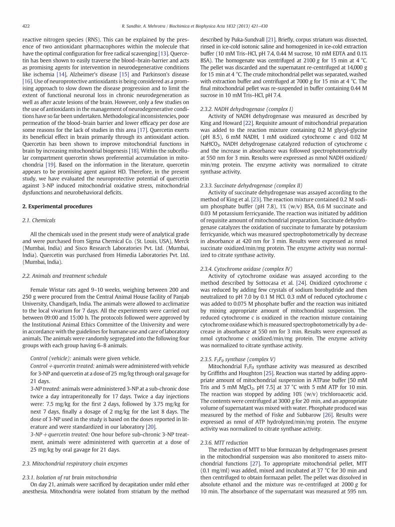
3.4.1. Locomotor activityThe locomotor activity was assessed in terms of photo beam counts
for duration of 180 s using actophotometer (Fig. 4). On day 0, animals inall the four groups had an average count of 166.5. Onday 1, the numbersof counts for 3-NP treated animals fell to an average of 125, suggesting asignificant impairment in locomotor functions. However, quercetinsupplementation to 3-NP treated animals increased the averagenumber of counts to 140. This trend of decline in locomotor functionsin 3-NP treated animals was followed for days 7, 14 and 21 following3-NP treatment with an average of 70, 51 and 80 counts respectively.Quercetin supplementation was able to significantly improved locomo-tor functions by recording an average of 134, 145 and 150 countsrespectively for days 7, 14 and 21 in 3-NP+quercetin animals.
Fig. 3. Effect of quercetin supplementation on mitochondrial total, low molecular weight(LMW) and protein thiols in mitochondria isolated from striatum of 3-NP treated rats.Values are expressed as mean±SEM; n=6/group. * Significantly different from controlgroup (pb0.05), † significantly different from 3-NP treated group (pb0.05).
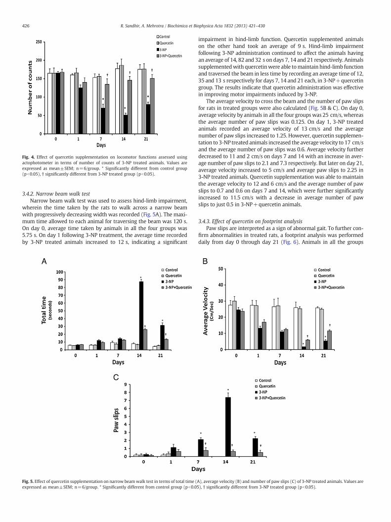
Fig. 4. Effect of quercetin supplementation on locomotor functions assessed usingactophotometer in terms of number of counts of 3-NP treated animals. Values areexpressed as mean±SEM; n=6/group. * Significantly different from control group(pb0.05), † significantly different from 3-NP treated group (pb0.05).
426 R. Sandhir, A. Mehrotra / Biochimica et Biophysica Acta 1832 (2013) 421–430
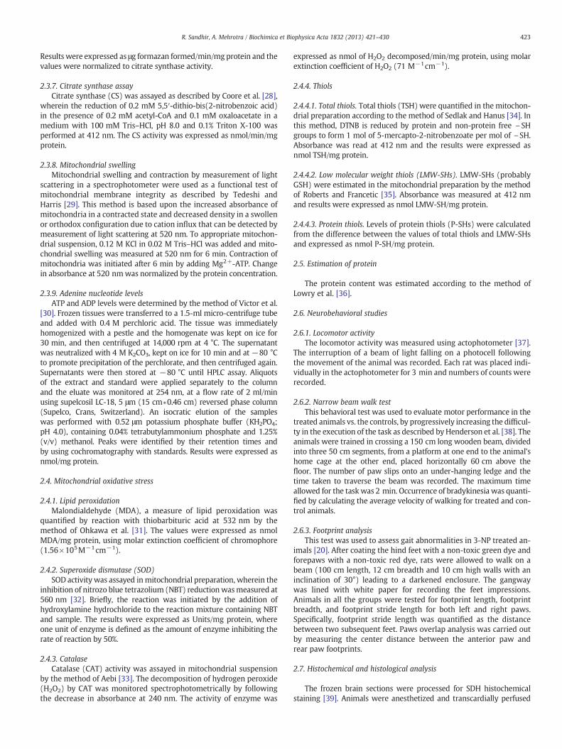
3.4.2. Narrow beam walk testNarrow beam walk test was used to assess hind-limb impairment,
wherein the time taken by the rats to walk across a narrow beamwith progressively decreasing width was recorded (Fig. 5A). The maxi-mum time allowed to each animal for traversing the beam was 120 s.On day 0, average time taken by animals in all the four groups was5.75 s. On day 1 following 3-NP treatment, the average time recordedby 3-NP treated animals increased to 12 s, indicating a significant
Fig. 5. Effect of quercetin supplementation on narrow beamwalk test in terms of total time (expressed as mean±SEM; n=6/group. * Significantly different from control group (pb0.0
impairment in hind-limb function. Quercetin supplemented animalson the other hand took an average of 9 s. Hind-limb impairmentfollowing 3-NP administration continued to affect the animals havingan average of 14, 82 and 32 s on days 7, 14 and 21 respectively. Animalssupplementedwith quercetinwere able tomaintain hind-limb functionand traversed the beam in less time by recording an average time of 12,35 and 13 s respectively for days 7, 14 and 21 each, in 3-NP+quercetingroup. The results indicate that quercetin administration was effectivein improving motor impairments induced by 3-NP.
The average velocity to cross the beam and the number of paw slipsfor rats in treated groups were also calculated (Fig. 5B & C). On day 0,average velocity by animals in all the four groupswas 25 cm/s, whereasthe average number of paw slips was 0.125. On day 1, 3-NP treatedanimals recorded an average velocity of 13 cm/s and the averagenumber of paw slips increased to 1.25. However, quercetin supplemen-tation to 3-NP treated animals increased the average velocity to 17 cm/sand the average number of paw slips was 0.6. Average velocity furtherdecreased to 11 and 2 cm/s on days 7 and 14 with an increase in aver-age number of paw slips to 2.1 and 7.3 respectively. But later on day 21,average velocity increased to 5 cm/s and average paw slips to 2.25 in3-NP treated animals. Quercetin supplementation was able to maintainthe average velocity to 12 and 6 cm/s and the average number of pawslips to 0.7 and 0.6 on days 7 and 14, which were further significantlyincreased to 11.5 cm/s with a decrease in average number of pawslips to just 0.5 in 3-NP+quercetin animals.
3.4.3. Effect of quercetin on footprint analysisPaw slips are interpreted as a sign of abnormal gait. To further con-
firm abnormalities in treated rats, a footprint analysis was performeddaily from day 0 through day 21 (Fig. 6). Animals in all the groups
A), average velocity (B) and number of paw slips (C) of 3-NP treated animals. Values are5), † significantly different from 3-NP treated group (pb0.05).
Fig. 6. Effect of quercetin supplementation on footprint test for gait analysis by control (A), quercetin (B), 3-NP (C) and 3-NP+quercetin (D) treated animals.
427R. Sandhir, A. Mehrotra / Biochimica et Biophysica Acta 1832 (2013) 421–430
were tested for footprint length, footprint breadth and footprint stridelength for both left and right paws. Out of six parameters that wereanalyzed only the stride length for both, left and right paws was signif-icantly affected in the animals that received 3-NP+quercetin supple-mentation in comparison to 3-NP treated animals (data not shown).However, the footprint length and footprint breadth for both leftand right paws remained unaffected (data not shown). An additionalmeasurement of walking pattern is the paws overlap which wascalculated as the minimum distance (in cm) between left and rightpaws overlap (Fig. 7). The rationale of this analysis lies on the fact thatrodents while walking usually tend to step their hind paw in the sameplace previously occupied by their front paw. Quantifying this aspectof themotor behavior could then be considered as ameasure of locomo-tor ability. On day 0, the average distance of left paw for animals in allthe four groups was 0.75 cm, whereas for right paw it was 0.9 cm. Ondays 7, 14 and 21, 3-NP treated animals recorded a significant increasein average distance of left paw overlap of 0.9, 0.6 and 0.6 cm respective-ly. Average overlap distance in right paw for days 7, 14 and 21 wassignificantly found to be 1.1, 0.66 and 0.61 cm respectively. However,quercetin supplementation in 3-NP+quercetin animals maintainedan overall average distance in left and right paws overlap between0.25 and 0.45 cm for the entire duration of 21 days, suggesting theability of quercetin in preventing gait abnormalities observed in 3-NPinduced HD.
Fig. 7. Effect of quercetin supplementation on gait analysis in terms of left paw (A) andright paws (B) overlap of 3-NP treated animals. Values are expressed as mean±SEM;n=6/group. * Significantly different from control group (pb0.05), † significantly differentfrom 3-NP treated group (pb0.05).
3.5. Effect of quercetin on histopathological changes
Histopathological changes in the striatum of control and 3-NPtreated animals are depicted in Fig. 8. The striatum of control ratsshows normal morphology. Quercetin supplementation to rats aloneexhibited normal appearance of striatum as similar to that of controlrats. Striatal sections of rats treated with 3-NP exhibited irregulardamaged cells with condensed and pyknotic nuclei (Fig. 8C). Querce-tin treatment to 3-NP animals resulted in modulation of the abnor-malities in the striatal histopathology near to normal (Fig. 8D).
4. Discussion
Defects in mitochondrial functions have been proposed to contrib-ute substantially to most of the neurodegenerative disorders includingHD. The activity of mitochondrial complexes-I, II, IV and F1F0 synthase

Fig. 8. Effect of quercetin supplementation on histopathological alterations in striatum of control and 3-NP treated rats. Sections were visualized under light microscope at a mag-nification of 40×. (A) Control rats showing normal striatal histology. (B) Histological appearance of striatal tissue from rats supplemented with quercetin alone shows normal cellnuclei similar to that of control animals. (C) 3-NP induced rats showing damaged and condensed pyknotic nuclei in striatum (PN). (D) Striatum of quercetin+3-NP administeredgroups showing lesser number of pyknotic nuclei (scale bar—100 μm).
428 R. Sandhir, A. Mehrotra / Biochimica et Biophysica Acta 1832 (2013) 421–430
was found to be significantly inhibited in 3-NP treated animals leadingto bioenergetic failure in terms of reduced ATP levels andATP/ADP ratio.This inhibition in activities might be an effect of hydroxyl radicalsgenerated due to 3-NP mediated SDH inhibition [41]. In addition, MTTreduction (a marker of mitochondrial functioning) was markedlyreduced in the striatum of 3-NP treated animals. Mitochondrialcomplex-II has been reported to, at least partly, involved in MTT reduc-tion by intact astrocytes and neurons, confirming inhibition of SDH by3-NP [42]. This metabolic defect imposed by 3-NP treatment might beclosely associated with ATP depletion, mitochondrial swelling, loss ofionic gradients, and alterations in mitochondrial membrane structureand function [43]. Preferential decrease of SDH activity in the lesionarea of 3-NP treated animals in the histochemical assay corroboratesthe earlier observations [39] and is in accordance with the biochemicalfindings in the present study showing inhibition in SDH activity. Quer-cetin supplementation to 3-NP treated animals restored SDH stainingin the striatum as compared to 3-NP treated animals. We also foundan increase in mitochondrial swelling on 3-NP administration whichmight be attributed to increased production of harmful ROS. Quercetinsupplementation to 3-NP treated animals restored the normal function-ing of ETC enzymes along with a significant increase in F1–F0 synthaseactivity, restoration of ATP levels and prevention of mitochondrialswelling. The beneficial effect of quercetin might be attributed toantioxidant activity of this flavonoid. Complex I and cytochrome c aresuggested to bemolecular targets of quercetin action thereby inhibitingH2O2 production [44]. Quercetin has been shown to prevent the open-ing of MPT pore thereby preventing mitochondrial swelling [19]. Quer-cetin has been reported to bind to a variety of cytosolic proteins andaccumulates in lipid compartments (i.e., membranes), suggesting that,hydrophobic compartments such as mitochondria, in which heme-containing proteins are largely expressed, may represent a site ofpreferential accumulation of the flavonoid [45]. Since, mitochondriarepresent a critical source of ROS and their ability to accumulate querce-tin may be functional for protection of mitochondrial function and
integrity. Quercetin has recently been reported to be most efficient inprotecting against indomethacin-induced mitochondrial dysfunctionsand this could be due to its ability to enter cells and accumulate inmitochondria [46]. Furthermore, quercetin supplementation has alsobeen shown to protect mitochondrial integrity and size along withmitochondrial functions by controlling succinate dehydrogenase andNADH oxidase activities [47].
Increased oxidative stress is attributed to alterations in antioxidantdefense system which includes lipid peroxidation and antioxidantenzymes. One of the most abundant products of lipid peroxidation isMDA. The values of mitochondrial MDA levels in 3-NP exposed animalswere found to be significantly higher. Quercetin supplementation to3-NP treated animals was able to significantly lower lipid peroxidelevels. These findings are supported by a study in which, protectiveeffect of quercetin is correlated with the capacity of this flavonoid todetoxify H2O2 and thereby effectively blocking MDA formation in thebrain [48]. Also, multiple methylation of the hydroxyl groups presentin quercetin increases lipophilic nature of the molecule, thereby in-creasing availability of the flavonoid in mitochondrial membrane andpreventing lipid peroxidation, thus enhancing the pharmacological po-tential of this flavonoid against such pathological processes [49].MnSOD is also regarded as a critical component of the enzymatic anti-oxidant defense system and in the present study SOD activity wasfound to be significantly lowered in 3-NP treated animals. This couldbe due to increased generation of superoxide radicals, leading todecrease in the activity of MnSOD. 3-NP administration also resultedin a significant decrease in catalase activity. However, quercetin supple-mentation to 3-NP induced HD animals for 21 days significantlyimproved the levels of MnSOD and catalase, suggesting that it is an ef-fective mechanism in ameliorating mitochondrial oxidative stress. Incontrast, quercetinpossesses bothpro- and anti-oxidantproperties [50],and it was therefore hypothesized that quercetin effects on MnSODactivity may be due to changes in expression of the MnSOD gene [51].Quercetin was shown to be a potent inhibitor of H2O2 production,
429R. Sandhir, A. Mehrotra / Biochimica et Biophysica Acta 1832 (2013) 421–430
evenwhen production rate of H2O2was stimulated by themitochondri-al inhibitors such as rotenone and antimycin A [45]. Quercetin has alsobeen shown to prevent oxidative nuclear and mitochondrial DNAdamage [52].
In biological systems, thiols play a central role in coordinating theantioxidant defense network by their ability to act as reducing agents[53]. Non-enzymatic antioxidant, GSH constitutes the first line ofdefense against free radicals. Mitochondrial total (TSH), low molecularweight (such as GSH) and protein thiols (PSH) were found to be signif-icantly lowered in 3-NP treated animals. The decrease in TSH content in3-NP induced HD animals might be due to a reduction in GSH levels, asit has been demonstrated that GSH oxidation causes extensive redoxchanges to mitochondrial thiol proteins [54]. The probability that acell will undergo apoptosis and ensue mitochondrial swelling is inpart dictated by the cellular redox potential, which is mainly deter-mined by the reduction and oxidation of mitochondrial thiol residues[55]. Thus a decrease in mitochondrial thiol content makes a cell moresusceptible to apoptosis causing osmotic swelling of mitochondria,which may be the case in 3-NP induced HD. Evidence of decreasedthiol level in 3-NP treated animals suggests that endogenous antioxi-dants may play a role in protecting against 3-NP induced oxidation[56]. Supplementation with quercetin to 3-NP treated animals wasfound to be effective in preventing decrease in mitochondrial thiolssuggesting direct neutralization of ROS by quercetin.
Neurobehavioral impairment is a characteristic symptom of striatalcell loss and is found in neurodegenerative disorders such as HD.Increased oxidative stress and mitochondrial dysfunctions following3-NP administration can result in poor cognitive and motor functions[43]. Our results showed consistent decrease in locomotor activity,increased time taken to cross the narrow beam and increased pawslipswhich resulted in decreased average velocity of 3-NP administeredanimals. Footprint analysis for gait abnormalities showed reduced steplength and significant decrease in stride length in 3-NP treated animalshence, clearly establishing gait abnormalities in the 3-NP model of HD.Among the other parameters of gait abnormalities, stride length hasbeen used as a reliable marker of basal ganglia dysfunction leading tomotor abnormalities [57]. In addition, the analysis of the beam testconfirmed that a progressive difficulty in accomplishing a task ofincreasing complexity could be due to 3-NP induced development ofgait abnormalities. Quercetin supplementation to 3-NP administeredanimals significantly improved motor functions and gait in animals,which reflects the ability of quercetin to prevent 3-NP-induced mito-chondrial dysfunctions.
3-NP-induced histopathological findings have been well character-ized in a number of previous reports [58]. In our study, 3-NP-inducedrats exhibited irregular damaged cells with condensed and pyknoticnuclei in striatum, which replicates the histological alterations similarto that in humanHD [59]. These histological changes could be attributedto increased oxidative stress following 3-NP administration. Quercetinhas shown protective effect by decreasing neuronal death and reducingreactive astrogliosis after ischemia–reperfusion injury, suggesting thatquercetin should provide therapeutic benefits for suppression ofinflammatory-related neuronal injury in neurodegenerative diseases[60]. The neuroprotective effect of quercetin was observed on animalstreated with 3-NP showing only milder degenerative changes com-pared to 3-NP treated animals. Quercetin improved respiratory chainfunctions, antioxidant status and neurobehavioral functions in 3-NPtreated animals whichmay be due to the presence of phenolic hydroxylgroups, which act as electron donors and are responsible for free radicalscavenging activity of quercetin. In particular, the catechol structurewhich possesses two hydroxyl groups at neighboring positions is re-markably superior to other dispositions in electron donating ability.The antioxidant activity of quercetin can also be explained by their che-lating action and the catechol group is recognized to contribute directlyto the chelating action of quercetin, reflecting its natural capability as atherapeutic agent. Furthermore, the beneficial effect of quercetin on
mitochondrial functionsmight involve increasedmitochondrial biogen-esis through regulation of sirtuins (SIRT1) or peroxisome proliferator-activated receptor gamma co-activator (PGC-1) [61,62].
5. Conclusions
In conclusion, our results reveal that quercetin prevents mitochon-drial dysfunctions, oxidative stress along with motor deficits in 3-NPinduced HD. Thus, suggesting the potential of quercetin as an antioxi-dant which can be engaged in themanagement of HD and other neuro-degenerative disorders wherein mitochondrial functions are perturbed.
Conflict of interest
There is no conflict of interest.
Acknowledgements
The authors acknowledge the financial assistance received fromthe Department of Science of Technology and the University GrantsCommission under the PURSE and SAP programs.
References
[1] J.P. Vonsattel, M. DiFiglia, Huntington disease, J. Neuropathol. Exp. Neurol. 57 (1998)369–384.
[2] The Huntington's Disease Collaborative Research Group, A novel gene containinga trinucleotide repeat that is expanded and unstable on Huntington's diseasechromosomes, Cell 72 (1993) 971–983.
[3] D.A. Maroof, A.L. Gross, J. Brandt, Modeling longitudinal change in motor andcognitive processing speed in presymptomatic Huntington's disease, J. Clin. Exp.Neuropsychol. 33 (2011) 901–909.
[4] Y.N. Jin, G.V. Johnson, The interrelationship between mitochondrial dysfunctionand transcriptional dysregulation in Huntington disease, J. Bioenerg. Biomembr.42 (2010) 199–205.
[5] S.H. Yang, P.H. Cheng, H. Banta, K. Piotrowska-Nitsche, J.J. Yang, E.C. Cheng, B.Snyder, K. Larkin, J. Liu, Towards a transgenic model of Huntington's disease ina non-human primate, Nature 453 (2008) 921–924.
[6] E. Brouillet, M.C. Guyot, V. Mittoux, S. Altairac, F. Conde, S. Palfi, P. Hantraye, Partialinhibition of brain succinate dehydrogenase by 3-nitropropionic acid is sufficientto initiate striatal degeneration in rat, J. Neurochem. 70 (1998) 794–805.
[7] M. Damiano, L. Galvan, N. Déglon, E. Brouillet, Mitochondria in Huntington'sdisease, Biochim. Biophys. Acta 1802 (2010) 52–61.
[8] E. Brouillet, F. Conde, M.F. Beal, P. Hantraye, Replicating Huntington's diseasephenotype in experimental animals, Prog. Neurobiol. 59 (1999) 427–468.
[9] A.V. Panov, C.A. Gutekunst, B.R. Leavitt, M.R. Hayden, J.R. Burke, W.J. Strittmatter,J.T. Greenamyre, Early mitochondrial calcium defects in Huntington's disease area direct effect of polyglutamines, Nat. Neurosci. 5 (2002) 731–736.
[10] S.M. Hersch, H.D. Rosas, Neuroprotection for Huntington's disease: ready, set,slow, Neurotherapeutics 5 (2008) 226–236.
[11] D. Vauzour, A.R. Mateos, G. Corona, M.J. Oruna-Concha, J.P.E. Spencer, Polyphenolsand human health: prevention of disease and mechanisms of action, Nutrients 2(2010) 1106–1131.
[12] J.G. Gallego, S.S. Camposy, M.J. Tunon, Anti-inflammatory properties of dietaryflavonoids, Nutr. Hosp. 22 (2007) 287–293.
[13] C.G. Heijnen, G.R. Haenen, J.A. Vekemans, A. Bast, Peroxynitrite scavenging offlavonoids: structure activity relationship, Environ. Toxicol. Pharmacol. 10 (2001)199–206.
[14] A. Simonyi, Q.Wang, R.L. Miller, M. Yusof, P.B. Shelat, A.Y. Sun, G.Y. Sun, Polyphenolsin cerebral ischemia: novel targets for neuroprotection, Mol. Neurobiol. 31 (2005)135–147.
[15] B.A. Silva, A.C. Dias, F. Ferreres, J.O. Malva, C.R. Oliveira, Neuroprotective effect ofH. perforatum extracts on beta-amyloid-induced neurotoxicity, Neurotox. Res. 6(2004) 119–130.
[16] N. Haleagrahara, C.J. Siew, N.K. Mitra, M. Kumari, Neuroprotective effect of biofla-vonoid quercetin in 6-hydroxydopamine-induced oxidative stress biomarkers inthe rat striatum, Neurosci. Lett. 15 (2011) 139–143.
[17] B. Moosmann, C. Behl, Cytoprotective antioxidant function of tyrosine and trypto-phan residues in transmembrane proteins, Eur. J. Biochem. 267 (2000) 5687–5692.
[18] J.M. Davis, E.A. Murphy, M.D. Carmichael, B. Davis, Quercetin increases brain andmuscle mitochondrial biogenesis and exercise tolerance, Am. J. Physiol. Regul.Integr. Comp. Physiol. 296 (2009) 1071–1077.
[19] M. Fiorani, A. Guidarelli, M. Blasa, C. Azzolini, M. Candiracci, E. Piatti, O. Cantoni, Mi-tochondria accumulate large amounts of quercetin: prevention of mitochondrialdamage and release upon oxidation of the extra-mitochondrial fraction of theflavonoid, J. Nutr. Biochem. 21 (2010) 397–404.
[20] G. Cirillo, N. Maggio, M.R. Bianco, C. Vollono, S. Sellitti, M. Papa, Discriminativebehavioral assessment unveils remarkable reactive astrocytosis and early molecular

430 R. Sandhir, A. Mehrotra / Biochimica et Biophysica Acta 1832 (2013) 421–430
correlates in basal ganglia of 3-nitropropionic acid subchronic treated rats,Neurochem. Int. 56 (2010) 152–160.
[21] M. Puka-Sundvall, C. Wallin, E. Gilland, U. Hallin, X. Wang, M. Sandberg, J.Karlsson, K. Blomgren, H. Hagberg, Impairment of mitochondrial respirationafter cerebral hypoxia–ischemia in immature rats: relationship to activation ofcaspase-3 and neuronal injury, Brain Res. Dev. Brain Res. 125 (2000) 43–50.
[22] T.E. King, R.L. Howard, Preparation and properties of soluble NADH dehydroge-nase from cardiac muscle, Methods Enzymol. 10 (1967) 322–331.
[23] T.E. King, T. Ohnishi, D.B. Winter, J.T. Wu, Biochemical and EPR probes for struc-ture–function studies of iron sulfur centers of succinate dehydrogenase, Adv.Exp. Med. Biol. 74 (1976) 182–227.
[24] G.L. Sottocasa, B. Kuylenstierna, L. Ernster, A. Bergstrand, An electron transportsystem associated with the outer membrane of liver mitochondria, a biochemicaland morphological study, J. Cell Biol. 32 (1967) 415–438.
[25] D.E. Griffiths, R.L. Houghton, Studies on energy linked reactions: modified mito-chondrial ATPase of oligomycin-resistant mutants of Saccharomyces cerevisiae,Eur. J. Biochem. 46 (1974) 157–167.
[26] C.H. Fiske, Y. Subbarow, The colorimetric determination of phosphorus, J. Biol.Chem. 66 (1925) 375–400.
[27] R. Sandhir, A. Mehrotra, S.S. Kamboj, Lycopene prevents 3-nitropropionicacid-induced mitochondrial oxidative stress and dysfunctions in nervous system,Neurochem. Int. 57 (2010) 579–587.
[28] H.G. Coore, R.M. Denton, B.R. Martin, P.J. Randle, Regulation of adipose tissuepyruvate dehydrogenase by insulin and other hormones, Biochem. J. 125 (1971)115–127.
[29] H. Tedeshi, D.L. Harris, Some observations on the photometric estimation of mito-chondrial volume, Biochim. Biophys. Acta 28 (1958) 392–402.
[30] T. Victor, A.M. Jordann, A.J. Bester, A. Lochner, A sensitive and rapid method forseparating adenine nucleotides and creatine phosphate by ion-pair reversed-phasehigh-performance liquid chromatography, J. Chromatogr. 389 (1987) 339–344.
[31] H. Ohkawa, N. Ohishi, K. Yagi, Assay for lipid peroxides in animal tissues bythiobarbituric acid reaction, Anal. Biochem. 95 (1979) 351–358.
[32] Y. Kono, Generation of superoxide radical during autoxidation of hydroxylamineand an assay for superoxide dismutase, Arch. Biochem. Biophys. 186 (1978)189–195.
[33] H. Aebi, Catalase in vitro, Methods Enzymol. 105 (1984) 121–126.[34] K.J. Sedlak, L. Hanus, Changes of glutathione and protein bound SH-groups
concentration in rat adrenals under acute and repeated stress, Endocrinol. Exp.16 (1982) 103–109.
[35] J.C. Roberts, D.J. Francetic, The importance of sample preparation and storage inglutathione analysis, Anal. Biochem. 211 (1993) 183–187.
[36] O.H. Lowry, N.J. Rosenbrough, A.L. Farr, R.J. Randall, Protein measurement withthe Folin phenol reagent, J. Biol. Chem. 193 (1951) 265–275.
[37] R. Frussa-Filho, V.C. Abílio, M. Bergamo, J. Palermo-Neto, Behavioural subsensitivityinduced by long-term administration of a low dose of haloperidol to rats, J. Pharm.Pharmacol. 49 (1997) 412–415.
[38] J.M. Henderson, S.B. Schleimer, H. Allbutt, V. Dabholkar, D. Abela, J. Jovic, M.Quinlivan, Behavioural effects of parafascicular thalamic lesions in an animalmodel of parkinsonism, Behav. Brain Res. 162 (2005) 222–232.
[39] E. Brouillet, M.C. Guyot, V. Mittoux, S. Altrairac, F. Conde, S. Palfi, P. Hantraye, Par-tial inhibition of succinate dehydrogenase by 3-nitropropionic acid is sufficient toinitiate striatal degeneration in rat, J. Neurochem. 70 (1998) 794–805.
[40] J.A. Kiernan, Histological and Histochemical Methods, fourth ed. Scion publishingLtd., Oxfordshire, UK, 2008.
[41] I. Tunez, I. Tasset, V. Perez-De La-Cruz, A. Santamaria, 3-Nitropropionic acid as atool to study the mechanisms involved in Huntington's disease: past, presentand future, Molecules 15 (2010) 878–916.
[42] K. Abe,H. Saito, Amyloid beta protein inhibits cellularMTT reduction not by suppres-sion ofmitochondrial succinate dehydrogenase but by acceleration ofMTT formazanexocytosis in cultured rat cortical astrocytes, Neurosci. Res. 31 (1998) 295–305.
[43] R. Sandhir, A. Sood, A. Mehrotra, S.S. Kamboj, N-Acetylcysteine reverses mitochon-drial dysfunctions and behavioral abnormalities in 3-nitropropionic acid-inducedHuntington's disease, Neurodegener Dis 9 (2012) 145–157.
[44] R. Lagoa, I. Graziani, C. Lopez-Sanchez, V. Garcia-Martinez, C. Gutierrez-Merino,Complex I and cytochrome c are molecular targets of flavonoids that inhibithydrogen peroxide production by mitochondria, Biochim. Biophys. Acta 1807(2011) 1562–1572.
[45] Y.J. Moon, X. Wang, M.E. Morris, Dietary flavonoids: effects on xenobiotic andcarcinogen metabolism, Toxicol. In Vitro 20 (2006) 187–210.
[46] C. Carrasco-Pozo, M.L. Mizgier, H. Speisky, M. Gotteland, Differential protectiveeffects of quercetin, resveratrol, rutin and epigallocatechin gallate againstmitochon-drial dysfunction induced by indomethacin in Caco-2 cells, Chem. Biol. Interact. 195(2011) 199–205.
[47] S. Chakraborty, S. Stalin, N. Das, S. Thakur-Choudhury, S. Ghosh, S. Swarnakar, Theuse of nano-quercetin to arrest mitochondrial damage and MMP-9 upregulationduring prevention of gastric inflammation induced by ethanol in rat, Biomaterials10 (2012) 2991–3001.
[48] J.L. Franco, H.C. Braga, J. Stringari, F.C. Missau, T. Posser, B.G. Mendes, R.B. Leal, A.R.Santos, A.L. Dafre, M.G. Pizzolatti, M. Farina, Mercurial-induced hydrogen perox-ide generation in mouse brain mitochondria: protective effects of quercetin,Chem. Res. Toxicol. 20 (2007) 1919–1926.
[49] Y. Hatefi, The mitochondrial electron transport and oxidative phosphorylationsystem, Annu. Rev. Biochem. 54 (1985) 1015–1069.
[50] D. Metodiewa, A.K. Jaiswal, N. Cenas, E. Dickancaité, J. Segura-Aguilar, Quercetinmay act as a cytotoxic prooxidant after its metabolic activation to semiquinoneand quinoidal product, Free Radic. Biol. Med. 26 (1999) 107–116.
[51] Y.F. Chang, Y.C. Hsu, H.F. Hung, H.J. Lee, W.Y. Lui, C.W. Chi, J.J. Wang, Quercetin in-duces oxidative stress and potentiates the apoptotic action of 2-methoxyestradiolin human hepatoma cells, Nutr. Cancer 61 (2009) 735–745.
[52] L. Potenza, C. Calcabrini, R. De Bellis, U. Mancini, L. Cucchiarini, M. Dachà, Effect ofquercetin on oxidative nuclear and mitochondrial DNA damage, Biofactors 33(2008) 33–48.
[53] C.K. Sen, Redox signaling and the emerging therapeutic potential of thiol antiox-idants, Biochem. Pharmacol. 55 (1998) 1747–1758.
[54] T.K. Lin, G. Hughes, A. Muratovska, F.H. Blaikie, P.S. Brookes, V. Darley-Usmar, R.A.Smith, M.P. Murphy, Specific modification of mitochondrial protein thiols in re-sponse to oxidative stress: a proteomics approach, J. Biol. Chem. 277 (2002)17048–17056.
[55] P. Marchetti, D. Decaudin, A. Macho, N. Zamzami, T. Hirsch, S.A. Susin, G. Kroemer,Redox regulation of apoptosis: impact of thiol oxidation status on mitochondrialfunction, Eur. J. Immunol. 27 (1997) 289–296.
[56] M.F. Beal, R.J. Ferrante, R. Henshaw, R.T. Matthews, P.H. Chan, N.W. Kowall, C.J.Epstein, J.B. Schulz, 3-Nitropropionic acid neurotoxicity is attenuated in copper/zincsuperoxide dismutase transgenic mice, J. Neurochem. 65 (1995) 919–922.
[57] P.O. Fernagut, E. Diguet, B. Labattu, F. Tison, A simple method to measure stridelength as an index of nigrostriatal dysfunction in mice, J. Neurosci. Methods 113(2002) 123–130.
[58] J.C. Vis, M.M. Verbeek, R.M. De Waal, H.J. Ten Donkelaar, H.P. Kremer,3-Nitropropionic acid induces a spectrum of Huntington's disease-like neuropa-thology in rat striatum, Neuropathol. Appl. Neurobiol. 25 (1999) 513–521.
[59] M.F. Beal, E. Brouillet, B.G. Jenkins, R.J. Ferrante, N.W. Kowall, J.M. Miller, E. Storey,R. Srivastava, B.R. Rosen, B.T. Hyman, Neurochemical and histologic characteriza-tion of striatal excitotoxic lesions produced by the mitochondrial toxin3-nitropropionic acid, J. Neurosci. 13 (1993) 4181–4192.
[60] J.C. Chen, F.M. Ho, P.L. Chao, C.P. Chen, K.C. Jeng, H.B. Hsu, S.T. Lee, W.T. Wu, W.W.Lin, Inhibition of iNOS gene expression by quercetin is mediated by the inhibitionof kappaB kinase, nuclear factor-kappa B and STAT1, and depends on hemeoxygenase-1 induction in mouse BV-2 microglia, Eur. J. Pharmacol. 521 (2005)9–20.
[61] S. Chung, H. Yao, S. Caito, J.W. Hwang, G. Arunachalam, I. Rahman, Regulation ofSIRT1 in cellular functions: role of polyphenols, Arch. Biochem. Biophys. 501(2010) 79–90.
[62] J. Bouitbir, A.L. Charles, A. Echaniz-Laguna, M. Kindo, F. Daussin, J. Auwerx, F.Piquard, B. Geny, J. Zoll, Opposite effects of statins on mitochondria of cardiacand skeletal muscles: a ‘mitohormesis’ mechanism involving reactive oxygenspecies and PGC-1, Eur. Heart J. 33 (2012) 1397–1407.


![Quercetin attenuates reduced uterine perfusion pressure ...Quercetin could be widely found in vegetables, fruits, and soybeans [9]. Various studies reported the effect of quercetin](https://static.fdocuments.us/doc/165x107/60fc3df128e11010ab38e9f6/quercetin-attenuates-reduced-uterine-perfusion-pressure-quercetin-could-be-widely.jpg)