Quantitative pulmonary - thorax.bmj.com · intra-acinar arteries andveins, ... the lamb fetal...
8
Thorax, 1977, 32, 121-128 Quantitative structural study of pulmonary circulation in the newborn with aortic atresia, stenosis, or coarctation SHEILA G. HAWORTH AND LYNNE REID From the Department of Experimental Pathology, Cardiothoracic Institute, Brompton Hospital, London SW3 Haworth, Sheila G. and Reid, Lynne (1977). Thorax, 32, 121-128. Quantitative structural study of pulmonary circulation in the newborn with aortic atresia, stenosis, or coarctation. Study of the structural features of the pulmonary circulation in various types of congenital heart disease makes it possible to correlate function and structure in the fetal and newborn lung. We applied quantitative morphometric techniques to the injected and inflated lungs of newborn infants who had died with obstruction to left ventricular outflow from aortic atresia, stenosis, or coarctation. The structure and development of the pulmonary circulation was judged by the number of arteries and veins and their size and wall structure, with particular attention to vessels within the respiratory unit. The study established for the first time that the structure of the pulmonary circulation is modified by the antenatal abnormalities in blood flow that occur through the heart and great vessels in the presence of congenital heart disease. Fetal multiplication of intra-acinar arteries in aortic atresia and stenosis is increased as also is the muscularity of both pre- and intra-acinar arteries and veins, muscle extending into smaller and more peripheral vessels than is normal at birth. When the pulmonary circulation is normal before birth but arterial pressure and flow are abnormally increased at birth, as in coarctation with patent ductus and ventricular septal defect, an increase in arterial diameter and muscularity is apparent within the first week of life. Since recent surgical advances have made more types of congenital heart disease amenable to surgery at an early age interest in the behaviour of the fetal circulation in the presence of con- genital heart disease has increased. For example, Rudolph et al. (1974) have studied the effect on the lamb fetal circulation of banding, in utero, either of the great vessels to produce experi- mentally aortic or pulmonary stenosis. If per- formed early in fetal life this operation produces structural changes in the heart similar to those found in the human: the haemodynamic changes can be measured while the animal is still in utero. These studies have not included an assessment of lung development. Previous reports do not agree on the structural changes found in the pulmonary vessels of infants dying with underdevelopment of the left heart. Wagenvoort and Edwards (1961) described an in- crease in the number of muscular pulmonary arteries at the lung periphery, and Naeye (1962), measuring medial area, reported an increase in wall thickness of individual pulmonary arteries. Ferencz and Dammann (1957) emphasised that medial hypertrophy in such cases was more severe than that seen in septal defects. In contrast, Rudolph (1974) reported four cases of hypoplastic left heart syndrome, two attributed to Thomas, and described the arteries at birth as dilated and relatively thin walled. The pulmonary veins also show increased musculagity (Samuelson et al.. 1970) and often resemble arteries (Wagenvoort, 1970). In severe coarctation of the aorta with patent ductus arteriosus Naeye (1961) demon- strated an increase in pulmonary arterial muscle, and considered that this had developed in utero. The present report describes in detail the structural features of the pulmonary circulation in a small series of newborn infants with lesions obstructing the left ventricular outflow-that is, 121 on 13 June 2019 by guest. Protected by copyright. http://thorax.bmj.com/ Thorax: first published as 10.1136/thx.32.2.121 on 1 April 1977. Downloaded from
Transcript of Quantitative pulmonary - thorax.bmj.com · intra-acinar arteries andveins, ... the lamb fetal...

Thorax, 1977, 32, 121-128
Quantitative structural study of pulmonarycirculation in the newborn with aortic atresia,stenosis, or coarctationSHEILA G. HAWORTH AND LYNNE REID
From the Department of Experimental Pathology, Cardiothoracic Institute, Brompton Hospital,London SW3
Haworth, Sheila G. and Reid, Lynne (1977). Thorax, 32, 121-128. Quantitative structural studyof pulmonary circulation in the newborn with aortic atresia, stenosis, or coarctation. Study of thestructural features of the pulmonary circulation in various types of congenital heart diseasemakes it possible to correlate function and structure in the fetal and newborn lung. We appliedquantitative morphometric techniques to the injected and inflated lungs of newborn infants whohad died with obstruction to left ventricular outflow from aortic atresia, stenosis, or coarctation.The structure and development of the pulmonary circulation was judged by the number ofarteries and veins and their size and wall structure, with particular attention to vessels withinthe respiratory unit. The study established for the first time that the structure of the pulmonarycirculation is modified by the antenatal abnormalities in blood flow that occur through the heartand great vessels in the presence of congenital heart disease. Fetal multiplication of intra-acinararteries in aortic atresia and stenosis is increased as also is the muscularity of both pre- andintra-acinar arteries and veins, muscle extending into smaller and more peripheral vessels thanis normal at birth. When the pulmonary circulation is normal before birth but arterial pressureand flow are abnormally increased at birth, as in coarctation with patent ductus and ventricularseptal defect, an increase in arterial diameter and muscularity is apparent within the first weekof life.
Since recent surgical advances have made moretypes of congenital heart disease amenable tosurgery at an early age interest in the behaviourof the fetal circulation in the presence of con-genital heart disease has increased. For example,Rudolph et al. (1974) have studied the effect onthe lamb fetal circulation of banding, in utero,either of the great vessels to produce experi-mentally aortic or pulmonary stenosis. If per-formed early in fetal life this operation producesstructural changes in the heart similar to thosefound in the human: the haemodynamic changescan be measured while the animal is still in utero.These studies have not included an assessment oflung development.
Previous reports do not agree on the structuralchanges found in the pulmonary vessels of infantsdying with underdevelopment of the left heart.Wagenvoort and Edwards (1961) described an in-crease in the number of muscular pulmonary
arteries at the lung periphery, and Naeye (1962),measuring medial area, reported an increase inwall thickness of individual pulmonary arteries.Ferencz and Dammann (1957) emphasised thatmedial hypertrophy in such cases was more severethan that seen in septal defects. In contrast,Rudolph (1974) reported four cases of hypoplasticleft heart syndrome, two attributed to Thomas,and described the arteries at birth as dilated andrelatively thin walled. The pulmonary veins alsoshow increased musculagity (Samuelson et al..1970) and often resemble arteries (Wagenvoort,1970). In severe coarctation of the aorta withpatent ductus arteriosus Naeye (1961) demon-strated an increase in pulmonary arterial muscle,and considered that this had developed in utero.The present report describes in detail the
structural features of the pulmonary circulationin a small series of newborn infants with lesionsobstructing the left ventricular outflow-that is,
121
on 13 June 2019 by guest. Protected by copyright.
http://thorax.bmj.com
/T
horax: first published as 10.1136/thx.32.2.121 on 1 April 1977. D
ownloaded from
122
aortic atresia, stenosis, and coarctation. Quantita-tive morphometric techniques were applied to theinjected and inflated lung in order to describe thedevelopment of the pulmonary circulation as
judged by vessel number, size, and structure,particular attention being given to the vesselswithin the respiratory unit of lung, the acinus.The structural changes in the pulmonary circula-tion were related so far as possible to haemo-dynamic features either as thought to be presentin utero or as measured after birth.
Patients and methods
CLINICAL FEATURESLung structure was examined in six patients: twowith aortic atresia, two with aortic stenosis, andtwo with coarctation of the aorta and patentductus arteriosus (PDA) with ventricular septaldefect (VSD). The two cases of aortic atresia(cases 1 and 2) and one of the cases of aorticstenosis (case 3) presented during the firstthree days of life and died by the third day(Table 1). The second case of aortic stenosis(case 4) presented on the tenth day of lifeand died at 21 days. Physical findings were
similar in these four cases-tachypnoea, peri-pheral pulses reduced in volume in upper andlower limbs, and peripheral oedema. One infantwith aortic atresia had a continuous murmur inthe left infraclavicular region (case 1) and theother a mid-systolic murmur at the upper leftsternal edge (case 2). One of the infants withaortic stenosis had no murmur (case 3) and onehad a short ejection murmur (case 4). In all fourcases the electrocardiogram showed right ventri-cular hypertrophy, with either a QR complex or a'pure' R wave exceeding 10 mm in leads V4 Rand Vl as compared with an R wave of 5 mm or
Sheila G. Haworth and Lynne Reid
less in V6. The chest radiographs showed pul-monary oedema and cardiac enlargement, thecardiothoracic ratio exceeding 63% in all infants.
Cardiac catheterisation was not performed inthe two cases of aortic atresia. In the infants withaortic stenosis the gradient across the aortic valvewas 4-39 kPa (33 mmHg) in case 3 and 9-31 kPa(70 mmHg) in case 4 and pulmonary arterial pres-sure was greater than systemic pressure in both.Left ventriculography in case 3 showed a smallleft ventricle. Surgical correction was attemptedin both cases but death occurred during operationin case 3 and eight days after operation in case 4.One of the two infants with coarctation of the
aorta (case 5) presented on the third and died onthe fifth day of life, and the other presented at 16days and died one day later after resection of thecoarctation and banding of the pulmonary artery.The electrocardiogram showed right ventricularhypertrophy in case 5 and left ventricular hyper-trophy in case 6. In both cases the chest radio-graph showed cardiac enlargement with a cardio-thoracic ratio greater than 73%. Pulmonaryvascular markings were normal in case 5 butincreased in case 6. In both infants the pulmonaryarterial pressure at cardiac catheterisation was atsystemic level, the ductus arteriosus was patent,and angiography demonstrated coarctation of theaorta and PDA with VSD.
In all six cases the diagnosis was confirmed atnecropsy. In the two cases of aortic atresia theleft ventricle was very small and in the two casesof aortic stenosis smaller than normal: all fourhad endocardial fibroelastosis of the left ventricle.In one case of aortic atresia (case 2) a secundumatrial septal defect was present and in the otherthree cases the foramen ovale was enlarged andcircular. In the cases with coarctation of the aortaand PDA an infracristal VSD was also present. It
Table 1 Summary of pathological findings
Case Age at Arterial size Arterial Arterial Peripheral VeinNo. death No. WT extension of WT
(days) Pre-acinar Intra-acinar arterial muscle
Aortic atresia I I N N A'ttiA2 3 N N tA
Aortic stenosis 3 3 N N A4 21 N N TA
Coarctation 5 5 j 4 N *, 4 N6 17 4 4 N *, 4 N
N=Normal.t =Increase above normal, number of arrows indicating degree.=Decrease below normal.
A='Arterialisation' (Wagenvoort, 1970).WT=Wall thickness as % of external diameter.* =Affecting only arteries > 250 &m in diameter.
on 13 June 2019 by guest. Protected by copyright.
http://thorax.bmj.com
/T
horax: first published as 10.1136/thx.32.2.121 on 1 April 1977. D
ownloaded from
Structural study of pulmonary circulation with aortic atresia, stenosis, or coarctation
was 4 mm in diameter in case 5 and 3 mm in case6; the foramen ovale was patent but not enlarged.
MATERIAL FOR PATHOLOGICAL STUDYIn cases 1 and 3 the pulmonary arteries to bothlungs were injected and in the others the rightpulmonary artery and left pulmonary vein. In eachlung either the pulmonary arteries or veins wereinjected with a Micropaque-gelatin suspension at60°C and at a pressure of 100 cm H20 (1-0 kPa):this injection technique fills and distends all vesselslarger than 15 ,um in diameter. The lungs werethen inflated with a buffered formol saline solutionat a pressure of 45 cm H20 (0 45 kPa) and allowedto fix: each lung was radiographed and sliced(Davies and Reid, 1970; Hislop and Reid, 1970)and blocks of tissue were selected by a randomsampling technique for microscopic examination(Dunnill, 1962).
QUANTITATIVE ANALYSIS OF STRUCTURAL FEATURESOF LUNGFrom the arteriogram the following features canbe measured: hilar and intrapulmonary pattern ofbranching, and the density of background hazewhich reflects the number of small peripheralarteries (Anderson et al., 1973); apicocaudal lunglength; length of posterobasal artery measuredalong its pathway from hilum to costophrenicangle; and lumen diameter of the arterial pathwayto the posterior basal segment measured at thehilum and at 25% intervals from hilum to pleuralsurface.
Microscopically the following features of peri-pheral arterial structure were studied: the externaldiameter of an individual artery, measured by tak-ing the distance between the external elasticlaminae across two diameters; wall thickness,measured from external to internal elasticlaminae at the four sites where the measureddiameters cut the wall-the mean wall thicknesswas then calculated. From these measurements2 Xwall thicknessexternal diameter X 100=% wall thickness
Obliquely sectioned arteries were measured onlyacross the smallest diameter, since this is the truediameter, and at this point the measurement ofwall thickness is also the accurate one. Thestructure of the arterial wall-whether muscular,partially muscular, or non-muscular-is estab-lished. The arteries found within a unit area canbe grouped by size and structural type and theproportion of each structural type determinedwithin a group of arteries of a given size-described as a 'population count'. When a small
artery accompanies a small airway it is alsocharacterised by reference to the type of airwayit accompanies. Thus the structure of the arterycan be related to airway level and in this wayextension of muscle along the arterial pathway isestablished. The number of arteries and alveoliare counted in the same area of lung section andthe results expressed as a ratio to correct for anydifference in the degree of inflation in differentlungs. The proportion of lung volume occupiedby various structures is established by a micro-scopic point-counting technique (Dunnill, 1962).Increased muscularity is thus apparent from anincreased wall thickness as related to size of anartery, or the presence of muscle in arteries whichare either smaller or in a more peripheral positionalong the arterial pathway than is normal.
Since in the first weeks of life the lung is grow-ing rapidly the findings for each patient arecompared with those of a normal infant of asimilar age.
Results
rhe structural features in the pulmonary circula-tion are summarised in Table 1. Since in the casesof aortic atresia and stenosis the findings, whilesimilar, were somewhat different from those in thetwo cases of coarctation and PDA with VSD theresults lrom the two groups of patients arepresented separately. Certain features of thearteriogram were similar and normal in all sixcases-the pulmonary arterial branching pattern,apicocaudal lung length, and lower lobe arterylength.
AORTIC ATRESIA AND STENOSISArterial sizeIn cases of aortic atresia and stenosis the lumendiameter of preacinar arteries was normal, exceptin case 1 where it was slightly increased. At thelung periphery external diameter of intra-acinararteries was in all cases normal.
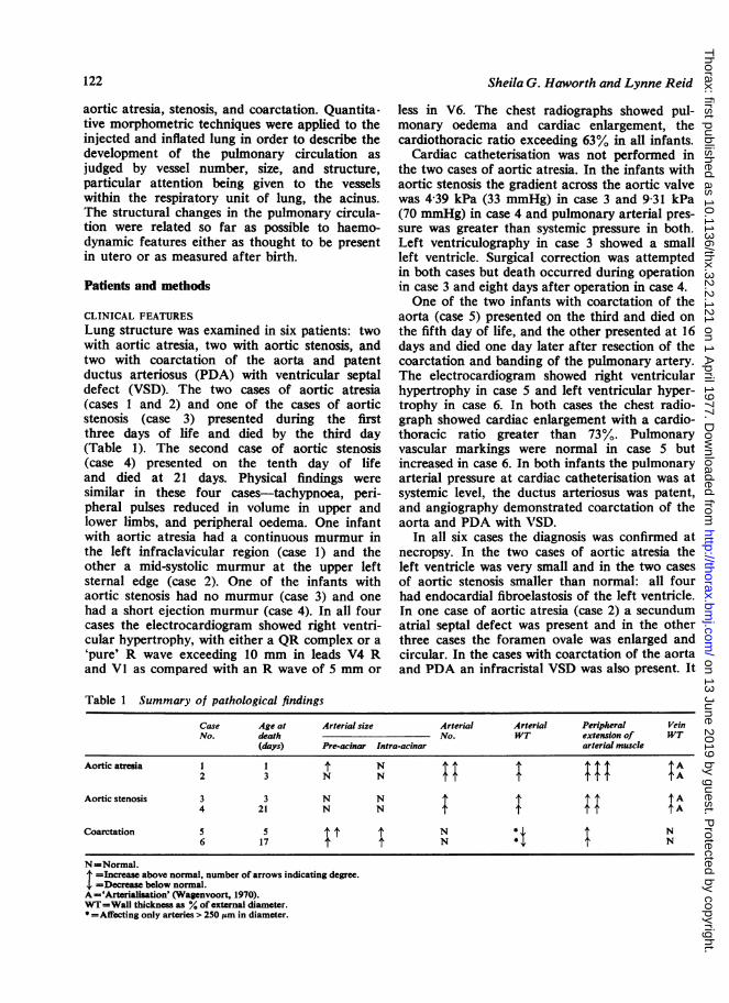
Number of intra-acinar arteriesIn all four cases the background haze on thearteriogram was denser than normal (Fig. 1), sug-gesting an increase in the number of peripheralarteries. The proportion of lung volume occupiedby arteries as assessed by microscopic point count-ing was also abnormally high. An increase innumber of arteries per unit area of lung explainedthese findings (Table 2). Because the alveolarnumber per unit area was normal the alveolar/arterial ratio was reduced.
123
on 13 June 2019 by guest. Protected by copyright.
http://thorax.bmj.com
/T
horax: first published as 10.1136/thx.32.2.121 on 1 April 1977. D
ownloaded from

Sheila G. Haworth and Lynne Reid
Fig. 1 A rteriogram of left lung in (left) a case of aortic atresia and (right) a normal infant showing anabnormal increase in density background haze in aortic atresia (XO95).
Table 2 Number of alveoli and arteries per unit areaand alveolar/arterial ratio
Alveoli Arteries Alveolar/ArterialCase No. (No.) (No.) ratio
1 15 324 1290 11-82 14 338 1001 14-33 15 884 812 19-64 10 931 782 14-05 12 467 556 22*46 8860 408 21P6
Normal values at:Birth 8863 468 18.93 days 12 722 625 20-34weeks 13196 516 2554 months 11 315 529 22-2
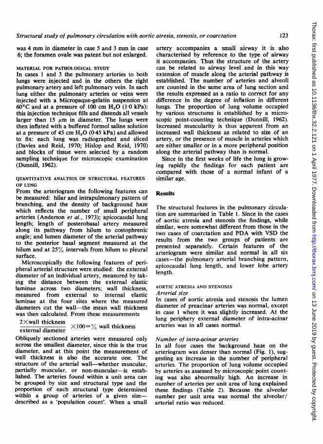
Arterial muscularityAll cases showed a considerable increase inmuscularity as judged by medial thickness and byextension of muscle into smaller and more peri-
pheral arteries than is normal (Fig. 2). In vesselsof all sizes percentage wall thickness was higherthan during fetal life (Hislop and Reid, 1972).Surprisingly, the mean medial thickness wasgreater in the cases of aortic stenosis than in thoseof aortic atresia, perhaps because one case ofaortic stenosis, the infant, had survived for 21days, time enough to develop considerable arterialmuscularity, while one of the cases of aorticatresia (case 2) had a secundum atrial septaldefect and therefore no obstruction to pulmonaryvenous return. The high wall thickness in arteriesless than 250 ,um in diameter suggests that thesevessels did not dilate normally at birth (Hislopand Reid, 1973a).
Examination of the structure of arteries accom-panying peripheral airways showed considerableextension of muscle along arterial pathways (Table3). In the normal lung at birth few arteries withinthe acinus have a muscular coat. During child-
124
on 13 June 2019 by guest. Protected by copyright.
http://thorax.bmj.com
/T
horax: first published as 10.1136/thx.32.2.121 on 1 April 1977. D
ownloaded from

Structural study of pulmonary circulation with aortic atresia, stenosis, or coarctation
kI__*:w' -.......
200 400 600 860 1000 1200External Diameter (,um)
Fig. 2 Mean percentage wall thickness of smallperipheral pulmonary arteries related to externaldiameter (um) showing increase in muscularity incases of aortic atresia and stenosis, and in cases ofcoarctation a decrease in wall thickness in arteriesgreater than 250 pm in diameter. * * Aorticstenosis. x x Aortic atresia. c o CoarctationPDA and VSD. @- -* Normal 3 days. 0 .... 0Mean of 6 normal children 10 months-JO years. I SE.
Table 3 Proportion of arteries of differentstructural types accompanying peripheral airways
Case Structural Terminal Respiratory AlveolarNo. type bronchioli bronchioli ducts
I nm - - -pm - 9 68m 100 91 32
2 nm Consolidation made accuratepm identification of peripheral airwaysm impossible
3 nm - - 28pm - 38 67m 100 62 5
4 nm - - 15pm 20 64 85m 80 36 -
5 nm - - 28pm 75 100 72m 25 - -
6 nm - 9 48pm - 78 52m 100 13 -
Normal values (Hislop and Reid 1973a) at:3 days nm - 30 66
pm 72 70 34m 28 - -
4months nm - 3 35pm - 30 18m 100 67 47
nm=non-muscular; pm=partially muscular; m=muscular.
hood muscle gradually extends along the arterialpathway, reaching alveolar wall vessels in lateadolescence (Hislop and Reid, 1973a). In all four
of the present cases, however, over 65% ofarteries accompanying alveolar ducts had apartially muscular wall and many alveolar wallarteries were completely muscularised.The available venograms, one of a case of
aortic atresia (case 2) and one of aortic stenosis(case 4), showed dilatation of the preacinarpathways. In all cases vein wall thickness was in-creased and an external elastic lamina had ap-peared. Thus, according to Wagenvoort's (1970)description, the veins had become 'arterialised'.
COARCTATION OF AORTA WITH PDA AND VSDArterial sizeIn contrast to the cases of aortic atresia andstenosis, in both cases of coarctation with PDAand VSD the preacinar arterial lumen diameterand the external diameter of intra-acinar arteriesat the level of the terminal bronchioli were greaterthan normal. The number of arteries per unitarea of lung and the alveolar/arterial ratio werenormal for the age of the child.
Arterial wall thickness was less than normal invessels above 250 MLm in diameter (Fig. 2) perhapsbecause of dilatation. In smaller arteries wallthickness was normal. Within the acinus muscleextended into smaller and more peripheral arteriesthan is normal (Table 1). In the younger of thetwo infants (case 5) the appearance of the musclecells present at alveolar duct level suggested im-maturity. The cells were arranged as a singlelayer, the media were only 1-13 ,um-1i96 Mmthick, and they stained poorly, if at all. While 72%of arteries at alveolar duct level were partiallymuscular none was entirely muscular. The infantin case 6 was 12 days older than the infant in case5 and the proportion of muscularised arteries wasincreased at all levels within the acinus: themuscle cells stained normally.
Discussion
In cases of both aortic atresia and stenosis therewas an increase in number of the intra-acinararteries and the arteries and veins were moremuscular. The muscularity was shown both by anincrease in wall thickness of normally muscularvessels and by extension of muscle into vesselsthat do not normally contain it at this age.Arterial size was generally normal. Since three ofthese babies died in the first three days of lifethese changes undoubtedly occurred before birth.In contrast, in coarctation of the aorta the pre-acinar pulmonary arteries were dilated and thinwalled but muscle was found more peripherallythan normal, since intra-acinar arteries had
12
Un 10.0
= 8.
3 6-
0
CP
0
040
a -
125
on 13 June 2019 by guest. Protected by copyright.
http://thorax.bmj.com
/T
horax: first published as 10.1136/thx.32.2.121 on 1 April 1977. D
ownloaded from

126
smooth muscle cells in their wall. In the youngestchild the muscle cells were of immature appear-ance, suggesting development after birth. Thesestructural differences probably reflect a differenceboth in haemodynamic abnormality and in thestage of fetal or postnatal life at which itdeveloped.
AORTIC ATRESIA AND STENOSISThe methods used in this study supplement theinformation obtained from previously used tech-niques. Wagenvoort and Edwards (1961), usinguninjected tissue, demonstrated an increase inarterial medial muscle area per unit of lung tissue,which they thought was due to an increase in thenumber of muscularised arteries. Naeye (1962),using a planimetric technique, demonstrated anincrease in medial muscle in individual vesselsalready muscularised, as we have also shown. Ourresults show that increase in the amount ofmuscle is due to an increase in muscle of arteriesalready muscularised and to an extension ofmuscle into arteries that are not normallymuscularised. Furthermore, the number of peri-pheral arteries 'available' for muscularisation isalso increased.
In the present study accurate measurements ofdiameter and wall thickness of individual arteriesrevealed not only the severity of medial hyper-trophy but also that arteries smaller than 250 ,umin diameter had failed to dilate normally at birth.We have also demonstrated a normal preacinarbranching pattern, the virtual absence of arterialdilatation, either pre- or intra-acinar, and an in-crease in arterial multiplication within the acinus.Thus in aortic atresia and stenosis lung develop-
ment has been affected before birth, presumablyby the abnormal pattern of blood flow throughthe heart and great vessels. At birth the rightventricular pressure is abnormally raised, althoughopinions differ about the level of pulmonaryarterial pressure before birth. Rudolph (1974)argues that pulmonary hypertension will developin utero in this condition only if the ductusarteriosus fails to dilate to accommodate ade-quately the systemic blood flow; right ventricularpressure would then increase in an attempt tomaintain systemic and placental perfusion. Ourfindings of increased pulmonary arterial muscu-larity suggest an abnormally high pulmonaryarterial pressure before birth, despite the fact thatin all cases the ductus arteriosus was large.The prenatal development of arterial medial
hypertrophy in aortic atresia and stenosis haspreviously been attributed either to pulmonaryvenous obstruction (Wagenvoort and Edwards,
Sheila G. Haworth and Lynne Reid
1961) or to a raised right ventricular pressure(Naeye, 1962). Our results favoured pulmonaryarterial hypertension as the cause. In the presentseries medial hypertrophy was always greater thanis usually seen in newborn infants with totalanomalous pulmonary venous return (personalobservation). Furthermore, the presence of veinwall hypertrophy does not necessarily implypulmonary venous obstruction since 'arterialisa-tion' of veins may occur in the presence of onlymoderate pulmonary venous hypertension, as isseen in VSD (Hislop et al., 1975).The arterial medial hypertrophy favours
diversion of blood away from the lungs, thusensuring adequate systemic perfusion. Musclehypertrophy encroaches on the lumen, and becausethe arteries are smaller before than after birthan increase in size or number of muscle cells willproduce a greater increase in resistance. At birthpulmonary vascular resistance must have fallenslightly but the high wall thickness of small pul-monary arteries found in the lungs of thesenewborn infants suggests that they did not dilatenormally at birth, and so a raised pulmonaryvascular resistance is maintained.
This discussion implies that the smooth musclehypertrophies in response to haemodynamicchanges in the pulmonary circulation and is not aprimary structural abnormality. In older childrenwith pulmonary hypertension there is an increasein muscularity of both pre- and intra-acinararteries, as here, while in newborn children withpersistent pulmonary hypertension (without con-genital heart disease) it is only the intra-acinararteries which are affected (Haworth and Reid,1976). A prenatal increase in number of intra-acinar arteries has not previously been reported.This increase could include some peripheral pre-acinar arteries, since at this level such vesselswould be included in the arterial count of smallarteries. Vessel number in post-natal life is influ-enced by haemodynamic changes, increasing intetralogy of Fallot (Hislop and Reid, 1973b) anddecreasing in VSD (Hislop et al., 1975). In thepresent study the mechanism responsible for theexcessive multiplication of intra-acinar arteries isnot understood, but the multiplication must occursome time before birth since at birth these vesselsare surrounded by a thick coat of mature musclecells. The simplest interpretation is that it is aresponse to increased flow.The development of an increased number of
arteries would increase the number of vessels avail-able for recruitment. If the distensibility of theperipheral arteries is reduced by muscle hyper-trophy recruitment of peripheral arteries may be
on 13 June 2019 by guest. Protected by copyright.
http://thorax.bmj.com
/T
horax: first published as 10.1136/thx.32.2.121 on 1 April 1977. D
ownloaded from

Structural study of pulmonary circulation with aortic atresia, stenosis, or coarctation
important in accommodating the increase in pul-monary blood volume occurring at birth. Recruit-ment of additional channels is thought to be moreimportant than distension in accommodating anincrease in blood volume (Maseri et al., 1972).
COARCTATION OF AORTA WITH PDA AND VSDIn both cases of coarctation with PDA and VSDthere was no increase either in the number ofintra-acinar arteries or in arterial wall thickness,suggesting that, unlike cases of aortic atresia andstenosis, the structure of the pulmonary circula-tion was probably normal at birth. This supportsthe contention that coarctation can develop afterbirth, arising when the aortic end of the ductusarteriosus constricts after birth and protrudesslightly into the lumen of the aorta, approachinga shelf of tissue on the posterolateral surface ofthe aortic wall (Rudolph et al., 1972). Had aorticobstruction been present before birth severemedial hypertrophy, similar to that seen in thepresent cases of aortic atresia and stenosis, wouldhave been expected even in the 5-day-old child.When infants are born with severe coarctation anincrease in arterial wall thickness may be found,as in the cases reported by Naeye (1961).
Reduction in wall thickness of small arteriesunder 250 ,um in diameter suggests that pulmonaryvascular resistance had fallen normally at birth,permitting a left-to-right shunt across the PDAand VSD. The muscle cells found in vesselsunder 250 ,um in diameter had almost certainlyappeared after birth, probably in response to thesecondary increase in arterial pressure caused bya PDA and VSD. In the older infant considerablemuscle hypertrophy had developed by 17 days ofage.
Although it has been suggested that regressionof pulmonary arterial muscle after birth is de-layed in the presence of an aortopulmonary com-munication (Hoffman and Rudolph, 1965), ourresults offer evidence that wall thickness of smallarteries can diminish normally during the first fewdays of life but that subsequently the develop-ment of new muscle cells occurs rapidly. At 5days of age immature muscle cells were seen. Inthe experimental animal they may be detectedafter only three days exposure to hypoxia (Hislop.personal communication). Possibly the rapiditywith which muscle cells develop has previouslybeen underestimated, and in some cases of con-genital heart disease early formation of musclecells may have been mistaken for persistence offetal muscle.Thus it seems that in aortic atresia and stenosis
the entire cardiac output is ejected by the right
ventricle into the pulmonary artery and thepulmonary arterial smooth muscle hypertrophies.Pulmonary vascular resistance is thereby increasedand systemic perfusion encouraged. This haemo-dynamic state persists after birth and peripheralpulmonary arteries remain thick walled. Themechanism of production and the functionaleffects of the excessive multiplication of intra-acinar arteries remain unexplained. By contrast,when coarctation develops after birth the pul-monary circulation is structurally normal in utero.At birth pulmonary arterial pressure falls normallyand the small vessels dilate, permitting a left toright shunt to develop across the PDA and VSD.As pulmonary blood flow increases the arteriesdilate and then, as pulmonary arterial pressureincreases, their muscle coats hypertrophy.
Aortic atresia and stenosis may increase thelungs' susceptibility to vasoconstrictor agents suchas hypoxia and acidosis. The rapid postnatal in-crease in muscularity in the presence of pulmonaryhypertension suggests that if the structure of thepulmonary circulation were the only considerationit would be better not to delay surgicalintervention.
We thank the physicians and surgeons of theBrompton Hospital, London, and the DeutschesHerzzentrum, Munich, Germany, for permissionto use material from their cases.
This work was supported by the WellcomeTrust.
References
Anderson, E. G., Simon, G., and Reid, L. (1973).Primary and thrombo-embolic pulmonary hyper-tension: a quantitative pathological study. Journalof Pathology, 110, 273-293.
Davies, G. and Reid, L. (1970). Growth of thealveoli and pulmonary arteries in childhood.Thorax, 25, 669-681.
Dunnill, M. S. (1962). Quantitative methods in thestudy of pulmonary pathology. Thorax, 17, 320-328.
Ferencz, C. and Dammann, J. F. Jr. (1957). Signifi-cance of the pulmonary vascular bed in congenitalheart disease. V. Lesions of the left side of theheart causing obstruction of the pulmonary venousreturn. Circulation, 16, 1046-1056.
Haworth, S. G. and Reid, L. (1976). Persistent fetalcirculation: Newly recognized structural features.Journal of Pediatrics, 88, 614-620.
Hislop, A., Haworth, S. G., Shinebourne, E. A., andReid, L. (1975). Quantitative structural analysis cfpulmonary vessels in isolated ventricular septaldefect in infancy. British Heart Journal, 37, 1014-1021.
127
on 13 June 2019 by guest. Protected by copyright.
http://thorax.bmj.com
/T
horax: first published as 10.1136/thx.32.2.121 on 1 April 1977. D
ownloaded from

Sheila G. Haworth and Lynne Reid
Hislop, A. and Reid, L. (1970). New pathological find-ings in emphysema of childhood: 1. Polyalveolarlobe with emphysema. Thorax, 25, 682-690.
Hislop, A. and Reid, L. (1972). Intra-pulmonaryarterial development during fetal life-branchingpattern and structure. Journal of Anatomy, 113,35-48.
Hislop, A. and Reid, L. (1973a). Pulmonary arterialdevelopment during childhood: branching patternand structure. Thorax, 28, 129-135.
Hislop, A. and Reid, L. (1973b). Structural changes inthe pulmonary arteries and veins in tetralogy ofFallot. British Heart Journal, 35, 1178-1183.
Hoffman, J. I. E. and Rudolph, A. M. (1965). Thenatural history of ventricular septal defects ininfancy. American Journal of Cardiology, 16, 634-653.
Maseri, A., Caldini, P., Harward, P., Joshi, R. C.,Permutt, S., and Zierler, K. L. (1972). Determinantsof pulmonary vascular volume: recruitment versusdistensibility. Circulation Research, 31, 218-228.
Naeye, R. L. (1961). Perinatal vascular changes incoarctation of the aorta with distal patent ductusarteriosus. Circulation, 24, 754-760.
Naeye, R. L. (1962). Perinatal vascular changesassociated with underdevelopment of the left heart.American Journal of Pathology, 41, 287-295.
Rudolph, A. M. (1974). In Congenital Diseases of theHeart, p. 552. Year Book Medical Publishers, NewYork, Chicago.
Rudolph, A. M., Fishman A., and Heymann, M. A.(1974). Cited by Rudolph, A. M. In CongenitalDiseases of the Heart, pp. 363 and 548. Year BookMedical Publishers, New York, Chicago.
Rudolph, A. M., Heymann, M. A., and Spitznas. U.(1972). Hemodynamic considerations in the develop-ment of narrowing of the aorta. American Journalof Cardiology. 30, 514-525.
Samuelson, A., Becker, A. E., and Wagenvoort, C. A.(1970). A morphometric study of pulmonary veinsin normal infants and infants with congenital heartdisease. Archives of Pathology, 90, 112-116.
Wagenvoort, C. A. (1970). Morphologic changes inthe intrapulmonary veins. Human Pathology, 1,205-213.
Wagenvoort, C. A. and Edwards, J. E. (1961). Thepulmonary arterial tree in aortic atresia with intactventricular septum. Laboratory Investigation, 10,924-933.
Requests for reprints to: Dr. Lynne Reid, Depart-ment of Pathology, Bldg. G-9, Children's HospitalMedical Center, 320 Longwood Avenue, Boston,Ma 02115, U.S.A.
128
on 13 June 2019 by guest. Protected by copyright.
http://thorax.bmj.com
/T
horax: first published as 10.1136/thx.32.2.121 on 1 April 1977. D
ownloaded from

![Isolated Retroperitoneal Hydatid Cyst Invading Splenic Hilum...the presence of cyst rupture, spread of protoscoleces, and bacterialinfection-relatedcomplications[1,6,7].Thedefini-](https://static.fdocuments.us/doc/165x107/5e4690d07bb29234947acf53/isolated-retroperitoneal-hydatid-cyst-invading-splenic-hilum-the-presence-of.jpg)