Quantitative assessment of placental morphology may ...
12
RESEARCH ARTICLE Open Access Quantitative assessment of placental morphology may identify specific causes of stillbirth Imogen Ptacek 1,2 , Anna Smith 3 , Ainslie Garrod 1,2 , Sian Bullough 1,2 , Nicola Bradley 1,2 , Gauri Batra 3 , Colin P. Sibley 1,2 , Rebecca L. Jones 1,2 , Paul Brownbill 1,2 and Alexander E. P. Heazell 1,2* Abstract Background: Stillbirth is frequently the result of pathological processes involving the placenta. Understanding the significance of specific lesions is hindered by qualitative subjective evaluation. We hypothesised that quantitative assessment of placental morphology would identify alterations between different causes of stillbirth and that placental phenotype would be independent of post-mortem effects and differ between live births and stillbirths with the same condition. Methods: Placental tissue was obtained from stillbirths with an established cause of death, those of unknown cause and live births. Image analysis was used to quantify different facets of placental structure including: syncytial nuclear aggregates (SNAs), proliferative cells, blood vessels, leukocytes and trophoblast area. These analyses were then applied to placental tissue from live births and stillbirths associated with fetal growth restriction (FGR), and to placental lobules before and after perfusion of the maternal side of the placental circulation to model post-mortem effects. Results: Different causes of stillbirth, particularly FGR, cord accident and hypertension had altered placental morphology compared to healthy live births. FGR stillbirths had increased SNAs and trophoblast area and reduced proliferation and villous vascularity; 2 out of 10 stillbirths of unknown cause had similar placental morphology to FGR. Stillbirths with FGR had reduced vascularity, proliferation and trophoblast area compared to FGR live births. Ex vivo perfusion did not reproduce the morphological findings of stillbirth. Conclusion: These preliminary data suggest that addition of quantitative assessment of placental morphology may distinguish between different causes of stillbirth; these changes do not appear to be due to post-mortem effects. Applying quantitative assessment in addition to qualitative assessment might reduce the proportion of unexplained stillbirths. Keywords: Stillbirth, Unexplained Stillbirth, Placental Morphometry, Fetal Growth Restriction, Villous vascularity, Avascular villi * Correspondence: [email protected] 1 Institute of Human Development, Faculty of Medical and Human Sciences, University of Manchester, Oxford Rd, Manchester M13 9PL, UK 2 Maternal and Fetal Health Research Centre, 5th floor (Research), St Mary’s Hospital, Oxford Road, Manchester M13 9WL, UK Full list of author information is available at the end of the article © 2016 Ptacek et al. Open Access This article is distributed under the terms of the Creative Commons Attribution 4.0 International License (http://creativecommons.org/licenses/by/4.0/), which permits unrestricted use, distribution, and reproduction in any medium, provided you give appropriate credit to the original author(s) and the source, provide a link to the Creative Commons license, and indicate if changes were made. The Creative Commons Public Domain Dedication waiver (http://creativecommons.org/publicdomain/zero/1.0/) applies to the data made available in this article, unless otherwise stated. Ptacek et al. BMC Clinical Pathology (2016) 16:1 DOI 10.1186/s12907-016-0023-y
Transcript of Quantitative assessment of placental morphology may ...

RESEARCH ARTICLE Open Access
Quantitative assessment of placentalmorphology may identify specific causes ofstillbirthImogen Ptacek1,2, Anna Smith3, Ainslie Garrod1,2, Sian Bullough1,2, Nicola Bradley1,2, Gauri Batra3, Colin P. Sibley1,2,Rebecca L. Jones1,2, Paul Brownbill1,2 and Alexander E. P. Heazell1,2*
Abstract
Background: Stillbirth is frequently the result of pathological processes involving the placenta. Understanding thesignificance of specific lesions is hindered by qualitative subjective evaluation. We hypothesised that quantitativeassessment of placental morphology would identify alterations between different causes of stillbirth and thatplacental phenotype would be independent of post-mortem effects and differ between live births and stillbirthswith the same condition.
Methods: Placental tissue was obtained from stillbirths with an established cause of death, those of unknowncause and live births. Image analysis was used to quantify different facets of placental structure including: syncytialnuclear aggregates (SNAs), proliferative cells, blood vessels, leukocytes and trophoblast area. These analyses werethen applied to placental tissue from live births and stillbirths associated with fetal growth restriction (FGR), and toplacental lobules before and after perfusion of the maternal side of the placental circulation to model post-mortemeffects.
Results: Different causes of stillbirth, particularly FGR, cord accident and hypertension had altered placentalmorphology compared to healthy live births. FGR stillbirths had increased SNAs and trophoblast area and reducedproliferation and villous vascularity; 2 out of 10 stillbirths of unknown cause had similar placental morphology toFGR. Stillbirths with FGR had reduced vascularity, proliferation and trophoblast area compared to FGR live births. Exvivo perfusion did not reproduce the morphological findings of stillbirth.
Conclusion: These preliminary data suggest that addition of quantitative assessment of placental morphology maydistinguish between different causes of stillbirth; these changes do not appear to be due to post-mortem effects.Applying quantitative assessment in addition to qualitative assessment might reduce the proportion of unexplainedstillbirths.
Keywords: Stillbirth, Unexplained Stillbirth, Placental Morphometry, Fetal Growth Restriction, Villous vascularity,Avascular villi
* Correspondence: [email protected] of Human Development, Faculty of Medical and Human Sciences,University of Manchester, Oxford Rd, Manchester M13 9PL, UK2Maternal and Fetal Health Research Centre, 5th floor (Research), St Mary’sHospital, Oxford Road, Manchester M13 9WL, UKFull list of author information is available at the end of the article
© 2016 Ptacek et al. Open Access This article is distributed under the terms of the Creative Commons Attribution 4.0International License (http://creativecommons.org/licenses/by/4.0/), which permits unrestricted use, distribution, andreproduction in any medium, provided you give appropriate credit to the original author(s) and the source, provide a link tothe Creative Commons license, and indicate if changes were made. The Creative Commons Public Domain Dedication waiver(http://creativecommons.org/publicdomain/zero/1.0/) applies to the data made available in this article, unless otherwise stated.
Ptacek et al. BMC Clinical Pathology (2016) 16:1 DOI 10.1186/s12907-016-0023-y

BackgroundHistological examination of the placenta is one of themost frequently performed investigations to identify thecause of death in cases of stillbirth [1]; its application inthis context is recommended by international guidelines[2–4]. A recent systematic review found large variationsin the methodological quality of studies of placentalexamination after stillbirth, with few studies of highquality [5]. Interpretation of the results of such studies isfurther complicated by the use of different classificationsystems and a lack of consensus in terminology used todescribe placental lesions which results in a large vari-ation in the proportion of stillbirths attributed to a pla-cental “cause” from 11–65 % [5]. Such qualitativeplacental assessment, combined with varied terminology,has some deficiencies. Firstly, qualitative assessment ofplacental lesions may introduce bias, particularly if as-sessors are not blinded to outcome. Furthermore, quali-tative assessment may lead to inter-observer variation indiagnoses, which ranged from 25–91 % in one study [6].The significance of specific abnormalities to stillbirthhas also been questioned by Pathak et al. who describeplacental abnormalities in a significant proportion of ap-parently healthy live-born infants [7]. Finally, identifica-tion of a specific lesion does not imply a single cause.For example, appearances of fetal thrombotic vasculopa-thy have been associated with various pathologies in-cluding: cytomegalovirus infection [8], umbilical cordaccidents [9] or specific patterns of umbilical cord coil-ing [10]. Similarly, changes of maternal underperfusionmay be related to hypertensive disorders [11] and anti-phospholipid syndrome [12].Recently, significant advances have been made in
the development of modern classification systems[13–15] that reduce the proportion of unexplainedstillbirths [16]. These classification systems have givengreater recognition to the role of placental pathologyin the aetiology of stillbirth [13–15] and progress hasbeen made in reducing the variation of placentalhistological findings [6]. However, these clinically-orientated descriptions of placental phenotype havenot yet been adopted into widespread practice, in partdue to continued debate about terminology whichvaries between clinical and research studies [17]. Re-search studies have employed quantitative descriptionsof placental morphology by stereology and morphom-etry to describe differences in placental structure inclinical conditions related to stillbirth such as fetalgrowth restriction (FGR) [18] and reduced fetal move-ments [19]. We aimed to use these quantitativemethods to objectively evaluate placental appearancesof different causes of stillbirth. Firstly, we hypothe-sised that specific causes of stillbirth would be associ-ated with a morphometric phenotype. Secondly, we
hypothesised that morphological abnormalities associ-ated with stillbirth would differ from live births withthe same condition. To be of diagnostic value, anyobserved changes should not represent artefacts ofstorage or cessation of fetal blood flow after death.Since we have already described the effects of placen-tal storage on placental structure [20], here we ad-dress a third hypothesis that there is an acute effectof post-stillbirth fetoplacental haemostasis on placen-tal morphology.
MethodsPlacental tissue samplesTo address the first hypothesis we obtained placentaltissue from cases of stillbirth, defined as the birth ofan infant with no signs of life after 24 weeks gesta-tion. Parents gave permission for the use of samplesfor research at the time of consent for post-mortemexamination. A favourable ethical opinion was givenby the Greater Manchester South Research EthicsCommittee (09/H1012/11) and approval given fromthe Research and Innovation Division of CentralManchester University Hospitals NHS FoundationTrust to conduct the study. Cases of stillbirth wereclassified using the ReCoDe system [15] by amultidisciplinary meeting following a full panel of in-vestigations including: post-mortem, histopathologicalexamination of the placenta, chromosomal analysisand maternal biochemical, haematological, immuno-logical and serological tests. We obtained samplesfrom the following classifications of stillbirth: cord ac-cident (n = 8), diabetes (n = 5), FGR (n = 10), hyperten-sion (n = 8), infection (n = 8) and from stillbirths of anunknown cause (n = 10). For comparison, matchedplacental samples were used from appropriately-grownlive born infants and preterm births (26-36 weeks)(demographics shown in Table 1). Samples from livebirths were collected following written informed con-sent as part of the Maternal and Fetal ResearchCentre (MFHRC) Biobank (08/H1010/55). To addressthe second hypothesis, placental samples were takenfrom a further cohort of stillbirths attributed to FGR(n = 13) and from live births with FGR (n = 13) withthe same ethical approvals as described above. FGRwas defined as a customised birthweight <5th centile(demographics shown in Table 2). To address thefinal hypothesis, placental tissue was obtained fromappropriately grown (n = 7) and FGR (n = 5) livebirths following written informed consent as part ofthe MFHRC biobank previously described. For allcases, maternal and infant demographic informationwas recorded from medical case notes and post-mortem reports (for stillbirths). An estimate of theduration of in utero retention was made according to
Ptacek et al. BMC Clinical Pathology (2016) 16:1 Page 2 of 12

Table 1 Demographic characteristics of samples from live births and stillbirths from known and unknown causes. Birthweight was significantly lower in stillbirths from FGR andhypertension than live births (P < 0.01); all other variables did not significantly differ between groups. Data are presented as median with range in parentheses except forestimated time of retention in utero where number of cases are presented
Live births Preterm birth Cord Diabetes Hypertension Infection FGR Unknown
Number of Samples 10 7 8 5 8 9 10 10
Maternal Age (years) 32 (28–37) 27 (18–41) 28 (21–36) 34 (31–40) 33 (27–37) 30 (25–32) 30 (22–32) 28 (21–31)
Gravidity 1 (1–5) 3 (1–6) 1 (1–1) 4 (2–7) 1 (1–2) 1 (1–3) 1 (1–4) 1 (1–2)
Parity 0 (0–4) 0 (0–4) 0 (0–0) 1 (1–2) 0 (0–0) 0 (0–2) 0 (0–2) 0 (0–0)
Gestation at delivery(weeks)
37 (37–38) 31 (26–36) 30 (27–38) 28 (28–40) 31 (26–35) 38 (24–41) 34 (26–38) 31 (27–39)
Birthweight (g) 3090 (2805–3430) 1821 (786–2760) 1300 (702–2750) 3120 (1473–3590) 1110 (399–1730) 2680 (494–2985) 1065 (564–2230) 2170 (892–3150)
Estimated in uteroretention time
0 h - <24 h N/A N/A 0 1 4 4 2 2
≥24 h - < 48 h 1 0 2 2 4 3
≥48 h - < 96 h 2 1 0 2 2 1
≥96 h - <1 week 3 1 1 0 0 2
≥1 week 2 2 1 1 2 2
Ptaceket
al.BMCClinicalPathology
(2016) 16:1 Page
3of
12
Genests’ descriptions of findings at post-mortem andfrom histopathological examination of the placenta[21–23].Tissue from live births was obtained within 30 min of
delivery. For assessment of placental morphology biop-sies of villous tissue were dissected from the centre,middle and edge of the placenta. Tissue was fixed in 4 %neutral buffered formalin for 24 h before being wax em-bedded. For stillbirth samples three blocks of placentaltissue not obtained from specific lesions were obtainedfor each placenta.
Placental perfusionUnless otherwise stated, all reagents were supplied bySigma-Aldrich Chemical Company (Poole, UK). Toexamine the acute impact of continued maternalblood flow in the absence of fetal blood flow single-sided (maternal) ex vivo human placental lobule per-fusion was adapted from the dual-sided perfusionmodel [24]. Perfusion was performed on placentasfrom normal pregnancy (n = 7) and placentas frompregnancies complicated by FGR (n = 5). An intactperipheral lobule was selected, devoid of post-partumtears, deep decidual damage and marginal membraneseparations. Prior to perfusion two villous biopsieswere sampled from neighbouring lobules taken 5 cmapart, and fixed immediately in 4 % neutral bufferedformalin forming “pre-perfusion samples”. The fetalartery and vein on the chorionic surface, serving thevillous trees within the lobule designated for perfu-sion, were each ligated using sutures (Mersilk 3/0,Ethicon, supplied by NuCare, UK) to confine a staticfetal blood pool within the fetal vasculature of the as-sociated cotyledons. The maternal surface was cannu-lated using five 10 cm lengths of polythene tubing(Smiths Medical, UK) arising from a perfusion mani-fold (Harvard Apparatus, UK). The distal ends of thecannulae were cut into apices and inserted throughthe decidual surface of the lobule with an even spatial
distribution. The perfusate was modified Earle's bicar-bonate buffer (EBB 117 mM NaCl, 10.7 mM KCl,5.6 mM D-glucose, 3.6 mM CaCl, 1.8 mM NaH2PO4,13.6 mM NaHCO3, 0.04 mM L-arginine, 0.8 mMMgSO4, 3.5 % (w/v) dextran, 0.1 % (w/v) bovineserum albumin, 5000 IU/L Heparin sodium) equili-brated with 95 % O2 / 5 % CO2 to pH 7.4 andwarmed to 37 °C, delivered by a roller pump (WatsonMarlow, UK) at 14 ml/min. Lobule preparations wereonly considered acceptable for experimentation whenmaternal-side perfusion was established within 30 minof delivery. Open-circuit perfusion was for 6 h, andthen the physiological buffer was switched to a 4 %neutral buffered formalin at T = 6 h for a 10 minmaternal-side perfusion fixation period. Followingthis, the lobule was excised and two further fullthickness (vertical and horizontal) biopsies slices weretaken as the “post-perfusion samples”. These wide tis-sue sections where then immersion fixed in 4 % neu-tral buffered formalin for 24 h before being waxembedded. Placental structure in these biopsies wasexamined as described above.
ImmunohistochemistryPlacental cell turnover, structure and vascularity wereassessed using antibodies specific for Ki67 (Dako, Ely,Cambridgeshire, UK; 0.16 μg/ml), cytokeratin 7 (Dako;0.9 μg/ml) and CD31 (Dako; 0.16 μg/ml). The num-ber of leukocytes was assessed by an antibody specificfor CD45 (Dako; 0.4 μg/ml). Negative controls wereperformed using non-immune mouse IgG (Dako) atmatching concentrations to the primary antibody.Immunohistochemistry was performed as previouslydescribed with antigen retrieval performed by micro-waving the sections for 10 min in 0.01 M sodium cit-rate buffer [19, 20].Quantification of syncytial nuclear aggregates (SNAs,
also known as syncytial knots) was conducted onsections stained with haematoxylin and eosin as
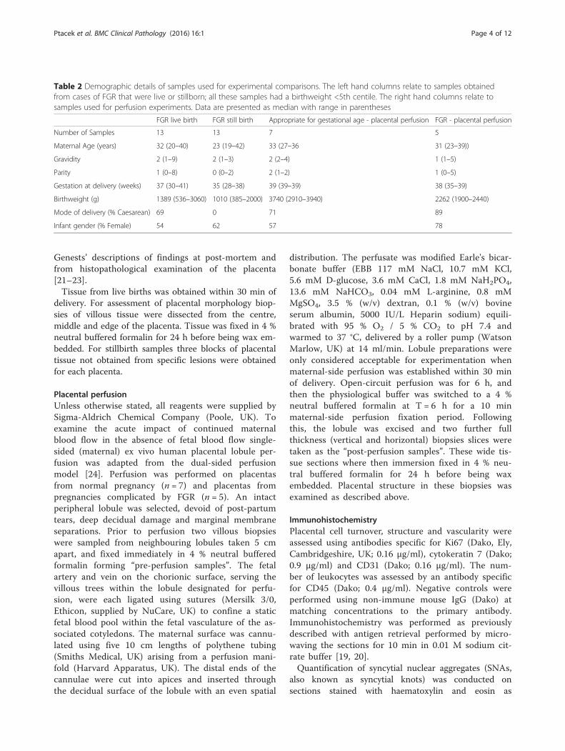
Table 2 Demographic details of samples used for experimental comparisons. The left hand columns relate to samples obtainedfrom cases of FGR that were live or stillborn; all these samples had a birthweight <5th centile. The right hand columns relate tosamples used for perfusion experiments. Data are presented as median with range in parentheses
FGR live birth FGR still birth Appropriate for gestational age - placental perfusion FGR - placental perfusion
Number of Samples 13 13 7 5
Maternal Age (years) 32 (20–40) 23 (19–42) 33 (27–36 31 (23–39))
Gravidity 2 (1–9) 2 (1–3) 2 (2–4) 1 (1–5)
Parity 1 (0–8) 0 (0–2) 2 (1–2) 1 (0–5)
Gestation at delivery (weeks) 37 (30–41) 35 (28–38) 39 (39–39) 38 (35–39)
Birthweight (g) 1389 (536–3060) 1010 (385–2000) 3740 (2910–3940) 2262 (1900–2440)
Mode of delivery (% Caesarean) 69 0 71 89
Infant gender (% Female) 54 62 57 78
Ptacek et al. BMC Clinical Pathology (2016) 16:1 Page 4 of 12

previously described [19, 20]. Dewaxed and rehydratedsections were stained with Harris’s haematoxylin for10 min before differentiation in acid-alcohol. Slides werestained with eosin for 2 min, rinsed in cold tap water,and dehydrated and mounted as described above.For all analyses images were captured using an
Olympus BX41 light microscope (Southend-on-Sea,UK) and QIcam Fast 1394 (QImaging, BC, Canada)and Image Pro Plus 6.0 and 7.0 (Media CyberneticsInc., MD, USA). During image acquisition and ana-lysis the presumed cause of stillbirth was concealed
from the observer. Between images the microscopewas taken out of focus to prevent selection bias, ifthe randomly selected image was not mostly of ter-minal villi another area was identified. Five randomimages of terminal villi were taken of each section,giving a total of 15 images per placenta for eachcomponent evaluated.
Assessment of placental structureThe number of SNAs were counted and total villousarea measured using image analysis software, expressed
Fig. 1 a Assessment of syncytial nuclear aggregates (SNAs) in different causes of stillbirth compared to healthy live births. SNAs are shown byopen arrows in representative images from normal pregnancy and stillbirth associated with hypertension. b Assessment of proliferation indifferent causes of stillbirth compared to healthy live births. c Assessment of trophoblast area in different causes of stillbirth compared to healthylive births. Negative control images shown in small panel beneath representative images of normal pregnancy and FGR. Scale bar = 50 μm in allimages. Graphs show median and range, * p < 0.05, ** p < 0.01, *** p < 0.001. Dotted line indicates median level of healthy control
Ptacek et al. BMC Clinical Pathology (2016) 16:1 Page 5 of 12

as the number of SNAs per mm2 of villous tissue as pre-viously described [25]. Proliferative index was the num-ber of Ki67 positive nuclei as a proportion of total nucleias previously described [19, 20]. Vascularity wasexpressed as the number of capillaries per terminal villusand the percentage of avascular villi (defined as a villuswith no evidence of CD-31 immunostaining or morpho-logical evidence of vessels) [19, 20]. Trophoblast areawas expressed as the proportion of villous area positivefor CK-7 immunostaining. The number of leukocyteswas assessed by the number of CD45 positive cells per1,000 nuclei.
Statistical analysisFor comparison of different causes of stillbirth data fromeach variable was compared to the median level inhealthy controls using Wilcoxon signed rank test. Casesof FGR that were live-born were compared to those whowere stillborn using Mann-Whitney U test. Data frompre- and post-perfusion samples were compared usingWilcoxon matched-pairs test. Demographic variableswere compared using Kruskal-Wallis test with Dunn’spost-hoc test for multiple comparisons and Mann-Whitney U test for single comparisons. For all statisticaltests a p-value of 0.05 was considered to be statistically
Fig. 2 a Assessment of villous vascularity in different causes of stillbirth compared to healthy live births. b Proportion of avascular villi in differentcauses of stillbirth compared to healthy live births. Avascular villi are highlighted in red. c Assessment of the number of leukocytes in differentcauses of stillbirth compared to healthy live births. Negative control images shown in small panel beneath representative images of normalpregnancy and FGR. Scale bar = 50 μm in all images. Graphs show median and range, * p < 0.05, ** p < 0.01, *** p < 0.001. Dotted line indicatesmedian level of healthy control
Ptacek et al. BMC Clinical Pathology (2016) 16:1 Page 6 of 12

Table 3 Pattern of placental morphology in placental samples from stillbirths of unknown cause (n = 10) demonstrating twosamples with a very similar pattern to samples from FGR (highlighted in grey)
Sample SNAs Proliferation Vascularity Avascular villi Trophoblast Leukocytes Profile
Unknown 1 Low High Unchanged High Low Low Not similar
Unknown 2 High Unchanged Low High Unchanged Unchanged Not similar
Unknown 3 High Low Low High High Unchanged Similar to FGR
Unknown 4 High Unchanged High Unchanged Unchanged Unchanged Not similar
Unknown 5 High Low Low High High Unchanged Similar to FGR
Unknown 6 High Unchanged Low High Unchanged Unchanged Not similar
Unknown 7 Unchanged Unchanged Low High Unchanged Unchanged Not similar
Unknown 8 Unchanged High Low High Increased Low Not similar
Unknown 9 High Unchanged Low High Increased High Not similar
Unknown 10 Unchanged Unchanged Low High Increased Unchanged Not similar
Fig. 3 Assessment of placental morphometry in live births associated with FGR compared to stillbirths associated with FGR. Graphs present datafor a) Syncytial nuclear aggregates (SNAs) b) Proliferation, c) Trophoblast area, d) Villous vascularity, e) Proportion of avascular villi and f) Numberof Leukocytes. Graphs show median and range, * p < 0.05, ** p < 0.01, *** p < 0.001
Ptacek et al. BMC Clinical Pathology (2016) 16:1 Page 7 of 12

significant. All statistical analyses were carried out usingGraphPad PRISM (Version 6, La Jolla, CA).
ResultsPlacental morphology in different causes of stillbirthIn comparison to normal pregnancy, SNAs were in-creased in stillbirths attributed to cord accident, hyper-tension, FGR and in stillbirths with an unknownaetiology (Fig. 1a). This was in contrast to fewer SNAs
seen in preterm live births. Proliferation was reduced inall cases of stillbirth, but was particularly reduced inthose cases attributed to cord accident or FGR (Fig. 1b).The median trophoblast area (measured as cytokeratin-7positive area) was increased in stillbirths attributed to in-fection and FGR (Fig. 1c). The median number of bloodvessels identified by CD31 immunostaining was signifi-cantly reduced in stillbirths attributed to FGR and thosewith an unknown cause (Fig. 2a). The number of
Fig. 4 Assessment of placental morphometry before and after maternal-side only placental perfusion in normal and FGR placentas. Graphspresent data for a reduction in a) syncytial nuclear aggregates (SNAs), but no change in b) Proliferation, c) Trophoblast area, d) Villous vascularity,e) Proportion of avascular villi and f) Number of Leukocytes. Graphs show median and range, * p < 0.05. Representative images of each featureare shown. Scale bar = 50 μm in all images
Ptacek et al. BMC Clinical Pathology (2016) 16:1 Page 8 of 12

avascular villi was significantly increased in these condi-tions, although an increase in avascular villi was alsoseen in stillbirths attributed to cord compression andhypertension (Fig. 2b). These changes were in contrastto an increase in vascularity and reduction in avascularvilli observed in preterm live births. The median numberof leukocytes was reduced in stillbirths attributed to ma-ternal hypertension and FGR compared to healthy con-trols (Fig. 2c). It is important to note that in somevariables, notably the number of leukocytes in cases ofinfection, there was a wide range in measurements ob-tained. The cause of stillbirth with the most placentaldifferences from healthy pregnancies was FGR, whichhas increased numbers of SNAs, reduced proliferation,increased trophoblast area, fewer blood vessels per vil-lus, more avascular villi and decreased numbers of leu-kocytes. Interestingly, the condition with next mostfrequent abnormalities was stillbirths of unknown cause.When the individual profiles of stillbirths from unknowncause are examined, two had a similar profile to thosewith FGR and others had similar features such as in-creased density of SNAs and reduced vascularity(Table 3). None of the features examined altered accord-ing to the estimated duration of in utero retention (Add-itional file 1: Figure S1).When compared to FGR live births, FGR stillbirths did
not have increased numbers of SNAs but had reducedproliferation and trophoblast area, fewer blood vesselsper villus and a greater proportion of avascular villi.Leukocytes were increased in FGR stillbirths comparedto FGR live births (Fig. 3).
Effect of short-term fetoplacental haemostasis onplacental morphologyTo assess changes that may happen in utero after cessa-tion of fetal blood flow, placental tissue was examinedbefore and after maternal-side only placental perfusionin placental tissue from healthy and FGR pregnancies.Perfusion in this manner for 6 h was not associated withany changes in proliferation, trophoblast area, villousvascularity or the proportion of avascular villi (Fig. 4).Maternal-side only perfusion was associated with a re-duction in the number of SNAs in normal tissue (Fig. 4a).There was a consistent trend towards lower numbers ofleukocytes in perfused tissue from both appropriately-grown and FGR pregnancies (p = 0.08).
DiscussionThis pilot study demonstrates that objective assess-ment of placental morphology may provide additionalinformation on placental villous structure in cases ofstillbirth and in some cases, such as in FGR, can dif-ferentiate between specific causes of stillbirth andhealthy live-born infants. In other cases, such as
stillbirths attributed to maternal diabetes, there wereno morphological differences from live-born infants,which is consistent with few histopathological abnor-malities in stillbirths related to diabetes [26]. Whenthe morphometric profile was applied to ten stillbirthsof unknown cause, two had a very similar placentalprofile to FGR, which suggests that some stillbirthsthat currently have no identified cause (despite inten-sive investigation) may actually result from FGR in afetus that was not small, i.e. infants who have a birth-weight >10th centile but whose growth rate was slow-ing down. These preliminary findings suggest thataddition of objective assessment of placental morph-ology to histological examination with the use of amodern classification system may further decrease theproportion of unexplained stillbirths. However, furtherresearch is needed to understand the role that abnor-malities of placental structure and function have inthe aetiology of stillbirth both in the presence of asmall fetus and when the birthweight is within an ac-cepted normal range [27].The findings in FGR stillbirths are consistent with other
stereological and morphometric assessment of FGR pla-centas including: increased SNAs [25], reduced villousvascularity [28], reduced proliferation [29] and number ofcytotrophoblasts [28]. The pattern seen in FGR stillbirthswas also consistent with the placental morphology inwomen with reduced fetal movements [19], who are at in-creased risk of stillbirth [30]. However, some results differfrom other studies, one of which describes a positive rela-tionship between trophoblast area and birthweight [31].The reduced trophoblast area in stillbirth FGR comparedto healthy live births contrasts with an increased area rela-tive to live born FGR infants. This observation may resultfrom thicker syncytiotrophoblast covering hypoplasticvilli; application of stereological techniques is required toexplore this observation in greater depth.The findings of this study are consistent with the Still-
birth Collaborative Research Network (SCRN) case-control study which found increased presence of placen-tal lesions in stillbirths, including: diffuse terminal vil-lous immaturity, inflammation, vascular degeneration inthe chorionic plate, intra-placental thrombi, avascularvilli and parenchymal infarction [32]. The SCRN studyfound differences in lesions depending on gestation.Avascular villi and fetal vascular thrombi were more fre-quently seen in term stillbirths than those occurring atearlier gestations. Whereas, chorioamnionitis was seenless frequently in stillbirths than live births at 24 weeks’gestation, but more frequently in term stillbirths com-pared to matched live births [32]. The SCRN study pro-vides evidence that different causes of stillbirth have adifferent placental phenotype and addition evidence thatgestation may affect the cause of stillbirth. Our study
Ptacek et al. BMC Clinical Pathology (2016) 16:1 Page 9 of 12

demonstrated that some features (SNAs and villous vas-cularity) were altered in preterm compared to term livebirths. Critically, these changes were in the opposite dir-ection to that seen in stillbirth, so the morphologychanges seen in cases of stillbirth cannot be attributedto their preterm gestation.The finding that the placental phenotype of FGR still-
births had reduced villous vascularity, increased avascu-lar villi and increased leukocyte infiltration compared tolive-born FGR may imply that FGR stillbirths result froma more severe placental phenotype. However, these dif-ferences must be interpreted cautiously, as differencesbetween live and stillbirth may also result from artefactsfrom cessation of fetoplacental blood flow after fetaldeath, differences in mode of delivery or from storageprior to fixation. Our previous experimental data suggestthat storage prior to fixation for ≤48 h does not alterany of the indices measured here [20]. Studying the ef-fects of in utero retention is more challenging. Weattempted to model this by maternal-side only placentalperfusion for 6 h, finding that this did not reproduce anyof the differences between FGR stillbirths and live births.However, the duration of perfusion was limited by theexperimental technique and it cannot reproduce the inutero environment (e.g. presence of the maternal im-mune system). Placental changes may be altered by theduration of in utero retention, as evident by changes inhistopathological appearances of the placenta in fetalmaceration [33]. Thus, further study is needed to deter-mine the effects of potential confounders, particularly inutero retention, on the quantitative measures used inthis study. This could be explored by evaluating themorphology of stillbirths with known in utero retentiontime (e.g. intrapartum events, feticide for structuralanomaly). The possibility that morphological changesmight arise from differences in mode of delivery shouldalso be considered, as Caesarean section is rarely used incases of stillbirth, but is frequently employed in live bornFGR infants; this can be resolved by detailed study ofplacental morphology after vaginal delivery and Caesar-ean section.The study reported here is strengthened by a detailed
assessment of multiple aspects of placental morphologywith blinding of assessor to study group or pregnancyoutcome. We also have compared cases of stillbirth toappropriately-grown healthy controls and preterm birthsprimarily recruited for research rather than clinical caseswith indication(s) for perinatal histology. The use of ob-jective techniques that also have been used to evaluaterelated conditions allows comparison between differentclinical situations. However, this study does have limita-tions: although 50 samples from well-characterised still-births have been analysed, this only amounts to ≤10samples per group and these were obtained from a single
Paediatric and Perinatal Pathology department. Unfortu-nately, at the time of collection the collection protocolsbetween the clinical histopathological service and re-search laboratories were slightly different resulting indifferent numbers of samples obtained per placenta po-tentially introducing a bias between samples from still-births and live births. However, we believe the chance ofselection bias to be low as placental tissue was randomlysampled and blocks of specific lesions were not used ineither protocol. Samples were divided into groups basedupon the classification of stillbirth determined by multi-disciplinary review (involving obstetricians, midwives,sonographers and pathologists) and, although this wasmade as robust as possible, it is possible that the causeof death was different from that attributed in the peri-natal review process.
ConclusionDue to the critical role played by the placenta in de-termining the outcome of pregnancy and the role ofplacenta failure in the aetiology of stillbirth [34], pla-cental histology is a frequently employed investigationthat can provide important information for cliniciansand parents regarding the reasons for their child’sdeath [35]. When combined with a modern classifica-tion system, histological examination of the placentareduces the proportion of unexplained stillbirths [36,37]. Our preliminary findings suggest that addition ofobjective measurement of placental structure may addto understanding of the cause of stillbirth. Thesequantitative observations need to be related to estab-lished qualitative descriptions; in some cases such asavascular villi and fetal thrombotic vasculopathy thismay be straightforward, in others, such as placentalmaturation disorders, this may be more complex.Prior to clinical application further studies are neededto develop normal ranges for these morphologicalcharacteristics at different gestational ages and todetermine the effects of in utero retention on theseindices. Then blinded studies of randomly sampledcases of stillbirth from multiple populations areneeded to ensure the findings presented here are suf-ficiently sensitive and specific for diagnostic use.
Additional file
Additional file 1: Figure S1. Assessment of placental morphometry instillbirths (irrespective of cause) grouped by estimated time of in uteroretention according to Genest’s criteria [21–23]. Graphs present data forA) Syncytial nuclear aggregates (SNAs) B) Proliferation, C) Trophoblastarea, D) Villous vascularity, E) Proportion of avascular villi and F) Numberof Leukocytes per 1,000 nuclei. Graphs present the median andinterquartile range for each group. There is no statistically significantdifference of the frequency of the morphological feature and the groupsdivided by in utero retention. (PPTX 197 kb)
Ptacek et al. BMC Clinical Pathology (2016) 16:1 Page 10 of 12

AbbreviationsEBB: Earles’ Bicarbonate Buffer; FGR: Fetal growth restriction;MFHRC: Maternal and Fetal Health Research Centre; ReCoDe: RelevantCondition at Death (Classification System); SCRN: Stillbirth CollaborativeResearch Network; SNA: Syncytial nuclear aggregate.
Competing interestsThe authors confirm that they have no conflicts of interest to report inrelation to this manuscript.
Authors’ contributionsAEPH, GB, CPS, RLJ and PB conceived the study and designed theexperiments. IP, SA, AG, SB, NB and AEPH conducted the experiments andcompleted the analysis. All authors contributed to the development andwriting of the manuscript.
AcknowledgementsThis work was funded by Tommy’s - The Baby Charity, Holly Martin StillbirthResearch Fund and Tunbridge Wells Sands. The Maternal and Fetal HealthResearch Centre is supported by funding from Tommy’s the Baby Charity, anAction Research Endowment Fund, the Manchester Biomedical ResearchCentre and the Greater Manchester Comprehensive Local Research Network.The funders did not have any role in the data acquisition, analysis, writing ofthe manuscript or decision to publish the findings. The authors wish toacknowledge all those who donated placental tissue, particularly bereavedparents who wished to contribute to research efforts to understand stillbirth.The authors wish to thank Mr James Horn for assistance withimmunoperoxidase staining.
Author details1Institute of Human Development, Faculty of Medical and Human Sciences,University of Manchester, Oxford Rd, Manchester M13 9PL, UK. 2Maternal andFetal Health Research Centre, 5th floor (Research), St Mary’s Hospital, OxfordRoad, Manchester M13 9WL, UK. 3Department of Histopathology, RoyalManchester Children’s Hospital, Central Manchester University Hospitals NHSFoundation Trust, Manchester Academic Health Science Centre, ManchesterM13 9WL, UK.
Received: 26 August 2015 Accepted: 4 February 2016
References1. Turner K, Sebire NJ, Evans M, on behalf of MBRRACE-UK. Pathological and
Histological Investigations. In: Draper ES, Kurinczuk JJ, Kenyon S, on behalfof MBRRACE-UK, editors. MBRRACE-UK Perinatal Confidential Enquiry: Term,singleton, normally formed antepartum stillbirth. Leicester: Department ofHealth Sciences, University of Leicester; 2015. p. 61–4.
2. American College of Obstetricians and Gynecologists. ACOG PracticeBulletin No. 102: management of stillbirth. Obstet Gynecol. 2009;113(3):748–61.
3. Royal College of Obstetricians and Gynaecologists. Green-Top Guideline 55 -Late Intrauterine Fetal Death and Stillbirth. London: Royal College ofObstetricians and Gynaecologists; 2010.
4. Flenady V, King J, Charles A, Gardener G, Ellwood D, Day K, McCowan L,Kent A, Tudehope D, Richardson R et al. PSANZ Clinical Practice Guidelinefor Perinatal Mortality. Version 2.2. 2009. http://www.psanzpnmsig.org
5. Ptacek I, Sebire NJ, Man JA, Brownbill P, Heazell AE. Systematic review ofplacental pathology reported in association with stillbirth. Placenta. 2014;35(8):552–62.
6. Turowski G, Berge LN, Helgadottir LB, Jacobsen EM, Roald B. A new,clinically oriented, unifying and simple placental classification system.Placenta. 2012;33(12):1026–35.
7. Pathak S, Lees CC, Hackett G, Jessop F, Sebire NJ. Frequency and clinicalsignificance of placental histological lesions in an unselected population ator near term. Virchows Arch. 2011;459(6):565–72.
8. Iwasenko JM, Howard J, Arbuckle S, Graf N, Hall B, Craig ME, et al. Humancytomegalovirus infection is detected frequently in stillbirths and isassociated with fetal thrombotic vasculopathy. J Infect Dis. 2011;203(11):1526–33.
9. Ryan WD, Trivedi N, Benirschke K, Lacoursiere DY, Parast MM. Placentalhistologic criteria for diagnosis of cord accident: sensitivity and specificity.Pediatr Dev Pathol. 2012;15(4):275–80.
10. Ernst LM, Minturn L, Huang MH, Curry E, Su EJ. Gross patterns of umbilicalcord coiling: correlations with placental histology and stillbirth. Placenta.2013;34(7):583–8.
11. Veerbeek JH, Nikkels PG, Torrance HL, Gravesteijn J, Post Uiterweer ED,Derks JB, et al. Placental pathology in early intrauterine growthrestriction associated with maternal hypertension. Placenta. 2014;35(9):696–701.
12. Viall CA, Chamley LW. Histopathology in the placentae of women withantiphospholipid antibodies: A systematic review of the literature.Autoimmun Rev. 2015;14(5):446–71.
13. Korteweg FJ, Gordijn SJ, Timmer A, Erwich JJ, Bergman KA, Bouman K, et al.The Tulip classification of perinatal mortality: introduction andmultidisciplinary inter-rater agreement. BJOG. 2006;113(4):393–401.
14. Flenady V, Froen JF, Pinar H, Torabi R, Saastad E, Guyon G, et al. Anevaluation of classification systems for stillbirth. BMC Pregnancy Childbirth.2009;9:24.
15. Gardosi J, Kady SM, McGeown P, Francis A, Tonks A. Classification ofstillbirth by relevant condition at death (ReCoDe): population based cohortstudy. Br Med J. 2005;331(7525):1113–7.
16. Vergani P, Cozzolino S, Pozzi E, Cuttin MS, Greco M, Ornaghi S, et al.Identifying the causes of stillbirth: a comparison of four classificationsystems. Am J Obstet Gynecol. 2008;199(3):319 e311–314.
17. Barbaux S, Erwich JJ, Favaron PO, Gil S, Gallot D, Golos TG, et al. IFPAmeeting 2014 workshop report: Animal models to study pregnancypathologies; new approaches to study human placental exposure toxenobiotics; biomarkers of pregnancy pathologies; placental genetics andepigenetics; the placenta and stillbirth and fetal growth restriction. Placenta.2015;36 Suppl 1:S5–10.
18. Mayhew TM, Manwani R, Ohadike C, Wijesekara J, Baker PN. The placenta inpre-eclampsia and intrauterine growth restriction: studies on exchangesurface areas, diffusion distances and villous membrane diffusiveconductances. Placenta. 2007;28(2-3):233–8.
19. Warrander LK, Batra G, Bernatavicius G, Greenwood SL, Dutton P, Jones RL,et al. Maternal perception of reduced fetal movements is associated withaltered placental structure and function. PLoS One. 2012;7(4):e34851.
20. Garrod A, Batra G, Ptacek I, Heazell AE. Duration and method of tissuestorage alters placental morphology - implications for clinical and researchpractice. Placenta. 2013;34(11):1116–9.
21. Genest DR. Estimating the time of death in stillborn fetuses: II. Histologicevaluation of the placenta; a study of 71 stillborns. Obstet Gynecol. 1992;80(4):585–92.
22. Genest DR, Singer DB. Estimating the time of death in stillborn fetuses: III.External fetal examination; a study of 86 stillborns. Obstet Gynecol. 1992;80(4):593–600.
23. Genest DR, Williams MA, Greene MF. Estimating the time of death instillborn fetuses: I. Histologic evaluation of fetal organs; an autopsy study of150 stillborns. Obstet Gynecol. 1992;80(4):575–84.
24. Brownbill P, McKeeman GC, Brockelsby JC, Crocker IP, Sibley CP. Vasoactiveand permeability effects of vascular endothelial growth factor-165 in theterm in vitro dually perfused human placental lobule. Endocrinology. 2007;148(10):4734–44.
25. Heazell AE, Moll SJ, Jones CJ, Baker PN, Crocker IP. Formation of syncytialknots is increased by hyperoxia, hypoxia and reactive oxygen species.Placenta. 2007;28(Supplement 1):S33–40.
26. Edwards A, Springett A, Padfield J, Dorling J, Bugg G, Mansell P. Differencesin post-mortem findings after stillbirth in women with and withoutdiabetes. Diabet Med. 2013;30(10):1219–24.
27. Heazell AE, Whitworth MK, Whitcombe J, Glover SW, Bevan C, Brewin J,et al. Research priorities for stillbirth: process overview and results from UKStillbirth Priority Setting Partnership. Ultrasound Obstet Gynecol. 2015;46(6):641–7.
28. Chen C-P, Bajoria R, Aplin JD. Decreased vascularization and cellproliferation in placentas of intrauterine growth-restricted fetuses withabnormal umbilical artery flow velocity waveforms. Am J Obstet Gynecol.2002;187(3):764–9.
29. Heazell AE, Sharp AN, Baker PN, Crocker IP. Intra-uterine growth restriction isassociated with increased apoptosis and altered expression of proteins inthe p53 pathway in villous trophoblast. Apoptosis. 2011;16:135–44.
Ptacek et al. BMC Clinical Pathology (2016) 16:1 Page 11 of 12

30. Heazell AE, Froen JF. Methods of fetal movement counting and thedetection of fetal compromise. J Obstet Gynaecol. 2008;28(2):147–54.
31. Daayana S, Baker P, Crocker I. An image analysis technique for theinvestigation of variations in placental morphology in pregnanciescomplicated by preeclampsia with and without intrauterine growthrestriction. J Soc Gynecol Investig. 2004;11(8):545–52.
32. Pinar H, Goldenberg RL, Koch MA, Heim-Hall J, Hawkins HK, Shehata B, et al.Placental findings in singleton stillbirths. Obstet Gynecol. 2014;123(2 Pt 1):325–36.
33. Stanek J, Biesiada J. Relation of placental diagnosis in stillbirth to fetalmaceration and gestational age at delivery. J Perinat Med. 2014;42(4):457–71.
34. Heazell AE, Worton SA, Higgins LE, Ingram E, Johnstone ED, Jones RL, et al.IFPA Gabor Than Award Lecture: Recognition of placental failure is key tosaving babies’ lives. Placenta. 2015;36 Suppl 1:S20–8.
35. Korteweg FJ, Erwich JJ, Holm JP, Ravise JM, van der Meer J, Veeger NJ, et al.Diverse placental pathologies as the main causes of fetal death. ObstetGynecol. 2009;114(4):809–17.
36. Heazell AE, Martindale EA. Can post-mortem examination of the placentahelp determine the cause of stillbirth? J Obstet Gynaecol. 2009;29(3):225–8.
37. Korteweg FJ, Erwich JJ, Timmer A, van der Meer J, Ravise JM, Veeger NJ,et al. Evaluation of 1025 fetal deaths: proposed diagnostic workup. Am JObstet Gynecol. 2012;206(1):53 e51–12.
• We accept pre-submission inquiries
• Our selector tool helps you to find the most relevant journal
• We provide round the clock customer support
• Convenient online submission
• Thorough peer review
• Inclusion in PubMed and all major indexing services
• Maximum visibility for your research
Submit your manuscript atwww.biomedcentral.com/submit
Submit your next manuscript to BioMed Central and we will help you at every step:
Ptacek et al. BMC Clinical Pathology (2016) 16:1 Page 12 of 12