Quantitative Analysis of Redox-Sensitive Proteome...
14
Quantitative Analysis of Redox-Sensitive Proteome with DIGE and ICAT Cexiong Fu, † Jun Hu, † Tong Liu, † Tetsuro Ago, ‡ Junichi Sadoshima, ‡ and Hong Li* ,† Center for Advanced Proteomics Research and Department of Biochemistry and Molecular Biology, UMDNJ-New Jersey Medical School Cancer Center, Newark, New Jersey 07103, and Cardiovascular Research Institute and Department of Cell Biology and Molecular Medicine, UMDNJ-New Jersey Medical School, Newark, New Jersey 07103 Received February 6, 2008 Oxidative modifications of protein thiols are important mechanisms for regulating protein functions. The present study aimed to compare the relative effectiveness of two thiol-specific quantitative proteomic techniques, difference gel electrophoresis (DIGE) and isotope coded affinity tag (ICAT), for the discovery of redox-sensitive proteins in heart tissues. We found that these two methods were largely complementary; each could be used to reveal a set of unique redox-sensitive proteins. Some of these proteins are low-abundant signaling proteins and membrane proteins. From DIGE analysis, we found that both NF-κB-repressing protein and epoxide hydrolase were sensitive to H 2 O 2 oxidation. In ICAT analysis, we found that specific cysteines within sacroplasmic endoplamic reticulum calcium ATPase 2 and voltage-dependent anion-selective channel protein 1 were sensitive to H 2 O 2 oxidation. From these analyses, we conclude that both methods should be employed for proteome-wide studies, to maximize the possibility of identifying proteins containing redox-sensitive cysteinyl thiols in complex biological systems. Keywords: oxidation • DIGE • ICAT • cysteine thiol • redox proteomics Introduction Oxidative modifications of protein thiols serve important roles in regulating cellular physiology. Under mild oxidative stress, reversible oxidation of selective protein residues may serve as redox sensors and signal transducers for conveying cellular anti-stress responses. When encountering severe oxida- tive insults, many proteins undergo irreversible oxidative modifications, causing protein degradation and cell death. 1 Among the many amino acids that are susceptible to oxidative stress, cysteine is particularly sensitive to oxidative insults, owing to its favorable nucleophilic property. 2 Given the unique chemical nature of protein thiols, they play essential roles in maintaining the integrity of protein structure and function. Redox-sensitive cysteines usually possess acidic pK a ’s and are likely to deprotonate under physiological pH, rendering them susceptible to oxidant challenge. Redox-sensitive cysteines have been identified in a wide spectrum of proteins including transcription factors: OxyR, 3 AP-1, 4 NF-κB, 5 Ref-1, 6 p53, 7 and HIF-1R; 8 signal transducers: cAMP-dependent protein kinase, 9 mitogen-activated protein kinases, 10 protein tyrosine phos- phatase, 11 and apoptosis signal-regulating kinase 1; 12 and stress response proteins, such as peroxiredoxins, 13 superoxide dis- mutase, 14 thioredoxin, 15 and heat shock proteins. 16,17 Oxidative modifications of these crucial cysteines have significant impacts on both protein structures and functions. Reactive oxygen species (ROS) and reactive nitrogen species (RNS) are the major driving forces for oxidizing proteins in general and cysteine thiols in particular. ROS and RNS, includ- ing hydrogen peroxide (H 2 O 2 ), superoxide ion (O 2 •- ), nitric oxide (NO • ), and hydroxyl radical (OH • ), can be produced in vivo from a wide range of cellular processes, such as metabo- lism, proliferation, inflammation, and senescence. Certain reversible cysteine oxidative modifications, such as disulfide bond formation, 18 sulfenation, 19,20 nitrosylation, 21 and glutathion- ylation, 22,23 are suggested to serve cellular protective functions to preserve crucial cysteine residues from irreversible oxidative damage or to regulate redox signal transduction. On the other hand, refractory and irreversible oxidative modifications of cysteine residues such as sulfonic acid formation and 4-hy- droxynonenal adducts formation often lead to the inhibition of protein function and/or protein degradation. 1 The labile, transient, and dynamic nature of reversible oxidative modifications poses enormous technical challenges for both accurate proteomic identification and sensitive quan- tification of cysteine thiols. Such difficulties also reside in the fact that many proteins of biological interest are present in relatively low abundance. A variety of gel-based proteomics methods have been developed to identify redox-sensitive proteins. 24–27 Diagonal electrophoresis 18 is designed to identify disulfide-bond interlaced proteins according to distinct gel * To whom correspondence should be addressed. Hong Li, Department of Biochemistry and Molecular Biology, UMDNJ-NJMS, 185 S. Orange Ave. MSB E-609, Newark, NJ 07103. Tel: 973-972-8396. Fax: 973-972-5594. E-mail: [email protected]. † Center for Advanced Proteomics Research. ‡ Cardiovascular Research Institute. 10.1021/pr800233r CCC: $40.75 2008 American Chemical Society Journal of Proteome Research 2008, 7, 3789–3802 3789 Published on Web 08/16/2008
Transcript of Quantitative Analysis of Redox-Sensitive Proteome...

Quantitative Analysis of Redox-Sensitive Proteome with DIGE and
ICAT
Cexiong Fu,† Jun Hu,† Tong Liu,† Tetsuro Ago,‡ Junichi Sadoshima,‡ and Hong Li*,†
Center for Advanced Proteomics Research and Department of Biochemistry and Molecular Biology,UMDNJ-New Jersey Medical School Cancer Center, Newark, New Jersey 07103, and Cardiovascular Research
Institute and Department of Cell Biology and Molecular Medicine, UMDNJ-New Jersey Medical School,Newark, New Jersey 07103
Received February 6, 2008
Oxidative modifications of protein thiols are important mechanisms for regulating protein functions.The present study aimed to compare the relative effectiveness of two thiol-specific quantitativeproteomic techniques, difference gel electrophoresis (DIGE) and isotope coded affinity tag (ICAT), forthe discovery of redox-sensitive proteins in heart tissues. We found that these two methods were largelycomplementary; each could be used to reveal a set of unique redox-sensitive proteins. Some of theseproteins are low-abundant signaling proteins and membrane proteins. From DIGE analysis, we foundthat both NF-κB-repressing protein and epoxide hydrolase were sensitive to H2O2 oxidation. In ICATanalysis, we found that specific cysteines within sacroplasmic endoplamic reticulum calcium ATPase2 and voltage-dependent anion-selective channel protein 1 were sensitive to H2O2 oxidation. Fromthese analyses, we conclude that both methods should be employed for proteome-wide studies, tomaximize the possibility of identifying proteins containing redox-sensitive cysteinyl thiols in complexbiological systems.
Keywords: oxidation • DIGE • ICAT • cysteine thiol • redox proteomics
Introduction
Oxidative modifications of protein thiols serve importantroles in regulating cellular physiology. Under mild oxidativestress, reversible oxidation of selective protein residues mayserve as redox sensors and signal transducers for conveyingcellular anti-stress responses. When encountering severe oxida-tive insults, many proteins undergo irreversible oxidativemodifications, causing protein degradation and cell death.1
Among the many amino acids that are susceptible to oxidativestress, cysteine is particularly sensitive to oxidative insults,owing to its favorable nucleophilic property.2 Given the uniquechemical nature of protein thiols, they play essential roles inmaintaining the integrity of protein structure and function.Redox-sensitive cysteines usually possess acidic pKa’s and arelikely to deprotonate under physiological pH, rendering themsusceptible to oxidant challenge. Redox-sensitive cysteines havebeen identified in a wide spectrum of proteins includingtranscription factors: OxyR,3 AP-1,4 NF-κB,5 Ref-1,6 p53,7 andHIF-1R;8 signal transducers: cAMP-dependent protein kinase,9
mitogen-activated protein kinases,10 protein tyrosine phos-phatase,11 and apoptosis signal-regulating kinase 1;12 and stressresponse proteins, such as peroxiredoxins,13 superoxide dis-
mutase,14 thioredoxin,15 and heat shock proteins.16,17 Oxidativemodifications of these crucial cysteines have significant impactson both protein structures and functions.
Reactive oxygen species (ROS) and reactive nitrogen species(RNS) are the major driving forces for oxidizing proteins ingeneral and cysteine thiols in particular. ROS and RNS, includ-ing hydrogen peroxide (H2O2), superoxide ion (O2
•-), nitricoxide (NO•), and hydroxyl radical (OH•), can be produced invivo from a wide range of cellular processes, such as metabo-lism, proliferation, inflammation, and senescence. Certainreversible cysteine oxidative modifications, such as disulfidebond formation,18sulfenation,19,20nitrosylation,21andglutathion-ylation,22,23 are suggested to serve cellular protective functionsto preserve crucial cysteine residues from irreversible oxidativedamage or to regulate redox signal transduction. On the otherhand, refractory and irreversible oxidative modifications ofcysteine residues such as sulfonic acid formation and 4-hy-droxynonenal adducts formation often lead to the inhibitionof protein function and/or protein degradation.1
The labile, transient, and dynamic nature of reversibleoxidative modifications poses enormous technical challengesfor both accurate proteomic identification and sensitive quan-tification of cysteine thiols. Such difficulties also reside in thefact that many proteins of biological interest are present inrelatively low abundance. A variety of gel-based proteomicsmethods have been developed to identify redox-sensitiveproteins.24–27 Diagonal electrophoresis18 is designed to identifydisulfide-bond interlaced proteins according to distinct gel
* To whom correspondence should be addressed. Hong Li, Departmentof Biochemistry and Molecular Biology, UMDNJ-NJMS, 185 S. Orange Ave.MSB E-609, Newark, NJ 07103. Tel: 973-972-8396. Fax: 973-972-5594. E-mail:[email protected].
† Center for Advanced Proteomics Research.‡ Cardiovascular Research Institute.
10.1021/pr800233r CCC: $40.75 2008 American Chemical Society Journal of Proteome Research 2008, 7, 3789–3802 3789Published on Web 08/16/2008

migration profiles of proteins with either intra- or intermo-lecular disulfide bond linkages, under sequential nonreducingand reducing electrophoresis conditions. Protein glutathiony-lation and nitrosylation could be detected by 2DE methods bytaking advantage of the availability of specific antibodies.28 Inaddition, Kim et al.27 devised a novel immunoblotting methodbased on the covalent attachment of a biotin-linked tag toreactive cysteines for immunoblotting detection. However,these immunoblotting methods suffer from moderate resolu-tion, nonspecific detection, and low throughput. Recently, gel-based methods employing cysteine-specific fluorochromes24–26
or radioactive probes22,29 have been developed to identifyredox-sensitive proteins within complex protein mixtures. Tominimize gel-to-gel variations, it is essential to be able to carryout multiplexed experiments in a high-throughput fashion.Unlike conventional 2DE methods, the saturation-labelingDIGE method has the potential for robust multiplexed analysisof protein thiols. This approach employs a pair of fluorescentCyDyes to specifically label free thiols in multiple proteinsamples. Mixtures of differentially labeled proteins can thenbe resolved simultaneously in the same gel, and quantitativedifferences can be ascertained from the dual channel fluores-cent images. Although DIGE was originally designed to measurethe alterations of protein expression, Hurd and co-workers30
have successfully tailored this technique to detect redox-sensitive proteins.
A gel-free mass spectrometry (MS)-based method, ICAT,31
is another thiol-specific proteomic technique with multiplexingcapability. Each ICAT reagent consists of three essential groups:a thiol-reactive group, an isotope-coded light or heavy linker,and a biotin segment to facilitate peptide enrichment. In anICAT experiment, protein samples are first labeled with eitherlight or heavy ICAT reagents on cysteine thiols. The mixturesof labeled proteins are then digested by trypsin and separatedthrough a multistep chromatographic separation procedure.Peptides are identified with tandem MS, and the relativequantifications of peptides are inferred from the integrated LCpeak areas of the heavy and light versions of the ICAT-labeledpeptides. Sethuraman et al.32 successfully applied the ICATtechnique to discover 18 potential H2O2-sensitive membraneproteins in rabbit heart.
Despite reports of the successful applications of both DIGEand ICAT methods for the identification of redox-sensitiveproteins, comparative studies of their strengths and limitationsfor analyzing redox proteomes have not been reported. Hereinwe used these two quantitative proteomic methods to screenfor heart proteins that are sensitive to H2O2 oxidation. To ourknowledge, this is the first direct comparison of DIGE and ICATmethods for the identification and quantification of redox-sensitive proteins in complex biological mixtures. We foundthat these two methods were largely complementary and eachwas able to reveal a set of unique low-abundant redox-sensitiveproteins. From DIGE analysis, we were able to identify tran-scriptional regulators and signaling molecules that were H2O2-sensitive, whereas from ICAT analysis, we were able to discoverseveral redox-sensitive membrane proteins. Based on thisstudy, we conclude that both methods should be employed forredox proteomics studies in order to comprehensively identifyredox-sensitive proteins as well as localize their reactivecysteine thiols within complex biological systems.
Materials and Methods
Chemicals and Reagents. HPLC grade acetonitrile (ACN) andwater were purchased from J. T. Baker (Phillipsburg, NJ). Tris,R-cyano-4-hydroxycinnamic acid (CHCA), trifluoroacetic acid(TFA), iodoacetamide, dithiothreitol (DTT), and protease in-hibitor cocktail were purchased from Sigma-Aldrich (St. Louis,MO). MS calibration standard peptides, Glu-fibrinopeptide, andhuman adrenocorticotropic hormone 18-39 were also boughtfrom Sigma-Aldrich. Cleavable ICAT reagent was obtained fromApplied Biosystems (ABI, Forster city, CA). Porcine malatedehydrogenase (MDH) was purchased from Roche AppliedScience (Indianapolis, IN).
Protein Extraction and Oxidation. Eight to ten week oldSwiss Webster mice heart tissues (Pel Freeze Biologicals, Rogers,AR) were weighted (from four different animals, ∼100 mg each)and lysed with 500 µL of lysis buffer containing 8 M urea, 2%CHAPS, and 0.1% Triton X-100 in 30 mM tris at pH 8.0. Aprotease inhibitor cocktail was added prior to homogenization.The homogenates were centrifuged at 14 000g at 4 °C for 30min. Protein concentrations in supernatants were measuredby Bradford assay (Bio-Rad, Hercules, CA). Protein solutionfrom each animal was divided into two aliquots of 500 µg each;one was treated with 500 µM H2O2 for 30 min in the dark, andthe other was treated with an equal volume of H2O. Proteinsamples were then desalted with a ReadyPrep 2D Cleanup Kit(Bio-Rad), and the protein pellets were reconstituted with eitherICAT or DIGE labeling buffers in accordance to respectiveexperiments.
DIGE Saturation Dye Labeling and 2DE. DIGE experimentswere carried out in an octaplexed fashion, where Cy3 male-imide (Cy3m) was used to label the four control and four H2O2
treated samples. In parallel, Cy5 maleimide (Cy5m) was usedto label eight equal aliquots of the internal standards. EachCy3m-labeled sample was then mixed pairwise with one Cy5m-labeled internal standard and used for subsequent 2DE analysis.The DIGE labeling procedure was adapted from the manufac-turer’s protocol, except that the tris(2-carboxyethyl)phosphine(TCEP) reduction step was skipped to preserve oxidized cys-teines. Briefly, protein samples were first resuspended in DIGElabeling buffer (8 M urea, 2% CHAPS, 0.1% Triton X-100, and30 mM tris at pH 8.0). Five micrograms of proteins from eithercontrol or H2O2-treated samples was labeled with 4 nmol ofCy3m each. Aliquots of 5 µg of the internal standards (preparedby pooling 20 µg of proteins each from all eight samples) werelabeled with 4 nmol of Cy5m separately. The labeling reactionswere carried out at 37 °C for 1 h and then quenched by addingequal volumes of sample buffer (8 M urea, 2% CHAPS, 2% DTT,and 2% ampholytes 5-8). Each Cy5m-labeled internal standardwas combined with a designated Cy3m-labeled sample (eithercontrol or H2O2 treated samples) for subsequent 2DE analysis.To achieve optimal DIGE labeling and prevent spontaneousthiol oxidations, all experiments were performed with minimallight exposure. In addition, each tube was flushed with nitrogenprior to labeling. The final volume of each combined reactionmixture was adjusted to 183 µL with rehydration buffer (8 Murea, 2% CHAPS, 1% DTT, and 1% ampholytes 5-8) plus 2 µLof 1% bromophenol blue. A pick gel sample was prepared bycombining 25 µg of proteins from all eight samples and pairedwith a Cy5m-labeled internal standard to facilitate spot match-ing. Samples were loaded onto 11 cm IPG strips (pH 5-8, Bio-Rad), rehydrated with the sample buffer for 2 h, and subse-quently focused in a Protean IEF cell (Bio-Rad) for at least
research articles Fu et al.
3790 Journal of Proteome Research • Vol. 7, No. 9, 2008

80 000 V-hr, at 7000 V maximum and a current limit of 50 µA/gel strip. The IEF-focused IPG strips were then sequentiallyequilibrated with 50 mM DTT and 50 mM iodoacetamide for15 min each in equilibration buffer (6 M urea, 2% SDS, and20% glycerol in 50 mM tris at pH 8.3). Proteins were thenseparated in the second dimension with 12.5% Criterion Tris-HCl gels using a Criterion Doceca cell (Bio-Rad). After elec-trophoresis, gels were first rinsed with Milli-Q water and thenfixed with 40% ethanol and 10% acetic acid. The gels were thenscanned on a Typhoon 9400 imager (GE Healthcare, Piscat-away, NJ) with 100 µm resolution and appropriate photomul-tiplier tube voltages to ensure no spot saturation. The pick gelwas stained overnight with Sypro Ruby (Bio-Rad) and scanned.DeCyder software (v5.0, GE Healthcare) was used to analyzethe gel images. Gel images were processed with 1500 expectedspots, and spots with areas smaller than 400 and slopes largerthan 1.5, mostly dust artifacts, were excluded from furtheranalysis. Changed protein spots with p-values e 0.05 werematched to the pick gel and were excised for identificationaccording to a protocol established in this laboratory.33 Trypticpeptides were desalted with C18 Ziptips (Millipore) and spottedonto a MALDI plate by mixing 1:1 with the matrix solution (6mg/ml CHCA in 60% ACN and 0.1% TFA). MS and MS/MSspectra were acquired using a 4700 MALDI-TOF/TOF massspectrometer (ABI). Protein identifications were performed byboth peptide mass fingerprinting (with Profound, http://prowl.rockefeller.edu/prowl-cgi/profound.exe) and MS/MS spec-tra matching (using embedded Mascot search engine v1.9 ona GPS server v3.5, ABI). Database search parameters were asfollowed: mass tolerance of 50 ppm for MS and 0.3 Da for MS/MS, trypsin digestion with maximum one missed cleavage,NCBI mouse database, variable modifications including me-thionine oxidation, and carboamidomethylation of cysteines.Proteins identified with sequence coverage above 15% and atleast two unique peptides identified with confidence interval(C. I.) values above 95% from the MS/MS search were consid-ered significant.
ICAT Labeling and Multidimensional Chromatography.Protein samples were reconstituted with the labeling buffersupplied with the ICAT Reagent Kit (ABI) and processedaccording to the manufacturer’s protocol, unless stated oth-erwise. Two ICAT labeling strategies were employed: (1) Aforward strategy in which free cysteines were directly labeledin the control and treated samples with either light or heavyICAT reagents respectively without the reduction of disulfidebonds. In brief, 100 µg of the control sample was labeled withthe light ICAT reagent, while the H2O2-treated sample waslabeled with the heavy ICAT reagent. The TCEP reduction stepwas avoided to preserve the native thiol redox-states. Thelabeling reactions were carried out at 37 °C for 2 h, and excessICAT reagents were quenched with additional 10 mM DTT. Thenewly reduced thiols were alkylated with 10 mM iodoaceta-mide. (2) A backward approach in which we first alkylated allfree cysteine thiols with iodoacetamide and reduced thereversibly oxidized cysteines with TCEP. For easier conceptualcomparison between the forward and backward labeling ICATresults, we used the light ICAT reagent to label the newlyexposed cysteine thiols from H2O2-treated samples and heavyICAT reagent for labeling the control samples. After the labelingreactions with either strategy, the light and heavy versions ofthe samples were combined and subjected to tryptic digestionat 50:1 (protein to enzyme) ratio overnight at 37 °C. Trypticpeptides were separated from the excess ICAT reagents and
detergents in the lysis buffer and were fractioned with strongcation exchange chromatography (SCX). SCX was carried outon a Biocad Sprint System (ABI) with a PolySulfoethyl A column(200 mm × 4.6 mm, PolyLC Inc., Columbia, MD). The ICAT-labeled peptides were first acidified with mobile phase A (10mM KH2PO4, 20% ACN at pH 2.7) to a final volume of 500 µLand injected. The peptides were first separated at 1.0 mL/minwith 0-25% mobile phase B (0.6 M KCl, 10 mM KH2PO4, and20% ACN at pH 2.7) for 30 min, followed by 25-100% B over20 min, and remained at 100% B for an additional 30 min.Peptide fractions were collected at 2 min intervals and dried.ICAT-labeled peptides were then enriched by avidin cartridges(ABI) and dried. Biotin moieties were cleaved off with TFA at37 °C for 2 h. After the cleavage, the peptides were dried andreconstituted in 5% ACN and 0.1% TFA (mobile phase A forreversed phase LC, RPLC) and were purified with a LC Packingscapillary HPLC system (Dionex, Sunnyvale, CA) on a PepMapC18 column (5 µm, 0.075 × 150 mm) along with an inlinetrapping column (5 µm, 0.3 × 5 mm). RPLC gradient consistedof first 5-30% mobile phase B (MPB, 95% ACN, and 0.1% TFA)over 75 min, then 30-90% MPB for 15 min, and maintained at90% MPB for an additional 10 min at a flow rate of 400 nL/min. The RPLC eluent was mixed at a 1:2 ratio with the MALDImatrix (6 mg/mL CHCA, 60% ACN, and 0.1% TFA) through amicro tee and spotted onto the MALDI plates with Probot(Dionex).34
Redox Analysis of Purified Malate Dehydrogenase. Onehundred micrograms of MDH was dissolved in 80 µL of thelysis buffer (identical to the buffer used for heart proteinextraction). Forty microliters each of MDH solution was treatedwith either H2O or 500 µM H2O2 for 30 min in the dark.Subsequently both DIGE and ICAT analyses were performedas described for the heart samples.
MS and Database Search. An ICAT-ratio-dependent acquisi-tion strategy was used for the quantification and identificationof peptides, where both MS and MS/MS spectra were acquiredon a 4700 MALDI-TOF/TOF mass spectrometer (ABI). ICAT ionpairs (heavy and light versions with a mass difference of 9.03( 0.03 Da) were quantified with the GPS Explorer software(v3.5, ABI). Relative ICAT ratios were computed with theintegrated chromatographic areas of the ICAT ions. Only ICATpairs with at least 20% intensity changes, plus with signal-to-noise ratios over 80 were submitted for MS/MS analysis andthe subsequent MASCOT database search. Only peptidesidentified with C. I. values at or above 95% and mass errorswithin 20 ppm were considered as confident identifications.The frequency of false identification in ICAT analysis wasevaluated with a target-decoy database search strategy pro-posed by Peng et al.35 The decoy database was constructed withall database protein entry sequences reversed. The false positiverate (FPR) was calculated with the equation FPR) (2 × FP)/(FP + TP). False positive (FP) hit equals to the total number ofpeptide assignments in the decoy database search with C. I. g95%, and true positive (TP) hit is the number of total hits afterthe subtraction of FP hits. The FPR calculated for this studywas 3.9%.
Results and Discussion
Establishment of a Model Redox System for the Compari-son of ICAT and DIGE. We chose to use hydrogen peroxidetreatment of extracted tissue proteins for this study was tocreate a robust model system with defined redox states for eachprotein, to compare the effectiveness of these two methods.
Redox Proteomics via DIGE and ICAT research articles
Journal of Proteome Research • Vol. 7, No. 9, 2008 3791

This model system is different from in vivo redox systems, inwhich many diverse and somewhat unpredictable oxidativemodifications are likely to exist, rendering head-to-head com-parisons of the ICAT and DIGE methods difficult. The H2O2
concentration selected was based on both our own observa-tions and previous published reports.36 We wanted to establisha model system that would demonstrate noticeable decreaseof free cysteine thiols following oxidative treatment, yet nottoo severe to induce protein degradation. To correlate therelationship between the degree of protein thiol oxidation andH2O2 dosage, we conducted a Western blotting analysis toassess the extent of thiol alkylation changes with biotinylatediodoacetamide, following the oxidation of the heart proteinswith serial-diluted H2O2. The same samples were also analyzedby 2DE to evaluate the degrees of protein degradation. To oursurprise, from 50 to 400 µM, no consistent and substantialprotein thiol oxidation was observed (data not shown). Above1 mM, we started to observe protein degradation (data notshown). It is known that H2O2 is not as potent as other agentsthat include hydroxyl radicals and peroxinitrite for proteinoxidation, both in vivo and in vitro.37 Additional reason whyappreciable protein oxidation was observed only at 500 µM ofH2O2 might also be due to the fact that, a small tripeptideantioxidant, GSH could be present in the protein extracts, andit would quench the effects of H2O2 at lower oxidant concen-trations. Given these observations, we chose 500 µM of H2O2
for this methodology comparison study.
Identification of H2O2-Sensitive Heart Proteins by DIGE.Saturation-labeling DIGE utilizes fluorescent CyDyes linked toa thiol-reactive maleimide group. Such that the Cy3m andCy5m CyDyes can label free thiols in protein samples, facilitat-ing simultaneous protein thiol measurement. Herein we usedDIGE method to identify redox-sensitive heart proteins. Indi-vidual control and H2O2-treated samples were paired with acommon internal standard for accurate gel-to-gel spot match-ing and quantification. Because we performed CyDye labelingreactions without protein reduction and alkylation, only freethiols should be labeled and their relative ratios measured. Thegel images were analyzed with the DeCyder software and thequantification of free thiol levels in the samples was normalizedto the pooled internal standards. Only significantly changed(p e 0.05) protein spots with at least 50% DIGE signal decreasewere subjected to MS identification. Overall, 1000 proteinsspots were detected and matched among the gels; 55 spots wereexcised for identification by MS. Forty-two spots containing26 unique proteins were confidently identified (SupplementalTable 1, Supporting Information). An example of the compari-son between either a control sample (green, Figure 1A) or aH2O2-treated sample (green, Figure 1B) and their respectiveinternal standards (red) is shown in Figure 1. Among theproteins having significantly decreased thiol signals upon H2O2
treatment, an example of DIGE analysis of malate dehydroge-nase is shown (Figure 1C-G). A treatment/control ratio of 0.26(p-value < 0.05) was observed. We observed that several proteinspots shifted to more acidic forms as a result of H2O2 treatment(Figure 1A and B), indicating the possible occurrence of acidicpost-translational modifications such as the oxidation of cys-teine thiols to sulfenic, sulfinic, or sulfonic acids. Recently,several studies have reported that key cysteines oxidized intothese more acidic forms may serve as redox sensors formodulating protein functions.38,39 Given our experimentaldesign, there may be two possibilities for the reduction offluorescence intensities observed for H2O2-treated proteins:
H2O2-induced protein degradation or cysteine thiol oxidation.To distinguish these two possibilities, we also analyzed thesamples by conventional 2DE and stained with Sypro Ruby.No significant changes at the protein expression levels wereobserved (data not shown). Therefore, it is likely that theselective reduction of thiol signals in H2O2-treated samples isprimarily due to the oxidation of redox-sensitive cysteines.
We also attempted to locate the specific CyDye-labeledcysteines in these protein spots by searching for Cy3m or Cy5mas well as other possible oxidative modifications of proteinthiols in both MS and MS/MS spectra. However, these effortswere not fruitful, probably due to (1) possible inefficientionization of CyDye-labeled peptides, (2) poor recovery of largepeptides from the in-gel digestion steps, and (3) the DTTreduction step during in-gel digestion may have reduced someoxidative modifications of cysteines. Although the exact sitesand types of oxidative modifications could not be determinedsolely by the DIGE method, it did provide a panoramic viewof proteins that are sensitive to H2O2 treatment. Many proteinsfound here have previously been reported as sensitive to redoxmodifications (Table 1). For example, many metabolic proteins,especially those involved in the glycolysis, pyruvate dehydro-genase complex, and the tricarboxylic acid cycle, were foundto be susceptible to H2O2 oxidation. In addition, proteinsessential for the mitochondrial electron transport chain alsounderwent oxidative modifications upon H2O2 treatment.Finally, we found a significant impact of H2O2 on antioxidantproteins, structural and signaling proteins as well. The wide-spread oxidation of proteins involved in such diverse biologicalprocesses suggested that oxidative stress may impact manycellular machineries within the cardiovascular system. Interest-ingly, a low-abundant nuclear protein, NF-κB repressing factor,was found oxidized upon H2O2 treatment. The finding maysuggest a novel mechanism for NF-κB regulation. The knownpathway for NF-κB activation by oxidative stress is initiated viathe oxidation-induced dissociation of NF-κB and its inhibitor,IκB in the cytoplasm. The activated NF-κB will translocate tothe nucleus and activate target gene transcription.5,40 Undernormal physiological conditions, NF-κB repressing factor is theguardian in the nucleus that prevents abnormal activation ofNF-κB by direct protein-protein interaction. The oxidation ofthe NF-κB repressing factor may abolish its NF-κB bindingcapability and pave the way for full activation of NF-κB.
Identification of H2O2-Sensitive Heart Proteins by ICAT.We adopted a forward ICAT labeling strategy previouslyreported by Sethuraman et al.41 to identify redox-sensitiveprotein thiols. Modifications (as described in the Materials andMethods) were made to enhance both protein digestion anddownstream MS/MS peptide fragmentation efficiency. Theidentification of the H2O2-sensitive cysteine-containing pep-tides must satisfy the followed criteria: (i) contain at least oneICAT modified cysteine; (ii) at least 20% decrease in ICAT MSion intensity following H2O2 treatment; (iii) C. I. g 95%, and(iv) each peptide assigned to only one protein withoutredundancy.
Over 2000 ICAT pairs were observed and quantified by MS;the histogram of all ICAT ratios showed a normal distributionwith a median of 0.98 (data not shown), suggesting nosignificant bias was introduced during sample preparation andlabeling. To assess the degree of random analytical variationsand to obtain a cutoff value of the ICAT ratio that representssignificantly oxidized peptides, a preliminary ICAT experimentof bovine serum albumin (BSA) standards in three predefined
research articles Fu et al.
3792 Journal of Proteome Research • Vol. 7, No. 9, 2008

Figure 1. Identification of H2O2-sensitive heart proteins with saturation-labeling DIGE. Mouse heart proteins were incubated with H2Oor 500 µM H2O2 and labeled with Cy3m (green), each sample was mixed with an internal standard labeled with Cy5m (red). A mixtureof a control sample and the internal standard was resolved in a 2D gel and the superimposed fluorescent image from the control andthe standard proteome is shown in (A). The same procedure was applied to a H2O2-treated sample and the gel image is shown in (B).More green spots were observed in the control sample (A) than the H2O2 treated sample (B), when both are compared to the Cy5mlabeled internal standard. This observation indicates that fewer free thiols were available for Cy3m labeling in the H2O2 treated sample(B). The white boxes in panels (A) and (B) highlight a group of succinate dehydrogenase isoforms that were sensitive to H2O2 oxidation(less green spots in B and confirmed with DeCyder quantification analysis). It is also noticeable that the acidic end of this group ofprotein isoforms appeared more red in A and greener in B, suggesting the appearance of more acidic isoforms under H2O2 challenge.Protein spots were detected and matched among the gels with the assistance of preassigned landmark spots. Consistent protein spotdetection and matching is demonstrated between the control (C) and the H2O2-treated gels (D). The highlighted spots in panels (C) and(D) represent a protein (later identified as malate dehydrogenase) with significant decrease of fluorescent intensity following H2O2
treatment. This change is more strikingly illustrated in the 3D comparison of the spot volumes between a control (E) and a H2O2-treated sample (F). Quantitative analysis was carried out after spot volume normalization with the internal standard. Statistical evaluation(p < 0.005) (G) confirmed the significant decrease of ∼70% of cysteine thiol in malate dehydrogenase following H2O2 treatment.
Redox Proteomics via DIGE and ICAT research articles
Journal of Proteome Research • Vol. 7, No. 9, 2008 3793

Tab
le1.
Red
ox-
Sen
siti
veP
rote
ins
Fou
nd
inT
his
Stu
dy
Usi
ng
the
Forw
ard
Lab
elin
gS
trat
egya
research articles Fu et al.
3794 Journal of Proteome Research • Vol. 7, No. 9, 2008

Tab
le1.
Co
nti
nu
ed
aT
CA
cycl
e,T
rica
rbo
xylic
acid
cycl
e;P
DH
C,
Pyr
uva
ted
ehyd
roge
nas
eco
mp
lex;
OX
PH
OS,
Oxi
dat
ive
ph
osp
ho
ryla
tio
n.
Redox Proteomics via DIGE and ICAT research articles
Journal of Proteome Research • Vol. 7, No. 9, 2008 3795

heavy-to-light ICAT ratios was performed. We found that 99%of the sample populations were located within two standarddeviations (∼7%) from the expected values (SupplementalFigure 1, Supporting Information). Therefore, at a stringentcutoff value of 20% ICAT ratio alteration, it was unlikely thatthe changes were due to random analytical variations. About300 ICAT ion pairs with at least 20% changes were subjectedto downstream MS/MS identification. In total, 71 ICAT-labeledpeptides from 50 H2O2-sensitive proteins were identified, outof which 15 were identified with at least 2 peptides (Supple-mental Table 2, Supporting Information). A large number ofthese proteins are metabolic enzymes, components of themitochondrial electron transport chain, antioxidants and stressresponse proteins, as well as structural and signaling proteins.A MS/MS spectrum of a peptide (204TIIPLISQCTPK215) fromMDH is presented with a rather complete series of y-ions forconfident identification (Figure 2). The observation of a massdifference of 339 Da between y3 and y4 ions matched the massof a heavy ICAT reagent modified cysteine. Quantification ofthiol levels was carried out at MS level (see insert in Figure 2).The hallmark of a pair of the heavy and light ICAT-labeledpeptides is a mass difference of 9.03 Da between the twoadjacent monoisotopic peaks. The relative ICAT ratio for thispeptide was 0.73, indicating ∼27% loss of the redox-sensitive
cysteine thiols upon H2O2 treatment. This is in contrast with∼74% loss of redox sensitive cysteine thiols found in DIGEanalysis (Figure 1C-G), perhaps due to the fact that many othercysteines within MDH may also be oxidized21,32 (see laterdiscussion). Many proteins found in our ICAT analysis havepreviously been identified as redox-sensitive, either with speci-fied or unspecified redox-reactive cysteines (Table 1). Sarco-plasmic/endoplasmic reticulum calcium ATPase 2 (SERCA2) isan important transmembrane calcium pump. The SERCA2pump machinery involves the formation of a redox-regulateddisulfide bond that gates the influx of calcium flow as well asoscillation frequency. A site-directed mutagenesis study42 hasidentified this pair of crucial cysteines (cys875 and cys887) andthe formation of the disulfide bond has been shown to bemodulated by endoplasmic reticulum (ER) protein disulfideisomerase. We found that the redox-sensitive cys875 in peptide864VSFYQLSHFLQCK876 (C: ICAT labeled) with a 50% declineof free thiol levels upon H2O2 treatment. The disulfide bondcounterpart Cys887 resides in a tryptic peptide spanning 47amino acid residues, which might be too large for efficient MS/MS fragmentation. Peroxiredoxins (PRX) are important anti-oxidant proteins that serve to detoxify peroxides. Unlike other2-cys motif-containing PRXs, PRX5 contains only one conservedcys96 at its redox-sensitive catalytic center,43 which may form
Figure 2. Example of ICAT identification of H2O2-sensitive heart proteins. A MS/MS spectrum for a peptide (204TIIPLISQCTPK215) fromMDH isolated from mouse heart is presented with a continuous series of y-ions for confident identification. The observation of a massdifference of 339 Da between y3 and y4 ions matched the mass of a heavy ICAT reagent-modified cysteine. The decrease of cysteinethiol was observed in the MS spectrum (see insert). The hallmark of a pair of the heavy and light ICAT-labeled peptides is a massdifference of 9.03 Da between the two adjacent monoisotopic peaks in the MS spectrum. The relative ICAT ratio for this peptide was0.73, indicating ∼27% loss of the redox-sensitive cysteine thiols upon H2O2 treatment.
research articles Fu et al.
3796 Journal of Proteome Research • Vol. 7, No. 9, 2008

an intramolecular disulfide bond with cys200. We identified theredox-sensitive cys96 in the ICAT-labeled peptide 83GVLFGVP-GAFTPGCSK98 with a 40% decline of free thiol levels upon H2O2
treatment. To the best of our knowledge, this study representsthe first unambiguous identification of this redox-sensitivecysteine in PRX5 by a proteomic method. Many proteinsidentified here, contain reactive cysteines concurring withpublished reports (Table 1). They included aconitase 2 (cys385),malate dehydrogenase (cys137), fructose-bisphosphate aldolaseC (cys178), creatine kinase (cys317), and HSP 60 (cys442). Redoxmodifications of these crucial cysteines lead to the modifica-tions of protein activities. Some redox-sensitive cysteine thiolsare possibly reported here for the first time, including cyto-chrome C oxidase VIb-1, sodium/potassium transportingATPase �-1, tropomyosin-1, and actinin R-2.
Comparing our ICAT study results with the observationsmade by Sethuraman et al.,32 both studies utilized a similar“forward” labeling strategy and have demonstrated the ef-fectiveness of the ICAT approach for redox studies. Sethuramanet al. focused on membrane proteins. Interestingly, severalidentical peptides were seen in both studies, including peptidesfrom creatine kinase, malate dehydrogenase, GAPDH, VDAC1,and SERCA2, suggesting that the ICAT approach is robust forthe detection of redox-sensitive proteins. Furthermore, thedegree of oxidation of these cysteines is comparable despitethe fact that we used only one-fourth of the H2O2 concentrationused in Sethuraman’s work. For example, creatine kinase wasidentified with 2 peptides, 173GLSLPPACSR182 (9.8% oxidized)and 311LGYILTCPSNLGTGLR326 (42.3% oxidized) in their studyand 8.0% and 37.0% in our report, respectively. Both studiesdemonstrated that cys317 was more sensitive to H2O2 oxidationthan cys180. It is notable that the 173GLSLPPACSR182 peptide isnot included in Supplemental Table 2 (Supporting Information)in this study, due to the requirement of at least a 20% decreasein ICAT signals.
In addition to identifying diminished free thiol levels uponH2O2 treatment, we also attempted to identify other reversiblyoxidized cysteines by including a DTT reduction step after ICATlabeling, followed by alkylation with iodoacetamide. Althoughin the present ICAT workflow, the opportunities to locate suchcysteines were limited by the requirement of the coexistenceof at least one ICAT-labeled cysteine in the same trypticpeptide; otherwise those carboamidomethyl modified cysteineswould have been washed off during the avidin affinity enrich-ment step. We did identify 6 proteins containing carboami-domethyl modifications, including GAPDH, in which cys150 haspreviously been shown to be essential for its function and thereversible oxidation of cys150 could down-regulate GAPDHactivity.44 In our study, we observed a carboamidomethylmodified cys150 together with an adjacent ICAT-labeled cys154
in peptide IVSNASC150TTNC154LAPLAK. It is possible thatGAPDH underwent reversible cysteine modification (e.g., sulfena-tion) to conserve the crucial cys150 under H2O2 challenge.Despite the exact identity of the reversible modification couldnot be revealed by our current method, a modified methodwith the inclusion of different reducing reagents, such asarsenite (for sulfenic acid)20 or ascorbic acid (for nitrosylation)21
could be used to identify such modifications. In summary, wehave shown that ICAT is a versatile and robust method to
identify redox-sensitive proteins with precise specification ofsites of oxidation.
Comparison of Forward and Backward Labeling Strategies.Recently, Hurd and co-workers30 reported a “backward” label-ing strategy to identify redox-sensitive proteins with the DIGEmethod. With this strategy, free thiols were initially blockedby alkylating reagents, and those reversibly oxidized thiols werethen reduced by DTT and labeled with different CyDyes. Incontrast to this “backward” labeling strategy, we used a“forward” DIGE labeling strategy in this study, where oxidizedthiols were maintained in their native states, and free thiolswere labeled with different saturation CyDyes for the quanti-fication of redox-sensitive protein thiols. Both strategies canidentify redox-sensitive proteins, but with their unique prosand cons. For example, the forward strategy could be affectedby spontaneous cysteine oxidation during sample extractionand analysis. Such oxidation events could be minimized withlow temperature, short processing time, and inert gas protec-tion. The backward strategy could be impeded by tediousreagent cleanup steps prior to the DIGE labeling, which is notdesirable for routine quantitative study.
Interestingly, despite using different DIGE labeling ap-proaches to unravel redox-sensitive proteins in mouse heartfollowing H2O2 treatments, a comparable number of redox-sensitive proteins were identified in both our and Hurd et al.’swork. Several proteins were identified by both strategies; forexample, oxoglutarate dehydrogenase, succinate dehydroge-nase, isocitrate dehydrogenase, electron transfer flavorprotein,acyl-CoA dehydrogenase, and superoxide dismutase (see Supple-mental Table 1, Supporting Information). On the other hand,each method also identified a unique set of proteins. Thisobservation may in part be due to the different isoelectricfocusing pI ranges used (pI 5-8 in our study and pI 3-10 inHurd’s study). Curiously, several proteins from the mitochon-drial electron transport chain as well as cardiac actin andaconitase 2 were not seen as sensitive to H2O2 treatment byHurd et al. However, we observed significant reduction of thiollevels in these proteins (either by DIGE or ICAT analysis), whichare in agreement with published reports of having redox-sensitive cysteines within these proteins.20,32 The reason thatthese proteins were not detected by the “backward” DIGElabeling strategy is unclear. It is notable that several basic and/or hydrophobic proteins, for example, creatine kinase andvoltage-dependent anion channel 1 (VDAC1), were observedby Hurd et al., but not by our forward DIGE strategy. We dididentify these proteins as H2O2-sensitive proteins, however,with the “forward” labeling strategy employed for the ICATanalysis.
One possible advantage of the backward labeling strategy isthe likelihood of revealing redox-sensitive cysteines buriedinside the proteins that may not be accessible by the bulkyDIGE or ICAT reagents using the forward strategy. To verifythis possibility, we conducted a comparative ICAT studybetween the forward and backward labeling strategies. It wasapparent that using the backward ICAT labeling strategy, wefound 35% more peptides sensitive to H2O2 oxidation thanusing the forward labeling strategy (Supplemental Tables 2 and3, Supporting Information). This result may be attributed tothe reduction of the disulfide bonds, after which cysteinesbecame more accessible for subsequent ICAT labeling reagents.Twenty-seven peptides (in 22 unique proteins) were found tobe H2O2-sensitive by both strategies. The common proteinsfound with both approaches included peptides within aspartate
Redox Proteomics via DIGE and ICAT research articles
Journal of Proteome Research • Vol. 7, No. 9, 2008 3797

aminotransferase, creatine kinase, PRX5, and triosephosphateisomerase etc. (Supplemental Tables 2 and 3, SupportingInformation). Similar to the comparison between forward andbackward DIGE strategies, each ICAT labeling strategy alsoenabled the discovery of a distinct list of nonoverlappingpeptides, implying the complementary nature of these twoapproaches.
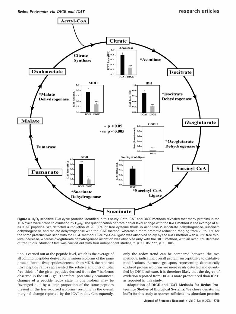
Comparison of DIGE and ICAT Methods for Thiol Oxida-tion Studies. In this study, we have compared two thiol-specificquantitative proteomic techniques for redox-sensitive proteinidentification. DIGE is a gel-base method and quantifies overallprotein thiol levels whereas ICAT utilizes multidimensionalchromatographic methods to selectively enrich and quantifyisotope-encoded cysteine-containing peptides. These two meth-ods appear to be complementary for the purpose of identifyingredox-sensitive proteins (Figure 3). There were 13 proteinscommonly found by both methods (Table 1). They includedactinin 2, creatine kinase, aconitase 2, malate dehydrogenase,ubiquinol-cytochrome-c reductase complex core protein 1, andsuperoxide dismutase. Many of these proteins are known tobe susceptible to oxidative stress in cells.30,45–47 There were 37redox-sensitive proteins identified solely by ICAT method. Wefound that some of these proteins are hydrophobic membraneproteins, which may be poorly resolved in 2DE. Examples ofthese proteins included SERCA2 and membrane glycoproteingp42. Another scenario for proteins detected only with ICATmay attribute to the fact that protein pI values exceed theisoelectric focusing range of 2DE (pI 5-8 used in this study).PRX5 (pI 9.1), succinyl-CoA ligase (pI 9.46), cytochrome coxidase VIb-1 (pI 8.96), CRP2 (pI 8.96), CRP3 (pI 8.90), andtropomyosin-1 alpha chain (pI 4.69) all fell into this category.Additional possibilities for ICAT-identified proteins that weremissed in DIGE analysis might be due to the comigration oflow and high abundance proteins to the same gel spots. It isconceivable that redox-sensitive proteins may not be identifiedin the background of abundant proteins. On the other hand,DIGE method also captured 13 proteins that were absent fromICAT analysis (Figure 3). They included oxoglutarate dehydro-genase, dihydrolipoamide S-acetyltransferase, epoxide hydro-lase, NF-κB-repressing factor, etc. (Supplemental Table 1,Supporting Information). We speculate that the lack of detec-tion of these proteins with ICAT could result from incompleteenzymatic digestion, weak retentions on the RPLC trappingcolumn, or poor ionization and/or fragmentation in MS.Furthermore, we compared the quantitative aspects of thesetwo methods with regard to the redox-sensitive proteins foundwithin the TCA cycle (Figure 4). Both methods found that
aconitase, isocitrate dehydrogenase, succinate dehydrogenase,and malate dehydrogenase were significantly oxidized uponH2O2 challenge. The ICAT method detected thiol reductionsof 20-30% whereas the DIGE method observed more dramaticchanges ranging from 70 to 90% of the thiol signals for the sameproteins. On the other hand, succinyl-CoA ligase was observedsolely in ICAT method with a 35% decrease, whereas oxoglu-tarate dehydrogenase oxidation appeared only in DIGE analysiswith an over 95% decrease of free thiol signal. Overall, the trendof protein/peptide thiol signal decreases followed the samedirection but to different extents. Almost in all cases, DIGEanalysis reported more significant reduction of free thiol levelswhereas ICAT only detected a moderate change. This isprobably due to the fact that DIGE signals represented thequantification of a specific protein isoform that may differ fromother isoforms of the same protein by having multiple oxida-tively modified cysteines. In contrast, ICAT analysis detectedthe averaged changes of a specific cysteine residue among allprotein isoforms.
To confirm the hypothesis that the quantitative discrepanciesbetween DIGE and ICAT originated from the formation of aseries of acidic protein isoforms following H2O2 oxidation, wehave performed additional ICAT and DIGE analyses of apurified malate dehydrogenase (MDH) following identical H2O2
oxidation treatment used for heart proteins. From DIGEanalysis, we found clear evidence of the appearance of ad-ditional acidic MDH isoforms (Figure 5, red gel spots 1-3) inH2O2-treated MDH. The 7 discrete 2DE spots appeared torepresent MDH isoforms with different extent of oxidativelyacidified cysteines. The most acidic protein spots were likelyto contain many but not all of the 8 MDH cysteines as sulfinic,sulfenic, or sulfonic acids, with the remaining cysteine thiolslabeled with CyDyes. The DIGE ratios shown in Figure 5 wereindicative of the relative amounts of the remaining cysteinethiol levels, likely barometers of the relative proportion of eachMDH isoform in the samples under comparison, assumingoverall MDH levels remained the same following H2O2 oxida-tion. The conclusions that can be drawn from DIGE analysisof MDH included that higher proportion of acidically oxidizedMDH isoforms were formed following H2O2 oxidation. It shouldbe mentioned that completely oxidized protein isoform wouldnot be labeled by CyDyes but may be revealed with Sypro Rubystaining of the 2D gels. In ICAT experiments, we successfullyidentified 5 cysteine-containing MDH peptides, with the re-maining peptide too large to be efficiently fragmented by MS/MS in our MS instrument. The quantification of these ICAT-labeled MDH peptides demonstrated varied susceptibility toH2O2 oxidation among the 5 cysteines (Table 2 and Supple-mental Figure 2, Supporting Information). As a simplisticinterpretation, Cys 69 was the most vulnerable to H2O2 oxida-tion, with 77% of its free thiol oxidized by H2O2. Cys 65 wasalso relatively sensitive to H2O2 oxidation, with over 66% offree thiol oxidized. Cys 188, Cys 251, and Cys 261 were onlymoderately sensitive to H2O2 oxidation (Table 2). Both DIGEand ICAT redox quantification results should be interpretedwith the understanding that related yet different biochemicalinformation are obtained with each technique. The quantifica-tion results from DIGE method is gel spot-based (or proteinisoform-specific) instead of simply protein-specific. From DIGEanalysis, we found the increase of MDH oxidative isoforms. Itwas an effective method for discovering a small fraction ofMDH isoforms that were particularly sensitive to redox regula-tion. On the other hand, with the ICAT method, the quantifica-
Figure 3. Venn diagram of H2O2-sensitive proteins discovered byDIGE and ICAT methods. We identified 50 proteins as potentialtargets of H2O2 oxidation by the ICAT method and 26 with theDIGE method. Of these, 13 proteins were identified with bothmethods. Overall, the ICAT technique enabled us to identify 24more redox-sensitive proteins than the DIGE method.
research articles Fu et al.
3798 Journal of Proteome Research • Vol. 7, No. 9, 2008
tion is carried out at the peptide level, which is the average ofall common peptides derived form various isoforms of the sameprotein. For the five peptides detected from MDH, the reportedICAT peptide ratios represented the relative amounts of totalfree thiols of the given peptides derived from the 7 isoformsobserved in the DIGE gel. Therefore, potentially pronouncedchanges of a peptide redox state in one isoform may be“averaged out” by a large proportion of the same peptidespresent in the less oxidized isoforms, resulting in the overallmarginal change reported by the ICAT ratios. Consequently,
only the redox trend can be compared between the twomethods, indicating overall protein susceptibility to oxidativemodifications. Because gel spots representing dramaticallyoxidized protein isoforms are more easily detected and quanti-fied by DIGE software, it is therefore likely that the degree ofoxidation reported from DIGE is more pronounced than ICAT,as reported in this study.
Adaptation of DIGE and ICAT Methods for Redox Pro-teomics Studies of Biological Systems. We chose denaturingbuffer for this study to recover sufficient low-abundant proteins
Figure 4. H2O2-sensitive TCA cycle proteins identified in this study. Both ICAT and DIGE methods revealed that many proteins in theTCA cycle were prone to oxidation by H2O2. The quantification of protein thiol level change with the ICAT method is the average of allits ICAT peptides. We detected a reduction of 20-30% of free cysteine thiols in aconitase 2, isocitrate dehydrogenase, succinatedehydrogenase, and malate dehydrogenase with the ICAT method, whereas a more dramatic reduction ranging from 70 to 90% forthe same proteins was seen with the DIGE method. Succinyl-CoA ligase was observed solely by the ICAT method with a 35% free thiollevel decrease, whereas oxoglutarate dehydrogenase oxidation was observed only with the DIGE method, with an over 95% decreaseof free thiols. Student t test was carried out with four independent studies, *, p < 0.05; ***, p < 0.005.
Redox Proteomics via DIGE and ICAT research articles
Journal of Proteome Research • Vol. 7, No. 9, 2008 3799

from the tissues, which is important for biological studies.Because in our model system the proteins were oxidized byH2O2 after denaturation, many cysteine thiols might notpossess their native protein structures in denaturing lysis buffer;therefore protein sensitivities to oxidant H2O2 reported in thisstudy may not be identical to in vivo conditions. However,because no reduction and alkylation steps were included inthe lysis buffer, select cysteine thiols might still contain somestructural environments that enabled their interactions withother vicinal amino acids, resulting in varied thiol sensitivityto H2O2 oxidation observed in this study (see example in Figure5). We can not claim unequivocally whether the isolated heartproteins oxidized by 500 µM of H2O2 are also sensitive tointracellular oxidants under pathophysiological conditions.However, from two subsequent independent studies of heartproteins isolated from animal models with deficiencies of redoxmodulators, we found many of the same proteins were differ-entially oxidized (manuscripts in preparation), confirming theeffectiveness of both DIGE and ICAT methods for the discoveryof distinct groups of redox-sensitive proteins.
Taken together, both methods have demonstrated the ef-fectiveness for the identification of both previously known andunknown redox-sensitive proteins. Meanwhile, each methodwas useful at discovering a unique set of novel redox-sensitiveproteins, and may pave the way for further mechanistic studies.DIGE could be useful for the identification of redox-inducedprotein isoform formations and ICAT method is more versatilefor identifying redox-sensitive peptides with exact localizationof the cysteine oxidized. Overall, these two methods arecomplementary for redox proteomics studies. The selection ofthe best method is task-dependent (Table 3). If the goal is toquantify the global redox proteomic changes, DIGE is the
method of choice. However, if the focus is to identify specificcysteines within key proteins for downstream structural andfunctional study, ICAT may be more advantageous. If the goalis to identify as many redox-sensitive proteins as possible, bothmethods may be needed.
Summary
A systematic comparison between DIGE and ICAT methodswas carried out for detecting redox-sensitive proteins followingthe H2O2 treatment of heart proteins. Each method comple-mented the other and can be used together to obtain acomprehensive understanding of the changes in heart redoxproteome. ICAT is an attractive tool for the precise localizationof redox-sensitive cysteine in low-abundant proteins, whereasDIGE is advantageous for easier implementation of multiplexedexperiments to discover redox proteomics patterns. Collectively,
Figure 5. DIGE analysis of MDH oxidized by H2O2. Cy3m (green)-labeled control sample and Cy5m (red)-labeled H2O2- treated porcineMDH. A series of MDH isoforms were observed with both untreated and H2O2-treated MDH. It was clear that the H2O2-treated MDHcontained additional isoforms distributed toward the acidic end of the 2DE gel (red spots 1-3), suggesting the acidification of MDH,likely in the forms of cysteine oxidation to sulfinic, sulfenic, or sulfonic acids. The untreated MDH maintained isoforms clustered aroundthe more basic pI range, as illustrated by the green gel spots 6-7. The relative MDHreduction/MDHoxidation ratios for all MDH isoformsare marked below the corresponding gel spots, with isoform #1 being the most oxidized and isoform #7 being the most reduced.Ratio*: Cy3m/Cy5m ratio represents relative free cysteine thiol levels in control/H2O2-treated MDH isoforms.
Table 2. Purified Malate Dehydrogenase Peptide Identificationand Thiol-Oxidative Quantification by ICAT
peptide sequencea C.I. %observed
masserror
(ppm)ICAT ratio
(H/L)b
55GYLGPEQLPDCLK67 100 1659.8 -11 0.3468GCDVVVIPAGVPR80 100 1508.8 -13 0.23180TIIPLISQCTPK191 100 1540.9 -16 0.49246EGVVECSFVK255 100 1323.5 5 0.45256SQETDCPYFSTPLLLGK272 100 2126.0 -4 0.47
a ICAT labeled cysteines are underlined. b H/L: H2O2 oxidized/untreated MDH peptides.
Table 3. Relative Merits of DIGE and ICAT Techniques forProtein Thiol Quantification
advantages disadvantages
DIGE • More confident proteinidentification, bettersequence coverage
• Not amenable foranalyzing basic,hydrophobic, and largeproteins
• More effective atidentifying proteinoxidative isoforms
• Unable to locate the exactsites of cysteine oxidativemodifications
• Easier implementation ofstatistics for multiplexedquantification
• Comigration of proteinsmay interfere with accurateprotein identification andthiol quantification
• Suitable for large scalescreening
• May overlooklow-abundant proteins
ICAT • Able to locate the exactsites of redox-sensitivecysteines
• Less protein sequencecoverage than 2DE
• Amenable tomultidimensionalseparation and enhancesthe detection oflow-abundant proteins
• Multiple ICAT experimentsare required to establishstatistical significance forquantification and may betime-consuming
• Suitable for large scalescreening
• Overlapping ions in MSspectra may interfere withaccurate quantification andidentification
research articles Fu et al.
3800 Journal of Proteome Research • Vol. 7, No. 9, 2008

we have identified 63 unique redox-sensitive proteins, includingsome previously reported and several novel H2O2 oxidationtargets, that may shed light into better understandings of theredox-mediated regulation of cellular processes.
Acknowledgment. This research is supported in partby a NIH grant NS046593 to H.L. and by a grant from thefoundation of UMDNJ to H.L. and J.S.
Supporting Information Available: SupplementalTables 1-3 and Supplemental Figures 1 and 2. This materialis available free of charge via the Internet at http://pubs.acs.org.
References(1) Ghezzi, P.; Bonetto, V. Redox proteomics: Identification of oxida-
tively, modified proteins. Proteomics 2003, 3 (7), 1145–53.(2) Di Simplicio, P.; Franconi, F.; Frosali, S.; Di Giuseppe, D. Thiolation
and nitrosation of cysteines in biological fluids and cells. AminoAcids 2003, 25 (3-4), 323–39.
(3) Zheng, M.; Aslund, F.; Storz, G. Activation of the OxyR transcriptionfactor by reversible disulfide bond formation. Science 1998, 279(5357), 1718–21.
(4) Abate, C.; Patel, L.; Rauscher, F. J.; Curran, T. Redox regulation offos and jun DNA-binding activity in vitro. Science 1990, 249 (4973),1157–61.
(5) Toledano, M. B.; Leonard, W. J. Modulation of transcription factorNF-kappa B binding activity by oxidation-reduction in vitro. Proc.Natl. Acad. Sci. U.S.A. 1991, 88 (10), 4328–32.
(6) Xanthoudakis, S.; Miao, G. G.; Curran, T. The redox and DNA-repair activities of Ref-1 are encoded by nonoverlapping domains.Proc. Natl. Acad. Sci. U.S.A. 1994, 91 (1), 23–7.
(7) Rainwater, R.; Parks, D.; Anderson, M. E.; Tegtmeyer, P.; Mann,K. Role of cysteine residues in regulation of p53 function. Mol.Cell. Biol. 1995, 15 (7), 3892–903.
(8) Gu, J.; Milligan, J.; Huang, L. E. Molecular mechanism of hypoxia-inducible factor 1alpha -p300 interaction. A leucine-rich interfaceregulated by a single cysteine. J. Biol. Chem. 2001, 276 (5), 3550–4.
(9) Humphries, K. M.; Juliano, C.; Taylor, S. S. Regulation of cAMP-dependent protein kinase activity by glutathionylation. J. Biol.Chem. 2002, 277 (45), 43505–11.
(10) Kamata, H.; Honda, S.; Maeda, S.; Chang, L.; Hirata, H.; Karin, M.Reactive oxygen species promote TNFalpha-induced death andsustained JNK activation by inhibiting MAP kinase phosphatases.Cell 2005, 120 (5), 649–61.
(11) Tonks, N. K. Redox redux: revisiting PTPs and the control of cellsignaling. Cell 2005, 121 (5), 667–70.
(12) Park, H. S.; Yu, J. W.; Cho, J. H.; Kim, M. S.; Huh, S. H.; Ryoo, K.;Choi, E. J. Inhibition of apoptosis signal-regulating kinase 1 bynitric oxide through a thiol redox mechanism. J. Biol. Chem. 2004,279 (9), 7584–90.
(13) Vivancos, A. P.; Castillo, E. A.; Biteau, B.; Nicot, C.; Ayte, J.;Toledano, M. B.; Hidalgo, E. A cysteine-sulfinic acid in peroxire-doxin regulates H2O2-sensing by the antioxidant Pap1 pathway.Proc. Natl. Acad. Sci. U.S.A. 2005, 102 (25), 8875–80.
(14) Fujiwara, N.; Nakano, M.; Kato, S.; Yoshihara, D.; Ookawara, T.;Eguchi, H.; Taniguchi, N.; Suzuki, K. Oxidative modification tocysteine sulfonic acid of Cys111 in human copper-zinc superoxidedismutase. J. Biol. Chem. 2007, 282 (49), 35933–44.
(15) Sumbayev, V. V. S-nitrosylation of thioredoxin mediates activationof apoptosis signal-regulating kinase 1. Arch. Biochem. Biophys.2003, 415 (1), 133–6.
(16) Graf, P. C.; Jakob, U. Redox-regulated molecular chaperones. Cell.Mol. Life Sci. 2002, 59 (10), 1624–31.
(17) Ilbert, M.; Horst, J.; Ahrens, S.; Winter, J.; Graf, P. C.; Lilie, H.; Jakob,U. The redox-switch domain of Hsp33 functions as dual stresssensor. Nat. Struct. Mol. Biol. 2007, 14 (6), 556–63.
(18) Brennan, J. P.; Wait, R.; Begum, S.; Bell, J. R.; Dunn, M. J.; Eaton,P. Detection and mapping of widespread intermolecular proteindisulfide formation during cardiac oxidative stress using proteom-ics with diagonal electrophoresis. J. Biol. Chem. 2004, 279 (40),41352–41360.
(19) Charles, R. L.; Schroder, E.; May, G.; Free, P.; Gaffney, P. R.; Wait,R.; Begum, S.; Heads, R. J.; Eaton, P. Protein sulfenation as a redoxsensor: proteomics studies using a novel biotinylated dimedoneanalogue. Mol. Cell. Proteomics 2007, 6 (9), 1473–84.
(20) Saurin, A. T.; Neubert, H.; Brennan, J. P.; Eaton, P. Widespreadsulfenic acid formation in tissues in response to hydrogenperoxide. Proc. Natl. Acad. Sci. U.S.A. 2004, 101 (52), 17982–87.
(21) Hao, G.; Derakhshan, B.; Shi, L.; Campagne, F.; Gross, S. S. SNOSID,a proteomic method for identification of cysteine S-nitrosylationsites in complex protein mixtures. Proc. Natl. Acad. Sci. U.S.A.2006, 103 (4), 1012–17.
(22) Fratelli, M.; Demol, H.; Puype, M.; Casagrande, S.; Eberini, I.;Salmona, M.; Bonetto, V.; Mengozzi, M.; Duffieux, F.; Miclet, E.;Bachi, A.; Vandekerckhove, J.; Gianazza, E.; Ghezzi, P. Identificationby redox proteomics of glutathionylated proteins in oxidativelystressed human T lymphocytes. Proc. Natl. Acad. Sci. U.S.A. 2002,99 (6), 3505–10.
(23) Michelet, L.; Zaffagnini, M.; Marchand, C.; Collin, V.; Decottignies,P.; Tsan, P.; Lancelin, J. M.; Trost, P.; Miginiac-Maslow, M.; Noctor,G.; Lemaire, S. D. Glutathionylation of chloroplast thioredoxin fis a redox signaling mechanism in plants. Proc. Natl. Acad. Sci.U.S.A. 2005, 102 (45), 16478–83.
(24) Yano, H.; Wong, J. H.; Lee, Y. M.; Cho, M. J.; Buchanan, B. B. Astrategy for the identification of proteins targeted by thioredoxin.Proc. Natl. Acad. Sci. U.S.A. 2001, 98 (8), 4794–99.
(25) Essex, D. W.; Li, M. R.; Miller, A.; Feinman, R. D. Protein disulfideisomerase and sulfhydryl-dependent pathways in platelet activa-tion. Biochemistry 2001, 40 (20), 6070–75.
(26) Baty, J. W.; Hampton, M. B.; Winterbourn, C. C. Detection ofoxidant sensitive thiol proteins by fluorescence labeling and two-dimensional electrophoresis. Proteomics 2002, 2 (9), 1261–66.
(27) Kim, J. R.; Yoon, H. W.; Kwon, K. S.; Lee, S. R.; Rhee, S. G.Identification of proteins containing cysteine residues that aresensitive to oxidation by hydrogen peroxide at neutral pH. Anal.Biochem. 2000, 283 (2), 214–21.
(28) Woo, H. A.; Kang, S. W.; Kim, H. K.; Yang, K. S.; Chae, H. Z.; Rhee,S. G. Reversible oxidation of the active site cysteine of peroxire-doxins to cysteine sulfinic acid. Immunoblot detection withantibodies specific for the hyperoxidized cysteine-containingsequence. J. Biol. Chem. 2003, 278 (48), 47361–4.
(29) Leichert, L. I.; Jakob, U. Protein thiol modifications visualized invivo. PLoS Biol. 2004, 2 (11), e333.
(30) Hurd, T. R.; Prime, T. A.; Harbour, M. E.; Lilley, K. S.; Murphy,M. P. Detection of ros-sensitive thiol proteins by redox-differencegel electrophoresis (redox-dige): Implications for mitochondrialredox signalling. J. Biol. Chem. 2007, 22040–51.
(31) Gygi, S. P.; Rist, B.; Gerber, S. A.; Turecek, F.; Gelb, M. H.;Aebersold, R. Quantitative analysis of complex protein mixturesusing isotope-coded affinity tags. Nat. Biotechnol. 1999, 17 (10),994–9.
(32) Sethuraman, M.; McComb, M. E.; Huang, H.; Huang, S. Q.;Heibeck, T.; Costello, C. E.; Cohen, R. A. Isotope-coded affinitytag (ICAT) approach to redox proteomics: Identification andquantitation of oxidant-sensitive cysteine thiols in complex proteinmixtures. J. Proteome Res. 2004, 3 (6), 1228–33.
(33) Christoffers, K. H.; Li, H.; Howells, R. D. Purification and massspectrometric analysis of the delta opioid receptor. Mol. Brain Res.2005, 136 (1-2), 54–64.
(34) Liu, T.; Donahue, K. C.; Hu, J.; Kurnellas, M. P.; Grant, J. E.; Li, H.;Elkabes, S. Identification of differentially expressed proteins inexperimental autoimmune encephalomyelitis (EAE) by proteomicanalysis of the spinal cord. J. Proteome Res. 2007, 6 (7), 2565–75.
(35) Peng, J.; Elias, J. E.; Thoreen, C. C.; Licklider, L. J.; Gygi, S. P.Evaluation of multidimensional chromatography coupled withtandem mass spectrometry (LC/LC-MS/MS) for large-scale proteinanalysis: the yeast proteome. J. Proteome Res. 2003, 2 (1), 43–50.
(36) Lee, C. K.; Park, H. J.; So, H. H.; Kim, H. J.; Lee, K. S.; Choi, W. S.;Lee, H. M.; Won, K. J.; Yoon, T. J.; Park, T. K.; Kim, B. Proteomicprofiling and identification of cofilin responding to oxidative stressin vascular smooth muscle. Proteomics 2006, 6 (24), 6455–75.
(37) Obata, T. Nitric oxide and depolarization induce hydroxyl radicalgeneration. Jpn. J. Pharmacol. 2002, 88 (1), 1–5.
(38) Yang, K.-S.; Kang, S. W.; Woo, H. A.; Hwang, S. C.; Chae, H. Z.;Kim, K.; Rhee, S. G. Inactivation of Human Peroxiredoxin I duringCatalysis as the Result of the Oxidation of the Catalytic SiteCysteine to Cysteine-sulfinic Acid. J. Biol. Chem. 2002, 277 (41),38029–36.
(39) Canet-Aviles, R. M.; Wilson, M. A.; Miller, D. W.; Ahmad, R.;McLendon, C.; Bandyopadhyay, S.; Baptista, M. J.; Ringe, D.;Petsko, G. A.; Cookson, M. R. The Parkinson’s disease proteinDJ-1 is neuroprotective due to cysteine-sulfinic acid-drivenmitochondrial localization. Proc. Natl. Acad. Sci. U.S.A. 2004,101 (24), 9103–8.
Redox Proteomics via DIGE and ICAT research articles
Journal of Proteome Research • Vol. 7, No. 9, 2008 3801

(40) Janssen-Heininger, Y. M.; Poynter, M. E.; Baeuerle, P. A. Recentadvances towards understanding redox mechanisms in the activa-tion of nuclear factor kappaB. Free Radic. Biol. Med. 2000, 28 (9),1317–27.
(41) Sethuraman, M.; McComb, M. E.; Heibeck, T.; Costello, C. E.;Cohen, R. A. Isotope-coded affinity tag approach to identify andquantify oxidant-sensitive protein thiols. Mol. Cell. Proteomics2004, 3 (3), 273–8.
(42) Li, Y.; Camacho, P. Ca2+-dependent redox modulation of SERCA2b by ERp57. J. Cell Biol. 2004, 164 (1), 35–46.
(43) Declercq, J. P.; Evrard, C.; Clippe, A.; Stricht, D. V.; Bernard, A.;Knoops, B. Crystal structure of human peroxiredoxin 5, a noveltype of mammalian peroxiredoxin at 1.5 A resolution. J. Mol. Biol.2001, 311 (4), 751–9.
(44) Nakajima, H.; Amano, W.; Fujita, A.; Fukuhara, A.; Azuma, Y.-T.;Hata, F.; Inui, T.; Takeuchi, T. The Active Site Cysteine of theProapoptotic Protein Glyceraldehyde-3-phosphate DehydrogenaseIs Essential in Oxidative Stress-induced Aggregation and CellDeath. J. Biol. Chem. 2007, 282 (36), 26562–74.
(45) Wolosker, H.; Panizzutti, R.; Engelender, S. Inhibition of creatinekinase by S-nitrosoglutathione. FEBS Lett. 1996, 392 (3), 274–6.
(46) Sharma, A. B.; Sun, J.; Howard, L. L.; Williams, A. G., Jr.; Mallet,R. T. Oxidative stress reversibly inactivates myocardial enzymesduring cardiac arrest. Am. J. Physiol. Heart Circ. Physiol. 2007, 292(1), H198–206.
(47) Inarrea, P.; Moini, H.; Rettori, D.; Han, D.; Martinez, J.; Garcia, I.;Fernandez-Vizarra, E.; Iturralde, M.; Cadenas, E. Redox activationof mitochondrial intermembrane space Cu,Zn-superoxide dismu-tase. Biochem. J. 2005, 387 (Pt 1), 203–9.
(48) Han, D.; Canali, R.; Garcia, J.; Aguilera, R.; Gallaher, T. K.; Cadenas,E. Sites and mechanisms of aconitase inactivation by peroxynitrite:modulation by citrate and glutathione. Biochemistry 2005, 44 (36),11986–96.
(49) Eaton, P.; Byers, H. L.; Leeds, N.; Ward, M. A.; Shattock, M. J.Detection, quantitation, purification, and identification of cardiacproteins S-thiolated during ischemia and reperfusion. J. Biol.Chem. 2002, 277 (12), 9806–11.
(50) Cabiscol, E.; Piulats, E.; Echave, P.; Herrero, E.; Ros, J. Oxidativestress promotes specific protein damage in Saccharomyces cer-evisiae. J. Biol. Chem. 2000, 275 (35), 27393–8.
(51) Chen, Y. R.; Chen, C. L.; Pfeiffer, D. R.; Zweier, J. L. Mitochondrialcomplex II in the post-ischemic heart: oxidative injury and therole of protein S-glutathionylation. J. Biol. Chem. 2007, 282 (45),32640–54.
(52) Meany, D. L.; Xie, H.; Thompson, L. V.; Arriaga, E. A.; Griffin, T. J.Identification of carbonylated proteins from enriched rat skeletalmuscle mitochondria using affinity chromatography-stable isotopelabeling and tandem mass spectrometry. Proteomics 2007, 7 (7),1150–63.
(53) Newman, S. F.; Sultana, R.; Perluigi, M.; Coccia, R.; Cai, J.; Pierce,W. M.; Klein, J. B.; Turner, D. M.; Butterfield, D. A. An increase inS-glutathionylated proteins in the Alzheimer’s disease inferiorparietal lobule, a proteomics approach. J. Neurosci. Res. 2007, 85(7), 1506–14.
(54) Schilling, B.; Yoo, C. B.; Collins, C. J.; Gibson, B. W. Determiningcysteine oxidation status using differential alkylation. Int. J. MassSpectrom. 2004, 236 (1-3), 117–27.
(55) Mohr, S.; Stamler, J. S.; Brune, B. Posttranslational modificationof glyceraldehyde-3-phosphate dehydrogenase by S-nitrosylationand subsequent NADH attachment. J. Biol. Chem. 1996, 271 (8),4209–14.
(56) Ishii, T.; Tatsuda, E.; Kumazawa, S.; Nakayama, T.; Uchida, K.Molecular basis of enzyme inactivation by an endogenous elec-trophile 4-hydroxy-2-nonenal: identification of modification sitesin glyceraldehyde-3-phosphate dehydrogenase. Biochemistry 2003,42 (12), 3474–80.
(57) Kim, B. J.; Hood, B. L.; Aragon, R. A.; Hardwick, J. P.; Conrads,T. P.; Veenstra, T. D.; Song, B. J. Increased oxidation and degrada-tion of cytosolic proteins in alcohol-exposed mouse liver andhepatoma cells. Proteomics 2006, 6 (4), 1250–60.
(58) Nagumo, Y.; Kakeya, H.; Shoji, M.; Hayashi, Y.; Dohmae, N.; Osada,H. Epolactaene binds human Hsp60 Cys442 resulting in theinhibition of chaperone activity. Biochem. J. 2005, 387 (Pt 3),835–40.
PR800233R
research articles Fu et al.
3802 Journal of Proteome Research • Vol. 7, No. 9, 2008