Q-absorb Study - Jarro · TM Jarrow FORMULAS® Co-Q10 TM Clinical Trial A Super Bioavailable...
28
TM Jarrow FORMULAS ® Co-Q10 TM Clinical Trial A Super Bioavailable xx x x Form of Co-Q10 Q-absorb ™ raised plasma Co-Q10 by 400% over baseline in humans. With exercise, Q-absorb raised plasma Co-Q10 by 493% 4.0 3.25 3.0 2.0 1.0 0.81 Q-absorb ™ + Exercise Q-absorb ™ Baseline 0.81 300 mg/day; 4 weeks; 23 subjects Co-Q 10 mcg / mL / plasma 3.25 4.0 Belardinelli R, et al. American Heart Association Supplement to Circulation. October 28, 2003. Vol.108 (17), pg.739 Lancisi Heart Institute, Ancona, and Institute of Biochemistry, University of Ancona, Italy With Background and Annotations by Dallas Clouatre, Ph.D. Coenzyme Q10 potentiates the effect of exercise training on the endothelium- dependent relaxation of the brachial artery in chronic heart failure Q-absorb
Transcript of Q-absorb Study - Jarro · TM Jarrow FORMULAS® Co-Q10 TM Clinical Trial A Super Bioavailable...
TM
JarrowFORMULAS®
Co-Q10
TM
Clinical Tr ial
A S u p e r B i o a va i l a b l e x x xx F o r m o f C o - Q 10
Q-absorb™ raised plasma Co-Q10 by 400% over baseline in humans. With exercise, Q-absorb raised plasma Co-Q10 by 493%
4.0
3.25
3.0
2.0
1.00.81
Q-absorb™ + Exercise
Q-absorb™
Baseline0.81
300 mg/day; 4 weeks; 23 subjects
Co-
Q10
mcg
/ m
L /
plas
ma
3.25
4.0
Belardinelli R, et al. American Heart Association Supplement to Circulation. October 28, 2003. Vol.108 (17), pg.739
Lancisi Heart Institute, Ancona, and Institute of Biochemistry, University of Ancona, ItalyWith Background and Annotations by Dallas Clouatre, Ph.D.
Coenzyme Q10 potentiates the effect of exercise training on the endothelium-dependent relaxation of the brachial artery in chronic heart failure
Q-absorb
1
Q-absorb™
in Chronic Heart Failure
Coenzyme Q10 potentiates the effect of exercise training on the endothelium-dependent relaxation of the brachial
artery in chronic heart failure
Belardinelli, RM et al. American Heart Association Supplement to Circulation.October 28, 2003. Vol. 108 (17), pg. 739
Lancisi Heart Institute, Ancona, and Institute of Biochemistry, University of Ancona, ItalyWith Background and Annotations by Dallas Clouatre, Ph.D.
American Heart AssociationOral Presentation
Scientific Sessions 2003November 9-12, Orlando, Fl
Q-absorb™
Enhanced Absorption Formula
2 3
Commentary by Dallas Clouatre, Ph.D.
Introduction by Jarrow L. Rogovin
© Copyright 2004 byDallas Clouatre, Ph.D. and Romualdo Belardinelli, MD, FESC, Andi Mucaj, MD, Francesca Lacalaprice, RT, Maridia Solenghi, MD, Federica Principi, MD, Francesco Mosca, MD, Maurizio Battino, MD, GianPaolo Littarru, MD
2 3
ACKNOWLEDGEMENTS
I express my gratitude and appreciation to Prof. Gian-Paolo Littarru, M.D., and Romualdo Belardinelli, M.D., FESC, their staffs and the twenty-three heart patients without whom this historic study could not have been undertaken. I also thank Kaneka Corporation, the makers of Jarrow Formulas’ 100% natural, all-trans coenzyme-Q10 by fermentation. Their assistance, encouragement and cooperation is greatly appreciated.
Los Angeles, January 2004
4 5
CONTENTS
Introduction: The Problem of Bioavailability of Co-Q10 . . . . . . . . . . . . . . . . . . . . . . . . . . 5
The Study . . . . . . . . . . . . . . . . . . . . . . . . . . . . . . . . . . . . . . . . . . . . . . . . . . 5
The American Heart Association Presentation:
Coenzyme Q10 Potentiates the Effect of Exercise Training
on the Endothelium-Dependent Relaxation of the Brachial
Artery in Chronic Heart Failure . . . . . . . . . . . . . . . . . . . . . . . . . . . . . . . . . . . . . . . . . . . 7
Background: Coenzyme Q10, a Molecule with Diverse Functions . . . . . . . . . . . . . . . . . 24
References for Background . . . . . . . . . . . . . . . . . . . . . . . . . . . . . . . . . . . . . . . . . . . . . . . 27
4 5
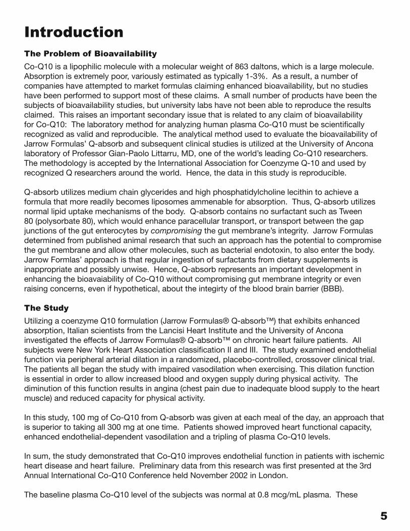
IntroductionThe Problem of Bioavailability
Co-Q10 is a lipophilic molecule with a molecular weight of 863 daltons, which is a large molecule. Absorption is extremely poor, variously estimated as typically 1-3%. As a result, a number of companies have attempted to market formulas claiming enhanced bioavailability, but no studies have been performed to support most of these claims. A small number of products have been the subjects of bioavailability studies, but university labs have not been able to reproduce the results claimed. This raises an important secondary issue that is related to any claim of bioavailability for Co-Q10: The laboratory method for analyzing human plasma Co-Q10 must be scientifically recognized as valid and reproducible. The analytical method used to evaluate the bioavailability of Jarrow Formulas’ Q-absorb and subsequent clinical studies is utilized at the University of Ancona laboratory of Professor Gian-Paolo Littarru, MD, one of the world’s leading Co-Q10 researchers. The methodology is accepted by the International Association for Coenzyme Q-10 and used by recognized Q researchers around the world. Hence, the data in this study is reproducible.
Q-absorb utilizes medium chain glycerides and high phosphatidylcholine lecithin to achieve a formula that more readily becomes liposomes ammenable for absorption. Thus, Q-absorb utilizes normal lipid uptake mechanisms of the body. Q-absorb contains no surfactant such as Tween 80 (polysorbate 80), which would enhance paracellular transport, or transport between the gap junctions of the gut enterocytes by compromising the gut membrane’s integrity. Jarrow Formulas determined from published animal research that such an approach has the potential to compromise the gut membrane and allow other molecules, such as bacterial endotoxin, to also enter the body. Jarrow Formlas’ approach is that regular ingestion of surfactants from dietary supplements is inappropriate and possibly unwise. Hence, Q-absorb represents an important development in enhancing the bioavaiability of Co-Q10 without compromising gut membrane integrity or even raising concerns, even if hypothetical, about the integirty of the blood brain barrier (BBB).
The Study
Utilizing a coenzyme Q10 formulation (Jarrow Formulas® Q-absorb™) that exhibits enhanced absorption, Italian scientists from the Lancisi Heart Institute and the University of Ancona investigated the effects of Jarrow Formulas® Q-absorb™ on chronic heart failure patients. All subjects were New York Heart Association classification II and III. The study examined endothelial function via peripheral arterial dilation in a randomized, placebo-controlled, crossover clinical trial. The patients all began the study with impaired vasodilation when exercising. This dilation function is essential in order to allow increased blood and oxygen supply during physical activity. The diminution of this function results in angina (chest pain due to inadequate blood supply to the heart muscle) and reduced capacity for physical activity.
In this study, 100 mg of Co-Q10 from Q-absorb was given at each meal of the day, an approach that is superior to taking all 300 mg at one time. Patients showed improved heart functional capacity, enhanced endothelial-dependent vasodilation and a tripling of plasma Co-Q10 levels.
In sum, the study demonstrated that Co-Q10 improves endothelial function in patients with ischemic heart disease and heart failure. Preliminary data from this research was first presented at the 3rd Annual International Co-Q10 Conference held November 2002 in London.
The baseline plasma Co-Q10 level of the subjects was normal at 0.8 mcg/mL plasma. These
6 7
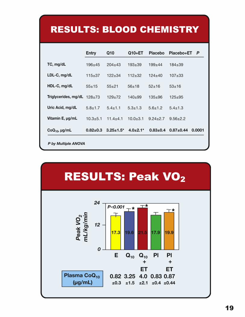
subjects had all been under careful medical supervision prior to entry. In the concluded study, Q-absorb administration resulted in significant elevations in plasma Co-Q10 averaging a remarkable 3.25 mcg/mL plasma without exercise and an even more remarkable 4.0 mcg/mL plasma with exercise; peripheral arterial dilation increased substantially; and a substantial increase in peak VO2 (volume of oxygen consumed) was achieved. The co-administration of Q-absorb and exercise therapy resulted in the greatest gains, with endothelial dysfunction being almost normalized.
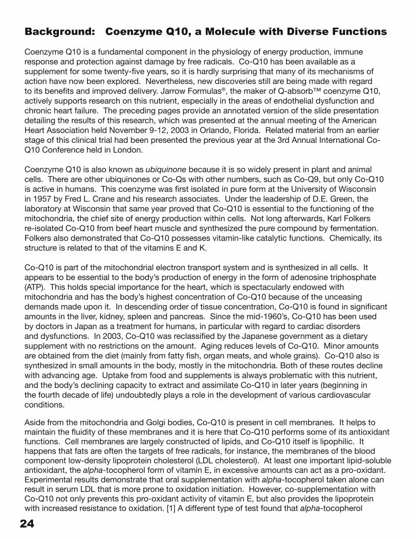
It is believed by the researchers that the primary effect of Co-Q10 in this study may have been it’s SOD (superoxide dismutase) mimetic effect that, by scavenging superoxide radical, spared the subjects’ NO levels. NO plays a critical role in endothelial relaxation and capillary dilation.
These results were presented by Romualdo Berlardinelli, MD, FESC, of the Lancisi Heart Institute, Ancona, Italy, at the annual meeting of the American Heart Association held November 9-12, 2003 in Orlando, Florida. That presentation is reproduced in the following pages.
6 7
American Heart Association Presentation:
Coenzyme Q10 Potentiates the Effect of Exercise Training on the Endothelium-Dependent Relaxation of
the Brachial Artery in Chronic Heart Failure
8 9
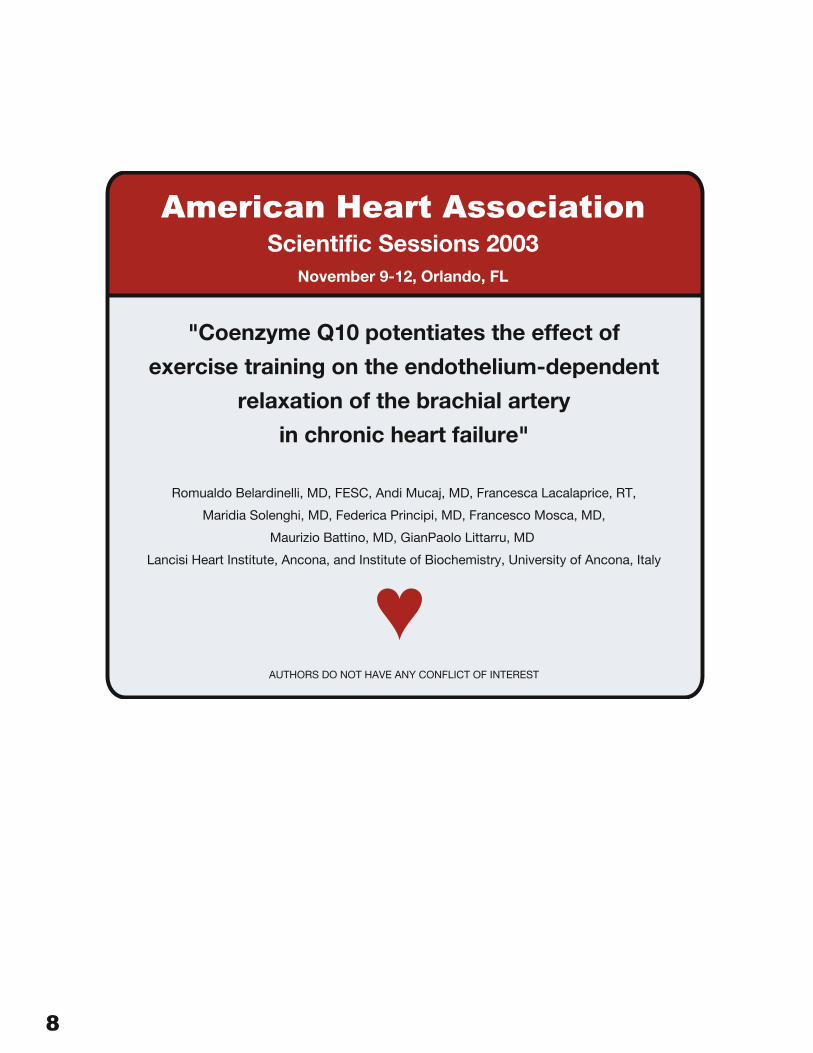
American Heart AssociationScientific Sessions 2003
November 9-12, Orlando, FL
"Coenzyme Q10 potentiates the effect of
exercise training on the endothelium-dependent
relaxation of the brachial artery
in chronic heart failure"
Romualdo Belardinelli, MD, FESC, Andi Mucaj, MD, Francesca Lacalaprice, RT,
Maridia Solenghi, MD, Federica Principi, MD, Francesco Mosca, MD,
Maurizio Battino, MD, GianPaolo Littarru, MD
Lancisi Heart Institute, Ancona, and Institute of Biochemistry, University of Ancona, Italy
AUTHORS DO NOT HAVE ANY CONFLICT OF INTEREST
8 9
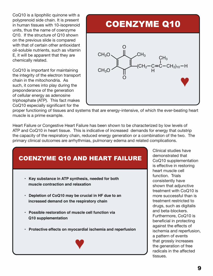
CoQ10 is a lipophilic quinone with a polyprenoid side chain. It is present in human tissues with 10-isoprenoid units, thus the name of coenzyme Q10. If the structure of Q10 shown on the previous slide is compared with that of certain other antioxidant oil-soluble nutrients, such as vitamin E, it will be apparent that they are chemically related.
CoQ10 is important for maintaining the integrity of the electron transport chain in the mitochondria. As such, it comes into play during the preponderance of the generation of cellular energy as adenosine triphosphate (ATP). This fact makes CoQ10 especially significant for the proper functioning of tissues and systems that are energy-intensive, of which the ever-beating heart muscle is a prime example.
Heart Failure or Congestive Heart Failure has been shown to be characterized by low levels of ATP and CoQ10 in heart tissue. This is indicative of increased demands for energy that outstrip the capacity of the respiratory chain, reduced energy generation or a combination of the two. The primary clinical outcomes are arrhythmias, pulmonary edema and related complications.
Clinical studies have demonstrated that CoQ10 supplementation is effective in restoring heart muscle cell function. Trials consistently have shown that adjunctive treatment with CoQ10 is more successful than is treatment restricted to drugs, such as digitalis and beta-blockers. Furthermore, CoQ10 is beneficial in protecting against the effects of ischemia and reperfusion, a pattern of events that grossly increases the generation of free radicals in the affected tissues.
COENZYME Q10 AND HEART FAILURE
• Key substance in ATP synthesis, needed for both muscle contraction and relaxation
• Depletion of CoQ10 may be crucial in HF due to an increased demand on the respiratory chain
• Possible restoration of muscle cell function via Q10 supplementation
• Protective effects on myocardial ischemia and reperfusion
COENZYME Q10
10 11
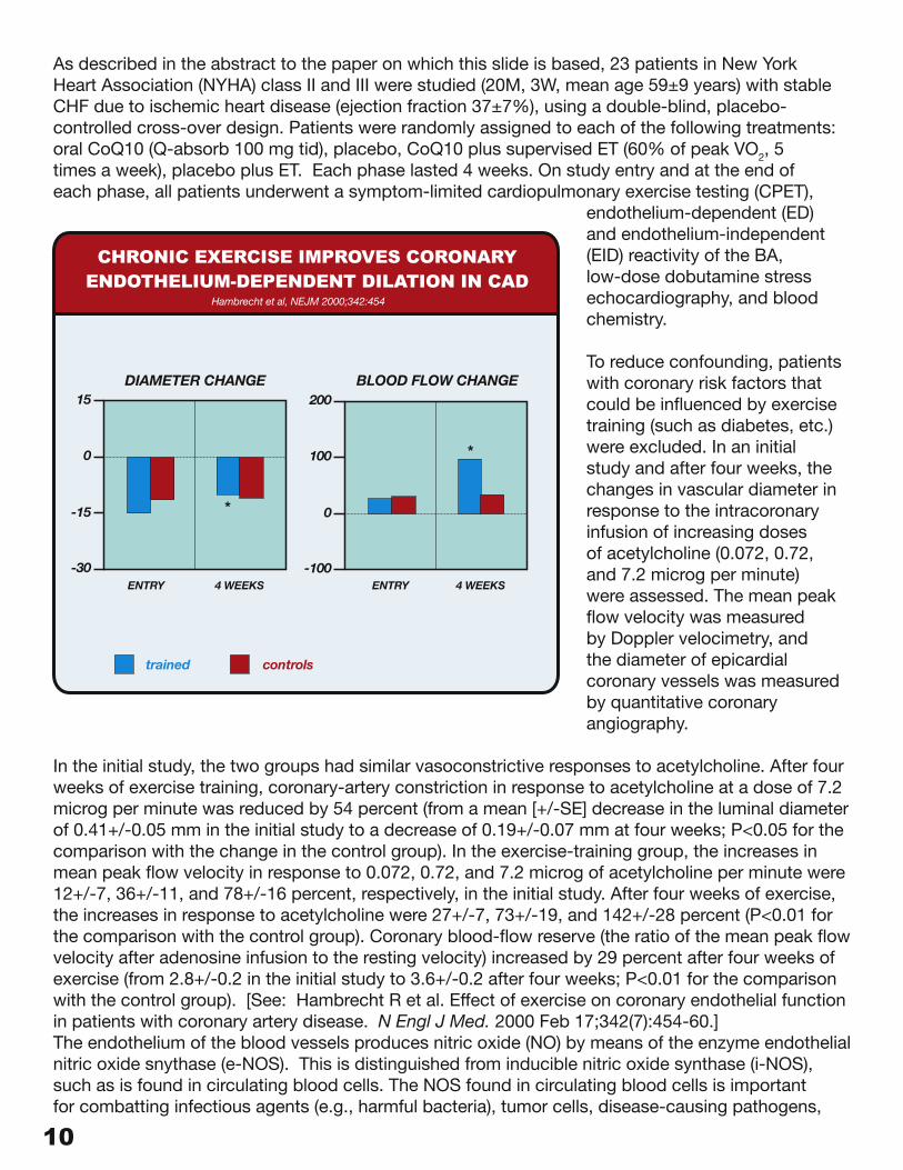
As described in the abstract to the paper on which this slide is based, 23 patients in New York Heart Association (NYHA) class II and III were studied (20M, 3W, mean age 59±9 years) with stable CHF due to ischemic heart disease (ejection fraction 37±7%), using a double-blind, placebo-controlled cross-over design. Patients were randomly assigned to each of the following treatments: oral CoQ10 (Q-absorb 100 mg tid), placebo, CoQ10 plus supervised ET (60% of peak VO2, 5 times a week), placebo plus ET. Each phase lasted 4 weeks. On study entry and at the end of each phase, all patients underwent a symptom-limited cardiopulmonary exercise testing (CPET),
endothelium-dependent (ED) and endothelium-independent (EID) reactivity of the BA, low-dose dobutamine stress echocardiography, and blood chemistry.
To reduce confounding, patients with coronary risk factors that could be influenced by exercise training (such as diabetes, etc.) were excluded. In an initial study and after four weeks, the changes in vascular diameter in response to the intracoronary infusion of increasing doses of acetylcholine (0.072, 0.72, and 7.2 microg per minute) were assessed. The mean peak flow velocity was measured by Doppler velocimetry, and the diameter of epicardial coronary vessels was measured by quantitative coronary angiography.
In the initial study, the two groups had similar vasoconstrictive responses to acetylcholine. After four weeks of exercise training, coronary-artery constriction in response to acetylcholine at a dose of 7.2 microg per minute was reduced by 54 percent (from a mean [+/-SE] decrease in the luminal diameter of 0.41+/-0.05 mm in the initial study to a decrease of 0.19+/-0.07 mm at four weeks; P<0.05 for the comparison with the change in the control group). In the exercise-training group, the increases in mean peak flow velocity in response to 0.072, 0.72, and 7.2 microg of acetylcholine per minute were 12+/-7, 36+/-11, and 78+/-16 percent, respectively, in the initial study. After four weeks of exercise, the increases in response to acetylcholine were 27+/-7, 73+/-19, and 142+/-28 percent (P<0.01 for the comparison with the control group). Coronary blood-flow reserve (the ratio of the mean peak flow velocity after adenosine infusion to the resting velocity) increased by 29 percent after four weeks of exercise (from 2.8+/-0.2 in the initial study to 3.6+/-0.2 after four weeks; P<0.01 for the comparison with the control group). [See: Hambrecht R et al. Effect of exercise on coronary endothelial function in patients with coronary artery disease. N Engl J Med. 2000 Feb 17;342(7):454-60.] The endothelium of the blood vessels produces nitric oxide (NO) by means of the enzyme endothelial nitric oxide snythase (e-NOS). This is distinguished from inducible nitric oxide synthase (i-NOS), such as is found in circulating blood cells. The NOS found in circulating blood cells is important for combatting infectious agents (e.g., harmful bacteria), tumor cells, disease-causing pathogens,
trained controls
CHRONIC EXERCISE IMPROVES CORONARYENDOTHELIUM-DEPENDENT DILATION IN CAD
Hambrecht et al, NEJM 2000;342:454
15
0
-15
-30ENTRY 4 WEEKS ENTRY 4 WEEKS
DIAMETER CHANGE BLOOD FLOW CHANGE
*
*
200
100
0
-100
10 11
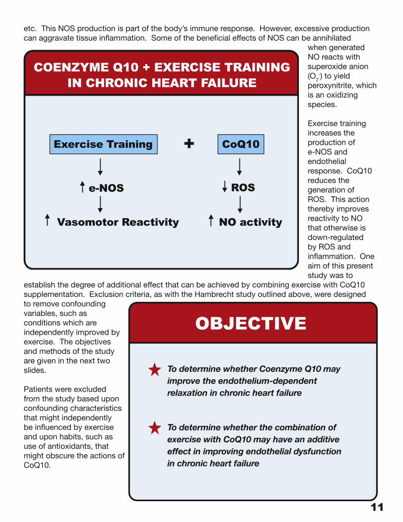
etc. This NOS production is part of the body’s immune response. However, excessive production can aggravate tissue inflammation. Some of the beneficial effects of NOS can be annihilated
when generated NO reacts with superoxide anion (O2
-) to yield peroxynitrite, which is an oxidizing species.
Exercise training increases the production of e-NOS and endothelial response. CoQ10 reduces the generation of ROS. This action thereby improves reactivity to NO that otherwise is down-regulated by ROS and inflammation. One aim of this present study was to
establish the degree of additional effect that can be achieved by combining exercise with CoQ10 supplementation. Exclusion criteria, as with the Hambrecht study outlined above, were designed to remove confounding variables, such as conditions which are independently improved by exercise. The objectives and methods of the study are given in the next two slides.
Patients were excluded from the study based upon confounding characteristics that might independently be influenced by exercise and upon habits, such as use of antioxidants, that might obscure the actions of CoQ10.
COENZYME Q10 + EXERCISE TRAININGIN CHRONIC HEART FAILURE
Exercise Training CoQ10
e-NOS ROS
Vasomotor Reactivity NO activity
+
OBJECTIVE
To determine whether Coenzyme Q10 mayimprove the endothelium-dependentrelaxation in chronic heart failure
To determine whether the combination ofexercise with CoQ10 may have an additiveeffect in improving endothelial dysfunctionin chronic heart failure
12 13
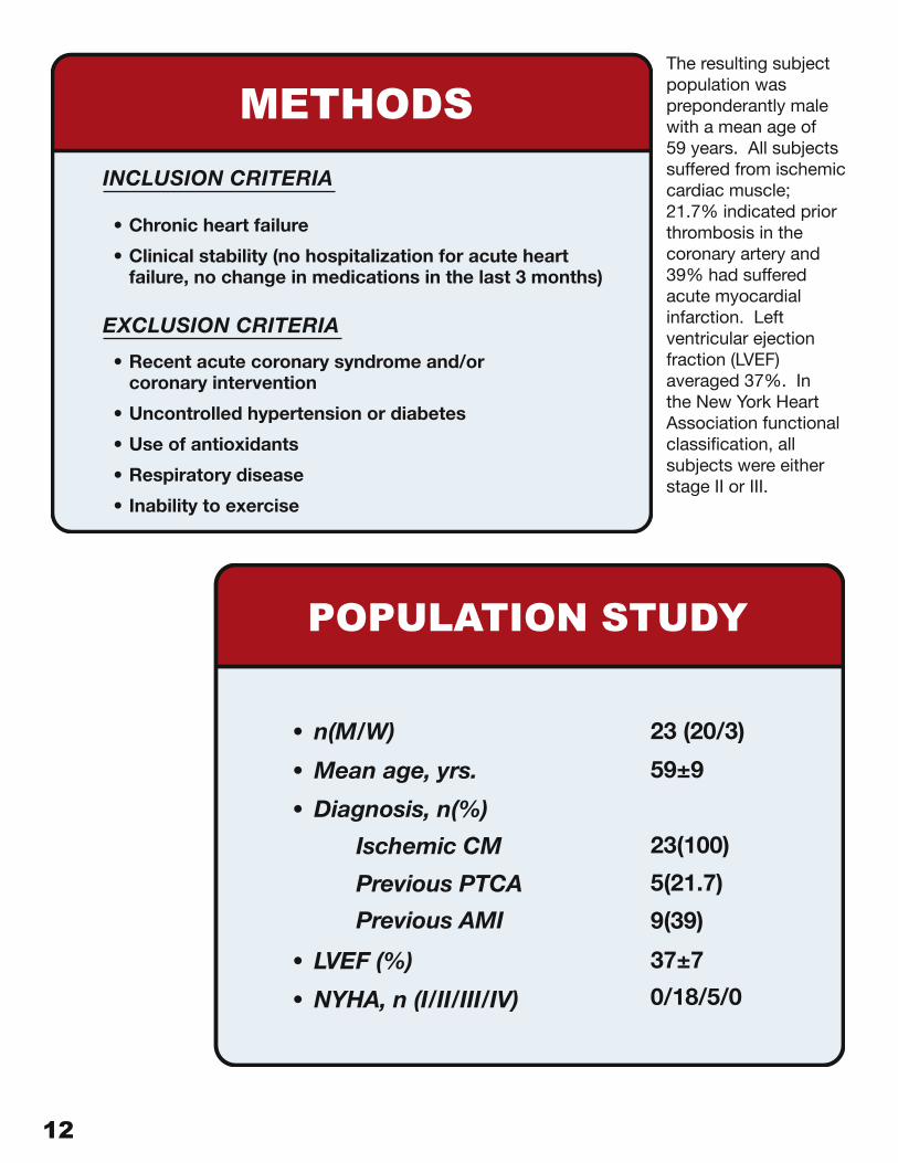
The resulting subject population was preponderantly male with a mean age of 59 years. All subjects suffered from ischemic cardiac muscle; 21.7% indicated prior thrombosis in the coronary artery and 39% had suffered acute myocardial infarction. Left ventricular ejection fraction (LVEF) averaged 37%. In the New York Heart Association functional classification, all subjects were either stage II or III.
POPULATION STUDY
• n(M/W)
• Mean age, yrs.
• Diagnosis, n(%)
Ischemic CM
Previous PTCA
Previous AMI
• LVEF (%)
• NYHA, n (I/II/III/IV)
23 (20/3)
59±9
23(100)
5(21.7)
9(39)
37±7
0/18/5/0
METHODS
INCLUSION CRITERIA
• Chronic heart failure
• Clinical stability (no hospitalization for acute heart failure, no change in medications in the last 3 months)
EXCLUSION CRITERIA
• Recent acute coronary syndrome and/or coronary intervention
• Uncontrolled hypertension or diabetes
• Use of antioxidants
• Respiratory disease
• Inability to exercise
12 13
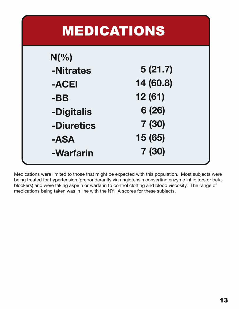
Medications were limited to those that might be expected with this population. Most subjects were being treated for hypertension (preponderantly via angiotensin converting enzyme inhibitors or beta-blockers) and were taking aspirin or warfarin to control clotting and blood viscosity. The range of medications being taken was in line with the NYHA scores for these subjects.
MEDICATIONS
N(%)-Nitrates-ACEI-BB-Digitalis-Diuretics-ASA
5 (21.7)14 (60.8)12 (61)
6 (26)7 (30)
15 (65)7 (30)-Warfarin
14 15
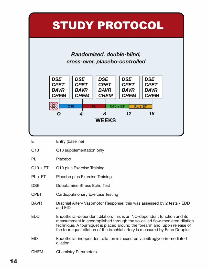
E Entry (baseline)
Q10 Q10 supplementation only
PL Placebo
Q10 + ET Q10 plus Exercise Training
PL + ET Placebo plus Exercise Training
DSE Dobutamine Stress Echo Test
CPET Cardiopulmonary Exercise Testing
BAVR Brachial Artery Vasomotor Response: this was assessed by 2 tests - EDD and EID
EDD Endothelial-dependent dilation: this is an NO-dependent function and its measurement in accomplished through the so-called flow-mediated dilation technique. A tourniquet is placed around the forearm and, upon release of the tourniquet dilation of the brachial artery is measured by Echo Doppler
EID Endothelial-independent dilation is measured via nitroglycerin-mediated dilation
CHEM Chemistry Parameters
STUDY PROTOCOL
Randomized, double-blind,cross-over, placebo-controlled
DSECPETBAVRCHEM
DSECPETBAVRCHEM
DSECPETBAVRCHEM
DSECPETBAVRCHEM
DSECPETBAVRCHEM
Q10 PL Q10 + ET PL + ETEO 4 8 12 16
WEEKS
14 15
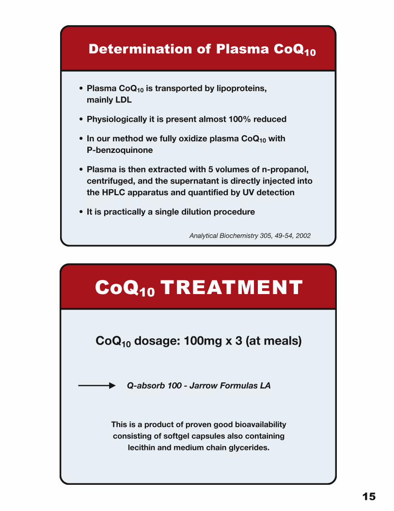
Determination of Plasma CoQ10
• Plasma CoQ10 is transported by lipoproteins, mainly LDL
• Physiologically it is present almost 100% reduced
• In our method we fully oxidize plasma CoQ10 with P-benzoquinone
• Plasma is then extracted with 5 volumes of n-propanol, centrifuged, and the supernatant is directly injected into the HPLC apparatus and quantified by UV detection
• It is practically a single dilution procedure
Analytical Biochemistry 305, 49-54, 2002
CoQ10 TREATMENT
CoQ10 dosage: 100mg x 3 (at meals)
Q-absorb 100 - Jarrow Formulas LA
This is a product of proven good bioavailability consisting of softgel capsules also containing
lecithin and medium chain glycerides.
16 17
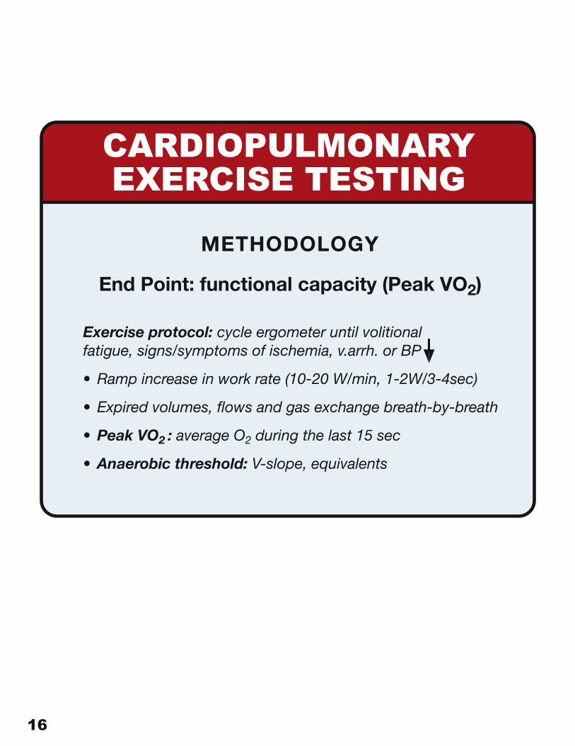
CARDIOPULMONARYEXERCISE TESTING
METHODOLOGY
End Point: functional capacity (Peak VO2)
Exercise protocol: cycle ergometer until volitional fatigue, signs/symptoms of ischemia, v.arrh. or BP
• Ramp increase in work rate (10-20 W/min, 1-2W/3-4sec)
• Expired volumes, flows and gas exchange breath-by-breath
• Peak VO2 : average O2 during the last 15 sec
• Anaerobic threshold: V-slope, equivalents
16 17
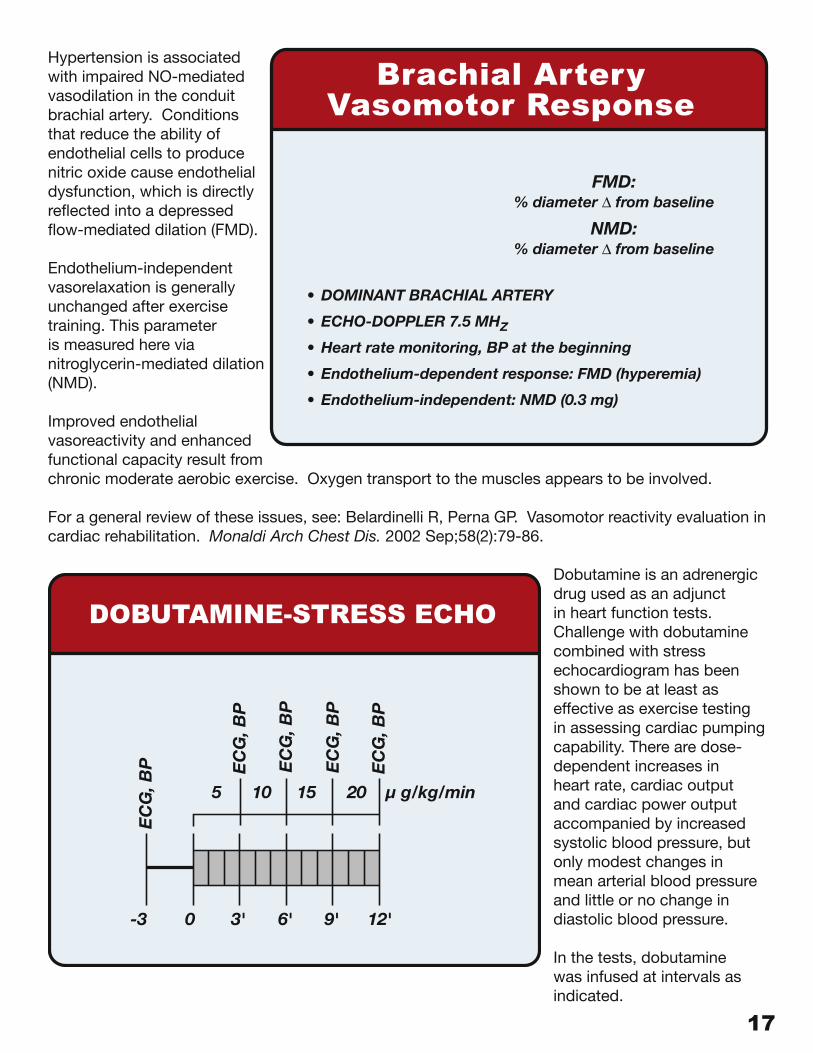
Hypertension is associated with impaired NO-mediated vasodilation in the conduit brachial artery. Conditions that reduce the ability of endothelial cells to produce nitric oxide cause endothelial dysfunction, which is directly reflected into a depressed flow-mediated dilation (FMD).
Endothelium-independent vasorelaxation is generally unchanged after exercise training. This parameter is measured here via nitroglycerin-mediated dilation (NMD).
Improved endothelial vasoreactivity and enhanced functional capacity result from chronic moderate aerobic exercise. Oxygen transport to the muscles appears to be involved.
For a general review of these issues, see: Belardinelli R, Perna GP. Vasomotor reactivity evaluation in cardiac rehabilitation. Monaldi Arch Chest Dis. 2002 Sep;58(2):79-86.
Dobutamine is an adrenergic drug used as an adjunct in heart function tests. Challenge with dobutamine combined with stress echocardiogram has been shown to be at least as effective as exercise testing in assessing cardiac pumping capability. There are dose-dependent increases in heart rate, cardiac output and cardiac power output accompanied by increased systolic blood pressure, but only modest changes in mean arterial blood pressure and little or no change in diastolic blood pressure.
In the tests, dobutamine was infused at intervals as indicated.
Brachial ArteryVasomotor Response
FMD:% diameter � from baseline
NMD:% diameter � from baseline
• DOMINANT BRACHIAL ARTERY
• ECHO-DOPPLER 7.5 MHZ
• Heart rate monitoring, BP at the beginning
• Endothelium-dependent response: FMD (hyperemia)
• Endothelium-independent: NMD (0.3 mg)
DOBUTAMINE-STRESS ECHO
EC
G, B
P EC
G, B
P
EC
G, B
P
EC
G, B
P
EC
G, B
P
5 10 15 20 µ g/kg/min
-3 0 3' 6' 9' 12'
18 19
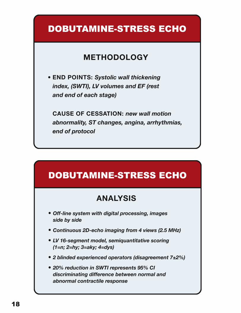
DOBUTAMINE-STRESS ECHO
METHODOLOGY
• END POINTS: Systolic wall thickening
index, (SWTI), LV volumes and EF (rest
and end of each stage)
CAUSE OF CESSATION: new wall motion
abnormality, ST changes, angina, arrhythmias,
end of protocol
DOBUTAMINE-STRESS ECHO
ANALYSIS
• Off-line system with digital processing, images side by side
• Continuous 2D-echo imaging from 4 views (2.5 MHz)
• LV 16-segment model, semiquantitative scoring (1=n; 2=hy; 3=aky; 4=dys)
• 2 blinded experienced operators (disagreement 7±2%)
• 20% reduction in SWTI represents 95% CI discriminating difference between normal and abnormal contractile response
18 19
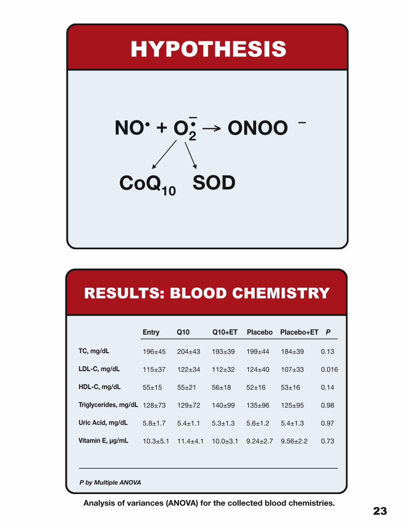
RESULTS: BLOOD CHEMISTRY
Entry Q10 Q10+ET Placebo Placebo+ET P
196±45
115±37
55±15
128±73
5.8±1.7
10.3±5.1
204±43
122±34
55±21
129±72
5.4±1.1
11.4±4.1
193±39
112±32
56±18
140±99
5.3±1.3
10.0±3.1
199±44
124±40
52±16
135±96
5.6±1.2
9.24±2.7
184±39
107±33
53±16
125±95
5.4±1.3
9.56±2.2
0.82±0.3 3.25±1.5* 4.0±2.1* 0.83±0.4 0.87±0.44 0.0001
TC, mg/dL
LDL-C, mg/dL
HDL-C, mg/dL
Triglycerides, mg/dL
Uric Acid, mg/dL
Vitamin E, µg/mL
P by Multiple ANOVA
CoQ10, µg/mL
RESULTS: Peak VO2
Pea
k V
O2
mL/
kg/m
in
24
12
0
P V 0.001
17.3 19.6 21.5 17.9 19.9
E Q10 Q10 Pl Pl
ET ET+ +
0.82 3.25 4.0 0.83 0.87±0.3 ±1.5 ±2.1 ±0.4 ±0.44
Plasma CoQ10
(µg/mL)
20 21
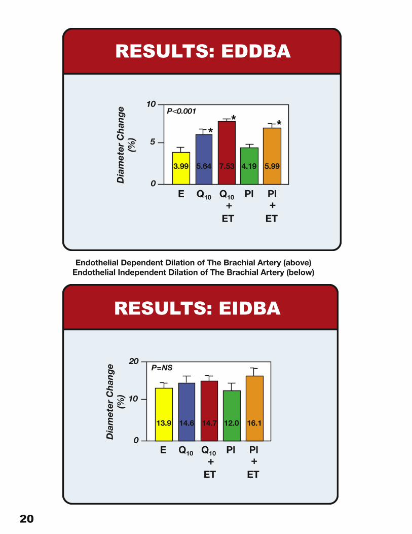
RESULTS: EDDBA
Dia
met
er C
han
ge
(%)
10
5
0
P V 0.001
3.99 5.64 7.53 4.19 5.99
E Q10 Q10 Pl Pl
ET ET+ +
RESULTS: EIDBA
Dia
met
er C
han
ge
(%)
20
10
0
P=NS
13.9 14.6 14.7 12.0 16.1
E Q10 Q10 Pl Pl
ET ET+ +
Endothelial Dependent Dilation of The Brachial Artery (above) Endothelial Independent Dilation of The Brachial Artery (below)
20 21
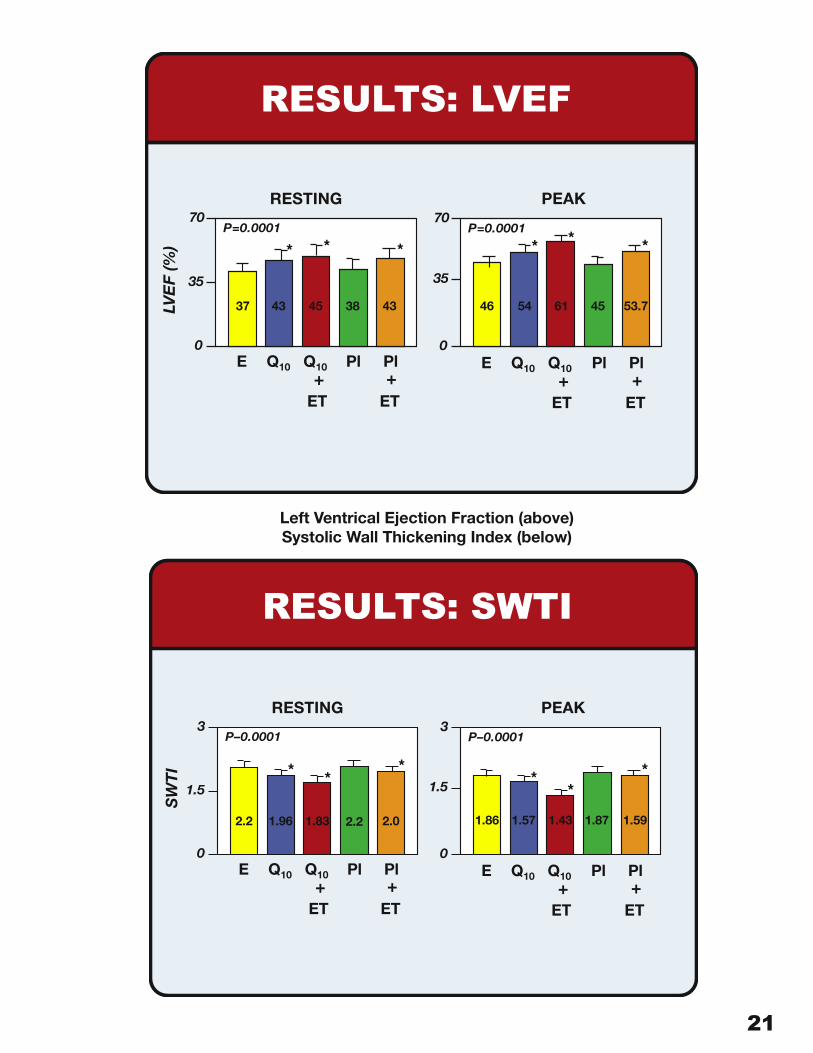
RESULTS: LVEF
LVE
F (%
)
E Q10 Q10 Pl Pl
ET ET+ +
0
70
35
P=0.0001
37 43 45 38 43
RESTING
0
70
35
P=0.0001
46 54 61 45 53.7
E Q10 Q10 Pl Pl
ET ET+ +
PEAK
RESULTS: SWTI
SW
TI
E Q10 Q10 Pl Pl
ET ET+ +
0
3
1.5
P–0.0001
2.2 1.96 1.83 2.2 2.0
RESTING
0
3
1.5
P–0.0001
1.86 1.57 1.43 1.87 1.59
E Q10 Q10 Pl Pl
ET ET+ +
PEAK
Left Ventrical Ejection Fraction (above)Systolic Wall Thickening Index (below)
22 23
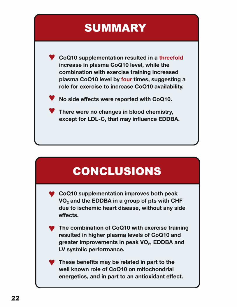
SUMMARY
CoQ10 supplementation resulted in a threefoldincrease in plasma CoQ10 level, while thecombination with exercise training increasedplasma CoQ10 level by four times, suggesting arole for exercise to increase CoQ10 availability.
No side effects were reported with CoQ10.
There were no changes in blood chemistry,except for LDL-C, that may influence EDDBA.
CONCLUSIONS
CoQ10 supplementation improves both peak VO2 and the EDDBA in a group of pts with CHF due to ischemic heart disease, without any side effects.
The combination of CoQ10 with exercise training resulted in higher plasma levels of CoQ10 and greater improvements in peak VO2, EDDBA and LV systolic performance.
These benefits may be related in part to the well known role of CoQ10 on mitochondrial energetics, and in part to an antioxidant effect.
22 23
RESULTS: BLOOD CHEMISTRY
Entry Q10 Q10+ET Placebo Placebo+ET P
196±45
115±37
55±15
128±73
5.8±1.7
10.3±5.1
204±43
122±34
55±21
129±72
5.4±1.1
11.4±4.1
193±39
112±32
56±18
140±99
5.3±1.3
10.0±3.1
199±44
124±40
52±16
135±96
5.6±1.2
9.24±2.7
184±39
107±33
53±16
125±95
5.4±1.3
9.56±2.2
TC, mg/dL
LDL-C, mg/dL
HDL-C, mg/dL
Triglycerides, mg/dL
Uric Acid, mg/dL
Vitamin E, µg/mL
P by Multiple ANOVA
0.13
0.016
0.14
0.98
0.97
0.73
HYPOTHESIS
NO O+2
10
ONOO
SODCoQ
Analysis of variances (ANOVA) for the collected blood chemistries.
24 25
Background: Coenzyme Q10, a Molecule with Diverse Functions
Coenzyme Q10 is a fundamental component in the physiology of energy production, immune response and protection against damage by free radicals. Co-Q10 has been available as a supplement for some twenty-five years, so it is hardly surprising that many of its mechanisms of action have now been explored. Nevertheless, new discoveries still are being made with regard to its benefits and improved delivery. Jarrow Formulas®, the maker of Q-absorb™ coenzyme Q10, actively supports research on this nutrient, especially in the areas of endothelial dysfunction and chronic heart failure. The preceding pages provide an annotated version of the slide presentation detailing the results of this research, which was presented at the annual meeting of the American Heart Association held November 9-12, 2003 in Orlando, Florida. Related material from an earlier stage of this clinical trial had been presented the previous year at the 3rd Annual International Co-Q10 Conference held in London.
Coenzyme Q10 is also known as ubiquinone because it is so widely present in plant and animal cells. There are other ubiquinones or Co-Qs with other numbers, such as Co-Q9, but only Co-Q10 is active in humans. This coenzyme was first isolated in pure form at the University of Wisconsin in 1957 by Fred L. Crane and his research associates. Under the leadership of D.E. Green, the laboratory at Wisconsin that same year proved that Co-Q10 is essential to the functioning of the mitochondria, the chief site of energy production within cells. Not long afterwards, Karl Folkers re-isolated Co-Q10 from beef heart muscle and synthesized the pure compound by fermentation. Folkers also demonstrated that Co-Q10 possesses vitamin-like catalytic functions. Chemically, its structure is related to that of the vitamins E and K.
Co-Q10 is part of the mitochondrial electron transport system and is synthesized in all cells. It appears to be essential to the body’s production of energy in the form of adenosine triphosphate (ATP). This holds special importance for the heart, which is spectacularly endowed with mitochondria and has the body’s highest concentration of Co-Q10 because of the unceasing demands made upon it. In descending order of tissue concentration, Co-Q10 is found in significant amounts in the liver, kidney, spleen and pancreas. Since the mid-1960’s, Co-Q10 has been used by doctors in Japan as a treatment for humans, in particular with regard to cardiac disorders and dysfunctions. In 2003, Co-Q10 was reclassified by the Japanese government as a dietary supplement with no restrictions on the amount. Aging reduces levels of Co-Q10. Minor amounts are obtained from the diet (mainly from fatty fish, organ meats, and whole grains). Co-Q10 also is synthesized in small amounts in the body, mostly in the mitochondria. Both of these routes decline with advancing age. Uptake from food and supplements is always problematic with this nutrient, and the body’s declining capacity to extract and assimilate Co-Q10 in later years (beginning in the fourth decade of life) undoubtedly plays a role in the development of various cardiovascular conditions.
Aside from the mitochondria and Golgi bodies, Co-Q10 is present in cell membranes. It helps to maintain the fluidity of these membranes and it is here that Co-Q10 performs some of its antioxidant functions. Cell membranes are largely constructed of lipids, and Co-Q10 itself is lipophilic. It happens that fats are often the targets of free radicals, for instance, the membranes of the blood component low-density lipoprotein cholesterol (LDL cholesterol). At least one important lipid-soluble antioxidant, the alpha-tocopherol form of vitamin E, in excessive amounts can act as a pro-oxidant. Experimental results demonstrate that oral supplementation with alpha-tocopherol taken alone can result in serum LDL that is more prone to oxidation initiation. However, co-supplementation with Co-Q10 not only prevents this pro-oxidant activity of vitamin E, but also provides the lipoprotein with increased resistance to oxidation. [1] A different type of test found that alpha-tocopherol
24 25
supplementation alone reduces blood Co-Q10 levels while improving antioxidant protection for blood lipids, thus indicating that Co-Q10 is required to regenerate the vitamin E radical. [2] This suggests that in order to fully realize the antioxidant benefits of vitamin E and avoid the exhaustion of serum Co-Q10, co-supplementation with Co-Q10 or another antioxidant with related functions, such as alpha-lipoic acid, may be required.
Co-Q10 has a multitude of benefits, some of which––but not all––are the result of its antioxidant qualities. It is often recommended to treat congestive heart failure, cardiac arrhythmias and ischemic injury. [3] These cardiovascular uses are its most thoroughly established employments and they have become even more important with the widespread prescription of statin and other cholesterol-lowering drugs for the treatment and prevention of cardiovascular diseases. Most such drugs act by inhibiting the enzyme HMG-CoA reductase. Inhibition of the HMG-CoA reductase enzyme, however, also blocks the biosynthesis of Co-Q10 and leads to reduced levels of Co-Q10 in blood plasma and tissues. This fact and the popularity of HMG-CoA inhibitors have been used to explain the increasing prevalence of congestive heart failure in the United States. Statins also increase muscle myopathies and mitochondrial dysfunctions (leading to muscle pain and weakness), another indication of Co-Q10 depletion. Supplementation with Co-Q10 therefore would appear to be prudent in conjunction with the use of HMG-CoA inhibitors. [4]
Co-Q10 provides many benefits that are hard to explain. This coenzyme has been observed to serve as an immune stimulant that boosts the capacities of existing immune cells as opposed to requiring the unleashing of additional immune components. How this is accomplished is unclear, but at least in part it is the result of reducing the burden on the immune system as a whole. The process goes something like this: Free radicals damage tissues which are then recognized by immune cells as “foreign.” This requires immune activation and, in turn, the release of yet more free radicals as one component of the response of the immune system. The resulting generalized low-level inflammation, finally, reduces the body’s immune response capacity. This is recognized, for instance, by an imbalance in the Th-1 versus Th-2 cells (T helper cell types) found in the system. As a general rule, with advancing years, free radical generation increases while immune competence declines, hence supplementation with Co-Q10 becomes more important.
Although Co-Q10 is not itself a weight-loss agent, it nevertheless may be significant for maintaining health in the overweight and in diabetics. Blood serum tests for levels of Co-Q10 indicate that these levels are deficient in almost 50% of obese subjects. [5] Moreover, there is some evidence that this quasi-vitamin can improve pancreatic beta-cell response and glycemic control in proto-diabetic and diabetic individuals. [6] It is not at all certain that such benefits, strictly speaking, are related to Co-Q10’s antioxidant functions.
Similarly, Co-Q10 helps in another of the Syndrome X conditions, hypertension. Again, it has been established that the blood pressure regulating benefits of Co-Q10 may be unrelated to its antioxidant benefits. [7] Aging causes structural and functional changes to the vascular wall of the cardiovascular system that result in endothelial dysfunction. [8] This endothelial dysfunction is characterized by a decrease in the capacity of the endothelium to properly dilate, that is, to relax and open the circulation. This may be a significant causative factor in the increased cardiovascular events seen in aging subjects. Studies conducted with animals and with patients have demonstrated a vascular effect from Co-Q10. [9,10]
As remarked at the outset, Jarrow Formulas® supports research on the topics of the bioavailability of Co-Q10 formulas, endothelial dysfunction and chronic heart failure. The phenomenon of endothelial dysfunction is typical of heart failure and is the subject of the research reported herein. After several years of work by Jarrow Formulas® and Prof. Littarru to develop and validate a Co-Q10 formulation that exhibits enhanced absorption, Italian scientists, headed by Romualdo Berlardinellli,
26 27
MD, FESC, of the Lancisi Heart Institute, Ancona, and at the University of Ancona School of Medicine conducted a study that investigated endothelial function via peripheral arterial dilation in a randomized, placebo-controlled, crossover clinical trial. It demonstrated that Jarrow Formulas® Q-absorb improves endothelial function in patients with ischemic heart disease and heart failure. This research was presented at the 3rd Annual International Co-Q10 Conference recently held in London. Q-absorb administration resulted in significant elevations in plasma Co-Q10 averaging a remarkable 3.7 mcg/mL plasma, peripheral arterial dilation, and an increase in peak VO2 (volume of oxygen consumed). The co-administration of Q-absorb and exercise therapy resulted in the greatest gains, with endothelial dysfunction being almost normalized. More recently, scientists from the Lancisi Heart Institute and the University of Ancona further investigated the effects of Jarrow Formulas® Q-absorb™ on chronic heart failure patients. In the study, 100 mg of Co-Q10 from Q-absorb was given at each meal of the day, an approach that is superior to taking all 300 mg at one time. Patients showed improved heart functional capacity, enhanced endothelial-dependent vasodilation and a tripling of plasma Co-Q10 levels. Results were presented at the annual meeting of the American Heart Association held November 9-12, 2003 in Orlando, Florida. That presentation is reproduced in the previous pages.
Coenzyme Q10 is poorly absorbed. Only 1–3 percent of the ingested nutrient is taken up into serum. Jarrow Formulas® Q-absorb™ employs a completely natural proliposome lipid soluble delivery system clinically shown in the earlier study in humans to increase Co-Q10 plasma levels 150 percent over baseline within two weeks when taken three times per day for a total supplementation of 120 milligrams. (Baseline is typically .60–.80 mcg/ml plasma.) These results are at least 28 percent higher than are realized with dry Co-Q10 capsules. Results within thirty days were more than 200% over baseline.
The current formulation of Q-absorb typically gives rise to Co-Q10 levels of 2.0–2.5 mcg/ml plasma. Q-absorb therefore reduces the amount of supplemental Co-Q10 required to raise the concentration found in the blood and it reduces the time required to see effects. In the study reported above, the daily amount of Co-Q10 given was 300 mg (100 mg t.i.d.) from Q-absorb. Plasma levels averaged a remarkable 3.70 mcg/ml plasma and ranged as high as 4 mcg/ml plasma.
Q-absorb uses no polysorbate or other detergent to achieve its improved bioavailability. It contains only 100 percent naturally-derived materials. This includes the Co-Q10 from Kaneka Corporation, which is derived from a natural biofermentation process and is 100% all natural, all-trans configuration.
Jarrow Formulas® continues to work to improve the uptake of its Q-absorb product and to demonstrate its utility for the health of its customers.
26 27
References for Background
[1] Thomas SR, Neuzil J, Stocker R. Cosupplementation with coenzyme Q prevents the prooxidant effect of alpha-tocopherol and increases the resistance of LDL to transition metal-dependent oxidation initiation. Arterioscler Thromb Vasc Biol. 1996 May;16(5):687-96.
[2] Kaikkonen J, Nyyssonen K, Tomasi A, Iannone A, Tuomainen TP, Porkkala-Sarataho E, Salonen JT. Antioxidative efficacy of parallel and combined supplementation with coenzyme Q10 and d-alpha-tocopherol in mildly hypercholesterolemic subjects: a randomized placebo-controlled clinical study. Free Radic Res. 2000 Sep;33(3):329-40.
[3] Rosenfeldt FL, Pepe S, Linnane A, Nagley P, Rowland M, Ou R, Marasco S, Lyon W, Esmore D. Coenzyme Q10 protects the aging heart against stress: studies in rats, human tissues, and patients. Ann N Y Acad Sci. 2002 Apr;959:355-9; discussion 463-5.
[4] De Pinieux G, Chariot P, Ammi-Said M, Louarn F, Lejonc JL, Astier A, Jacotot B, Gherardi R. Lipid-lowering drugs and mitochondrial function: effects of HMG-CoA reductase inhibitors on serum ubiquinone and blood lactate/pyruvate ratio. Br J Clin Pharmacol. 1996 Sep;42(3):333-7.
[5] Emile G. Bliznakov and Gerald L. Hunt, The Miracle Nutrient Coenzyme CoQ10 (New York: Bantam Books, 1986) 150-154 citing the Belgian studies of Dr. Luc Van Gaal and his colleagues.
[6] McCarty MF. Toward practical prevention of type 2 diabetes. Med Hypotheses. 2000 May;54(5):786-93.
[7] Hodgson JM, Watts GF, Playford DA, Burke V, Croft KD. Coenzyme Q(10) improves blood pressure and glycaemic control: a controlled trial in subjects with type 2 diabetes. Eur J Clin Nutr. 2002 Nov;56(11):1137-42.
[8] Matz RL, Andriantsitohaina R. Age-related endothelial dysfunction: potential implications for pharmacotherapy. Drugs Aging 2003;20(7)527-50.
[9] Lonnrot K, Porsti I, Alho H, Wu X, Hervonen A, Tolvanen JP. Control of arterial tone after long-term coenzyme Q10 supplementation in senescent rats. Br J Pharmacol. 1998 Aug;124(7):1500-6. Erratum in: Br J Pharmacol 1998 Dec;125(8):17.
[10] Digiesi V, Cantini F, Oradei A, Bisi G, Guarino GC, Brocchi A, Bellandi F, Mancini M, Littarru GP. Coenzyme Q10 in essential hypertension. Mol Aspects Med. 1994;15 Suppl:s257-6.